Review

Transient Receptor Potential Ankyrin 1 (TRPA1) Channel as a Sensor of Oxidative Stress in Cancer Cells

by
Francesco Moccia
1,* and
Daniela Montagna
2,3
1
Laboratory of General Physiology, Department of Biology and Biotechnology “L. Spallanzani”, University of Pavia, 27100 Pavia, Italy
2
Department of Sciences Clinic-Surgical, Diagnostic and Pediatric, University of Pavia, 27100 Pavia, Italy
3
Pediatric Clinic, Foundation IRCCS Policlinico San Matteo, 27100 Pavia, Italy
*
Author to whom correspondence should be addressed.
Cells 2023, 12(9), 1261; https://doi.org/10.3390/cells12091261
Submission received: 20 March 2023 / Revised: 20 April 2023 / Accepted: 20 April 2023 / Published: 26 April 2023

Abstract

:
Moderate levels of reactive oxygen species (ROS), such as hydrogen peroxide (H2O2), fuel tumor metastasis and invasion in a variety of cancer types. Conversely, excessive ROS levels can impair tumor growth and metastasis by triggering cancer cell death. In order to cope with the oxidative stress imposed by the tumor microenvironment, malignant cells exploit a sophisticated network of antioxidant defense mechanisms. Targeting the antioxidant capacity of cancer cells or enhancing their sensitivity to ROS-dependent cell death represent a promising strategy for alternative anticancer treatments. Transient Receptor Potential Ankyrin 1 (TRPA1) is a redox-sensitive non-selective cation channel that mediates extracellular Ca2+ entry upon an increase in intracellular ROS levels. The ensuing increase in intracellular Ca2+ concentration can in turn engage a non-canonical antioxidant defense program or induce mitochondrial Ca2+ dysfunction and apoptotic cell death depending on the cancer type. Herein, we sought to describe the opposing effects of ROS-dependent TRPA1 activation on cancer cell fate and propose the pharmacological manipulation of TRPA1 as an alternative therapeutic strategy to enhance cancer cell sensitivity to oxidative stress.

1. Introduction

Reactive oxygen species (ROS) comprise a group of highly reactive oxygen-containing molecules, including the non-radical, hydrogen peroxide (H2O2), the free-radicals, hydroxyl radical (OH), superoxide anion (O2), peroxides (RO), and oxides of nitrogen (RO) [1,2,3,4]. Aberrant redox homeostasis represents a hallmark of cancer cells, since moderate ROS (e.g., 10–50 µM H2O2 and O2 in the nanomolar range) stimulate cell transformation, hyperproliferation, invasion, metastasis, and angiogenesis (Figure 1) [1,2,3,5,6,7,8]. The rate of basal ROS production in cancer cells is enhanced by multiple mechanisms [2,9], such as metabolic disturbances, adaptation to hypoxia, oncogene activation, and loss of tumor suppressors. However, excessive ROS levels (e.g., >100 µM H2O2; O2 in the micromolar range; ≈30 µM OH) could impair tumor development and spread by triggering cell apoptosis, ferroptosis, or senescence [8,10,11,12,13]. Therefore, cancer cells cope with oxidative stress by exploiting a sophisticated network of antioxidant defense mechanisms [10,14]. Non-enzymatic small molecules directly scavenge ROS and comprise the endogenously synthesized glutathione (GSH), melatonin, and melanin, as well as the exogenously derived vitamin C, vitamin E, and β-carotene [10]. Noteworthily, GSH expression is up-regulated in a variety of cancer cell types [15], thereby increasing their antioxidant capacity. Enzymatic mechanisms include catalase (CAT), which degrades H2O2 to H2O and oxygen (O2), peroxiredoxins (PRXs), and glutathione peroxidases (GPXs), which reduce H2O2 to O2, and superoxide dismutases (SODs), which catalyze the conversion of O2 to H2O and O2 (Figure 1) [10]. Six PRX isoforms have been described not only in the cytosol but also in multiple organelles, including mitochondria, the endoplasmic reticulum (ER), and peroxisomes, whereas eight GPX isoforms scavenge H2O2 in the cytosol and mitochondria [10,16]. Reduced thioredoxin (TRX) and reduced glutathione (GSH), respectively, serve as cofactors for PRXs- and GPXs-mediated reduction of H2O2 to H2O. Additionally, GSH is used by glutathione-S-transferases (GSTs) to detoxify reactive compounds generated by oxidative stress [10,16]. Also SODs are spatially distributed in different subcellular compartments to favor O2 elimination: Cytoplasmic SOD (SOD-1 or Cu/Zn-SOD), mitochondrial SOD (SOD-2 or Mn-SOD), and extracellular SOD (SOD-3 or EC SOD) [17]. SOD-1 and SOD-2 rapidly dismutate O2 into H2O2, which is less reactive and is reduced to O2 and H2O2 by catalase or converted to H2O2 and oxidized glutathione by GPx [17].
The transcription factor, nuclear factor-erythroid 2 p45-related factor 2 (NRF2), is the master regulator of redox homeostasis in cancer cells [1,10,14]. NRF2 activity is finely regulated by kelch-like ECH-associated protein 1 (KEAP1) and the Cul3-based E3 ubiquitin ligase, which target NRF2 for proteosomal degradation. High ROS levels cause the oxidation of redox-sensitive cysteine residues in KEAP1, thereby preventing the physical interaction with and subsequent degradation of NRF2. The latter can in turn translocate into the nucleus and drive the expression of numerous antioxidant genes [10,16]. These include ROS-detoxifying enzymes, e.g., GPXs, GSTs, and PRXs, as well as the two subunits comprising the glutamate–cysteine ligase (GCL), i.e., the catalytic subunit (GCLC) and the modifier subunit (GCLM), which catalyzes the rate-limiting step in GSH biosynthesis [1,10,14]. Therefore, an alternative anticancer strategy could be designed by either increasing ROS production or reducing the antioxidant capacity of cancer cells [1,10,14,18].
Transient Receptor Potential Ankyrin 1 (TRPA1), the unique member of the mammalian TRPA sub-family, is a Ca2+-permeable, non-selective cation channel that is able to integrate thermal, mechanical, and chemical signals [19,20]. Among its multiple endogenous agonists, ROS are crucial to activate TRPA1 in many disorders featured by oxidative stress, including neuropathic pain, inflammation, osteoarthritis, migraine, postischemic dysesthesia, diabetes, and respiratory diseases [19,21,22]. TRPA1-dependent extracellular Ca2+ entry can support several cancer hallmarks, including hyperproliferation, survival against pro-apoptotic stimuli, and invasive behavior [16,23,24,25]. In addition, TRPA1-mediated depolarization of peripheral nociceptors is involved in cancer-induced bone pain and cancer-related neuropathic pain [26,27]. A recent series of studies showed that TRPA1 may also serve as a crucial sensor of redox signaling in cancer cells and that extracellular Ca2+ entry through TRPA1 can promote either cell survival [28,29,30] or cell death [31,32,33] in response to oxidative stress. Herein, we first summarize the current knowledge about the structure and gating mechanisms of TRPA1. Then, we briefly survey the contribution of TRPA1-mediated intracellular Ca2+ signals to cancer cell proliferation, migration, and angiogenesis. Finally, we discuss how the redox-sensing capability of TRPA1 could be used by certain cancer cells to engage a non-canonical antioxidant defense program, while ROS-dependent TRPA1 activation leads to intracellular Ca2+ overload, mitochondrial dysfunction, and apoptosis in other cancer types. Therefore, TRPA1 channels could represent a novel molecular target for anticancer therapy that could be exploited either to dampen the antioxidant capacity of malignant cells or to exacerbate their sensitivity to ROS signaling.

2. TRPA1: Molecular Structure, Biophysical Properties, and Pharmacological Sensitivity

The mammalian TRP superfamily of non-selective cation channels encompasses 28 members that are subdivided into 6 sub-families based on their sequence homology: Canonical (TRPC1-7), melastatin (TRPM1-8), vanilloid (TRPV1-6), ankyrin (TRPA1), polycystin (TRPP), and mucolipin (TRPML1-3). The TRPP sub-family consists of eight members, but only TRPP2, TRPP3, and TRPP5 function as ion channels [34,35]. TRPA1 is the sole member of the TRPA subfamily and has originally been detected in a subpopulation of Aδ- and C-fiber nociceptive sensory neurons, in which it can serve as a chemical, mechanical, and thermal nocisensor [19,20]. Subsequently, TRPA1 has been found in other cell types such as epithelial cells, fibroblasts, enterochromaffin cells, mast cells, melanocytes, odontoblasts, and β-cells of the Langerhans islets, which may serve as sensory cells and interact with adjoining nociceptors [19,20]. More recently, TRPA1 expression has been confirmed in the central nervous system [36] and in cancer cells [16]. Interestingly, in the dorsal root ganglion, TRPA1 channels are also located in endolysosomes, thereby contributing to mediating intracellular Ca2+ release [37].

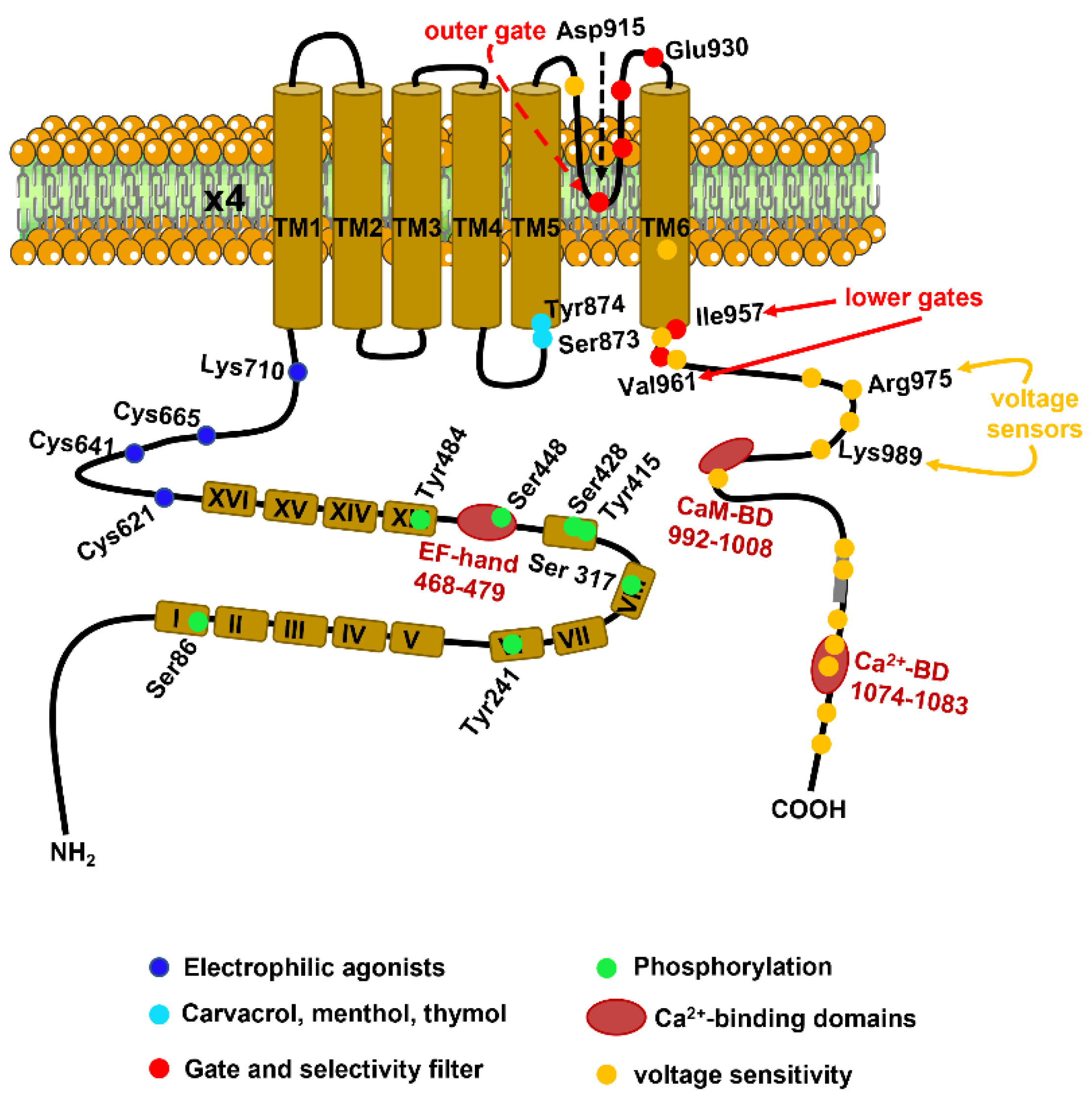
2.1. The Molecular Structure of TRPA1

The TRPA1 gene is located in band q21.13 of chromosome 8 in humans and consists of 73.635 bases and 29 exons [38]. The protein channel encoded by the TRPA1 gene presents an estimated molecular weight of ~127 kDa and long cytosolic NH2- and COOH-terminal tails, which collectively account for ~80% of the total protein mass [38,39]. The functional TRPA1 channel protein results from the assembly of four subunits into a homotetramer through ‘domain-swap’ interactions [39]. The molecular architecture of the TRPA1 protein has been recently solved at a near-atomic resolution (~4 Å) by using single-particle electron cryo-microscopy [39]. As predicted by the cDNA sequence [38], each subunit consists of six transmembrane (S1–S6) domains and presents an extracellular re-entrant pore loop between S5 and S6 (Figure 2) [39]. The long NH2-terminal of TRPA1 protein houses the most extensive ankyrin repeat domain (ARD) of the TRP superfamily, which comprises 14–16 ankyrin repeats (Figure 2), each consisting of a ~33 amino acids-long α-helix-β-turn-α-helix motif [39]. The ARD is connected to TM1 via the pre-S1 region, containing some cysteine residues (e.g., Cys621, Cys641, and Cys665) that are critical for TRPA1 activation by electrophilic agonists (Figure 2) [39,40]. In addition, the proximal portion of the COOH-terminus contains two residues, i.e., Arg975 and Lys989 (Figure 2), which control the voltage-dependent activation of TRPA1 at highly depolarizing potentials (>+100 mV) [41]. The ion conduction pathway of the TRPA1 channel is featured by two major constrictions, or gates, that resemble those also identified in the central cavity of TRP Vanilloid 1 (TRPV1). The outer gate is contributed to by diagonally opposed Asp915 residues, which are 7 Å apart and control Ca2+ permeability (Figure 2). The inner gate is formed by two hydrophobic seals established by Ile957 and Val961, which narrow the funnel to ~4 Å and thereby constrain the permeation of rehydrated cations (Figure 2). The outer pore domain of TRPA1 contains two α-helices, with a string of acidic amino acids (Glu920, Glu924, and Glu930) in the second α-helix that is likely to serve as a negatively charged conduit to repel anions and attract cations (Figure 2) [39].

2.2. Biophysical Properties and Gating Mechanisms of TRPA1

TRPA1 is a non-selective cation channel that is permeable to both monovalent (e.g., Na+ and K+) and divalent (e.g., Mg2+) cations and may be open in the absence of exogenous stimulation [19,42,43]. In the presence of extracellular Ca2+, TRPA1 presents a single-channel conductance of ~65 pS and ~110 pS for negative and positive membrane potentials, respectively [44]. Non-stimulated TRPA1 channels display a pore diameter of ~11 Å, a monovalent cation permeability sequence of Rb+ > K+ > Cs+ > Na+ > Li+, high permeability for Ca2+ over Na (PCa/PNa ~6), and a fractional Ca2+ current of ~17% [42,45]. Stimulation with electrophilic agonists, such as mustard oil, enhances the PCa/PNa to ~9 and the fractional Ca2+ current to ~23% and changes the monovalent cation permeability sequence to Ca2+ > Ba2+ > Mg2+ > NH4+ > Li+ > Na+ > K+ > Rb+ > Cs+ [42,45]. In addition, agonist exposure can change the dimensions of the selectivity filter, thereby resulting in the progressive, but reversible, dilation of the channel pore (by 1–3 Å), which can become permeable to large organic cations, such as N-methyl-D-glucamine and the cationic dye Yo-Pro [19,45,46,47]. An alternative model proposed that the increase in permeability to large molecules does not reflect a change in channel permeability, but rather an elevation in the intracellular ion concentration [48].

2.3. Ca2+-Dependent Regulation of TRPA1 Activity

Interestingly, TRPA1 activity can be modulated by both extracellular and intracellular Ca2+ [36,44]. An increase in [Ca2+]i could initiate and augment TRPA1-mediated inward currents elicited by several compounds, such as Δ9-tetrahydrocannabinol (THC) and allyl isothiocyanate (AITC) [49]. Similarly, elevating the extracellular Ca2+ concentration also potentiated TRPA1 channel activation and inactivation [50]. These observations led to a model according to which extracellular Ca2+ permeates the channel pore and thereafter regulates TRPA1 activity by binding to a site that is located within or very close to the pore [19]. The NH2 tail contains a putative EF-hand Ca2+-sensing domain that is located on ARD12 and is likely to mediate the Ca2+-dependent activation of TRPA1 via the residues Asp466, Leu474 and Asp477 (Figure 2) [19,36,42,51]. An additional Ca2+-binding site, which is involved in the Ca2+-dependent activation and inactivation of TRPA1, is located at the distal COOH-terminal region and formed by Glu1077 and Asp1080-Asp1082 (Figure 2) [52]. Intriguingly, calmodulin (CaM) may be associated with TRPA1 in the presence of Ca2+ via the physical interaction with a CaM-binding domain in the COOH-tail of TRPA1 (Figure 2) [53]. It has been proposed that CaM promotes TRPA1 inactivation following extracellular Ca2+ influx [53].

2.4. TRPA1 Activity Is Stimulated by Physical Stimuli: Voltage, Temperature and Membrane Deformation

The gating of TRPA1 channels can be modulated by multiple physical stimuli, including changes in membrane potential, temperature, and plasma membrane tension [19]. TRPA1 can be activated by highly depolarizing voltages (>+100 mV) and presents a half-maximal voltage (V1/2) of channel activation ranging between +90 mV and +170 mV [42,44]. As anticipated in Section 2.1, the voltage-dependent gating of TRPA1 depends on four positively charged residues, i.e., Lys969, Arg975, Lys988, and Lys989, which are located within the most proximal helix (H1) of the COOH-terminus (Figure 2) [41]. The voltage sensitivity of the channel is also sensitive to single alanine mutations of multiple residues located in the predicted helix that is centered around Lys1048 and Lys1052 [41]. Membrane depolarization could promote the physical interaction between the distal region and the more proximal voltage sensor, thereby resulting in a conformation change that causes TRPA1 activation [42]. However, the voltage-dependent gating of TRPA1 can be shifted towards more physiological membrane potentials by Ca2+ (see Section 2.3), electrophilic and non-electrophilic agonists, and cold or hot temperatures [19,36,42]. In accord, human TRPA1 is intrinsically sensitive to both noxious cold (<12 °C) and noxious (>43 °C) heat [54,55], although its thermosensitive profile may depend on the species [56]. Of note, the heat and cold responsiveness of the human TRPA1 (hTRPA1) is finely tuned by its redox environment [56]. ROS produced in response to dramatic changes in the local temperature could represent the driving force underlying the activation of hTRPA1 by noxious cold or hot temperatures [54]. A recent investigation proposed that the COOH-terminal region of hTRPA1 harbors temperature-sensitive modules that are allosterically coupled to the S5–S6 pore region and the S1–S4 transmembrane domain [55]. The temperature sensitivity of hTRPA1 can be modulated by the NH2-terminal ARD [57] and is lost under reducing conditions [54]. Finally, TRPA1 channels present mechanosensitive activity that has largely been observed in C fibers exposed to large membrane deformation and in other cell types involved in mechanosensation, such as sensory neurons and Merkel cells [19,58]. Emerging evidence indicates that hTRPA1 is intrinsically mechanosensitive: when reconstituted in artificial lipid bilayers, its single-channel current activity increased in response to an increase in the lipid tension stress [59]. Interestingly, the mechanosensitivity of the human TRPA1 also does not require the NH2-terminal ARD but is finely tuned by its redox state and is favored by a pro-oxidant environment [59].

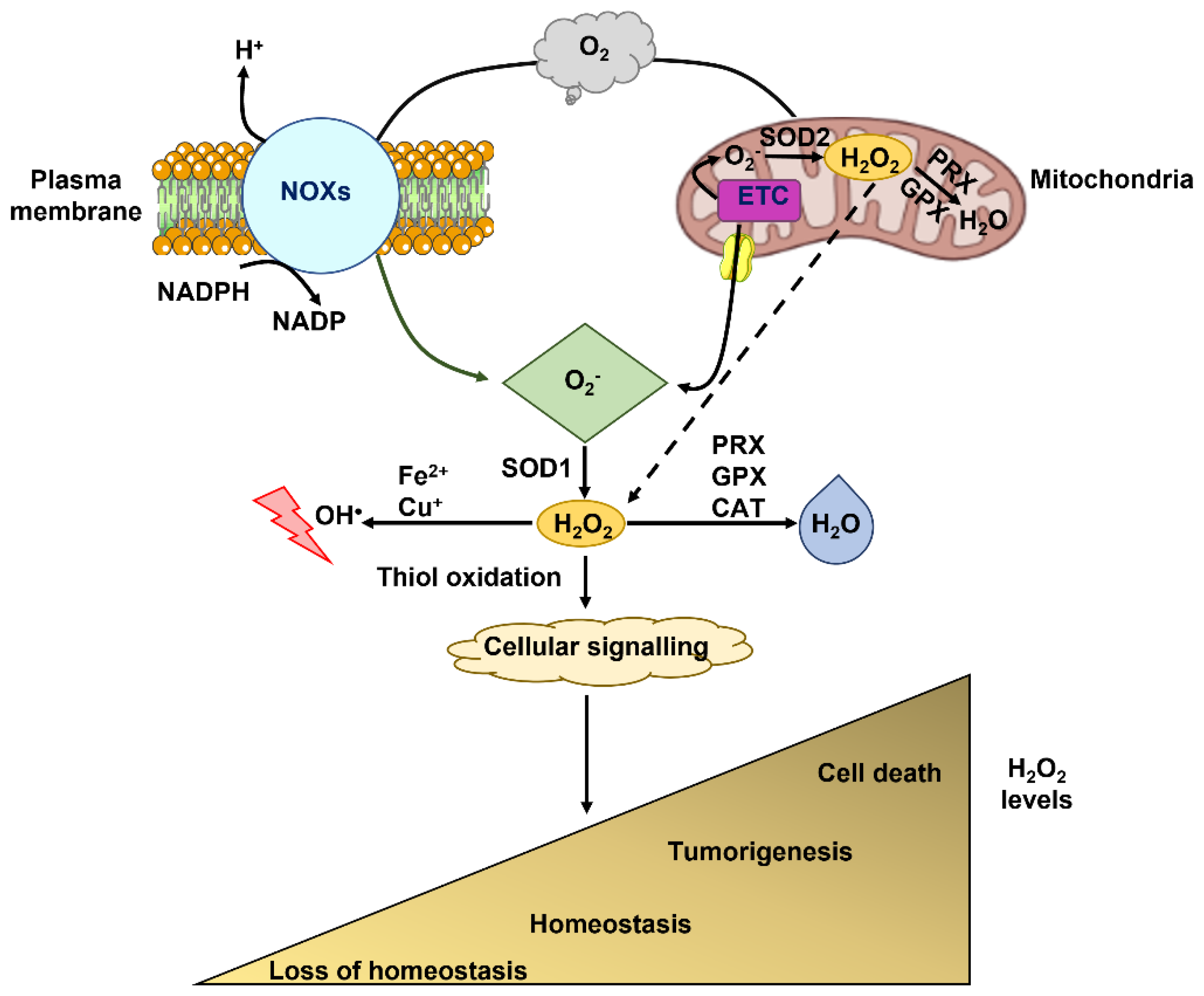
2.5. TRPA1 As a Sensor of Redox Signaling

ROS and ROS metabolites, as well as reactive nitrogen species (RNS), are the main endogenous regulators of TRPA1 activity under physiological conditions [17,19,36,42,44]. Therefore, TRPA1 belongs to the class of TRP channels that serve as sensors of the cellular redox state [60] and include TRP Melastatin 2 (TRPM2) [61,62] and TRP Vanilloid 1 (TRPV1) [63,64]. TRPA1 can be directly activated by H2O2 [65,66], OH [66], the cyclopentenone prostaglandin 15-deoxy-delta(12,14)-prostaglandin J(2) [15d-PGJ(2)] [65], nitric oxide (NO) [67], and peroxynitrite (ONOO), [68]. In addition, TRPA1 channels can be stimulated by multiple endogenous aldehydes that are produced in response to lipid peroxidation [4,69], such as 4-hydroxynonenal (4-HNE), 4-oxo-nonenal, and 4-hydroxyhexenal [43,65]. Similarly, TRPA1 activity is enhanced by nitro-oleic acid, which is produced during the nitration of plasma membrane phospholipids [70]. ROS increase the open probability of TRPA1 via covalent modifications of three cysteine residues, i.e., Cys621, Cys641, and Cys665, which are situated within the pre-TM1 region at the NH2-terminal of the channel protein (Figure 2) [71]. The exceptional reactivity of Cys621 towards electrophiles is facilitated by Lys620, whereas full TRPA1 activation by oxidative stress requires the covalent modification of Cys665 [71]. The combination of cryo-electron microscopy, which solved the full-length structure of hTRPA1 at ~a 4 Å resolution [39], and molecular modeling [72] suggested that the electrophilic reactive Cys621, Cys641, and C665 could form a ligand-binding pocket by coming in close proximity with each other [42]. The pre-TM1 region contains three additional cysteine residues, i.e., Cys619, Cys639, and Cys663, and to a lesser extent Lys708, that are not directly modified by ROS, but react to other electrophilic TRPA1 agonists, such as AITC [73]. The electrophilic activation of TRPA1 channels can also be facilitated by the disruption or formation of disulfide bonds between these, as well as other, cysteine residues at the NH2-terminal [42,72]. Finally, the TM core of TRPA1 protein presents cysteine and lysine residues, i.e., Cys727, Lys771, and Cys834, that are likely to be exposed to the lipid environment and could, therefore, be reactive to lipophilic electrophiles [36,39].
Quantitative analysis demonstrated that TRPA1 is the most highly redox-sensitive TRP channel and can, therefore, uniquely serve as a sensor of molecular oxygen (O2) [74]. Under normoxic conditions (~20% O2), TRPA1 activity is tonically inhibited through hydroxylation of Pro394 in the NH2-terminal ARD by prolyl hydroxylases (PHDs) [74], which function as the main O2 sensor of the cell and regulate the stability of hypoxia-inducible transcription factors (HIFs) [75]. The hydroxylation activity of PHDs is decreased upon a reduction in O2 concentration, thereby relieving the channel from inhibition and leading to TRPA1 activation under hypoxia [74]. In addition, hypoxia can induce the insertion of non-hydroxylated TRPA1 channels into the plasma membrane [74]. On the other hand, hyperoxia can activate TRPA1 through the O2-dependent oxidation of two cysteine residues at the NH2-terminal of the channel protein, i.e., Cys633 and Cys656 [74]. Additionally, although seemingly paradoxical, an increase in oxidative stress could occur even during hypoxia. In accord, electron transfer from ubisemiquinone to O2 at the Q0 site of the mitochondrial complex III dramatically enhances ROS generation in hypoxic cells [76]. Therefore, TRPA1 may also serve as a molecular sensor of the local changes in the O2 concentration [77].

3. TRPA1-Mediated Ca2+ Signals Support Tumorigenesis

Because of the versatility of its gating mechanisms, TRPA1 is uniquely suited to detect the increase in ROS production that supports neoplastic transformation and dissemination [16]. An additional feature of solid malignancies is represented by hypoxia, which is due to their abnormal vascular network, comprising leaky, highly disorganized, and compressed capillary vessels [78,79]. Therefore, the cancer microenvironment has been recognized as the ideal milieu to activate TRPA1 [16,26,60]. In the present section, we will briefly survey the mechanisms whereby extracellular Ca2+ entry via TRPA1 channels promotes cancer cell proliferation, survival, migration, and angiogenesis. In Section 4, we will specifically address how redox-sensitive TRPA1 activation could either engage non-canonical antioxidant defense programs or induce apoptosis in cancer cells. In Section 5, we focus on the pharmacology of TRPA1 channels and describe how stimulating or inhibiting TRPA1 activity could represent a novel therapeutic strategy to induce ROS-dependent cancer cell death.
Remodeling of the Ca2+ handling machinery, including multiple members of the TRP superfamily, contributes to many cancer hallmarks, such as aberrant proliferation, tissue invasion and metastasis, resistance to pro-apoptotic chemotherapeutics, and sustained angiogenesis [80,81,82,83,84,85,86,87,88]. Early work showed that the TRPA1 protein was upregulated in several cell lines and in tumor samples of human small-cell lung cancer (SCLC) [33]. TRPA1-mediated extracellular Ca2+ entry prevented starvation-induced SCLC cell apoptosis by recruiting extracellular signal-regulated kinases 1/2 (ERK 1/2) in an Src-dependent manner [33]. A parallel investigation demonstrated that TRPA1 expression on the plasma membrane of human lung adenocarcinoma A549 cells can be increased by inflammatory cytokines, such as interleukin (IL)-1α, IL-1β, and tumor necrosis factor α (TNFα) [89]. Similarly, TRPA1 was expressed in prostate cancer stromal cells expanded from different patients, but not in healthy primary cultured prostate epithelial cells [90]. This study showed that the antibacterial agent, triclostan, stimulated TRPA1 to mediate extracellular Ca2+ entry, thereby resulting in vascular endothelial growth factor (VEGF) secretion and prostate cancer cell proliferation [90]. In addition, VEGF could target adjacent endothelial cells to induce sprouting angiogenesis and favor prostate cancer vascularization [91]. Interestingly, TRPA1 protein was also largely expressed in prostate tumor-derived endothelial cells (PTECs), while it was absent in its normal counterpart [92]. TRPA1-mediated intracellular Ca2+ signals stimulated PTECs to migrate and assemble in capillary-like networks both in vitro and in vivo [92]. Additionally, the TRPA1 protein was expressed and mediated an increase in the intracellular Ca2+ concentration ([Ca2+]i) in human prostate cancer-associated fibroblasts (CAFs) [93]. In prostate CAFs, TRPA1 could be activated by the natural polyphenolic antioxidant, resveratrol, and induced the secretion of VEGF and hepatocyte growth factor (HGF), which in turn reduced resveratrol-induced apoptosis in co-cultured human prostate cancer cells [93]. TRPA1 was also detected in human pancreatic ductal adenocarcinoma cells (PDACs), which displayed higher levels of TRPA1 mRNA expression as compared to non-neoplastic cells [24]. TRPA1 activity, both under basal conditions and in the presence of the electrophilic agonist AITC, reduced PDAC cell migration and caused changes in cell cycle progression, i.e., induced a shift from G0/G1 to a sub-G1 phase [24]. TRPA1 could also regulate PDAC cell motility in a flux-independent manner, i.e., without the requirements for extracellular Ca2+ entry, as recently suggested for other TRP channels [94,95] and ligand-gated ion channels [96,97,98]. In agreement with this hypothesis, TRPA1 can physically associate with the fibroblast growth factor receptor 2 (FGFR2) via its NH2-terminal ARD and thereby stimulate lung adenocarcinoma (LUAD) progression and metastatic spreading in a Ca2+-independent manner [99]. A subsequent report, however, showed that FGFR2 expression is rather low in TRPA1-expressin lung cancer cells and that the TRPA1–FGFR2 interaction is likely to be a rare event in LUAD [29]. Therefore, TRPA1 can contribute to tumorigenesis, although its effect can vary depending on the cancer type, e.g., TRPA1-mediated Ca2+ signals stimulate and inhibit migration in PTECs and PDACs, respectively, as discussed above. TRPA1 expression and functional activity have also been reported in human uveal melanoma 92.1 cells [25], human neuroblastoma IMR-32 cells [100], and human oral squamous cell carcinoma (OSCC) samples [23]. Certainly, the validation of TRPA1 as a novel molecular target for anticancer strategies would benefit from a wider knowledge of the impact of TRPA1-mediated Ca2+ signals in a more extensive array of cancer types.

4. TRPA1-Mediates ROS-Dependent Intracellular Ca2+ Signals in Cancer Cells: Survival vs. Apoptosis

Redox signaling has long been known to regulate cellular fate via distinct spatio-temporal Ca2+ signatures [17,64,101,102]. Low-to-moderate ROS levels can induce intracellular Ca2+ oscillations that regulate proliferation [103,104,105], gene expression [103,106], and mitochondrial bioenergetics [107,108], while excessive ROS production results in a continual and persistent rise in [Ca2+]i that stimulates cell death [109,110]. Based on the evidence that TRPA1 presents a high redox-sensing capability and that intracellular Ca2+ signaling finely tunes tumorigenesis, recent investigations sought to unravel whether and how TRPA1 confers cancer cells the ability to cope with or succumb to oxidative stress.

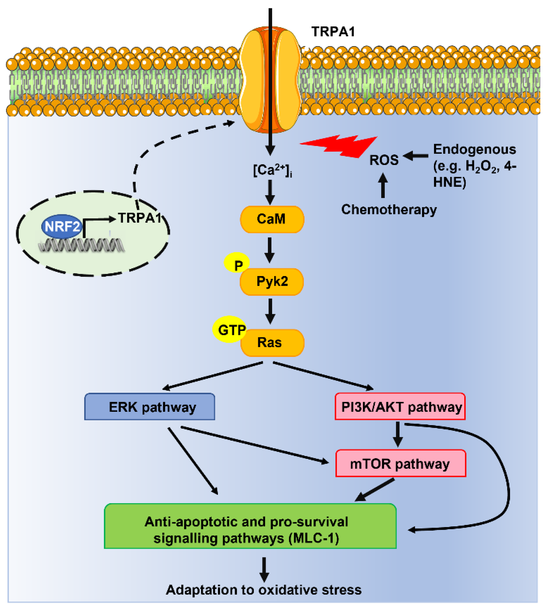
4.1. TRPA1-Mediated Ca2+ Influx Promotes Cancer Cell Survival to Oxidative Stress

Takahashi and coworkers recently carried out a systematic investigation to assess the role of extracellular Ca2+ entry through TRPA1 in the engagement of an antioxidant defense program in breast and lung cancer cells [29]. This report showed that the TRPA1 transcript and protein were up-regulated in breast and lung tumors as compared to adjacent normal tissue. Furthermore, TRPA1 activation with another electrophilic agonist, i.e., mustard oil, induced a long-lasting increase in [Ca2+]i that was abolished by the removal of extracellular Ca2+ and genetic (via a selective short hairpin RNA) or pharmacological (via AP-18) blockade of TRPA1 [29]. Intriguingly, the same approach demonstrated that TRPA1 mediated H2O2-evoked intracellular Ca2+ oscillations in breast and lung cancer cell lines [29]. In agreement with the pro-survival role of Ca2+ spiking in cancer cells [111,112], extracellular Ca2+ entry via TRPA1 was required to promote cell survival in TRPA1-enriched cancer cells challenged with H2O2 [29]. In addition, ectopic expression of TRPA1 rescued cell survival and prevented apoptosis in H2O2-treated TRPA1-low-expressing cancer cells [29]. By using a more physiological context, the authors unveiled an increase in ROS production in the inner region of breast and lung cancer spheroids, which led to TRPA1-mediated Ca2+ entry and resistance to oxidative stress. Importantly, TRPA1 activation did not reduce ROS levels, thereby indicating that TRPA1 does not contribute to scavenging ROS but rather to engaging an antioxidative defense program [29]. Furthermore, TRPA1-mediated Ca2+ influx promoted resistance to anoikis [29], i.e., the mode of apoptotic cell death that may occur when cells detach from the extracellular matrix (ECM) and migrate to a distant point to metastasize [113]. ROS generation is crucial to induce anoikis, but TRPA1 activation underlay detachment-induced Ca2+ signals and anoikis resistance in the inner region of tumor spheroids without reducing intracellular ROS levels [29]. Additionally, TRPA1-mediated Ca2+ entry promoted breast and lung cancer cell resistance to ROS-producing chemotherapeutics, such as carboplatin, doxorubicin, and paclitaxel [29]. In agreement with in vitro findings, this study demonstrated that genetic or pharmacological blockade of TRPA1 retarded breast and lung cancer growth and suppressed chemoresistance in immunocompromised mice and confirmed that tumor cells were also exposed to higher oxidative stress in vivo. Significant levels of 8-hydroxyguanosine (8-OHdG) and 4-HNE, which are common readouts of oxidative stress, were detected in cancer cells [29]. For instance, the 4-HNE concentration can increase up to the low micromolar range in the tumor microenvironment with potential pro-apoptotic effects against cancer cells [8]. Notably, 4-HNE has long been known to stimulate TRPA1-dependent intracellular Ca2+ signals [114,115,116]. The authors then exploited a reverse-phase protein array to unravel the Ca2+-dependent effectors that mediate the antioxidant defense triggered by TRPA1. They reported that TRPA1-mediated extracellular Ca2+ entry recruits the Ca2+/calmodulin-dependent proline-rich tyrosine kinase 2 (Pyk2) [117], which in turn engages several pro-survival signaling pathways, such as RAS-ERK, phosphatidylinositol 3-kinase (PI3K)/protein kinase B (AKT), and mammalian target of rapamycin (mTOR), and increases the expression of the anti-apoptotic protein MLC-1 (Figure 3) [29]. Finally, the authors demonstrated that TRPA1 expression in breast and lung cancer cells was regulated by NRF2, which can therefore prevent ROS-induced apoptosis by inducing the expression of both canonical (e.g., antioxidant) and non-canonical (e.g., TRPA1) oxidative stress defense proteins (Figure 3) [16,29]. Interestingly, mutations in NFE2L2 and KEAP1 genes, which, respectively, encode for NRF2 and KEAP1, were associated with higher TRPA1 expression in lung tumors and head-neck squamous carcinoma [29]. Genetic silencing of NRF2 reduced TRPA1 expression in lung cancer cell lines, while it did not affect the expression levels of other TRP isoforms, such as TRPC3 and TRPV1 [29]. This observation is rather interesting since TRPV1 is also sensitive to ROS signaling [64,103,106], while TRPC3 is primarily regulated by diacylglycerol [118]. Therefore, NRF2 is likely to selectively control TRPA1 expression, although NRF2-dependent regulation of other ROS-sensitive TRP isoforms, such as TRPM2 [105] and TRPV4 [119], should also be investigated. Chromatin immunoprecipitation (ChIP)-coupled deep sequencing (ChIP-Seq) identified three putative NRF2-binding sites (Peak 1 to Peak 3) around the TRPA1 gene locus [29]. However, only NRF2 binding to thPeak1 region was able to induce TRPA1 expression [29].
A recent investigation suggested that H2O2-induced Ca2+ entry via TRPA1 could also engage an antioxidant defense program in melanoma, which presents a rather high oxidative stress in the tumor microenvironment [30]. The authors showed that the number of intratumoral and peritumoral M2 macrophages and the amount of 4-HNE progressively increased with tumor severity in cutaneous melanoma samples, while TRPA1 protein expression remained unchanged [30]. In addition, genetic (via a selective small interfering RNA) and pharmacological (via A967079) blockade of TRPA1 suppressed H2O2-evoked intracellular Ca2+ signals in the human melanoma cell lines WM266-4 and SK-MEL-28 [30]. Furthermore, TRPA1-mediated Ca2+ signals exacerbated H2O2-dependent ROS production [30], which could reflect the Ca2+-dependent recruitment of NOX4 [17]. Therefore, TRPA1 activation could amplify oxidative stress in cutaneous melanoma to amplify tumor progression [30]. Future work will have to assess whether TRPA1-induced ROS production delivers a pro-tumorigenic input or somehow tempers the efficacy of the non-canonical defense program engaged by TRPA1-mediated Ca2+ signals. Addressing this issue would be instrumental to better predicting the therapeutic outcome of TRPA1 manipulation (see also Section 5.3).

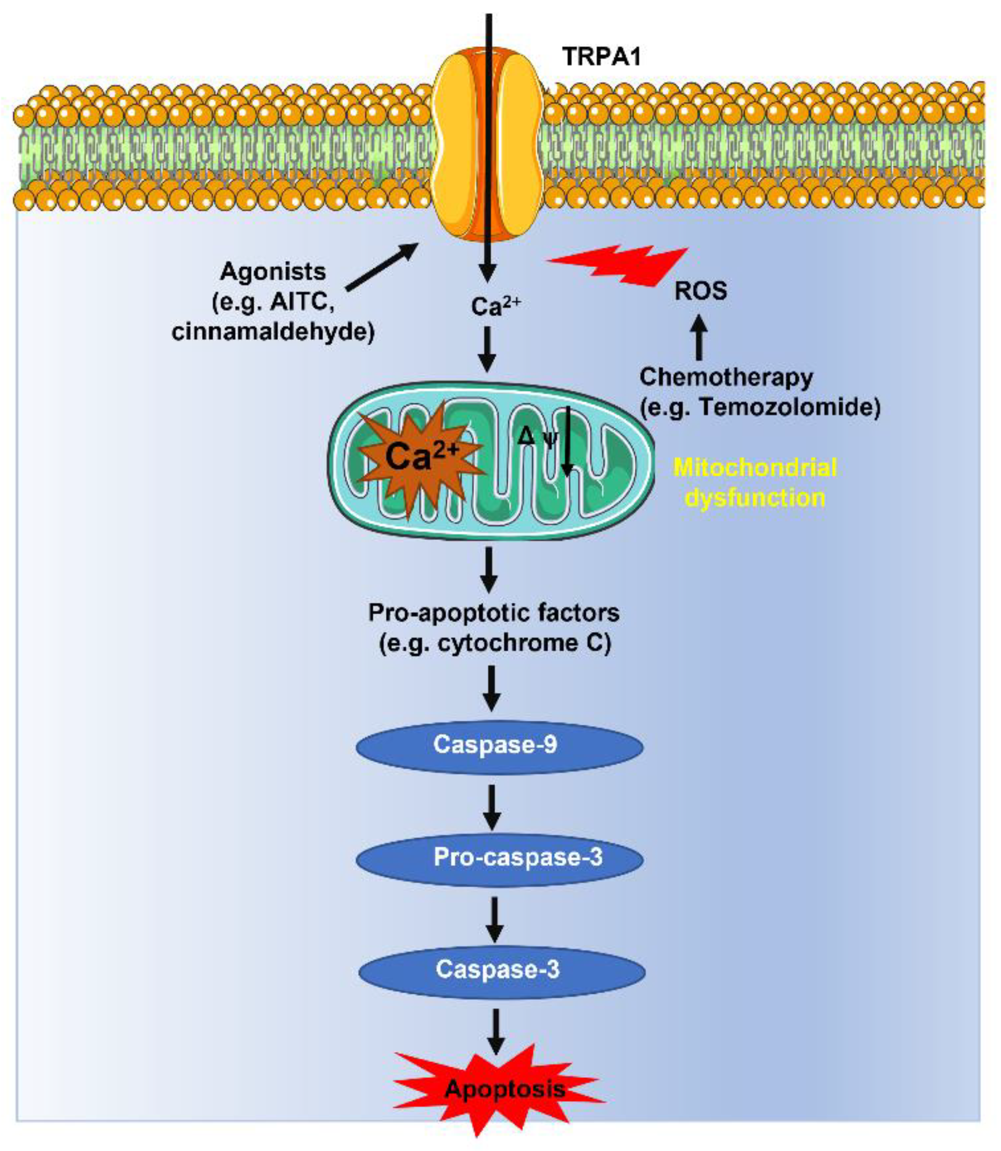
4.2. TRPA1-Mediated Ca2+ Influx Promotes ROS-Dependent Apoptosis in Cancer Cells

The findings described in [29] supported the prevailing view that extracellular Ca2+ entry via TRPA1 exerts an antioxidant defense effect in breast and lung cancer cells [16]. Nevertheless, parallel investigations provided evidence that the TRPA1-dependent increase in [Ca2+]i may also support H2O2-induced apoptosis in other types of cancer cells. Temozolomide (TMZ) therapy represents the standard of care for the treatment of glioblastoma by inducing lethal DNA damage and subsequent ROS production [120,121]. Unfortunately, the development of TMZ resistance severely hampers its therapeutic efficacy and leads to patients’ death [122]. A recent report showed that TMZ induced the expression of O6-methylguanine DNA-methyltransferase (MGMT), a DNA repair enzyme that favors glioblastoma cell resistance, and MnSOD, an antioxidant gene, in the glioblastoma cell lines SHG-44 and U251 [28]. However, previous activation of TRPA1 with Compound 16a (PF-4840154) increased ROS production, enhanced apoptosis, and reduced MGMT/MnSOD expression, thereby reducing TMZ resistance [28]. Similar results were obtained by the ectopic expression of TRPA1 in U521 cells exposed to TMZ. Mechanistic analysis revealed that TRPA1-mediated Ca2+ entry boosted ROS production by exacerbating TMZ-dependent damage to mitochondrial dynamics [28]. Consistently, an independent study showed that hypoxia increased TRPA1-dependent membrane currents in another human glioblastoma cell line, i.e., DBTRG, thereby inducing cytosolic Ca2+ overload, mitochondrial depolarization, caspase-3 and caspase-9 activation, and apoptosis (Figure 4) [32]. TMZ is also employed to treat relapsed or refractory neuroblastoma [121]. Interestingly, TMZ induced apoptosis in SH-SY5Y neuroblastoma cells via the ROS-dependent activation of TRPA1 followed by mitochondrial dysfunction and caspase activation (Figure 4) [123]. Therefore, these findings indicate that TRPA1 stimulation could represent a promising therapeutic strategy to sensitize certain cancer types to ROS-induced apoptosis. This hypothesis has been supported by two recent investigations showing that AITC induced cytosolic Ca2+ overload and reduced viability in OSCC PE/CA-PJ41 cells [23], whereas cinnamaldehyde, another selective electrophilic TRPA1 agonist, induced ROS-dependent apoptosis in colon cancer cells [31].

4.3. Why Does ROS-Sensitive TRPA1-Mediated Ca2+ Influx Exert both Anti-Cancer and Pro-Tumorigenic Effects in Cancer Cells?

The highly heterogeneous spatio-temporal profile of Ca2+ signals orchestrates the recruitment of downstream Ca2+-dependent effectors [126] and determines whether an increase in [Ca2+]i induces proliferation [111,127] or senescence [128,129], apoptosis [130,131,132] or autophagy [84,133], sensitivity [130,131] or resistance [125,134] to anticancer strategies. Therefore, the bimodal pro- and anti-oncogenic effect of ROS-dependent Ca2+ influx via TRPA1 in cancer cells is not surprising. Takahashi and coworkers demonstrated that TRPA1 triggers intracellular Ca2+ oscillations to promote ROS resistance in lung and breast cancers [29]. Conversely, preliminary evidence suggested that TRPA1 activation led to more protracted (and pervasive) Ca2+ elevations in OSCC [23] and glioblastoma [28,32], thereby resulting in cancer cell apoptosis. In accord, repetitive oscillations in [Ca2+]i are nicely suited to recruit Ca2+-dependent effectors that promote cancer cell proliferation and survival, including Pyk2 [135,136,137,138], while avoiding mitochondrial Ca2+ overload [127,130,139,140,141]. It is unclear why TRPA1 activation evoked pro-oncogenic repetitive Ca2+ spikes in some, e.g., breast and lung, but not all tumor types that have been examined so far. Extracellular Ca2+ entry via other TRP channels, such as TRPM2 [142], TRPV1 [64], and TRPV4 [143], can elicit rhythmic Ca2+ release from the ER via Ca2+-induced Ca2+ release (CICR) through inositol-1,4,5-trisphosphate (InsP3) receptors (InsP3Rs). Future work should investigate whether TRPA1 channel protein is selectively coupled to ER-located InsP3Rs in lung and breast cancers rather than in OSCC, glioblastoma, and colorectal carcinoma. Furthermore, TRPA1 has also been detected in acidic lysosomal vesicles [37], which represent an emerging pro-oncogenic Ca2+ releasing organelle in cancer cells [84,127] and stimulate InsP3-dependent ER Ca2+ spikes via CICR [127,144,145]. Takahashi and coworkers showed that the removal of extracellular Ca2+ abolished H2O2-evoked, TRPA1-mediated intracellular Ca2+ oscillations in lung and breast cancer cells [29]. Nevertheless, the lysosomal expression of TRPA1 in cancer cells surely deserves future investigation.

5. Targeting TRPA1 to Sensitize Cancer Cells to Oxidative Stress

The bimodal effect of the TRPA1-mediated Ca2+ influx on the ability of cancer cells to cope or not with oxidative stress could be appropriately exploited for therapeutic purposes. The pharmacological blockade of TRPA1 could provide an effective strategy to reduce the Ca2+-dependent recruitment of antioxidant defense and pro-survival signaling pathways in lung and breast cancers, as recently shown in [146]. Conversely, stimulating TRPA1-mediated Ca2+ entry could represent a valuable tool to sensitize ROS-dependent cytosolic Ca2+ overload and apoptosis in OSCC, glioblastoma, and colorectal carcinoma. TRPA1 channels are the most broadly tuned chemosensory channels identified so far and are sensitive to a vast panel of small molecular drugs and natural compounds, which can either stimulate or inhibit their activity with a rather high selectivity [19,36,44].

5.1. TRPA1 Activators

TRPA1 channels can be stimulated not only by ROS but also by synthetic drugs, local anesthetics, environmental irritants, and plant-derived pungent compounds [19,36,44,147]. TRPA1 agonists can be broadly categorized as electrophilic compounds, which increase the open probability through covalent modifications, and non-electrophilic activators, which exert a non-covalent modulation of the channel. Electrophilic agonists stimulate TRPA1 activity by targeting the reactive thiol groups of cysteine and lysine at the NH2-terminal domain that are also able to sense intracellular ROS (see Section 2.5). These compounds include AITC, cinnamaldehyde, allicin, mustard oil, hydrogen sulphide, diallyl sulfide, acrolein, and JT010 [19,36,44,147,148]. Non-electrophilic modulators induce TRPA1-mediated Ca2+ entry without inducing covalent modifications of the channel protein and include menthol, thymol, carvacrol, local anesthetics (e.g., lidocaine, tetracaine, and procaine), anesthetic agents (e.g., propofol and etomidate), nonsteroidal anti-inflammatory drugs (e.g., acetaminophen), and several compounds exploited in cosmetics and therapeutics (e.g., alkyl esters of p-hydroxybenzoate or parabens) [19,36,44,147]. The mechanism whereby some non-electrophilic activators stimulate TRPA1 has been elucidated [44]. The non-covalent agonist, GNE551, was recently shown to increase the single-channel open probability of TRPA1 by interacting with a hydrophobic transmembrane binding site (Gln940) [149]. In contrast, the cell-penetrating peptidergic scorpion toxin (WaTx) can prolong the single-channel opening of TRPA1 by associating with an intracellular electrophile ligand-binding domain contributed to by Cys621 and Cys641 [150]. Therefore, a variety of agonists are potentially available to stimulate TRPA1 and enhance ROS-dependent apoptosis in cancer cell types that succumb in response to sustained Ca2+ entry via TRPA1. A comprehensive list of electrophilic and non-electrophilic TRPA1 agonists is presented in Table 1.

5.2. TRPA1 Blockers

The involvement of TRPA1 in a growing number of disorders (see Section 1) has favored the development of specific TRPA1 antagonists [19,36,44,147], which are listed in Table 2. The first and most widespread TRPA1 antagonist was synthesized by the Hydra Company based on its xanthine structure and was named HC-030031 [147]. Chembridge-5861528 is a derivative of HC-030031 that presents similar potency and specificity, but improved solubility [36,44]. However, the most potent available TRPA1 inhibitors are Compound 10, Compound 31, A-967079, and Glenmark 10, 15, 37 (synthesized by Glenmark), which display half-maximal inhibitory concentration (IC50) values within the nanomolar range [19,36,44]. In addition, TRPA1 activity is sensitive to the paracetamol analog (also termed acetaminophen) 6a/b, a novel battery of α-aryl pyrrolidine sulfonamides, and the analgesics tramadol and its metabolite M1 [19]. The Sambucus ebulus L. (SEB) fruit extract, which can exert beneficial therapeutic effects against inflammation and ER stress-related disorders [163], has recently been shown to inhibit TRPA1 [31]. However, SEB, as well as early TRPA1 inhibitors such as camphor and ruthenium red, can target other TRP channels and is therefore not selective [31]. On the other hand, the partial agonist AP-18 inhibits TRPA1-mediated Ca2+ entry by desensitizing the channel [36,44].

5.3. Could TRPA1 Manipulation Directly Affect Oxidative Stress in the Tumor Microenvironment?

A recent investigation demonstrated that H2O2-dependent TRPA1 activation can amplify oxidative stress in melanoma cell lines [30]. It is, however, still unclear whether TRPA1-induced ROS production delivers a pro-tumorigenic input or somehow tempers the efficacy of the non-canonical defense program engaged by TRPA1-mediated Ca2+ signals. Addressing this issue would be instrumental in better predicting the therapeutic outcome of TRPA1 manipulation. In accord, selective TRPA1 stimulation to promote caspase activation and cell death could exacerbate oxidative stress (e.g., in brain tumors), possibly further boosting cancer cell elimination. Nevertheless, if TRPA1-induced ROS production rather facilitates tumor progression, the therapeutic efficacy of this approach could be hampered by a negative-feedback mechanism. Conversely, blocking TRPA1-mediated Ca2+ signals to prevent the recruitment of non-canonical antioxidant programs (e.g., in lung and breast cancers) would limit TRPA1-induced ROS production, thereby either increasing (if TRPA1-dependent ROS are pro-tumorigenic) or tempering (if TRPA1-dependent ROS are pro-apoptotic) the therapeutic impact of TRPA1 manipulation.

6. Conclusions

Emerging evidence indicates that the Ca2+-permeable, non-selective cation channel TRPA1 is upregulated in cancer cells. TRPA1 is the most highly redox-sensitive TRP isoform and therefore its ability to mediate Ca2+ entry translates the high oxidative stress of the tumor microenvironment in an intracellular Ca2+ signal that can dramatically impact cancer cell fate. In breast and lung cancer, ROS-induced TRPA1 activation leads to intracellular Ca2+ oscillations that engage antioxidant and pro-survival signaling pathways, resulting in cancer cell tolerance to oxidative stress. TRPA1-mediated Ca2+ entry could therefore promote resistance to pro-oxidant therapies based on ROS-producing drugs, such as carboplatin, doxorubicin, and paclitaxel. In other solid malignancies, including glioblastoma and neuroblastoma, ROS-induced TRPA1 activation results in a persistent increase in [Ca2+]i that causes mitochondrial Ca2+ overload and damage, thereby leading to caspase 3 activation and apoptotic cell death. These findings support the notion that the pharmacological manipulation of TRPA1-mediated Ca2+ signals could represent an alternative anticancer strategy. For instance, selective TRPA1 agonists (e.g., brain tumors) or blockers (e.g., lung and breast cancers) could be administered as adjuvant drugs of ROS-producing therapeutics, such as carboplatin, doxorubicin, and paclitaxel, to increase cancer cell sensitivity to oxidative stress.

Author Contributions

Conceptualization, F.M.; methodology, F.M. and D.M.; writing—original draft preparation, F.M.; writing—review and editing, F.M. and D.M.; supervision, F.M.; funding acquisition, F.M. and D.M. All authors have read and agreed to the published version of the manuscript.

Funding

This research was funded by the Italian Ministry of Education, University and Research (MIUR): Dipartimenti di Eccellenza Program (2018–2022)-Dept. of Biology and Biotechnology “L. Spallanzani”, University of Pavia (F.M.), Fondo Ricerca Giovani from the University of Pavia (F.M.), Program “Ricerca Corrente 08059819” of the Foundation IRCCS Policlinico San Matteo, Pavia (D.M.).

Institutional Review Board Statement

Not applicable.

Informed Consent Statement

Not applicable.

Data Availability Statement

Not applicable.

Acknowledgments

The authors gratefully acknowledge all members of their laboratories for their commitment to cancer research.

Conflicts of Interest

The authors declare no conflict of interest.

References

  1. Aboelella, N.S.; Brandle, C.; Kim, T.; Ding, Z.C.; Zhou, G. Oxidative Stress in the Tumor Microenvironment and Its Relevance to Cancer Immunotherapy. Cancers 2021, 13, 986. [Google Scholar] [CrossRef]
  2. Rezcek, C.R.; Chandel, N.S. The Two Faces of Reactive Oxygen Species in Cancer. Annu. Rev. Cancer Biol. 2017, 1, 79–89. [Google Scholar] [CrossRef]
  3. Cheung, E.C.; Vousden, K.H. The role of ROS in tumour development and progression. Nat. Rev. Cancer 2022, 22, 280–297. [Google Scholar] [CrossRef] [PubMed]
  4. Remigante, A.; Morabito, R.; Spinelli, S.; Trichilo, V.; Loddo, S.; Sarikas, A.; Dossena, S.; Marino, A. d-Galactose Decreases Anion Exchange Capability through Band 3 Protein in Human Erythrocytes. Antioxidants 2020, 9, 689. [Google Scholar] [CrossRef]
  5. Panieri, E.; Santoro, M.M. ROS homeostasis and metabolism: A dangerous liason in cancer cells. Cell Death Dis. 2016, 7, e2253. [Google Scholar] [CrossRef]
  6. Perillo, B.; Di Donato, M.; Pezone, A.; Di Zazzo, E.; Giovannelli, P.; Galasso, G.; Castoria, G.; Migliaccio, A. ROS in cancer therapy: The bright side of the moon. Exp. Mol. Med. 2020, 52, 192–203. [Google Scholar] [CrossRef]
  7. Doskey, C.M.; Buranasudja, V.; Wagner, B.A.; Wilkes, J.G.; Du, J.; Cullen, J.J.; Buettner, G.R. Tumor cells have decreased ability to metabolize H(2)O(2): Implications for pharmacological ascorbate in cancer therapy. Redox Biol. 2016, 10, 274–284. [Google Scholar] [CrossRef] [PubMed]
  8. Burdon, R.H. Superoxide and hydrogen peroxide in relation to mammalian cell proliferation. Free Radic. Biol. Med. 1995, 18, 775–794. [Google Scholar] [CrossRef] [PubMed]
  9. Sullivan, L.B.; Chandel, N.S. Mitochondrial reactive oxygen species and cancer. Cancer Metab. 2014, 2, 17. [Google Scholar] [CrossRef]
  10. Hayes, J.D.; Dinkova-Kostova, A.T.; Tew, K.D. Oxidative Stress in Cancer. Cancer Cell 2020, 38, 167–197. [Google Scholar] [CrossRef] [PubMed]
  11. Redza-Dutordoir, M.; Averill-Bates, D.A. Activation of apoptosis signalling pathways by reactive oxygen species. Biochim. Biophys. Acta 2016, 1863, 2977–2992. [Google Scholar] [CrossRef] [PubMed]
  12. Ahmad, K.A.; Iskandar, K.B.; Hirpara, J.L.; Clement, M.V.; Pervaiz, S. Hydrogen peroxide-mediated cytosolic acidification is a signal for mitochondrial translocation of Bax during drug-induced apoptosis of tumor cells. Cancer Res. 2004, 64, 7867–7878. [Google Scholar] [CrossRef] [PubMed]
  13. Wu, L.; Ishigaki, Y.; Zeng, W.; Harimoto, T.; Yin, B.; Chen, Y.; Liao, S.; Liu, Y.; Sun, Y.; Zhang, X.; et al. Generation of hydroxyl radical-activatable ratiometric near-infrared bimodal probes for early monitoring of tumor response to therapy. Nat. Commun. 2021, 12, 6145. [Google Scholar] [CrossRef] [PubMed]
  14. Gorrini, C.; Harris, I.S.; Mak, T.W. Modulation of oxidative stress as an anticancer strategy. Nat. Rev. Drug Discov. 2013, 12, 931–947. [Google Scholar] [CrossRef] [PubMed]
  15. Gamcsik, M.P.; Kasibhatla, M.S.; Teeter, S.D.; Colvin, O.M. Glutathione levels in human tumors. Biomarkers 2012, 17, 671–691. [Google Scholar] [CrossRef]
  16. Reczek, C.R.; Chandel, N.S. ROS Promotes Cancer Cell Survival through Calcium Signaling. Cancer Cell 2018, 33, 949–951. [Google Scholar] [CrossRef]
  17. Negri, S.; Faris, P.; Moccia, F. Reactive Oxygen Species and Endothelial Ca(2+) Signaling: Brothers in Arms or Partners in Crime? Int. J. Mol. Sci. 2021, 22, 9821. [Google Scholar] [CrossRef]
  18. Luo, M.; Zhou, L.; Huang, Z.; Li, B.; Nice, E.C.; Xu, J.; Huang, C. Antioxidant Therapy in Cancer: Rationale and Progress. Antioxidants 2022, 11, 1128. [Google Scholar] [CrossRef]
  19. Talavera, K.; Startek, J.B.; Alvarez-Collazo, J.; Boonen, B.; Alpizar, Y.A.; Sanchez, A.; Naert, R.; Nilius, B. Mammalian Transient Receptor Potential TRPA1 Channels: From Structure to Disease. Physiol. Rev. 2020, 100, 725–803. [Google Scholar] [CrossRef]
  20. Zygmunt, P.M.; Hogestatt, E.D. Trpa1. Handb. Exp. Pharmacol. 2014, 222, 583–630. [Google Scholar] [CrossRef]
  21. Singh, R.; Adhya, P.; Sharma, S.S. Redox-sensitive TRP channels: A promising pharmacological target in chemotherapy-induced peripheral neuropathy. Expert Opin. Ther. Targets 2021, 25, 529–545. [Google Scholar] [CrossRef] [PubMed]
  22. Yamamoto, S.; Shimizu, S. Significance of TRP channels in oxidative stress. Eur. J. Pharmacol. 2016, 793, 109–111. [Google Scholar] [CrossRef] [PubMed]
  23. Kiss, F.; Kormos, V.; Szoke, E.; Kecskes, A.; Toth, N.; Steib, A.; Szallasi, A.; Scheich, B.; Gaszner, B.; Kun, J.; et al. Functional Transient Receptor Potential Ankyrin 1 and Vanilloid 1 Ion Channels Are Overexpressed in Human Oral Squamous Cell Carcinoma. Int. J. Mol. Sci. 2022, 23, 1921. [Google Scholar] [CrossRef] [PubMed]
  24. Cojocaru, F.; Selescu, T.; Domocos, D.; Marutescu, L.; Chiritoiu, G.; Chelaru, N.R.; Dima, S.; Mihailescu, D.; Babes, A.; Cucu, D. Functional expression of the transient receptor potential ankyrin type 1 channel in pancreatic adenocarcinoma cells. Sci. Rep. 2021, 11, 2018. [Google Scholar] [CrossRef]
  25. Mergler, S.; Derckx, R.; Reinach, P.S.; Garreis, F.; Bohm, A.; Schmelzer, L.; Skosyrski, S.; Ramesh, N.; Abdelmessih, S.; Polat, O.K.; et al. Calcium regulation by temperature-sensitive transient receptor potential channels in human uveal melanoma cells. Cell. Signal. 2014, 26, 56–69. [Google Scholar] [CrossRef]
  26. Duitama, M.; Moreno, Y.; Santander, S.P.; Casas, Z.; Sutachan, J.J.; Torres, Y.P.; Albarracin, S.L. TRP Channels as Molecular Targets to Relieve Cancer Pain. Biomolecules 2021, 12, 1. [Google Scholar] [CrossRef]
  27. de Almeida, A.S.; Bernardes, L.B.; Trevisan, G. TRP channels in cancer pain. Eur. J. Pharmacol. 2021, 904, 174185. [Google Scholar] [CrossRef]
  28. Chen, H.; Li, C.; Hu, H.; Zhang, B. Activated TRPA1 plays a therapeutic role in TMZ resistance in glioblastoma by altering mitochondrial dynamics. BMC Mol. Cell Biol. 2022, 23, 38. [Google Scholar] [CrossRef]
  29. Takahashi, N.; Chen, H.Y.; Harris, I.S.; Stover, D.G.; Selfors, L.M.; Bronson, R.T.; Deraedt, T.; Cichowski, K.; Welm, A.L.; Mori, Y.; et al. Cancer Cells Co-opt the Neuronal Redox-Sensing Channel TRPA1 to Promote Oxidative-Stress Tolerance. Cancer Cell 2018, 33, 985–1003.e1007. [Google Scholar] [CrossRef]
  30. De Logu, F.; Souza Monteiro de Araujo, D.; Ugolini, F.; Iannone, L.F.; Vannucchi, M.; Portelli, F.; Landini, L.; Titiz, M.; De Giorgi, V.; Geppetti, P.; et al. The TRPA1 Channel Amplifies the Oxidative Stress Signal in Melanoma. Cells 2021, 10, 3131. [Google Scholar] [CrossRef]
  31. Kaya, M.M.; Kaya, I.; Naziroglu, M. Transient receptor potential channel stimulation induced oxidative stress and apoptosis in the colon of mice with colitis-associated colon cancer: Modulator role of Sambucus ebulus L. Mol. Biol. Rep. 2022, 50, 2207–2220. [Google Scholar] [CrossRef] [PubMed]
  32. Deveci, H.A.; Akyuva, Y.; Nur, G.; Naziroglu, M. Alpha lipoic acid attenuates hypoxia-induced apoptosis, inflammation and mitochondrial oxidative stress via inhibition of TRPA1 channel in human glioblastoma cell line. Biomed. Pharmacother. 2019, 111, 292–304. [Google Scholar] [CrossRef] [PubMed]
  33. Schaefer, E.A.; Stohr, S.; Meister, M.; Aigner, A.; Gudermann, T.; Buech, T.R. Stimulation of the chemosensory TRPA1 cation channel by volatile toxic substances promotes cell survival of small cell lung cancer cells. Biochem. Pharmacol. 2013, 85, 426–438. [Google Scholar] [CrossRef]
  34. Gees, M.; Colsoul, B.; Nilius, B. The role of transient receptor potential cation channels in Ca2+ signaling. Cold Spring Harb. Perspect. Biol. 2010, 2, a003962. [Google Scholar] [CrossRef] [PubMed]
  35. Himmel, N.J.; Cox, D.N. Transient receptor potential channels: Current perspectives on evolution, structure, function and nomenclature. Proc. Biol. Sci. 2020, 287, 20201309. [Google Scholar] [CrossRef]
  36. Alvarado, M.G.; Thakore, P.; Earley, S. Transient Receptor Potential Channel Ankyrin 1: A Unique Regulator of Vascular Function. Cells 2021, 10, 1167. [Google Scholar] [CrossRef] [PubMed]
  37. Shang, S.; Zhu, F.; Liu, B.; Chai, Z.; Wu, Q.; Hu, M.; Wang, Y.; Huang, R.; Zhang, X.; Wu, X.; et al. Intracellular TRPA1 mediates Ca2+ release from lysosomes in dorsal root ganglion neurons. J. Cell Biol. 2016, 215, 369–381. [Google Scholar] [CrossRef]
  38. Jaquemar, D.; Schenker, T.; Trueb, B. An ankyrin-like protein with transmembrane domains is specifically lost after oncogenic transformation of human fibroblasts. J. Biol. Chem. 1999, 274, 7325–7333. [Google Scholar] [CrossRef]
  39. Paulsen, C.E.; Armache, J.P.; Gao, Y.; Cheng, Y.; Julius, D. Structure of the TRPA1 ion channel suggests regulatory mechanisms. Nature 2015, 520, 511–517. [Google Scholar] [CrossRef]
  40. Cordero-Morales, J.F.; Gracheva, E.O.; Julius, D. Cytoplasmic ankyrin repeats of transient receptor potential A1 (TRPA1) dictate sensitivity to thermal and chemical stimuli. Proc. Natl. Acad. Sci. USA 2011, 108, E1184–E1191. [Google Scholar] [CrossRef]
  41. Samad, A.; Sura, L.; Benedikt, J.; Ettrich, R.; Minofar, B.; Teisinger, J.; Vlachova, V. The C-terminal basic residues contribute to the chemical- and voltage-dependent activation of TRPA1. Biochem. J. 2011, 433, 197–204. [Google Scholar] [CrossRef]
  42. Meents, J.E.; Ciotu, C.I.; Fischer, M.J.M. TRPA1: A molecular view. J. Neurophysiol. 2019, 121, 427–443. [Google Scholar] [CrossRef] [PubMed]
  43. Thakore, P.; Alvarado, M.G.; Ali, S.; Mughal, A.; Pires, P.W.; Yamasaki, E.; Pritchard, H.A.; Isakson, B.E.; Tran, C.H.T.; Earley, S. Brain endothelial cell TRPA1 channels initiate neurovascular coupling. eLife 2021, 10, e63040. [Google Scholar] [CrossRef] [PubMed]
  44. Thakore, P.; Ali, S.; Earley, S. Regulation of vascular tone by transient receptor potential ankyrin 1 channels. Curr. Top. Membr. 2020, 85, 119–150. [Google Scholar] [CrossRef] [PubMed]
  45. Karashima, Y.; Prenen, J.; Talavera, K.; Janssens, A.; Voets, T.; Nilius, B. Agonist-induced changes in Ca(2+) permeation through the nociceptor cation channel TRPA1. Biophys. J. 2010, 98, 773–783. [Google Scholar] [CrossRef]
  46. Banke, T.G.; Chaplan, S.R.; Wickenden, A.D. Dynamic changes in the TRPA1 selectivity filter lead to progressive but reversible pore dilation. Am. J. Physiol. Cell Physiol. 2010, 298, C1457–C1468. [Google Scholar] [CrossRef]
  47. Chen, J.; Kim, D.; Bianchi, B.R.; Cavanaugh, E.J.; Faltynek, C.R.; Kym, P.R.; Reilly, R.M. Pore dilation occurs in TRPA1 but not in TRPM8 channels. Mol. Pain 2009, 5, 3. [Google Scholar] [CrossRef]
  48. Li, M.; Toombes, G.E.; Silberberg, S.D.; Swartz, K.J. Physical basis of apparent pore dilation of ATP-activated P2X receptor channels. Nat. Neurosci. 2015, 18, 1577–1583. [Google Scholar] [CrossRef]
  49. Jordt, S.E.; Bautista, D.M.; Chuang, H.H.; McKemy, D.D.; Zygmunt, P.M.; Hogestatt, E.D.; Meng, I.D.; Julius, D. Mustard oils and cannabinoids excite sensory nerve fibres through the TRP channel ANKTM1. Nature 2004, 427, 260–265. [Google Scholar] [CrossRef]
  50. Wang, Y.Y.; Chang, R.B.; Waters, H.N.; McKemy, D.D.; Liman, E.R. The nociceptor ion channel TRPA1 is potentiated and inactivated by permeating calcium ions. J. Biol. Chem. 2008, 283, 32691–32703. [Google Scholar] [CrossRef]
  51. Doerner, J.F.; Gisselmann, G.; Hatt, H.; Wetzel, C.H. Transient receptor potential channel A1 is directly gated by calcium ions. J. Biol. Chem. 2007, 282, 13180–13189. [Google Scholar] [CrossRef]
  52. Sura, L.; Zima, V.; Marsakova, L.; Hynkova, A.; Barvik, I.; Vlachova, V. C-terminal acidic cluster is involved in Ca2+-induced regulation of human transient receptor potential ankyrin 1 channel. J. Biol. Chem. 2012, 287, 18067–18077. [Google Scholar] [CrossRef] [PubMed]
  53. Hasan, R.; Leeson-Payne, A.T.; Jaggar, J.H.; Zhang, X. Calmodulin is responsible for Ca(2+)-dependent regulation of TRPA1 Channels. Sci. Rep. 2017, 7, 45098. [Google Scholar] [CrossRef]
  54. Moparthi, L.; Kichko, T.I.; Eberhardt, M.; Hogestatt, E.D.; Kjellbom, P.; Johanson, U.; Reeh, P.W.; Leffler, A.; Filipovic, M.R.; Zygmunt, P.M. Human TRPA1 is a heat sensor displaying intrinsic U-shaped thermosensitivity. Sci. Rep. 2016, 6, 28763. [Google Scholar] [CrossRef]
  55. Moparthi, L.; Sinica, V.; Moparthi, V.K.; Kreir, M.; Vignane, T.; Filipovic, M.R.; Vlachova, V.; Zygmunt, P.M. The human TRPA1 intrinsic cold and heat sensitivity involves separate channel structures beyond the N-ARD domain. Nat. Commun. 2022, 13, 6113. [Google Scholar] [CrossRef]
  56. Sinica, V.; Vlachova, V. Transient receptor potential ankyrin 1 channel: An evolutionarily tuned thermosensor. Physiol. Res. 2021, 70, 363–381. [Google Scholar] [CrossRef] [PubMed]
  57. Moparthi, L.; Survery, S.; Kreir, M.; Simonsen, C.; Kjellbom, P.; Hogestatt, E.D.; Johanson, U.; Zygmunt, P.M. Human TRPA1 is intrinsically cold- and chemosensitive with and without its N-terminal ankyrin repeat domain. Proc. Natl. Acad. Sci. USA 2014, 111, 16901–16906. [Google Scholar] [CrossRef] [PubMed]
  58. Reeh, P.W.; Fischer, M.J.M. Nobel somatosensations and pain. Pflug. Arch. 2022, 474, 405–420. [Google Scholar] [CrossRef]
  59. Moparthi, L.; Zygmunt, P.M. Human TRPA1 is an inherently mechanosensitive bilayer-gated ion channel. Cell Calcium 2020, 91, 102255. [Google Scholar] [CrossRef]
  60. Sakaguchi, R.; Mori, Y. Transient receptor potential (TRP) channels: Biosensors for redox environmental stimuli and cellular status. Free Radic. Biol. Med. 2020, 146, 36–44. [Google Scholar] [CrossRef]
  61. Ferrera, L.; Barbieri, R.; Picco, C.; Zuccolini, P.; Remigante, A.; Bertelli, S.; Fumagalli, M.R.; Zifarelli, G.; La Porta, C.A.M.; Gavazzo, P.; et al. TRPM2 Oxidation Activates Two Distinct Potassium Channels in Melanoma Cells through Intracellular Calcium Increase. Int. J. Mol. Sci. 2021, 22, 8359. [Google Scholar] [CrossRef]
  62. Remigante, A.; Spinelli, S.; Marino, A.; Pusch, M.; Morabito, R.; Dossena, S. Oxidative Stress and Immune Response in Melanoma: Ion Channels as Targets of Therapy. Int. J. Mol. Sci. 2023, 24, 887. [Google Scholar] [CrossRef]
  63. Ogawa, N.; Kurokawa, T.; Fujiwara, K.; Polat, O.K.; Badr, H.; Takahashi, N.; Mori, Y. Functional and Structural Divergence in Human TRPV1 Channel Subunits by Oxidative Cysteine Modification. J. Biol. Chem. 2016, 291, 4197–4210. [Google Scholar] [CrossRef] [PubMed]
  64. Negri, S.; Faris, P.; Tullii, G.; Vismara, M.; Pellegata, A.F.; Lodola, F.; Guidetti, G.; Rosti, V.; Antognazza, M.R.; Moccia, F. Conjugated polymers mediate intracellular Ca(2+) signals in circulating endothelial colony forming cells through the reactive oxygen species-dependent activation of Transient Receptor Potential Vanilloid 1 (TRPV1). Cell Calcium 2022, 101, 102502. [Google Scholar] [CrossRef]
  65. Andersson, D.A.; Gentry, C.; Moss, S.; Bevan, S. Transient receptor potential A1 is a sensory receptor for multiple products of oxidative stress. J. Neurosci. 2008, 28, 2485–2494. [Google Scholar] [CrossRef]
  66. Bessac, B.F.; Sivula, M.; von Hehn, C.A.; Escalera, J.; Cohn, L.; Jordt, S.E. TRPA1 is a major oxidant sensor in murine airway sensory neurons. J. Clin. Investig. 2008, 118, 1899–1910. [Google Scholar] [CrossRef]
  67. Miyamoto, T.; Dubin, A.E.; Petrus, M.J.; Patapoutian, A. TRPV1 and TRPA1 mediate peripheral nitric oxide-induced nociception in mice. PLoS ONE 2009, 4, e7596. [Google Scholar] [CrossRef] [PubMed]
  68. Andersson, D.A.; Filipovic, M.R.; Gentry, C.; Eberhardt, M.; Vastani, N.; Leffler, A.; Reeh, P.; Bevan, S. Streptozotocin Stimulates the Ion Channel TRPA1 Directly: Involvement of peroxynitrite. J. Biol. Chem. 2015, 290, 15185–15196. [Google Scholar] [CrossRef] [PubMed]
  69. Crupi, R.; Morabito, R.; Remigante, A.; Gugliandolo, E.; Britti, D.; Cuzzocrea, S.; Marino, A. Susceptibility of erythrocytes from different sources to xenobiotics-induced lysis. Comp. Biochem. Physiol. C Toxicol. Pharmacol. 2019, 221, 68–72. [Google Scholar] [CrossRef]
  70. Taylor-Clark, T.E.; Ghatta, S.; Bettner, W.; Undem, B.J. Nitrooleic acid, an endogenous product of nitrative stress, activates nociceptive sensory nerves via the direct activation of TRPA1. Mol. Pharmacol. 2009, 75, 820–829. [Google Scholar] [CrossRef]
  71. Bahia, P.K.; Parks, T.A.; Stanford, K.R.; Mitchell, D.A.; Varma, S.; Stevens, S.M., Jr.; Taylor-Clark, T.E. The exceptionally high reactivity of Cys 621 is critical for electrophilic activation of the sensory nerve ion channel TRPA1. J. Gen. Physiol. 2016, 147, 451–465. [Google Scholar] [CrossRef]
  72. Zayats, V.; Samad, A.; Minofar, B.; Roelofs, K.E.; Stockner, T.; Ettrich, R. Regulation of the transient receptor potential channel TRPA1 by its N-terminal ankyrin repeat domain. J. Mol. Model. 2013, 19, 4689–4700. [Google Scholar] [CrossRef] [PubMed]
  73. Hinman, A.; Chuang, H.H.; Bautista, D.M.; Julius, D. TRP channel activation by reversible covalent modification. Proc. Natl. Acad. Sci. USA 2006, 103, 19564–19568. [Google Scholar] [CrossRef] [PubMed]
  74. Takahashi, N.; Kuwaki, T.; Kiyonaka, S.; Numata, T.; Kozai, D.; Mizuno, Y.; Yamamoto, S.; Naito, S.; Knevels, E.; Carmeliet, P.; et al. TRPA1 underlies a sensing mechanism for O2. Nat. Chem. Biol. 2011, 7, 701–711. [Google Scholar] [CrossRef]
  75. Nguyen, T.L.; Duran, R.V. Prolyl hydroxylase domain enzymes and their role in cell signaling and cancer metabolism. Int. J. Biochem. Cell Biol. 2016, 80, 71–80. [Google Scholar] [CrossRef]
  76. Chandel, N.S.; McClintock, D.S.; Feliciano, C.E.; Wood, T.M.; Melendez, J.A.; Rodriguez, A.M.; Schumacker, P.T. Reactive oxygen species generated at mitochondrial complex III stabilize hypoxia-inducible factor-1alpha during hypoxia: A mechanism of O2 sensing. J. Biol. Chem. 2000, 275, 25130–25138. [Google Scholar] [CrossRef]
  77. Mori, Y.; Takahashi, N.; Kurokawa, T.; Kiyonaka, S. TRP channels in oxygen physiology: Distinctive functional properties and roles of TRPA1 in O(2) sensing. Proc. Jpn. Acad. Ser. B Phys. Biol. Sci. 2017, 93, 464–482. [Google Scholar] [CrossRef] [PubMed]
  78. Eelen, G.; Treps, L.; Li, X.; Carmeliet, P. Basic and Therapeutic Aspects of Angiogenesis Updated. Circ. Res. 2020, 127, 310–329. [Google Scholar] [CrossRef]
  79. Armani, G.; Pozzi, E.; Pagani, A.; Porta, C.; Rizzo, M.; Cicognini, D.; Rovati, B.; Moccia, F.; Pedrazzoli, P.; Ferraris, E. The heterogeneity of cancer endothelium: The relevance of angiogenesis and endothelial progenitor cells in cancer microenvironment. Microvasc. Res. 2021, 138, 104189. [Google Scholar] [CrossRef]
  80. Monteith, G.R.; Prevarskaya, N.; Roberts-Thomson, S.J. The calcium-cancer signalling nexus. Nat. Rev. Cancer 2017, 17, 367–380. [Google Scholar] [CrossRef] [PubMed]
  81. Prevarskaya, N.; Skryma, R.; Shuba, Y. Ion Channels in Cancer: Are Cancer Hallmarks Oncochannelopathies? Physiol. Rev. 2018, 98, 559–621. [Google Scholar] [CrossRef] [PubMed]
  82. Scarpellino, G.; Genova, T.; Avanzato, D.; Bernardini, M.; Bianco, S.; Petrillo, S.; Tolosano, E.; de Almeida Vieira, J.R.; Bussolati, B.; Fiorio Pla, A.; et al. Purinergic Calcium Signals in Tumor-Derived Endothelium. Cancers 2019, 11, 766. [Google Scholar] [CrossRef]
  83. Scarpellino, G.; Munaron, L.; Cantelmo, A.R.; Fiorio Pla, A. Calcium-Permeable Channels in Tumor Vascularization: Peculiar, Sensors of Microenvironmental Chemical and Physical Cues. Rev. Physiol. Biochem. Pharmacol. 2020, 182, 111–137. [Google Scholar] [CrossRef]
  84. Faris, P.; Shekha, M.; Montagna, D.; Guerra, G.; Moccia, F. Endolysosomal Ca(2+) Signalling and Cancer Hallmarks: Two-Pore Channels on the Move, TRPML1 Lags Behind! Cancers 2018, 11, 27. [Google Scholar] [CrossRef]
  85. Zhong, T.; Zhang, W.; Guo, H.; Pan, X.; Chen, X.; He, Q.; Yang, B.; Ding, L. The regulatory and modulatory roles of TRP family channels in malignant tumors and relevant therapeutic strategies. Acta Pharm. Sin. B 2022, 12, 1761–1780. [Google Scholar] [CrossRef] [PubMed]
  86. Lodola, F.; Laforenza, U.; Cattaneo, F.; Ruffinatti, F.A.; Poletto, V.; Massa, M.; Tancredi, R.; Zuccolo, E.; Khdar, A.D.; Riccardi, A.; et al. VEGF-induced intracellular Ca2+ oscillations are down-regulated and do not stimulate angiogenesis in breast cancer-derived endothelial colony forming cells. Oncotarget 2017, 8, 95223–95246. [Google Scholar] [CrossRef] [PubMed]
  87. Fliniaux, I.; Germain, E.; Farfariello, V.; Prevarskaya, N. TRPs and Ca(2+) in cell death and survival. Cell Calcium 2018, 69, 4–18. [Google Scholar] [CrossRef]
  88. Perna, A.; Sellitto, C.; Komici, K.; Hay, E.; Rocca, A.; De Blasiis, P.; Lucariello, A.; Moccia, F.; Guerra, G. Transient Receptor Potential (TRP) Channels in Tumor Vascularization. Int. J. Mol. Sci. 2022, 23, 4253. [Google Scholar] [CrossRef] [PubMed]
  89. Takahashi, K.; Ohta, T. Membrane translocation of transient receptor potential ankyrin 1 induced by inflammatory cytokines in lung cancer cells. Biochem. Biophys. Res. Commun. 2017, 490, 587–593. [Google Scholar] [CrossRef]
  90. Derouiche, S.; Mariot, P.; Warnier, M.; Vancauwenberghe, E.; Bidaux, G.; Gosset, P.; Mauroy, B.; Bonnal, J.L.; Slomianny, C.; Delcourt, P.; et al. Activation of TRPA1 Channel by Antibacterial Agent Triclosan Induces VEGF Secretion in Human Prostate Cancer Stromal Cells. Cancer Prev. Res. (Phila.) 2017, 10, 177–187. [Google Scholar] [CrossRef]
  91. Moccia, F.; Poletto, V. May the remodeling of the Ca(2)(+) toolkit in endothelial progenitor cells derived from cancer patients suggest alternative targets for anti-angiogenic treatment? Biochim. Biophys. Acta 2015, 1853, 1958–1973. [Google Scholar] [CrossRef] [PubMed]
  92. Bernardini, M.; Brossa, A.; Chinigo, G.; Grolez, G.P.; Trimaglio, G.; Allart, L.; Hulot, A.; Marot, G.; Genova, T.; Joshi, A.; et al. Transient Receptor Potential Channel Expression Signatures in Tumor-Derived Endothelial Cells: Functional Roles in Prostate Cancer Angiogenesis. Cancers 2019, 11, 956. [Google Scholar] [CrossRef] [PubMed]
  93. Vancauwenberghe, E.; Noyer, L.; Derouiche, S.; Lemonnier, L.; Gosset, P.; Sadofsky, L.R.; Mariot, P.; Warnier, M.; Bokhobza, A.; Slomianny, C.; et al. Activation of mutated TRPA1 ion channel by resveratrol in human prostate cancer associated fibroblasts (CAF). Mol. Carcinog. 2017, 56, 1851–1867. [Google Scholar] [CrossRef]
  94. Genova, T.; Grolez, G.P.; Camillo, C.; Bernardini, M.; Bokhobza, A.; Richard, E.; Scianna, M.; Lemonnier, L.; Valdembri, D.; Munaron, L.; et al. TRPM8 inhibits endothelial cell migration via a non-channel function by trapping the small GTPase Rap1. J. Cell Biol. 2017, 216, 2107–2130. [Google Scholar] [CrossRef]
  95. Grolez, G.P.; Chinigo, G.; Barras, A.; Hammadi, M.; Noyer, L.; Kondratska, K.; Bulk, E.; Oullier, T.; Marionneau-Lambot, S.; Le Mee, M.; et al. TRPM8 as an Anti-Tumoral Target in Prostate Cancer Growth and Metastasis Dissemination. Int. J. Mol. Sci. 2022, 23, 6672. [Google Scholar] [CrossRef] [PubMed]
  96. Soda, T.; Brunetti, V.; Berra-Romani, R.; Moccia, F. The Emerging Role of N-Methyl-D-Aspartate (NMDA) Receptors in the Cardiovascular System: Physiological Implications, Pathological Consequences, and Therapeutic Perspectives. Int. J. Mol. Sci. 2023, 24, 3914. [Google Scholar] [CrossRef] [PubMed]
  97. Negri, S.; Scolari, F.; Vismara, M.; Brunetti, V.; Faris, P.; Terribile, G.; Sancini, G.; Berra-Romani, R.; Moccia, F. GABA(A) and GABA(B) Receptors Mediate GABA-Induced Intracellular Ca(2+) Signals in Human Brain Microvascular Endothelial Cells. Cells 2022, 11, 3860. [Google Scholar] [CrossRef]
  98. Negri, S.; Faris, P.; Maniezzi, C.; Pellavio, G.; Spaiardi, P.; Botta, L.; Laforenza, U.; Biella, G.; Moccia, D.F. NMDA receptors elicit flux-independent intracellular Ca(2+) signals via metabotropic glutamate receptors and flux-dependent nitric oxide release in human brain microvascular endothelial cells. Cell Calcium 2021, 99, 102454. [Google Scholar] [CrossRef]
  99. Berrout, J.; Kyriakopoulou, E.; Moparthi, L.; Hogea, A.S.; Berrout, L.; Ivan, C.; Lorger, M.; Boyle, J.; Peers, C.; Muench, S.; et al. TRPA1-FGFR2 binding event is a regulatory oncogenic driver modulated by miRNA-142-3p. Nat. Commun. 2017, 8, 947. [Google Scholar] [CrossRef]
  100. Louhivuori, L.M.; Bart, G.; Larsson, K.P.; Louhivuori, V.; Nasman, J.; Nordstrom, T.; Koivisto, A.P.; Akerman, K.E. Differentiation dependent expression of TRPA1 and TRPM8 channels in IMR-32 human neuroblastoma cells. J. Cell. Physiol. 2009, 221, 67–74. [Google Scholar] [CrossRef]
  101. Bansaghi, S.; Golenar, T.; Madesh, M.; Csordas, G.; RamachandraRao, S.; Sharma, K.; Yule, D.I.; Joseph, S.K.; Hajnoczky, G. Isoform- and species-specific control of inositol 1,4,5-trisphosphate (IP3) receptors by reactive oxygen species. J. Biol. Chem. 2014, 289, 8170–8181. [Google Scholar] [CrossRef]
  102. Joseph, S.K.; Booth, D.M.; Young, M.P.; Hajnoczky, G. Redox regulation of ER and mitochondrial Ca(2+) signaling in cell survival and death. Cell Calcium 2019, 79, 89–97. [Google Scholar] [CrossRef] [PubMed]
  103. Lodola, F.; Rosti, V.; Tullii, G.; Desii, A.; Tapella, L.; Catarsi, P.; Lim, D.; Moccia, F.; Antognazza, M.R. Conjugated polymers optically regulate the fate of endothelial colony-forming cells. Sci. Adv. 2019, 5, eaav4620. [Google Scholar] [CrossRef] [PubMed]
  104. Negri, S.; Faris, P.; Rosti, V.; Antognazza, M.R.; Lodola, F.; Moccia, F. Endothelial TRPV1 as an Emerging Molecular Target to Promote Therapeutic Angiogenesis. Cells 2020, 9, 1341. [Google Scholar] [CrossRef] [PubMed]
  105. Martinotti, S.; Laforenza, U.; Patrone, M.; Moccia, F.; Ranzato, E. Honey-Mediated Wound Healing: H(2)O(2) Entry through AQP3 Determines Extracellular Ca(2+) Influx. Int. J. Mol. Sci. 2019, 20, 764. [Google Scholar] [CrossRef]
  106. Moccia, F.; Negri, S.; Faris, P.; Ronchi, C.; Lodola, F. Optical excitation of organic semiconductors as a highly selective strategy to induce vascular regeneration and tissue repair. Vascul. Pharmacol. 2022, 144, 106998. [Google Scholar] [CrossRef]
  107. Booth, D.M.; Varnai, P.; Joseph, S.K.; Hajnoczky, G. Oxidative bursts of single mitochondria mediate retrograde signaling toward the ER. Mol. Cell 2021, 81, 3866–3876. [Google Scholar] [CrossRef]
  108. Booth, D.M.; Enyedi, B.; Geiszt, M.; Varnai, P.; Hajnoczky, G. Redox Nanodomains Are Induced by and Control Calcium Signaling at the ER-Mitochondrial Interface. Mol. Cell 2016, 63, 240–248. [Google Scholar] [CrossRef]
  109. Wang, S.; Lv, J.; Pang, Y.; Hu, S.; Lin, Y.; Li, M. Ion channel-targeting near-infrared photothermal switch with synergistic effect for specific cancer therapy. J. Mater. Chem. B 2022, 10, 748–756. [Google Scholar] [CrossRef]
  110. Nardin, C.; Peres, C.; Mazzarda, F.; Ziraldo, G.; Salvatore, A.M.; Mammano, F. Photosensitizer Activation Drives Apoptosis by Interorganellar Ca(2+) Transfer and Superoxide Production in Bystander Cancer Cells. Cells 2019, 8, 1175. [Google Scholar] [CrossRef]
  111. Hausmann, D.; Hoffmann, D.C.; Venkataramani, V.; Jung, E.; Horschitz, S.; Tetzlaff, S.K.; Jabali, A.; Hai, L.; Kessler, T.; Azorin, D.D.; et al. Autonomous rhythmic activity in glioma networks drives brain tumour growth. Nature 2023, 613, 179–186. [Google Scholar] [CrossRef]
  112. Rosa, N.; Ivanova, H.; Wagner, L.E., 2nd; Kale, J.; La Rovere, R.; Welkenhuyzen, K.; Louros, N.; Karamanou, S.; Shabardina, V.; Lemmens, I.; et al. Bcl-xL acts as an inhibitor of IP(3)R channels, thereby antagonizing Ca(2+)-driven apoptosis. Cell Death Differ. 2022, 29, 788–805. [Google Scholar] [CrossRef] [PubMed]
  113. Adeshakin, F.O.; Adeshakin, A.O.; Afolabi, L.O.; Yan, D.; Zhang, G.; Wan, X. Mechanisms for Modulating Anoikis Resistance in Cancer and the Relevance of Metabolic Reprogramming. Front. Oncol. 2021, 11, 626577. [Google Scholar] [CrossRef] [PubMed]
  114. Sullivan, M.N.; Gonzales, A.L.; Pires, P.W.; Bruhl, A.; Leo, M.D.; Li, W.; Oulidi, A.; Boop, F.A.; Feng, Y.; Jaggar, J.H.; et al. Localized TRPA1 channel Ca2+ signals stimulated by reactive oxygen species promote cerebral artery dilation. Sci. Signal. 2015, 8, ra2. [Google Scholar] [CrossRef]
  115. Oehler, B.; Kloka, J.; Mohammadi, M.; Ben-Kraiem, A.; Rittner, H.L. D-4F, an ApoA-I mimetic peptide ameliorating TRPA1-mediated nocifensive behaviour in a model of neurogenic inflammation. Mol. Pain 2020, 16, 1744806920903848. [Google Scholar] [CrossRef] [PubMed]
  116. Negri, S.; Faris, P.; Soda, T.; Moccia, F. Endothelial signaling at the core of neurovascular coupling: The emerging role of endothelial inward-rectifier K(+) (Kir2.1) channels and N-methyl-d-aspartate receptors in the regulation of cerebral blood flow. Int. J. Biochem. Cell Biol. 2021, 135, 105983. [Google Scholar] [CrossRef]
  117. Moccia, F.; Zuccolo, E.; Poletto, V.; Turin, I.; Guerra, G.; Pedrazzoli, P.; Rosti, V.; Porta, C.; Montagna, D. Targeting Stim and Orai Proteins as an Alternative Approach in Anticancer Therapy. Curr. Med. Chem. 2016, 23, 3450–3480. [Google Scholar] [CrossRef]
  118. Moccia, F.; Lucariello, A.; Guerra, G. TRPC3-mediated Ca(2+) signals as a promising strategy to boost therapeutic angiogenesis in failing hearts: The role of autologous endothelial colony forming cells. J. Cell. Physiol. 2018, 233, 3901–3917. [Google Scholar] [CrossRef]
  119. Suresh, K.; Servinsky, L.; Reyes, J.; Baksh, S.; Undem, C.; Caterina, M.; Pearse, D.B.; Shimoda, L.A. Hydrogen peroxide-induced calcium influx in lung microvascular endothelial cells involves TRPV4. Am. J. Physiol. Lung Cell. Mol. Physiol. 2015, 309, L1467–L1477. [Google Scholar] [CrossRef]
  120. Pandey, A.; Tripathi, S.C.; Mai, J.; Hanash, S.M.; Shen, H.; Mitra, S.; Rostomily, R.C. Combinatorial Effect of PLK1 Inhibition with Temozolomide and Radiation in Glioblastoma. Cancers 2021, 13, 5114. [Google Scholar] [CrossRef]
  121. Thomas, A.; Tanaka, M.; Trepel, J.; Reinhold, W.C.; Rajapakse, V.N.; Pommier, Y. Temozolomide in the Era of Precision Medicine. Cancer Res. 2017, 77, 823–826. [Google Scholar] [CrossRef]
  122. Singh, N.; Miner, A.; Hennis, L.; Mittal, S. Mechanisms of temozolomide resistance in glioblastoma—A comprehensive review. Cancer Drug Resist. 2021, 4, 17–43. [Google Scholar] [CrossRef]
  123. Ozkal, B.; Ovey, I.S. Selenium enhances TRPA1 channel-mediated activity of temozolomide in SH-SY5Y neuroblastoma cells. Childs Nerv. Syst. 2020, 36, 1283–1292. [Google Scholar] [CrossRef]
  124. Bonora, M.; Giorgi, C.; Pinton, P. Molecular mechanisms and consequences of mitochondrial permeability transition. Nat. Rev. Mol. Cell Biol. 2022, 23, 266–285. [Google Scholar] [CrossRef]
  125. Patergnani, S.; Danese, A.; Bouhamida, E.; Aguiari, G.; Previati, M.; Pinton, P.; Giorgi, C. Various Aspects of Calcium Signaling in the Regulation of Apoptosis, Autophagy, Cell Proliferation, and Cancer. Int. J. Mol. Sci. 2020, 21, 8323. [Google Scholar] [CrossRef]
  126. Bootman, M.D.; Bultynck, G. Fundamentals of Cellular Calcium Signaling: A Primer. Cold Spring Harb. Perspect. Biol. 2020, 12, a038802. [Google Scholar] [CrossRef] [PubMed]
  127. Faris, P.; Pellavio, G.; Ferulli, F.; Di Nezza, F.; Shekha, M.; Lim, D.; Maestri, M.; Guerra, G.; Ambrosone, L.; Pedrazzoli, P.; et al. Nicotinic Acid Adenine Dinucleotide Phosphate (NAADP) Induces Intracellular Ca(2+) Release through the Two-Pore Channel TPC1 in Metastatic Colorectal Cancer Cells. Cancers 2019, 11, 542. [Google Scholar] [CrossRef] [PubMed]
  128. Farfariello, V.; Gordienko, D.V.; Mesilmany, L.; Touil, Y.; Germain, E.; Fliniaux, I.; Desruelles, E.; Gkika, D.; Roudbaraki, M.; Shapovalov, G.; et al. TRPC3 shapes the ER-mitochondria Ca(2+) transfer characterizing tumour-promoting senescence. Nat. Commun. 2022, 13, 956. [Google Scholar] [CrossRef]
  129. Ziegler, D.V.; Vindrieux, D.; Goehrig, D.; Jaber, S.; Collin, G.; Griveau, A.; Wiel, C.; Bendridi, N.; Djebali, S.; Farfariello, V.; et al. Calcium channel ITPR2 and mitochondria-ER contacts promote cellular senescence and aging. Nat. Commun. 2021, 12, 720. [Google Scholar] [CrossRef] [PubMed]
  130. Kerkhofs, M.; Bittremieux, M.; Morciano, G.; Giorgi, C.; Pinton, P.; Parys, J.B.; Bultynck, G. Emerging molecular mechanisms in chemotherapy: Ca(2+) signaling at the mitochondria-associated endoplasmic reticulum membranes. Cell Death Dis. 2018, 9, 334. [Google Scholar] [CrossRef]
  131. Morciano, G.; Marchi, S.; Morganti, C.; Sbano, L.; Bittremieux, M.; Kerkhofs, M.; Corricelli, M.; Danese, A.; Karkucinska-Wieckowska, A.; Wieckowski, M.R.; et al. Role of Mitochondria-Associated ER Membranes in Calcium Regulation in Cancer-Specific Settings. Neoplasia 2018, 20, 510–523. [Google Scholar] [CrossRef] [PubMed]
  132. Astesana, V.; Faris, P.; Ferrari, B.; Siciliani, S.; Lim, D.; Biggiogera, M.; De Pascali, S.A.; Fanizzi, F.P.; Roda, E.; Moccia, F.; et al. [Pt(O,O′-acac)(gamma-acac)(DMS)]: Alternative Strategies to Overcome Cisplatin-Induced Side Effects and Resistance in T98G Glioma Cells. Cell. Mol. Neurobiol. 2020, 41, 563–587. [Google Scholar] [CrossRef] [PubMed]
  133. Dubois, C.; Kondratskyi, A.; Bidaux, G.; Noyer, L.; Vancauwenberghe, E.; Farfariello, V.; Toillon, R.A.; Roudbaraki, M.; Tierny, D.; Bonnal, J.L.; et al. Co-targeting Mitochondrial Ca(2+) Homeostasis and Autophagy Enhances Cancer Cells’ Chemosensitivity. iScience 2020, 23, 101263. [Google Scholar] [CrossRef] [PubMed]
  134. Tandl, D.; Sponagel, T.; Alansary, D.; Fuck, S.; Smit, T.; Hehlgans, S.; Jakob, B.; Fournier, C.; Niemeyer, B.A.; Rodel, F.; et al. X-ray irradiation triggers immune response in human T-lymphocytes via store-operated Ca2+ entry and NFAT activation. J. Gen. Physiol. 2022, 154, e202112865. [Google Scholar] [CrossRef]
  135. Lu, F.; Sun, J.; Zheng, Q.; Li, J.; Hu, Y.; Yu, P.; He, H.; Zhao, Y.; Wang, X.; Yang, S.; et al. Imaging elemental events of store-operated Ca(2+) entry in invading cancer cells with plasmalemmal targeted sensors. J. Cell Sci. 2019, 132, 224923. [Google Scholar] [CrossRef]
  136. Lee, D.; Hong, J.H. Activated PyK2 and Its Associated Molecules Transduce Cellular Signaling from the Cancerous Milieu for Cancer Metastasis. Int. J. Mol. Sci. 2022, 23, 15475. [Google Scholar] [CrossRef]
  137. Chen, Y.F.; Chiu, W.T.; Chen, Y.T.; Lin, P.Y.; Huang, H.J.; Chou, C.Y.; Chang, H.C.; Tang, M.J.; Shen, M.R. Calcium store sensor stromal-interaction molecule 1-dependent signaling plays an important role in cervical cancer growth, migration, and angiogenesis. Proc. Natl. Acad. Sci. USA 2011, 108, 15225–15230. [Google Scholar] [CrossRef]
  138. Vismara, M.; Negri, S.; Scolari, F.; Brunetti, V.; Trivigno, S.M.G.; Faris, P.; Galgano, L.; Soda, T.; Berra-Romani, R.; Canobbio, I.; et al. Platelet-Derived Extracellular Vesicles Stimulate Migration through Partial Remodelling of the Ca(2+) Handling Machinery in MDA-MB-231 Breast Cancer Cells. Cells 2022, 11, 3120. [Google Scholar] [CrossRef]
  139. Rhythmic Ca2+ Communication Promotes Glioma Cell Proliferation. Cancer Discov. 2023, 13, 259. [CrossRef]
  140. Alharbi, A.; Zhang, Y.; Parrington, J. Deciphering the Role of Ca(2+) Signalling in Cancer Metastasis: From the Bench to the Bedside. Cancers 2021, 13, 179. [Google Scholar] [CrossRef]
  141. Sharma, A.; Ramena, G.T.; Elble, R.C. Advances in Intracellular Calcium Signaling Reveal Untapped Targets for Cancer Therapy. Biomedicines 2021, 9, 1077. [Google Scholar] [CrossRef] [PubMed]
  142. Tao, R.; Sun, H.Y.; Lau, C.P.; Tse, H.F.; Lee, H.C.; Li, G.R. Cyclic ADP ribose is a novel regulator of intracellular Ca2+ oscillations in human bone marrow mesenchymal stem cells. J. Cell. Mol. Med. 2011, 15, 2684–2696. [Google Scholar] [CrossRef] [PubMed]
  143. Balducci, V.; Faris, P.; Balbi, C.; Costa, A.; Negri, S.; Rosti, V.; Bollini, S.; Moccia, F. The human amniotic fluid stem cell secretome triggers intracellular Ca(2+) oscillations, NF-kappaB nuclear translocation and tube formation in human endothelial colony-forming cells. J. Cell. Mol. Med. 2021, 25, 8074–8086. [Google Scholar] [CrossRef] [PubMed]
  144. Galione, A.; Davis, L.C.; Martucci, L.L.; Morgan, A.J. NAADP-Mediated Ca(2+) Signalling. Handb. Exp. Pharmacol. 2022, 1–32. [Google Scholar] [CrossRef]
  145. Zuccolo, E.; Kheder, D.A.; Lim, D.; Perna, A.; Nezza, F.D.; Botta, L.; Scarpellino, G.; Negri, S.; Martinotti, S.; Soda, T.; et al. Glutamate triggers intracellular Ca(2+) oscillations and nitric oxide release by inducing NAADP- and InsP3 -dependent Ca(2+) release in mouse brain endothelial cells. J. Cell. Physiol. 2019, 234, 3538–3554. [Google Scholar] [CrossRef]
  146. Li, B.; Ren, S.; Gao, D.; Li, N.; Wu, M.; Yuan, H.; Zhou, M.; Xing, C. Photothermal Conjugated Polymer Nanoparticles for Suppressing Breast Tumor Growth by Regulating TRPA1 Ion Channels. Adv. Healthc. Mater. 2022, 11, e2102506. [Google Scholar] [CrossRef] [PubMed]
  147. Mahajan, N.; Khare, P.; Kondepudi, K.K.; Bishnoi, M. TRPA1: Pharmacology, natural activators and role in obesity prevention. Eur. J. Pharmacol. 2021, 912, 174553. [Google Scholar] [CrossRef]
  148. Munaron, L.; Avanzato, D.; Moccia, F.; Mancardi, D. Hydrogen sulfide as a regulator of calcium channels. Cell Calcium 2013, 53, 77–84. [Google Scholar] [CrossRef]
  149. Liu, C.; Reese, R.; Vu, S.; Rouge, L.; Shields, S.D.; Kakiuchi-Kiyota, S.; Chen, H.; Johnson, K.; Shi, Y.P.; Chernov-Rogan, T.; et al. A Non-covalent Ligand Reveals Biased Agonism of the TRPA1 Ion Channel. Neuron 2021, 109, 273–284.e274. [Google Scholar] [CrossRef]
  150. Lin King, J.V.; Emrick, J.J.; Kelly, M.J.S.; Herzig, V.; King, G.F.; Medzihradszky, K.F.; Julius, D. A Cell-Penetrating Scorpion Toxin Enables Mode-Specific Modulation of TRPA1 and Pain. Cell 2019, 178, 1362–1374.e1316. [Google Scholar] [CrossRef]
  151. Hu, H.; Tian, J.; Zhu, Y.; Wang, C.; Xiao, R.; Herz, J.M.; Wood, J.D.; Zhu, M.X. Activation of TRPA1 channels by fenamate nonsteroidal anti-inflammatory drugs. Pflug. Arch. 2010, 459, 579–592. [Google Scholar] [CrossRef] [PubMed]
  152. Bautista, D.M.; Movahed, P.; Hinman, A.; Axelsson, H.E.; Sterner, O.; Hogestatt, E.D.; Julius, D.; Jordt, S.E.; Zygmunt, P.M. Pungent products from garlic activate the sensory ion channel TRPA1. Proc. Natl. Acad. Sci. USA 2005, 102, 12248–12252. [Google Scholar] [CrossRef] [PubMed]
  153. Andersson, D.A.; Gentry, C.; Bevan, S. TRPA1 has a key role in the somatic pro-nociceptive actions of hydrogen sulfide. PLoS ONE 2012, 7, e46917. [Google Scholar] [CrossRef] [PubMed]
  154. Koizumi, K.; Iwasaki, Y.; Narukawa, M.; Iitsuka, Y.; Fukao, T.; Seki, T.; Ariga, T.; Watanabe, T. Diallyl sulfides in garlic activate both TRPA1 and TRPV1. Biochem. Biophys. Res. Commun. 2009, 382, 545–548. [Google Scholar] [CrossRef]
  155. Bautista, D.M.; Jordt, S.E.; Nikai, T.; Tsuruda, P.R.; Read, A.J.; Poblete, J.; Yamoah, E.N.; Basbaum, A.I.; Julius, D. TRPA1 mediates the inflammatory actions of environmental irritants and proalgesic agents. Cell 2006, 124, 1269–1282. [Google Scholar] [CrossRef] [PubMed]
  156. Takaya, J.; Mio, K.; Shiraishi, T.; Kurokawa, T.; Otsuka, S.; Mori, Y.; Uesugi, M. A Potent and Site-Selective Agonist of TRPA1. J. Am. Chem. Soc. 2015, 137, 15859–15864. [Google Scholar] [CrossRef]
  157. Bessac, B.F.; Jordt, S.E. Breathtaking TRP channels: TRPA1 and TRPV1 in airway chemosensation and reflex control. Physiology 2008, 23, 360–370. [Google Scholar] [CrossRef] [PubMed]
  158. Taylor-Clark, T.E.; McAlexander, M.A.; Nassenstein, C.; Sheardown, S.A.; Wilson, S.; Thornton, J.; Carr, M.J.; Undem, B.J. Relative contributions of TRPA1 and TRPV1 channels in the activation of vagal bronchopulmonary C-fibres by the endogenous autacoid 4-oxononenal. J. Physiol. 2008, 586, 3447–3459. [Google Scholar] [CrossRef]
  159. Bianchi, B.R.; Zhang, X.F.; Reilly, R.M.; Kym, P.R.; Yao, B.B.; Chen, J. Species comparison and pharmacological characterization of human, monkey, rat, and mouse TRPA1 channels. J. Pharmacol. Exp. Ther. 2012, 341, 360–368. [Google Scholar] [CrossRef]
  160. Lee, S.P.; Buber, M.T.; Yang, Q.; Cerne, R.; Cortes, R.Y.; Sprous, D.G.; Bryant, R.W. Thymol and related alkyl phenols activate the hTRPA1 channel. Br. J. Pharmacol. 2008, 153, 1739–1749. [Google Scholar] [CrossRef] [PubMed]
  161. Leffler, A.; Lattrell, A.; Kronewald, S.; Niedermirtl, F.; Nau, C. Activation of TRPA1 by membrane permeable local anesthetics. Mol. Pain 2011, 7, 62. [Google Scholar] [CrossRef] [PubMed]
  162. Nishimoto, R.; Kashio, M.; Tominaga, M. Propofol-induced pain sensation involves multiple mechanisms in sensory neurons. Pflug. Arch. 2015, 467, 2011–2020. [Google Scholar] [CrossRef] [PubMed]
  163. Tasinov, O.; Dincheva, I.; Badjakov, I.; Kiselova-Kaneva, Y.; Galunska, B.; Nogueiras, R.; Ivanova, D. Phytochemical Composition, Anti-Inflammatory and ER Stress-Reducing Potential of Sambucus ebulus L. Fruit Extract. Plants 2021, 10, 2446. [Google Scholar] [CrossRef] [PubMed]
  164. McNamara, C.R.; Mandel-Brehm, J.; Bautista, D.M.; Siemens, J.; Deranian, K.L.; Zhao, M.; Hayward, N.J.; Chong, J.A.; Julius, D.; Moran, M.M.; et al. TRPA1 mediates formalin-induced pain. Proc. Natl. Acad. Sci. USA 2007, 104, 13525–13530. [Google Scholar] [CrossRef]
  165. Wei, H.; Chapman, H.; Saarnilehto, M.; Kuokkanen, K.; Koivisto, A.; Pertovaara, A. Roles of cutaneous versus spinal TRPA1 channels in mechanical hypersensitivity in the diabetic or mustard oil-treated non-diabetic rat. Neuropharmacology 2010, 58, 578–584. [Google Scholar] [CrossRef]
  166. Defalco, J.; Steiger, D.; Gustafson, A.; Emerling, D.E.; Kelly, M.G.; Duncton, M.A. Oxime derivatives related to AP18: Agonists and antagonists of the TRPA1 receptor. Bioorg. Med. Chem. Lett. 2010, 20, 276–279. [Google Scholar] [CrossRef]
  167. McGaraughty, S.; Chu, K.L.; Perner, R.J.; Didomenico, S.; Kort, M.E.; Kym, P.R. TRPA1 modulation of spontaneous and mechanically evoked firing of spinal neurons in uninjured, osteoarthritic, and inflamed rats. Mol. Pain 2010, 6, 14. [Google Scholar] [CrossRef]
  168. Copeland, K.W.; Boezio, A.A.; Cheung, E.; Lee, J.; Olivieri, P.; Schenkel, L.B.; Wan, Q.; Wang, W.; Wells, M.C.; Youngblood, B.; et al. Development of novel azabenzofuran TRPA1 antagonists as in vivo tools. Bioorg. Med. Chem. Lett. 2014, 24, 3464–3468. [Google Scholar] [CrossRef]
  169. Rooney, L.; Vidal, A.; D’Souza, A.M.; Devereux, N.; Masick, B.; Boissel, V.; West, R.; Head, V.; Stringer, R.; Lao, J.; et al. Discovery, optimization, and biological evaluation of 5-(2-(trifluoromethyl)phenyl)indazoles as a novel class of transient receptor potential A1 (TRPA1) antagonists. J. Med. Chem. 2014, 57, 5129–5140. [Google Scholar] [CrossRef]
Figure 1. Generation and role of reactive oxygen species (ROS) in cancer cells. The membrane-bound NADPH oxidases (NOXs) and mitochondria are the main ROS generators in mammalian cells. NOXs catalyze the generation of intracellular superoxide anion (O2) by operating the transfer of electrons from cytosolic NADPH to molecular oxygen (O2). Mitochondria produce ROS during cellular respiration: 1–2% of the electrons that are orderly transferred to the terminal electron acceptor, O2, via the electron transport chain (ETC) leak from the ETC and directly react with O2 thereby forming O2. Mitochondria-derived O2 can be released into the intermembrane space, thereby traversing the voltage-dependent anion channel into the cytosol. Herein, NOXs- and mitochondrial-derived O2 are converted into hydrogen peroxide (H2O2) by cytosolic superoxide dismutase 1 (SOD1). Alternately, mitochondrial-derived O2 is released into the mitochondrial matrix, where it is converted into H2O2 by the superoxide dismutase 2 (SOD2). H2O2 may freely diffuse across the mitochondrial membranes into the cytosol, or it can be detoxified into water (H2O) in the mitochondrial matrix by glutathione peroxidase (GPX) and peroxiredoxin (PRX). In the cytosol, H2O2 can stimulate cellular signaling via thiol oxidation of target proteins; it can be detoxified to H2O by PRX, GPX, and catalase (CAT); and it can interact with metal cations (Fe2+ and Cu+) to generate hydroxyl radical (OH), which induce cellular damage by reacting with DNA, proteins, and lipids. A reduction in cytosolic H2O2 levels can disrupt cellular signaling and result in a loss of cellular homeostasis. Conversely, excessive H2O2 levels can lead to aberrant cellular signaling and favor tumorigenesis. Uncontrolled H2O2 levels can result in oxidative stress and cell death.
Cells 12 01261 g001
Figure 2. Molecular architecture of the human TRPA1 (hTRPA1) channel protein. Each hTRPA1 monomer comprises six transmembrane (TM) domains, a membrane-reentrant loop lining the channel pore between TM5 and TM6, and cytosolic NH2- and COOH-terminal tails. Brown rounded square shapes indicate the ankyrin repeat domains (ARDs), each indicated by a Roman numeral. ARD9 and ARD10 are not shown. Putative gates selectivity filter (red circles), voltage sensors (orange circles), and Ca2+-binding domains (BD) (dark red circles) are indicated. Single amino acid residues that regulate hTRPA1 channel function, underlie hTRPA1 modulation by various intracellular signaling pathways (e.g., phosphorylation and Ca2+-binding), or underlie agonist- or voltage-dependent gating are shown.
Cells 12 01261 g002
Figure 3. Extracellular Ca2+ entry via TRPA1 channels can induce adaptation to oxidative stress in cancer cells. ROS produced by tumor microenvironment (e.g., H2O2 and 4-HNE) or in response to chemotherapeutics (e.g., carboplatin, doxorubicin, and paclitaxel) activate TRPA1 on the plasma membrane. Extracellular Ca2+ entry, in turn, engages the Ca2+/CaM-dependent Pyk2, which stimulates the monomeric G-protein, RAS, to recruit several anti-apoptotic and pro-survival signaling pathways and thereby induce adaptation to oxidative stress. Furthermore, the redox-sensitive antioxidant transcription factor, NRF2, promotes TRPA1 expression, thereby triggering a positive feedback loop to enhance tolerance to oxidative stress and promote cancer cell survival.
Cells 12 01261 g003
Figure 4. Extracellular Ca2+ entry via TRPA1 channels can induce mitochondrial Ca2+ overload, caspase-3 activation, and cell death in oxidative stress in cancer cells. ROS produced in response to chemotherapeutic treatments (e.g., Temozolomide) or selective agonists (e.g., AITC and cinnamaldehyde, but see also Table 1) can stimulate TRPA1 channels on the plasma membrane. The following influx of Ca2+ can induce mitochondrial Ca2+ overload and mitochondrial depolarization as well as the Ca2+-dependent assembly of the mitochondrial permeability transition pore (not shown), thereby releasing pro-apoptotic factors (e.g., cytochrome C and apoptosis-inducing factor, or AIF) into the cytoplasm [124]. Herein, cytochrome C interacts with apoptosis-activating factor 1 (Apaf-1, not shown) to form a supermolecular protein complex that recruits and activates the initiator caspase-9. Caspase-9, in turn, cleaves and activates the executioner, caspase-3 [125].
Cells 12 01261 g004
Table 1. List of hTRPA1 activators.
AgonistSourceChemical NatureEC50Reference
Allyl isothiocyanate (AITC)MustardElectrophilic64 ± 3 µM[73]
CinnamaldehydeCinnamonElectrophilic400 ± 40 µM[151]
AllicinGarlicElectrophilic7.5 ± 0.4 µM[152]
Hydrogen sulphideGarlicElectrophilic1.8 ± 0.08 mM NaHS (mouse TRPA1)[153]
Diallyl sulfideGarlicElectrophilic254[154]
AcroleinAir pollutant Electrophilic5 ± 1 µM[155]
JT-010SyntheticElectrophilic0.047 µM[156]
H2O2ROSElectrophilic290 ± 90 µM[157]
4-HNEROSElectrophilic5 µM[158]
PGJ2ROSElectrophilic5.6 µM (mouse TRPA1)[65]
4-oxononenal (4-ONE)ROSElectrophilic5.8 µM[158]
4-hydroxyhexenal (4-HHE)ROSElectrophilic≥4.3 µM[158]
MentholMintNon-electrophilic278 ± 30 µM[159]
ThymolThymeNon-electrophilic127 µM[160]
CarvacrolThymeNon-electrophilic7 µM[160]
LidocaineAnestheticNon-electrophilic24.000 ± 600 µM[161]
PropofolAnestheticNon-electrophilic17 µM[162]
WaTxSyntheticNon-electrophilic16 µM[150]
Table 2. List of hTRPA1 inhibitors.
InhibitorIC50Reference
HC-030315.3–6.2 µM[164]
Chembridge-586152814.3–18.7 µM[165]
AP-183.1 µM[166]
A-96707967 nM[167]
Compound 10170 nM[168]
Compound 3115 nM[169]
Disclaimer/Publisher’s Note: The statements, opinions and data contained in all publications are solely those of the individual author(s) and contributor(s) and not of MDPI and/or the editor(s). MDPI and/or the editor(s) disclaim responsibility for any injury to people or property resulting from any ideas, methods, instructions or products referred to in the content.

