
Ocular Delivery of Therapeutic Agents by Cell-Penetrating Peptides

Abstract
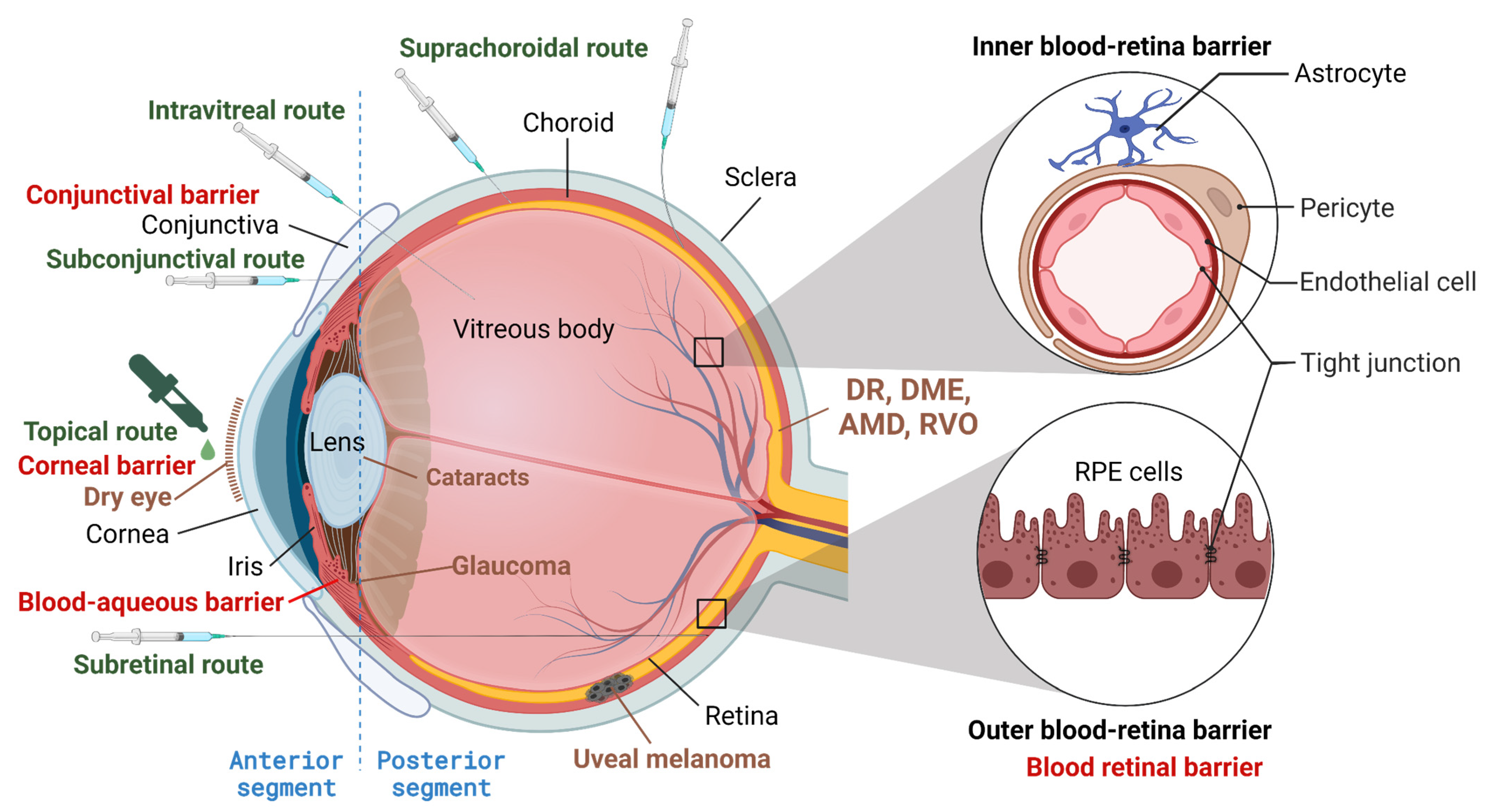
:1. Introduction
2. The Strategies for Cell Penetration
2.1. Cationic CPPs
2.2. Amphiphilic Peptides
2.3. Hydrophobic Peptides
2.4. Others
3. CPP Is Promising for Ocular Drug Delivery
4. Advantages of Peptide-Based Therapeutics
5. Successfully Used Peptide/Protein Drugs for Eye Diseases
6. Peptide/Protein Drugs for Eye Diseases in Clinical Trials
6.1. Anti-VEGF Therapy
6.2. Anti-Ang2
6.3. Anti-PDGF Therapy
6.4. Anti-Inflammation Therapy
6.5. Drugs Targeting Other Molecules
7. Pre-Clinical Peptide Drug Development
8. Future Perspective
9. Conclusions
Author Contributions
Funding
Institutional Review Board Statement
Informed Consent Statement
Data Availability Statement
Conflicts of Interest
References
- World Report on Vision. Licence: CC BY-NC-SA 3.0 IGO; World Health Organization: Geneva, Switzerland, 2019.
- Common Eye Disorders and Diseases; CDC: Atlanta, GA, USA, 2022.
- Gaudana, R.; Ananthula, H.K.; Parenky, A.; Mitra, A.K. Ocular drug delivery. AAPS J. 2010, 12, 348–360. [Google Scholar] [CrossRef] [PubMed]
- Balevic, S.J.; Rabinovich, C.E. Profile of adalimumab and its potential in the treatment of uveitis. Drug Des. Devel. Ther. 2016, 10, 2997–3003. [Google Scholar] [CrossRef] [PubMed] [Green Version]
- Bande, M.F.; Mansilla, R.; Pata, M.P.; Fernandez, M.; Blanco-Teijeiro, M.J.; Pineiro, A.; Gomez-Ulla, F. Intravitreal injections of anti-VEGF agents and antibiotic prophylaxis for endophthalmitis: A systematic review and meta-analysis. Sci. Rep. 2017, 7, 18088. [Google Scholar] [CrossRef] [PubMed] [Green Version]
- Detorakis, E.T.; Agorogiannis, G.; Drakonaki, E.E.; Tsilimbaris, M.K.; Pallikaris, I.G. Successful management of choroidal metastasis with intravitreal ranibizumab injections. Ophthalmic Surg. Lasers Imaging 2012, 43, e47–e51. [Google Scholar] [CrossRef]
- Falavarjani, K.G.; Modarres, M.; Nazari, H. Therapeutic effect of bevacizumab injected into the silicone oil in eyes with neovascular glaucoma after vitrectomy for advanced diabetic retinopathy. Eye 2010, 24, 717–719. [Google Scholar] [CrossRef]
- Nicholson, B.P.; Schachat, A.P. A review of clinical trials of anti-VEGF agents for diabetic retinopathy. Graefe’s Arch. Clin. Exp. Ophthalmol. 2010, 248, 915–930. [Google Scholar] [CrossRef]
- Wong, T.Y.; Liew, G.; Mitchell, P. Clinical update: New treatments for age-related macular degeneration. Lancet 2007, 370, 204–206. [Google Scholar] [CrossRef]
- Sun, Y.J.; Lin, C.H.; Wu, M.R.; Lee, S.H.; Yang, J.; Kunchur, C.R.; Mujica, E.M.; Chiang, B.; Jung, Y.S.; Wang, S.; et al. An intravitreal implant injection method for sustained drug delivery into mouse eyes. Cell Rep. Methods 2021, 1, 100125. [Google Scholar] [CrossRef]
- Olsen, T.W.; Feng, X.; Wabner, K.; Csaky, K.; Pambuccian, S.; Cameron, J.D. Pharmacokinetics of pars plana intravitreal injections versus microcannula suprachoroidal injections of bevacizumab in a porcine model. Invest. Ophthalmol. Vis. Sci. 2011, 52, 4749–4756. [Google Scholar] [CrossRef] [Green Version]
- Olsen, T.W.; Feng, X.; Wabner, K.; Conston, S.R.; Sierra, D.H.; Folden, D.V.; Smith, M.E.; Cameron, J.D. Cannulation of the suprachoroidal space: A novel drug delivery methodology to the posterior segment. Am. J. Ophthalmol. 2006, 142, 777–787. [Google Scholar] [CrossRef]
- Rizzo, S.; Ebert, F.G.; Bartolo, E.D.; Barca, F.; Cresti, F.; Augustin, C.; Augustin, A. Suprachoroidal drug infusion for the treatment of severe subfoveal hard exudates. Retina 2012, 32, 776–784. [Google Scholar] [CrossRef] [PubMed]
- Thakur Singh, R.R.; Tekko, I.; McAvoy, K.; McMillan, H.; Jones, D.; Donnelly, R.F. Minimally invasive microneedles for ocular drug delivery. Expert Opin. Drug Deliv. 2017, 14, 525–537. [Google Scholar] [CrossRef] [Green Version]
- Gupta, P.; Yadav, K.S. Applications of microneedles in delivering drugs for various ocular diseases. Life Sci. 2019, 237, 116907. [Google Scholar] [CrossRef] [PubMed]
- Schultz, C.; Breaux, J.; Schentag, J.; Morck, D. Drug delivery to the posterior segment of the eye through hydrogel contact lenses. Clin. Exp. Optom. 2011, 94, 212–218. [Google Scholar] [CrossRef] [PubMed]
- Nomoto, H.; Shiraga, F.; Kuno, N.; Kimura, E.; Fujii, S.; Shinomiya, K.; Nugent, A.K.; Hirooka, K.; Baba, T. Pharmacokinetics of bevacizumab after topical, subconjunctival, and intravitreal administration in rabbits. Invest. Ophthalmol. Vis. Sci. 2009, 50, 4807–4813. [Google Scholar] [CrossRef]
- Lim, M.; Jacobs, D.S.; Rosenthal, P.; Carrasquillo, K.G. The Boston Ocular Surface Prosthesis as a novel drug delivery system for bevacizumab. Semin. Ophthalmol. 2009, 24, 149–155. [Google Scholar] [CrossRef]
- Ross, A.E.; Bengani, L.C.; Tulsan, R.; Maidana, D.E.; Salvador-Culla, B.; Kobashi, H.; Kolovou, P.E.; Zhai, H.; Taghizadeh, K.; Kuang, L.; et al. Topical sustained drug delivery to the retina with a drug-eluting contact lens. Biomaterials 2019, 217, 119285. [Google Scholar] [CrossRef]
- Sidman, R.L.; Li, J.; Lawrence, M.; Hu, W.; Musso, G.F.; Giordano, R.J.; Cardo-Vila, M.; Pasqualini, R.; Arap, W. The peptidomimetic Vasotide targets two retinal VEGF receptors and reduces pathological angiogenesis in murine and nonhuman primate models of retinal disease. Sci. Transl. Med. 2015, 7, 309ra165. [Google Scholar] [CrossRef] [Green Version]
- Dugel, P.U.; Jaffe, G.J.; Sallstig, P.; Warburton, J.; Weichselberger, A.; Wieland, M.; Singerman, L. Brolucizumab Versus Aflibercept in Participants with Neovascular Age-Related Macular Degeneration: A Randomized Trial. Ophthalmology 2017, 124, 1296–1304. [Google Scholar] [CrossRef]
- Dugel, P.U.; Koh, A.; Ogura, Y.; Jaffe, G.J.; Schmidt-Erfurth, U.; Brown, D.M.; Gomes, A.V.; Warburton, J.; Weichselberger, A.; Holz, F.G.; et al. HAWK and HARRIER: Phase 3, Multicenter, Randomized, Double-Masked Trials of Brolucizumab for Neovascular Age-Related Macular Degeneration. Ophthalmology 2020, 127, 72–84. [Google Scholar] [CrossRef]
- Mones, J.; Srivastava, S.K.; Jaffe, G.J.; Tadayoni, R.; Albini, T.A.; Kaiser, P.K.; Holz, F.G.; Korobelnik, J.F.; Kim, I.K.; Pruente, C.; et al. Risk of Inflammation, Retinal Vasculitis, and Retinal Occlusion-Related Events with Brolucizumab: Post Hoc Review of HAWK and HARRIER. Ophthalmology 2021, 128, 1050–1059. [Google Scholar] [CrossRef] [PubMed]
- Woo, S.J.; Veith, M.; Hamouz, J.; Ernest, J.; Zalewski, D.; Studnicka, J.; Vajas, A.; Papp, A.; Gabor, V.; Luu, J.; et al. Efficacy and Safety of a Proposed Ranibizumab Biosimilar Product vs a Reference Ranibizumab Product for Patients With Neovascular Age-Related Macular Degeneration: A Randomized Clinical Trial. JAMA Ophthalmol. 2021, 139, 68–76. [Google Scholar] [CrossRef] [PubMed]
- Bressler, N.M.; Veith, M.; Hamouz, J.; Ernest, J.; Zalewski, D.; Studnicka, J.; Vajas, A.; Papp, A.; Vogt, G.; Luu, J.; et al. Biosimilar SB11 versus reference ranibizumab in neovascular age-related macular degeneration: 1-year phase III randomised clinical trial outcomes. Br. J. Ophthalmol. 2021, 107, 384–391. [Google Scholar] [CrossRef] [PubMed]
- Koh, G.Y.; Augustin, H.G.; Campochiaro, P.A. Viewpoints: Dual-blocking antibody against VEGF-A and angiopoietin-2 for treating vascular diseases of the eye. Trends Mol. Med. 2022, 28, 347–349. [Google Scholar] [CrossRef]
- Wykoff, C.C.; Abreu, F.; Adamis, A.P.; Basu, K.; Eichenbaum, D.A.; Haskova, Z.; Lin, H.; Loewenstein, A.; Mohan, S.; Pearce, I.A.; et al. Efficacy, durability, and safety of intravitreal faricimab with extended dosing up to every 16 weeks in patients with diabetic macular oedema (YOSEMITE and RHINE): Two randomised, double-masked, phase 3 trials. Lancet 2022, 399, 741–755. [Google Scholar] [CrossRef]
- Heier, J.S.; Khanani, A.M.; Quezada Ruiz, C.; Basu, K.; Ferrone, P.J.; Brittain, C.; Figueroa, M.S.; Lin, H.; Holz, F.G.; Patel, V.; et al. Efficacy, durability, and safety of intravitreal faricimab up to every 16 weeks for neovascular age-related macular degeneration (TENAYA and LUCERNE): Two randomised, double-masked, phase 3, non-inferiority trials. Lancet 2022, 399, 729–740. [Google Scholar] [CrossRef]
- Holekamp, N.M.; Campochiaro, P.A.; Chang, M.A.; Miller, D.; Pieramici, D.; Adamis, A.P.; Brittain, C.; Evans, E.; Kaufman, D.; Maass, K.F.; et al. Archway Randomized Phase 3 Trial of the Port Delivery System with Ranibizumab for Neovascular Age-Related Macular Degeneration. Ophthalmology 2022, 129, 295–307. [Google Scholar] [CrossRef]
- Rosenfeld, P.J.; Brown, D.M.; Heier, J.S.; Boyer, D.S.; Kaiser, P.K.; Chung, C.Y.; Kim, R.Y.; Group, M.S. Ranibizumab for neovascular age-related macular degeneration. N. Engl. J. Med. 2006, 355, 1419–1431. [Google Scholar] [CrossRef] [Green Version]
- Regillo, C.D.; Brown, D.M.; Abraham, P.; Yue, H.; Ianchulev, T.; Schneider, S.; Shams, N. Randomized, double-masked, sham-controlled trial of ranibizumab for neovascular age-related macular degeneration: PIER Study year 1. Am. J. Ophthalmol. 2008, 145, 239–248. [Google Scholar] [CrossRef]
- Brown, D.M.; Michels, M.; Kaiser, P.K.; Heier, J.S.; Sy, J.P.; Ianchulev, T.; Group, A.S. Ranibizumab versus verteporfin photodynamic therapy for neovascular age-related macular degeneration: Two-year results of the ANCHOR study. Ophthalmology 2009, 116, 57–65.e5. [Google Scholar] [CrossRef]
- Dixon, J.A.; Oliver, S.C.; Olson, J.L.; Mandava, N. VEGF Trap-Eye for the treatment of neovascular age-related macular degeneration. Expert Opin. Investig. Drugs 2009, 18, 1573–1580. [Google Scholar] [CrossRef] [PubMed]
- Eylea® (Aflibercept) Injection Approved as the First Pharmacologic Treatment for Preterm Infants with Retinopathy of Prematurity (ROP) by the FDA. Available online: https://investor.regeneron.com/news-releases/news-release-details/eylear-aflibercept-injection-approved-first-pharmacologic (accessed on 21 February 2023).
- Stahl, A.; Sukgen, E.A.; Wu, W.C.; Lepore, D.; Nakanishi, H.; Mazela, J.; Moshfeghi, D.M.; Vitti, R.; Athanikar, A.; Chu, K.; et al. Effect of Intravitreal Aflibercept vs Laser Photocoagulation on Treatment Success of Retinopathy of Prematurity: The FIREFLEYE Randomized Clinical Trial. JAMA Ophthalmol. 2022, 328, 348–359. [Google Scholar] [CrossRef] [PubMed]
- de Cogan, F.; Hill, L.J.; Lynch, A.; Morgan-Warren, P.J.; Lechner, J.; Berwick, M.R.; Peacock, A.F.A.; Chen, M.; Scott, R.A.H.; Xu, H.; et al. Topical Delivery of Anti-VEGF Drugs to the Ocular Posterior Segment Using Cell-Penetrating Peptides. Invest. Ophthalmol. Vis. Sci. 2017, 58, 2578–2590. [Google Scholar] [CrossRef] [Green Version]
- Heitz, F.; Morris, M.C.; Divita, G. Twenty years of cell-penetrating peptides: From molecular mechanisms to therapeutics. Br. J. Pharmacol. 2009, 157, 195–206. [Google Scholar] [CrossRef] [PubMed] [Green Version]
- Borrelli, A.; Tornesello, A.L.; Tornesello, M.L.; Buonaguro, F.M. Cell Penetrating Peptides as Molecular Carriers for Anti-Cancer Agents. Molecules 2018, 23, 295. [Google Scholar] [CrossRef] [PubMed] [Green Version]
- Guidotti, G.; Brambilla, L.; Rossi, D. Cell-Penetrating Peptides: From Basic Research to Clinics. Trends Pharmacol. Sci. 2017, 38, 406–424. [Google Scholar] [CrossRef]
- Nikoi, N.D.; Berwick, M.; Bryant, J.A.; Riordan, L.; Slope, L.; Peacock, A.F.A.; de Cogan, F. Stability of Cell-Penetrating Peptide anti-VEGF Formulations for the Treatment of Age-Related Macular Degeneration. Curr. Eye Res. 2021, 46, 751–757. [Google Scholar] [CrossRef]
- Frankel, A.D.; Pabo, C.O. Cellular uptake of the tat protein from human immunodeficiency virus. Cell 1988, 55, 1189–1193. [Google Scholar] [CrossRef]
- Derossi, D.; Joliot, A.H.; Chassaing, G.; Prochiantz, A. The third helix of the Antennapedia homeodomain translocates through biological membranes. J. Biol. Chem. 1994, 269, 10444–10450. [Google Scholar] [CrossRef]
- Elmquist, A.; Lindgren, M.; Bartfai, T.; Langel, U. VE-cadherin-derived cell-penetrating peptide, pVEC, with carrier functions. Exp. Cell Res. 2001, 269, 237–244. [Google Scholar] [CrossRef]
- Sani, M.A.; Separovic, F. How Membrane-Active Peptides Get into Lipid Membranes. Acc. Chem. Res. 2016, 49, 1130–1138. [Google Scholar] [CrossRef] [PubMed]
- Copolovici, D.M.; Langel, K.; Eriste, E.; Langel, U. Cell-penetrating peptides: Design, synthesis, and applications. ACS Nano 2014, 8, 1972–1994. [Google Scholar] [CrossRef] [PubMed]
- Green, M.; Loewenstein, P.M. Autonomous functional domains of chemically synthesized human immunodeficiency virus tat trans-activator protein. Cell 1988, 55, 1179–1188. [Google Scholar] [CrossRef] [PubMed]
- Lattig-Tunnemann, G.; Prinz, M.; Hoffmann, D.; Behlke, J.; Palm-Apergi, C.; Morano, I.; Herce, H.D.; Cardoso, M.C. Backbone rigidity and static presentation of guanidinium groups increases cellular uptake of arginine-rich cell-penetrating peptides. Nat. Commun. 2011, 2, 453. [Google Scholar] [CrossRef] [PubMed] [Green Version]
- Lein, M.; deRonde, B.M.; Sgolastra, F.; Tew, G.N.; Holden, M.A. Protein transport across membranes: Comparison between lysine and guanidinium-rich carriers. Biochim. Biophys. Acta 2015, 1848, 2980–2984. [Google Scholar] [CrossRef] [Green Version]
- Wender, P.A.; Mitchell, D.J.; Pattabiraman, K.; Pelkey, E.T.; Steinman, L.; Rothbard, J.B. The design, synthesis, and evaluation of molecules that enable or enhance cellular uptake: Peptoid molecular transporters. Proc. Natl. Acad. Sci. USA 2000, 97, 13003–13008. [Google Scholar] [CrossRef] [Green Version]
- Rothbard, J.B.; Jessop, T.C.; Lewis, R.S.; Murray, B.A.; Wender, P.A. Role of membrane potential and hydrogen bonding in the mechanism of translocation of guanidinium-rich peptides into cells. J. Am. Chem. Soc. 2004, 126, 9506–9507. [Google Scholar] [CrossRef]
- Esbjorner, E.K.; Lincoln, P.; Norden, B. Counterion-mediated membrane penetration: Cationic cell-penetrating peptides overcome Born energy barrier by ion-pairing with phospholipids. Biochim. Biophys. Acta 2007, 1768, 1550–1558. [Google Scholar] [CrossRef] [Green Version]
- Tunnemann, G.; Ter-Avetisyan, G.; Martin, R.M.; Stockl, M.; Herrmann, A.; Cardoso, M.C. Live-cell analysis of cell penetration ability and toxicity of oligo-arginines. J. Pept. Sci. 2008, 14, 469–476. [Google Scholar] [CrossRef]
- Wadia, J.S.; Stan, R.V.; Dowdy, S.F. Transducible TAT-HA fusogenic peptide enhances escape of TAT-fusion proteins after lipid raft macropinocytosis. Nat. Med. 2004, 10, 310–315. [Google Scholar] [CrossRef]
- Ciobanasu, C.; Siebrasse, J.P.; Kubitscheck, U. Cell-penetrating HIV1 TAT peptides can generate pores in model membranes. Biophys. J. 2010, 99, 153–162. [Google Scholar] [CrossRef] [PubMed] [Green Version]
- Herce, H.D.; Garcia, A.E.; Cardoso, M.C. Fundamental molecular mechanism for the cellular uptake of guanidinium-rich molecules. J. Am. Chem. Soc. 2014, 136, 17459–17467. [Google Scholar] [CrossRef] [PubMed]
- Sun, D.; Forsman, J.; Lund, M.; Woodward, C.E. Effect of arginine-rich cell penetrating peptides on membrane pore formation and life-times: A molecular simulation study. Phys. Chem. Chem. Phys. 2014, 16, 20785–20795. [Google Scholar] [CrossRef] [PubMed] [Green Version]
- Allolio, C.; Magarkar, A.; Jurkiewicz, P.; Baxova, K.; Javanainen, M.; Mason, P.E.; Sachl, R.; Cebecauer, M.; Hof, M.; Horinek, D.; et al. Arginine-rich cell-penetrating peptides induce membrane multilamellarity and subsequently enter via formation of a fusion pore. Proc. Natl. Acad. Sci. USA 2018, 115, 11923–11928. [Google Scholar] [CrossRef] [PubMed] [Green Version]
- Chiquet, C.; Aptel, F.; Creuzot-Garcher, C.; Berrod, J.P.; Kodjikian, L.; Massin, P.; Deloche, C.; Perino, J.; Kirwan, B.A.; de Brouwer, S.; et al. Postoperative Ocular Inflammation: A Single Subconjunctival Injection of XG-102 Compared to Dexamethasone Drops in a Randomized Trial. Am. J. Ophthalmol. 2017, 174, 76–84. [Google Scholar] [CrossRef] [Green Version]
- Staecker, H.; Jokovic, G.; Karpishchenko, S.; Kienle-Gogolok, A.; Krzyzaniak, A.; Lin, C.D.; Navratil, P.; Tzvetkov, V.; Wright, N.; Meyer, T. Efficacy and Safety of AM-111 in the Treatment of Acute Unilateral Sudden Deafness-A Double-blind, Randomized, Placebo-controlled Phase 3 Study. Otol. NeurOtol. 2019, 40, 584–594. [Google Scholar] [CrossRef]
- He, H.; Ye, J.; Liu, E.; Liang, Q.; Liu, Q.; Yang, V.C. Low molecular weight protamine (LMWP): A nontoxic protamine substitute and an effective cell-penetrating peptide. J. Control. Release 2014, 193, 63–73. [Google Scholar] [CrossRef]
- He, H.; Sheng, J.; David, A.E.; Kwon, Y.M.; Zhang, J.; Huang, Y.; Wang, J.; Yang, V.C. The use of low molecular weight protamine chemical chimera to enhance monomeric insulin intestinal absorption. Biomaterials 2013, 34, 7733–7743. [Google Scholar] [CrossRef] [Green Version]
- Suh, J.S.; Lee, H.J.; Nam, H.; Jo, B.S.; Lee, D.W.; Kim, J.H.; Lee, J.Y.; Chung, C.P.; Lee, G.; Park, Y.J. Control of cancer stem cell like population by intracellular target identification followed by the treatment with peptide-siRNA complex. BioChem. Biophys. Res. Commun. 2017, 491, 827–833. [Google Scholar] [CrossRef]
- Xia, H.; Gu, G.; Hu, Q.; Liu, Z.; Jiang, M.; Kang, T.; Miao, D.; Song, Q.; Yao, L.; Tu, Y.; et al. Activatable cell penetrating peptide-conjugated nanoparticles with enhanced permeability for site-specific targeting delivery of anticancer drug. Bioconjug. Chem. 2013, 24, 419–430. [Google Scholar] [CrossRef]
- Wang, H.; Zhao, Y.; Wang, H.; Gong, J.; He, H.; Shin, M.C.; Yang, V.C.; Huang, Y. Low-molecular-weight protamine-modified PLGA nanoparticles for overcoming drug-resistant breast cancer. J. Control. Release 2014, 192, 47–56. [Google Scholar] [CrossRef] [PubMed]
- Xia, H.; Gao, X.; Gu, G.; Liu, Z.; Zeng, N.; Hu, Q.; Song, Q.; Yao, L.; Pang, Z.; Jiang, X.; et al. Low molecular weight protamine-functionalized nanoparticles for drug delivery to the brain after intranasal administration. Biomaterials 2011, 32, 9888–9898. [Google Scholar] [CrossRef]
- Milletti, F. Cell-penetrating peptides: Classes, origin, and current landscape. Drug Discov. Today 2012, 17, 850–860. [Google Scholar] [CrossRef]
- Kalafatovic, D.; Giralt, E. Cell-Penetrating Peptides: Design Strategies beyond Primary Structure and Amphipathicity. Molecules 2017, 22, 1929. [Google Scholar] [CrossRef] [PubMed] [Green Version]
- Morris, M.C.; Deshayes, S.; Heitz, F.; Divita, G. Cell-penetrating peptides: From molecular mechanisms to therapeutics. Biol. Cell 2008, 100, 201–217. [Google Scholar] [CrossRef] [PubMed] [Green Version]
- Oehlke, J.; Scheller, A.; Wiesner, B.; Krause, E.; Beyermann, M.; Klauschenz, E.; Melzig, M.; Bienert, M. Cellular uptake of an alpha-helical amphipathic model peptide with the potential to deliver polar compounds into the cell interior non-endocytically. Biochim. Biophys. Acta 1998, 1414, 127–139. [Google Scholar] [CrossRef] [Green Version]
- Pooga, M.; Soomets, U.; Hallbrink, M.; Valkna, A.; Saar, K.; Rezaei, K.; Kahl, U.; Hao, J.X.; Xu, X.J.; Wiesenfeld-Hallin, Z.; et al. Cell penetrating PNA constructs regulate galanin receptor levels and modify pain transmission in vivo. Nat. Biotechnol. 1998, 16, 857–861. [Google Scholar] [CrossRef]
- Sadler, K.; Eom, K.D.; Yang, J.L.; Dimitrova, Y.; Tam, J.P. Translocating proline-rich peptides from the antimicrobial peptide bactenecin 7. Biochemistry 2002, 41, 14150–14157. [Google Scholar] [CrossRef]
- Pujals, S.; Giralt, E. Proline-rich, amphipathic cell-penetrating peptides. Adv. Drug Deliv. Rev. 2008, 60, 473–484. [Google Scholar] [CrossRef]
- Fernandez-Carneado, J.; Kogan, M.J.; Pujals, S.; Giralt, E. Amphipathic peptides and drug delivery. Biopolymers 2004, 76, 196–203. [Google Scholar] [CrossRef]
- Feger, G.; Angelov, B.; Angelova, A. Prediction of Amphiphilic Cell-Penetrating Peptide Building Blocks from Protein-Derived Amino Acid Sequences for Engineering of Drug Delivery Nanoassemblies. J. Phys. Chem. B 2020, 124, 4069–4078. [Google Scholar] [CrossRef] [PubMed]
- Krishnamurthy, S.; Wohlford-Lenane, C.; Kandimalla, S.; Sartre, G.; Meyerholz, D.K.; Theberge, V.; Hallee, S.; Duperre, A.M.; Del’Guidice, T.; Lepetit-Stoffaes, J.P.; et al. Engineered amphiphilic peptides enable delivery of proteins and CRISPR-associated nucleases to airway epithelia. Nat. Commun. 2019, 10, 4906. [Google Scholar] [CrossRef] [PubMed] [Green Version]
- Rhee, M.; Davis, P. Mechanism of uptake of C105Y, a novel cell-penetrating peptide. J. Biol. Chem. 2006, 281, 1233–1240. [Google Scholar] [CrossRef] [PubMed] [Green Version]
- Marks, J.R.; Placone, J.; Hristova, K.; Wimley, W.C. Spontaneous membrane-translocating peptides by orthogonal high-throughput screening. J. Am. Chem. Soc. 2011, 133, 8995–9004. [Google Scholar] [CrossRef] [PubMed] [Green Version]
- Gasparini, G.; Sargsyan, G.; Bang, E.K.; Sakai, N.; Matile, S. Ring Tension Applied to Thiol-Mediated Cellular Uptake. Angew Chem. Int. Ed. Engl. 2015, 54, 7328–7331. [Google Scholar] [CrossRef]
- Aubry, S.; Burlina, F.; Dupont, E.; Delaroche, D.; Joliot, A.; Lavielle, S.; Chassaing, G.; Sagan, S. Cell-surface thiols affect cell entry of disulfide-conjugated peptides. FASEB J. 2009, 23, 2956–2967. [Google Scholar] [CrossRef]
- Schneider, A.F.L.; Kithil, M.; Cardoso, M.C.; Lehmann, M.; Hackenberger, C.P.R. Cellular uptake of large biomolecules enabled by cell-surface-reactive cell-penetrating peptide additives. Nat. Chem. 2021, 13, 530–539. [Google Scholar] [CrossRef]
- Lostale-Seijo, I.; Louzao, I.; Juanes, M.; Montenegro, J. Peptide/Cas9 nanostructures for ribonucleoprotein cell membrane transport and gene edition. Chem. Sci. 2017, 8, 7923–7931. [Google Scholar] [CrossRef] [Green Version]
- Lee, Y.J.; Erazo-Oliveras, A.; Pellois, J.P. Delivery of macromolecules into live cells by simple co-incubation with a peptide. Chembiochem 2010, 11, 325–330. [Google Scholar] [CrossRef] [Green Version]
- Sato, H.; Feix, J.B. Peptide-membrane interactions and mechanisms of membrane destruction by amphipathic alpha-helical antimicrobial peptides. Biochim. Biophys. Acta 2006, 1758, 1245–1256. [Google Scholar] [CrossRef] [Green Version]
- Bird, G.H.; Mazzola, E.; Opoku-Nsiah, K.; Lammert, M.A.; Godes, M.; Neuberg, D.S.; Walensky, L.D. Biophysical determinants for cellular uptake of hydrocarbon-stapled peptide helices. Nat. Chem. Biol. 2016, 12, 845–852. [Google Scholar] [CrossRef] [PubMed] [Green Version]
- Mo, R.H.; Zaro, J.L.; Shen, W.C. Comparison of cationic and amphipathic cell penetrating peptides for siRNA delivery and efficacy. Mol. Pharm. 2012, 9, 299–309. [Google Scholar] [CrossRef] [PubMed]
- Gote, V.; Sikder, S.; Sicotte, J.; Pal, D. Ocular Drug Delivery: Present Innovations and Future Challenges. J. Pharmacol. Exp. Ther. 2019, 370, 602–624. [Google Scholar] [CrossRef] [PubMed]
- Pescina, S.; Ostacolo, C.; Gomez-Monterrey, I.M.; Sala, M.; Bertamino, A.; Sonvico, F.; Padula, C.; Santi, P.; Bianchera, A.; Nicoli, S. Cell penetrating peptides in ocular drug delivery: State of the art. J. Control. Release 2018, 284, 84–102. [Google Scholar] [CrossRef] [PubMed]
- Chakravarthy, U.; Armendariz, B.G.; Fauser, S. 15 years of anti-VEGF treatment for nAMD: Success or failure or something in between? Eye 2022, 36, 2232–2233. [Google Scholar] [CrossRef]
- Bohni, S.C.; Bittner, M.; Howell, J.P.; Bachmann, L.M.; Faes, L.; Schmid, M.K. Comparison of Eylea(R) with Lucentis(R) as first-line therapy in patients with treatment-naive neovascular age-related macular degeneration in real-life clinical practice: Retrospective case-series analysis. BMC Ophthalmol. 2015, 15, 109. [Google Scholar] [CrossRef] [Green Version]
- Chang, J.H.; Garg, N.K.; Lunde, E.; Han, K.Y.; Jain, S.; Azar, D.T. Corneal neovascularization: An anti-VEGF therapy review. Surv. Ophthalmol. 2012, 57, 415–429. [Google Scholar] [CrossRef] [Green Version]
- Ferrara, N. Vascular endothelial growth factor and age-related macular degeneration: From basic science to therapy. Nat. Med. 2010, 16, 1107–1111. [Google Scholar] [CrossRef]
- Holash, J.; Davis, S.; Papadopoulos, N.; Croll, S.D.; Ho, L.; Russell, M.; Boland, P.; Leidich, R.; Hylton, D.; Burova, E.; et al. VEGF-Trap: A VEGF blocker with potent antitumor effects. Proc. Natl. Acad. Sci. USA 2002, 99, 11393–11398. [Google Scholar] [CrossRef] [Green Version]
- Ferrara, N.; Hillan, K.J.; Gerber, H.P.; Novotny, W. Discovery and development of bevacizumab, an anti-VEGF antibody for treating cancer. Nat. Rev. Drug Discov. 2004, 3, 391–400. [Google Scholar] [CrossRef]
- Imai, K.; Takaoka, A. Comparing antibody and small-molecule therapies for cancer. Nat. Rev. Cancer 2006, 6, 714–727. [Google Scholar] [CrossRef] [PubMed]
- Vargason, A.M.; Anselmo, A.C.; Mitragotri, S. The evolution of commercial drug delivery technologies. Nat. Biomed. Eng. 2021, 5, 951–967. [Google Scholar] [CrossRef] [PubMed]
- Craik, D.J.; Fairlie, D.P.; Liras, S.; Price, D. The future of peptide-based drugs. Chem. Biol. Drug Des. 2013, 81, 136–147. [Google Scholar] [CrossRef] [PubMed]
- Bruno, B.J.; Miller, G.D.; Lim, C.S. Basics and recent advances in peptide and protein drug delivery. Ther. Deliv. 2013, 4, 1443–1467. [Google Scholar] [CrossRef] [Green Version]
- Lau, J.L.; Dunn, M.K. Therapeutic peptides: Historical perspectives, current development trends, and future directions. Bioorg. Med. Chem. 2018, 26, 2700–2707. [Google Scholar] [CrossRef]
- Wang, L.; Wang, N.; Zhang, W.; Cheng, X.; Yan, Z.; Shao, G.; Wang, X.; Wang, R.; Fu, C. Therapeutic peptides: Current applications and future directions. Signal. Transduct. Target. Ther. 2022, 7, 48. [Google Scholar] [CrossRef]
- Fosgerau, K.; Hoffmann, T. Peptide therapeutics: Current status and future directions. Drug Discov. Today 2015, 20, 122–128. [Google Scholar] [CrossRef] [Green Version]
- Di, L. Strategic approaches to optimizing peptide ADME properties. AAPS J. 2015, 17, 134–143. [Google Scholar] [CrossRef] [Green Version]
- Yamada, K.H. A novel approach in preventing vascular leakage and angiogenesis in wet age-related macular degeneration. Neural Regen. Res. 2022, 17, 1751–1752. [Google Scholar] [CrossRef]
- Yoon, C.K.; Oh, J.; Bae, K.; Park, U.C.; Yu, K.S.; Yu, H.G. Efficacy and safety of a new ranibizumab biosimilar CKD-701 using a pro re nata treatment regimen in neovascular age-related macular degeneration: A phase 3 randomized clinical trial. PLoS ONE 2022, 17, e0275611. [Google Scholar] [CrossRef]
- Zhang, Z.; Wu, Y.; Lyu, Y.L.; Chang, M.Q.; Xu, Q.J.; Liu, Y.M.; Kang, W.Y.; Wang, Q.Y.; Li, C.L. Efficacy and safety of intravitreal HLX04-O, an anti-VEGF monoclonal antibody, for the treatment of wet age-related macular degeneration. Int. J. Ophthalmol. 2022, 15, 1549–1553. [Google Scholar] [CrossRef] [PubMed]
- Li, X.; Xu, G.; Wang, Y.; Xu, X.; Liu, X.; Tang, S.; Zhang, F.; Zhang, J.; Tang, L.; Wu, Q.; et al. Safety and efficacy of conbercept in neovascular age-related macular degeneration: Results from a 12-month randomized phase 2 study: AURORA study. Ophthalmology 2014, 121, 1740–1747. [Google Scholar] [CrossRef] [PubMed] [Green Version]
- Liu, K.; Song, Y.; Xu, G.; Ye, J.; Wu, Z.; Liu, X.; Dong, X.; Zhang, M.; Xing, Y.; Zhu, S.; et al. Conbercept for Treatment of Neovascular Age-related Macular Degeneration: Results of the Randomized Phase 3 PHOENIX Study. Am. J. Ophthalmol. 2019, 197, 156–167. [Google Scholar] [CrossRef]
- Sun, Z.; Zhou, H.; Lin, B.; Jiao, X.; Luo, Y.; Zhang, F.; Tao, S.; Wu, Q.; Ke, Z.; Liu, X. Efficacy and Safety of Intravitreal Conbercept Injections in Macular Edema Secondary to Retinal Vein Occlusion. Retina 2017, 37, 1723–1730. [Google Scholar] [CrossRef] [PubMed]
- Shalchi, Z.; Mahroo, O.; Bunce, C.; Mitry, D. Anti-vascular endothelial growth factor for macular oedema secondary to branch retinal vein occlusion. Cochrane Database Syst. Rev. 2020, 7, CD009510. [Google Scholar] [CrossRef]
- Slakter, J.S.; Coleman, H.R.; Wykoff, C.C.; Price, C.; Baldwin, M.E.; Jackson, T.L. Efficacy and Safety of OPT-302 in combination with Ranibizumab for Polypoidal Choroidal Vasculopathy. IOVS 2022, 63, 382-F0213. [Google Scholar]
- Jackson, T.L.; Slakter, J.; Buyse, M.; Wang, K.; Dugel, P.U.; Wykoff, C.C.; Boyer, D.S.; Gerometta, M.; Baldwin, M.E.; Price, C.F. A randomized controlled trial of OPT-302, a VEGF-C/D inhibitor for neovascular age-related macular degeneration. Ophthalmology 2023, in press. [Google Scholar] [CrossRef]
- Kunimoto, D.; Yoon, Y.H.; Wykoff, C.C.; Chang, A.; Khurana, R.N.; Maturi, R.K.; Agostini, H.; Souied, E.; Chow, D.R.; Lotery, A.J.; et al. Efficacy and Safety of Abicipar in Neovascular Age-Related Macular Degeneration: 52-Week Results of Phase 3 Randomized Controlled Study. Ophthalmology 2020, 127, 1331–1344. [Google Scholar] [CrossRef]
- Wolf, S.; Souied, E.H.; Mauget-Faysse, M.; Devin, F.; Patel, M.; Wolf-Schnurrbusch, U.E.; Stumpp, M.; MP0112 wet AMD study group. Phase I Mp0112 Wet AMD Study: Results Of A Single Escalating Dose Study With DARPin® MP0112 In Wet AMD. IOVS 2011, 52, 1655. [Google Scholar]
- Brown, D.M.; Boyer, D.S.; Csaky, K.; Vitti, R.; Perlee, L.; Chu, K.W.; Asmus, F.; Leal, S.; Zeitz, O.; Cheng, Y.; et al. Intravitreal Nesvacumab (Antiangiopoietin 2) Plus Aflibercept in Diabetic Macular Edema: Phase 2 RUBY Randomized Trial. Retina 2022, 42, 1111–1120. [Google Scholar] [CrossRef]
- AffaMed Therapeutics Announces First Patient Dosed in the US Phase 1 Clinical Trial of AM712 in Retinal Disease. Available online: https://www.affamed.com/press-releases-37 (accessed on 9 February 2023).
- Dunn, E.N.; Hariprasad, S.M.; Sheth, V.S. An Overview of the Fovista and Rinucumab Trials and the Fate of Anti-PDGF Medications. Ophthalmic Surg. Lasers Imaging Retin. 2017, 48, 100–104. [Google Scholar] [CrossRef] [PubMed] [Green Version]
- Jaffe, G.J.; Ciulla, T.A.; Ciardella, A.P.; Devin, F.; Dugel, P.U.; Eandi, C.M.; Masonson, H.; Mones, J.; Pearlman, J.A.; Quaranta-El Maftouhi, M.; et al. Dual Antagonism of PDGF and VEGF in Neovascular Age-Related Macular Degeneration: A Phase IIb, Multicenter, Randomized Controlled Trial. Ophthalmology 2017, 124, 224–234. [Google Scholar] [CrossRef] [PubMed] [Green Version]
- Qin, S.; Dong, N.; Yang, M.; Wang, J.; Feng, X.; Wang, Y. Complement Inhibitors in Age-Related Macular Degeneration: A Potential Therapeutic Option. J. Immunol. Res. 2021, 2021, 9945725. [Google Scholar] [CrossRef] [PubMed]
- Holz, F.G.; Sadda, S.R.; Busbee, B.; Chew, E.Y.; Mitchell, P.; Tufail, A.; Brittain, C.; Ferrara, D.; Gray, S.; Honigberg, L.; et al. Efficacy and Safety of Lampalizumab for Geographic Atrophy Due to Age-Related Macular Degeneration: Chroma and Spectri Phase 3 Randomized Clinical Trials. JAMA Ophthalmol. 2018, 136, 666–677. [Google Scholar] [CrossRef] [Green Version]
- Yehoshua, Z.; de Amorim Garcia Filho, C.A.; Nunes, R.P.; Gregori, G.; Penha, F.M.; Moshfeghi, A.A.; Zhang, K.; Sadda, S.; Feuer, W.; Rosenfeld, P.J. Systemic complement inhibition with eculizumab for geographic atrophy in age-related macular degeneration: The COMPLETE study. Ophthalmology 2014, 121, 693–701. [Google Scholar] [CrossRef] [Green Version]
- Khanani, A.M.; Maturi, R.K.; Bagheri, N.; Bakall, B.; Boyer, D.S.; Couvillion, S.S.; Dhoot, D.S.; Holekamp, N.M.; Jamal, K.N.; Marcus, D.M.; et al. A Phase I, Single Ascending Dose Study of GEM103 (Recombinant Human Complement Factor H) in Patients with Geographic Atrophy. Ophthalmol. Sci. 2022, 2, 100154. [Google Scholar] [CrossRef]
- Lashkari, K.; Teague, G.; Chen, H.; Lin, Y.Q.; Kumar, S.; McLaughlin, M.M.; Lopez, F.J. A monoclonal antibody targeting amyloid beta (Abeta) restores complement factor I bioactivity: Potential implications in age-related macular degeneration and Alzheimer’s disease. PLoS ONE 2018, 13, e0195751. [Google Scholar] [CrossRef] [Green Version]
- Cabral de Guimaraes, T.A.; Daich Varela, M.; Georgiou, M.; Michaelides, M. Treatments for dry age-related macular degeneration: Therapeutic avenues, clinical trials and future directions. Br. J. Ophthalmol. 2022, 106, 297–304. [Google Scholar] [CrossRef]
- Rosenfeld, P.J.; Berger, B.; Reichel, E.; Danis, R.P.; Gress, A.; Ye, L.; Magee, M.; Parham, L.R.; McLaughlin, M.M. A Randomized Phase 2 Study of an Anti-Amyloid beta Monoclonal Antibody in Geographic Atrophy Secondary to Age-Related Macular Degeneration. Ophthalmol. Retin. 2018, 2, 1028–1040. [Google Scholar] [CrossRef]
- David, S.; Boyer, P.J.R. New Pathways for Dry AMD Treatment several potential therapies are in clinical trials. Retin. Physician 2019, 16, 24–25. [Google Scholar]
- Apellis Completes Enrollment in Two Phase 3 Studies of the Targeted C3 Therapy, Pegcetacoplan, in Patients with Geographic Atrophy (GA). Available online: https://investors.apellis.com/news-releases/news-release-details/apellis-completes-enrollment-two-phase-3-studies-targeted-c3 (accessed on 7 February 2023).
- Yang, S.; Li, T.; Jia, H.; Gao, M.; Li, Y.; Wan, X.; Huang, Z.; Li, M.; Zhai, Y.; Li, X.; et al. Targeting C3b/C4b and VEGF with a bispecific fusion protein optimized for neovascular age-related macular degeneration therapy. Sci. Transl. Med. 2022, 14, eabj2177. [Google Scholar] [CrossRef] [PubMed]
- Jia, H.; Li, T.; Sun, J.; Gong, Y.; Liu, H.; Wang, H.; Chen, J.; Liu, W.; Lu, S.; Feng, L.; et al. A Novel Bispecific Fusion Protein Targeting C3b/C4b and VEGF in Patients With nAMD: A Randomized, Open-Label, Phase 1b Study. Am. J. Ophthalmol. 2022, 248, 8–15. [Google Scholar] [CrossRef] [PubMed]
- Thomas, C.N.; Sim, D.A.; Lee, W.H.; Alfahad, N.; Dick, A.D.; Denniston, A.K.; Hill, L.J. Emerging therapies and their delivery for treating age-related macular degeneration. Br. J. Pharmacol. 2022, 179, 1908–1937. [Google Scholar] [CrossRef] [PubMed]
- Gonzalez, V.H.; Berger, B.; Goldberg, R.; Gordon, C.M.; Khurana, R.N.; Angeles, R.; Shams, N. Safety and Tolerability of Intravitreal Carotuximab (DE-122) in Patients With Persistent Exudative Age-Related Macular Degeneration: A Phase I Study. Transl. Vis. Sci. Technol. 2021, 10, 27. [Google Scholar] [CrossRef] [PubMed]
- Lpath’s iSONEP Fails in Phase II Wet-AMD Study. Available online: https://www.thepharmaletter.com/article/lpath-s-isonep-fails-in-phase-ii-wet-amd-study (accessed on 7 February 2023).
- Late-Onset Retinal Degeneration Clinical Trials. Available online: https://www.medifind.com/conditions/late-onset-retinal-degeneration/4620/clinical-trial/6047940 (accessed on 9 February 2023).
- Wells, J.A., III; Berger, B.B.; Gonzales, C.; Gonzales, V.H.; Johnson, D.L.; Sippy, B.D.; Soni, M. Multicenter Phase 1 Clinical Trial Targeting Tissue Factor for the Treatment of Neovascular AMD. IOVS 2012, 53, 450. [Google Scholar]
- Ghanchi, F.; Bourne, R.; Downes, S.M.; Gale, R.; Rennie, C.; Tapply, I.; Sivaprasad, S. An update on long-acting therapies in chronic sight-threatening eye diseases of the posterior segment: AMD, DMO, RVO, uveitis and glaucoma. Eye 2022, 36, 1154–1167. [Google Scholar] [CrossRef]
- Grosskreutz, C.L.; Hockey, H.U.; Serra, D.; Dryja, T.P. Dry Eye Signs and Symptoms Persist During Systemic Neutralization of IL-1beta by Canakinumab or IL-17A by Secukinumab. Cornea 2015, 34, 1551–1556. [Google Scholar] [CrossRef] [Green Version]
- Hariprasad, S.M.; Gale, R.P.; Weng, C.Y.; Ebbers, H.C.; Rezk, M.F.; Tadayoni, R. An Introduction to Biosimilars for the Treatment of Retinal Diseases: A Narrative Review. Ophthalmol. Ther. 2022, 11, 959–982. [Google Scholar] [CrossRef]
- Kapur, M.; Nirula, S.; Naik, M.P. Future of anti-VEGF: Biosimilars and biobetters. Int. J. Retin. Vitr. 2022, 8, 2. [Google Scholar] [CrossRef]
- Yang, J.Y.; Wang, Q.; Chen, M.X.; Yan, Y.N.; Zhou, W.J.; Liu, Y.M.; Wei, W.B. Retinal Microvascular Changes in Uveal Melanoma Following Conbercept Injection after Plaque Radiotherapy as Detected by Optical Coherence Tomography Angiography. Retina 2021, 41, 2605–2611. [Google Scholar] [CrossRef]
- Moisseiev, E.; Loewenstein, A. Abicipar pegol-a novel anti-VEGF therapy with a long duration of action. Eye 2020, 34, 605–606. [Google Scholar] [CrossRef] [PubMed] [Green Version]
- Rodrigues, G.A.; Mason, M.; Christie, L.A.; Hansen, C.; Hernandez, L.M.; Burke, J.; Luhrs, K.A.; Hohman, T.C. Functional Characterization of Abicipar-Pegol, an Anti-VEGF DARPin Therapeutic That Potently Inhibits Angiogenesis and Vascular Permeability. Invest. Ophthalmol. Vis. Sci. 2018, 59, 5836–5846. [Google Scholar] [CrossRef] [PubMed] [Green Version]
- Holekamp, N.M. Abicipar Pegol not Approved for Treatment of Wet AMD—Healio. Available online: https://www.healio.com/news/ophthalmology/20200626/abicipar-pegol-not-approved-for-treatment-of-wet-amd (accessed on 7 February 2023).
- Papadopoulos, K.P.; Kelley, R.K.; Tolcher, A.W.; Razak, A.R.; Van Loon, K.; Patnaik, A.; Bedard, P.L.; Alfaro, A.A.; Beeram, M.; Adriaens, L.; et al. A Phase I First-in-Human Study of Nesvacumab (REGN910), a Fully Human Anti-Angiopoietin-2 (Ang2) Monoclonal Antibody, in Patients with Advanced Solid Tumors. Clin. Cancer Res. 2016, 22, 1348–1355. [Google Scholar] [CrossRef] [PubMed] [Green Version]
- Rasmussen, A.; Bloch, S.B.; Fuchs, J.; Hansen, L.H.; Larsen, M.; LaCour, M.; Lund-Andersen, H.; Sander, B. A 4-year longitudinal study of 555 patients treated with ranibizumab for neovascular age-related macular degeneration. Ophthalmology 2013, 120, 2630–2636. [Google Scholar] [CrossRef]
- Rakic, J.M.; Leys, A.; Brie, H.; Denhaerynck, K.; Pacheco, C.; Vancayzeele, S.; Hermans, C.; Macdonald, K.; Abraham, I. Real-world variability in ranibizumab treatment and associated clinical, quality of life, and safety outcomes over 24 months in patients with neovascular age-related macular degeneration: The HELIOS study. Clin. Ophthalmol. 2013, 7, 1849–1858. [Google Scholar] [CrossRef] [Green Version]
- Holz, F.G.; Tadayoni, R.; Beatty, S.; Berger, A.; Cereda, M.G.; Cortez, R.; Hoyng, C.B.; Hykin, P.; Staurenghi, G.; Heldner, S.; et al. Multi-country real-life experience of anti-vascular endothelial growth factor therapy for wet age-related macular degeneration. Br. J. Ophthalmol. 2015, 99, 220–226. [Google Scholar] [CrossRef]
- Rubner, R.; Li, K.V.; Canto-Soler, M.V. Progress of clinical therapies for dry age-related macular degeneration. Int. J. Ophthalmol. 2022, 15, 157–166. [Google Scholar] [CrossRef]
- Sivaprasad, S.; Chong, N.V. The complement system and age-related macular degeneration. Eye 2006, 20, 867–872. [Google Scholar] [CrossRef]
- Hoy, S.M. Pegcetacoplan: First Approval. Drugs 2021, 81, 1423–1430. [Google Scholar] [CrossRef]
- Ren, X.; Li, J.; Xu, X.; Wang, C.; Cheng, Y. IBI302, a promising candidate for AMD treatment, targeting both the VEGF and complement system with high binding affinity in vitro and effective targeting of the ocular tissue in healthy rhesus monkeys. Exp. Eye Res. 2016, 145, 352–358. [Google Scholar] [CrossRef]
- Isumi, Y.; Hayashi, S.; Inoue, T.; Yoshigae, Y.; Sato, T.; Hasegawa, J.; Agatsuma, T. DS-7080a, a Selective Anti-ROBO4 Antibody, Shows Anti-Angiogenic Efficacy with Distinctly Different Profiles from Anti-VEGF Agents. Transl. Vis. Sci. Technol. 2020, 9, 7. [Google Scholar] [CrossRef]
- Shaw, L.T.; Mackin, A.; Shah, R.; Jain, S.; Jain, P.; Nayak, R.; Hariprasad, S.M. Risuteganib-a novel integrin inhibitor for the treatment of non-exudative (dry) age-related macular degeneration and diabetic macular edema. Expert Opin. Investig. Drugs 2020, 29, 547–554. [Google Scholar] [CrossRef] [PubMed]
- Bonny, C.; Oberson, A.; Negri, S.; Sauser, C.; Schorderet, D.F. Cell-permeable peptide inhibitors of JNK: Novel blockers of beta-cell death. Diabetes 2001, 50, 77–82. [Google Scholar] [CrossRef] [PubMed] [Green Version]
- Rafiei, F.; Tabesh, H.; Farzad, F. Sustained subconjunctival drug delivery systems: Current trends and future perspectives. Int. Ophthalmol. 2020, 40, 2385–2401. [Google Scholar] [CrossRef] [PubMed]
- Yamada, K.H.; Nakajima, Y.; Geyer, M.; Wary, K.K.; Ushio-Fukai, M.; Komarova, Y.; Malik, A.B. KIF13B regulates angiogenesis through Golgi to plasma membrane trafficking of VEGFR2. J. Cell Sci. 2014, 127, 4518–4530. [Google Scholar] [CrossRef] [Green Version]
- Yamada, K.H.; Kang, H.; Malik, A.B. Antiangiogenic Therapeutic Potential of Peptides Derived from the Molecular Motor KIF13B that Transports VEGFR2 to Plasmalemma in Endothelial Cells. Am. J. Pathol. 2017, 187, 214–224. [Google Scholar] [CrossRef] [Green Version]
- Waters, S.B.; Zhou, C.; Nguyen, T.; Zelkha, R.; Lee, H.; Kazlauskas, A.; Rosenblatt, M.I.; Malik, A.B.; Yamada, K.H. VEGFR2 Trafficking by KIF13B Is a Novel Therapeutic Target for Wet Age-Related Macular Degeneration. Invest. Ophthalmol. Vis. Sci. 2021, 62, 5. [Google Scholar] [CrossRef]
- Johnson, L.N.; Cashman, S.M.; Kumar-Singh, R. Cell-penetrating peptide for enhanced delivery of nucleic acids and drugs to ocular tissues including retina and cornea. Mol. Ther. 2008, 16, 107–114. [Google Scholar] [CrossRef]
- Johnson, L.N.; Cashman, S.M.; Read, S.P.; Kumar-Singh, R. Cell penetrating peptide POD mediates delivery of recombinant proteins to retina, cornea and skin. Vis. Res. 2010, 50, 686–697. [Google Scholar] [CrossRef] [Green Version]
- Wang, Y.; Lin, H.; Lin, S.; Qu, J.; Xiao, J.; Huang, Y.; Xiao, Y.; Fu, X.; Yang, Y.; Li, X. Cell-penetrating peptide TAT-mediated delivery of acidic FGF to retina and protection against ischemia-reperfusion injury in rats. J. Cell Mol. Med. 2010, 14, 1998–2005. [Google Scholar] [CrossRef]
- Ozaki, T.; Nakazawa, M.; Yamashita, T.; Ishiguro, S. Delivery of Topically Applied Calpain Inhibitory Peptide to the Posterior Segment of the Rat Eye. PLoS ONE 2015, 10, e0130986. [Google Scholar] [CrossRef] [PubMed]
- Ozaki, T.; Ishiguro, S.; Hirano, S.; Baba, A.; Yamashita, T.; Tomita, H.; Nakazawa, M. Inhibitory peptide of mitochondrial mu-calpain protects against photoreceptor degeneration in rhodopsin transgenic S334ter and P23H rats. PLoS ONE 2013, 8, e71650. [Google Scholar] [CrossRef]
- Ozaki, T.; Nakazawa, M.; Yamashita, T.; Sorimachi, H.; Hata, S.; Tomita, H.; Isago, H.; Baba, A.; Ishiguro, S. Intravitreal injection or topical eye-drop application of a mu-calpain C2L domain peptide protects against photoreceptor cell death in Royal College of Surgeons’ rats, a model of retinitis pigmentosa. Biochim. Biophys. Acta 2012, 1822, 1783–1795. [Google Scholar] [CrossRef] [PubMed] [Green Version]


| Generic Name | Brand Name | Format | Company | Year | Conditions | Ref. |
|---|---|---|---|---|---|---|
| Ranibizumab | Lucentis | Anti-VEGF-A antibody | Genentech | 2006 | Wet AMD, DR, DME, mCNV, RVO | [30,31,32] |
| Aflibercept | Eylea | Recombinant protein targeting VEGF | Regeneron | 2011 | Wet AMD, ROP | [33,34,35] |
| Brolucizumab | Beovu | Anti-VEGF-A antibody | Novartis | 2019 | Wet AMD | [21,22] |
| Susvimo | Ocular implant for ranibizumab | Genentech | 2021 | Wet AMD | [29] | |
| Ranibizumab-nuna (SB11) | Byooviz | Biosimilar of ranibizumab | Biogen/Samsung Bioepis | 2021 | Wet AMD, DME, RVO, mCNV | [24,25] |
| Faricimab | Vabysmo | Ab targeting both VEGF-A and Ang2 | Roche/Genentech | 2022 | Wet AMD, DME | [27,28] |
| Name | Type | Format | Clinical Trials | Route | Conditions | Ref. |
|---|---|---|---|---|---|---|
| CKD-701 | Antibody | Anti-VEGF antibody fragment | Phase 3 (NCT04857177) | IVT | Wet AMD | [103] |
| HLX04-O | Antibody | Anti-VEGF antibody | Phase 1/2 (NCT04993352), phase 3 (NCT05003245, NCT04740671) | IVT | Wet AMD | [104] |
| Conbercept/Lumitin/KH902 | Protein | A recombinant protein targeting all VEGF isoforms and PlGF | Phase 2 (NCT01157715), phase 3 (NCT01436864), Phase 2 (NCT01809236) and phase 3 (NCT03108352), phase 3 PANDA-1 and PANDA-2 (NCT03630952 and NCT03577899) | IVT | Wet AMD, DME, RVO | [105,106,107,108] |
| OPT-302 | Protein | A recombinant protein targeting VEGF-C and VEGF-D | Phase 2 (NCT03345082), phase 3 (NCT04757636, NCT04757610) | IVT | Wet AMD | [109,110] |
| Abicipar | Protein | Engineered protein with Ankyrin repeat targeting VEGF | Phase 3 (NCT02462928, NCT02462486) | IVT | Wet AMD | [111] |
| MP0112 | Protein | DARPin, a long-acting VEGF inhibitor | Phase 1 (NCT01086761) | IVT | Wet AMD | [112] |
| Nesvacumab/REGN910/SAR307746 | Antibody | Anti-Ang2 | Phase 2 (NCT02712008) | IVT | DME | [113] |
| AM712/ASKG712 | Protein | A bifunctional molecule targeting VEGF and Ang2 | Phase 1 (NCT05345769, NCT05456828) | IVT | Wet AMD | [114] |
| Rinucumab/REGN2176 | Antibody | Anti-PDGFRβ | Phase 1 (NCT02061865), phase 2 (NCT02418754) | IVT | Wet AMD | [115] |
| Fovista/pegpleranib | Antibody | Anti-PDGF | Phase 2 (NCT01089517) | IVT | Wet AMD | [116] |
| Tesidolumab/LFG316 | Antibody | An antibody that prevents the cleavage of C5 | Phase 2 trials for wet AMD (NCT01535950, NCT01624636), for dry AMD (NCT01527500) | IVT | Wet AMD, dry AMD | [117] |
| Lampalizumab | Antibody | Anti-factor D | Phase 3 (NCT02247531, NCT02247479) | IVT | Dry AMD | [118] |
| Eculizumab | Antibody | Anti- C5 | Phase 2 (NCT00935883) | Systemic | Dry AMD | [119] |
| GEM103 | Protein | A recombinant human complement factor H | Phase 1 (NCT04246866), phase 2 (NCT04643886, NCT04684394) | Dry AMD | [120] | |
| GSK933776 | Antibody | Anti-amyloid β antibody | Phase 2 (NCT01342926) | IVT | Dry AMD | [121,122,123] |
| RN6G/PF-43829223 | Antibody | Anti-amyloid β antibody | Phase 2 trial for dry AMD (NCT01577381) | IVT | Dry AMD | [124] |
| Pegcetacoplan/APL-2/Empaveli | Peptide | C3 inhibitor | Phase 3 trials DERBY and OAKS for dry AMD (NCT03525613, NCT03525600) | IVT | Dry AMD | [125] |
| Efdamrofusp alpha/IBI302 | Protein | Bispecific decoy receptor fusion protein for VEGF and complement | Phase 1 (NCT03814291, NCT04370379) | IVT | Wet AMD | [126,127] |
| RC28-E | Protein | Dual decoy receptor targeting VEGF and bFGF | Phase 1/2 clinical trial (NCT04270669), Phase 2 (NCT04782128, NCT04782115) | IVT | DR, DME, wet AMD | [128] |
| DE-122/Carotuximab/TRC105 | Antibody | An antibody for endoglin | Phase 1/2 for AMD (NCT02555306), phase 2 (NCT03211234). | IVT | AMD | [129] |
| iSONEP | Protein | Anti-S1P | Phase 2 (NCT01414153) | [130] | ||
| DS-7080a | Antibody | Anti-ROBO4 | Phase 1 (NCT02530918) | IVT | Wet AMD, DMED | [131] |
| HI-con1 is an antibody-like molecule targeted against tissue factor (TF), composed of two human factor VII | Antibody | A factor VII-IgGFc chimeric protein targeting tissue factor | Phase 1/2 (NCT01485588), phase 2 (NCT02358889) | IVT | Wet AMD | [132] |
| ALG-1001/Risuteganib/Luminate | Peptide | Anti-integrin oligopeptide | Phase 2 for dry AMD (NCT03626636), phase 1/2 for AMD (NCT01749891), phase 2 for DME (NCT02348918), phase 2 for vitreomacular adhesion (NCT02153476) | IVT | Dry AMD, wet AMD, DMO, vitreomacular adhesion | [133] |
| XG-102/AM-111/brimapitide/D-JNKI-1 | Peptide | A TAT-coupled JNK inhibitor | Phase 3 (NCT02235272, NCT02508337) | IVT | Postoperative ocular inflammation | [58] |
| Canakinumab/ACZ885/ILARISs | Antibody | Anti-IL-1β antibody | Phase 2 (NCT01250171) | IVT | Dry AMD | [134] |
Disclaimer/Publisher’s Note: The statements, opinions and data contained in all publications are solely those of the individual author(s) and contributor(s) and not of MDPI and/or the editor(s). MDPI and/or the editor(s) disclaim responsibility for any injury to people or property resulting from any ideas, methods, instructions or products referred to in the content. |
© 2023 by the authors. Licensee MDPI, Basel, Switzerland. This article is an open access article distributed under the terms and conditions of the Creative Commons Attribution (CC BY) license (https://creativecommons.org/licenses/by/4.0/).
Share and Cite
Nhàn, N.T.T.; Maidana, D.E.; Yamada, K.H. Ocular Delivery of Therapeutic Agents by Cell-Penetrating Peptides. Cells 2023, 12, 1071. https://doi.org/10.3390/cells12071071
Nhàn NTT, Maidana DE, Yamada KH. Ocular Delivery of Therapeutic Agents by Cell-Penetrating Peptides. Cells. 2023; 12(7):1071. https://doi.org/10.3390/cells12071071
Chicago/Turabian StyleNhàn, Nguyễn Thị Thanh, Daniel E. Maidana, and Kaori H. Yamada. 2023. "Ocular Delivery of Therapeutic Agents by Cell-Penetrating Peptides" Cells 12, no. 7: 1071. https://doi.org/10.3390/cells12071071