Review

Experimentally Created Magnetic Force in Microbiological Space and On-Earth Studies: Perspectives and Restrictions

by
Svetlana A. Ermolaeva
1,*,
Vladislav A. Parfenov
2,3,
Pavel A. Karalkin
4,
Yusef D. Khesuani
5 and
Pavel A. Domnin
1
1
Gamaleya National Research Centre for Epidemiology and Microbiology, 123098 Moscow, Russia
2
Institute of Metallurgy and Material Science, Russian Academy of Sciences, 119334 Moscow, Russia
3
National Research Nuclear University MEPhI (Moscow Engineering Physics Institute), 115409 Moscow, Russia
4
Institute of Cluster Oncology, Sechenov First Moscow State Medical University, 127473 Moscow, Russia
5
3D Bioprinting Solutions Ltd., 115409 Moscow, Russia
*
Author to whom correspondence should be addressed.
Cells 2023, 12(2), 338; https://doi.org/10.3390/cells12020338
Submission received: 30 November 2022 / Revised: 7 January 2023 / Accepted: 13 January 2023 / Published: 16 January 2023
(This article belongs to the Special Issue New Insights into Microgravity and Space Biology)

Abstract

:
Magnetic force and gravity are two fundamental forces affecting all living organisms, including bacteria. On Earth, experimentally created magnetic force can be used to counterbalance gravity and place living organisms in conditions of magnetic levitation. Under conditions of microgravity, magnetic force becomes the only force that moves bacteria, providing an acceleration towards areas of the lowest magnetic field and locking cells in this area. In this review, we consider basic principles and experimental systems used to create a magnetic force strong enough to balance gravity. Further, we describe how magnetic levitation is applied in on-Earth microbiological studies. Next, we consider bacterial behavior under combined conditions of microgravity and magnetic force onboard a spacecraft. At last, we discuss restrictions on applications of magnetic force in microbiological studies and the impact of these restrictions on biotechnological applications under space and on-Earth conditions.

1. Introduction

Microbiologic studies hold an important position in space exploration due to the significance of the role that bacteria play in human life. Considerable efforts have been made in microbiological studies related to human health (for a review see [1,2,3]). Another important trend in microbiology-related space studies deals with multiple practical applications from waste utilization to development of microbial cell factories under spaceflight conditions [4,5,6]. Significant efforts made in these areas have revealed noticeable changes in the metabolism and physiology of bacteria under spaceflight conditions. Spaceflight provides unique conditions of microgravity unavailable on Earth. Bacteria were shown to reply to microgravity by changes in growth rates, cell sizes, motility, biofilm formation, antibiotic resistance, and virulence [7,8,9,10,11,12,13,14]. In the last years, an increasing number of microbiological studies use “omics” technologies to analyze changes in gene expression, protein production, and metabolic pathways. Microgravity conditions change expression of stress response systems and factors, induce changes in genes associated with biofilm formation, affect surface structures including transporter proteins and pili, and noticeably affect spectra of secondary metabolites [15,16,17,18,19,20,21]. Besides the obvious importance of fundamental knowledge on changes in bacterial physiology and metabolism, applied applications of microgravity effects might be useful in future projects performed in space and on Earth and provide advantageous biotechnological platforms allowing for rational conversion of metabolic processes and improvement of bioprocessing efficiency.
For more than 50 years, manned spacecraft and especially orbital stations Mir and the International Space Station (ISS) have provided onboard conditions for multiple microbiological experiments [1,22,23]. However, financial, safety and time restrictions on space studies have required development of on-Earth models to study specific microgravity features. For bacteria growing in liquid media, spaceflight conditions provide weightlessness, lack of sedimentation, and low shear conditions due to the absence of convectional flows within the liquid [1,24]. A number of on-Earth models were developed that mimic these hallmarks of microgravity. The devices based on rotation over one or two axes, designated depending on the construction as clinostats, random positioning machines (RPM) or rotating wall vessels (RWV) are widely used in simulated microgravity studies (for a review see [25,26,27]). These devices provide low fluid shear and lack of sedimentation. Cells placed in rotating devices undergo a combined force provided by the sum of the gravity and centripetal force vectors, which is characterized by a continuous change in the module and direction with the temporal average equal to zero.
Another on-Earth simulated microgravity model is based on the effect of magnetic levitation, which is provided by balancing the gravity vector with magnetic force [28,29,30]. Magnetic levitation (MagLev) is widely used in applied studies to create conditions in which an object is suspended with no support other than magnetic fields to counteract gravitational effects. In microbiological research, MagLev allows for solving different tasks including cell separation, differentiation, or vice versa cell accumulation and integration depending on the system configuration [30,31,32]. Using MagLev systems in space studies is at its beginning, but application of the magnetic force under spaceflight conditions provides novel possibilities to modify bacterial growth conditions according to research and biotechnological necessity. In this review, we consider the physical nature of magnetic force, applications and restrictions on the magnetic levitation models and discuss how magnetic force affects bacteria under spaceflight conditions.

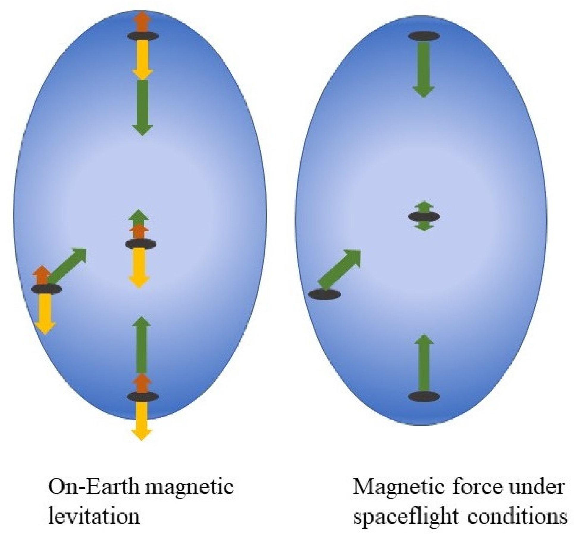
2. Magnetic Force vs. Gravity

Magnetic force appears as a result of quantum effects of the non-homogenous magnetic field on atomic and molecular electrons. If the magnetic force affecting the diamagnetic object is upward directed and strong enough to counterbalance gravitational force conditions, (dia)magnetic levitation (MagLev) can be reached on Earth. Water and all living creatures are diamagnetic, which is why they can levitate in the magnetic field. Below, we give a short explanation of those aspects of magnetism that are important for understanding bacterial behavior in the presence of magnetic force. For a deeper view of the nature of magnetic force and magnetism, we address the reader to excellent reviews that contemplate these phenomena from physical and biological points of view [31,32,33,34,35].
When the external magnetic field is applied, magnetization of a certain material depends on its atomic properties, such as presence of unpaired electrons. Electron magnetic moments respond to the external magnetic field being attracted or pushed off. The magnitude of magnetization is quantitatively expressed via the magnetic susceptibility χ. Magnetic susceptibility χ of diamagnetic materials is negative (χ < 0) and have low values of approximately χ ≈ −10−5. Being placed in the magnetic field gradient, the diamagnetic object is pushed off to the area of the lowest magnetic field. In paramagnetic materials, the magnetic susceptibility is positive (χ > 0) and typically ranges from 10−5 to 10−3 [33,34]. When the external magnetic field is applied, paramagnetic objects are attracted to the area of a higher magnetic field similar to ferromagnetic but with incomparably less power. Thus, magnetic effects in diamagnetic materials is different from effects in paramagnetic materials.
The magnetic force gives an acceleration to any object equal to a m = ( χ ρ μ ° ) B d B d r , where χ is a magnetic susceptibility, ρ is density of the objects, μ0 is a vacuum permeability, B is a magnetic flux density, the value characterizing magnetic field at a given point of the working volume, and d B d r   is its gradient at the same point. For diamagnetic objects, the magnetic force and acceleration a m is directed to areas with lower magnetic field.
The magnetic force acts throughout the object’s volume, so the force supporting the object within the magnetic field gradient is a volume integral of forces affecting internal structures according to their magnetic susceptibilities. Magnetic susceptibility of a cell is close to the susceptibility of water, which is an important part of the living cell. Guevorkian and Valles experimentally determined magnetic susceptibilities for unicellular eukaryotes Paramecium suspended in the water-based medium as χc = −0.911 × 10⁻⁵ and χm = −0.904 × 10⁻⁵ for cells and medium, respectively [36]. Magnetic susceptibilities of cell cytosolic and lipid fractions of the protist cell differ from magnetic susceptibility of water by 2% and 10%, respectively. Still, in some cases, the complicated composition of the living cell can affect the magnetic force distribution due to differences in magnetic properties of cellular structures. Mainly, a distinct response can be seen from specific intracellular structures with highly different magnetic susceptibilities, such as ferrimagnetic crystals of magnetotactic bacteria that have a magnetic susceptibility χ ≈ 106 [37,38]. Besides such special structures, basic physical principles suggest that magnetic fields interact only weakly with biological matter itself [39,40]. Subtle effects associated with the presence of magnetic ions, such as iron Fe3+, magnesium isotope 25Mg2+ or zinc isotope 67Zn2+, can change rates of reactions catalyzed by enzymes, which include these metals in their active center and affect cell metabolism but do not provide forces able to move cells as wholes [41,42].
If we consider bacteria suspended in the liquid medium and placed within the non-homogenous CMF, the bacterial cell undergoes effects of three forces, which are gravitational, Archimedes and magnetic forces. To provide the MagL effect, the vector sum of the Archimedes and magnetic forces has to counterbalance the gravitational force (Figure 1). Among these three forces, the gravitational and magnetic forces operate throughout the volume, while the Archimedes force acts at the surface (for a detailed view, see [26]). In this way, magnetic levitation is different from the neutral buoyance that takes place for microorganisms floating in the ocean when the Archimedes force counterbalance the gravitational force and is closer to weightlessness. Dijkstra and co-authors experimentally evaluated values of the Archimedes and magnetic forces that affect E. coli suspended in a water-based medium placed in a superconducting magnet and demonstrated that these values are comparable [43]. For bacterial suspension in paramagnetic liquids (see below) the difference between the Archimedes and magnetic forces might be different than for water suspension although detailed data are unavailable yet. Therefore, the MagLev when applied to bacterial suspension provides partial weightlessness and can be applied to study the bacterial response on these conditions [44]. This fact together with numerous practical applications makes MagLev a very attractive method in microbiological studies.
Now consider forces affecting bacteria growing in the liquid culture onboard a spacecraft when the culture is subjected to the non-homogenous external CMF. All things onboard the spacecraft are essentially weightless. The weightlessness is due to the orbital centrifugal acceleration that balances the gravitational force putting all things in continuous “free fall”. The Archimedes force, which appears as a consequence of the difference between the cell’s weight and the weight of fluid displaced by the cell, is zero at these conditions. The magnetic force remains the only force that affects bacteria giving them an acceleration to move toward to areas with a minimal magnetic flux density (Figure 1). Therefore, the presence of a magnetic force changes conditions of bacterial growth at the spacecraft board where there are no other forces providing acceleration of the bacterial cell. From this point of view, an application of the magnetic force at the spacecraft board provides conditions of simulated gravity. If such a simulated gravity model is used some restrictions that result from the effects of the magnetic field itself, and its gradient should be taken into account. In the next sections, we consider systems used in microbiological studies to create a magnetic force, and their applications and restrictions.

3. Superconducting Magnets for Magnetic Levitation of Living Objects

Superconducting magnets used for magnetic levitation of living objects are organized as vertically oriented solenoids, with a bore ranging from 32 mm up to 195 mm, that provide strong spatially variated magnetic field reaching 33 T and more (for a review see [26,31]. Inside the bore, the magnetic field is strongest in the center of the solenoid and decreases upward and downward from the center. At the point of the strongest field, the gradient of the magnetic field is zero, therefore the magnetic force is zero, too. The magnetic force, which always directs to the area of the lower magnetic field, directs upward in the upper part of the solenoid and downward in the lower part. Levitation of diamagnetic material is observed above the center of the solenoid where the magnetic force counterbalances gravity. The values of the magnetic flux density at which levitation is observed ranges from 8 to 16 T depending on the solenoid particular configuration [29,43,45,46]. In contrast, the product of the magnetic field and its gradient B d B d r is theoretically calculated for a drop of water from the equation a m = g = ( χ ρ μ ° ) B d B d r , where the gravitational acceleration g   = 9.8 m s−2, water density ρ = 1000 kg m−3, and water magnetic susceptibility χ = −0.9 × 10 −5 [31]. This value is near 1370 T2 m−1 at the levitation point. The superconducting solenoid systems were successfully used to levitate frogs and their eggs, plants, protozoa and bacteria and to study their physiology and metabolism [29,36,43,45]. Still, such systems are not portative, especially when we think about space studies, and have restricted parameters of the of the levitation area (configuration of the magnetic force direction, square and the volume) [47].
A number of works investigated restrictions and artefacts generated by application of superconducting magnets in MagLev research that hide effects of simulated microgravity. The most important impact that might overshadow effects of weightlessness is the high magnetic field itself. Although superstrong magnetic fields (>10 T) were shown not to disturb exponential bacterial growth [48], such fields provide subtle effects on gene expression that affect bacterial growth at the stationary phase, cell morphology and production of secondary metabolites [48,49,50]. The magnetic field gradient can provide further effects on physiology and metabolism, although such effects were mainly described for mammalian cells and observed at high values of the gradient from 103 to 109 T m−1 [51]. Besides these direct effects associated with cell response on the magnetic field and its gradient, indirect effects changing bacterial growth were described. The magnetic field gradient forming in the superconducting magnets drives flows of oxygen that improves bacterial growth besides effects of weightlessness itself [43]. Potential gradients of nutritive substances and/or secondary metabolites expelled by bacteria have not yet been explored, although such gradients could affect the growth of bacteria. Supermagnet cooling systems affect temperature gradient that in turn raise convection flows that can also influence bacterial growth and is different from spaceflight microgravity conditions with the absence of convection flows [31]. Still, thorough studies showed that effects of the high magnetic fields and artificial microgravity can be separated, supporting suitability of the MagLev as a simulated microgravity model [50,52,53,54]. Taken together, these data suggest that the right controls play the most important role in experiments on magnetic levitation as a model to study microgravity effects on living objects.

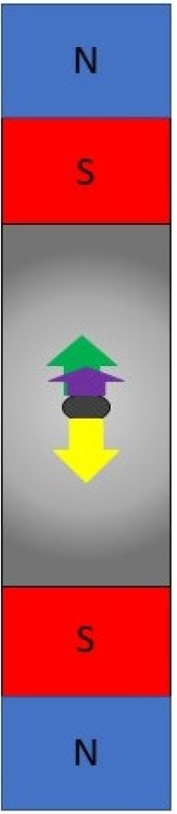
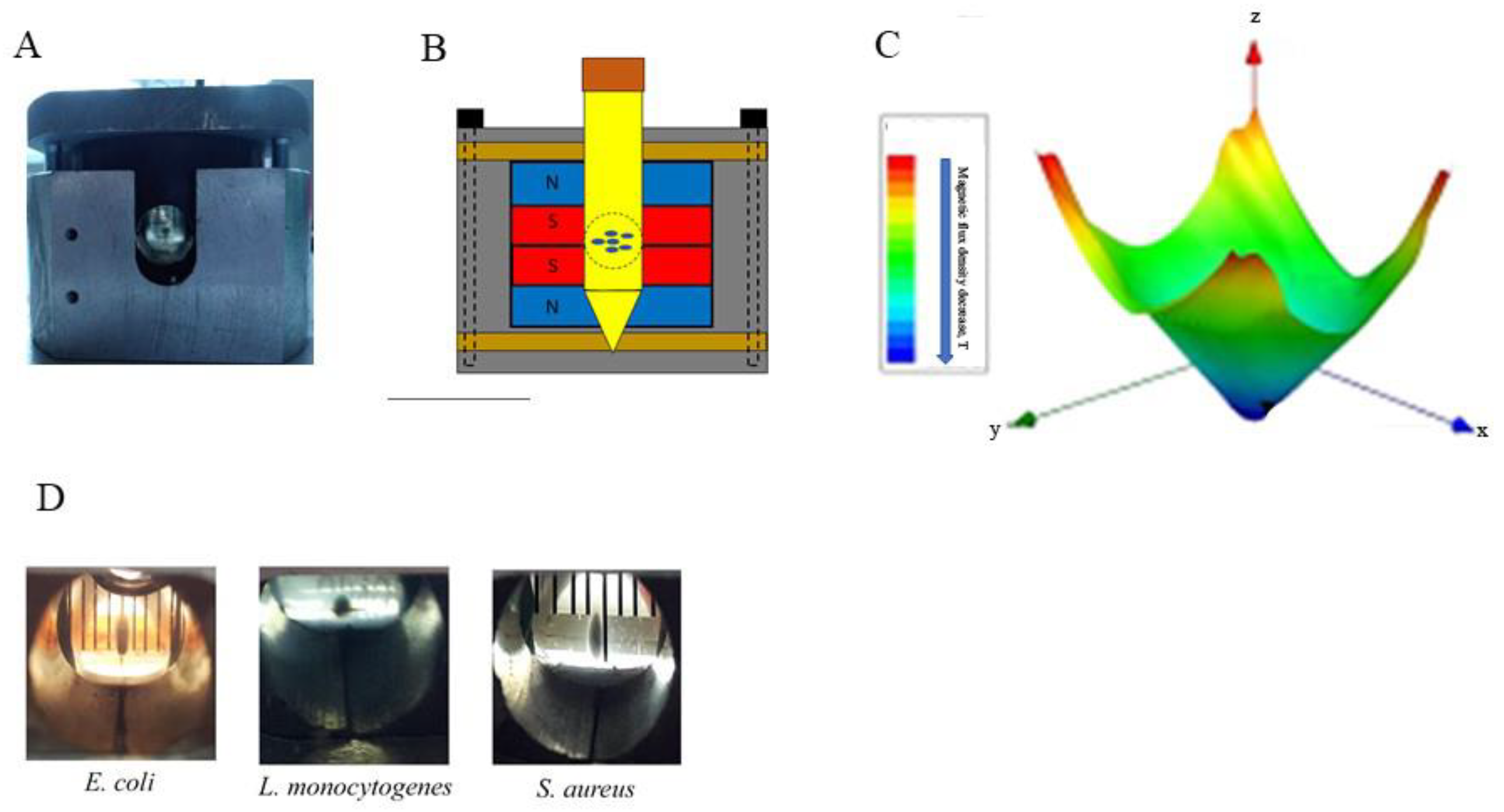
4. MagLev Systems Based on Permanent Magnets

Strong permanent magnets made from rare earth element-containing alloys avoid some disadvantages of superconducting magnets. The particular benefits of permanent magnets include their relatively low cost, small sizes, independence on external infrastructure, and portability. Further, permanent magnets operate at lower magnetic fields and do not disturb uniform temperature distribution inside the working volume that prevents appearance of convection flows due to the temperature gradient. At last, diverse mutual arrangement of permanent magnets provides distinct spatial distribution of magnetic flux gradients to make it suitable for different tasks.
Neodymium-containing magnets, although the strongest among permanent magnets, do not allow for obtaining values of the magnetic flux densities higher than 2 T and cannot per se create a magnetic force sufficient for magnetic levitation [55]. The problem of low magnetic fields was successfully overcome for suspension of diamagnetic objects, such as bacteria by using paramagnetic or supermagnetic fluids [36,52,56,57,58,59]. The magnetic force affecting the diamagnetic object floated in the paramagnetic medium increases proportionally to the difference between magnetic susceptibilities of the cell and the medium [36,59]. Guevorkian and Valles experimentally demonstrated that the difference between magnetic susceptibilities of the unicellular eukaryotes Paramecium and standard cultivation medium is only 0.007 × 10⁻⁵, while supplementation of the medium with the 4 mM solution of gadolinium salt Gd-DTPA (gadolinium diethylene-triamine-pentaacetate) increases the difference 100-fold up to 0.764 × 10⁻⁵, which decreases the required value of the parameter B d B d r from 1370 T2 m−1 to 510 T2 m−1 [36]. Gadolinium is a rare-earth element widely applied in medical applications being included into complex chemical substances. Parfenov et al. demonstrated that supplementation of the liquid medium with another gadolinium-based substance, gadobutrol ([10-[2,3-dihydroxi-1-(hydroxymethyl)propil]-1,4,7,10-tetraazacyclododexan-1,4,7-triacetin(3-)-N1, N4, N7, N10,O1, O4, O7]gadolinium), taken in concentration of 10 mM provides levitation of human cells in the static magnetic field created with the NdFeB magnets with a remnant magnetization of 1.21 T [59,60]. Similar conditions allowed for magnetic levitation of bacterial cells [52]. The same system with the medium supplemented with 20 mM gadobutrol provides levitation of the macroscopic non-attached aggregates formed by bacteria and the bacterium-produced extracellular matrix [52]. MagLev in the 50 mM gadobutrol allowed for separation of viable and antibiotic killed E. coli according to their buoyancy [58].
Thus, medium supplementation with gadolinium derivatives provides magnetic forces sufficient for levitation of bacteria, bacterial aggregates, and mammalian cells using magnetic fields created by permanent magnets. However, when using these systems, potential toxic effects of the gadolinium salts should be considered as well as effects of the strong magnetic field and its gradient that are described above. Gadolinium derivatives are widely used as contrast agents in magnetic resonance imaging (MRI) techniques so their toxicity has been extensively studied [61,62,63]. An ability of the Gd3+ to compete with Ca2+ in active centers of Ca2+-binding enzymes underlies gadolinium toxicity because Ca2+ substitution with Gd3+ can interrupt critically important Ca2+-dependent pathways [61]. To decrease toxicity, gadolinium-based substances, which are used in MRI, enclose Gd3+ in the complex that is expected to remain chelated in the human body and be excreted intact. Fast and almost unchanged excretion was demonstrated for FDA approved contrast agents such as gadobutrol [62,63]. In bacteria, although Ca2+ ions do not play such a critical role as in mammals, Ca2+ dependent proteins take part in such cellular functions as motility and chemotaxis, virulence, and maintenance of membrane integrity [64,65,66]. Potential toxic effects of gadolinium-based substances should be taken into account in MagLev studies of bacterial physiology.
Another limitation of the described approach is changes in the nature of the magnetic force action. The introduction of a paramagnetic fluid provides an effect similar to the effect of the Archimedes force, when a force is associated with displacement of the fluid volume. As a result, the force acts at the surface but not throughout the volume. Thus, although we obtain the neutral buoyancy, simulated microgravity is restricted. This effect limits using MagLev systems based on permanent magnets in studies of physiological changes in response to microgravity. On the other hand, permanent magnet based MagLev systems are effectively used in other applications.

5. Using the Magnetic Force for Cell Separation and Differentiation

The important benefit of permanent magnets is an ability to form different 3D patterns of the magnetic field gradient in accordance with the particular aim of the study by changing mutual arrangements of magnets. Depending on the system configuration, MagLev allows for solving different tasks, including cell separation, configuration or integration [30]. The basic MagLev scheme uses two straightforward magnets oriented to each other with the same poles [58,67] (Figure 2). This system has been improved by using parallel ring-shaped magnets or extended to use with 96-well plates [68,69,70]. Such systems are successfully used for cell separation studies. Distinct cell densities provide differences in Archimedes force that in turn affects requirements of the magnetic force balancing the cell, so cells with distinct densities levitate at different heights. Alternatively, levitation at discrete heights can be caused by distinct magnetic susceptibilities. Durmus and co-workers demonstrated that changes in bacterial membrane permeability caused by antibiotic treatment provide changes in levitation height that seems to be due to penetration of paramagnetic medium into the cytoplasm that in turn change both cell density and magnetic susceptibility [58]. Thus, the use of MagLev systems coupled with introduction of specific magnetic markers opens new perspectives in microbiological research.
To our knowledge, MagLev systems suggested for cell separation have not yet been used in space experiments. Under spaceflight conditions, the magnetic force is the only force pushing bacteria that would prevent cell balancing. Still, such systems could be efficiently applied in simulated gravity modeling in the course of space exploration along with centrifuges which are now used.

6. Using a Magnetic Force for Cell Integration and Aggregation

While the above described systems are used for cell separation and differentiation, alternative mutual arrangement of magnets provides cell integration. Parfenov and co-authors reported a magnetic bioassembler that includes two round-shaped magnets located directly next to each other [59] (Figure 3). Such mutual arrangement of the magnets results in creation of a sharp gradient of the magnetic field decreasing by the center of the working volume where the magnetic field is the lowest. Bacterial and cell suspensions in the medium supplemented with gadobutrol, when placed into the bioassembler, gather near the center of the working volume [52]. Such an approach when applied to human cells provides scaffold-free, label-free and nozzle-free bioassembly of 3D tissues [59,71]. If applied to bacterial cells, the bioassembler stimulates a process of autoaggregation resulting in formation of non-attached bacterial aggregates [52].
Bacterial flocculation and autoaggregation within the liquid volume is a process typical for natural aquatic ecosystems, which is used industrially for waste-water treatments and other applications [72,73,74,75,76]. Non-attached aggregates formed by pathogenic bacteria are described in chronic infections [77,78,79,80]. Formed non-attached aggregates represent 3D multicellular structures that include bacteria and a self-produced matrix. Morphologically, such aggregates are similar to well-known biofilms. Biofilms are defined as surface-attached 3D multilayer structures, while non-attached aggregates float freely within the liquid bulk. Mechanisms of non-attached aggregate formation are relatively poorly studied because experimental studies are impeded by the lack of models preventing fast aggregate sedimentation. The described bioassambler has been successfully used to demonstrate that strains of well-forming biofilms might be deficient in non-attached aggregate formation, and vice-versa, non-attached aggregate forming strains do not form biofilms [52]. These data suggest that mechanisms underlying formation of biofilms and non-attached aggregates are different, at least partly. The MagLev approach provides a unique possibility to study molecular mechanisms underlying non-attached aggregate formation.

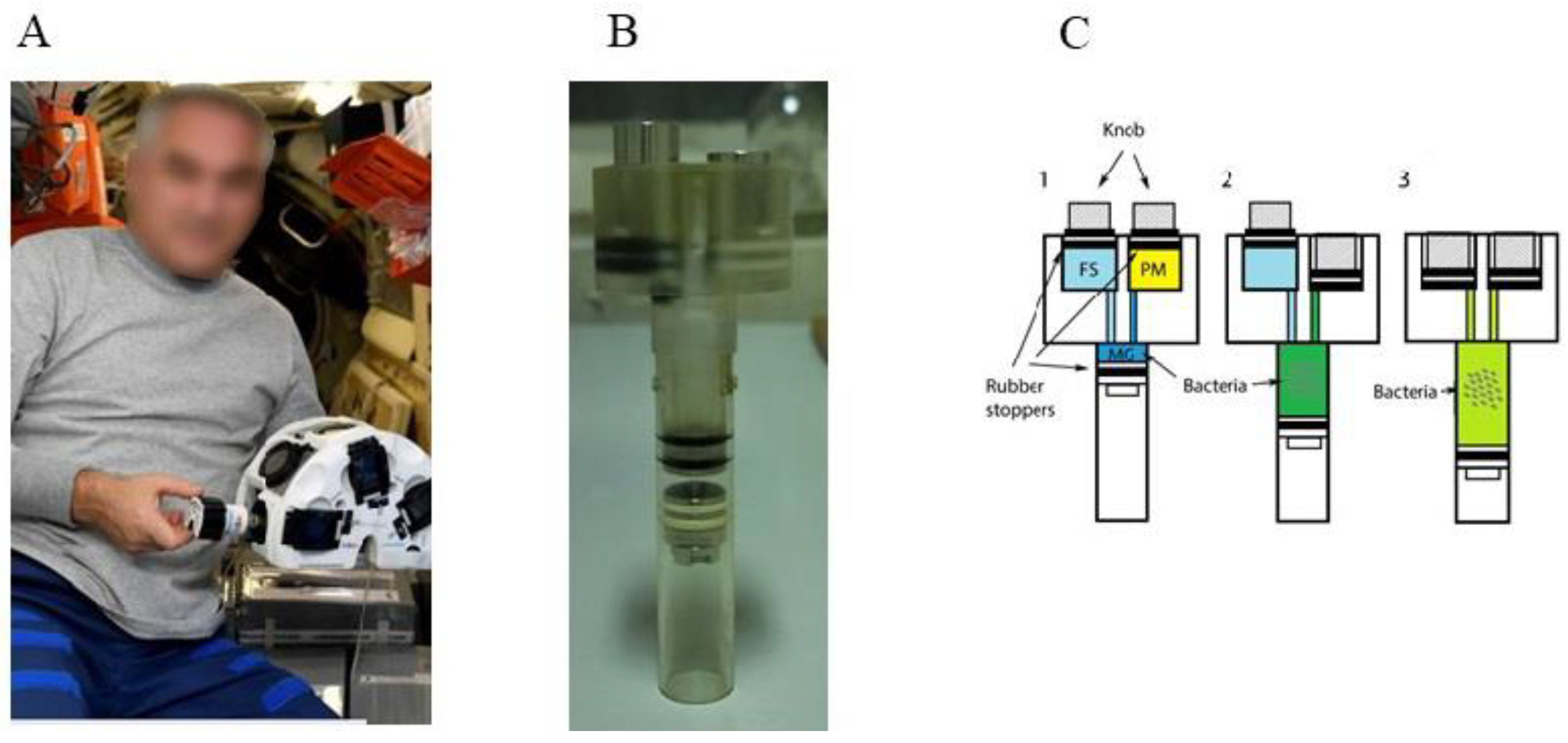
7. Magnetic Force Application in Space Studies

The bioassambler “Organ.Aut” developed on the basis of described bioassambler was delivered to ISS at the end of 2019 [19,81]. “Organ.Aut” has six sockets each and includes two magnets arranged in the same way as in the ground bioassambler to allow for cell integration and aggregation, and with similar parameters (Figure 4). In the course of the space experiment, the bioassambler “Organ.Aut” was used to perform biofabrication of 3D structures under the combined impact of spaceflight conditions and the magnetic force [81]. In the bacterial part of the space experiment, the culture of the Escherichia coli probiotic strain M17 was placed in the bioassambler “Organ.Aut” [19]. To provide the magnetic force, the rich LB nutritive medium was supplemented with 20 mM gadobutrol. Bacteria in control cuvettes placed in “Organ.Aut” grew in LB without gadobutrol, so they experienced the magnetic field and its gradient but the magnetic force was negligible to provide bacterial accumulation.
The net effect of microgravity on bacteria may be distinct in its dependence on such parameters as a tested strain, medium composition or a growth stage (reviewed in [2,4,24,82]. Better availability of nutrients for non-sedimenting bacteria provide their better growth at exponential and early stationary growth. In contrast, absence of convective flows results in toxic by-product accumulation in the bacterial vicinity and exhaustion of nutrients and oxygen that is observed at later stages of growth [83]. Stimulated by the magnetic force bacterial accumulation in a small part of the working volume results in strengthening microgravity effects caused by the absence of convective flows. Particularly, bacterial response on nutrition and oxygen limitations during prolonged cultivation was more clearly manifested under combined conditions of microgravity and the magnetic force than conditions of microgravity only Proteomic analysis demonstrated that the response included the down-regulation of glycolysis and TCA enzymes and the up-regulation of methylglyoxal bypass that were more prominent under combined impact of microgravity and the magnetic force than microgravity only [19].
Autoaggregation is an established phenomenon observed in bacteria cultivated under real and simulated microgravity [8,9,17,84,85]. The bioassambler in space provided formation of visible macroscopic structures that included bacteria and non-cellular matrix and had morphology similar to morphology of non-attached aggregates obtained under MagLev conditions in the bioassambler on Earth [19,52]. Proteomic data revealed similar mechanisms that accompanied autoaggregation under conditions of real microgravity and MagLev [19]. Particularly, the surface protein Ag43 known to provide E. coli autoaggregation by direct protein-protein interactions was similarly up-regulated under all listed conditions [19,86]. Another feature similar for bacterial growth under MagLev and spaceflight conditions was upregulation of glyoxylate shunt enzymes and Vitamin B12 transporter BtuB [19]. Both the glyoxylate shunt and the transporter BtuB are active under anaerobic conditions and can be involved in utilization of metabolites released by destroyed bacteria [87,88,89,90,91,92]. The glyoxylate shunt is essential when acetyl-CoA is a direct product of metabolic pathways, for example, via degradation of acetate, fatty acids, and alkanes supplied by lysed cells [87,88]. The BtuB is the transporter of vitamin B12, which is an essential co-factor of ethanolamine deaminase, the enzyme involved in degradation of bacterial membrane phospholipids [92,93,94]. Utilization of metabolites supplied by lysed cells has to be effective in autoaggregates where dead and alive bacteria are in close proximity while it might be useless for a planktonic culture cultivated under standard gravity conditions when dead bacteria sank to the bottom. Thus, microbial response on low shear conditions and microgravity utilizes mechanisms developed to form non-attached aggregates on Earth.
Establishment of a direct bacterial response on weightlessness (gravitaxis) besides indirect effects of low shear conditions accompanying absence of convective flows is a challenging task that has not yet been fully resolved [95]. From this point of view, the maximal interest would represent inverse effects of the magnetic force under magnetic levitation conditions and in space. Under MagLev conditions, the magnetic force counterbalances the gravitation force and the cell experiences partial weightlessness as it was discussed above. Under spaceflight conditions, the magnetic force simulates a gravitational force. We believe that future studies with MagLev systems under spaceflight and On-Earth conditions would be successful to provide evidences on gravitaxis in bacteria.

8. Future Industrial Applications of the Magnetic Force in Microbiological Studies

Microbial secondary metabolites that represent raw material important for medicine and/or industry amount to several thousands and include antimicrobial, antiparasitic, and antitumor compounds, growth regulators, building blocks of industrially relevant chemicals, etc. [96,97,98]. Multiple studies are devoted to industrial strain selection and/or creation of microorganisms that have been recognized as potential producers of bulk chemicals [99,100,101,102]. Besides strain selection, industrial metabolite production requires optimization of growth conditions. Real and simulated microgravity was tested to analyze how microgravity conditions affect production of important secondary metabolites. Obtained results are contradictory and demonstrated that production of secondary metabolites can be up-regulated, inhibited, or changed in its secretion depending on a substance, a strain, a growth phase, medium composition, and a microgravity model used (reviewed in [4,24]). There are only a few studies where effects of the magnetic force on secondary metabolite production were studied. Liu and co-authors used the superconducting magnet-based MagLev model to study microgravity effects on production of the anthelmintic agent avermectin by Streptomyces avermitilis [50]. Plating bacteria on the solid medium prevented development of indirect MagLev effects observed in liquid media but provided more space for a detailed analysis of direct gravitaxis and effects of the magnetic field and its gradient. Obtained results demonstrated increased production of avermectin under MagLev conditions and provided evidence that this increase is attributed to the strong magnetic field. These results suggest that even in the absence of direct and non-direct effects of the magnetic force, the MagLev systems might improve production of secondary metabolites via magnetic field effects.
Using MagLev bioassambler and its space analog “Organ.Aut”, we demonstrated that the bacterial aggregation due to the magnetic force within the liquid bulk up-regulates metabolic pathways leading to production of such important molecules as succinate [19]. Succinic acid is widely used in the food industry and is an important precursor of many industrially relevant chemicals used in biomedicine and biochemistry [96,97]. Bio-based production of succinic acid is a well-established process that includes different natural and genetically-modified microorganisms. Succinic acid accumulates as a final product under anaerobic conditions, and a number of obligate anaerobes, such as Anaerobiospirillum succiniciproducens, Actinobacillus succinogenes, Mannheimia succiniciproducens, were described as industrially important producers of the succinic acid [103,104,105,106,107]. Still, easy cultivation makes genetically modified E. coli although it is not obligate anaerobe to be an attractive succinic acid producer [107]. Succinic acid production in E. coli uses dual-phase fermentations, which comprise an initial aerobic growth phase followed by an anaerobic production phase. Using MagLev system bacterial gathering in the center of the bioassmbler and corresponding changes in the oxygen availability provide a potential alternative to systems used now for dual-change fermentation of succinic acid and other potential bioproducts, which require limited oxygen availability. The unique capabilities of Maglev systems to change bacterial growth conditions on Earth and in space opens up a wide field for development of novel biotechnologies and provides ample opportunities for future research.

9. Conclusions

The current development in superconductive and powerful permanent magnets offers unique possibilities to create the magnetic force strong enough to provide cell movement, assembly and congregation. In microbial on-Earth studies, the strong magnetic force allows for creating MagLev systems that can be widely used in practical applications, such as test-systems for bacterial separation and identification, characterization of antibiotic resistance, and formation of non-attached bacterial aggregates. Application of magnetic force under spaceflight conditions is only at its beginning. Still, initial results suggest that the introduction of MagLev systems into real microgravity conditions opens a wide field for future research and practical applications from models of simulated gravity to bacterial factories producing substances to meet the various needs of mankind.

Author Contributions

Conceptualization, S.A.E., V.A.P., P.A.K. and Y.D.K.; writing—original draft preparation, S.A.E.; writing—review and editing, V.A.P., P.A.K. and Y.D.K.; visualization, P.A.D. All authors have read and agreed to the published version of the manuscript.

Funding

S.A.E. lab research is supported by the Russian Ministry of Health (grant 056-00093-22-04).

Institutional Review Board Statement

Not applicable.

Informed Consent Statement

Not applicable.

Data Availability Statement

Not applicable.

Acknowledgments

The authors thank all participants and collaborators that took part in the development of the bioassembler and its testing in microbiological studies on Earth and at the ISS board.

Conflicts of Interest

The authors declare no conflict of interest.

References

  1. Nickerson, C.A.; Ott, C.M.; Wilson, J.W.; Ramamurthy, R.; Pierson, D.L. Microbial Responses to Microgravity and Other Low-Shear Environments. Microbiol. Mol. Biol. Rev. 2004, 68, 345–361. [Google Scholar] [CrossRef] [PubMed] [Green Version]
  2. Higginson, E.E.; Galen, J.E.; Levine, M.M.; Tennant, S.M. Microgravity as a biological tool to examine host-pathogen interactions and to guide development of therapeutics and preventatives that target pathogenic bacteria. Pathog. Dis. 2016, 74, 95. [Google Scholar] [CrossRef] [PubMed] [Green Version]
  3. Senatore, G.; Mastroleo, F.; Leys, N.; Mauriello, G. Effect of microgravity & space radiation on microbes. Future Microbiol. 2018, 13, 831–847. [Google Scholar] [CrossRef] [PubMed]
  4. Huang, B.; Li, D.G.; Huang, Y.; Liu, C.T. Effects of spaceflight and simulated microgravity on microbial growth and secondary metabolism. Mil. Med. Res. 2018, 5, 18. [Google Scholar] [CrossRef] [PubMed] [Green Version]
  5. Bijlani, S.; Stephens, E.; Singh, N.K.; Venkateswaran, K.; Wang, C.C.C. Advances in space microbiology. iScience 2021, 24, 102395. [Google Scholar] [CrossRef] [PubMed]
  6. Klimarev, S.; Il’in, V.; Starkova, L. Choice of a method and a type of device for water decontamination and warming within physical-chemical life support systems. Av. Ekol. Med. 2008, 42, 3–14. [Google Scholar]
  7. Horneck, G.; Klaus, D.M.; Mancinelli, R.L. Space Microbiology. Microbiol. Mol. Biol. Rev. 2010, 74, 121–156. [Google Scholar] [CrossRef] [Green Version]
  8. Zea, L.; Larsen, M.; Estante, F.; Qvortrup, K.; Moeller, R.; de Oliveira, S.D.; Stodieck, L.; Klaus, D. Phenotypic changes exhibited by E. coli cultured in space. Front. Microbiol. 2017, 8, 1598. [Google Scholar] [CrossRef] [Green Version]
  9. Crabbé, A.; Nielsen-Preiss, S.M.; Woolley, C.M.; Barrila, J.; Buchanan, K.; McCracken, J.; Inglis, D.O.; Searles, S.C.; Nelman-Gonzalez, M.A.; Ott, C.M.; et al. Spaceflight enhances cell aggregation and random budding in Candida albicans. PLoS ONE 2013, 8, e80677. [Google Scholar] [CrossRef] [Green Version]
  10. Gasset, G.; Tixador, R.; Eche, B.; Lapchine, L.; Moatti, N.; Toorop, P.; Woldringh, C. Growth and division of Escherichia coli under microgravity conditions. Res. Microbiol. 1994, 145, 111–120. [Google Scholar] [CrossRef] [Green Version]
  11. Tixador, R.; Gasset, G.; Eche, B.; Moatti, N.; Lapchine, L.; Woldringh, C.; Toorop, P.; Moatti, J.P.; Delmotte, F.; Tap, G. Behavior of bacteria and antibiotics under space conditions. Aviat. Space Environ. Med. 1994, 65, 551–556. [Google Scholar] [PubMed]
  12. Fajardo-Cavazos, P.; Leehan, J.D.; Nicholson, W.L. Alterations in the spectrum of spontaneous rifampicin-resistance mutations in the Bacillus subtilis rpoB gene after cultivation in the human spaceflight environment. Front. Microbiol. 2018, 9, 192. [Google Scholar] [CrossRef] [PubMed] [Green Version]
  13. Tirumalai, M.R.; Karouia, F.; Tran, Q.; Stepanov, V.G.; Bruce, R.J.; Ott, C.M.; Pierson, D.L.; Fox, G.E. Evaluation of acquired antibiotic resistance in Escherichia coli exposed to long-term low-shear modeled microgravity and background antibiotic exposure. MBio 2019, 10, e02637-18. [Google Scholar] [CrossRef] [PubMed] [Green Version]
  14. Taylor, P.W. Impact of space flight on bacterial virulence and antibiotic susceptibility. Infect. Drug Resist. 2015, 8, 249–262. [Google Scholar] [CrossRef] [PubMed] [Green Version]
  15. Li, J.; Liu, F.; Wang, Q.; Ge, P.; Woo, P.C.Y.; Yan, J.; Zhao, Y.; Gao, G.F.; Liu, C.H.; Liu, C. Genomic and transcriptomic analysis of NDM-1 Klebsiella pneumoniae in spaceflight reveal mechanisms underlying environmental adaptability. Sci. Rep. 2014, 4, 6216. [Google Scholar] [CrossRef] [Green Version]
  16. Vukanti, R.; Model, M.A.; Leff, L.G. Effect of modeled reduced gravity conditions on bacterial morphology and physiology. BMC Microbiol. 2012, 12, 4. [Google Scholar] [CrossRef] [Green Version]
  17. Wilson, J.W.; Ott, C.M.; Höner Zu Bentrup, K.; Ramamurthy, R.; Quick, L.; Porwollik, S.; Cheng, P.; McClelland, M.; Tsaprailis, G.; Radabaugh, T.; et al. Space flight alters bacterial gene expression and virulence and reveals a role for global regulator Hfq. Proc. Natl. Acad. Sci. USA 2007, 104, 16299–16304. [Google Scholar] [CrossRef] [Green Version]
  18. Yim, J.; Cho, S.W.; Kim, B.; Park, S.; Han, Y.H.; Seo, S.W. Transcriptional profiling of the probiotic Escherichia Coli nissle 1917 strain under simulated microgravity. Int. J. Mol. Sci. 2020, 21, 2666. [Google Scholar] [CrossRef]
  19. Domnin, P.A.; Parfenov, V.A.; Kononikhin, A.S.; Petrov, S.V.; Shevlyagina, N.V.; Arkhipova, A.Y.; Koudan, E.V.; Nezhurina, E.K.; Brzhozovskiy, A.G.; Bugrova, A.E.; et al. Combined Impact of Magnetic Force and Spaceflight Conditions on Escherichia Coli Physiology. Int. J. Mol. Sci. 2022, 23, 1837. [Google Scholar] [CrossRef]
  20. Arunasri, K.; Adil, M.; Venu Charan, K.; Suvro, C.; Himabindu Reddy, S.; Shivaji, S. Effect of Simulated Microgravity on E. coli K12 MG1655 Growth and Gene Expression. PLoS ONE 2013, 8, e57860. [Google Scholar] [CrossRef] [Green Version]
  21. Morrison, M.D.; Fajardo-Cavazos, P.; Nicholson, W.L. Comparison of Bacillus subtilis transcriptome profiles from two separate missions to the International Space Station. NPJ Microgravity 2019, 5, 1. [Google Scholar] [CrossRef] [PubMed]
  22. Tixador, R.; Richoilley, G.; Gasset, G.; Planel, H.; Moatti, N.; Lapchine, L.; Enjalbert, L.; Raffin, J.; Bost, R.; Zaloguev, S.N.; et al. Preliminary results of cytos 2 experiment. Acta Astronaut. 1985, 12, 131–134. [Google Scholar] [CrossRef] [PubMed]
  23. Juergensmeyer, M.A.; Juergensmeyer, E.A.; Guikema, J.A. Long-term exposure to spaceflight conditions affects bacterial response to antibiotics. Microgravity Sci. Technol. 1999, 12, 41–47. [Google Scholar] [PubMed]
  24. Sharma, G.; Curtis, P.D. The Impacts of Microgravity on Bacterial Metabolism. Life 2022, 12, 774. [Google Scholar] [CrossRef] [PubMed]
  25. Brown, R.B.; Klaus, D.; Todd, P. Effects of space flight, clinorotation, and centrifugation on the substrate utilization efficiency of E. coli. Microgravity Sci. Technol. 2002, 13, 24–29. [Google Scholar] [CrossRef]
  26. Herranz, R.; Anken, R.; Boonstra, J.; Braun, M.; Christianen, P.C.M.; De Geest, M.; Hauslage, J.; Hilbig, R.; Hill, R.J.A.; Lebert, M.; et al. Ground-based facilities for simulation of microgravity: Organism-specific recommendations for their use, and recommended terminology. Astrobiology 2013, 13, 1–17. [Google Scholar] [CrossRef] [Green Version]
  27. Kiss, J.Z.; Wolverton, C.; Wyatt, S.E.; Hasenstein, K.H.; van Loon, J.J.W.A. Comparison of Microgravity Analogs to Spaceflight in Studies of Plant Growth and Development. Front. Plant Sci. 2019, 10, 1577. [Google Scholar] [CrossRef] [Green Version]
  28. Valles, J.M.; Guevorkian, K. Low gravity on earth by magnetic levitation of biological material. J. Gravit. Physiol. 2002, 9, 11–14. [Google Scholar]
  29. Berry, M.V.; Geim, A.K. Of flying frogs and levitrons. Eur. J. Phys. 1997, 18, 307–313. [Google Scholar] [CrossRef]
  30. Ashkarran, A.A.; Mahmoudi, M. Magnetic Levitation Systems for Disease Diagnostics. Trends Biotechnol. 2021, 39, 311–321. [Google Scholar] [CrossRef]
  31. Brooks, J.S.; Reavis, J.A.; Medwood, R.A.; Stalcup, T.F.; Meisel, M.W.; Steinberg, E.; Arnowitz, L.; Stover, C.C.; Perenboom, J.A.A.J. New opportunities in science, materials, and biological systems in the low-gravity (magnetic levitation) environment (invited). J. Appl. Phys. 2000, 87, 6194. [Google Scholar] [CrossRef] [Green Version]
  32. Panina, L.V.; Gurevich, A.; Beklemisheva, A.; Omelyanchik, A.; Levada, K.; Rodionova, V. Spatial Manipulation of Particles and Cells at Micro-and Nanoscale via Magnetic Forces. Cells 2022, 11, 950. [Google Scholar] [CrossRef] [PubMed]
  33. Khalil, I.S.M.; Klingner, A.; Misra, S. Theory of electromagnetics: Soft-magnetic bodies. In Mathematical Modelling of Swiming Soft Microrobots; Academic Press: Cambridge, MA, USA, 2021; pp. 43–59. [Google Scholar] [CrossRef]
  34. Gaeta, M.; Cavallaro, M.; Vinci, S.L.; Mormina, E.; Blandino, A.; Marino, M.A.; Granata, F.; Tessitore, A.; Galletta, K.; D’Angelo, T.; et al. Magnetism of materials: Theory and practice in magnetic resonance imaging. Insights Imaging 2021, 12, 179. [Google Scholar] [CrossRef] [PubMed]
  35. Yamato, M.; Kimura, T. Magnetic Processing of Diamagnetic Materials. Polymers 2020, 12, 1491. [Google Scholar] [CrossRef] [PubMed]
  36. Guevorkian, K.; Valles, J.M. Swimming Paramecium in magnetically simulated enhanced, reduced, and inverted gravity environments. Proc. Natl. Acad. Sci. USA 2006, 103, 13051–13056. [Google Scholar] [CrossRef] [Green Version]
  37. Barber-Zucker, S.; Zarivach, R. A Look into the Biochemistry of Magnetosome Biosynthesis in Magnetotactic Bacteria. ACS Chem. Biol. 2017, 12, 13–22. [Google Scholar] [CrossRef]
  38. Goswami, P.; He, K.; Li, J.; Pan, Y.; Roberts, A.P.; Lin, W. Magnetotactic bacteria and magnetofossils: Ecology, evolution and environmental implications. NPJ Biofilms Microb. 2022, 8, 43. [Google Scholar] [CrossRef]
  39. Meister, M. Physical limits to magnetogenetics. Elife 2016, 5, e17210. [Google Scholar] [CrossRef]
  40. Schenck, J.F. Physical interactions of static magnetic fields with living tissues. Prog. Biophys. Mol. Biol. 2005, 87, 185–204. [Google Scholar] [CrossRef]
  41. Letuta, U.G.; Berdinskiy, V.L. Biological effects of static magnetic fields and zinc isotopes on E. coli bacteria. Bioelectromagnetics 2019, 40, 62–73. [Google Scholar] [CrossRef] [Green Version]
  42. Letuta, U.G.; Avdeeva, E.I. Magnetic-dependent ATP pool in Escherichia coli. Dokl. Biochem. Biophys. 2017, 474, 196–199. [Google Scholar] [CrossRef] [PubMed]
  43. Dijkstra, C.E.; Larkin, O.J.; Anthony, P.; Davey, M.R.; Eaves, L.; Rees, C.E.D.; Hill, R.J.A. Diamagnetic levitation enhances growth of liquid bacterial cultures by increasing oxygen availability. J. R. Soc. Interface 2011, 8, 334–344. [Google Scholar] [CrossRef] [PubMed] [Green Version]
  44. Valles, J.M.; Maris, H.J.; Seidel, G.M.; Tang, J.; Yao, W. Magnetic levitation-based Martian and Lunar gravity simulator. Adv. Space Res. 2005, 36, 114–118. [Google Scholar] [CrossRef]
  45. Valles, J.M.; Lin, K.; Denegre, J.M.; Mowry, K.L. Stable magnetic field gradient levitation of Xenopus laevis: Toward low-gravity simulation. Biophys. J. 1997, 73, 1130–1133. [Google Scholar] [CrossRef] [Green Version]
  46. Manzano, A.I.; van Loon, J.J.W.A.; Christianen, P.C.M.; Gonzalez-Rubio, J.M.; Medina, F.J.; Herranz, R. Gravitational and magnetic field variations synergize to cause subtle variations in the global transcriptional state of Arabidopsis in vitro callus cultures. BMC Genom. 2012, 13, 105. [Google Scholar] [CrossRef] [PubMed] [Green Version]
  47. Sanavandi, H.; Guo, W. A magnetic levitation based low-gravity simulator with an unprecedented large functional volume. NPJ microgravity 2021, 7, 40. [Google Scholar] [CrossRef]
  48. Tsuchiya, K.; Nakamura, K.; Okuno, K.; Ano, T.; Shoda, M. Effect of homogeneous and inhomogeneous high magnetic fields on the growth of Escherichia coli. J. Ferment. Bioeng. 1996, 81, 343–346. [Google Scholar] [CrossRef]
  49. Tsuchiya, K.; Okuno, K.; Ano, T.; Tanaka, K.; Takahashi, H.; Shoda, M. High magnetic field enhances stationary phase-specific transcription activity of Escherichia coli. Bioelectrochem. Bioenerg. 1999, 48, 383–387. [Google Scholar] [CrossRef]
  50. Liu, M.; Gao, H.; Shang, P.; Zhou, X.; Ashforth, E.; Zhuo, Y.; Chen, D.; Ren, B.; Liu, Z.; Zhang, L. Magnetic field is the dominant factor to induce the response of Streptomyces avermitilis in altered gravity simulated by diamagnetic levitation. PLoS ONE 2011, 6, e24697. [Google Scholar] [CrossRef]
  51. Zablotskii, V.; Polyakova, T.; Lunov, O.; Dejneka, A. How a High-Gradient Magnetic Field Could Affect Cell Life. Sci. Rep. 2016, 6, 37407. [Google Scholar] [CrossRef] [Green Version]
  52. Domnin, P.; Arkhipov, A.; Petrov, S.; Sysolyatina, E.; Parfenov, V.; Karalkin, P.; Mukhachev, A.; Gusarov, A.; Moisenovich, M.; Khesuani, Y.; et al. An In Vitro Model of Nonattached Biofilm-Like Bacterial Aggregates Based on Magnetic Levitation. Appl. Environ. Microbiol. 2020, 86, e01074-20. [Google Scholar] [CrossRef] [PubMed]
  53. Herranz, R.; Larkin, O.J.; Dijkstra, C.E.; Hill, R.J.A.; Anthony, P.; Davey, M.R.; Eaves, L.; van Loon, J.J.W.A.; Medina, F.J.; Marco, R. Microgravity simulation by diamagnetic levitation: Effects of a strong gradient magnetic field on the transcriptional profile of Drosophila melanogaster. BMC Genom. 2012, 13, 52. [Google Scholar] [CrossRef] [PubMed] [Green Version]
  54. Wang, S.; Zhang, N.; Di, J.; Zhao, W.; Shi, G.; Xie, R.; Hu, B.; Yang, H. Analysis of the effects of magnetic levitation to simulate microgravity environment on the Arp2/3 complex pathway in macrophage. J. Biol. Phys. 2021, 47, 323–335. [Google Scholar] [CrossRef] [PubMed]
  55. Lee, K.; Go, G.; Yoo, A.; Kang, B.; Choi, E.; Park, J.O.; Kim, C.S. Wearable Fixation Device for a Magnetically Controllable Therapeutic Agent Carrier: Application to Cartilage Repair. Pharmaceutics 2020, 12, 593. [Google Scholar] [CrossRef] [PubMed]
  56. Mirica, K.A.; Ilievski, F.; Ellerbee, A.K.; Shevkoplyas, S.S.; Whitesides, G.M. Using Magnetic Levitation for Three Dimensional Self-Assembly. Adv. Mater. 2011, 23, 4134–4140. [Google Scholar] [CrossRef] [PubMed]
  57. Subramaniam, A.B.; Yang, D.; Yu, H.D.; Nemiroski, A.; Tricard, S.; Ellerbee, A.K.; Soh, S.; Whitesides, G.M. Noncontact orientation of objects in three-dimensional space using magnetic levitation. Proc. Natl. Acad. Sci. USA 2014, 111, 12980–12985. [Google Scholar] [CrossRef] [PubMed] [Green Version]
  58. Durmus, N.G.; Tekin, H.C.; Guven, S.; Sridhar, K.; Yildiz, A.A.; Calibasi, G.; Ghiran, I.; Davis, R.W.; Steinmetz, L.M.; Demirci, U. Magnetic levitation of single cells. Proc. Natl. Acad. Sci. USA 2015, 112, E3661–E3668. [Google Scholar] [CrossRef] [Green Version]
  59. Parfenov, V.A.; Koudan, E.V.; Bulanova, E.A.; Karalkin, P.A.; Das Pereira, F.; Norkin, N.E.; Knyazeva, A.D.; Gryadunova, A.A.; Petrov, O.F.; Vasiliev, M.M.; et al. Scaffold-free, label-free and nozzle-free biofabrication technology using magnetic levitational assembly. Biofabrication 2018, 10, 034104. [Google Scholar] [CrossRef]
  60. Parfenov, V.A.; Petrov, S.V.; Pereira, F.D.A.S.; Levin, A.A.; Koudan, E.V.; Nezhurina, E.K.; Karalkin, P.A.; Vasiliev, M.M.; Petrov, O.F.; Komlev, V.S.; et al. Scaffold-free, Label-free, and Nozzle-free Magnetic Levitational Bioassembler for Rapid Formative Biofabrication of 3D Tissues and Organs. Int. J. Bioprint. 2020, 6, 110–119. [Google Scholar] [CrossRef]
  61. Do, Q.N.; Lenkinski, R.E.; Tircso, G.; Kovacs, Z. How the Chemical Properties of GBCAs Influence Their Safety Profiles In Vivo. Molecules 2021, 27, 58. [Google Scholar] [CrossRef]
  62. Anderhalten, L.; Silva, R.V.; Morr, A.; Wang, S.; Smorodchenko, A.; Saatz, J.; Traub, H.; Mueller, S.; Boehm-Sturm, P.; Rodriguez-Sillke, Y.; et al. Different Impact of Gadopentetate and Gadobutrol on Inflammation-Promoted Retention and Toxicity of Gadolinium Within the Mouse Brain. Investig. Radiol. 2022, 57, 677–688. [Google Scholar] [CrossRef] [PubMed]
  63. Erdoğan, M.A.; Apaydin, M.; Armagan, G.; Taskiran, D. Evaluation of toxicity of gadolinium-based contrast agents on neuronal cells. Acta Radiol. 2021, 62, 206–214. [Google Scholar] [CrossRef] [PubMed]
  64. Tisa, L.S.; Adler, J. Calcium ions are involved in Escherichia coli chemotaxis. Proc. Natl. Acad. Sci. USA 1992, 89, 11804–11808. [Google Scholar] [CrossRef] [PubMed] [Green Version]
  65. Orans, J.; Johnson, M.D.L.; Coggan, K.A.; Sperlazza, J.R.; Heiniger, R.W.; Wolfgang, M.C.; Redinbo, M.R. Crystal structure analysis reveals Pseudomonas PilY1 as an essential calcium-dependent regulator of bacterial surface motility. Proc. Natl. Acad. Sci. USA 2010, 107, 1065–1070. [Google Scholar] [CrossRef] [PubMed] [Green Version]
  66. Gupta, H.K.; Shrivastava, S.; Sharma, R. A novel calcium uptake transporter of uncharacterized P-type ATPase family supplies calcium for cell surface integrity in Mycobacterium smegmatis. Mbio 2017, 8, e01388-17. [Google Scholar] [CrossRef] [Green Version]
  67. Nemiroski, A.; Soh, S.; Kwok, S.W.; Yu, H.D.; Whitesides, G.M. Tilted Magnetic Levitation Enables Measurement of the Complete Range of Densities of Materials with Low Magnetic Permeability. J. Am. Chem. Soc. 2016, 138, 1252–1257. [Google Scholar] [CrossRef] [Green Version]
  68. Ge, S.; Whitesides, G.M. “Axial” Magnetic Levitation Using Ring Magnets Enables Simple Density-Based Analysis, Separation, and Manipulation. Anal. Chem. 2018, 90, 12239–12245. [Google Scholar] [CrossRef]
  69. Ge, S.; Wang, Y.; Deshler, N.J.; Preston, D.J.; Whitesides, G.M. High-Throughput Density Measurement Using Magnetic Levitation. J. Am. Chem. Soc. 2018, 140, 7510–7518. [Google Scholar] [CrossRef]
  70. Zhang, C.; Zhao, P.; Tang, D.; Xia, N.; Zhang, X.; Nie, J.; Gu, F.; Zhou, H.; Fu, J. Axial magnetic levitation: A high-sensitive and maneuverable density-based analysis device. Sens. Actuators B Chem. 2020, 304, 127362. [Google Scholar] [CrossRef]
  71. Parfenov, V.A.; Mironov, V.A.; Van Kampen, K.A.; Karalkin, P.A.; Koudan, E.V.; Pereira, F.D.A.S.; Petrov, S.V.; Nezhurina, E.K.; Petrov, O.F.; Myasnikov, M.I.; et al. Scaffold-free and label-free biofabrication technology using levitational assembly in a high magnetic field. Biofabrication 2020, 12, 045022. [Google Scholar] [CrossRef]
  72. Zhang, Z. The flocculation mechanism and treatment of oily wastewater by flocculation. Water Sci. Technol. 2017, 76, 2630–2637. [Google Scholar] [CrossRef] [PubMed]
  73. Heidari, M. Role of Natural Flocculation in Eliminating Toxic Metals. Arch. Environ. Contam. Toxicol. 2019, 76, 366–374. [Google Scholar] [CrossRef] [PubMed]
  74. Salehizadeh, H.; Yan, N.; Farnood, R. Recent advances in polysaccharide bio-based flocculants. Biotechnol. Adv. 2018, 36, 92–119. [Google Scholar] [CrossRef] [PubMed]
  75. Vandamme, D.; Foubert, I.; Muylaert, K. Flocculation as a low-cost method for harvesting microalgae for bulk biomass production. Trends Biotechnol. 2013, 31, 233–239. [Google Scholar] [CrossRef] [Green Version]
  76. Walch, H.; von der Kammer, F.; Hofmann, T. Freshwater suspended particulate matter—Key components and processes in floc formation and dynamics. Water Res. 2022, 220, 118655. [Google Scholar] [CrossRef]
  77. Alhede, M.; Kragh, K.N.; Qvortrup, K.; Allesen-Holm, M.; van Gennip, M.; Christensen, L.D.; Jensen, P.Ø.; Nielsen, A.K.; Parsek, M.; Wozniak, D.; et al. Phenotypes of non-attached pseudomonas aeruginosa aggregates resemble surface attached biofilm. PLoS ONE 2011, 6, e27943. [Google Scholar] [CrossRef]
  78. Tremblay, Y.D.N.; Labrie, J.; Chénier, S.; Jacques, M. Actinobacillus pleuropneumoniae grows as aggregates in the lung of pigs: Is it time to refine our in vitro biofilm assays? Microb. Biotechnol. 2017, 10, 756–760. [Google Scholar] [CrossRef]
  79. Anderson, G.G.; Palermo, J.J.; Schilling, J.D.; Roth, R.; Heuser, J.; Hultgren, S.J. Intracellular bacterial biofilm-like pods in urinary tract infections. Science 2003, 301, 105–107. [Google Scholar] [CrossRef] [Green Version]
  80. Demirdjian, S.; Sanchez, H.; Hopkins, D.; Berwin, B. Motility-Independent Formation of Antibiotic-Tolerant Pseudomonas aeruginosa Aggregates. Appl. Environ. Microbiol. 2019, 85, e00844-19. [Google Scholar] [CrossRef] [Green Version]
  81. Parfenov, V.A.; Khesuani, Y.D.; Petrov, S.V.; Karalkin, P.A.; Koudan, E.V.; Nezhurina, E.K.; Pereira, F.D.A.S.; Krokhmal, A.A.; Gryadunova, A.A.; Bulanova, E.A.; et al. Magnetic levitational bioassembly of 3D tissue construct in space. Sci. Adv. 2020, 6, eaba4174. [Google Scholar] [CrossRef]
  82. Acres, J.M.; Youngapelian, M.J.; Nadeau, J. The influence of spaceflight and simulated microgravity on bacterial motility and chemotaxis. NPJ Microgravity 2021, 7, 7. [Google Scholar] [CrossRef] [PubMed]
  83. Klaus, D.; Simske, S.; Todd, P.; Stodieck, L. Investigation of space flight effects on Escherichia coil and a proposed model of underlying physical mechanisms. Microbiology 1997, 143, 449–455. [Google Scholar] [CrossRef] [Green Version]
  84. Crabbé, A.; De Boever, P.; Van Houdt, R.; Moors, H.; Mergeay, M.; Cornelis, P. Use of the rotating wall vessel technology to study the effect of shear stress on growth behaviour of Pseudomonas aeruginosa PA01. Environ. Microbiol. 2008, 10, 2098–2110. [Google Scholar] [CrossRef] [PubMed]
  85. Castro, S.L.; Nelman-Gonzalez, M.; Nickerson, C.A.; Ott, C.M. Induction of attachment-independent biofilm formation and repression of hfq expression by low-fluid-shear culture of Staphylococcus aureus. Appl. Environ. Microbiol. 2011, 77, 6368–6378. [Google Scholar] [CrossRef] [PubMed] [Green Version]
  86. Kjaergaard, K.; Schembri, M.A.; Hasman, H.; Klemm, P. Antigen 43 from Escherichia coli induces inter- and intraspecies cell aggregation and changes in colony morphology of Pseudomonas fluorescens. J. Bacteriol. 2000, 182, 4789–4796. [Google Scholar] [CrossRef] [Green Version]
  87. Maloy, S.R.; Bohlander, M.; Nunn, W.D. Elevated levels of glyoxylate shunt enzymes in Escherichia coli strains constitutive for fatty acid degradation. J. Bacteriol. 1980, 143, 720–725. [Google Scholar] [CrossRef] [PubMed] [Green Version]
  88. Renilla, S.; Bernal, V.; Fuhrer, T.; Castaño-Cerezo, S.; Pastor, J.M.; Iborra, J.L.; Sauer, U.; Cánovas, M. Acetate scavenging activity in Escherichia coli: Interplay of acetyl-CoA synthetase and the PEP-glyoxylate cycle in chemostat cultures. Appl. Microbiol. Biotechnol. 2012, 93, 2109–2124. [Google Scholar] [CrossRef] [PubMed]
  89. Zhang, S.; Bryant, D.A. Biochemical validation of the glyoxylate cycle in the cyanobacterium Chlorogloeopsis fritschii strain PCC 9212. J. Biol. Chem. 2015, 290, 14019–14030. [Google Scholar] [CrossRef] [PubMed] [Green Version]
  90. Kaval, K.G.; Garsin, D.A. Ethanolamine utilization in bacteria. MBio 2018, 9, e00066-18. [Google Scholar] [CrossRef] [Green Version]
  91. Garsin, D.A. Ethanolamine utilization in bacterial pathogens: Roles and regulation. Nat. Rev. Microbiol. 2010, 8, 290–295. [Google Scholar] [CrossRef] [Green Version]
  92. Lee, K.M.; Go, J.; Yoon, M.Y.; Park, Y.; Kim, S.C.; Yong, D.E.; Yoon, S.S. Vitamin B 12-Mediated restoration of defective anaerobic growth leads to reduced biofilm formation in Pseudomonas aeruginosa. Infect. Immun. 2012, 80, 1639–1649. [Google Scholar] [CrossRef] [PubMed]
  93. Chang, G.W.; Chang, J.T. Evidence for the B12-dependent enzyme ethanolamine deaminase in Salmonella. Nature 1975, 254, 150–151. [Google Scholar] [CrossRef] [PubMed]
  94. Blackwell, C.M.; Turner, J.M. Microbial metabolism of amino alcohols. Purification and properties of coenzyme B12-dependent ethanolamine ammonia-lyase of Escherichia coli. Biochem. J. 1978, 175, 555–563. [Google Scholar] [CrossRef] [PubMed] [Green Version]
  95. Fajardo-Cavazos, P.; Nicholson, W.L. Mechanotransduction in Prokaryotes: A Possible Mechanism of Spaceflight Adaptation. Life 2021, 11, 33. [Google Scholar] [CrossRef]
  96. Zeikus, J.G.; Jain, M.K.; Elankovan, P. Biotechnology of succinic acid production and markets for derived industrial products. Appl. Microbiol. Biotechnol. 1999, 51, 545–552. [Google Scholar] [CrossRef]
  97. Birajdar, M.S.; Joo, H.; Koh, W.G.; Park, H. Natural bio-based monomers for biomedical applications: A review. Biomater. Res. 2021, 25, 8. [Google Scholar] [CrossRef]
  98. Pham, J.V.; Yilma, M.A.; Feliz, A.; Majid, M.T.; Maffetone, N.; Walker, J.R.; Kim, E.; Cho, H.J.; Reynolds, J.M.; Song, M.C.; et al. A review of the microbial production of bioactive natural products and biologics. Front. Microbiol. 2019, 10, 4881–4905. [Google Scholar] [CrossRef] [Green Version]
  99. Yin, X.; Li, J.; Shin, H.-D.; Du, G.; Liu, L.; Chen, J. Metabolic engineering in the biotechnological production of organic acids in the tricarboxylic acid cycle of microorganisms: Advances and prospects. Biotechnol. Adv. 2015, 33, 830–841. [Google Scholar] [CrossRef]
  100. Vuoristo, K.S.; Mars, A.E.; Sanders, J.P.M.; Eggink, G.; Weusthuis, R.A. Metabolic Engineering of TCA Cycle for Production of Chemicals. Trends Biotechnol. 2016, 34, 191–197. [Google Scholar] [CrossRef]
  101. Liu, J.; Wu, X.; Yao, M.; Xiao, W.; Zha, J. Chassis engineering for microbial production of chemicals: From natural microbes to synthetic organisms. Curr. Opin. Biotechnol. 2020, 66, 105–112. [Google Scholar] [CrossRef]
  102. Huangfu, J.; Zhang, G.; Li, J.; Li, C. Advances in engineered microorganisms for improving metabolic conversion via microgravity effects. Bioeng. 2015, 6, 251–255. [Google Scholar] [CrossRef] [PubMed]
  103. Lee, S.J.; Song, H.; Lee, S.Y. Genome-based metabolic engineering of Mannheimia succiniciproducens for succinic acid production. Appl. Environ. Microbiol. 2006, 72, 1939–1948. [Google Scholar] [CrossRef] [PubMed] [Green Version]
  104. Nghiem, N.; Davison, B.; Suttle, B.; Richardson, G. Production of succinic acid by Anaerobiospirillum succiniciproducens. Appl. Biochem. Biotechnol. 1997, 63–65, 565–576. [Google Scholar] [CrossRef] [PubMed]
  105. Louasté, B.; Eloutassi, N. Succinic acid production from whey and lactose by Actinobacillus succinogenes 130Z in batch fermentation. Biotechnol. Rep. 2020, 27, e00481. [Google Scholar] [CrossRef] [PubMed]
  106. Bretz, K.; Kabasci, S. Feed-control development for succinic acid production with Anaerobiospirillum succiniciproducens. Biotechnol. Bioeng. 2012, 109, 1187–1192. [Google Scholar] [CrossRef]
  107. Vemuri, G.N.; Eiteman, M.A.; Altman, E. Succinate production in dual-phase Escherichia coli fermentations depends on the time of transition from aerobic to anaerobic conditions. J. Ind. Microbiol. Biotechnol. 2002, 28, 325–332. [Google Scholar] [CrossRef]
Figure 1. Forces that affect the bacterium in suspension within the non-homogenous magnetic field on Earth and under microgravity (spaceflight) conditions. On Earth, the bacterium experiences three forces, which are gravitational, Archimedes, and magnetic. Magnetic levitation is achieved at the point where the vector sum of the magnetic and Archimedes forces counterbalances gravity. Under spaceflight conditions, the magnetic force is the only force, it pushes the bacterium to the area of the lowest magnetic field. Green arrows—the magnetic force; yellow arrows—gravity; brown arrow—the Archimedes force. The gradient of the blue represents the magnetic field gradient: the magnetic flux density decreases as the distance from magnets increases and is the lowest in the center. (The figure modified [19]).
Cells 12 00338 g001
Figure 2. Scheme of the MagLev system used to separate suspended diamagnetic particles including bacteria. Arrows represent forces affecting the particle: green is a magnetic force; yellow is a gravitational force; violet is an Archimedes force. The gradient of the gray represents the magnetic field strength that decreases as the distance from the magnets. The particle levitates at the point where the vector sum of the magnetic and Archimedes forces counterbalances gravity. Particles are separated depending on their density.
Cells 12 00338 g002
Figure 3. The MagLev system for bacterial aggregation. (A)—the magnetic bioassembler; (B)—the scheme of the bioassembler; (C)—the calculated magnetic flux formed within the bioaasembler [52]; (D)—non-attached aggregates formed by different bacterial species using the bioassembler.
Cells 12 00338 g003
Figure 4. The magnetic bioassambler “Organ.Aut” used in space studies. (A)—the cosmonaut of Roscosmos, performing the experiment with “Organ.Aut” aboard the ISS; (B)—the working cuvette; (C)—the scheme of cuvette using: 1: before the start of the experiment bacteria were placed in the Melbiol hydrogel (MG), the nutritive paramagnetic medium (LB + 1 M Gadovist, PM) and fixation solution (FS) were in the isolated chambers; 2: then the cosmonaut pushed a button to mix MG and PM, and bacteria started to grow; 3: at the end of the experiment, the cosmonaut pushed a second button to fix bacteria; the total system hermetically sealed without air bubbles. Figure modified from [19].
Cells 12 00338 g004
Disclaimer/Publisher’s Note: The statements, opinions and data contained in all publications are solely those of the individual author(s) and contributor(s) and not of MDPI and/or the editor(s). MDPI and/or the editor(s) disclaim responsibility for any injury to people or property resulting from any ideas, methods, instructions or products referred to in the content.

