Next Issue
Volume 11, May-2
Previous Issue
Volume 11, April-2


Cells, Volume 11, Issue 9 (May-1 2022) – 217 articles

Cover Story (view full-size image): 
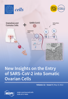
Severe acute respiratory syndrome coronavirus 2 (SARS-CoV-2) may affect female reproductive health. Herein, we provided evidence for the susceptibility of human ovarian cells and granulosa (GCs) and cumulus (CCs) cells to SARS-CoV-2 infection. Indeed, we demonstrated that both GCs and CCs express cell host factors ACE2, TRPMSS2, BSG, and CTSL, which are pivotal for the virus life cycle. Moreover, we demonstrated the ability of SARS-CoV-2 to productively infect the follicular microenvironment and we highlighted the presence of full-size virions attached to the membrane and located inside the cytoplasm of the infected cells. This in vitro study reveals the susceptibility of human ovarian cells to SARS-CoV-2 infection, suggesting a potential detrimental effect of COVID-19 infection on female human fertility. View this paper.
  • Issues are regarded as officially published after their release is announced to the table of contents alert mailing list.
  • You may sign up for e-mail alerts to receive table of contents of newly released issues.
  • PDF is the official format for papers published in both, html and pdf forms. To view the papers in pdf format, click on the "PDF Full-text" link, and use the free Adobe Reader to open them.
Order results
Result details
Section
Select all
Export citation of selected articles as:
21 pages, 42843 KiB  
Article
The Effects of Taurocholic Acid on Biliary Damage and Liver Fibrosis Are Mediated by Calcitonin-Gene-Related Peptide Signaling
by Romina Mancinelli, Ludovica Ceci, Lindsey Kennedy, Heather Francis, Vik Meadows, Lixian Chen, Guido Carpino, Konstantina Kyritsi, Nan Wu, Tianhao Zhou, Keisaku Sato, Luigi Pannarale, Shannon Glaser, Sanjukta Chakraborty, Gianfranco Alpini, Eugenio Gaudio, Paolo Onori and Antonio Franchitto
Cells 2022, 11(9), 1591; https://doi.org/10.3390/cells11091591 - 09 May 2022
Cited by 6 | Viewed by 2749
Abstract
Background & aims: Cholangiocytes are the target cells of liver diseases that are characterized by biliary senescence (evidenced by enhanced levels of senescence-associated secretory phenotype, SASP, e.g., TGF-β1), and liver inflammation and fibrosis accompanied by altered bile acid (BA) homeostasis. Taurocholic acid (TC) [...] Read more.
Background & aims: Cholangiocytes are the target cells of liver diseases that are characterized by biliary senescence (evidenced by enhanced levels of senescence-associated secretory phenotype, SASP, e.g., TGF-β1), and liver inflammation and fibrosis accompanied by altered bile acid (BA) homeostasis. Taurocholic acid (TC) stimulates biliary hyperplasia by activation of 3′,5′-cyclic cyclic adenosine monophosphate (cAMP) signaling, thereby preventing biliary damage (caused by cholinergic/adrenergic denervation) through enhanced liver angiogenesis. Also: (i) α-calcitonin gene-related peptide (α-CGRP, which activates the calcitonin receptor-like receptor, CRLR), stimulates biliary proliferation/senescence and liver fibrosis by enhanced biliary secretion of SASPs; and (ii) knock-out of α-CGRP reduces these phenotypes by decreased cAMP levels in cholestatic models. We aimed to demonstrate that TC effects on liver phenotypes are dependent on changes in the α-CGRP/CALCRL/cAMP/PKA/ERK1/2/TGF-β1/VEGF axis. Methods: Wild-type and α-CGRP−/− mice were fed with a control (BAC) or TC diet for 1 or 2 wk. We measured: (i) CGRP levels by both ELISA kits in serum and by qPCR in isolated cholangiocytes (CALCA gene for α-CGRP); (ii) CALCRL immunoreactivity by immunohistochemistry (IHC) in liver sections; (iii) liver histology, intrahepatic biliary mass, biliary senescence (by β-GAL staining and double immunofluorescence (IF) for p16/CK19), and liver fibrosis (by Red Sirius staining and double IF for collagen/CK19 in liver sections), as well as by qPCR for senescence markers in isolated cholangiocytes; and (iv) phosphorylation of PKA/ERK1/2, immunoreactivity of TGF-β1/TGF- βRI and angiogenic factors by IHC/immunofluorescence in liver sections and qPCR in isolated cholangiocytes. We measured changes in BA composition in total liver by liquid chromatography/mass spectrometry. Results: TC feeding increased CALCA expression, biliary damage, and liver inflammation and fibrosis, as well as phenotypes that were associated with enhanced immunoreactivity of the PKA/ERK1/2/TGF-β1/TGF-βRI/VEGF axis compared to BAC-fed mice and phenotypes that were reversed in α-CGRP−/− mice fed TC coupled with changes in hepatic BA composition. Conclusion: Modulation of the TC/ α-CGRP/CALCRL/PKA/ERK1/2/TGF-β1/VEGF axis may be important in the management of cholangiopathies characterized by BA accumulation. Full article
(This article belongs to the Special Issue New Aspects and Mechanisms in Liver Diseases)
Show Figures

Figure 1

11 pages, 2049 KiB  
Commentary
Cellular and Molecular Signaling as Targets for Cancer Vaccine Therapeutics
by Wen-Chi Wei, Lie-Fen Shyur and Ning-Sun Yang
Cells 2022, 11(9), 1590; https://doi.org/10.3390/cells11091590 - 09 May 2022
Viewed by 1911
Abstract
Plenty of evidence has recently shown that various inflammatory activities at the local tissue, organ, or even the whole body (systemic) level are strongly linked to many life-threatening chronic diseases, most notably various cancers. However, only very limited information is available for making [...] Read more.
Plenty of evidence has recently shown that various inflammatory activities at the local tissue, organ, or even the whole body (systemic) level are strongly linked to many life-threatening chronic diseases, most notably various cancers. However, only very limited information is available for making good use of our supporting immune-modulatory therapeutics for the treatment of cancers. This may result from a lack of studies on specific remedies for efficacious control or modulatory suppression of inflammation-related cancerous diseases. Our group and laboratories were fortunate to have initiated and consistently pursued an integrated team-work program project, aimed at investigating selected medicinal herbs and the derived, purified phytochemical compounds. We focused on the study of key and specific immune-signaling mechanisms at the cellular and molecular levels. We were fortunate to obtain a series of fruitful research results. We believe that our key findings reported herein may be helpful for proposing future thematic and integrated research projects that aim to develop future phytochemical drugs against cancers. The mechanisms of the cellular and molecular systems involved in inflammation are becoming increasingly recognized as keystones for the development of future therapeutic approaches for many chronic and cancerous diseases. Recently, the immune checkpoint inhibitors such as antibodies against PD-1 and/or PD-L1 have been shown to be too expensive for general clinical use, and their effects far from optimal, often showing little or no effect or only short-term efficacy. These results point to the need for developing future immune-regulatory or modulatory therapeutics. Full article
(This article belongs to the Special Issue Immunogenic Cell Death in Cancer and Infectious Disease)
Show Figures

Figure 1

16 pages, 3452 KiB  
Article
Defining the Skeletal Myogenic Lineage in Human Pluripotent Stem Cell-Derived Teratomas
by Matthew P. Pappas, Ning Xie, Jacqueline S. Penaloza and Sunny S. K. Chan
Cells 2022, 11(9), 1589; https://doi.org/10.3390/cells11091589 - 09 May 2022
Cited by 3 | Viewed by 2314
Abstract
Skeletal muscle stem cells are essential to muscle homeostasis and regeneration after injury, and have emerged as a promising cell source for treating skeletal disorders. An attractive approach to obtain these cells utilizes differentiation of pluripotent stem cells (PSCs). We recently reported that [...] Read more.
Skeletal muscle stem cells are essential to muscle homeostasis and regeneration after injury, and have emerged as a promising cell source for treating skeletal disorders. An attractive approach to obtain these cells utilizes differentiation of pluripotent stem cells (PSCs). We recently reported that teratomas derived from mouse PSCs are a rich source of skeletal muscle stem cells. Here, we showed that teratoma formation is also capable of producing skeletal myogenic progenitors from human PSCs. Using single-cell transcriptomics, we discovered several distinct skeletal myogenic subpopulations that represent progressive developmental stages of the skeletal myogenic lineage and recapitulate human embryonic skeletal myogenesis. We further discovered that ERBB3 and CD82 are effective surface markers for prospective isolation of the skeletal myogenic lineage in human PSC-derived teratomas. Therefore, teratoma formation provides an accessible model for obtaining human skeletal myogenic progenitors from PSCs. Full article
(This article belongs to the Collection Stem Cells in Tissue Engineering and Regeneration)
Show Figures

Figure 1

26 pages, 1502 KiB  
Review
The Role of Non-Coding RNAs in the Human Placenta
by Milena Žarković, Franziska Hufsky, Udo R. Markert and Manja Marz
Cells 2022, 11(9), 1588; https://doi.org/10.3390/cells11091588 - 09 May 2022
Cited by 11 | Viewed by 2853
Abstract
Non-coding RNAs (ncRNAs) play a central and regulatory role in almost all cells, organs, and species, which has been broadly recognized since the human ENCODE project and several other genome projects. Nevertheless, a small fraction of ncRNAs have been identified, and in the [...] Read more.
Non-coding RNAs (ncRNAs) play a central and regulatory role in almost all cells, organs, and species, which has been broadly recognized since the human ENCODE project and several other genome projects. Nevertheless, a small fraction of ncRNAs have been identified, and in the placenta they have been investigated very marginally. To date, most examples of ncRNAs which have been identified to be specific for fetal tissues, including placenta, are members of the group of microRNAs (miRNAs). Due to their quantity, it can be expected that the fairly larger group of other ncRNAs exerts far stronger effects than miRNAs. The syncytiotrophoblast of fetal origin forms the interface between fetus and mother, and releases permanently extracellular vesicles (EVs) into the maternal circulation which contain fetal proteins and RNA, including ncRNA, for communication with neighboring and distant maternal cells. Disorders of ncRNA in placental tissue, especially in trophoblast cells, and in EVs seem to be involved in pregnancy disorders, potentially as a cause or consequence. This review summarizes the current knowledge on placental ncRNA, their transport in EVs, and their involvement and pregnancy pathologies, as well as their potential for novel diagnostic tools. Full article
(This article belongs to the Special Issue Extracellular Vesicle-Associated Non-Coding RNAs)
Show Figures

Figure 1

19 pages, 2187 KiB  
Article
Surface Hydration Protects Cystic Fibrosis Airways from Infection by Restoring Junctional Networks
by Juliette L. Simonin, Alexandre Luscher, Davide Losa, Mehdi Badaoui, Christian van Delden, Thilo Köhler and Marc Chanson
Cells 2022, 11(9), 1587; https://doi.org/10.3390/cells11091587 - 09 May 2022
Cited by 5 | Viewed by 5457
Abstract
Defective hydration of airway surface mucosa is associated with recurrent lung infection in cystic fibrosis (CF), a disease caused by CF transmembrane conductance regulator (CFTR) gene mutations. Whether the composition and/or presence of an airway surface liquid (ASL) is sufficient to [...] Read more.
Defective hydration of airway surface mucosa is associated with recurrent lung infection in cystic fibrosis (CF), a disease caused by CF transmembrane conductance regulator (CFTR) gene mutations. Whether the composition and/or presence of an airway surface liquid (ASL) is sufficient to prevent infection remains unclear. The susceptibility to infection of polarized wild type and CFTR knockdown (CFTR-KD) airway epithelial cells was determined in the presence or absence of a healthy ASL or physiological saline. CFTR-KD epithelia exhibited strong ASL volume reduction, enhanced susceptibility to infection, and reduced junctional integrity. Interestingly, the presence of an apical physiological saline alleviated disruption of the airway epithelial barrier by stimulating essential junctional protein expression. Thus, rehydrated CFTR-KD cells were protected from infection despite normally intense bacterial growth. This study indicates that an epithelial integrity gatekeeper is modulated by the presence of an apical liquid volume, irrespective of the liquid’s composition and of expression of a functional CFTR. Full article
(This article belongs to the Collection Cystic Fibrosis: Cells, Physiopathology and Emerging Therapies)
Show Figures

Figure 1

4 pages, 464 KiB  
Editorial
Code Red in the Supply Center: The Impact of Immune Activation on Hematopoiesis
by Katherine C. MacNamara and Martijn A. Nolte
Cells 2022, 11(9), 1586; https://doi.org/10.3390/cells11091586 - 09 May 2022
Viewed by 1376
Abstract
This Special Issue entitled “The Impact of Immune Activation on Hematopoiesis” aims to bring together review and primary articles focused on distinct types of immune activation that impact hematopoiesis [...] Full article
(This article belongs to the Special Issue The Impact of Immune Activation on Hematopoiesis)
Show Figures

Figure 1

18 pages, 8330 KiB  
Article
FKBP51, AmotL2 and IQGAP1 Involvement in Cilastatin Prevention of Cisplatin-Induced Tubular Nephrotoxicity in Rats
by Rebeca González-Fernández, María Ángeles González-Nicolás, Manuel Morales, Julio Ávila, Alberto Lázaro and Pablo Martín-Vasallo
Cells 2022, 11(9), 1585; https://doi.org/10.3390/cells11091585 - 09 May 2022
Cited by 2 | Viewed by 2298
Abstract
The immunophilin FKBP51, the angiomotin AmotL2, and the scaffoldin IQGAP1 are overexpressed in many types of cancer, with the highest increase in leucocytes from patients undergoing oxaliplatin chemotherapy. Inflammation is involved in the pathogenesis of nephrotoxicity induced by platinum analogs. Cilastatin prevents renal [...] Read more.
The immunophilin FKBP51, the angiomotin AmotL2, and the scaffoldin IQGAP1 are overexpressed in many types of cancer, with the highest increase in leucocytes from patients undergoing oxaliplatin chemotherapy. Inflammation is involved in the pathogenesis of nephrotoxicity induced by platinum analogs. Cilastatin prevents renal damage caused by cisplatin. This functional and confocal microscopy study shows the renal focal-segmental expression of TNFα after cisplatin administration in rats, predominantly of tubular localization and mostly prevented by co-administration of cilastatin. FKBP51, AmotL2 and IQGAP1 protein expression increases slightly with cilastatin administration and to a much higher extent with cisplatin, in a cellular- and subcellular-specific manner. Kidney tubule cells expressing FKBP51 show either very low or no expression of TNFα, while cells expressing TNFα have low levels of FKBP51. AmotL2 and TNFα seem to colocalize and their expression is increased in tubular cells. IQGAP1 fluorescence increases with cilastatin, cisplatin and joint cilastatin-cisplatin treatment, and does not correlate with TNFα expression or localization. These data suggest a role for FKBP51, AmotL2 and IQGAP1 in cisplatin toxicity in kidney tubules and in the protective effect of cilastatin through inhibition of dehydropeptidase-I. Full article
(This article belongs to the Special Issue Scaffold Proteins in Health, Disease, and Therapy)
Show Figures

Figure 1

15 pages, 2707 KiB  
Article
Safety Profile and Issues of Subcutaneous Immunotherapy in the Treatment of Children with Allergic Rhinitis
by Anang Endaryanto and Ricardo Adrian Nugraha
Cells 2022, 11(9), 1584; https://doi.org/10.3390/cells11091584 - 09 May 2022
Cited by 5 | Viewed by 1846
Abstract
This study aims to evaluate safety issues of house dust mite subcutaneous immunotherapy (SCIT) among allergic rhinitis (AR) children. A retrospective cohort study was done between 2015 and 2020 to investigate the side effects of SCIT among AR children caused by a house [...] Read more.
This study aims to evaluate safety issues of house dust mite subcutaneous immunotherapy (SCIT) among allergic rhinitis (AR) children. A retrospective cohort study was done between 2015 and 2020 to investigate the side effects of SCIT among AR children caused by a house dust mite allergy. Among 1098 patients who received house dust mite subcutaneous immunotherapy injections, 284 patients (25.87%) had side effects (SE). SE were found to be 699 times higher or in 2.27% of the 30,744 subcutaneous immunotherapy injections. A total of 17.9% of the patients had local SE during SCIT administration. Systemic side effects occurred in 8.38% of children receiving SCIT and in 0.53% of the total population who received SCIT injections. Only 2/92 (2.18%) of patients suffered an allergic reaction within 30 minutes of injection and these patients responded well to antiallergic medication. Severe anaphylaxis occurred in 0.091% of the 1098 patients in the SCIT group and in 0.0033% of the 30,774 SCIT injections. Systemic SE after SCIT occurred in 8.38% of patients receiving SCIT or 0.53% of the total number of SCIT injections. Anaphylactic episodes occurred in 16 patients (1.46%) and 15 patients (1.37%) who had first and second episodes. One severe attack was found and it was resolved with adrenaline. This study demonstrates that in pediatric patients with AR who received HDM SCIT for 18 months with high adherence, some experienced significant local SE and systemic SE caused by SCIT, but this did not interfere with the course of AR treatment or the effectiveness of SCIT. Full article
(This article belongs to the Section Stem Cells)
Show Figures

Figure 1

15 pages, 2618 KiB  
Article
An Ex Vivo 3D Tumor Microenvironment-Mimicry Culture to Study TAM Modulation of Cancer Immunotherapy
by Yan-Ruide Li, Yanqi Yu, Adam Kramer, Ryan Hon, Matthew Wilson, James Brown and Lili Yang
Cells 2022, 11(9), 1583; https://doi.org/10.3390/cells11091583 - 08 May 2022
Cited by 10 | Viewed by 3850
Abstract
Tumor-associated macrophages (TAMs) accumulate in the solid tumor microenvironment (TME) and have been shown to promote tumor growth and dampen antitumor immune responses. TAM-mediated suppression of T-cell antitumor reactivity is considered to be a major obstacle for many immunotherapies, including immune checkpoint blockade [...] Read more.
Tumor-associated macrophages (TAMs) accumulate in the solid tumor microenvironment (TME) and have been shown to promote tumor growth and dampen antitumor immune responses. TAM-mediated suppression of T-cell antitumor reactivity is considered to be a major obstacle for many immunotherapies, including immune checkpoint blockade and adoptive T/CAR-T-cell therapies. An ex vivo culture system closely mimicking the TME can greatly facilitate the study of cancer immunotherapies. Here, we report the development of a 3D TME-mimicry culture that is comprised of the three major components of a human TME, including human tumor cells, TAMs, and tumor antigen-specific T cells. This TME-mimicry culture can readout the TAM-mediated suppression of T-cell antitumor reactivity, and therefore can be used to study TAM modulation of T-cell-based cancer immunotherapy. As a proof-of-principle, the studies of a PD-1/PD-L1 blockade therapy and a MAO-A blockade therapy were performed and validated. Full article
(This article belongs to the Special Issue Cell-Based Models of Diseases for Drug Discovery)
Show Figures

Figure 1

22 pages, 15009 KiB  
Article
Electrospun Membrane Surface Modification by Sonocoating with HA and ZnO:Ag Nanoparticles—Characterization and Evaluation of Osteoblasts and Bacterial Cell Behavior In Vitro
by Julia Higuchi, Katarzyna Klimek, Jacek Wojnarowicz, Agnieszka Opalińska, Agnieszka Chodara, Urszula Szałaj, Sylwia Dąbrowska, Damian Fudala and Grazyna Ginalska
Cells 2022, 11(9), 1582; https://doi.org/10.3390/cells11091582 - 08 May 2022
Cited by 15 | Viewed by 6064
Abstract
Guided tissue regeneration and guided bone regeneration membranes are some of the most common products used for bone regeneration in periodontal dentistry. The main disadvantage of commercially available membranes is their lack of bone cell stimulation and easy bacterial colonization. The aim of [...] Read more.
Guided tissue regeneration and guided bone regeneration membranes are some of the most common products used for bone regeneration in periodontal dentistry. The main disadvantage of commercially available membranes is their lack of bone cell stimulation and easy bacterial colonization. The aim of this work was to design and fabricate a new membrane construct composed of electrospun poly (D,L-lactic acid)/poly (lactic-co-glycolic acid) fibers sonocoated with layers of nanoparticles with specific properties, i.e., hydroxyapatite and bimetallic nanocomposite of zinc oxide–silver. Thus, within this study, four different variants of biomaterials were evaluated, namely: poly (D,L-lactic acid)/poly (lactic-co-glycolic acid) biomaterial, poly(D,L-lactic acid)/poly (lactic-co-glycolic acid)/nano hydroxyapatite biomaterial, poly (D,L-lactic acid)/poly (lactic-co-glycolic acid)/nano zinc oxide–silver biomaterial, and poly (D,L-lactic acid)/poly (lactic-co-glycolic acid)/nano hydroxyapatite/nano zinc oxide–silver biomaterial. First, it was demonstrated that the wettability of biomaterials—a prerequisite property important for ensuring desired biological response—was highly increased after the sonocoating process. Moreover, it was indicated that biomaterials composed of poly (D,L-lactic acid)/poly (lactic-co-glycolic acid) with or without a nano hydroxyapatite layer allowed proper osteoblast growth and proliferation, but did not have antibacterial properties. Addition of a nano zinc oxide–silver layer to the biomaterial inhibited growth of bacterial cells around the membrane, but at the same time induced very high cytotoxicity towards osteoblasts. Most importantly, enrichment of this biomaterial with a supplementary underlayer of nano hydroxyapatite allowed for the preservation of antibacterial properties and also a decrease in the cytotoxicity towards bone cells, associated with the presence of a nano zinc oxide–silver layer. Thus, the final structure of the composite poly (D,L-lactic acid)/poly (lactic-co-glycolic acid)/nano hydroxyapatite/nano zinc oxide–silver seems to be a promising construct for tissue engineering products, especially guided tissue regeneration/guided bone regeneration membranes. Nevertheless, additional research is needed in order to improve the developed construct, which will simultaneously protect the biomaterial from bacterial colonization and enhance the bone regeneration properties. Full article
(This article belongs to the Special Issue Cellular Response to Biomaterials Designed for Tissue Engineering)
Show Figures

Graphical abstract

22 pages, 5646 KiB  
Article
Multifaceted and Intricate Oncogenic Mechanisms of NDRG1 in Head and Neck Cancer Depend on Its C-Terminal 3R-Motif
by Guo-Rung You, Joseph T. Chang, Hsiao-Fan Li and Ann-Joy Cheng
Cells 2022, 11(9), 1581; https://doi.org/10.3390/cells11091581 - 07 May 2022
Cited by 4 | Viewed by 2203
Abstract
N-Myc downstream-regulated 1 (NDRG1) has inconsistent oncogenic functions in various cancers. We surveyed and characterized the role of NDRG1 in head and neck cancer (HNC). Cellular methods included spheroid cell formation, clonogenic survival, cell viability, and Matrigel invasion assays. Molecular techniques included transcriptomic [...] Read more.
N-Myc downstream-regulated 1 (NDRG1) has inconsistent oncogenic functions in various cancers. We surveyed and characterized the role of NDRG1 in head and neck cancer (HNC). Cellular methods included spheroid cell formation, clonogenic survival, cell viability, and Matrigel invasion assays. Molecular techniques included transcriptomic profiling, RT-qPCR, immunoblotting, in vitro phosphorylation, immunofluorescent staining, and confocal microscopy. Prognostic significance was assessed by Kaplan–Meier analysis. NDRG1 participated in diverse oncogenic functions in HNC cells, mainly stress response and cell motility. Notably, NDRG1 contributed to spheroid cell growth, radio-chemoresistance, and upregulation of stemness-related markers (CD44 and Twist1). NDRG1 facilitated cell migration and invasion, and was associated with modulation of the extracellular matrix molecules (fibronectin, vimentin). Characterizing the 3R-motif in NDRG1 revealed its mechanism in the differential regulation of the phenotypes. The 3R-motif displayed minimal effect on cancer stemness but was crucial for cell motility. Phosphorylating the motif by GSK3b at serine residues led to its nuclear translocation to promote motility. Clinical analyses supported the oncogenic function of NDRG1, which was overexpressed in HNC and associated with poor prognosis. The data elucidate the multifaceted and intricate mechanisms of NDRG1 in HNC. NDRG1 may be a prognostic indicator or therapeutic target for refractory HNC. Full article
(This article belongs to the Special Issue New Insights into Head and Neck Squamous Cell Carcinoma)
Show Figures

Graphical abstract

16 pages, 1318 KiB  
Review
The Molecular Mechanisms Governing the Assembly of the Immuno- and Thymoproteasomes in the Presence of Constitutive Proteasomes
by Ayaka Watanabe, Hideki Yashiroda, Satoshi Ishihara, Megan Lo and Shigeo Murata
Cells 2022, 11(9), 1580; https://doi.org/10.3390/cells11091580 - 07 May 2022
Cited by 5 | Viewed by 2454
Abstract
The proteasome is a large protein complex responsible for proteolysis in cells. Though the proteasome is widely conserved in all eukaryotes, vertebrates additionally possess tissue-specific proteasomes, termed immunoproteasomes and thymoproteasomes. These specialized proteasomes diverge from constitutive proteasomes in the makeup of their catalytic [...] Read more.
The proteasome is a large protein complex responsible for proteolysis in cells. Though the proteasome is widely conserved in all eukaryotes, vertebrates additionally possess tissue-specific proteasomes, termed immunoproteasomes and thymoproteasomes. These specialized proteasomes diverge from constitutive proteasomes in the makeup of their catalytic 20S core particle (CP), whereby the constitutive β1, β2, and β5 catalytic subunits are replaced by β1i, β2i, and β5i in immunoproteasomes, or β1i, β2i, and β5t in thymoproteasomes. However, as constitutive β1, β2, and β5 are also present in tissues and cells expressing immuno- and thymoproteasomes, the specialized proteasomes must be able to selectively incorporate their specific subunits. Here, we review the mechanisms governing the assembly of constitutive and specialized proteasomes elucidated thus far. Studies have revealed that β1i and β2i are added onto the α-ring of the CP prior to the other β subunits. Furthermore, β5i and β5t can be incorporated independent of β4, whereas constitutive β5 incorporation is dependent on β4. These mechanisms allow the immuno- and thymoproteasomes to integrate tissue-specific β-subunits without contamination from constitutive β1, β2, and β5. We end the review with a brief discussion on the diseases caused by mutations to the immunoproteasome and the proteins involved with its assembly. Full article
(This article belongs to the Special Issue The Immunoproteasome in Health and Disease)
Show Figures

Graphical abstract

15 pages, 2001 KiB  
Article
Proteomic, Biochemical, and Morphological Analyses of the Effect of Silver Nanoparticles Mixed with Organic and Inorganic Chemicals on Wheat Growth
by Setsuko Komatsu, Hisateru Yamaguchi, Keisuke Hitachi and Kunihiro Tsuchida
Cells 2022, 11(9), 1579; https://doi.org/10.3390/cells11091579 - 07 May 2022
Cited by 4 | Viewed by 1766
Abstract
Wheat is vulnerable to numerous diseases; on the other hand, silver nanoparticles (AgNPs) exhibit a sterilizing action. To understand the combined effects of AgNPs with nicotinate and potassium nitrate (KNO3) for plant growth and sterilization, a gel- and label-free proteomics was [...] Read more.
Wheat is vulnerable to numerous diseases; on the other hand, silver nanoparticles (AgNPs) exhibit a sterilizing action. To understand the combined effects of AgNPs with nicotinate and potassium nitrate (KNO3) for plant growth and sterilization, a gel- and label-free proteomics was performed. Root weight was promoted by the treatment of AgNPs mixed with nicotinate and KNO3. From a total of 5557 detected proteins, 90 proteins were changed by the mixture of AgNPs, nicotinate, and KNO3; among them, 25 and 65 proteins increased and decreased, respectively. The changed proteins were mainly associated with redox and biotic stress in the functional categorization. By immunoblot analysis, the abundance of glutathione reductase/peroxiredoxin and pathogen-related protein three significantly decreased with the mixture. Furthermore, from the changed proteins, the abundance of starch synthase and lipoxygenase significantly increased and decreased, respectively. Through biochemical analysis, the starch contents increased with the mixture. The application of esculetin, which is a lipoxygenase inhibitor, increased the weight and length of the root. These results suggest that the AgNPs mixed with nicotinate and KNO3 cause positive effects on wheat seedlings by regulating pathogen-related protein and reactive-oxygen species scavenging. Furthermore, increasing starch and decreasing lipoxygenase might improve wheat growth. Full article
(This article belongs to the Collection Deciphering the Proteome in Cell Biology and Diseases)
Show Figures

Figure 1

38 pages, 1590 KiB  
Review
Glatiramer Acetate Immunomodulation: Evidence of Neuroprotection and Cognitive Preservation
by Arielle Kasindi, Dieu-Trang Fuchs, Yosef Koronyo, Altan Rentsendorj, Keith L. Black and Maya Koronyo-Hamaoui
Cells 2022, 11(9), 1578; https://doi.org/10.3390/cells11091578 - 07 May 2022
Cited by 18 | Viewed by 4107
Abstract
Novel, neuroprotective uses of Copaxone (generic name: glatiramer acetate—GA) are being examined, primarily in neurological conditions involving cognitive decline. GA is a well-studied synthetic copolymer that is FDA-approved for immune-based treatment of relapsing remitting multiple sclerosis (RRMS). Clinical studies have explored the potential [...] Read more.
Novel, neuroprotective uses of Copaxone (generic name: glatiramer acetate—GA) are being examined, primarily in neurological conditions involving cognitive decline. GA is a well-studied synthetic copolymer that is FDA-approved for immune-based treatment of relapsing remitting multiple sclerosis (RRMS). Clinical studies have explored the potential mechanism of action (MOA) and outcomes of GA immunization in patients. Furthermore, results from these and animal studies suggest that GA has a direct immunomodulatory effect on adaptive and innate immune cell phenotypes and responses. These MOAs have been postulated to have a common neuroprotective impact in several neuroinflammatory and neurodegenerative diseases. Notably, several clinical studies report that the use of GA mitigated MS-associated cognitive decline. Its propensity to ameliorate neuro-proinflammatory and degenerative processes ignites increased interest in potential alternate uses such as in age-related macular degeneration (AMD), amyotrophic lateral sclerosis (ALS), and Alzheimer’s disease (AD). Preclinical studies are exploring less frequent subcutaneous administration of GA, such as once weekly or monthly or a single dosing regimen. Indeed, cognitive functions were found to be either preserved, reversed, or improved after the less frequent treatment regimens with GA in animal models of AD. In this systematic review, we examine the potential novel uses of GA across clinical and pre-clinical studies, with evidence for its beneficial impact on cognition. Future investigation in large-size, double-blind clinical trials is warranted to establish the impact of GA immunomodulation on neuroprotection and cognitive preservation in various neurological conditions. Full article
(This article belongs to the Special Issue Mechanisms of Neurodevelopment and Neurodegeneration)
Show Figures

Graphical abstract

30 pages, 4374 KiB  
Article
Biodegradable Poly(D-L-lactide-co-glycolide) (PLGA)-Infiltrated Bioactive Glass (CAR12N) Scaffolds Maintain Mesenchymal Stem Cell Chondrogenesis for Cartilage Tissue Engineering
by Clemens Gögele, Silvana Müller, Svetlana Belov, Andreas Pradel, Sven Wiltzsch, Armin Lenhart, Markus Hornfeck, Vera Kerling, Achim Rübling, Hannes Kühl, Kerstin Schäfer-Eckart, Bernd Minnich, Thomas Martin Weiger and Gundula Schulze-Tanzil
Cells 2022, 11(9), 1577; https://doi.org/10.3390/cells11091577 - 07 May 2022
Cited by 5 | Viewed by 2496
Abstract
Regeneration of articular cartilage remains challenging. The aim of this study was to increase the stability of pure bioactive glass (BG) scaffolds by means of solvent phase polymer infiltration and to maintain cell adherence on the glass struts. Therefore, BG scaffolds either pure [...] Read more.
Regeneration of articular cartilage remains challenging. The aim of this study was to increase the stability of pure bioactive glass (BG) scaffolds by means of solvent phase polymer infiltration and to maintain cell adherence on the glass struts. Therefore, BG scaffolds either pure or enhanced with three different amounts of poly(D-L-lactide-co-glycolide) (PLGA) were characterized in detail. Scaffolds were seeded with primary porcine articular chondrocytes (pACs) and human mesenchymal stem cells (hMSCs) in a dynamic long-term culture (35 days). Light microscopy evaluations showed that PLGA was detectable in every region of the scaffold. Porosity was greater than 70%. The biomechanical stability was increased by polymer infiltration. PLGA infiltration did not result in a decrease in viability of both cell types, but increased DNA and sulfated glycosaminoglycan (sGAG) contents of hMSCs-colonized scaffolds. Successful chondrogenesis of hMSC-colonized scaffolds was demonstrated by immunocytochemical staining of collagen type II, cartilage proteoglycans and the transcription factor SOX9. PLGA-infiltrated scaffolds showed a higher relative expression of cartilage related genes not only of pAC-, but also of hMSC-colonized scaffolds in comparison to the pure BG. Based on the novel data, our recommendation is BG scaffolds with single infiltrated PLGA for cartilage tissue engineering. Full article
(This article belongs to the Special Issue Cell Therapies in Orthopaedics)
Show Figures

Figure 1

13 pages, 1998 KiB  
Review
A Long-Lasting PARP1-Activation Mediates Signal-Induced Gene Expression
by Malka Cohen-Armon
Cells 2022, 11(9), 1576; https://doi.org/10.3390/cells11091576 - 07 May 2022
Cited by 3 | Viewed by 3585
Abstract
This overview presents recent evidence for a long-lasting PARP1 activation by a variety of signal transduction mechanisms, mediating signal-induced gene expression and chromatin remodeling. This mode of PARP1 activation has been reported in a variety of cell types, under physiological conditions. In this [...] Read more.
This overview presents recent evidence for a long-lasting PARP1 activation by a variety of signal transduction mechanisms, mediating signal-induced gene expression and chromatin remodeling. This mode of PARP1 activation has been reported in a variety of cell types, under physiological conditions. In this mechanism, PARP1 is not transiently activated by binding to DNA breaks. Moreover, damaged DNA interfered with this long-lasting PARP1 activation. Full article
Show Figures

Figure 1

8 pages, 1375 KiB  
Communication
Glycogene Expression Profile of Human Limbal Epithelial Cells with Distinct Clonogenic Potential
by Damien Guindolet, Ashley M. Woodward, Eric E. Gabison and Pablo Argüeso
Cells 2022, 11(9), 1575; https://doi.org/10.3390/cells11091575 - 07 May 2022
Cited by 2 | Viewed by 1767
Abstract
Glycans function as valuable markers of stem cells but also regulate the ability of these cells to self-renew and differentiate. Approximately 2% of the human genome encodes for proteins that are involved in the biosynthesis and recognition of glycans. In the present study, [...] Read more.
Glycans function as valuable markers of stem cells but also regulate the ability of these cells to self-renew and differentiate. Approximately 2% of the human genome encodes for proteins that are involved in the biosynthesis and recognition of glycans. In the present study, we evaluated the expression of a small subset of glycogenes in human limbal epithelial cells with distinct clonogenic potential. Individual clones were classified as abortive or clonogenic, based on the fraction of the terminal colonies produced; clones leading exclusively to terminal colonies were referred to as abortive while those with half or fewer terminal colonies were referred to as clonogenic. An analysis of glycogene expression in clonogenic cultures revealed a high content of transcripts regulating the galactose and mannose metabolic pathways. Abortive clones were characterized by increased levels of GCNT4 and FUCA2, genes that are responsible for the branching of mucin-type O-glycans and the hydrolysis of fucose residues on N-glycans, respectively. The expansion of primary cultures of human limbal epithelial cells for 10 days resulted in stratification and a concomitant increase in MUC16, GCNT4 and FUCA2 expression. These data indicate that the clonogenic potential of human limbal epithelial cells is associated with specific glycosylation pathways. Mucin-type O-glycan branching and increased fucose metabolism are linked to limbal epithelial cell differentiation. Full article
(This article belongs to the Special Issue Ocular Stem Cells and Therapies)
Show Figures

Figure 1

22 pages, 17872 KiB  
Article
Growth Response and Differentiation of Bone Marrow-Derived Mesenchymal Stem/Stromal Cells in the Presence of Novel Multiple Myeloma Drug Melflufen
by Arjen Gebraad, Roope Ohlsbom, Juho J. Miettinen, Promise Emeh, Toni-Karri Pakarinen, Mikko Manninen, Antti Eskelinen, Kirsi Kuismanen, Ana Slipicevic, Fredrik Lehmann, Nina N. Nupponen, Caroline A. Heckman and Susanna Miettinen
Cells 2022, 11(9), 1574; https://doi.org/10.3390/cells11091574 - 07 May 2022
Cited by 2 | Viewed by 2686
Abstract
Mesenchymal stem/stromal cells (MSCs) are self-renewing and multipotent progenitors, which constitute the main cellular compartment of the bone marrow stroma. Because MSCs have an important role in the pathogenesis of multiple myeloma, it is essential to know if novel drugs target MSCs. Melflufen [...] Read more.
Mesenchymal stem/stromal cells (MSCs) are self-renewing and multipotent progenitors, which constitute the main cellular compartment of the bone marrow stroma. Because MSCs have an important role in the pathogenesis of multiple myeloma, it is essential to know if novel drugs target MSCs. Melflufen is a novel anticancer peptide–drug conjugate compound for patients with relapsed refractory multiple myeloma. Here, we studied the cytotoxicity of melflufen, melphalan and doxorubicin in healthy human bone marrow-derived MSCs (BMSCs) and how these drugs affect BMSC proliferation. We established co-cultures of BMSCs with MM.1S myeloma cells to see if BMSCs increase or decrease the cytotoxicity of melflufen, melphalan, bortezomib and doxorubicin. We evaluated how the drugs affect BMSC differentiation into adipocytes and osteoblasts and the BMSC-supported formation of vascular networks. Our results showed that BMSCs were more sensitive to melflufen than to melphalan. The cytotoxicity of melflufen in myeloma cells was not affected by the co-culture with BMSCs, as was the case for melphalan, bortezomib and doxorubicin. Adipogenesis, osteogenesis and BMSC-mediated angiogenesis were all affected by melflufen. Melphalan and doxorubicin affected BMSC differentiation in similar ways. The effects on adipogenesis and osteogenesis were not solely because of effects on proliferation, seen from the differential expression of differentiation markers normalized by cell number. Overall, our results indicate that melflufen has a significant impact on BMSCs, which could possibly affect therapy outcome. Full article
Show Figures

Figure 1

18 pages, 4918 KiB  
Article
Nano-Sized Extracellular Vesicles Secreted from GATA-4 Modified Mesenchymal Stem Cells Promote Angiogenesis by Delivering Let-7 miRNAs
by Min Gong, Min Wang, Jie Xu, Bin Yu, Yi-Gang Wang, Min Liu, Muhammad Ashraf and Meifeng Xu
Cells 2022, 11(9), 1573; https://doi.org/10.3390/cells11091573 - 07 May 2022
Cited by 9 | Viewed by 2265
Abstract
We demonstrated previously that extracellular vesicles (EVs) released from mesenchymal stem cells (MSCs) play a critical role in angiogenesis. Here, we examine whether this pro-angiogenic efficacy is enhanced in EVs derived from MSCs overexpressing GATA-4 (MSCGATA−4). Methods and Results. EVs were [...] Read more.
We demonstrated previously that extracellular vesicles (EVs) released from mesenchymal stem cells (MSCs) play a critical role in angiogenesis. Here, we examine whether this pro-angiogenic efficacy is enhanced in EVs derived from MSCs overexpressing GATA-4 (MSCGATA−4). Methods and Results. EVs were isolated from MSCGATA-4 (EVGATA-4) and control MSCs transduced with an empty vector (EVnull). EVs from both cell types were of the same size and displayed similar molecular markers. Compared with EVnull, EVGATA-4 increased both a tube-like structure formation and spheroid-based sprouting of human umbilical vein endothelial cells (HUVECs). The EVGATA-4 increased the numbers of CD31-positive cells and hemoglobin content inside Matrigel plugs subcutaneously transplanted into mice for 2 weeks. Moreover, EVGATA-4 encapsulated higher levels of let-7 family miRs compared to EVnull. The transfer of exosomal let-7 miRs into HUVECs was recorded with an accompanied down-regulation of thrombospondin-1 (THBS1) expression, a major endogenous angiogenesis inhibitor. The loss-and-gain of function studies of let-7 miRs showed that let-7f knockdown significantly decreased EVGATA-4-mediated vascularization inside Matrigel plugs. In contrast, let-7f overexpression promoted HUVEC migration and tube formation. Conclusion. Our results indicate that EVs derived from genetically modified MSCs with GATA-4 overexpression had increased pro-angiogenic capacity due to the delivery of let-7 miRs that targeted THBS1 in endothelial cells. Full article
(This article belongs to the Special Issue Mesenchymal Stem Cell-Derived Extracellular Vesicles)
Show Figures

Figure 1

30 pages, 3513 KiB  
Article
Adverse Childhood Experiences Predict the Phenome of Affective Disorders and These Effects Are Mediated by Staging, Neuroimmunotoxic and Growth Factor Profiles
by Michael Maes, Muanpetch Rachayon, Ketsupar Jirakran, Pimpayao Sodsai, Siriwan Klinchanhom, Monojit Debnath, Agnieska Basta-Kaim, Marta Kubera, Abbas F. Almulla and Atapol Sughondhabirom
Cells 2022, 11(9), 1564; https://doi.org/10.3390/cells11091564 - 07 May 2022
Cited by 18 | Viewed by 2957
Abstract
Adverse childhood experiences (ACEs) enhance pro-inflammatory and pro-oxidant responses. In affective disorders, recent precision nomothetic psychiatry studies disclosed new pathway phenotypes, including an ROI—reoccurrence of illness (ROI)—oxidative stress latent construct. The aim of the present study is to delineate a) whether ACEs sensitize [...] Read more.
Adverse childhood experiences (ACEs) enhance pro-inflammatory and pro-oxidant responses. In affective disorders, recent precision nomothetic psychiatry studies disclosed new pathway phenotypes, including an ROI—reoccurrence of illness (ROI)—oxidative stress latent construct. The aim of the present study is to delineate a) whether ACEs sensitize the M1 macrophage, the T helper cells (Th)1, Th2, and Th17, the IRS (immune-inflammatory-responses system), the CIRS (compensatory immunoregulatory system), and the neuroimmunotoxic and growth factor (GF) profiles and whether they are associated with ROI and the phenome of affective disorders and b) the molecular pathways underpinning the effects of the ACEs. We collected supernatants of stimulated (5 μg/mL of PHA and 25 μg/mL of LPS) and unstimulated diluted whole blood in 20 healthy controls and 30 depressed patients and measured a panel of 27 cytokines/GF using a Luminex method. ACEs (comprising mental and physical trauma, mental neglect, domestic violence, family history of mental disease, and parent loss) are accompanied by the increased stimulated, but not unstimulated, production of M1, Th1, Th2, Th17, IRS, neuroimmunotoxic, and GF profiles and are strongly correlated with ROI and the phenome. A latent vector extracted from the ROI features (recurrent episodes and suicidal behaviors) and the IRS/neuroimmunotoxic/GF profiles explains 66.8% of the variance in the phenome and completely mediates the effects of ACEs on the phenome. Enrichment analysis showed that the ACE-associated sensitization of immune/GF profiles involves JAK-STAT, nuclear factor-κB, tumor necrosis factor-α, G-protein coupled receptor, PI3K/Akt/RAS/MAPK, and hypoxia signaling. In summary, the ACE-induced sensitization of immune pathways and secondary immune hits predicts the phenome of affective disorders. Full article
Show Figures

Figure 1

42 pages, 13834 KiB  
Article
Multiomics Approach Reveals an Important Role of BNIP3 in Myocardial Remodeling and the Pathogenesis of Heart Failure with Reduced Ejection Fraction
by Antoine H. Chaanine, LeeAnn Higgins, Lothar Lauterboeck, Todd Markowski, Qinglin Yang and Patrice Delafontaine
Cells 2022, 11(9), 1572; https://doi.org/10.3390/cells11091572 - 06 May 2022
Cited by 6 | Viewed by 2629
Abstract
Previous work showed a role of BNIP3 in myocardial remodeling and progression to HFrEF. We utilized a multiomics approach to unravel BNIP3-related molecular mechanisms in the pathogenesis of HFrEF. BNIP3 knockdown in HFrEF improved glycolysis, pyruvate metabolism, branched-chain amino acid catabolism, and oxidative [...] Read more.
Previous work showed a role of BNIP3 in myocardial remodeling and progression to HFrEF. We utilized a multiomics approach to unravel BNIP3-related molecular mechanisms in the pathogenesis of HFrEF. BNIP3 knockdown in HFrEF improved glycolysis, pyruvate metabolism, branched-chain amino acid catabolism, and oxidative phosphorylation, and restored endoplasmic reticulum (ER)–mitochondrial (mt) calcium and ion homeostasis. These effects of BNIP3 on cardiac metabolism were related to its interaction and downregulation, and/or phosphorylation, of specific mt-proteins involved in the aforementioned metabolic pathways, including the MICOS and SLC25A families of carrier proteins. BNIP3 affected ER–mt-calcium and ion homeostasis via its interaction-induced VDAC1 dimerization and modulation of VDAC1 phosphorylation at Ser104 and Ser241, and the downregulation of LETM1. At the ER level, BNIP3 interacted with the enzyme SERCA2a and the PKA signaling complex, leading to the downregulation of SERCA2a and PKA-mediated Ser16 phospholamban phosphorylation. Additionally, BNIP3 attenuated AMPK and PRKCE activity by modulating AMPK phosphorylation at Ser485/491 and Ser377 residues, and PRKCE phosphorylation at Thr521 and Thr710 residues. BNIP3 also interacted with sarcomeric, cytoskeletal, and cellular transcription and translation proteins, and affected their expression and/or phosphorylation. In conclusion, BNIP3 modulates multiple pathobiological processes and constitutes an attractive therapeutic target in HFrEF. Full article
(This article belongs to the Topic Cell Signaling Pathways)
Show Figures

Figure 1

30 pages, 13578 KiB  
Review
Genetic Kidney Diseases (GKDs) Modeling Using Genome Editing Technologies
by Fernando Gómez-García, Raquel Martínez-Pulleiro, Noa Carrera, Catarina Allegue and Miguel A. Garcia-Gonzalez
Cells 2022, 11(9), 1571; https://doi.org/10.3390/cells11091571 - 06 May 2022
Cited by 2 | Viewed by 3835
Abstract
Genetic kidney diseases (GKDs) are a group of rare diseases, affecting approximately about 60 to 80 per 100,000 individuals, for which there is currently no treatment that can cure them (in many cases). GKDs usually leads to early-onset chronic kidney disease, which results [...] Read more.
Genetic kidney diseases (GKDs) are a group of rare diseases, affecting approximately about 60 to 80 per 100,000 individuals, for which there is currently no treatment that can cure them (in many cases). GKDs usually leads to early-onset chronic kidney disease, which results in patients having to undergo dialysis or kidney transplant. Here, we briefly describe genetic causes and phenotypic effects of six GKDs representative of different ranges of prevalence and renal involvement (ciliopathy, glomerulopathy, and tubulopathy). One of the shared characteristics of GKDs is that most of them are monogenic. This characteristic makes it possible to use site-specific nuclease systems to edit the genes that cause GKDs and generate in vitro and in vivo models that reflect the genetic abnormalities of GKDs. We describe and compare these site-specific nuclease systems (zinc finger nucleases (ZFNs), transcription activator-like effect nucleases (TALENs) and regularly clustered short palindromic repeat-associated protein (CRISPR-Cas9)) and review how these systems have allowed the generation of cellular and animal GKDs models and how they have contributed to shed light on many still unknown fields in GKDs. We also indicate the main obstacles limiting the application of these systems in a more efficient way. The information provided here will be useful to gain an accurate understanding of the technological advances in the field of genome editing for GKDs, as well as to serve as a guide for the selection of both the genome editing tool and the gene delivery method most suitable for the successful development of GKDs models. Full article
Show Figures

Figure 1

25 pages, 4172 KiB  
Article
Transcriptional Programs and Regulators Underlying Age-Dependent and Dark-Induced Senescence in Medicago truncatula
by Kashif Mahmood, Ivone Torres-Jerez, Nick Krom, Wei Liu and Michael K. Udvardi
Cells 2022, 11(9), 1570; https://doi.org/10.3390/cells11091570 - 06 May 2022
Cited by 3 | Viewed by 2058
Abstract
In forage crops, age-dependent and stress-induced senescence reduces forage yield and quality. Therefore, delaying leaf senescence may be a way to improve forage yield and quality as well as plant resilience to stresses. Here, we used RNA-sequencing to determine the molecular bases of [...] Read more.
In forage crops, age-dependent and stress-induced senescence reduces forage yield and quality. Therefore, delaying leaf senescence may be a way to improve forage yield and quality as well as plant resilience to stresses. Here, we used RNA-sequencing to determine the molecular bases of age-dependent and dark-induced leaf senescence in Medicago truncatula. We identified 6845 differentially expressed genes (DEGs) in M3 leaves associated with age-dependent leaf senescence. An even larger number (14219) of DEGs were associated with dark-induced senescence. Upregulated genes identified during age-dependent and dark-induced senescence were over-represented in oxidation–reduction processes and amino acid, carboxylic acid and chlorophyll catabolic processes. Dark-specific upregulated genes also over-represented autophagy, senescence and cell death. Mitochondrial functions were strongly inhibited by dark-treatment while these remained active during age-dependent senescence. Additionally, 391 DE transcription factors (TFs) belonging to various TF families were identified, including a core set of 74 TFs during age-dependent senescence while 759 DE TFs including a core set of 338 TFs were identified during dark-induced senescence. The heterologous expression of several senescence-induced TFs belonging to NAC, WKRY, bZIP, MYB and HD-zip TF families promoted senescence in tobacco leaves. This study revealed the dynamics of transcriptomic responses to age- and dark-induced senescence in M. truncatula and identified senescence-associated TFs that are attractive targets for future work to control senescence in forage legumes. Full article
(This article belongs to the Collection Feature Papers in Plant, Algae and Fungi Cell Biology)
Show Figures

Figure 1

23 pages, 1930 KiB  
Review
Obstructive Sleep Apnea as a Risk Factor for COVID-19 Severity—The Gut Microbiome as a Common Player Mediating Systemic Inflammation via Gut Barrier Dysfunction
by Saif Mashaqi, Rekha Kallamadi, Abhishek Matta, Stuart F. Quan, Salma I. Patel, Daniel Combs, Lauren Estep, Joyce Lee-Iannotti, Charles Smith, Sairam Parthasarathy and David Gozal
Cells 2022, 11(9), 1569; https://doi.org/10.3390/cells11091569 - 06 May 2022
Cited by 9 | Viewed by 3805
Abstract
The novel corona virus that is now known as (SARS-CoV-2) has killed more than six million people worldwide. The disease presentation varies from mild respiratory symptoms to acute respiratory distress syndrome and ultimately death. Several risk factors have been shown to worsen the [...] Read more.
The novel corona virus that is now known as (SARS-CoV-2) has killed more than six million people worldwide. The disease presentation varies from mild respiratory symptoms to acute respiratory distress syndrome and ultimately death. Several risk factors have been shown to worsen the severity of COVID-19 outcomes (such as age, hypertension, diabetes mellitus, and obesity). Since many of these risk factors are known to be influenced by obstructive sleep apnea, this raises the possibility that OSA might be an independent risk factor for COVID-19 severity. A shift in the gut microbiota has been proposed to contribute to outcomes in both COVID-19 and OSA. To further evaluate the potential triangular interrelationships between these three elements, we conducted a thorough literature review attempting to elucidate these interactions. From this review, it is concluded that OSA may be a risk factor for worse COVID-19 clinical outcomes, and the shifts in gut microbiota associated with both COVID-19 and OSA may mediate processes leading to bacterial translocation via a defective gut barrier which can then foster systemic inflammation. Thus, targeting biomarkers of intestinal tight junction dysfunction in conjunction with restoring gut dysbiosis may provide novel avenues for both risk detection and adjuvant therapy. Full article
(This article belongs to the Special Issue Gut Microbiota in Nutrition and Health)
Show Figures

Figure 1

19 pages, 1000 KiB  
Review
Nutrient-Response Pathways in Healthspan and Lifespan Regulation
by Aleksandra Dabrowska, Juhi Kumar and Charalampos Rallis
Cells 2022, 11(9), 1568; https://doi.org/10.3390/cells11091568 - 06 May 2022
Cited by 2 | Viewed by 3887
Abstract
Cellular, small invertebrate and vertebrate models are a driving force in biogerontology studies. Using various models, such as yeasts, appropriate tissue culture cells, Drosophila, the nematode Caenorhabditis elegans and the mouse, has tremendously increased our knowledge around the relationship between diet, nutrient-response signaling [...] Read more.
Cellular, small invertebrate and vertebrate models are a driving force in biogerontology studies. Using various models, such as yeasts, appropriate tissue culture cells, Drosophila, the nematode Caenorhabditis elegans and the mouse, has tremendously increased our knowledge around the relationship between diet, nutrient-response signaling pathways and lifespan regulation. In recent years, combinatorial drug treatments combined with mutagenesis, high-throughput screens, as well as multi-omics approaches, have provided unprecedented insights in cellular metabolism, development, differentiation, and aging. Scientists are, therefore, moving towards characterizing the fine architecture and cross-talks of growth and stress pathways towards identifying possible interventions that could lead to healthy aging and the amelioration of age-related diseases in humans. In this short review, we briefly examine recently uncovered knowledge around nutrient-response pathways, such as the Insulin Growth Factor (IGF) and the mechanistic Target of Rapamycin signaling pathways, as well as specific GWAS and some EWAS studies on lifespan and age-related disease that have enhanced our current understanding within the aging and biogerontology fields. We discuss what is learned from the rich and diverse generated data, as well as challenges and next frontiers in these scientific disciplines. Full article
(This article belongs to the Special Issue Cell Biology: State-of-the-Art and Perspectives in the British Isles)
Show Figures

Graphical abstract

20 pages, 1559 KiB  
Review
Mechanistic and Therapeutic Insights into Ataxic Disorders with Pentanucleotide Expansions
by Nan Zhang and Tetsuo Ashizawa
Cells 2022, 11(9), 1567; https://doi.org/10.3390/cells11091567 - 06 May 2022
Cited by 4 | Viewed by 2185
Abstract
Pentanucleotide expansion diseases constitute a special class of neurodegeneration. The repeat expansions occur in non-coding regions, have likely arisen from Alu elements, and often result in autosomal dominant or recessive phenotypes with underlying cerebellar neuropathology. When transcribed (potentially bidirectionally), the expanded RNA forms [...] Read more.
Pentanucleotide expansion diseases constitute a special class of neurodegeneration. The repeat expansions occur in non-coding regions, have likely arisen from Alu elements, and often result in autosomal dominant or recessive phenotypes with underlying cerebellar neuropathology. When transcribed (potentially bidirectionally), the expanded RNA forms complex secondary and tertiary structures that can give rise to RNA-mediated toxicity, including protein sequestration, pentapeptide synthesis, and mRNA dysregulation. Since several of these diseases have recently been discovered, our understanding of their pathological mechanisms is limited, and their therapeutic interventions underexplored. This review aims to highlight new in vitro and in vivo insights into these incurable diseases. Full article
Show Figures

Figure 1

12 pages, 1667 KiB  
Article
Fibronectin as a Marker of Disease Severity in Critically Ill COVID-19 Patients
by Anna Lemańska-Perek, Dorota Krzyżanowska-Gołąb, Barbara Dragan, Maciej Tyszko and Barbara Adamik
Cells 2022, 11(9), 1566; https://doi.org/10.3390/cells11091566 - 06 May 2022
Cited by 6 | Viewed by 1927
Abstract
The SARS-CoV-2 virus alters the expression of genes for extracellular matrix proteins, including fibronectin. The aim of the study was to establish the relationship between different forms of fibronectin, such as plasma (pFN), cellular (EDA-FN), and proteolytic FN-fragments, and disease severity and mortality [...] Read more.
The SARS-CoV-2 virus alters the expression of genes for extracellular matrix proteins, including fibronectin. The aim of the study was to establish the relationship between different forms of fibronectin, such as plasma (pFN), cellular (EDA-FN), and proteolytic FN-fragments, and disease severity and mortality of critically ill patients treated in the intensive care unit. The levels of pFN, EDA-FN, and FN-fragments were measured in patients with a viral (N = 43, COVID-19) or bacterial (N = 41, sepsis) infection, using immunoblotting and ELISA. The level of EDA-FN, but not pFN, was related to the treatment outcome and was significantly higher in COVID-19 Non-survivors than in Survivors. Furthermore, EDA-FN levels correlated with APACHE II and SOFA scores. FN-fragments were detected in 95% of COVID-19 samples and the amount was significantly higher in Non-survivors than in Survivors. Interestingly, FN-fragments were present in only 56% of samples from patients with bacterial sepsis, with no significant differences between Non-survivors and Survivors. The new knowledge gained from our research will help to understand the differences in immune response depending on the etiology of the infection. Fibronectin is a potential biomarker that can be used in clinical settings to monitor the condition of COVID-19 patients and predict treatment outcomes. Full article
(This article belongs to the Section Intracellular and Plasma Membranes)
Show Figures

Graphical abstract

10 pages, 2692 KiB  
Article
Tany-Seq: Integrated Analysis of the Mouse Tanycyte Transcriptome
by Andrew I. Sullivan, Matthew J. Potthoff and Kyle H. Flippo
Cells 2022, 11(9), 1565; https://doi.org/10.3390/cells11091565 - 06 May 2022
Cited by 5 | Viewed by 2755
Abstract
The ability to maintain energy homeostasis is necessary for survival. Recently, an emerging role for ependymogial cells, which line the third ventricle in the hypothalamus in the regulation of energy homeostasis, has been appreciated. These cells are called tanycytes and are physically at [...] Read more.
The ability to maintain energy homeostasis is necessary for survival. Recently, an emerging role for ependymogial cells, which line the third ventricle in the hypothalamus in the regulation of energy homeostasis, has been appreciated. These cells are called tanycytes and are physically at the interface of brain communication with peripheral organs and have been proposed to mediate the transport of circulating hormones from the third ventricle into the parenchyma of the hypothalamus. Despite the important role tanycytes have been proposed to play in mediating communication from the periphery to the brain, we understand very little about the ontology and function of these cells due to their limited abundance and lack of ability to genetically target this cell population reliably. To overcome these hurdles, we integrated existing hypothalamic single cell RNA sequencing data, focusing on tanycytes, to allow for more in-depth characterization of tanycytic cell types and their putative functions. Overall, we expect this dataset to serve as a resource for the research community. Full article
(This article belongs to the Special Issue 10th Anniversary of Cells—Advances in Cells of the Nervous System)
Show Figures

Figure 1

23 pages, 5480 KiB  
Article
Intraovarian, Isoform-Specific Transcriptional Roles of Progesterone Receptor in Ovulation
by Kirsten M. Smith, Doan T. Dinh, Lisa K. Akison, Matilda Nicholls, Kylie R. Dunning, Atsushi Morimoto, John P. Lydon, Darryl L. Russell and Rebecca L. Robker
Cells 2022, 11(9), 1563; https://doi.org/10.3390/cells11091563 - 05 May 2022
Cited by 5 | Viewed by 2472
Abstract
Progesterone receptor (PGR) activity is obligatory for mammalian ovulation; however, there is no established direct functional pathway explaining how progesterone receptor completely and specifically regulates oocyte release. This study examined the overarching cell- and isoform-specific effects of the PGR within each cellular compartment [...] Read more.
Progesterone receptor (PGR) activity is obligatory for mammalian ovulation; however, there is no established direct functional pathway explaining how progesterone receptor completely and specifically regulates oocyte release. This study examined the overarching cell- and isoform-specific effects of the PGR within each cellular compartment of the ovary, using mice null for the PGR (PRKO), as well as isoform-specific null mice. The PGR was expressed in ovarian granulosa and stromal cells and although PRKO ovaries showed no visible histological changes in preovulatory ovarian morphology, follicle rupture did not occur. Reciprocal ovarian transplant experiments established the necessity of ovarian PGR expression for ovulation. Cumulus–oocyte complexes of PRKO mice exhibited normal morphology but showed some altered gene expression. The examination of mitochondrial activity showed subtle differences in PRKO oocytes but no differences in granulosa cell respiration, glycolysis or β-oxidation. Concurrently, RNA-seq identified novel functional pathways through which the PGR may regulate ovulation. PGR-A was the predominant transcriptionally active isoform in granulosa cells and 154 key PGR-dependent genes were identified, including a secondary network of transcription factors. In addition, the PGR regulated unique gene networks in the ovarian stroma. Collectively, we establish the effector pathways activated by the PGR across the ovarian cell types and conclude that PGR coordinates gene expression in the cumulus, granulosa and stromal cells at ovulation. Identifying these networks linking the PGR to ovulation provides novel targets for fertility therapeutics and nonhormonal contraceptive development. Full article
(This article belongs to the Special Issue Progesterone Receptor Signaling)
Show Figures

Figure 1

26 pages, 17394 KiB  
Article
Allogeneic Serum and Macromolecular Crowding Maintain Native Equine Tenocyte Function in Culture
by Andrea Rampin, Ioannis Skoufos, Michael Raghunath, Athina Tzora, Nikolaos Diakakis, Nikitas Prassinos and Dimitrios I. Zeugolis
Cells 2022, 11(9), 1562; https://doi.org/10.3390/cells11091562 - 05 May 2022
Cited by 3 | Viewed by 2089
Abstract
The absence of a native extracellular matrix and the use of xenogeneic sera are often associated with rapid tenocyte function losses during in vitro culture. Herein, we assessed the influence of different sera (equine serum and foetal bovine serum) on equine tenocyte morphology, [...] Read more.
The absence of a native extracellular matrix and the use of xenogeneic sera are often associated with rapid tenocyte function losses during in vitro culture. Herein, we assessed the influence of different sera (equine serum and foetal bovine serum) on equine tenocyte morphology, viability, metabolic activity, proliferation and protein synthesis as a function of tissue-specific extracellular matrix deposition (induced via macromolecular crowding), aging (passages 3, 6, 9) and time in culture (days 3, 5, 7). In comparison to cells at passage 3, at day 3, in foetal bovine serum and without macromolecular crowding (traditional equine tenocyte culture), the highest number of significantly decreased readouts were observed for cells in foetal bovine serum, at passage 3, at day 5 and day 7 and without macromolecular crowding. Again, in comparison to traditional equine tenocyte culture, the highest number of significantly increased readouts were observed for cells in equine serum, at passage 3 and passage 6, at day 7 and with macromolecular crowding. Our data advocate the use of an allogeneic serum and tissue-specific extracellular matrix for effective expansion of equine tenocytes. Full article
(This article belongs to the Collection Advances in Cell Culture and Tissue Engineering)
Show Figures

Graphical abstract

Previous Issue
Next Issue
Back to TopTop