Next Issue
Volume 11, April-1
Previous Issue
Volume 11, March-1


Cells, Volume 11, Issue 6 (March-2 2022) – 142 articles

Cover Story (view full-size image): 
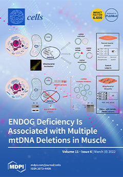
In the manuscript by Nasca et al., biallelic ENDOG variants were identified by NGS in a patient with mitochondrial myopathy progressive external ophthalmoplegia. He presented with multiple mitochondrial DNA deletions in muscle. Endonuclease G (ENDOG) is a nuclear-encoded mitochondrial-localized nuclease, whose exact biological function remains unclear but suggested to participate in mtDNA replication, metabolism, and maintenance. The absence of ENDOG protein in patient’s muscle and fibroblasts indicates that the identified variants are pathogenic. The presence of multiple mtDNA deletions supports the role of ENDOG in mtDNA maintenance; moreover, the patient’s clinical presentation is very similar to mitochondrial diseases caused by mutations in other genes involved in mtDNA homeostasis. This report provides evidence about the association of ENDOG variants with mitochondrial myopathy. View this paper
  • Issues are regarded as officially published after their release is announced to the table of contents alert mailing list.
  • You may sign up for e-mail alerts to receive table of contents of newly released issues.
  • PDF is the official format for papers published in both, html and pdf forms. To view the papers in pdf format, click on the "PDF Full-text" link, and use the free Adobe Reader to open them.
Order results
Result details
Section
Select all
Export citation of selected articles as:
22 pages, 1808 KiB  
Review
One Size Does Not Fit All: Heterogeneity in Developmental Hematopoiesis
by Cristiana Barone, Roberto Orsenigo, Raffaella Meneveri, Silvia Brunelli and Emanuele Azzoni
Cells 2022, 11(6), 1061; https://doi.org/10.3390/cells11061061 - 21 Mar 2022
Cited by 5 | Viewed by 4710
Abstract
Our knowledge of the complexity of the developing hematopoietic system has dramatically expanded over the course of the last few decades. We now know that, while hematopoietic stem cells (HSCs) firmly reside at the top of the adult hematopoietic hierarchy, multiple HSC-independent progenitor [...] Read more.
Our knowledge of the complexity of the developing hematopoietic system has dramatically expanded over the course of the last few decades. We now know that, while hematopoietic stem cells (HSCs) firmly reside at the top of the adult hematopoietic hierarchy, multiple HSC-independent progenitor populations play variegated and fundamental roles during fetal life, which reflect on adult physiology and can lead to disease if subject to perturbations. The importance of obtaining a high-resolution picture of the mechanisms by which the developing embryo establishes a functional hematopoietic system is demonstrated by many recent indications showing that ontogeny is a primary determinant of function of multiple critical cell types. This review will specifically focus on exploring the diversity of hematopoietic stem and progenitor cells unique to embryonic and fetal life. We will initially examine the evidence demonstrating heterogeneity within the hemogenic endothelium, precursor to all definitive hematopoietic cells. Next, we will summarize the dynamics and characteristics of the so-called “hematopoietic waves” taking place during vertebrate development. For each of these waves, we will define the cellular identities of their components, the extent and relevance of their respective contributions as well as potential drivers of heterogeneity. Full article
Show Figures

Figure 1

13 pages, 3273 KiB  
Article
A Temporary Pause in the Replication Licensing Restriction Leads to Rereplication during Early Human Cell Differentiation
by Marie Minet, Masood Abu-Halima, Yiqing Du, Julia Doerr, Christina Isted, Nicole Ludwig, Andreas Keller, Eckart Meese and Ulrike Fischer
Cells 2022, 11(6), 1060; https://doi.org/10.3390/cells11061060 - 21 Mar 2022
Cited by 1 | Viewed by 2088
Abstract
Gene amplifications in amphibians and flies are known to occur during development and have been well characterized, unlike in mammalian cells, where they are predominantly investigated as an attribute of tumors. Recently, we first described gene amplifications in human and mouse neural stem [...] Read more.
Gene amplifications in amphibians and flies are known to occur during development and have been well characterized, unlike in mammalian cells, where they are predominantly investigated as an attribute of tumors. Recently, we first described gene amplifications in human and mouse neural stem cells, myoblasts, and mesenchymal stem cells during differentiation. The mechanism leading to gene amplifications in amphibians and flies depends on endocycles and multiple origin-firings. So far, there is no knowledge about a comparable mechanism in normal human cells. Here, we describe rereplication during the early myotube differentiation of human skeletal myoblast cells, using fiber combing and pulse-treatment with EdU (5′-Ethynyl-2′-deoxyuridine)/CldU (5-Chlor-2′-deoxyuridine) and IdU (5-Iodo-2′-deoxyuridine)/CldU. We found rereplication during a restricted time window between 2 h and 8 h after differentiation induction. Rereplication was detected in cells simultaneously with the amplification of the MDM2 gene. Our findings support rereplication as a mechanism enabling gene amplification in normal human cells. Full article
(This article belongs to the Collection Skeletal Muscle Differentiation and Epigenetics)
Show Figures

Figure 1

16 pages, 5348 KiB  
Article
Fusion of Normoxic- and Hypoxic-Preconditioned Myoblasts Leads to Increased Hypertrophy
by Tamara Pircher, Henning Wackerhage, Elif Akova, Wolfgang Böcker, Attila Aszodi and Maximilian M. Saller
Cells 2022, 11(6), 1059; https://doi.org/10.3390/cells11061059 - 21 Mar 2022
Cited by 3 | Viewed by 2487
Abstract
Injuries, high altitude, and endurance exercise lead to hypoxic conditions in skeletal muscle and sometimes to hypoxia-induced local tissue damage. Thus, regenerative myoblasts/satellite cells are exposed to different levels and durations of partial oxygen pressure depending on the spatial distance from the blood [...] Read more.
Injuries, high altitude, and endurance exercise lead to hypoxic conditions in skeletal muscle and sometimes to hypoxia-induced local tissue damage. Thus, regenerative myoblasts/satellite cells are exposed to different levels and durations of partial oxygen pressure depending on the spatial distance from the blood vessels. To date, it is unclear how hypoxia affects myoblasts proliferation, differentiation, and particularly fusion with normoxic myoblasts. To study this, we investigated how 21% and 2% oxygen affects C2C12 myoblast morphology, proliferation, and myogenic differentiation and evaluated the fusion of normoxic- or hypoxic-preconditioned C2C12 cells in 21% or 2% oxygen in vitro. Out data show that the long-term hypoxic culture condition does not affect the proliferation of C2C12 cells but leads to rounder cells and reduced myotube formation when compared with myoblasts exposed to normoxia. However, when normoxic- and hypoxic-preconditioned myoblasts were differentiated together, the resultant myotubes were significantly larger than the control myotubes. Whole transcriptome sequencing analysis revealed several novel candidate genes that are differentially regulated during the differentiation under normoxia and hypoxia in mixed culture conditions and may thus be involved in the increase in myotube size. Taken together, oxygen-dependent adaption and interaction of myoblasts may represent a novel approach for the development of innovative therapeutic targets. Full article
Show Figures

Figure 1

22 pages, 1199 KiB  
Review
Ubiquitin-Proteasome System–Regulated Protein Degradation in Spermatogenesis
by Yi Xiong, Chao Yu and Qianting Zhang
Cells 2022, 11(6), 1058; https://doi.org/10.3390/cells11061058 - 21 Mar 2022
Cited by 15 | Viewed by 4614
Abstract
Spermatogenesis is a prolonged and highly ordered physiological process that produces haploid male germ cells through more than 40 steps and experiences dramatic morphological and cellular transformations. The ubiquitin proteasome system (UPS) plays central roles in the precise control of protein homeostasis to [...] Read more.
Spermatogenesis is a prolonged and highly ordered physiological process that produces haploid male germ cells through more than 40 steps and experiences dramatic morphological and cellular transformations. The ubiquitin proteasome system (UPS) plays central roles in the precise control of protein homeostasis to ensure the effectiveness of certain protein groups at a given stage and the inactivation of them after this stage. Many UPS components have been demonstrated to regulate the progression of spermatogenesis at different levels. Especially in recent years, novel testis-specific proteasome isoforms have been identified to be essential and unique for spermatogenesis. In this review, we set out to discuss our current knowledge in functions of diverse USP components in mammalian spermatogenesis through: (1) the composition of proteasome isoforms at each stage of spermatogenesis; (2) the specificity of each proteasome isoform and the associated degradation events; (3) the E3 ubiquitin ligases mediating protein ubiquitination in male germ cells; and (4) the deubiquitinases involved in spermatogenesis and male fertility. Exploring the functions of UPS machineries in spermatogenesis provides a global picture of the proteome dynamics during male germ cell production and shed light on the etiology and pathogenesis of human male infertility. Full article
Show Figures

Figure 1

17 pages, 1450 KiB  
Article
Cumulative Metabolic and Epigenetic Effects of Paternal and/or Maternal Supplementation with Arachidonic Acid across Three Consecutive Generations in Mice
by Carmen de la Rocha, Dalia Rodríguez-Ríos, Enrique Ramírez-Chávez, Jorge Molina-Torres, José de Jesús Flores-Sierra, Luis M. Orozco-Castellanos, Juan P. Galván-Chía, Atenea Vázquez Sánchez, Silvio Zaina and Gertrud Lund
Cells 2022, 11(6), 1057; https://doi.org/10.3390/cells11061057 - 21 Mar 2022
Cited by 7 | Viewed by 2625
Abstract
Apart from the known associations between arachidonic acid (AA), weight gain, and neurological and immune function, AA exposure leads to alterations in global and gene-specific DNA methylation (DNAm) and fatty acid (FA) content in human cultured cells. However, it is unknown as to [...] Read more.
Apart from the known associations between arachidonic acid (AA), weight gain, and neurological and immune function, AA exposure leads to alterations in global and gene-specific DNA methylation (DNAm) and fatty acid (FA) content in human cultured cells. However, it is unknown as to whether the latter effects occur in vivo and are maintained over extended periods of time and across generations. To address this issue, we asked whether AA supplementation for three consecutive generations (prior to coitus in sires or in utero in dams) affected offspring growth phenotypes, in addition to liver DNAm and FA profiles in mice. Twelve-week-old BALB/c mice were exposed daily to AA dissolved in soybean oil (vehicle, VH), or VH only, for 10 days prior to mating or during the entire pregnancy (20 days). On average, 15 mice were supplemented per generation, followed by analysis of offspring body weight and liver traits (x average = 36 and 10 per generation, respectively). Body weight cumulatively increased in F2 and F3 offspring generations and positively correlated with milligrams of paternal or maternal offspring AA exposure. A concomitant increase in liver weight was observed. Notably, akin to AA-challenged cultured cells, global DNAm and cis-7-hexadecenoic acid (16:1n-9), an anti-inflammatory FA that is dependent on stearoyl-CoA desaturase 1 (SCD1) activity, increased with milligrams of AA exposure. In accordance, liver Scd1 promoter methylation decreased with milligrams of germline AA exposure and was negatively correlated with liver weight. Our results show that mice retain cellular memories of AA exposure across generations that could potentially be beneficial to the innate immune system. Full article
Show Figures

Figure 1

17 pages, 3054 KiB  
Article
S100A4 Is a Strong Negative Prognostic Marker and Potential Therapeutic Target in Adenocarcinoma of the Stomach and Esophagus
by Christoph Treese, Kimberly Hartl, Michelle Pötzsch, Matthias Dahlmann, Moritz von Winterfeld, Erika Berg, Michael Hummel, Lena Timm, Beate Rau, Wolfgang Walther, Severin Daum, Dennis Kobelt and Ulrike Stein
Cells 2022, 11(6), 1056; https://doi.org/10.3390/cells11061056 - 21 Mar 2022
Cited by 3 | Viewed by 2637
Abstract
Deregulated Wnt-signaling is a key mechanism driving metastasis in adenocarcinoma of the gastroesophageal junction and stomach (AGE/S). The oncogene S100A4 was identified as a Wnt-signaling target gene and is known to promote metastasis. In this project, we illuminate the role of S100A4 for [...] Read more.
Deregulated Wnt-signaling is a key mechanism driving metastasis in adenocarcinoma of the gastroesophageal junction and stomach (AGE/S). The oncogene S100A4 was identified as a Wnt-signaling target gene and is known to promote metastasis. In this project, we illuminate the role of S100A4 for metastases development and disease prognosis of AGE/S. Five gastric cancer cell lines were assessed for S100A4 expression. Two cell lines with endogenous high S100A4 expression were used for functional phenotyping including analysis of proliferation and migration after stable S100A4 knock-down. The prognostic value of S100A4 was evaluated by analyzing the S100A4 expression of tissue microarrays with samples of 277 patients with AGE/S. S100A4 knock-down induced lower migration in FLO1 and NCI-N87 cells. Treatment with niclosamide in these cells led to partial inhibition of S100A4 and to reduced migration. Patients with high S100A4 expression showed lower 5-year overall and disease-specific survival. In addition, a larger share of patients in the S100A4 high expressing group suffered from metachronous metastasis. This study identifies S100A4 as a negative prognostic marker for patients with AGE/S. The strong correlation between S100A4 expression, metastases development and patient survival might open opportunities to use S100A4 to improve the prognosis of these patients and as a therapeutic target for intervention in this tumor entity. Full article
(This article belongs to the Special Issue Cellular and Molecular Mechanisms of Cancer Metastasis)
Show Figures

Figure 1

27 pages, 5397 KiB  
Article
The Role of CD200–CD200 Receptor in Human Blood and Lymphatic Endothelial Cells in the Regulation of Skin Tissue Inflammation
by Dominic Rütsche, Katarzyna Michalak-Micka, Dominika Zielinska, Hannah Moll, Ueli Moehrlen, Thomas Biedermann and Agnes S. Klar
Cells 2022, 11(6), 1055; https://doi.org/10.3390/cells11061055 - 21 Mar 2022
Cited by 5 | Viewed by 3321
Abstract
CD200 is a cell membrane glycoprotein that interacts with its structurally related receptor (CD200R) expressed on immune cells. We characterized CD200–CD200R interactions in human adult/juvenile (j/a) and fetal (f) skin and in in vivo prevascularized skin substitutes (vascDESS) prepared by co-culturing human dermal [...] Read more.
CD200 is a cell membrane glycoprotein that interacts with its structurally related receptor (CD200R) expressed on immune cells. We characterized CD200–CD200R interactions in human adult/juvenile (j/a) and fetal (f) skin and in in vivo prevascularized skin substitutes (vascDESS) prepared by co-culturing human dermal microvascular endothelial cells (HDMEC), containing both blood (BEC) and lymphatic (LEC) EC. We detected the highest expression of CD200 on lymphatic capillaries in j/a and f skin as well as in vascDESS in vivo, whereas it was only weakly expressed on blood capillaries. Notably, the highest CD200 levels were detected on LEC with enhanced Podoplanin expression, while reduced expression was observed on Podoplanin-low LEC. Further, qRT-PCR analysis revealed upregulated expression of some chemokines, including CC-chemokine ligand 21 (CCL21) in j/aCD200+ LEC, as compared to j/aCD200 LEC. The expression of CD200R was mainly detected on myeloid cells such as granulocytes, monocytes/macrophages, T cells in human peripheral blood, and human and rat skin. Functional immunoassays demonstrated specific binding of skin-derived CD200+ HDMEC to myeloid CD200R+ cells in vitro. Importantly, we confirmed enhanced CD200–CD200R interaction in vascDESS in vivo. We concluded that the CD200–CD200R axis plays a crucial role in regulating tissue inflammation during skin wound healing. Full article
(This article belongs to the Collection Molecular Determinants of Skin Integrity)
Show Figures

Figure 1

6 pages, 1116 KiB  
Opinion
NAADP Signaling: New Kids on the Block
by Andreas H. Guse
Cells 2022, 11(6), 1054; https://doi.org/10.3390/cells11061054 - 21 Mar 2022
Cited by 4 | Viewed by 2535
Abstract
Nicotinic acid adenine dinucleotide phosphate (NAADP) is a universal Ca2+ mobilizing second messenger essential for initiation of Ca2+ signaling. Recently, novel molecular mechanisms of both its rapid formation upon receptor stimulation and its mode of action were discovered. Dual NADPH oxidase [...] Read more.
Nicotinic acid adenine dinucleotide phosphate (NAADP) is a universal Ca2+ mobilizing second messenger essential for initiation of Ca2+ signaling. Recently, novel molecular mechanisms of both its rapid formation upon receptor stimulation and its mode of action were discovered. Dual NADPH oxidase 2 (DUOX2) and hematological and neurological expressed 1-like protein (HN1L)/Jupiter microtubule-associated homolog 2 (JPT2) were discovered as NAADP-forming enzyme and NAADP receptor/binding protein—the new kids on the block. These novel aspects are reviewed and integrated into the previous view of NAADP signaling. Full article
(This article belongs to the Section Intracellular and Plasma Membranes)
Show Figures

Figure 1

11 pages, 2548 KiB  
Article
Mitochondrial Phenotypes in Genetically Diverse Neurodegenerative Diseases and Their Response to Mitofusin Activation
by Xiawei Dang, Emily K. Walton, Barbara Zablocka, Robert H. Baloh, Michael E. Shy and Gerald W. Dorn II
Cells 2022, 11(6), 1053; https://doi.org/10.3390/cells11061053 - 21 Mar 2022
Cited by 10 | Viewed by 3243
Abstract
Mitochondrial fusion is essential to mitochondrial fitness and cellular health. Neurons of patients with genetic neurodegenerative diseases often exhibit mitochondrial fragmentation, reflecting an imbalance in mitochondrial fusion and fission (mitochondrial dysdynamism). Charcot–Marie–Tooth (CMT) disease type 2A is the prototypical disorder of impaired mitochondrial [...] Read more.
Mitochondrial fusion is essential to mitochondrial fitness and cellular health. Neurons of patients with genetic neurodegenerative diseases often exhibit mitochondrial fragmentation, reflecting an imbalance in mitochondrial fusion and fission (mitochondrial dysdynamism). Charcot–Marie–Tooth (CMT) disease type 2A is the prototypical disorder of impaired mitochondrial fusion caused by mutations in the fusion protein mitofusin (MFN)2. Yet, cultured CMT2A patient fibroblast mitochondria are often reported as morphologically normal. Metabolic stress might evoke pathological mitochondrial phenotypes in cultured patient fibroblasts, providing a platform for the pre-clinical individualized evaluation of investigational therapeutics. Here, substitution of galactose for glucose in culture media was used to redirect CMT2A patient fibroblasts (MFN2 T105M, R274W, H361Y, R364W) from glycolytic metabolism to mitochondrial oxidative phosphorylation, which provoked characteristic mitochondrial fragmentation and depolarization and induced a distinct transcriptional signature. Pharmacological MFN activation of metabolically reprogrammed fibroblasts partially reversed the mitochondrial abnormalities in CMT2A and CMT1 and a subset of Parkinson’s and Alzheimer’s disease patients, implicating addressable mitochondrial dysdynamism in these illnesses. Full article
(This article belongs to the Collection Determinants of Neuronal Susceptibility to Mitochondrial Disease)
Show Figures

Figure 1

16 pages, 3293 KiB  
Article
Silencing of Ago-2 Interacting Protein SERBP1 Relieves KCC2 Repression by miR-92 in Neurons
by Christian Barbato, Paola Frisone, Laura Braccini, Simona D’Aguanno, Luisa Pieroni, Maria Teresa Ciotti, Caterina Catalanotto, Carlo Cogoni and Francesca Ruberti
Cells 2022, 11(6), 1052; https://doi.org/10.3390/cells11061052 - 20 Mar 2022
Cited by 5 | Viewed by 3220
Abstract
RNA-binding proteins (RBPs) play important roles in modulating miRNA-mediated mRNA target repression. Argonaute2 (Ago2) is an essential component of the RNA-induced silencing complex (RISC) that plays a central role in silencing mechanisms via small non-coding RNA molecules known as siRNAs and miRNAs. Small [...] Read more.
RNA-binding proteins (RBPs) play important roles in modulating miRNA-mediated mRNA target repression. Argonaute2 (Ago2) is an essential component of the RNA-induced silencing complex (RISC) that plays a central role in silencing mechanisms via small non-coding RNA molecules known as siRNAs and miRNAs. Small RNAs loaded into Argonaute proteins catalyze endoribonucleolytic cleavage of target RNAs or recruit factors responsible for translational silencing and mRNA target destabilization. In previous studies we have shown that KCC2, a neuronal Cl (−) extruding K (+) Cl (−) co-transporter 2, is regulated by miR-92 in neuronal cells. Searching for Ago2 partners by immunoprecipitation and LC-MS/MS analysis, we isolated among other proteins the Serpine mRNA binding protein 1 (SERBP1) from SH-SY5Y neuroblastoma cells. Exploring the role of SERBP1 in miRNA-mediated gene silencing in SH-SY5Y cells and primary hippocampal neurons, we demonstrated that SERBP1 silencing regulates KCC2 expression through the 3′ untranslated region (UTR). In addition, we found that SERBP1 as well as Ago2/miR-92 complex bind to KCC2 3′UTR. Finally, we demonstrated the attenuation of miR-92-mediated repression of KCC2 3′UTR by SERBP1 silencing. These findings advance our knowledge regarding the miR-92-mediated modulation of KCC2 translation in neuronal cells and highlight SERBP1 as a key component of this gene regulation. Full article
Show Figures

Graphical abstract

10 pages, 1611 KiB  
Review
Role of Damage-Associated Molecular Patterns (DAMPs/Alarmins) in Severe Ocular Allergic Diseases
by Ken Fukuda, Waka Ishida, Tatsuma Kishimoto, Isana Nakajima, Yusaku Miura, Tamaki Sumi and Kenji Yamashiro
Cells 2022, 11(6), 1051; https://doi.org/10.3390/cells11061051 - 20 Mar 2022
Cited by 5 | Viewed by 2483
Abstract
Severe ocular allergic diseases, such as atopic keratoconjunctivitis and vernal keratoconjunctivitis, cause severe allergic inflammation in the conjunctiva and corneal epithelial damage, resulting in visual disturbances. The involvement of damage (danger)-associated molecular patterns (DAMPs/alarmins) in the pathogenesis of these diseases has been recognized. [...] Read more.
Severe ocular allergic diseases, such as atopic keratoconjunctivitis and vernal keratoconjunctivitis, cause severe allergic inflammation in the conjunctiva and corneal epithelial damage, resulting in visual disturbances. The involvement of damage (danger)-associated molecular patterns (DAMPs/alarmins) in the pathogenesis of these diseases has been recognized. Alarmins released from damaged corneal epithelial cells or eosinophils play a critical role in the induction of corneal lesions, vicious loop of corneal injury, and exacerbation of conjunctival allergic inflammation. Alarmins in the conjunctiva also play an essential role in the development of both allergic inflammation, based on the acquired immune system, and type 2 inflammation by innate immune responses in the ocular surface. Therefore, alarmins may be a potentially important therapeutic target in severe refractory ocular allergic diseases. Full article
(This article belongs to the Collection Retina in Health and Disease)
Show Figures

Figure 1

27 pages, 3294 KiB  
Review
Emerging Roles of Airway Epithelial Cells in Idiopathic Pulmonary Fibrosis
by Ashesh Chakraborty, Michal Mastalerz, Meshal Ansari, Herbert B. Schiller and Claudia A. Staab-Weijnitz
Cells 2022, 11(6), 1050; https://doi.org/10.3390/cells11061050 - 19 Mar 2022
Cited by 22 | Viewed by 8267
Abstract
Idiopathic pulmonary fibrosis (IPF) is a fatal disease with incompletely understood aetiology and limited treatment options. Traditionally, IPF was believed to be mainly caused by repetitive injuries to the alveolar epithelium. Several recent lines of evidence, however, suggest that IPF equally involves an [...] Read more.
Idiopathic pulmonary fibrosis (IPF) is a fatal disease with incompletely understood aetiology and limited treatment options. Traditionally, IPF was believed to be mainly caused by repetitive injuries to the alveolar epithelium. Several recent lines of evidence, however, suggest that IPF equally involves an aberrant airway epithelial response, which contributes significantly to disease development and progression. In this review, based on recent clinical, high-resolution imaging, genetic, and single-cell RNA sequencing data, we summarize alterations in airway structure, function, and cell type composition in IPF. We furthermore give a comprehensive overview on the genetic and mechanistic evidence pointing towards an essential role of airway epithelial cells in IPF pathogenesis and describe potentially implicated aberrant epithelial signalling pathways and regulation mechanisms in this context. The collected evidence argues for the investigation of possible therapeutic avenues targeting these processes, which thus represent important future directions of research. Full article
(This article belongs to the Special Issue State of the Art in Idiopathic Pulmonary Fibrosis)
Show Figures

Figure 1

12 pages, 1860 KiB  
Review
Predicting Mitochondrial Dynamic Behavior in Genetically Defined Neurodegenerative Diseases
by Gerald W. Dorn II and Xiawei Dang
Cells 2022, 11(6), 1049; https://doi.org/10.3390/cells11061049 - 19 Mar 2022
Cited by 10 | Viewed by 5143
Abstract
Mitochondrial dynamics encompass mitochondrial fusion, fission, and movement. Mitochondrial fission and fusion are seemingly ubiquitous, whereas mitochondrial movement is especially important for organelle transport through neuronal axons. Here, we review the roles of different mitochondrial dynamic processes in mitochondrial quantity and quality control, [...] Read more.
Mitochondrial dynamics encompass mitochondrial fusion, fission, and movement. Mitochondrial fission and fusion are seemingly ubiquitous, whereas mitochondrial movement is especially important for organelle transport through neuronal axons. Here, we review the roles of different mitochondrial dynamic processes in mitochondrial quantity and quality control, emphasizing their impact on the neurological system in Charcot–Marie–Tooth disease type 2A, amyotrophic lateral sclerosis, Friedrich’s ataxia, dominant optic atrophy, and Alzheimer’s, Huntington’s, and Parkinson’s diseases. In addition to mechanisms and concepts, we explore in detail different technical approaches for measuring mitochondrial dynamic dysfunction in vitro, describe how results from tissue culture studies may be applied to a better understanding of mitochondrial dysdynamism in human neurodegenerative diseases, and suggest how this experimental platform can be used to evaluate candidate therapeutics in different diseases or in individual patients sharing the same clinical diagnosis. Full article
(This article belongs to the Collection Determinants of Neuronal Susceptibility to Mitochondrial Disease)
Show Figures

Figure 1

21 pages, 1733 KiB  
Review
Transposable Elements: Major Players in Shaping Genomic and Evolutionary Patterns
by Nunzia Colonna Romano and Laura Fanti
Cells 2022, 11(6), 1048; https://doi.org/10.3390/cells11061048 - 19 Mar 2022
Cited by 15 | Viewed by 6511
Abstract
Transposable elements (TEs) are ubiquitous genetic elements, able to jump from one location of the genome to another, in all organisms. For this reason, on the one hand, TEs can induce deleterious mutations, causing dysfunction, disease and even lethality in individuals. On the [...] Read more.
Transposable elements (TEs) are ubiquitous genetic elements, able to jump from one location of the genome to another, in all organisms. For this reason, on the one hand, TEs can induce deleterious mutations, causing dysfunction, disease and even lethality in individuals. On the other hand, TEs can increase genetic variability, making populations better equipped to respond adaptively to environmental change. To counteract the deleterious effects of TEs, organisms have evolved strategies to avoid their activation. However, their mobilization does occur. Usually, TEs are maintained silent through several mechanisms, but they can be reactivated during certain developmental windows. Moreover, TEs can become de-repressed because of drastic changes in the external environment. Here, we describe the ‘double life’ of TEs, being both ‘parasites’ and ‘symbionts’ of the genome. We also argue that the transposition of TEs contributes to two important evolutionary processes: the temporal dynamic of evolution and the induction of genetic variability. Finally, we discuss how the interplay between two TE-dependent phenomena, insertional mutagenesis and epigenetic plasticity, plays a role in the process of evolution. Full article
Show Figures

Figure 1

21 pages, 4317 KiB  
Article
Non-Linear Frequency Dependence of Neurovascular Coupling in the Cerebellar Cortex Implies Vasodilation–Vasoconstriction Competition
by Giuseppe Gagliano, Anita Monteverdi, Stefano Casali, Umberto Laforenza, Claudia A. M. Gandini Wheeler-Kingshott, Egidio D’Angelo and Lisa Mapelli
Cells 2022, 11(6), 1047; https://doi.org/10.3390/cells11061047 - 19 Mar 2022
Cited by 8 | Viewed by 4242
Abstract
Neurovascular coupling (NVC) is the process associating local cerebral blood flow (CBF) to neuronal activity (NA). Although NVC provides the basis for the blood oxygen level dependent (BOLD) effect used in functional MRI (fMRI), the relationship between NVC and NA is still unclear. [...] Read more.
Neurovascular coupling (NVC) is the process associating local cerebral blood flow (CBF) to neuronal activity (NA). Although NVC provides the basis for the blood oxygen level dependent (BOLD) effect used in functional MRI (fMRI), the relationship between NVC and NA is still unclear. Since recent studies reported cerebellar non-linearities in BOLD signals during motor tasks execution, we investigated the NVC/NA relationship using a range of input frequencies in acute mouse cerebellar slices of vermis and hemisphere. The capillary diameter increased in response to mossy fiber activation in the 6–300 Hz range, with a marked inflection around 50 Hz (vermis) and 100 Hz (hemisphere). The corresponding NA was recorded using high-density multi-electrode arrays and correlated to capillary dynamics through a computational model dissecting the main components of granular layer activity. Here, NVC is known to involve a balance between the NMDAR-NO pathway driving vasodilation and the mGluRs-20HETE pathway driving vasoconstriction. Simulations showed that the NMDAR-mediated component of NA was sufficient to explain the time course of the capillary dilation but not its non-linear frequency dependence, suggesting that the mGluRs-20HETE pathway plays a role at intermediate frequencies. These parallel control pathways imply a vasodilation–vasoconstriction competition hypothesis that could adapt local hemodynamics at the microscale bearing implications for fMRI signals interpretation. Full article
(This article belongs to the Section Cells of the Nervous System)
Show Figures

Graphical abstract

12 pages, 2569 KiB  
Article
The Heat Shock Protein 90 Inhibitor, AT13387, Protects the Alveolo-Capillary Barrier and Prevents HCl-Induced Chronic Lung Injury and Pulmonary Fibrosis
by Ruben M. L. Colunga Biancatelli, Pavel Solopov, Christiana Dimitropoulou, Betsy Gregory, Tierney Day and John D. Catravas
Cells 2022, 11(6), 1046; https://doi.org/10.3390/cells11061046 - 19 Mar 2022
Cited by 11 | Viewed by 2649
Abstract
Hydrochloric acid (HCl) exposure causes asthma-like conditions, reactive airways dysfunction syndrome, and pulmonary fibrosis. Heat Shock Protein 90 (HSP90) is a molecular chaperone that regulates multiple cellular processes. HSP90 inhibitors are undergoing clinical trials for cancer and are also being studied in various [...] Read more.
Hydrochloric acid (HCl) exposure causes asthma-like conditions, reactive airways dysfunction syndrome, and pulmonary fibrosis. Heat Shock Protein 90 (HSP90) is a molecular chaperone that regulates multiple cellular processes. HSP90 inhibitors are undergoing clinical trials for cancer and are also being studied in various pre-clinical settings for their anti-inflammatory and anti-fibrotic effects. Here we investigated the ability of the heat shock protein 90 (HSP90) inhibitor AT13387 to prevent chronic lung injury induced by exposure to HCl in vivo and its protective role in the endothelial barrier in vitro. We instilled C57Bl/6J mice with 0.1N HCl (2 µL/g body weight, intratracheally) and after 24 h began treatment with vehicle or AT13387 (10 or 15 mg/kg, SC), administered 3×/week; we analyzed histological, functional, and molecular markers 30 days after HCl. In addition, we monitored transendothelial electrical resistance (TER) and protein expression in a monolayer of human lung microvascular endothelial cells (HLMVEC) exposed to HCl (0.02 N) and treated with vehicle or AT13387 (2 µM). HCl provoked persistent alveolar inflammation; activation of profibrotic pathways (MAPK/ERK, HSP90); increased deposition of collagen, fibronectin and elastin; histological evidence of fibrosis; and a decline in lung function reflected in a downward shift in pressure–volume curves, increased respiratory system resistance (Rrs), elastance (Ers), tissue damping (G), and hyperresponsiveness to methacholine. Treatment with 15 mg/kg AT13387reduced alveolar inflammation, fibrosis, and NLRP3 staining; blocked activation of ERK and HSP90; and attenuated the deposition of collagen and the development of chronic lung injury and airway hyperreactivity. In vitro, AT13387 prevented HCl-induced loss of barrier function and AKT, ERK, and ROCK1 activation, and restored HSP70 and cofilin expression. The HSP90 inhibitor, AT13387, represents a promising drug candidate for chronic lung injury that can be administered subcutaneously in the field, and at low, non-toxic doses. Full article
Show Figures

Figure 1

21 pages, 9764 KiB  
Article
Cardiac Ischemia On-a-Chip: Antiarrhythmic Effect of Levosimendan on Ischemic Human-Induced Pluripotent Stem Cell-Derived Cardiomyocytes
by Mahmoud Gaballah, Kirsi Penttinen, Joose Kreutzer, Antti-Juhana Mäki, Pasi Kallio and Katriina Aalto-Setälä
Cells 2022, 11(6), 1045; https://doi.org/10.3390/cells11061045 - 19 Mar 2022
Cited by 4 | Viewed by 3805
Abstract
Ischemic heart disease (IHD) is one of the leading causes of mortality worldwide. Preserving functionality and preventing arrhythmias of the heart are key principles in the management of patients with IHD. Levosimendan, a unique calcium (Ca2+) enhancer with inotropic activity, has [...] Read more.
Ischemic heart disease (IHD) is one of the leading causes of mortality worldwide. Preserving functionality and preventing arrhythmias of the heart are key principles in the management of patients with IHD. Levosimendan, a unique calcium (Ca2+) enhancer with inotropic activity, has been introduced into clinical usage for heart failure treatment. Human-induced pluripotent cell-derived cardiomyocytes (hiPSC-CMs) offer an opportunity to better understand the pathophysiological mechanisms of the disease as well as to serve as a platform for drug screening. Here, we developed an in vitro IHD model using hiPSC-CMs in hypoxic conditions and defined the effects of the subsequent hypoxic stress on CMs functionality. Furthermore, the effect of levosimendan on hiPSC-CMs functionality was evaluated during and after hypoxic stress. The morphology, contractile, Ca2+-handling, and gene expression properties of hiPSC-CMs were investigated in response to hypoxia. Hypoxia resulted in significant cardiac arrhythmia and decreased Ca2+ transient amplitude. In addition, disorganization of sarcomere structure was observed after hypoxia induction. Interestingly, levosimendan presented significant antiarrhythmic properties, as the arrhythmia was abolished or markedly reduced with levosimendan treatment either during or after the hypoxic stress. Moreover, levosimendan presented significant protection from the sarcomere alterations induced by hypoxia. In conclusion, this chip model appears to be a suitable preclinical representation of IHD. With this hypoxia platform, detailed knowledge of the disease pathophysiology can be obtained. The antiarrhythmic effect of levosimendan was clearly observed, suggesting a possible new clinical use for the drug. Full article
(This article belongs to the Special Issue Cell-Based Models of Diseases for Drug Discovery)
Show Figures

Graphical abstract

7 pages, 229 KiB  
Opinion
The Use of “Retardation” in FRAXA, FMRP, FMR1 and Other Designations
by Jonathan Herring, Kirsten Johnson and Jörg Richstein
Cells 2022, 11(6), 1044; https://doi.org/10.3390/cells11061044 - 19 Mar 2022
Cited by 11 | Viewed by 4423
Abstract
The European Fragile X Network met in Wroclaw, Poland, November 2021, and agreed to work towards the eradication of the word “retardation” in regard to the naming of the fragile X gene (FRAXA) and protein (FMRP). There are further genes which have “retardation” [...] Read more.
The European Fragile X Network met in Wroclaw, Poland, November 2021, and agreed to work towards the eradication of the word “retardation” in regard to the naming of the fragile X gene (FRAXA) and protein (FMRP). There are further genes which have “retardation” or abbreviations for “retardation” in their names or full designations, including FMR1, FMR2, FXR1, FXR2, NUFIP1, AFF1, CYFIP1, etc. “Retardation” was commonly used as a term in years past, but now any reference, even in an abbreviation, is offensive. This article discusses the stigmatisation associated with “retardation”, which leads to discrimination; the inaccuracy of using “retardation” in these designations; and the breadth of fragile X syndrome being beyond that of neurodiversity. A more inclusive terminology is called for, one which ceases to use any reference to “retardation”. Precedents for offensive gene names being altered is set out. The proposal is to approach the HGNC (HUGO [Human Genome Organisation] Gene Nomenclature Committee) for new terminology to be enacted. Ideas from other researchers in the field are welcomed. Full article
15 pages, 3822 KiB  
Article
Preservation of Smooth Muscle Cell Integrity and Function: A Target for Limiting Abdominal Aortic Aneurysm Expansion?
by Emily R. Clark, Rebecca J. Helliwell, Marc A. Bailey, Karen E. Hemmings, Katherine I. Bridge, Kathryn J. Griffin, D. Julian A. Scott, Louise M. Jennings, Kirsten Riches-Suman and Karen E. Porter
Cells 2022, 11(6), 1043; https://doi.org/10.3390/cells11061043 - 19 Mar 2022
Viewed by 2669
Abstract
(1) Abdominal aortic aneurysm (AAA) is a silent, progressive disease with significant mortality from rupture. Whilst screening programmes are now able to detect this pathology early in its development, no therapeutic intervention has yet been identified to halt or retard aortic expansion. The [...] Read more.
(1) Abdominal aortic aneurysm (AAA) is a silent, progressive disease with significant mortality from rupture. Whilst screening programmes are now able to detect this pathology early in its development, no therapeutic intervention has yet been identified to halt or retard aortic expansion. The inability to obtain aortic tissue from humans at early stages has created a necessity for laboratory models, yet it is essential to create a timeline of events from EARLY to END stage AAA progression. (2) We used a previously validated ex vivo porcine bioreactor model pre-treated with protease enzyme to create “aneurysm” tissue. Mechanical properties, histological changes in the intact vessel wall, and phenotype/function of vascular smooth muscle cells (SMC) cultured from the same vessels were investigated. (3) The principal finding was significant hyperproliferation of SMC from EARLY stage vessels, but without obvious histological or SMC aberrancies. END stage tissue exhibited histological loss of α-smooth muscle actin and elastin; mechanical impairment; and, in SMC, multiple indications of senescence. (4) Aortic SMC may offer a therapeutic target for intervention, although detailed studies incorporating intervening time points between EARLY and END stage are required. Such investigations may reveal mechanisms of SMC dysfunction in AAA development and hence a therapeutic window during which SMC differentiation could be preserved or reinstated. Full article
(This article belongs to the Special Issue Cell Biology: State-of-the-Art and Perspectives in the British Isles)
Show Figures

Figure 1

13 pages, 1683 KiB  
Article
Extracellular Vesicle-Mediated IL-1 Signaling in Response to Doxorubicin Activates PD-L1 Expression in Osteosarcoma Models
by Su Yati, Atiruj Silathapanasakul, Chakrarin Thakaeng, Mayuree Chanasakulniyom, Napat Songtawee, Sureerut Porntadavity, Peraphan Pothacharoen, Dumnoensun Pruksakorn, Prachya Kongtawelert, Pa-thai Yenchitsomanus and Theerawut Chanmee
Cells 2022, 11(6), 1042; https://doi.org/10.3390/cells11061042 - 18 Mar 2022
Cited by 11 | Viewed by 3075
Abstract
The expression of programmed cell death ligand 1 (PD-L1) in tumors is associated with tumor cell escape from T-cell cytotoxicity, and is considered a crucial effector in chemoresistance and tumor relapse. Although PD-L1 induction has been observed in patients after chemotherapy treatment, the [...] Read more.
The expression of programmed cell death ligand 1 (PD-L1) in tumors is associated with tumor cell escape from T-cell cytotoxicity, and is considered a crucial effector in chemoresistance and tumor relapse. Although PD-L1 induction has been observed in patients after chemotherapy treatment, the mechanism by which the drug activates PD-L1 expression remains elusive. Here, we identified the extracellular vesicles (EVs) as a molecular mediator that determines the effect of doxorubicin on PD-L1 expression in osteosarcoma models. Mechanistically, doxorubicin dependently stimulates the release of extracellular vesicles, which mediate autocrine/paracrine signals in osteosarcoma cells. The recipient cells were stimulated by these EVs and acquired the ability to promote the expression of inflammatory cytokines interleukin (IL)-1β and IL-6. In response to doxorubicin, IL-1β, but not IL-6, allowed- osteosarcoma cells to promote the expression of PD-L1, and the elimination of IL-1β/IL-1 receptor signaling with IL-1 receptor antagonist reduced PD-L1 expression. Together, these findings provided insights into the role of EV release in response to chemotherapy that mediates PD-L1 expression via the IL-1 signaling pathway, and suggested that the combination of a drug targeting IL-1 or PD-L1 with chemotherapy could be an effective treatment option for osteosarcoma patients. Full article
(This article belongs to the Special Issue Osteosarcoma: From Molecular Mechanisms to Therapeutic Opportunities)
Show Figures

Graphical abstract

21 pages, 2549 KiB  
Review
Tumor Cell Glycolysis—At the Crossroad of Epithelial–Mesenchymal Transition and Autophagy
by Fabrizio Marcucci and Cristiano Rumio
Cells 2022, 11(6), 1041; https://doi.org/10.3390/cells11061041 - 18 Mar 2022
Cited by 17 | Viewed by 3887
Abstract
Upregulation of glycolysis, induction of epithelial–mesenchymal transition (EMT) and macroautophagy (hereafter autophagy), are phenotypic changes that occur in tumor cells, in response to similar stimuli, either tumor cell-autonomous or from the tumor microenvironment. Available evidence, herein reviewed, suggests that glycolysis can play a [...] Read more.
Upregulation of glycolysis, induction of epithelial–mesenchymal transition (EMT) and macroautophagy (hereafter autophagy), are phenotypic changes that occur in tumor cells, in response to similar stimuli, either tumor cell-autonomous or from the tumor microenvironment. Available evidence, herein reviewed, suggests that glycolysis can play a causative role in the induction of EMT and autophagy in tumor cells. Thus, glycolysis has been shown to induce EMT and either induce or inhibit autophagy. Glycolysis-induced autophagy occurs both in the presence (glucose starvation) or absence (glucose sufficiency) of metabolic stress. In order to explain these, in part, contradictory experimental observations, we propose that in the presence of stimuli, tumor cells respond by upregulating glycolysis, which will then induce EMT and inhibit autophagy. In the presence of stimuli and glucose starvation, upregulated glycolysis leads to adenosine monophosphate-activated protein kinase (AMPK) activation and autophagy induction. In the presence of stimuli and glucose sufficiency, upregulated glycolytic enzymes (e.g., aldolase or glyceraldehyde 3-phosphate dehydrogenase) or decreased levels of glycolytic metabolites (e.g., dihydroxyacetone phosphate) may mimic a situation of metabolic stress (herein referred to as “pseudostarvation”), leading, directly or indirectly, to AMPK activation and autophagy induction. We also discuss possible mechanisms, whereby glycolysis can induce a mixed mesenchymal/autophagic phenotype in tumor cells. Subsequently, we address unresolved problems in this field and possible therapeutic consequences. Full article
(This article belongs to the Section Cellular Metabolism)
Show Figures

Figure 1

16 pages, 455 KiB  
Review
Review of Mechanisms and Treatment of Cancer-Induced Cardiac Cachexia
by Vignesh Vudatha, Teja Devarakonda, Christopher Liu, Devon C. Freudenberger, Andrea N. Riner, Kelly M. Herremans and Jose G. Trevino
Cells 2022, 11(6), 1040; https://doi.org/10.3390/cells11061040 - 18 Mar 2022
Cited by 6 | Viewed by 2625
Abstract
Cancer cachexia is a multifactorial, paraneoplastic syndrome that impacts roughly half of all cancer patients. It can negatively impact patient quality of life and prognosis by causing physical impairment, reducing chemotherapy tolerance, and precluding them as surgical candidates. While there is substantial research [...] Read more.
Cancer cachexia is a multifactorial, paraneoplastic syndrome that impacts roughly half of all cancer patients. It can negatively impact patient quality of life and prognosis by causing physical impairment, reducing chemotherapy tolerance, and precluding them as surgical candidates. While there is substantial research on cancer-induced skeletal muscle cachexia, there are comparatively fewer studies and therapies regarding cardiac cachexia in the setting of malignancy. A literature review was performed using the PubMed database to identify original articles pertaining to cancer-induced cardiac cachexia, including its mechanisms and potential therapeutic modalities. Seventy studies were identified by two independent reviewers based on inclusion and exclusion criteria. While there are multiple studies addressing the pathophysiology of cardiac-induced cancer cachexia, there are no studies evaluating therapeutic options in the clinical setting. Many treatment modalities including nutrition, heart failure medication, cancer drugs, exercise, and gene therapy have been explored in in vitro and mice models with varying degrees of success. While these may be beneficial in cancer patients, further prospective studies specifically focusing on the assessment and treatment of the cardiac component of cachexia are needed. Full article
(This article belongs to the Special Issue Cancer-Induced Cachexia)
Show Figures

Figure 1

22 pages, 1327 KiB  
Review
Interconnection between Cardiac Cachexia and Heart Failure—Protective Role of Cardiac Obesity
by María Elena Soto, Israel Pérez-Torres, María Esther Rubio-Ruiz, Linaloe Manzano-Pech and Verónica Guarner-Lans
Cells 2022, 11(6), 1039; https://doi.org/10.3390/cells11061039 - 18 Mar 2022
Cited by 6 | Viewed by 6339
Abstract
Cachexia may be caused by congestive heart failure, and it is then called cardiac cachexia, which leads to increased morbidity and mortality. Cardiac cachexia also worsens skeletal muscle degradation. Cardiac cachexia is the loss of edema-free muscle mass with or without affecting fat [...] Read more.
Cachexia may be caused by congestive heart failure, and it is then called cardiac cachexia, which leads to increased morbidity and mortality. Cardiac cachexia also worsens skeletal muscle degradation. Cardiac cachexia is the loss of edema-free muscle mass with or without affecting fat tissue. It is mainly caused by a loss of balance between protein synthesis and degradation, or it may result from intestinal malabsorption. The loss of balance in protein synthesis and degradation may be the consequence of altered endocrine mediators such as insulin, insulin-like growth factor 1, leptin, ghrelin, melanocortin, growth hormone and neuropeptide Y. In contrast to many other health problems, fat accumulation in the heart is protective in this condition. Fat in the heart can be divided into epicardial, myocardial and cardiac steatosis. In this review, we describe and discuss these topics, pointing out the interconnection between heart failure and cardiac cachexia and the protective role of cardiac obesity. We also set the basis for possible screening methods that may allow for a timely diagnosis of cardiac cachexia, since there is still no cure for this condition. Several therapeutic procedures are discussed including exercise, nutritional proposals, myostatin antibodies, ghrelin, anabolic steroids, anti-inflammatory substances, beta-adrenergic agonists, medroxyprogesterone acetate, megestrol acetate, cannabinoids, statins, thalidomide, proteasome inhibitors and pentoxifylline. However, to this date, there is no cure for cachexia. Full article
(This article belongs to the Special Issue Cancer-Induced Cachexia)
Show Figures

Graphical abstract

13 pages, 2397 KiB  
Article
DHEA Protects Human Cholangiocytes and Hepatocytes against Apoptosis and Oxidative Stress
by Ewa Kilanczyk, Dagmara Ruminkiewicz, Jesus M. Banales, Piotr Milkiewicz and Małgorzata Milkiewicz
Cells 2022, 11(6), 1038; https://doi.org/10.3390/cells11061038 - 18 Mar 2022
Cited by 8 | Viewed by 2385
Abstract
Primary biliary cholangitis (PBC) is a rare chronic cholestatic and immune-mediated liver disease of unknown aetiology that targets intrahepatic bile duct cells (cholangiocytes) and primarily affects postmenopausal women, when their estrogen levels sharply decrease. An impaired cholangiocyte response to estrogen characterizes the terminal [...] Read more.
Primary biliary cholangitis (PBC) is a rare chronic cholestatic and immune-mediated liver disease of unknown aetiology that targets intrahepatic bile duct cells (cholangiocytes) and primarily affects postmenopausal women, when their estrogen levels sharply decrease. An impaired cholangiocyte response to estrogen characterizes the terminal stage of the disease, as this is when an inefficiency of cholangiocyte proliferation, in balancing the loss of intrahepatic bile ducts, is observed. Here, we report that the estrogen precursor dehydroepiandrosterone (DHEA) and its sulfate metabolites, DHEA-S and 17 β-estradiol, enhance the proliferation of cholangiocytes and hepatocytes in vitro. Flow cytometry analysis showed that DHEA and DHEA-S decreased glyco-chenodeoxycholic acid (GCDC)-driven apoptosis in cholangiocytes. Cell viability assay (MTT) indicated that ER-α, -β, and the G-protein-coupled estrogen receptor, are involved in the protection of DHEA against oxidative stress in cholangiocytes. Finally, immunoblot analysis showed an elevated level of steroid sulfatase and a reduced level of sulfotransferase 1E1 enzymes, involved in the desulfation/sulfation process of estrogens in cirrhotic PBC, and primary sclerosis cholangitis (PSC) liver tissues, another type of chronic cholestatic and immune-mediated liver disease. Taken together, these results suggest that DHEA can prevent the deleterious effects of certain potentially toxic bile acids and reactive oxygen species, delaying the onset of liver disease. Full article
(This article belongs to the Special Issue Estrogen Receptor Hormone Action)
Show Figures

Graphical abstract

15 pages, 5474 KiB  
Article
The Atypical Chemerin Receptor GPR1 Displays Different Modes of Interaction with β-Arrestins in Humans and Mice with Important Consequences on Subcellular Localization and Trafficking
by Gaetan-Nagim Degroot, Valentin Lepage, Marc Parmentier and Jean-Yves Springael
Cells 2022, 11(6), 1037; https://doi.org/10.3390/cells11061037 - 18 Mar 2022
Cited by 4 | Viewed by 1861
Abstract
Atypical chemokine receptors (ACKRs) have emerged as a subfamily of chemokine receptors regulating the local bioavailability of their ligands through scavenging, concentration, or transport. The biological roles of ACKRs in human physiology and diseases are often studied by using transgenic mouse models. However, [...] Read more.
Atypical chemokine receptors (ACKRs) have emerged as a subfamily of chemokine receptors regulating the local bioavailability of their ligands through scavenging, concentration, or transport. The biological roles of ACKRs in human physiology and diseases are often studied by using transgenic mouse models. However, it is unknown whether mouse and human ACKRs share the same properties. In this study, we compared the properties of the human and mouse atypical chemerin receptor GPR1 and showed that they behave differently regarding their interaction with β-arrestins. Human hGPR1 interacts with β-arrestins as a result of chemerin stimulation, whereas its mouse orthologue mGPR1 displays a strong constitutive interaction with β-arrestins in basal conditions. The constitutive interaction of mGPR1 with β-arrestins is accompanied by a redistribution of the receptor from the plasma membrane to early and recycling endosomes. In addition, β-arrestins appear mandatory for the chemerin-induced internalization of mGPR1, whereas they are dispensable for the trafficking of hGPR1. However, mGPR1 scavenges chemerin and activates MAP kinases ERK1/2 similarly to hGPR1. Finally, we showed that the constitutive interaction of mGPR1 with β-arrestins required different structural constituents, including the receptor C-terminus and arginine 3.50 in the second intracellular loop. Altogether, our results show that sequence variations within cytosolic regions of GPR1 orthologues influence their ability to interact with β-arrestins, with important consequences on GPR1 subcellular distribution and trafficking. Full article
(This article belongs to the Special Issue Molecular Mechanisms of Chemokine Receptor Signaling and Trafficking)
Show Figures

Figure 1

22 pages, 2095 KiB  
Article
Predation Stress Causes Excessive Aggression in Female Mice with Partial Genetic Inactivation of Tryptophan Hydroxylase-2: Evidence for Altered Myelination-Related Processes
by Evgeniy Svirin, Ekaterina Veniaminova, João Pedro Costa-Nunes, Anna Gorlova, Aleksei Umriukhin, Allan V. Kalueff, Andrey Proshin, Daniel C. Anthony, Andrey Nedorubov, Anna Chung Kwan Tse, Susanne Walitza, Lee Wei Lim, Klaus-Peter Lesch and Tatyana Strekalova
Cells 2022, 11(6), 1036; https://doi.org/10.3390/cells11061036 - 18 Mar 2022
Cited by 4 | Viewed by 2777
Abstract
The interaction between brain serotonin (5-HT) deficiency and environmental adversity may predispose females to excessive aggression. Specifically, complete inactivation of the gene encoding tryptophan hydroxylase-2 (Tph2) results in the absence of neuronal 5-HT synthesis and excessive aggressiveness in both male and [...] Read more.
The interaction between brain serotonin (5-HT) deficiency and environmental adversity may predispose females to excessive aggression. Specifically, complete inactivation of the gene encoding tryptophan hydroxylase-2 (Tph2) results in the absence of neuronal 5-HT synthesis and excessive aggressiveness in both male and female null mutant (Tph2−/−) mice. In heterozygous male mice (Tph2+/−), there is a moderate reduction in brain 5-HT levels, and when they are exposed to stress, they exhibit increased aggression. Here, we exposed female Tph2+/− mice to a five-day rat predation stress paradigm and assessed their emotionality and social interaction/aggression-like behaviors. Tph2+/− females exhibited excessive aggression and increased dominant behavior. Stressed mutants displayed altered gene expression of the 5-HT receptors Htr1a and Htr2a, glycogen synthase kinase-3 β (GSK-3β), and c-fos as well as myelination-related transcripts in the prefrontal cortex: myelin basic protein (Mbp), proteolipid protein 1 (Plp1), myelin-associated glycoprotein (Mag), and myelin oligodendrocyte glycoprotein (Mog). The expression of the plasticity markers synaptophysin (Syp) and cAMP response element binding protein (Creb), but not AMPA receptor subunit A2 (GluA2), were affected by genotype. Moreover, in a separate experiment, naïve female Tph2+/− mice showed signs of enhanced stress resilience in the modified swim test with repeated swimming sessions. Taken together, the combination of a moderate reduction in brain 5-HT with environmental challenges results in behavioral changes in female mice that resemble the aggression-related behavior and resilience seen in stressed male mutants; additionally, the combination is comparable to the phenotype of null mutants lacking neuronal 5-HT. Changes in myelination-associated processes are suspected to underpin the molecular mechanisms leading to aggressive behavior. Full article
Show Figures

Figure 1

11 pages, 839 KiB  
Article
The Effect of Interaction NGF/p75NTR in Sperm Cells: A Rabbit Model
by Cesare Castellini, Simona Mattioli, Elisa Cotozzolo, Alessandra Pistilli, Mario Rende, Desirée Bartolini, Gabriele Di Sante, Laura Menchetti, Alessandro Dal Bosco and Anna Maria Stabile
Cells 2022, 11(6), 1035; https://doi.org/10.3390/cells11061035 - 18 Mar 2022
Cited by 8 | Viewed by 2162
Abstract
Background: Nerve Growth Factor (NGF) plays an important role in the reproductive system through its receptor’s interaction (p75NTR). This paper aims to analyze the impact of NGF p75NTR in epididymal and ejaculated rabbit semen during in vitro sperm storage. Methods: [...] Read more.
Background: Nerve Growth Factor (NGF) plays an important role in the reproductive system through its receptor’s interaction (p75NTR). This paper aims to analyze the impact of NGF p75NTR in epididymal and ejaculated rabbit semen during in vitro sperm storage. Methods: Semen samples from 10 adult rabbit bucks were collected four times (n = 40) and analyzed. NGF was quantified in seminal plasma, and the basal expression of p75NTR in sperm was established (time 0). Moreover, we evaluated p75NTR, the apoptotic rates, and the main sperm parameters, at times 2–4 and 6 h with or without the administration of exogenous NGF. Results: Based on the level of p75NTR, we defined the threshold value (25.6%), and sperm were divided into High (H) and Normal (N). During sperm storage, p75NTR of H samples significantly modulated some relevant sperm parameters. Specifically, comparing H samples with N ones, we observed a reduction in motility and non-capacitated cell number, together with an increased percentage of dead and apoptotic cells. Notably, the N group showed a reduction in dead and apoptotic cells after NGF treatment. Conversely, the NGF administration on H sperm did not change either the percentage of dead cells or the apoptotic rate. Conclusion: The concentration of p75NTR on ejaculated sperm modulates many semen outcomes (motility, apoptosis, viability) through NGF interaction affecting the senescence of sperm. Full article
Show Figures

Graphical abstract

14 pages, 1829 KiB  
Article
Automatic Deep-Learning Segmentation of Epicardial Adipose Tissue from Low-Dose Chest CT and Prognosis Impact on COVID-19
by Axel Bartoli, Joris Fournel, Léa Ait-Yahia, Farah Cadour, Farouk Tradi, Badih Ghattas, Sébastien Cortaredona, Matthieu Million, Adèle Lasbleiz, Anne Dutour, Bénédicte Gaborit and Alexis Jacquier
Cells 2022, 11(6), 1034; https://doi.org/10.3390/cells11061034 - 18 Mar 2022
Cited by 2 | Viewed by 2605
Abstract
Background: To develop a deep-learning (DL) pipeline that allowed an automated segmentation of epicardial adipose tissue (EAT) from low-dose computed tomography (LDCT) and investigate the link between EAT and COVID-19 clinical outcomes. Methods: This monocentric retrospective study included 353 patients: 95 for training, [...] Read more.
Background: To develop a deep-learning (DL) pipeline that allowed an automated segmentation of epicardial adipose tissue (EAT) from low-dose computed tomography (LDCT) and investigate the link between EAT and COVID-19 clinical outcomes. Methods: This monocentric retrospective study included 353 patients: 95 for training, 20 for testing, and 238 for prognosis evaluation. EAT segmentation was obtained after thresholding on a manually segmented pericardial volume. The model was evaluated with Dice coefficient (DSC), inter-and intraobserver reproducibility, and clinical measures. Uni-and multi-variate analyzes were conducted to assess the prognosis value of the EAT volume, EAT extent, and lung lesion extent on clinical outcomes, including hospitalization, oxygen therapy, intensive care unit admission and death. Results: The mean DSC for EAT volumes was 0.85 ± 0.05. For EAT volume, the mean absolute error was 11.7 ± 8.1 cm3 with a non-significant bias of −4.0 ± 13.9 cm3 and a correlation of 0.963 with the manual measures (p < 0.01). The multivariate model providing the higher AUC to predict adverse outcome include both EAT extent and lung lesion extent (AUC = 0.805). Conclusions: A DL algorithm was developed and evaluated to obtain reproducible and precise EAT segmentation on LDCT. EAT extent in association with lung lesion extent was associated with adverse clinical outcomes with an AUC = 0.805. Full article
(This article belongs to the Special Issue Molecular and Cellular Basis of Ectopic Fat Deposition in the Heart)
Show Figures

Figure 1

17 pages, 745 KiB  
Review
On the Cutting Edge of Oral Cancer Prevention: Finding Risk-Predictive Markers in Precancerous Lesions by Longitudinal Studies
by Madeleine Crawford, Eliza H. Johnson, Kelly Y. P. Liu, Catherine Poh and Robert Y. L. Tsai
Cells 2022, 11(6), 1033; https://doi.org/10.3390/cells11061033 - 18 Mar 2022
Cited by 5 | Viewed by 5872
Abstract
Early identification and management of precancerous lesions at high risk of developing cancers is the most effective and economical way to reduce the incidence, mortality, and morbidity of cancers as well as minimizing treatment-related complications, including pain, impaired functions, and disfiguration. Reliable cancer-risk-predictive [...] Read more.
Early identification and management of precancerous lesions at high risk of developing cancers is the most effective and economical way to reduce the incidence, mortality, and morbidity of cancers as well as minimizing treatment-related complications, including pain, impaired functions, and disfiguration. Reliable cancer-risk-predictive markers play an important role in enabling evidence-based decision making as well as providing mechanistic insight into the malignant conversion of precancerous lesions. The focus of this article is to review updates on markers that may predict the risk of oral premalignant lesions (OPLs) in developing into oral squamous cell carcinomas (OSCCs), which can logically be discovered only by prospective or retrospective longitudinal studies that analyze pre-progression OPL samples with long-term follow-up outcomes. These risk-predictive markers are different from those that prognosticate the survival outcome of cancers after they have been diagnosed and treated, or those that differentiate between different lesion types and stages. Up-to-date knowledge on cancer-risk-predictive markers discovered by longitudinally followed studies will be reviewed. The goal of this endeavor is to use this information as a starting point to address some key challenges limiting our progress in this area in the hope of achieving effective translation of research discoveries into new clinical interventions. Full article
(This article belongs to the Special Issue Mechanism of Anti-tumor Immunity of Cells and Immunotherapy)
Show Figures

Graphical abstract

22 pages, 5652 KiB  
Article
Lipopolysaccharide-Induced Strain-Specific Differences in Neuroinflammation and MHC-I Pathway Regulation in the Brains of Bl6 and 129Sv Mice
by Maria Piirsalu, Keerthana Chithanathan, Mohan Jayaram, Tanel Visnapuu, Kersti Lilleväli, Mihkel Zilmer and Eero Vasar
Cells 2022, 11(6), 1032; https://doi.org/10.3390/cells11061032 - 18 Mar 2022
Cited by 4 | Viewed by 1960
Abstract
Many studies have demonstrated significant mouse-strain-specific differences in behavior and response to pathogenic and pharmacological agents. This study seeks to characterize possible differences in microglia activation and overall severity of neuroinflammation in two widely used mouse strains, C57BL/6NTac (Bl6) and 129S6/SvEvTac (129Sv), in [...] Read more.
Many studies have demonstrated significant mouse-strain-specific differences in behavior and response to pathogenic and pharmacological agents. This study seeks to characterize possible differences in microglia activation and overall severity of neuroinflammation in two widely used mouse strains, C57BL/6NTac (Bl6) and 129S6/SvEvTac (129Sv), in response to acute lipopolysaccharide (LPS) administration. Locomotor activity within the open field arena revealed similar 24 h motor activity decline in both strains. Both strains also exhibited significant bodyweight loss due to LPS treatment, although it was more severe in the Bl6 strain. Furthermore, LPS induced a hypothermic response in Bl6 mice, which was not seen in 129Sv. We found that 24 h LPS challenge significantly increased the inflammatory status of microglia in 129Sv mice. On the other hand, we observed that, under physiological conditions, microglia of Bl6 seemed to be in a higher immune-alert state. Gene and protein expression analysis revealed that LPS induces a significantly stronger upregulation of MHC-I-pathway-related components in the brain of Bl6 compared to 129Sv mice. The most striking difference was detected in the olfactory bulb, where we observed significant LPS-induced upregulation of MHC-I pathway components in Bl6 mice, whereas no alterations were observed in 129Sv. We observed significant positive correlations between bodyweight decline and expressions of MHC-I components in the olfactory bulbs of Bl6 mice and the frontal cortex of 129Sv, highlighting different brain regions most affected by LPS in these strains. Our findings suggest that the brains of Bl6 mice exist in a more immunocompetent state compared to 129Sv mice. Full article
(This article belongs to the Special Issue Frontiers in Neuroinflammation)
Show Figures

Figure 1

Previous Issue
Next Issue
Back to TopTop