Article

Analytical Performance of Next-Generation Sequencing and RT-PCR on Formalin-Fixed Paraffin-Embedded Tumor Tissues for PIK3CA Testing in HR+/HER2− Breast Cancer

by
Konstantinos Venetis
1,2,†,
Francesco Pepe
3,†,
Elisabetta Munzone
4,
Elham Sajjadi
1,2,
Gianluca Russo
3,
Pasquale Pisapia
3,
Mariia Ivanova
1,
Giuseppina Bonizzi
1,
Davide Vacirca
1,
Alessandra Rappa
1,
Alberto Ranghiero
1,
Sergio Vincenzo Taormina
1,
Giuseppe Viale
1,2,
Giancarlo Troncone
3,
Massimo Barberis
1,
Elena Guerini-Rocco
1,2,
Umberto Malapelle
3,*,‡ and
Nicola Fusco
1,2,*,‡
1
Division of Pathology, IEO, European Institute of Oncology IRCCS, Via Giuseppe Ripamonti 435, 20141 Milan, Italy
2
Department of Oncology and Hemato-Oncology, University of Milan, Via Festa del Perdono 7, 20122 Milan, Italy
3
Department of Public Health, Federico II University of Naples, Via S. Pansini, 5, 80131 Naples, Italy
4
Division of Medical Senology, IEO, European Institute of Oncology IRCCS, Via Giuseppe Ripamonti 435, 20141 Milan, Italy
*
Authors to whom correspondence should be addressed.
These authors contributed equally to this work.
These authors jointly directed this work.
Cells 2022, 11(22), 3545; https://doi.org/10.3390/cells11223545
Submission received: 14 September 2022 / Revised: 28 October 2022 / Accepted: 28 October 2022 / Published: 9 November 2022
(This article belongs to the Special Issue Predictive Molecular Pathology in Breast Cancer)

Abstract

:
Somatic mutations in PIK3CA are present in ~40% breast cancers (BC); their detection in hormone receptor (HR)+/HER2− tumors allows for selecting patients with advanced disease eligible for PIK3CA targeting with alpelisib. The choice of what type of PIK3CA testing approach to adopt and which tissue sample to analyze is a new task in breast pathology. In this methodological study, we sought to assess the performance of next-generation sequencing (NGS) and RT-PCR for PIK3CA testing on archival formalin-fixed paraffin-embedded (FFPE) primary tumors and corresponding metastases. Sixteen HR+/HER2− BC with known PIK3CA-mutated status (ex. 7, 9, and 20) on metastatic samples by means of amplicon-based targeted NGS were selected, and n = 13 of these samples were re-tested with a commercially available CE-IVD RT-PCR assay. All available primary tumors (n = 8) were tested with both methods. NGS detected mutations in all samples, while RT-PCR in n = 2 sample-pairs and overall, in n = 5/8 (62.5%) primary tumors and 7/13 (53.8%) metastases (κ = 0.09; 95% CI, −0.69–0.87). Slight agreement (κ = 0; 95% CI, −0.59–0.59) was observed between NGS and RT-PCR, with the former being generally more sensitive in cases with low DNA quality and quantity. Post hoc visual inspection of the RT-PCR data increased the concordance to 76.9%. Targeted NGS offers reliable and robust PIK3CA testing on both tumor and metastasis FFPE samples; the accuracy of RT-PCR depends on the DNA quantity and quality. In HR+/HER2− BC, both the selection of the PIK3CA testing strategy of FFPE tissues and which sample to analyze should consider several technical parameters and should be tailored for each case.

1. Introduction

Breast cancer is the most prevalent type of tumor and the primary cause of cancer-related deaths among women globally [1]. Approximately two-thirds of these tumors express hormone receptors (HR) and lack HER2 overexpression and/or amplification [2,3]. For these patients, endocrine therapy (ET) combined with cyclin-dependent kinases (CDK)4/6 inhibitors is the gold standard combination in the metastatic setting [4,5]. A remarkable proportion of patients, regrettably, might experience ET resistance for several reasons, including the upregulation of phosphoinositide 3 kinase (PI3K)/Akt/mammalian target of rapamycin (mTOR) signaling [6,7,8]. Activating mutations in PIK3CA, the gene encoding the p110α subunit of PI3K, occur in ~40% of breast cancers, and they are driver events for tumorigenesis and tumor progression [9,10,11,12]. The SOLAR-1 study was a phase III randomized trial investigating the efficacy of the α-selective PI3K inhibitor and degrader alpelisib (BYL719; Novartis Pharma AG) plus fulvestrant in patients with HR+/HER2− advanced breast cancer who had received prior ET [2,13,14]. Significant clinical benefits were observed in the PIK3CA-mutated cohort compared to the control group. These results led to the clearance of this drug for the treatment of patients with HR+/HER2− PIK3CA-mutated advanced breast cancer by both the U.S. Food and Drug Administration (FDA) and European Medicines Agency (EMA) [15].
The approval of alpelisib was accompanied by an RT-PCR-based companion diagnostic test for PIK3CA status assessment (therascreen PIK3CA RGQ PCR Kit CE-IVD, Qiagen) [16,17]. In addition to the SOLAR-1, another study (i.e., BYLieve) showed that PIK3CA testing can be carried out on tumor tissue specimens (primary tumor or metastasis) and/or in circulating tumor DNA (ctDNA) [14,18]. Given that this type of mutational analysis is new in breast cancer predictive pathology, adopting the most appropriate diagnostic strategy in the real-world clinical practice is not trivial [19,20]. In this scenario, the issues related to tissue availability in biopsy specimens and the improved knowledge of the mutational landscape of breast cancer encouraged the widespread diffusion of next-generation sequencing (NGS) platforms in the diagnostic routine [16,21,22]. However, the differences in the analytical performance of NGS and RT-PCR, the selection of the most appropriate tissue sample (i.e., primary site vs. metastasis), and the stability of PIK3CA mutations over the course of the disease have not been well documented so far in breast cancer.
Formalin-fixed paraffin-embedded (FFPE) tumor tissues represent the most employed biosources for molecular testing in pathology laboratories worldwide [23]. Therefore, the optimization of PIK3CA molecular analysis in breast cancer FFPE samples, either primary or metastasis, is a prerequisite for the implementation of this test and its integration with liquid biopsies. This study aims to investigate the concordance rate of NGS and RT-PCR in detecting PIK3CA mutations and to evaluate whether primary tumor samples would be able to recapitulate the mutational status of the metastases.

2. Materials and Methods
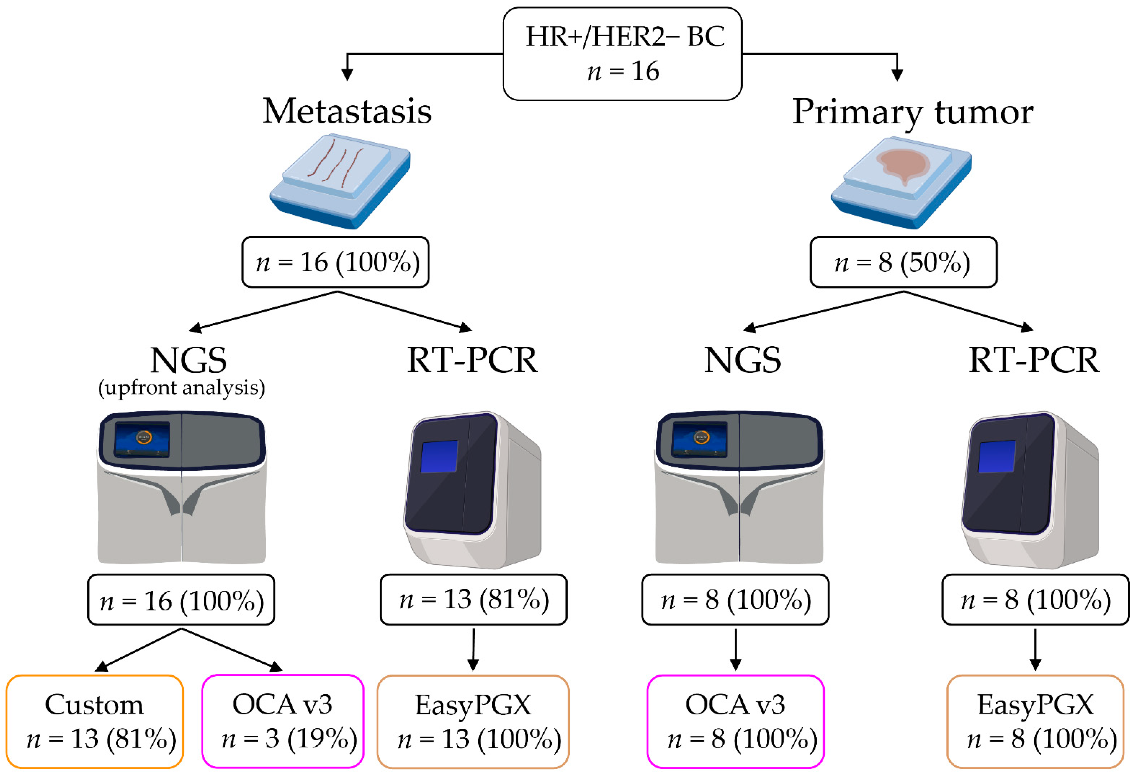
2.1. Study Design

This study was approved by the local Ethics Committee under approval number #UID3472; written informed consent was obtained from patients for use of tissue samples. According to the European Union’s General Data Protection Regulation (GDPR), all information regarding the recruited patients was pseudoanonymized [24]; the samples were handled in compliance with the Helsinki Declaration (http://www.wma.net/en/30publications/10policies/b3/, accessed on 14 September 2022). The study was designed to test the PIK3CA mutational status on different types of samples using different techniques, as depicted in Figure 1. The predictivity of PIK3CA mutations has been previously demonstrated [2] and was out of the scope of the present investigation.
From the institutional pathology archives of the IEO, European Institute of Oncology IRCCS, Milan, Italy a total of n = 16 PIK3CA-mutant HR+/HER2− metastatic breast cancer patients were retrospectively selected. The PIK3CA status of the metastatic samples was available for all patients because it was routinely assessed by NGS using either a custom panel or the OCA v3 (Oncomine Comprehensive Assay v3), both Thermo Fisher Scientific (Waltham, MA, USA) (Figure 2). All cases with residual material (n = 13) for further analyses were re-tested with an RT-PCR semi-closed assay (EasyPGX, Diatech Pharmacogenetics, Ancona, Italy) covering 10 PIK3CA regions, including the 5 regions covered by the therascreen (Figure 2). The available corresponding primary tumors (n = 8) were subjected to PIK3CA testing with both NGS and RT-PCR.

2.2. Patients and Tissue Specimens

All patients were diagnosed and managed at the aforementioned institution between 2017 and 2021. Hematoxylin and eosin-stained serial sections of each case were centrally reviewed, re-classified, and re-graded according to the latest WHO recommendations and the Nottingham histologic grading system, respectively [25,26]. HR and HER2 were re-tested and re-analyzed according to the latest ASCO/CAP recommendations, with particular attention to identifying HR-low and HER2-low cases [27,28,29]. Pathologic re-staging was performed following the 8th edition of the American Joint Committee on Cancer (AJCC) Cancer Staging Manual [30].

2.3. Nucleic Acids Purification

Seven unstained slides at four-μm-thick sections from representative FFPE tissue blocks were used for the analyses. In 10 out of 24 samples (42%), manual microdissection was performed before nucleic acid isolation to enrich tumor cell content using a sterile scalpel. DNA was extracted using the Maxwell® RSC DNA FFPE Kit (Promega, Madison, WI, USA) following the manufacturer’s instructions and then quantified by the QuantiFluor® ONE dsDNA System (Promega) on the Quantus™ Fluorometer (Promega).

2.4. Next-Generation Sequencing (NGS) Analysis

For the custom NGS panel, library amplification was carried out on automatized Ion Chef system (ThermoFisher Scientific) according to manufacturer instructions. A total of 15 ng was dispensed on Ion Code plates and amplified with Ion AmpliSeq DL8 kit (ThermoFisher Scientific) following standardized thermal conditions (23 cycles for amplification step, 4 min each). Then, purified libraries were diluted at 30 pM and newly loaded into the Ion Chef instrument for automatic template preparation and loading chip. Finally, libraries were automatically loaded on an Ion 520™ Chip and sequenced on an Ion S5™ System (ThermoFisher Scientific) following manufacturer instructions. Data analysis was performed as follows: after alignment to the hg19 human reference genome, coverage analysis with custom bed-files was assessed on coverage plug-in (v.5.0.2.0) from Torrent Suite [v.5.0.2]; variant Caller plug-in was carried out with a dedicated workflow on Ion Reporter Torrent Suite 5.16. Molecular alterations found in PIK3CA with a minimum coverage depth of 500×, allele coverage and a quality score ≥ 20, and minimum variant frequency of 1% were annotated. For OCA assay, libraries were prepared by using an Ion AmpliSeq DL8 kit (ThermoFisher Scientific) on an Ion Chef system (ThermoFisher Scientific) following manufacturer instructions. After library reamplification and barcoding, libraries were diluted at 30 pM and newly loaded into the Ion Chef instrument for automatic template preparation and loading chip. Finally, the barcoded chip was sequenced on an Ion S5™ System (ThermoFisher Scientific) following manufacturer instructions. The sequence data analysis was carried out by using customized analysis parameters on Torrent Suite™ 5.16. Only PIK3CA single nucleotide variants with a minimum coverage depth of 500×, allele coverage and a quality score ≥ 20, and a minimum variant frequency of 1% were reported.

2.5. RT-PCR Analysis

RT-PCR was conducted using the EasyPGX® ready PIK3CA kit (Diatech Pharmacogenetics) on the thermalcycler AriaDx Real-Time PCR System (Agilent, Santa Clara, CA, USA), according to the manufacturer’s instructions. This assay enables the detection of the most clinically relevant exons 9–20 PIK3CA alterations. An optimum of 15–30 ng/well is required to successfully perform the sequencing run. Data were analyzed using proprietary software (Agilent Aria software version 4.08) that automatically evaluates positive or negative results starting from the quantification cycle (Cq) and ΔCq values inspection.

2.6. Statistical Analyses

All statistical analyses were performed using SPSS software ver. 26.0 (IBM). The agreement between testing methods on the same biological sample and between different biospecimens using the same technical approach was evaluated with Cohen’s Kappa coefficient (κ).

3. Results

3.1. Clinicopathological Features

The median age at diagnosis of the patients included in this study was 52.5 years (range, 32–73 years; mean ± standard deviation (SD), 52.5 ± 11.2 years). Except for one case with lobular histology, all cases were invasive carcinomas of no special type. All cases were confirmed to be ER+, while eight patients had an ER+/PgR− tumor. Taken together, all of the cases included in this study at the time of the primary tumor diagnosis were HR+/HER2−. Among these, one patient developed an HER2+ disease after tumor progression. Furthermore, six (38.0%) HER2-negative cases displayed an HER2-low phenotype, i.e., immunohistochemistry (IHC) score 1+ or 2+ with no gene amplification. All other samples were HER2-zero. Among the metastatic sites, the liver was the most commonly affected (n = 8, 50.0%), while metastases were also observed in pleura (n = 2, 12.5%), iliac crest (n = 2, 12.5%), and distant lymph nodes (n = 2, 12.5%). Other metastatic sites included the pelvis and thorax (n = 1, 6.2%, respectively). Therapy with CDK4/6 inhibitors was administered to six (37.5%) patients. The clinicopathological characteristics of the patients included in this study are summarized in Table 1.
The time interval between disease diagnosis and metastasis in patients for whom both primary tumor and metastasis FFPE samples were available ranged from 38 to 127 months, as shown in Table 2.

3.2. Concordance between Primary Tumor and Metastasis Samples Using NGS

To define the mutational status of PIK3CA using NGS and to assess whether this can be recapitulated by the primary tissue, we analyzed both the primary tumor samples when available (n = 8, 50.0%) and all the metastases (n = 16, 100%). In the latter, NGS multigene panels detected PIK3CA ex. 7, 9, and 20 mutations. The mutations captured by this analysis include E453K (n = 1, 6.2%), E542K (n = 1, 6.2%), E545K (n = 3, 18.8%), H1047L (n = 1, 6.2%), H1047R (n = 6, 38%), N1044K (n = 1, 6.2%), N1068Kfs*5 (n = 1, 6.2%), Q546K (n = 1, 6.2%), and Q546R (n = 1, 6.2%). A high agreement between primary tumor samples and metastases analyzed with NGS and a concordance rate of 100% was observed regardless of samples’ technical characteristics and limitations (e.g., low DNA concentration, potential DNA fragmentation due to long-term archival tissues, and low tumor cell content). The overall results of this comparison and the specific characteristics of the samples are included in Table 3. This finding suggests the high reproducibility of NGS both in primary tumors and metastases. In addition, it suggests that the former, even if extensively stored, represents a valuable source for PIK3CA testing in the absence of metastasis samples.

3.3. Concordance between Primary Tumor and Metastasis Samples Using RT-PCR

Then, we asked whether the detection of PIK3CA alterations would be identifiable in primary tumor samples and metastases using RT-PCR assays. Based on the quantity and quality of the available material (i.e., archival slides and blocks, residual extracted DNA), all primary tumors (100%) and 13 out of 16 (81.3%) metastases were analyzed. Regarding the former, PIK3CA mutations were detected in five (62.5%) cases, while three (37.5%) cases were reported as wild-type, with two of them (66.6%) being out of the reference range of the assay (Table 4). By analyzing the metastasis samples, the RT-PCR assay managed to identify PIK3CA mutations in seven (53.8%) cases. In three (23.1%) of them reported as wild-type, two (66.6%) were not included in the reference range of the assay (Table 4). Moreover, the analysis software failed to provide results in three3 (23.1%) cases due to low DNA concentration. In these, despite the failure of the system in reporting the alteration, visual inspection of the raw data confirmed the presence of the mutation (Supplementary Figure S1). Overall, in three cases (i.e., PIK_004, PIK_007, and PIK_016), the RT-PCR assay identified mutations in the primary tumor, whereas these were not found in the corresponding metastases (i.e., failed or wild-type). Instead, in only one case, the mutation identified in the metastasis was not recapitulated by the primary tumor. The concordance rate between primary tumor and corresponding metastasis tested by RT-PCR was 40.0% (n = 2/5) with a Cohen kappa=0.09 (95% CI, −0.69–0.87), suggesting a slight agreement.

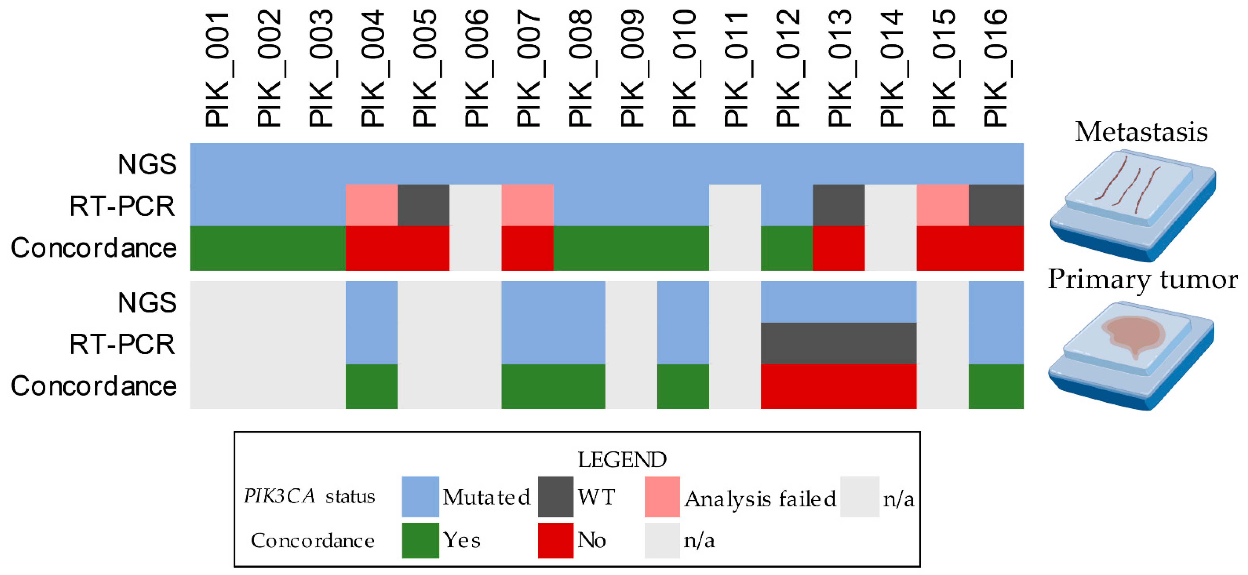
3.4. Comparison of NGS and RT-PCR

We next compared the results obtained from each technology to evaluate their analytical performance and their capability of detecting PIK3CA mutations both in primary tumor and metastasis samples. Concerning the analysis of the primary tumors, RT-PCR verified the mutations identified by NGS in five cases, again confirming a slight agreement with a Cohen kappa = 0 (95% CI, −0.89–0.89) and an overall concordance rate of 62.5%. Regarding the metastasis samples, the results of the EasyPGX system were concordant with those of the NGS panels in 7 out of 13 cases (53.8%), and a slight agreement was observed, with a Cohen kappa = 0 (95% CI, −0.59–0.59). Among these, exons 9 and 20 of PIK3CA harbored three (23.1%) and four (30.7%) molecular alterations, respectively. When the raw data of RT-PCR analysis were visually inspected, PIK3CA hotspot mutations were observed in 3/13 (23.1%) additional samples, improving the concordance rate to 76.9%. The results of the overall comparison between the two testing methods in both sets of samples are summarized in Figure 3.

4. Discussion

This proof-of-principle study evaluated the concordance rate for PIK3CA assessment between breast cancer primary tumors and matched metastases and the consensus among NGS and RT-PCR for this type of molecular testing. Our analyses confirm the robustness of targeted NGS and the reliability of the RT-PCR for PIK3CA testing. Additionally, we showed the high analytical performance of NGS-based panels able to detect PIK3CA pathogenic mutations both in surgical resections and biopsy specimens. Furthermore, we demonstrated the moderate efficacy of the RT-PCR assay to confirm the PIK3CA alterations identified by NGS due to technical limitations of the former in the analysis of challenging samples. Given the substantial overlap of PIK3CA status in primary tumors and metastases, the choice of what sample to test in HR+/HER2− breast cancer should be primarily driven by the quality and quantity of FFPE material available, regardless of the anatomical site. However, the interval between the diagnosis of the primary tumor and the occurrence of the metastasis as well as the therapy administered to each patient should be considered.
PIK3CA molecular testing has rapidly emerged as a new clinical request at the sight of data from the SOLAR-1 study regarding the efficacy of alpelisib in combination with fulvestrant in patients with advanced HR+/HER2− breast cancer previously treated with endocrine therapy [2,14]. Despite the approval of alpelisib that the FDA approved alongside the Therascreen PIK3CA mutation assay as a companion diagnostic, the increasing number of clinically relevant molecular alterations able to select PIK3CA-mutated breast cancer patients escalates the need for highly performant molecular testing methods [10]. Thus, the identification of the most suitable testing strategy according to internal expertise and available biospecimens represents an opening challenge in each institution performing molecular tests [16]. In this context, primary tumor and metastasis samples feature distinct technical burdens that drastically impact the analytical performance of the currently available testing methods. The latter is usually characterized by low DNA concentration that derives from moderate tumor cell content [20,31]. Given the overall long-term survivorship and long time to disease progression in patients with HR+/HER2− breast cancers, primary tumor samples are frequently old and may potentially be characterized by a high level of DNA fragmentation [32]. Despite these hindrances, in our cohort, NGS approaches adopted for PIK3CA molecular analysis managed to successfully identify mutations in all cases demonstrating an absolute concordance between primary and metastasis samples. In a recent study, a droplet digital PCR (ddPCR)-based approach highlighted a slightly lower concordance rate of 87.8% between primary tumors and metastasis samples [33]. This could either be related to intratumor heterogeneity or the evolution of the disease [21,34]. Moreover, the timing of biopsy sampling can influence the detection rate of molecular alterations [35]. However, in the recently published analyses from the AURORA study which analyzed 381 primary tumors and matched metastasis samples, an enrichment of PIK3CA mutations in metastasis was observed [36]. Thus, it is relevant to have a higher number of PIK3CA mutations in progressive or recurrent diseases [37]. Taken together, both the selection of the most appropriate genomic assay and the biospecimen to be analyzed should be considered when PIK3CA molecular testing is carried out.
The majority of molecular biology laboratories can evaluate the mutational status of PIK3CA using RT-PCR-based assays [38]. Accordingly, we investigated the analytical performance of a commercially available RT-PCR assay to identify mutations detected by NGS panels. Overall, the mutational analysis of PIK3CA status in primary tumor and metastasis samples by RT-PCR showed a low concordance rate of 33.3%. Failures occurred irrespective of the sample type and specifically, in the metastases, they were mainly related to low DNA concentration (n = 2, 15.4%). Importantly, in both cases, the mutation was found in the primary tumor sample in which DNA concentration was higher (0.26 vs. 9.11 and 0.18 vs. 0.54 ng/μL, respectively). Therefore, RT-PCR assays should be chosen carefully based on their technical characteristics and should be used with caution in samples with low DNA concentration. In such cases, complementary tests on matched primary tumors, which are usually richer in terms of material, should not be excluded to avoid false negative results.
By comparing the analytical performance of the two platforms both in primary tumors and matched metastases, we found discrepancies that need to be contextualized to better establish the advantages and limitations of each method. In the metastases, the moderate concordance between NGS and RT-PCR (overall concordance rate 53.8%) was entirely associated with failures in the analyses of the latter. In 4/6 (66.6%) of the discordant cases, which had low DNA concentration, this test either failed to detect the PIK3CA mutation or reported a false negative result, highlighting again that the quantity of nucleic acids drastically impacts the successful detection of genetic alterations. The lack of consensus in the other two cases (33.3%) was due to the limited reference range of RT-PCR that did not include these mutations. In the era of precision medicine, the increasing number of molecular alterations that should be tested in clinical practice requires the implementation of highly sensitive and specific assays; thus, the restricted reference range represents a crucial limitation of all the currently available RT-PCR assays, including therascreen® which covers 72% of all PIK3CA mutations [10,39]. The advent of the recent therapeutical implications of PIK3CA has further pointed out this limitation, despite older studies considering RT-PCR assays as valid options for this molecular test [40]. Interestingly though, the concordance between the two methods increased to 76.9% when the raw RT-PCR data of three discordant cases were visually inspected, confirming the result of NGS. This finding suggests that coupling the automatized analytical workflow with manual data analysis improves the outcomes of RT-PCR. However, it requires trained personnel for data analysis to overcome false negative results, and in those cases, further validation with another technique is warranted. Importantly, when the analytical performance of the two methods was examined in the primary tumor samples, a similar concordance rate (62.5%) was observed.
Our study has some limitations, including the small sample size, the lack of a validation set, and the fact that not all the metastasis samples had available material for being tested with both NGS and RT-PCR. Additionally, considering that primary tumor samples are not always present due to diagnosis at the later stages of the disease, in our study, primary tumors were analyzed in half of the selected cases. The adoption of an RT-PCR assay different from the companion diagnostic test therascreen® PIK3CA may not be in line with the approval by the FDA; however, it was chosen due to its slightly broader reference range. Finally, cases in which the RT-PCR assay either failed or reported a potential false negative result were not tested with another assay due to the limited residual DNA.

5. Conclusions

Targeted NGS for PIK3CA mutational testing on FFPE tumor and metastasis samples from patients with HR+/HER2− metastatic breast cancers is a reliable and robust diagnostic strategy. On the other hand, a careful selection of cases based on the quantity and quality of the DNA allows for solid RT-PCR-based PIK3CA testing both in primary tumors and matched metastases. Given that multiplexing assays allow the simultaneous analysis of several clinically actionable biomarkers, they require a relatively low nucleic acid input and generally have wide reference ranges. In the case of available high-quality material, based on the optimization of the laboratory workflow, RT-PCR would allow for short turnaround times and cheaper tests. Our findings should be expanded to larger cohorts of patients and compared with additional NGS and RT-PCR assays to establish tailored diagnostic algorithms and select the most appropriate test on the most appropriate FFPE samples at a single-patient level.

Supplementary Materials

The following supporting information can be downloaded at: https://www.mdpi.com/article/10.3390/cells11223545/s1; Figure S1: Visual representation of the RT-PCR raw data of cases reported as failed by the EasyPGX® analysis software.

Author Contributions

Conceptualization, U.M. and N.F.; methodology, K.V., F.P., D.V., A.R. (Alessandra Rappa), A.R. (Alberto Ranghiero), S.V.T., E.G.-R., U.M. and N.F.; software, K.V., F.P., G.B., U.M. and N.F.; validation, E.M., G.R. and P.P.; formal analysis, K.V. and F.P.; investigation, K.V., F.P., E.M., E.S., E.G.-R., U.M. and N.F.; resources, G.V., G.T., M.B., U.M. and N.F.; data curation, K.V., F.P., E.S., M.I. and G.B.; writing—original draft preparation, K.V. and F.P.; writing—review and editing, E.M., E.S., P.P., M.I., G.T, E.G.-R., U.M. and N.F.; visualization, K.V., F.P., U.M. and N.F.; supervision, U.M. and N.F.; project administration, U.M. and N.F.; funding acquisition, U.M. and N.F. All authors have read and agreed to the published version of the manuscript.

Funding

This study was funded by Novartis under the program for independent non-clinical investigator-initiated research.

Institutional Review Board Statement

The study was conducted in accordance with the Declaration of Helsinki, and approved by the Institutional Ethics Committee of European Institute of Oncology (IEO) IRCCS, Milan, Italy under the approval number #UID3472, approved on 16 December 2021; According to the European Union’s General Data Protection Regulation (GDPR), all information regarding the recruited patients were pseudoanonymized.

Informed Consent Statement

Written informed consent was obtained from all subjects involved in the study.

Data Availability Statement

The raw data of this study will be made available upon reasonable request to the corresponding author.

Conflicts of Interest

E.M. has acted as a consultant and/or has received funding for travel expenses and/or accommodation to Pierre Fabre, Genomic Health, Celgene, Roche, Eisai, Mylan, Exact Sciences, Merck Sharp & Dohme (MSD) Oncology, AstraZeneca, Daiichi Sankyo, Lilly, Novartis, Seagen, and Pfizer. E.S. is currently attending an unpaid fixed-term internship in Novartis during a University of Milan Ph.D. Program in Translational Medicine. D.V. has received personal fees from Novartis. A.R. has received personal fees from Novartis. G.V. has received honoraria for consulting, advisory role, and/or speaker bureau from MSD Oncology, Pfizer, Dako, Roche/Genetech, Astellas Pharma, Novartis, Bayer, Daiichi; Sankyo, Menarini, Ventana Medical Systems Dako/Agilent Technologies, Cepheid, and Celgene. E.G.R. from Thermo Fisher Scientific, Novartis, AstraZeneca, Roche, Biocartis, Exact Science, GlaxoSmithKline (GSK), and Illumina. U.M. has received personal fees (as consultant and/or speaker bureau) from Boehringer Ingelheim, Roche, MSD, Amgen, Thermo Fisher Scientifics, Eli Lilly, Diaceutics, GSK, Merck and AstraZeneca, Janssen, Diatech, Novartis and Hedera unrelated to the current work. N.F. has received honoraria for consulting, advisory role, and/or speaker bureau from MSD, Boehringer Ingelheim, Novartis, AstraZeneca, Daiichi Sankyo, GSK, Gilead, and Diaceutics. These companies had no role in the design of the study; in the collection, analyses, or interpretation of data; in the writing of the manuscript, automatic in the decision to publish the results. All other authors declare no potential conflicts of interest.

References

  1. Sung, H.; Ferlay, J.; Siegel, R.L.; Laversanne, M.; Soerjomataram, I.; Jemal, A.; Bray, F. Global cancer statistics 2020: GLOBOCAN estimates of incidence and mortality worldwide for 36 cancers in 185 countries. CA Cancer J. Clin. 2021, 71, 209–249. [Google Scholar] [CrossRef] [PubMed]
  2. André, F.; Ciruelos, E.; Rubovszky, G.; Campone, M.; Loibl, S.; Rugo, H.S.; Iwata, H.; Conte, P.; Mayer, I.A.; Kaufman, B.; et al. Alpelisib for PIK3CA-Mutated, Hormone Receptor-Positive Advanced Breast Cancer. N. Engl. J. Med. 2019, 380, 1929–1940. [Google Scholar] [CrossRef] [PubMed]
  3. Lopez, G.; Costanza, J.; Colleoni, M.; Fontana, L.; Ferrero, S.; Miozzo, M.; Fusco, N. Molecular Insights into the Classification of Luminal Breast Cancers: The Genomic Heterogeneity of Progesterone-Negative Tumors. Int. J. Mol. Sci. 2019, 20, 510. [Google Scholar] [CrossRef] [PubMed] [Green Version]
  4. Cardoso, F.; Paluch-Shimon, S.; Senkus, E.; Curigliano, G.; Aapro, M.S.; André, F.; Barrios, C.H.; Bergh, J.; Bhattacharyya, G.S.; Biganzoli, L.; et al. 5th ESO-ESMO international consensus guidelines for advanced breast cancer (ABC 5). Ann. Oncol. 2020, 31, 1623–1649. [Google Scholar] [CrossRef]
  5. Hui, R.; de Boer, R.; Lim, E.; Yeo, B.; Lynch, J. CDK4/6 inhibitor plus endocrine therapy for hormone receptor-positive, HER2-negative metastatic breast cancer: The new standard of care. Asia Pac. J. Clin. Oncol. 2021, 17 (Suppl. 1), 3–14. [Google Scholar] [CrossRef] [PubMed]
  6. Miller, T.W.; Balko, J.M.; Arteaga, C.L. Phosphatidylinositol 3-kinase and antiestrogen resistance in breast cancer. J. Clin. Oncol. 2011, 29, 4452–4461. [Google Scholar] [CrossRef] [Green Version]
  7. Mosele, F.; Stefanovska, B.; Lusque, A.; Tran Dien, A.; Garberis, I.; Droin, N.; Le Tourneau, C.; Sablin, M.P.; Lacroix, L.; Enrico, D.; et al. Outcome and molecular landscape of patients with PIK3CA-mutated metastatic breast cancer. Ann. Oncol. 2020, 31, 377–386. [Google Scholar] [CrossRef] [Green Version]
  8. Grizzi, G.; Ghidini, M.; Botticelli, A.; Tomasello, G.; Ghidini, A.; Grossi, F.; Fusco, N.; Cabiddu, M.; Savio, T.; Petrelli, F. Strategies for Increasing the Effectiveness of Aromatase Inhibitors in Locally Advanced Breast Cancer: An Evidence-Based Review on Current Options. Cancer Manag. Res. 2020, 12, 675–686. [Google Scholar] [CrossRef] [Green Version]
  9. Moynahan, M.E.; Chen, D.; He, W.; Sung, P.; Samoila, A.; You, D.; Bhatt, T.; Patel, P.; Ringeisen, F.; Hortobagyi, G.N.; et al. Correlation between PIK3CA mutations in cell-free DNA and everolimus efficacy in HR(+), HER2(-) advanced breast cancer: Results from BOLERO-2. Br. J. Cancer 2017, 116, 726–730. [Google Scholar] [CrossRef] [Green Version]
  10. Martínez-Sáez, O.; Chic, N.; Pascual, T.; Adamo, B.; Vidal, M.; González-Farré, B.; Sanfeliu, E.; Schettini, F.; Conte, B.; Brasó-Maristany, F.; et al. Frequency and spectrum of PIK3CA somatic mutations in breast cancer. Breast Cancer Res. 2020, 22, 45. [Google Scholar] [CrossRef]
  11. Goncalves, M.D.; Hopkins, B.D.; Cantley, L.C. Phosphatidylinositol 3-Kinase, Growth Disorders, and Cancer. N. Engl. J. Med. 2018, 379, 2052–2062. [Google Scholar] [CrossRef] [PubMed]
  12. Fritsch, C.; Huang, A.; Chatenay-Rivauday, C.; Schnell, C.; Reddy, A.; Liu, M.; Kauffmann, A.; Guthy, D.; Erdmann, D.; De Pover, A.; et al. Characterization of the novel and specific PI3Kα inhibitor NVP-BYL719 and development of the patient stratification strategy for clinical trials. Mol. Cancer Ther. 2014, 13, 1117–1129. [Google Scholar] [CrossRef] [PubMed] [Green Version]
  13. Fritsch, C.; Pfister, E.; Ebel, N.; Guthy, D.; Schnell, C.; Hofmann, F. Abstract 3934: Determination of the PI3Kα selective inhibitor alpelisib mechanism of action and efficacy in ER+/PIK3CA mutant breast cancer preclinical models. Cancer Res. 2018, 78, 3934. [Google Scholar] [CrossRef]
  14. André, F.; Ciruelos, E.M.; Juric, D.; Loibl, S.; Campone, M.; Mayer, I.A.; Rubovszky, G.; Yamashita, T.; Kaufman, B.; Lu, Y.S.; et al. Alpelisib plus fulvestrant for PIK3CA-mutated, hormone receptor-positive, human epidermal growth factor receptor-2-negative advanced breast cancer: Final overall survival results from SOLAR-1. Ann. Oncol. 2021, 32, 208–217. [Google Scholar] [CrossRef]
  15. Copur, M.S. Alpelisib to treat breast cancer. Drugs Today (Barc.) 2020, 56, 357–363. [Google Scholar] [CrossRef]
  16. Fusco, N.; Malapelle, U.; Fassan, M.; Marchiò, C.; Buglioni, S.; Zupo, S.; Criscitiello, C.; Vigneri, P.; Dei Tos, A.P.; Maiorano, E.; et al. PIK3CA Mutations as a Molecular Target for Hormone Receptor-Positive, HER2-Negative Metastatic Breast Cancer. Front. Oncol. 2021, 11, 644737. [Google Scholar] [CrossRef]
  17. Chan, A.C.; Weiss, L.M.; Bender, R. Abstract P4-09-15: A large study of PIK3CA mutations in the community setting identifies varying degree of mutation positivity rates across age groups in advanced HR+, HER2- breast cancer patients using an FDA RT-PCR cleared test. Cancer Res. 2020, 80, P4-09-15. [Google Scholar] [CrossRef]
  18. Rugo, H.S.; Lerebours, F.; Ciruelos, E.; Drullinsky, P.; Ruiz-Borrego, M.; Neven, P.; Park, Y.H.; Prat, A.; Bachelot, T.; Juric, D.; et al. Alpelisib plus fulvestrant in PIK3CA-mutated, hormone receptor-positive advanced breast cancer after a CDK4/6 inhibitor (BYLieve): One cohort of a phase 2, multicentre, open-label, non-comparative study. Lancet Oncol. 2021, 22, 489–498. [Google Scholar] [CrossRef]
  19. Angerilli, V.; Galuppini, F.; Pagni, F.; Fusco, N.; Malapelle, U.; Fassan, M. The Role of the Pathologist in the Next-Generation Era of Tumor Molecular Characterization. Diagnostics 2021, 11, 339. [Google Scholar] [CrossRef]
  20. Cappello, F.; Angerilli, V.; Munari, G.; Ceccon, C.; Sabbadin, M.; Pagni, F.; Fusco, N.; Malapelle, U.; Fassan, M. FFPE-Based NGS Approaches into Clinical Practice: The Limits of Glory from a Pathologist Viewpoint. J. Pers. Med. 2022, 12, 750. [Google Scholar] [CrossRef]
  21. Kalinsky, K.; Heguy, A.; Bhanot, U.K.; Patil, S.; Moynahan, M.E. PIK3CA mutations rarely demonstrate genotypic intratumoral heterogeneity and are selected for in breast cancer progression. Breast Cancer Res. Treat. 2011, 129, 635. [Google Scholar] [CrossRef] [PubMed]
  22. Zardavas, D.; te Marvelde, L.; Milne, R.L.; Fumagalli, D.; Fountzilas, G.; Kotoula, V.; Razis, E.; Papaxoinis, G.; Joensuu, H.; Moynahan, M.E.; et al. Tumor PIK3CA Genotype and Prognosis in Early-Stage Breast Cancer: A Pooled Analysis of Individual Patient Data. J. Clin. Oncol. 2018, 36, 981–990. [Google Scholar] [CrossRef] [PubMed] [Green Version]
  23. Berrino, E.; Annaratone, L.; Miglio, U.; Maldi, E.; Piccinelli, C.; Peano, E.; Balmativola, D.; Cassoni, P.; Pisacane, A.; Sarotto, I.; et al. Cold Formalin Fixation Guarantees DNA Integrity in Formalin Fixed Paraffin Embedded Tissues: Premises for a Better Quality of Diagnostic and Experimental Pathology with a Specific Impact on Breast Cancer. Front. Oncol. 2020, 10, 173. [Google Scholar] [CrossRef] [PubMed] [Green Version]
  24. General Data Protection Regulation (GDPR). 2018. OJ L 119, 04.05.2016; cor. OJ L 127, 23.5.2018. Available online: https://www.epsu.org/sites/default/files/article/files/GDPR_FINAL_EPSU.pdf (accessed on 14 September 2022).
  25. WHO Classification of Tumours Editorial Board. WHO Classification of Breast Tumours: WHO Classification of Tumours; World Health Organization: Geneva, Switzerland, 2019; Volume 2. [Google Scholar]
  26. Rakha, E.A.; El-Sayed, M.E.; Lee, A.H.; Elston, C.W.; Grainge, M.J.; Hodi, Z.; Blamey, R.W.; Ellis, I.O. Prognostic significance of Nottingham histologic grade in invasive breast carcinoma. J. Clin. Oncol. 2008, 26, 3153–3158. [Google Scholar] [CrossRef]
  27. Allison, K.H.; Hammond, M.E.H.; Dowsett, M.; McKernin, S.E.; Carey, L.A.; Fitzgibbons, P.L.; Hayes, D.F.; Lakhani, S.R.; Chavez-MacGregor, M.; Perlmutter, J.; et al. Estrogen and Progesterone Receptor Testing in Breast Cancer: ASCO/CAP Guideline Update. J. Clin. Oncol. 2020, 38, 1346–1366. [Google Scholar] [CrossRef]
  28. Fusco, N.; Ragazzi, M.; Sajjadi, E.; Venetis, K.; Piciotti, R.; Morganti, S.; Santandrea, G.; Fanelli, G.N.; Despini, L.; Invernizzi, M.; et al. Assessment of estrogen receptor low positive status in breast cancer: Implications for pathologists and oncologists. Histol. Histopathol 2021, 36, 18376. [Google Scholar] [CrossRef]
  29. Venetis, K.; Crimini, E.; Sajjadi, E.; Corti, C.; Guerini-Rocco, E.; Viale, G.; Curigliano, G.; Criscitiello, C.; Fusco, N. HER2 low, ultra-low, and novel complementary biomarkers: Expanding the spectrum of HER2 positivity in breast cancer. Front. Mol. Biosci. 2022, 9, 834651. [Google Scholar] [CrossRef]
  30. Amin, M.B.; Edge, S.B.; Greene, F.L.; Byrd, D.R.; Brookland, R.K.; Washington, M.K.; Gershenwald, J.E.; Compton, C.C.; Hess, K.R.; Sullivan, D.C. AJCC Cancer Staging Manual, 8th ed.; Springer International Publishing: Berlin/Heidelberg, Germany, 2017. [Google Scholar]
  31. Cho, M.; Ahn, S.; Hong, M.; Bang, H.; Van Vrancken, M.; Kim, S.; Lee, J.; Park, S.H.; Park, J.O.; Park, Y.S.; et al. Tissue recommendations for precision cancer therapy using next generation sequencing: A comprehensive single cancer center’s experiences. Oncotarget 2017, 8, 42478–42486. [Google Scholar] [CrossRef] [Green Version]
  32. Groelz, D.; Viertler, C.; Pabst, D.; Dettmann, N.; Zatloukal, K. Impact of storage conditions on the quality of nucleic acids in paraffin embedded tissues. PLoS ONE 2018, 13, e0203608. [Google Scholar] [CrossRef] [Green Version]
  33. Park, J.; Cho, S.Y.; Chang, E.S.; Sung, M.; Song, J.Y.; Jung, K.; Kim, S.S.; Shin, Y.K.; Choi, Y.L. Analysis of PIK3CA Mutation Concordance and Frequency in Primary and Different Distant Metastatic Sites in Breast Cancer. Cancer Res. Treat 2022. [Google Scholar] [CrossRef]
  34. Zardavas, D.; Irrthum, A.; Swanton, C.; Piccart, M. Clinical management of breast cancer heterogeneity. Nat. Rev. Clin. Oncol. 2015, 12, 381–394. [Google Scholar] [CrossRef] [PubMed]
  35. Suppan, C.; Graf, R.; Jahn, S.; Zhou, Q.; Klocker, E.V.; Bartsch, R.; Terbuch, A.; Kashofer, K.; Regitnig, P.; Lindenmann, J.; et al. Sensitive and robust liquid biopsy-based detection of PIK3CA mutations in hormone-receptor-positive metastatic breast cancer patients. Br. J. Cancer 2022, 126, 456–463. [Google Scholar] [CrossRef] [PubMed]
  36. Aftimos, P.; Oliveira, M.; Irrthum, A.; Fumagalli, D.; Sotiriou, C.; Gal-Yam, E.N.; Robson, M.E.; Ndozeng, J.; Di Leo, A.; Ciruelos, E.M.; et al. Genomic and Transcriptomic Analyses of Breast Cancer Primaries and Matched Metastases in AURORA, the Breast International Group (BIG) Molecular Screening Initiative. Cancer Discov. 2021, 11, 2796–2811. [Google Scholar] [CrossRef]
  37. Markou, A.; Farkona, S.; Schiza, C.; Efstathiou, T.; Kounelis, S.; Malamos, N.; Georgoulias, V.; Lianidou, E. PIK3CA Mutational Status in Circulating Tumor Cells Can Change During Disease Recurrence or Progression in Patients with Breast Cancer. Clin. Cancer Res. 2014, 20, 5823–5834. [Google Scholar] [CrossRef] [PubMed] [Green Version]
  38. Anderson, E.J.; Mollon, L.E.; Dean, J.L.; Warholak, T.L.; Aizer, A.; Platt, E.A.; Tang, D.H.; Davis, L.E. A Systematic Review of the Prevalence and Diagnostic Workup of PIK3CA Mutations in HR+/HER2- Metastatic Breast Cancer. Int. J. Breast. Cancer 2020, 2020, 3759179. [Google Scholar] [CrossRef]
  39. Kerr, K.M.; Bibeau, F.; Thunnissen, E.; Botling, J.; Ryška, A.; Wolf, J.; Öhrling, K.; Burdon, P.; Malapelle, U.; Büttner, R. The evolving landscape of biomarker testing for non-small cell lung cancer in Europe. Lung Cancer 2021, 154, 161–175. [Google Scholar] [CrossRef]
  40. Alvarez-Garcia, V.; Bartos, C.; Keraite, I.; Trivedi, U.; Brennan, P.M.; Kersaudy-Kerhoas, M.; Gharbi, K.; Oikonomidou, O.; Leslie, N.R. A simple and robust real-time qPCR method for the detection of PIK3CA mutations. Sci. Rep. 2018, 8, 4290. [Google Scholar] [CrossRef]
Figure 1. Study design. HR, hormone receptor; HER2, human epidermal growth factor receptor 2; BC, breast cancer; NGS, next-generation sequencing; Custom, custom panel (targeted sequencing); OCA v3, Oncomine Comprehensive Assay v3 (Thermo Fisher Scientific); EasyPGX, RT-PCR semi-closed assay (Diatech Pharmacogenetics).
Cells 11 03545 g001
Figure 2. Exon structure of PIK3CA gene with reference range of the assays employed in this study and the therascreen PIK3CA RGQ PCR Kit test employed in the SOLAR-1 trial. EasyPGX, EasyPGX PIK3CA testing, Diatech Pharmacogenetics; NGS, next-generation sequencing; OCA, Oncomine™ Comprehensive Assay v3M, ThermoFisher Scientific; Custom, custom targeted panel, ThermoFisher Scientific, Waltham, MA, USA.
Cells 11 03545 g002
Figure 3. Heatmap illustrating the results of the analysis at a single-patient level for both primary tumors and metastasis samples. Each column represents a patient, and each row represents the type of assay with a concordance of both if applicable, color-coded according to the legend below. WT, wild-type; n/a, non-applicable.
Cells 11 03545 g003
Table 1. Clinicopathological features of the patients included in this study according to the diagnosis of the primary tumor. NST, invasive carcinoma of no special type (aka ductal); ER, estrogen receptor; PgR, progesterone receptor; CDK4/6, cyclin-dependent kinase 4 and 6 inhibitors.
Patients (n = 16)
Age at diagnosis, range (median)32–73 (52.5)
Histology, n (%)
NST15 (93.7)
Lobular1 (6.3)
ER, n (%)
Positive16 (100)
Negative0
PgR, n (%)
Positive8 (50.0)
Negative8 (50.0)
HER2, n (%)
Low7 (43.8)
Zero9 (56.2)
Metastatic site, n (%)
Bone (iliac crest)2 (12.5)
Bone (pelvis)1 (6.3)
Lymph node2 (12.5)
Liver8 (50.0)
Pleura2 (12.5)
Thoracic wall1 (6.3)
Therapy with CDK4/6, n (%)
Yes6 (37.5)
No10 (62.5)
Table 2. Clinicopathological features at a single-case level and time interval between primary tumor and metastasis of the patients with both samples available. All cases were of no special histological subtype.
#CaseAge at Original DiagnosisTime between Primary Tumor and Metastasis (Months)Sample TypeER (%)PgR (%)HER2 (%)
PIK_0042669Primary95951+
Metastasis9501+
PIK_00746102Primary95701+
Metastasis9022+ not amplified
PIK_0084554Primary9501+
Metastasis9502+ not amplified
PIK_01053126Primary9501+
Metastasis9502+ not amplified
PIK_0124338Primary95202+ not amplified
Metastasis95101+
PIK_01344117Primary95950
Metastasis80900
PIK_01441116Primary95951+
Metastasis9500
PIK_01640127Primary95950
Metastasis6000
Table 3. Summary of the PIK3CA mutations detected using NGS on primary tumor and metastasis samples. Mutations are annotated according to both amino acid change and coding. VAF, variant allele frequency; NGS, next-generation sequencing.
MetastasesPrimary Tumors
#Case[DNA] (ng/µL)Tumor Cell Content (%)MutationVAF (%)[DNA] (ng/µL)Tumor Cell Content (%)MutationVAF (%)
PIK_0010.2620p.E545K/c.1633G>A3----
PIK_0020.8460p.H1047R/c.3140A>G29----
PIK_0030.3760p.H1047R/c.3140A>G67----
PIK_0040.2470p.H1047R/c.3140A>G382.9960p.H1047R/c.3140A>G42
PIK_0050.4480p.N1068Kfs*5/c.3203_3204insA20----
PIK_0060.4270p.E545K/c.1633G>A35----
PIK_0070.3630p.H1047R/c.3140A>G100.5420p.H1047R/c.3140A>G41
PIK_0084.0685p.H1047R/c.3140A>G560.4135p.H1047R/c.3140A>G22
PIK_0090.4370p.H1047L/c.3140A>T24----
PIK_0100.2470p.E545K/c.1633G>A250.6660p.E545K/c.1633G>A49
PIK_0114.5260p.H1047R/c.3140A>G44----
PIK_0120.4860p.E542K/c.1624G>A180.3180p.E542K/c.1624G>A4
PIK_013113.0080p.E453K/c.1357G>A133.0675p.E453K/c.1357G>A37
PIK_0140.4250p.N1044K/c.3132T>A541.8375p.N1044K/c.3132T>A37
PIK_0150.1290p.Q546K/c.1636C>A37----
PIK_0160.0780p.Q546R/c.1637A>G330.9740p.Q546R/c.1637A>G45
Table 4. Summary of the PIK3CA mutations detected using RT-PCR on primary tumor and metastasis samples. Mutations are annotated according to the amino acid change; WT, wild-type; # mutation out of the EasyPGX reference range; * Failed due to low DNA quantity but mutation visible in the individual report.
MetastasesPrimary Tumors
#Case[DNA]
(ng/µL)
Tumor Cell Content (%)Mutation[DNA] (ng/µL)Tumor Cell Content (%)Mutation
PIK_0010.2620E545x---
PIK_0020.8460H1047x---
PIK_0030.3760H1047x---
PIK_0040.2635Failed *9.1160H1047x
PIK_0050.4420WT #---
PIK_006------
PIK_0070.1840Failed *0.5420H1047x
PIK_0084.0685H1047x0.4190H1047x
PIK_0090.4370H1047x---
PIK_0100.2470E545x2.4930E545x
PIK_011------
PIK_0120.4860E542x0.4790WT
PIK_01328.0070WT #12.0070WT #
PIK_014---7.3135WT #
PIK_0150.1290Failed *---
PIK_0160.1480WT0.9740Q546x
Publisher’s Note: MDPI stays neutral with regard to jurisdictional claims in published maps and institutional affiliations.

