Next Issue
Volume 11, August-1
Previous Issue
Volume 11, July-1


Cells, Volume 11, Issue 14 (July-2 2022) – 137 articles

Cover Story (view full-size image): 
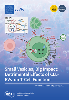
Chronic lymphocytic leukemia (CLL) is characterized by a clonal expansion of malignant B-cells and immunological defects, including T-cell dysfunctions. This leads to severe infectious complications and inefficient tumor immune surveillance. Although direct CLL-cell–T-cell interactions have been reported, the entirety of their complex interplay is incompletely understood. Here, we show that CLL-cells secrete bioactive extracellular vesicles (EVs) that carry a plethora of immune checkpoints (ICs) contributing to these T-cell deficiencies. CLL-EVs hamper T-cell viability, proliferation, activation, and metabolism while fostering their exhaustion and the formation of regulatory subsets. The variety of ICs in CLL-EVs could represent a barrier for immunotherapies, and our findings may pave the way to improving antitumor immunity by targeting EV formation or multiple ICs simultaneously. View this paper
  • Issues are regarded as officially published after their release is announced to the table of contents alert mailing list.
  • You may sign up for e-mail alerts to receive table of contents of newly released issues.
  • PDF is the official format for papers published in both, html and pdf forms. To view the papers in pdf format, click on the "PDF Full-text" link, and use the free Adobe Reader to open them.
Order results
Result details
Section
Select all
Export citation of selected articles as:
11 pages, 2717 KiB  
Article
Distinct PKA Signaling in Cytosolic and Mitochondrial Compartments in Electrically Paced Atrial Myocytes
by Noa Kirschner Peretz, Sofia Segal, Ido Weiser-Bitoun and Yael Yaniv
Cells 2022, 11(14), 2261; https://doi.org/10.3390/cells11142261 - 21 Jul 2022
Cited by 1 | Viewed by 1706
Abstract
Protein kinase A (PKA) is a key nodal signaling molecule that regulates a wide range of cellular functions in the cytosol and mitochondria. The distribution of A-kinase anchoring proteins that tether PKA, the local interaction with degradation molecules, and regulation by Ca2+ [...] Read more.
Protein kinase A (PKA) is a key nodal signaling molecule that regulates a wide range of cellular functions in the cytosol and mitochondria. The distribution of A-kinase anchoring proteins that tether PKA, the local interaction with degradation molecules, and regulation by Ca2+, may lead to distinct spatiotemporal cAMP/PKA signaling in these compartments. In this work, FRET-based sensors were used to investigate PKA signaling in the cytosol, outer mitochondrial membrane (OMM), and mitochondrial matrix (MM) and its crosstalk with Ca2+ in response to electrical stimulation of cultured rabbit atrial cells. A gradual decrease in PKA activity eliminating the ability of the atrial cells to respond to physiological electrical stimulation, was observed upon treatment of cells with H-89. Chelation of intracellular Ca2+ by BAPTA reduced PKA activity and diminished its response to forskolin, an AC stimulator. Under basal conditions, PKA activity in response to forskolin was lower in the OMM compared to the cytosol and MM. In response to electrical stimulation in the presence of ISO, distinct compartmentalization of PKA activity was observed, with higher activity in the cytosol and MM than in the OMM. Thus, distinct Ca2+-dependent spatiotemporal cAMP/PKA signaling exists in atrial cells, likely mediating its excitation and mitochondrial function. Full article
(This article belongs to the Special Issue Cyclic AMP/PKA/Epac Signaling in Health and Disease)
Show Figures

Figure 1

12 pages, 1220 KiB  
Review
Hedgehog Signaling in CNS Remyelination
by Minxi Fang, Tao Tang, Mengsheng Qiu and Xiaofeng Xu
Cells 2022, 11(14), 2260; https://doi.org/10.3390/cells11142260 - 21 Jul 2022
Cited by 3 | Viewed by 2089
Abstract
Remyelination is a fundamental repair process in the central nervous system (CNS) that is triggered by demyelinating events. In demyelinating diseases, oligodendrocytes (OLs) are targeted, leading to myelin loss, axonal damage, and severe functional impairment. While spontaneous remyelination often fails in the progression [...] Read more.
Remyelination is a fundamental repair process in the central nervous system (CNS) that is triggered by demyelinating events. In demyelinating diseases, oligodendrocytes (OLs) are targeted, leading to myelin loss, axonal damage, and severe functional impairment. While spontaneous remyelination often fails in the progression of demyelinating diseases, increased understanding of the mechanisms and identification of targets that regulate myelin regeneration becomes crucial. To date, several signaling pathways have been implicated in the remyelination process, including the Hedgehog (Hh) signaling pathway. This review summarizes the current data concerning the complicated roles of the Hh signaling pathway in the context of remyelination. We will highlight the open issues that have to be clarified prior to bringing molecules targeting the Hh signaling to demyelinating therapy. Full article
Show Figures

Figure 1

19 pages, 5100 KiB  
Article
Functional Expression of Multidrug-Resistance (MDR) Transporters in Developing Human Fetal Brain Endothelial Cells
by Phetcharawan Lye, Enrrico Bloise, Guinever E. Imperio, David Chitayat and Stephen G. Matthews
Cells 2022, 11(14), 2259; https://doi.org/10.3390/cells11142259 - 21 Jul 2022
Cited by 5 | Viewed by 2170
Abstract
There is little information about the functional expression of the multidrug resistance (MDR) transporters P-glycoprotein (P-gp, encoded by ABCB1) and breast cancer resistance protein (BCRP/ABCG2) in the developing blood–brain barrier (BBB). We isolated and cultured primary human fetal brain endothelial [...] Read more.
There is little information about the functional expression of the multidrug resistance (MDR) transporters P-glycoprotein (P-gp, encoded by ABCB1) and breast cancer resistance protein (BCRP/ABCG2) in the developing blood–brain barrier (BBB). We isolated and cultured primary human fetal brain endothelial cells (hfBECs) from early and mid-gestation brains and assessed P-gp/ABCB1 and BCRP/ABCG2 expression and function, as well as tube formation capability. Immunolocalization of the von Willebrand factor (marker of endothelial cells), zonula occludens-1 and claudin-5 (tight junctions) was detected in early and mid-gestation-derived hfBECs, which also formed capillary-like tube structures, confirming their BEC phenotype. P-gp and BCRP immunostaining was detected in capillary-like tubes and in the cytoplasm and nucleus of hfBECs. P-gp protein levels in the plasma membrane and nuclear protein fractions, as well as P-gp protein/ABCB1 mRNA and BCRP protein levels decreased (p < 0.05) in hfBECs, from early to mid-gestation. No differences in P-gp or BCRP activity in hfBECs were observed between the two age groups. The hfBECs from early and mid-gestation express functionally competent P-gp and BCRP drug transporters and may thus contribute to the BBB protective phenotype in the conceptus from early stages of pregnancy. Full article
(This article belongs to the Section Reproductive Cells and Development)
Show Figures

Figure 1

21 pages, 2391 KiB  
Article
Citalopram Neuroendocrine Challenge Shows Altered Tryptophan and Kynurenine Metabolism in Migraine
by Kinga Gecse, Andrea Edit Édes, Tamás Nagy, Adrienn Katalin Demeter, Dávid Virág, Márton Király, Borbála Dalmadi Kiss, Krisztina Ludányi, Zsuzsanna Környei, Adam Denes, Gyorgy Bagdy and Gabriella Juhasz
Cells 2022, 11(14), 2258; https://doi.org/10.3390/cells11142258 - 21 Jul 2022
Cited by 5 | Viewed by 2934
Abstract
Altered tryptophan (TRP) metabolism may have an important role in migraine susceptibility through its main metabolites, serotonin and kynurenine (KYN). Both affect pain processing and stress response by interfering with neural and brain hypersensitivity and by interacting with chemokines and cytokines that control [...] Read more.
Altered tryptophan (TRP) metabolism may have an important role in migraine susceptibility through its main metabolites, serotonin and kynurenine (KYN). Both affect pain processing and stress response by interfering with neural and brain hypersensitivity and by interacting with chemokines and cytokines that control vascular and inflammatory processes. The involvement of these pathways in migraine has been widely studied, but acute citalopram neuroendocrine challenge on TRP metabolism and cytokine profile has not been investigated yet. In our study, females with episodic migraine without aura and healthy controls were studied before and after acute citalopram or placebo in a double-blind setting. At baseline, increased TRP/large neutral amino acid (LNAA) ratio and decreased RANTES chemokine concentration were detected in migraine patients compared to controls. The challenge induced a significant increase in TRP, KYN, and TRP/LNAA in healthy controls, but not in migraine patients. Furthermore, migraine attack frequency negatively correlated with KYN/TRP ratio and positively correlated with the neuroendocrine-challenge-induced KYN concentration increase. Our results support a decreased breakdown of TRP via KYN pathway and a failure to modulate TRP–KYN pathway during citalopram-induced acute stress together with an increased vascular sensitivity in migraine. These mechanisms may provide useful drug targets for future drug development. Full article
(This article belongs to the Special Issue Migraine Neuroscience: From Experimental Models to Target Therapy)
Show Figures

Figure 1

17 pages, 855 KiB  
Review
Interleukin-11: A Potential Biomarker and Molecular Therapeutic Target in Non-Small Cell Lung Cancer
by Jason Hongting Leung, Benjamin Ng and Wei-Wen Lim
Cells 2022, 11(14), 2257; https://doi.org/10.3390/cells11142257 - 21 Jul 2022
Cited by 7 | Viewed by 3403
Abstract
Non-small cell lung cancer (NSCLC) accounts for 85% of lung cancer and is a fast progressive disease when left untreated. Identification of potential biomarkers in NSCLC is an ongoing area of research that aims to detect, diagnose, and prognosticate patients early to optimize [...] Read more.
Non-small cell lung cancer (NSCLC) accounts for 85% of lung cancer and is a fast progressive disease when left untreated. Identification of potential biomarkers in NSCLC is an ongoing area of research that aims to detect, diagnose, and prognosticate patients early to optimize treatment. We review the role of interleukin-11 (IL11), a stromal-cell derived pleiotropic cytokine with profibrotic and cellular remodeling properties, as a potential biomarker in NSCLC. This review identifies the need for biomarkers in NSCLC, the potential sources of IL11, and summarizes the available information leveraging upon published literature, publicly available datasets, and online tools. We identify accumulating evidence suggesting IL11 to be a potential biomarker in NSCLC patients. Further in-depth studies into the pathophysiological effects of IL11 on stromal-tumor interaction in NSCLC are warranted and current available literature highlights the potential value of IL11 detection as a diagnostic and prognostic biomarker in NSCLC. Full article
Show Figures

Figure 1

15 pages, 4310 KiB  
Article
Large-Scale Profiling on lncRNAs in Human Platelets: Correlation with Platelet Reactivity
by Yeying Sun, Rongrong Liu, Xiangwen Xia, Luchuan Xing, Jing Jiang, Weihua Bian, Wendy Zhang, Chunhua Wang and Chunxiang Zhang
Cells 2022, 11(14), 2256; https://doi.org/10.3390/cells11142256 - 21 Jul 2022
Cited by 9 | Viewed by 1644
Abstract
Recently, long noncoding RNAs (lncRNAs) have been key regulators for both mRNAs and proteins in nucleated cells. However, the expression profiles of lncRNAs in non-nucleated cells such as platelets are currently unclear. In this study, we determined the expression profiles of lncRNAs in [...] Read more.
Recently, long noncoding RNAs (lncRNAs) have been key regulators for both mRNAs and proteins in nucleated cells. However, the expression profiles of lncRNAs in non-nucleated cells such as platelets are currently unclear. In this study, we determined the expression profiles of lncRNAs in human platelets. We found that 6109 lncRNAs were expressed in human platelets. Interestingly, 338 lncRNAs were differentially expressed in hyperreactive and hyporeactive platelets. Bioinformatics’ analysis revealed that these aberrantly expressed lncRNAs might be related to platelet activity and other platelet functions. To provide a proof of concept, we measured the expression levels of PARLncRNA-1, a down-regulated lncRNA of hyperreactive platelets, in platelets from 12 patients with acute myocardial infarction and their controls. We found that the lncRNA was also significantly down-regulated in platelets from patients, which was partially reversed by treatment with aspirin a known antiplatelet drug. LncRNAs may represent a novel class of modulators for platelet functions. Full article
Show Figures

Figure 1

19 pages, 4967 KiB  
Article
TRPM7 Modulates Human Pancreatic Stellate Cell Activation
by Julie Auwercx, Philippe Kischel, Thibaut Lefebvre, Nicolas Jonckheere, Alison Vanlaeys, Stéphanie Guénin, Silviya Radoslavova, Isabelle Van Seuningen, Halima Ouadid-Ahidouch, Hemant M. Kocher, Isabelle Dhennin-Duthille and Mathieu Gautier
Cells 2022, 11(14), 2255; https://doi.org/10.3390/cells11142255 - 21 Jul 2022
Cited by 4 | Viewed by 2192
Abstract
Pancreatic diseases, such as pancreatitis or pancreatic ductal adenocarcinoma, are characterized by the presence of activated pancreatic stellate cells (PSCs). These cells represent key actors in the tumor stroma, as they actively participate in disease development and progression: reprograming these PSCs into a [...] Read more.
Pancreatic diseases, such as pancreatitis or pancreatic ductal adenocarcinoma, are characterized by the presence of activated pancreatic stellate cells (PSCs). These cells represent key actors in the tumor stroma, as they actively participate in disease development and progression: reprograming these PSCs into a quiescent phenotype has even been proposed as a promising strategy for restoring the hallmarks of a healthy pancreas. Since TRPM7 channels have been shown to regulate hepatic stellate cells proliferation and survival, we aimed to study the role of these magnesium channels in PSC activation and proliferation. PS-1 cells (isolated from a healthy pancreas) were used as a model of healthy PSCs: quiescence or activation were induced using all-trans retinoic acid or conditioned media of pancreatic cancer cells, respectively. The role of TRPM7 was studied by RNA silencing or by pharmacological inhibition. TRPM7 expression was found to be correlated with the activation status of PS-1 cells. TRPM7 expression was able to regulate proliferation through modulation of cell cycle regulators and most importantly p53, via the PI3K/Akt pathway, in a magnesium-dependent manner. Finally, the analysis of TCGA database showed the overexpression of TRPM7 in cancer-associated fibroblasts. Taken together, we provide strong evidences that TRPM7 can be considered as a marker of activated PSCs. Full article
(This article belongs to the Collection Advance in Ion Channel Signaling in Cancer Cells)
Show Figures

Figure 1

14 pages, 2036 KiB  
Article
Cerebral Autoregulation Assessment Using the Near Infrared Spectroscopy ‘NIRS-Only’ High Frequency Methodology in Critically Ill Patients: A Prospective Cross-Sectional Study
by Jeanette Tas, Nick Eleveld, Melisa Borg, Kirsten D. J. Bos, Anne P. Langermans, Sander M. J. van Kuijk, Iwan C. C. van der Horst, Jan Willem J. Elting and Marcel J. H. Aries
Cells 2022, 11(14), 2254; https://doi.org/10.3390/cells11142254 - 21 Jul 2022
Cited by 2 | Viewed by 2033
Abstract
Impairments in cerebral autoregulation (CA) are related to poor clinical outcome. Near infrared spectroscopy (NIRS) is a non-invasive technique applied to estimate CA. Our general purpose was to study the clinical feasibility of a previously published ‘NIRS-only’ CA methodology in a critically ill [...] Read more.
Impairments in cerebral autoregulation (CA) are related to poor clinical outcome. Near infrared spectroscopy (NIRS) is a non-invasive technique applied to estimate CA. Our general purpose was to study the clinical feasibility of a previously published ‘NIRS-only’ CA methodology in a critically ill intensive care unit (ICU) population and determine its relationship with clinical outcome. Bilateral NIRS measurements were performed for 1–2 h. Data segments of ten-minutes were used to calculate transfer function analyses (TFA) CA estimates between high frequency oxyhemoglobin (oxyHb) and deoxyhemoglobin (deoxyHb) signals. The phase shift was corrected for serial time shifts. Criteria were defined to select TFA phase plot segments (segments) with ‘high-pass filter’ characteristics. In 54 patients, 490 out of 729 segments were automatically selected (67%). In 34 primary neurology patients the median (q1–q3) low frequency (LF) phase shift was higher in 19 survivors compared to 15 non-survivors (13° (6.3–35) versus 0.83° (−2.8–13), p = 0.0167). CA estimation using the NIRS-only methodology seems feasible in an ICU population using segment selection for more robust and consistent CA estimations. The ‘NIRS-only’ methodology needs further validation, but has the advantage of being non-invasive without the need for arterial blood pressure monitoring. Full article
(This article belongs to the Special Issue Brain Injury, Microcirculation and Tissue Perfusion)
Show Figures

Graphical abstract

12 pages, 1987 KiB  
Article
Ssc-MiR-21-5p and Ssc-MiR-615 Regulates the Proliferation and Apoptosis of Leydig Cells by Targeting SOX5
by Qi Tang, Yanghai Zhang, Linxiu Yue, Hongying Ren and Chuanying Pan
Cells 2022, 11(14), 2253; https://doi.org/10.3390/cells11142253 - 21 Jul 2022
Cited by 7 | Viewed by 1749
Abstract
Leydig cells (LCs) are the predominant cells of androgen production, which plays key roles in spermatogenesis and maintaining male secondary sexual characteristics. Abnormal development of LCs affects androgen levels in vivo, affects fertility and may even lead to infertility. Little is known about [...] Read more.
Leydig cells (LCs) are the predominant cells of androgen production, which plays key roles in spermatogenesis and maintaining male secondary sexual characteristics. Abnormal development of LCs affects androgen levels in vivo, affects fertility and may even lead to infertility. Little is known about the regulation mechanism on LCs’ development and maturation in domestic animals, especially the regulation of non-coding RNAs. In this study, we continued to dig deeper in the previous RNA-seq data of porcine LCs from our group, combined with detecting the expression profiles in different tissues and different types of cells in the testis, to screen out candidate microRNAs (miRNAs) that may affect the regulation of LCs. A total of two miRNAs, ssc-miR-21-5p and ssc-miR-615 (“ssc” is omitted below), were finally determined. After overexpression and interference of miRNAs in vitro, the effects of candidate miRNAs on the proliferation and apoptosis of TM3 (mouse Leydig cell line) were explored. The results showed that miR-21-5p led to a decrease in TM3 cell density and p53 (apoptosis related protein) expression. Meanwhile, miR-21-5p decreased EdU positive cell numbers, but increased TUNEL positive cell numbers, suggesting miR-21-5p could inhibit proliferation and promote apoptosis. Conversely, miR-615 could increase TM3 cell density. Western blot and TUNEL assay indicated miR-615 inhibited apoptosis, but had no effect on proliferation. In addition, Sox5 was identified a potential target gene of these two miRNAs by Dual-Luciferase reporter system assay. Our findings about functions of miRNAs in TM3 and the mapping of miRNAs-target gene regulatory network would provide an important basis for the further elucidation of miRNAs in regulating pig LCs. Full article
(This article belongs to the Collection Regulatory Functions of microRNAs)
Show Figures

Figure 1

17 pages, 3935 KiB  
Article
Therapeutic Potential of Human Immature Dental Pulp Stem Cells Observed in Mouse Model for Acquired Aplastic Anemia
by Vivian Fonseca Gonzaga, Cristiane Valverde Wenceslau, Daniel Perez Vieira, Bruna de Oliveira Policiquio, Charbel Khalil, Rodrigo Pinheiro Araldi and Irina Kerkis
Cells 2022, 11(14), 2252; https://doi.org/10.3390/cells11142252 - 21 Jul 2022
Cited by 4 | Viewed by 2148
Abstract
Aplastic anemia (AA) is a rare and serious disorder of hematopoietic stem cells (HSCs) that results in the loss of blood cells due to the failure of the bone marrow (BM). Although BM transplantation is used to treat AA, its use is limited [...] Read more.
Aplastic anemia (AA) is a rare and serious disorder of hematopoietic stem cells (HSCs) that results in the loss of blood cells due to the failure of the bone marrow (BM). Although BM transplantation is used to treat AA, its use is limited by donor availability. In this sense, mesenchymal stem cells (MSCs) can offer a novel therapeutic approach for AA. This is because the MSCs contribute to the hematopoietic niche organization through their repopulating. In our study, we used the human immature dental pulp stem cell (hIDPSC), an MSC-like cell, to explore an alternative therapeutic approach for AA. For this, isogenic C57BL/6 mice were exposed to total body irradiation (TBI) to induce the AA. After 48 h of TBI, the mice were intraperitoneally treated with hIDPSC. The immunohistochemistry analyses confirmed that the hIDPSCs migrated and grafted in the mouse bone marrow (BM) and spleen, providing rapid support to hematopoiesis recovery compared to the group exposed to radiation, but not to those treated with the cells as well as the hematological parameters. Six months after the last hIDPSC transplantation, the BM showed long-term stable hematopoiesis. Our data highlight the therapeutic plasticity and hematoprotective role of hIDPSC for AA and potentially for other hematopoietic failures. Full article
(This article belongs to the Special Issue Dental Pulp Stem Cells and Regenerative Medicine)
Show Figures

Figure 1

15 pages, 1330 KiB  
Article
Maternal Fibroblast Growth Factor 21 Levels Decrease during Early Pregnancy in Normotensive Pregnant Women but Are Higher in Preeclamptic Women—A Longitudinal Study
by Julieth Daniela Buell-Acosta, Maria Fernanda Garces, Arturo José Parada-Baños, Edith Angel-Muller, Maria Carolina Paez, Javier Eslava-Schmalbach, Franklin Escobar-Cordoba, Sofia Alexandra Caminos-Cepeda, Ezequiel Lacunza, Justo P. Castaño, Rubén Nogueiras, Carlos Dieguez, Ariel Iván Ruiz-Parra and Jorge Eduardo Caminos
Cells 2022, 11(14), 2251; https://doi.org/10.3390/cells11142251 - 21 Jul 2022
Cited by 3 | Viewed by 3288
Abstract
(1) Background: Fibroblast growth factor 21 (FGF-21) is an endocrine factor involved in glucose and lipid metabolism that exerts pleiotropic effects. The aim of this study was to investigate the serum FGF-21 profile in healthy and mild preeclamptic pregnant women at each trimester [...] Read more.
(1) Background: Fibroblast growth factor 21 (FGF-21) is an endocrine factor involved in glucose and lipid metabolism that exerts pleiotropic effects. The aim of this study was to investigate the serum FGF-21 profile in healthy and mild preeclamptic pregnant women at each trimester of pregnancy; (2) Methods: Serum FGF-21 levels were determined by ELISA in a nested case-control study within a longitudinal cohort study that included healthy (n = 54) and mild preeclamptic (n = 20) pregnant women, women at three months after delivery (n = 20) and eumenorrheic women during the menstrual cycle (n = 20); (3) Results: FGF-21 levels were significantly lower in the mid-luteal phase compared to the early follicular phase of the menstrual cycle in eumenorrheic women (p < 0.01). Maternal levels of FGF-21 were significantly lower in the first and second trimesters and peaked during the third trimester in healthy pregnant women (p < 0.01). Serum levels of FGF-21 in healthy pregnant were significantly lower in the first and second trimester of pregnancy compared with the follicular phase of the menstrual cycle and postpartum (p < 0.01). Serum FGF-21 levels were significantly higher in preeclamptic compared to healthy pregnant women during pregnancy (p < 0.01); (4) Conclusions: These results suggest that a peak of FGF-21 towards the end of pregnancy in healthy pregnancy and higher levels in preeclamptic women might play a critical role that contributes to protecting against the negatives effects of high concentrations of non-esterified fatty acids (NEFA) and hypertensive disorder. Furthermore, FGF-21 might play an important role in reproductive function in healthy eumenorrheic women during the menstrual cycle. Full article
Show Figures

Graphical abstract

18 pages, 3389 KiB  
Review
Chaperone-Mediated Autophagy in Neurodegenerative Diseases: Molecular Mechanisms and Pharmacological Opportunities
by Yi-Ting Wang and Jia-Hong Lu
Cells 2022, 11(14), 2250; https://doi.org/10.3390/cells11142250 - 20 Jul 2022
Cited by 17 | Viewed by 3623
Abstract
Chaperone-mediated autophagy (CMA) is a protein degradation mechanism through lysosomes. By targeting the KFERQ motif of the substrate, CMA is responsible for the degradation of about 30% of cytosolic proteins, including a series of proteins associated with neurodegenerative diseases (NDs). The fact that [...] Read more.
Chaperone-mediated autophagy (CMA) is a protein degradation mechanism through lysosomes. By targeting the KFERQ motif of the substrate, CMA is responsible for the degradation of about 30% of cytosolic proteins, including a series of proteins associated with neurodegenerative diseases (NDs). The fact that decreased activity of CMA is observed in NDs, and ND-associated mutant proteins, including alpha-synuclein and Tau, directly impair CMA activity reveals a possible vicious cycle of CMA impairment and pathogenic protein accumulation in ND development. Given the intrinsic connection between CMA dysfunction and ND, enhancement of CMA has been regarded as a strategy to counteract ND. Indeed, genetic and pharmacological approaches to modulate CMA have been shown to promote the degradation of ND-associated proteins and alleviate ND phenotypes in multiple ND models. This review summarizes the current knowledge on the mechanism of CMA with a focus on its relationship with NDs and discusses the therapeutic potential of CMA modulation for ND. Full article
(This article belongs to the Special Issue Pharmacological Modulation of Autophagy)
Show Figures

Figure 1

17 pages, 850 KiB  
Review
Immune Checkpoint Inhibition in Acute Myeloid Leukemia and Myelodysplastic Syndromes
by Yasmin Abaza and Amer M. Zeidan
Cells 2022, 11(14), 2249; https://doi.org/10.3390/cells11142249 - 20 Jul 2022
Cited by 22 | Viewed by 5411
Abstract
Immune checkpoint inhibitors (ICIs) have revolutionized the treatment of many solid tumors, with limited progress made in the area of myeloid malignancies. The low mutational burden of acute myeloid leukemia (AML) is one potential reason behind the lack of activity of T-cell harnessing [...] Read more.
Immune checkpoint inhibitors (ICIs) have revolutionized the treatment of many solid tumors, with limited progress made in the area of myeloid malignancies. The low mutational burden of acute myeloid leukemia (AML) is one potential reason behind the lack of activity of T-cell harnessing ICIs, particularly CTLA-4 and PD-1 inhibitors. Innate immune checkpoints play a critical role in the immune escape of AML and myelodysplastic syndromes (MDS). The CD47 targeting agent, magrolimab, has shown promising activity when combined with azacitidine in early phase trials conducted in AML and higher-risk MDS, especially among patients harboring a TP53 mutation. Similarly, sabatolimab (an anti-TIM-3 monoclonal antibody) plus hypomethylating agents have shown durable responses in higher-risk MDS and AML in early clinical trials. Randomized trials are currently ongoing to confirm the efficacy of these agents. In this review, we will present the current progress and future directions of immune checkpoint inhibition in AML and MDS. Full article
(This article belongs to the Special Issue Inhibiting Immune Checkpoint-Expressing Cells for Tumor Therapy)
Show Figures

Figure 1

10 pages, 1206 KiB  
Article
Evidence for a Hepatitis B Virus Short RNA Fragment Directly Targeting the Cellular RRM2 Gene
by Karin Broennimann, Inna Ricardo-Lax, Julia Adler and Yosef Shaul
Cells 2022, 11(14), 2248; https://doi.org/10.3390/cells11142248 - 20 Jul 2022
Viewed by 1638
Abstract
The hepatitis B virus (HBV) is one of the smallest but most highly infectious human pathogens. With a DNA genome of only 3.2 kb and only four genes, HBV successfully completes its life cycle by using intricate processes to hijack the host machinery. [...] Read more.
The hepatitis B virus (HBV) is one of the smallest but most highly infectious human pathogens. With a DNA genome of only 3.2 kb and only four genes, HBV successfully completes its life cycle by using intricate processes to hijack the host machinery. HBV infects non-dividing liver cells in which dNTPs are limited. As a DNA virus, HBV requires dNTPs for its replication. HBV induces the ATR-mediated cellular DNA damage response pathway to overcome this constraint. This pathway upregulates R2 (RRM2) expression in generating an active RNR holoenzyme catalyzing de novo dNTP synthesis. Previously we reported that ERE, a small RNA fragment within the HBx ORF, is sufficient to induce R2 upregulation. Interestingly, there is high sequence similarity between ERE and a region within the R2 5′UTR that we named R2-box. Here, we established a mutant cell line in the R2-box region of the R2 gene using CRISPR-Cas9 technology to investigate the R2 regulation by ERE. This cell line expresses a much lower R2 level than the parental cell line. Interestingly, the HBV infection and life cycle were severely impaired. These cells became permissive to HBV infection upon ectopically R2 expression. These results validate the requirement of the R2 gene expression for HBV replication. Remarkably, the R2-box mutated cells became ERE refractory, suggesting that the homology region between ERE and R2 gene is critical for ERE-mediated R2 upregulation. Thus, along with the induction of the ATR pathway of the DNA damage response, ERE might also directly target the R2 gene via the R2-box. Full article
Show Figures

Graphical abstract

14 pages, 2517 KiB  
Article
Indirect Negative Effect of Mutant Ataxin-1 on Short- and Long-Term Synaptic Plasticity in Mouse Models of Spinocerebellar Ataxia Type 1
by Anton N. Shuvaev, Olga S. Belozor, Oleg I. Mozhei, Andrey N. Shuvaev, Yana V. Fritsler, Elena D. Khilazheva, Angelina I. Mosyagina, Hirokazu Hirai, Anja G. Teschemacher and Sergey Kasparov
Cells 2022, 11(14), 2247; https://doi.org/10.3390/cells11142247 - 20 Jul 2022
Cited by 5 | Viewed by 1819
Abstract
Spinocerebellar ataxia type 1 (SCA1) is an intractable progressive neurodegenerative disease that leads to a range of movement and motor defects and is eventually lethal. Purkinje cells (PC) are typically the first to show signs of degeneration. SCA1 is caused by an expansion [...] Read more.
Spinocerebellar ataxia type 1 (SCA1) is an intractable progressive neurodegenerative disease that leads to a range of movement and motor defects and is eventually lethal. Purkinje cells (PC) are typically the first to show signs of degeneration. SCA1 is caused by an expansion of the polyglutamine tract in the ATXN1 gene and the subsequent buildup of mutant Ataxin-1 protein. In addition to its toxicity, mutant Ataxin-1 protein interferes with gene expression and signal transduction in cells. Recently, it is evident that ATXN1 is not only expressed in neurons but also in glia, however, it is unclear the extent to which either contributes to the overall pathology of SCA1. There are various ways to model SCA1 in mice. Here, functional deficits at cerebellar synapses were investigated in two mouse models of SCA1 in which mutant ATXN1 is either nonspecifically expressed in all cell types of the cerebellum (SCA1 knock-in (KI)), or specifically in Bergmann glia with lentiviral vectors expressing mutant ATXN1 under the control of the astrocyte-specific GFAP promoter. We report impairment of motor performance in both SCA1 models. In both cases, prominent signs of astrocytosis were found using immunohistochemistry. Electrophysiological experiments revealed alteration of presynaptic plasticity at synapses between parallel fibers and PCs, and climbing fibers and PCs in SCA1 KI mice, which is not observed in animals expressing mutant ATXN1 solely in Bergmann glia. In contrast, short- and long-term synaptic plasticity was affected in both SCA1 KI mice and glia-targeted SCA1 mice. Thus, non-neuronal mechanisms may underlie some aspects of SCA1 pathology in the cerebellum. By combining the outcomes of our current work with our previous data from the B05 SCA1 model, we further our understanding of the mechanisms of SCA1. Full article
Show Figures

Graphical abstract

18 pages, 2930 KiB  
Article
A Drug Repurposing Screen Identifies Fludarabine Phosphate as a Potential Therapeutic Agent for N-MYC Overexpressing Neuroendocrine Prostate Cancers
by Hussain Elhasasna, Raymond Khan, Kalpana K. Bhanumathy, Frederick S. Vizeacoumar, Prachi Walke, Maricris Bautista, Dinesh K. Dahiya, Vincent Maranda, Hardikkumar Patel, Amrutha Balagopal, Nezeka Alli, Anand Krishnan, Andrew Freywald and Franco J. Vizeacoumar
Cells 2022, 11(14), 2246; https://doi.org/10.3390/cells11142246 - 20 Jul 2022
Cited by 5 | Viewed by 2660
Abstract
Neuroendocrine prostate cancer (NEPC) represents a highly aggressive form of prostate tumors. NEPC results from trans-differentiated castration-resistant prostate cancer (CRPC) with increasing evidence indicating that the incidence of NEPC often results from the adaptive response to androgen deprivation therapy. Recent studies have shown [...] Read more.
Neuroendocrine prostate cancer (NEPC) represents a highly aggressive form of prostate tumors. NEPC results from trans-differentiated castration-resistant prostate cancer (CRPC) with increasing evidence indicating that the incidence of NEPC often results from the adaptive response to androgen deprivation therapy. Recent studies have shown that a subset of NEPC exhibits overexpression of the MYCN oncogene along with the loss of tumor suppressing TP53 and RB1 activities. N-MYC is structurally disordered with no binding pockets available on its surface and so far, no clinically approved drug is available. We adopted a drug-repurposing strategy, screened ~1800 drug molecules, and identified fludarabine phosphate to preferentially inhibit the proliferation of N-MYC overexpressing NEPC cells by inducing reactive oxygen species (ROS). We also show that fludarabine phosphate affects N-MYC protein levels and N-MYC transcriptional targets in NEPC cells. Moreover, enhanced ROS production destabilizes N-MYC protein by inhibiting AKT signaling and is responsible for the reduced survival of NEPC cells and tumors. Our results indicate that increasing ROS production by the administration of fludarabine phosphate may represent an effective treatment option for patients with N-MYC overexpressing NEPC tumors. Full article
(This article belongs to the Special Issue Molecular Biology Associated with c-Myc)
Show Figures

Figure 1

18 pages, 553 KiB  
Review
The Role of Pannexin-1 Channels in HIV and NeuroHIV Pathogenesis
by Cristian A. Hernandez and Eugenin Eliseo
Cells 2022, 11(14), 2245; https://doi.org/10.3390/cells11142245 - 20 Jul 2022
Cited by 3 | Viewed by 2748
Abstract
The human immunodeficiency virus-1 (HIV) enters the brain shortly after infection, leading to long-term neurological complications in half of the HIV-infected population, even in the current anti-retroviral therapy (ART) era. Despite decades of research, no biomarkers can objectively measure and, more importantly, predict [...] Read more.
The human immunodeficiency virus-1 (HIV) enters the brain shortly after infection, leading to long-term neurological complications in half of the HIV-infected population, even in the current anti-retroviral therapy (ART) era. Despite decades of research, no biomarkers can objectively measure and, more importantly, predict the onset of HIV-associated neurocognitive disorders. Several biomarkers have been proposed; however, most of them only reflect late events of neuronal damage. Our laboratory recently identified that ATP and PGE2, inflammatory molecules released through Pannexin-1 channels, are elevated in the serum of HIV-infected individuals compared to uninfected individuals and other inflammatory diseases. More importantly, high circulating ATP levels, but not PGE2, can predict a decline in cognition, suggesting that HIV-infected individuals have impaired ATP metabolism and associated signaling. We identified that Pannexin-1 channel opening contributes to the high serological ATP levels, and ATP in the circulation could be used as a biomarker of HIV-associated cognitive impairment. In addition, we believe that ATP is a major contributor to chronic inflammation in the HIV-infected population, even in the anti-retroviral era. Here, we discuss the mechanisms associated with Pannexin-1 channel opening within the circulation, as well as within the resident viral reservoirs, ATP dysregulation, and cognitive disease observed in the HIV-infected population. Full article
(This article belongs to the Special Issue The Past, Present and Future of NeuroHIV: A Perspective to A Cure)
Show Figures

Figure 1

12 pages, 2337 KiB  
Article
ChromoEnhancer: An Artificial-Intelligence-Based Tool to Enhance Neoplastic Karyograms as an Aid for Effective Analysis
by Yahya Bokhari, Areej Alhareeri, Abdulrhman Aljouie, Aziza Alkhaldi, Mamoon Rashid, Mohammed Alawad, Raghad Alhassnan, Saad Samargandy, Aliakbar Panahi, Wolfgang Heidrich and Tomasz Arodz
Cells 2022, 11(14), 2244; https://doi.org/10.3390/cells11142244 - 20 Jul 2022
Cited by 4 | Viewed by 2550
Abstract
Cytogenetics laboratory tests are among the most important procedures for the diagnosis of genetic diseases, especially in the area of hematological malignancies. Manual chromosomal karyotyping methods are time consuming and labor intensive and, hence, expensive. Therefore, to alleviate the process of analysis, several [...] Read more.
Cytogenetics laboratory tests are among the most important procedures for the diagnosis of genetic diseases, especially in the area of hematological malignancies. Manual chromosomal karyotyping methods are time consuming and labor intensive and, hence, expensive. Therefore, to alleviate the process of analysis, several attempts have been made to enhance karyograms. The current chromosomal image enhancement is based on classical image processing. This approach has its limitations, one of which is that it has a mandatory application to all chromosomes, where customized application to each chromosome is ideal. Moreover, each chromosome needs a different level of enhancement, depending on whether a given area is from the chromosome itself or it is just an artifact from staining. The analysis of poor-quality karyograms, which is a difficulty faced often in preparations from cancer samples, is time consuming and might result in missing the abnormality or difficulty in reporting the exact breakpoint within the chromosome. We developed ChromoEnhancer, a novel artificial-intelligence-based method to enhance neoplastic karyogram images. The method is based on Generative Adversarial Networks (GANs) with a data-centric approach. GANs are known for the conversion of one image domain to another. We used GANs to convert poor-quality karyograms into good-quality images. Our method of karyogram enhancement led to robust routine cytogenetic analysis and, therefore, to accurate detection of cryptic chromosomal abnormalities. To evaluate ChromoEnahancer, we randomly assigned a subset of the enhanced images and their corresponding original (unenhanced) images to two independent cytogeneticists to measure the karyogram quality and the elapsed time to complete the analysis, using four rating criteria, each scaled from 1 to 5. Furthermore, we compared the enhanced images with our method to the original ones, using quantitative measures (PSNR and SSIM metrics). Full article
(This article belongs to the Special Issue Bioinformatics and Cells)
Show Figures

Figure 1

16 pages, 2672 KiB  
Article
Compensatory Upregulation of LPA2 and Activation of the PI3K-Akt Pathway Prevent LPA5-Dependent Loss of Intestinal Epithelial Cells in Intestinal Organoids
by Zhongxing Liang and C. Chris Yun
Cells 2022, 11(14), 2243; https://doi.org/10.3390/cells11142243 - 20 Jul 2022
Cited by 4 | Viewed by 1825
Abstract
Renewal of the intestinal epithelium is orchestrated by regenerative epithelial proliferation within crypts. Recent studies have shown that lysophosphatidic acid (LPA) can maintain intestinal epithelial renewal in vitro and conditional deletion of Lpar5 (Lpar5iKO) in mice ablates the intestinal epithelium [...] Read more.
Renewal of the intestinal epithelium is orchestrated by regenerative epithelial proliferation within crypts. Recent studies have shown that lysophosphatidic acid (LPA) can maintain intestinal epithelial renewal in vitro and conditional deletion of Lpar5 (Lpar5iKO) in mice ablates the intestinal epithelium and increases morbidity. In contrast, constitutive Lpar5 deletion (Lpar5cKO) does not cause a defect in intestinal crypt regeneration. In this study, we investigated whether another LPA receptor (LPAR) compensates for constitutive loss of LPA5 function to allow regeneration of intestinal epithelium. In Lpar5cKO intestinal epithelial cells (IECs), Lpar2 was upregulated and blocking LPA2 function reduced proliferation and increased apoptosis of Lpar5cKO IECs. Similar to Lpar5cKO mice, the absence of Lpar2 (Lpar2−/−) resulted in upregulation of Lpar5 in IECs, indicating that LPA2 and LPA5 reciprocally compensate for the loss of each other. Blocking LPA2 in Lpar5cKO enteroids reduced phosphorylation of Akt, indicating that LPA2 maintains the growth of Lpar5cKO enteroids through activation of the PI3K-Akt pathway. The present study provides evidence that loss of an LPAR can be compensated by another LPAR. This ability to compensate needs to be considered in studies aimed to define receptor functions or test the efficacy of a LPAR-targeting drug using genetically engineered animal models. Full article
Show Figures

Figure 1

25 pages, 8238 KiB  
Article
Induced Endothelial Cell-Integrated Liver Assembloids Promote Hepatic Maturation and Therapeutic Effect on Cholestatic Liver Fibrosis
by Donggyu Nam, Myung Rae Park, Hyunah Lee, Sung Chul Bae, Daniela Gerovska, Marcos J. Araúzo-Bravo, Holm Zaehres, Hans R. Schöler and Jeong Beom Kim
Cells 2022, 11(14), 2242; https://doi.org/10.3390/cells11142242 - 19 Jul 2022
Cited by 4 | Viewed by 2857
Abstract
The transplantation of pluripotent stem cell (PSC)-derived liver organoids has been studied to solve the current donor shortage. However, the differentiation of unintended cell populations, difficulty in generating multi-lineage organoids, and tumorigenicity of PSC-derived organoids are challenges. However, direct conversion technology has allowed [...] Read more.
The transplantation of pluripotent stem cell (PSC)-derived liver organoids has been studied to solve the current donor shortage. However, the differentiation of unintended cell populations, difficulty in generating multi-lineage organoids, and tumorigenicity of PSC-derived organoids are challenges. However, direct conversion technology has allowed for the generation lineage-restricted induced stem cells from somatic cells bypassing the pluripotent state, thereby eliminating tumorigenic risks. Here, liver assembloids (iHEAs) were generated by integrating induced endothelial cells (iECs) into the liver organoids (iHLOs) generated with induced hepatic stem cells (iHepSCs). Liver assembloids showed enhanced functional maturity compared to iHLOs in vitro and improved therapeutic effects on cholestatic liver fibrosis animals in vivo. Mechanistically, FN1 expressed from iECs led to the upregulation of Itgα5/β1 and Hnf4α in iHEAs and were correlated to the decreased expression of genes related to hepatic stellate cell activation such as Lox and Spp1 in the cholestatic liver fibrosis animals. In conclusion, our study demonstrates the possibility of generating transplantable iHEAs with directly converted cells, and our results evidence that integrating iECs allows iHEAs to have enhanced hepatic maturation compared to iHLOs. Full article
(This article belongs to the Section Stem Cells)
Show Figures

Figure 1

18 pages, 2667 KiB  
Article
Muramyl Dipeptide Administration Delays Alzheimer’s Disease Physiopathology via NOD2 Receptors
by Pierre-Alexandre Piec, Vincent Pons, Paul Préfontaine and Serge Rivest
Cells 2022, 11(14), 2241; https://doi.org/10.3390/cells11142241 - 19 Jul 2022
Cited by 6 | Viewed by 2614
Abstract
Alzheimer’s disease (AD) is the most common form of dementia in the world. The prevalence is steadily increasing due to an aging population and the lack of effective treatments. However, modulation of innate immune cells is a new therapeutic avenue, which is quite [...] Read more.
Alzheimer’s disease (AD) is the most common form of dementia in the world. The prevalence is steadily increasing due to an aging population and the lack of effective treatments. However, modulation of innate immune cells is a new therapeutic avenue, which is quite effective at delaying disease onset and improving cognitive decline. Methods: We studied the effect of the NOD2 receptor ligand muramyl dipeptide (MDP) on the modulation of the innate immune cells, namely patrolling monocytes and microglia. We administrated MDP once a week for 3 months in an APPswe/PS1 mouse model in both sexes. We started the treatment at 3 months before plaque formation and evaluated its effects at 6 months. Results: We showed that the MDP injections delay cognitive decline in both sexes via different mechanisms and protect the blood brain barrier (BBB). In males, MDP triggers the sink effect from the BBB, leading to a diminution in the amyloid load in the brain. This phenomenon is underlined by the increased expression of phagocytosis markers such as TREM2, CD68, and LAMP2 and a higher expression of ABCB1 and LRP1 at the BBB level. The beneficial effect seems more restricted to the brain in females treated with MDP, where microglia surround amyloid plaques and prevent the spreading of amyloid peptides. This phenomenon is also associated with an increase in TREM2 expression. Interestingly, both treated groups showed an increase in Arg-1 expression compared to controls, suggesting that MDP modulates the inflammatory response. Conclusion: These results indicate that stimulation of the NOD2 receptor in innate immune cells is a promising therapeutic avenue with potential different mechanisms between males and females. Full article
(This article belongs to the Special Issue Frontiers in Neuroinflammation)
Show Figures

Graphical abstract

17 pages, 4859 KiB  
Article
IDO1, FAT10, IFI6, and GILT Are Involved in the Antiretroviral Activity of γ-Interferon and IDO1 Restricts Retrovirus Infection by Autophagy Enhancement
by Yoshinao Kubo, Kiyoshi Yasui, Mai Izumida, Hideki Hayashi and Toshifumi Matsuyama
Cells 2022, 11(14), 2240; https://doi.org/10.3390/cells11142240 - 19 Jul 2022
Cited by 3 | Viewed by 1906
Abstract
Gamma-interferon (γ-IFN) significantly inhibits infection by replication-defective viral vectors derived from the human immunodeficiency virus type 1 (HIV-1) or murine leukemia virus (MLV) but the underlying mechanism remains unclear. Previously we reported that knockdown of γ-IFN-inducible lysosomal thiolreductase (GILT) abrogates the antiviral activity [...] Read more.
Gamma-interferon (γ-IFN) significantly inhibits infection by replication-defective viral vectors derived from the human immunodeficiency virus type 1 (HIV-1) or murine leukemia virus (MLV) but the underlying mechanism remains unclear. Previously we reported that knockdown of γ-IFN-inducible lysosomal thiolreductase (GILT) abrogates the antiviral activity of γ-IFN in TE671 cells but not in HeLa cells, suggesting that other γ-IFN-inducible host factors are involved in its antiviral activity in HeLa cells. We identified cellular factors, the expression of which are induced by γ-IFN in HeLa cells, using a microarray, and analyzed the effects of 11 γ-IFN-induced factors on retroviral vector infection. Our results showed that the exogenous expression of FAT10, IFI6, or IDO1 significantly inhibits both HIV-1- and MLV-based vector infections. The antiviral activity of γ-IFN was decreased in HeLa cells, in which the function of IDO1, IFI6, FAT10, and GILT were simultaneously inhibited. IDO1 is an enzyme that metabolizes an essential amino acid, tryptophan. However, IDO1 did not restrict retroviral vector infection in Atg3-silencing HeLa cells, in which autophagy did not occur. This study found that IDO1, IFI6, FAT10, and GILT are involved in the antiviral activity of γ-IFN, and IDO1 inhibits retroviral infection by inducing autophagy. Full article
(This article belongs to the Section Autophagy)
Show Figures

Figure 1

25 pages, 3287 KiB  
Article
Cyclic Hypoxia Induces Transcriptomic Changes in Mast Cells Leading to a Hyperresponsive Phenotype after FcεRI Cross-Linking
by Deisy Segura-Villalobos, Monica Lamas and Claudia González-Espinosa
Cells 2022, 11(14), 2239; https://doi.org/10.3390/cells11142239 - 19 Jul 2022
Cited by 2 | Viewed by 2166
Abstract
Mast cells (MCs) play important roles in tumor development, executing pro- or antitumoral functions depending on tumor type and tumor microenvironment (TME) conditions. Cyclic hypoxia (cyH) is a common feature of TME since tumor blood vessels fail to provide a continuous supply of [...] Read more.
Mast cells (MCs) play important roles in tumor development, executing pro- or antitumoral functions depending on tumor type and tumor microenvironment (TME) conditions. Cyclic hypoxia (cyH) is a common feature of TME since tumor blood vessels fail to provide a continuous supply of oxygen to the tumor mass. Here, we hypothesized that the localization of MCs in cyH regions within solid tumors could modify their transcriptional profile and activation parameters. Using confocal microscopy, we found an important number of MCs in cyH zones of murine melanoma B16-F1 tumors. Applying microarray analysis to examine the transcriptome of murine bone-marrow-derived MCs (BMMCs) exposed to interleaved cycles of hypoxia and re-oxygenation, we identified altered expression of 2512 genes. Functional enrichment analysis revealed that the transcriptional signature of MCs exposed to cyH is associated with oxidative phosphorylation and the FcεRI signaling pathway. Interestingly, FcεRI-dependent degranulation, calcium mobilization, and PLC-γ activity, as well as Tnf-α, Il-4, and Il-2 gene expression after IgE/antigen challenge were increased in BMMCs exposed to cyH compared with those maintained in normoxia. Taken together, our findings indicate that cyH causes an important phenotypic change in MCs that should be considered in the design of inflammation-targeted therapies to control tumor growth. Full article
Show Figures

Figure 1

23 pages, 1952 KiB  
Review
Sex Steroids Effects on Asthma: A Network Perspective of Immune and Airway Cells
by Niyati A. Borkar, Colin Kelly Combs and Venkatachalem Sathish
Cells 2022, 11(14), 2238; https://doi.org/10.3390/cells11142238 - 19 Jul 2022
Cited by 8 | Viewed by 3091
Abstract
A multitude of evidence has suggested the differential incidence, prevalence and severity of asthma between males and females. A compilation of recent literature recognized sex differences as a significant non-modifiable risk factor in asthma pathogenesis. Understanding the cellular and mechanistic basis of sex [...] Read more.
A multitude of evidence has suggested the differential incidence, prevalence and severity of asthma between males and females. A compilation of recent literature recognized sex differences as a significant non-modifiable risk factor in asthma pathogenesis. Understanding the cellular and mechanistic basis of sex differences remains complex and the pivotal point of this ever elusive quest, which remains to be clarified in the current scenario. Sex steroids are an integral part of human development and evolution while also playing a critical role in the conditioning of the immune system and thereby influencing the function of peripheral organs. Classical perspectives suggest a pre-defined effect of sex steroids, generalizing estrogens popularly under the “estrogen paradox” due to conflicting reports associating estrogen with a pro- and anti-inflammatory role. On the other hand, androgens are classified as “anti-inflammatory,” serving a protective role in mitigating inflammation. Although considered mainstream and simplistic, this observation remains valid for numerous reasons, as elaborated in the current review. Women appear immune-favored with stronger and more responsive immune elements than men. However, the remarkable female predominance of diverse autoimmune and allergic diseases contradicts this observation suggesting that hormonal differences between the sexes might modulate the normal and dysfunctional regulation of the immune system. This review illustrates the potential relationship between key elements of the immune cell system and their interplay with sex steroids, relevant to structural cells in the pathophysiology of asthma and many other lung diseases. Here, we discuss established and emerging paradigms in the clarification of observed sex differences in asthma in the context of the immune system, which will deepen our understanding of asthma etiopathology. Full article
(This article belongs to the Special Issue Airway Smooth Muscle and Asthma)
Show Figures

Figure 1

12 pages, 2076 KiB  
Article
A Deep-Learning Based System for Rapid Genus Identification of Pathogens under Hyperspectral Microscopic Images
by Chenglong Tao, Jian Du, Yingxin Tang, Junjie Wang, Ke Dong, Ming Yang, Bingliang Hu and Zhoufeng Zhang
Cells 2022, 11(14), 2237; https://doi.org/10.3390/cells11142237 - 19 Jul 2022
Cited by 11 | Viewed by 2455
Abstract
Infectious diseases have always been a major threat to the survival of humanity. Additionally, they bring an enormous economic burden to society. The conventional methods for bacteria identification are expensive, time-consuming and laborious. Therefore, it is of great importance to automatically rapidly identify [...] Read more.
Infectious diseases have always been a major threat to the survival of humanity. Additionally, they bring an enormous economic burden to society. The conventional methods for bacteria identification are expensive, time-consuming and laborious. Therefore, it is of great importance to automatically rapidly identify pathogenic bacteria in a short time. Here, we constructed an AI-assisted system for automating rapid bacteria genus identification, combining the hyperspectral microscopic technology and a deep-learning-based algorithm Buffer Net. After being trained and validated in the self-built dataset, which consists of 11 genera with over 130,000 hyperspectral images, the accuracy of the algorithm could achieve 94.9%, which outperformed 1D-CNN, 2D-CNN and 3D-ResNet. The AI-assisted system we developed has great potential in assisting clinicians in identifying pathogenic bacteria at the single-cell level with high accuracy in a cheap, rapid and automatic way. Since the AI-assisted system can identify the pathogenic genus rapidly (about 30 s per hyperspectral microscopic image) at the single-cell level, it can shorten the time or even eliminate the demand for cultivating. Additionally, the system is user-friendly for novices. Full article
(This article belongs to the Collection Computational Imaging for Biophotonics and Biomedicine)
Show Figures

Figure 1

23 pages, 4136 KiB  
Article
The Time Sequence of Gene Expression Changes after Spinal Cord Injury
by Seyoung Mun, Kyudong Han and Jung Keun Hyun
Cells 2022, 11(14), 2236; https://doi.org/10.3390/cells11142236 - 18 Jul 2022
Cited by 2 | Viewed by 2439
Abstract
Gene expression changes following spinal cord injury (SCI) are time-dependent, and an accurate understanding of these changes can be crucial in determining time-based treatment options in a clinical setting. We performed RNA sequencing of the contused spinal cord of rats at five different [...] Read more.
Gene expression changes following spinal cord injury (SCI) are time-dependent, and an accurate understanding of these changes can be crucial in determining time-based treatment options in a clinical setting. We performed RNA sequencing of the contused spinal cord of rats at five different time points from the very acute to chronic stages (1 hour, 1 day, 1 week, 1 month, and 3 months) following SCI. We identified differentially expressed genes (DEGs) and Gene Ontology (GO) terms at each time point, and 14,257 genes were commonly expressed at all time points. The biological process of the inflammatory response was increased at 1 hour and 1 day, and the cellular component of the integral component of the synaptic membrane was increased at 1 day. DEGs associated with cell activation and the innate immune response were highly enriched at 1 week and 1 month, respectively. A total of 2841 DEGs were differentially expressed at any of the five time points, and 18 genes (17 upregulated and 1 downregulated) showed common expression differences at all time points. We found that interleukin signaling, neutrophil degranulation, eukaryotic translation, collagen degradation, LGI–ADAM interactions, GABA receptor, and L1CAM-ankyrin interactions were prominent after SCI depending on the time post injury. We also performed gene–drug network analysis and found several potential antagonists and agonists which can be used to treat SCI. We expect to discover effective treatments in the clinical field through further studies revealing the efficacy and safety of potential drugs. Full article
(This article belongs to the Collection Cell Biology of Spinal Cord Injury and Repair)
Show Figures

Figure 1

18 pages, 1451 KiB  
Review
Integrin Signaling Shaping BTK-Inhibitor Resistance
by Laura Polcik, Svenja Dannewitz Prosseda, Federico Pozzo, Antonella Zucchetto, Valter Gattei and Tanja Nicole Hartmann
Cells 2022, 11(14), 2235; https://doi.org/10.3390/cells11142235 - 18 Jul 2022
Cited by 2 | Viewed by 3388
Abstract
Integrins are adhesion molecules that function as anchors in retaining tumor cells in supportive tissues and facilitating metastasis. Beta1 integrins are known to contribute to cell adhesion-mediated drug resistance in cancer. Very late antigen-4 (VLA-4), a CD49d/CD29 heterodimer, is a beta1 integrin implicated [...] Read more.
Integrins are adhesion molecules that function as anchors in retaining tumor cells in supportive tissues and facilitating metastasis. Beta1 integrins are known to contribute to cell adhesion-mediated drug resistance in cancer. Very late antigen-4 (VLA-4), a CD49d/CD29 heterodimer, is a beta1 integrin implicated in therapy resistance in both solid tumors and haematological malignancies such as chronic lymphocytic leukemia (CLL). A complex inside-out signaling mechanism activates VLA-4, which might include several therapeutic targets for CLL. Treatment regimens for this disease have recently shifted towards novel agents targeting BCR signaling. Bruton’s tyrosine kinase (BTK) is a component of B cell receptor signaling and BTK inhibitors such as ibrutinib are highly successful; however, their limitations include indefinite drug administration, the development of therapy resistance, and toxicities. VLA-4 might be activated independently of BTK, resulting in an ongoing interaction of CD49d-expressing leukemic cells with their surrounding tissue, which may reduce the success of therapy with BTK inhibitors and increases the need for alternative therapies. In this context, we discuss the inside-out signaling cascade culminating in VLA-4 activation, consider the advantages and disadvantages of BTK inhibitors in CLL and elucidate the mechanisms behind cell adhesion-mediated drug resistance. Full article
(This article belongs to the Special Issue New Insights into Tyrosine Kinase Alterations in Human Diseases)
Show Figures

Figure 1

18 pages, 767 KiB  
Review
MicroRNAs and Long Non-Coding RNAs in Adrenocortical Carcinoma
by Mario Detomas, Claudia Pivonello, Bianca Pellegrini, Laura-Sophie Landwehr, Silviu Sbiera, Rosario Pivonello, Cristina L. Ronchi, Annamaria Colao, Barbara Altieri and Maria Cristina De Martino
Cells 2022, 11(14), 2234; https://doi.org/10.3390/cells11142234 - 18 Jul 2022
Cited by 5 | Viewed by 2155
Abstract
Non-coding RNAs (ncRNAs) are a type of genetic material that do not encode proteins but regulate the gene expression at an epigenetic level, such as microRNAs (miRNAs) and long non-coding RNAs (lncRNAs). The role played by ncRNAs in many physiological and pathological processes [...] Read more.
Non-coding RNAs (ncRNAs) are a type of genetic material that do not encode proteins but regulate the gene expression at an epigenetic level, such as microRNAs (miRNAs) and long non-coding RNAs (lncRNAs). The role played by ncRNAs in many physiological and pathological processes has gained attention during the last few decades, as they might be useful in the diagnosis, treatment and management of several human disorders, including endocrine and oncological diseases. Adrenocortical carcinoma (ACC) is a rare and aggressive endocrine cancer, still characterized by high mortality and morbidity due to both endocrine and oncological complications. Despite the rarity of this disease, recently, the role of ncRNA has been quite extensively evaluated in ACC. In order to better explore the role of the ncRNA in human ACC, this review summarizes the current knowledge on ncRNA dysregulation in ACC and its potential role in the diagnosis, treatment, and management of this tumor. Full article
Show Figures

Figure 1

17 pages, 8366 KiB  
Article
A Novel Technique for the Generation of Substantial Numbers of Functional Resident T Cells from Kidney Tissue
by Michiel G. H. Betjes, Frederique Prevoo, Thierry P. P. van den Bosch, Mariska Klepper and Nicolle H. R. Litjens
Cells 2022, 11(14), 2233; https://doi.org/10.3390/cells11142233 - 18 Jul 2022
Viewed by 2074
Abstract
Studying functionality and antigen-specificity of resident kidney T cells derived from a kidney biopsy is hampered by the lack of sufficient numbers of T cells obtained by the standard method of enzymatic tissue dissociation. Enzymatic dissociation of kidney tissue was compared to a [...] Read more.
Studying functionality and antigen-specificity of resident kidney T cells derived from a kidney biopsy is hampered by the lack of sufficient numbers of T cells obtained by the standard method of enzymatic tissue dissociation. Enzymatic dissociation of kidney tissue was compared to a novel method of whole kidney tissue culture allowing T cells to migrate into the medium in the presence of exogenous IL-2 and IL-15. T cell numbers were quantified and phenotype of resident T cells (CD69+CD103+/−), TCR Vβ repertoire and functional characteristics were analyzed with multi-parameter flow cytometry. Renal tissue culture for four weeks in the presence of exogenous IL-2 and IL-15 yielded significantly higher numbers of T cells (1.3 × 10⁴/mm3) when compared to cultures without exogenous cytokines (71/mm3) or direct isolation by enzymatic dissociation (662/mm3 T cells, p < 0.05). The proportion of T cells with a resident phenotype did not change in the tissue culture; percentages amounted to 87.2% and 85.1%, respectively. In addition, frequencies of CD4+, CD8+, CD4−CD8−, T cells and MAIT T cells remained similar. For both CD4+ and CD8+, T cells had a more differentiated memory phenotype after tissue culture, but the distribution of TCR Vβ families did not change. In addition, the predominant Th1 cytokine secretion profile and poly-functionality of resident kidney T cell remained intact. T cell proliferation potential was not affected, excluding exhaustion and enrichment of BKV- and CMV-reactive resident T cells was observed. In conclusion, the kidney tissue culture method yields significantly increased numbers of resident T cells without major effects on composition and functionality. Full article
(This article belongs to the Section Cell Methods)
Show Figures

Graphical abstract

26 pages, 1129 KiB  
Review
Emergence of Edible Plant-Derived Nanovesicles as Functional Food Components and Nanocarriers for Therapeutics Delivery: Potentials in Human Health and Disease
by Sora Q. Kim and Kee-Hong Kim
Cells 2022, 11(14), 2232; https://doi.org/10.3390/cells11142232 - 18 Jul 2022
Cited by 18 | Viewed by 3830
Abstract
Extracellular vesicles (EVs) are a highly heterogeneous population of membranous particles that are secreted by almost all types of cells across different domains of life, including plants. In recent years, studies on plant-derived nanovesicles (PDNVs) showed that they could modulate metabolic reactions of [...] Read more.
Extracellular vesicles (EVs) are a highly heterogeneous population of membranous particles that are secreted by almost all types of cells across different domains of life, including plants. In recent years, studies on plant-derived nanovesicles (PDNVs) showed that they could modulate metabolic reactions of the recipient cells, affecting (patho)physiology with health benefits in a trans-kingdom manner. In addition to its bioactivity, PDNV has advantages over conventional nanocarriers, making its application promising for therapeutics delivery. Here, we discuss the characteristics of PDNV and highlight up-to-date pre-clinical and clinical evidence, focusing on therapeutic application. Full article
(This article belongs to the Special Issue Extracellular Vesicles as Nutrition Messengers in Health and Diseases)
Show Figures

Figure 1

Previous Issue
Next Issue
Back to TopTop