Next Issue
Volume 10, October
Previous Issue
Volume 10, August


Cells, Volume 10, Issue 9 (September 2021) – 332 articles

Cover Story (view full-size image): 
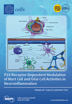
Localisation of MCs at the abluminal side of blood vessels in the brain favours their interaction with glial cells, such as microglia, astrocytes, and oligodendrocytes. In neuropathological conditions, such as Alzheimer’s disease, increased concentrations of ATP will be released into the extracellular space of the brain by resident cells, activating P2X7 on MCs. This leads to the release of mediators such as histamine, tryptase, IL-6, IL-13, TNF-α and IL-33, affecting adjacent glial cells and influencing their function. Conversely, activation of P2X7 on glial cells results in the release of IL-33, TNF-α and IL-6, which can influence MC behaviour. There is therefore a potential that BBB-permeable P2X7 antagonists could ameliorate ATP-driven pathological neuroinflammation and the detrimental consequences on glial cells. View this paper
  • Issues are regarded as officially published after their release is announced to the table of contents alert mailing list.
  • You may sign up for e-mail alerts to receive table of contents of newly released issues.
  • PDF is the official format for papers published in both, html and pdf forms. To view the papers in pdf format, click on the "PDF Full-text" link, and use the free Adobe Reader to open them.
Order results
Result details
Section
Select all
Export citation of selected articles as:
44 pages, 4935 KiB  
Review
Beyond the X Factor: Relevance of Sex Hormones in NAFLD Pathophysiology
by Sara Della Torre
Cells 2021, 10(9), 2502; https://doi.org/10.3390/cells10092502 - 21 Sep 2021
Cited by 30 | Viewed by 3981
Abstract
Non-alcoholic fatty liver disease (NAFLD) is a major health issue worldwide, being frequently associated with obesity, unbalanced dietary regimens, and reduced physical activity. Despite their greater adiposity and reduced physical activity, women show a lower risk of developing NAFLD in comparison to men, [...] Read more.
Non-alcoholic fatty liver disease (NAFLD) is a major health issue worldwide, being frequently associated with obesity, unbalanced dietary regimens, and reduced physical activity. Despite their greater adiposity and reduced physical activity, women show a lower risk of developing NAFLD in comparison to men, likely a consequence of a sex-specific regulation of liver metabolism. In the liver, sex differences in the uptake, synthesis, oxidation, deposition, and mobilization of lipids, as well as in the regulation of inflammation, are associated with differences in NAFLD prevalence and progression between men and women. Given the major role of sex hormones in driving hepatic sexual dimorphism, this review will focus on the role of sex hormones and their signaling in the regulation of hepatic metabolism and in the molecular mechanisms triggering NAFLD development and progression. Full article
(This article belongs to the Special Issue Cellular and Molecular Mechanisms Governing Liver Pathophysiology)
Show Figures

Figure 1

21 pages, 2955 KiB  
Review
Are Interactions between Epicardial Adipose Tissue, Cardiac Fibroblasts and Cardiac Myocytes Instrumental in Atrial Fibrosis and Atrial Fibrillation?
by Anirudh Krishnan, Emily Chilton, Jaishankar Raman, Pankaj Saxena, Craig McFarlane, Alexandra F. Trollope, Robert Kinobe and Lisa Chilton
Cells 2021, 10(9), 2501; https://doi.org/10.3390/cells10092501 - 21 Sep 2021
Cited by 27 | Viewed by 4468
Abstract
Atrial fibrillation is very common among the elderly and/or obese. While myocardial fibrosis is associated with atrial fibrillation, the exact mechanisms within atrial myocytes and surrounding non-myocytes are not fully understood. This review considers the potential roles of myocardial fibroblasts and myofibroblasts in [...] Read more.
Atrial fibrillation is very common among the elderly and/or obese. While myocardial fibrosis is associated with atrial fibrillation, the exact mechanisms within atrial myocytes and surrounding non-myocytes are not fully understood. This review considers the potential roles of myocardial fibroblasts and myofibroblasts in fibrosis and modulating myocyte electrophysiology through electrotonic interactions. Coupling with (myo)fibroblasts in vitro and in silico prolonged myocyte action potential duration and caused resting depolarization; an optogenetic study has verified in vivo that fibroblasts depolarized when coupled myocytes produced action potentials. This review also introduces another non-myocyte which may modulate both myocardial (myo)fibroblasts and myocytes: epicardial adipose tissue. Epicardial adipocytes are in intimate contact with myocytes and (myo)fibroblasts and may infiltrate the myocardium. Adipocytes secrete numerous adipokines which modulate (myo)fibroblast and myocyte physiology. These adipokines are protective in healthy hearts, preventing inflammation and fibrosis. However, adipokines secreted from adipocytes may switch to pro-inflammatory and pro-fibrotic, associated with reactive oxygen species generation. Pro-fibrotic adipokines stimulate myofibroblast differentiation, causing pronounced fibrosis in the epicardial adipose tissue and the myocardium. Adipose tissue also influences myocyte electrophysiology, via the adipokines and/or through electrotonic interactions. Deeper understanding of the interactions between myocytes and non-myocytes is important to understand and manage atrial fibrillation. Full article
(This article belongs to the Special Issue Cardiac Fibroblasts, Fibrosis and Cardiovascular Disease)
Show Figures

Figure 1

11 pages, 1876 KiB  
Article
Interleukin-26 Has Synergistic Catabolic Effects with Palmitate in Human Articular Chondrocytes via the TLR4-ERK1/2-c-Jun Signaling Pathway
by Yi-Ting Chen, Chih-Chien Wang, Chia-Pi Cheng, Feng-Cheng Liu, Chian-Her Lee, Herng-Sheng Lee and Yi-Jen Peng
Cells 2021, 10(9), 2500; https://doi.org/10.3390/cells10092500 - 21 Sep 2021
Cited by 4 | Viewed by 2402
Abstract
The inflammatory cytokine interleukin-26 (IL-26) is highly expressed in the serum and synovial fluid of patients with inflammatory arthritis. The effect of IL-26 on human articular chondrocytes (HACs) remains unclear. Obesity is associated with disability of patients with rheumatoid arthritis and disease activity [...] Read more.
The inflammatory cytokine interleukin-26 (IL-26) is highly expressed in the serum and synovial fluid of patients with inflammatory arthritis. The effect of IL-26 on human articular chondrocytes (HACs) remains unclear. Obesity is associated with disability of patients with rheumatoid arthritis and disease activity in those with ankylosing spondylitis. The saturated free fatty acid palmitate with IL-1β can synergistically induce catabolic effects in HACs. The aim of this study was to evaluate the effects of IL-26 and palmitate in HACs. In this study, palmitate markedly synergizes the IL-26-induced proinflammatory effects and matrix protease, including COX-2, IL-6, and MMP-1, in HACs via the toll-like receptor 4 (TLR4)-ERK1/2-c-Jun signal transduction pathway. The synergistic catabolic effects of palmitate and IL-26 were attenuated by inhibitors of TLR4 (TAK242), ERK1/2 (U0126), or c-Jun (SP600125) in HACs and cartilage matrix. In addition, metformin, a potential inhibitor of TLR4, also decreased expression of COX-2 and IL-6 induced by co-incubation with IL-26 and palmitate. IL-26 and palmitate synergistically induced expression of inflammatory and catabolic mediators, resulting in articular cartilage matrix breakdown. The present study also revealed a possible mechanism and therapeutic targets against articular cartilage degradation by increased saturated fatty acids in patients with inflammatory arthritis. Full article
(This article belongs to the Special Issue Innate Immune System in Immune-Mediated Inflammatory Diseases (IMIDs))
Show Figures

Figure 1

14 pages, 2701 KiB  
Article
Isolation and Characterization of Tissue Resident CD29-Positive Progenitor Cells in Livestock to Generate a Three-Dimensional Meat Bud
by Yuna Naraoka, Yo Mabuchi, Yosuke Yoneyama, Eriko Grace Suto, Daisuke Hisamatsu, Mami Ikeda, Risa Ito, Tetsuya Nakamura, Takanori Takebe and Chihiro Akazawa
Cells 2021, 10(9), 2499; https://doi.org/10.3390/cells10092499 - 21 Sep 2021
Cited by 8 | Viewed by 5428
Abstract
The current process of meat production using livestock has significant effects on the global environment, including high emissions of greenhouse gases. In recent years, cultured meat has attracted attention as a way to acquire animal proteins. However, the lack of markers that isolate [...] Read more.
The current process of meat production using livestock has significant effects on the global environment, including high emissions of greenhouse gases. In recent years, cultured meat has attracted attention as a way to acquire animal proteins. However, the lack of markers that isolate proliferating cells from bovine tissues and the complex structure of the meat make it difficult to culture meat in a dish. In this study, we screened 246 cell-surface antibodies by fluorescence-activated cell sorting for their capacity to form colonies and their suitability to construct spheroid “meat buds”. CD29+ cells (Ha2/5 clone) have a high potency to form colonies and efficiently proliferate on fibronectin-coated dishes. Furthermore, the meat buds created from CD29+ cells could differentiate into muscle and adipose cells in a three-dimensional structure. The meat buds embedded in the collagen gel proliferated in the matrix and formed large aggregates. Approximately 10 trillion cells can theoretically be obtained from 100 g of bovine tissue by culturing and amplifying them using these methods. The CD29+ cell characteristics of bovine tissue provide insights into the production of meat alternatives in vitro. Full article
(This article belongs to the Collection Signaling Pathways in Cell Generation and Reprogramming)
Show Figures

Figure 1

15 pages, 963 KiB  
Article
Response to Cysteamine in Osteoclasts Obtained from Patients with Nephropathic Cystinosis: A Genotype/Phenotype Correlation
by Thomas Quinaux, Aurélia Bertholet-Thomas, Aude Servais, Olivia Boyer, Isabelle Vrillon, Julien Hogan, Sandrine Lemoine, Ségolène Gaillard, Candide Alioli, Sophie Vasseur, Cécile Acquaviva, Olivier Peyruchaud, Irma Machuca-Gayet and Justine Bacchetta
Cells 2021, 10(9), 2498; https://doi.org/10.3390/cells10092498 - 21 Sep 2021
Cited by 4 | Viewed by 2065
Abstract
Bone complications of cystinosis have been recently described. The main objectives of this paper were to determine in vitro the impact of CTNS mutations and cysteamine therapy on human osteoclasts and to carry out a genotype-phenotype analysis related to osteoclastic differentiation. Human osteoclasts [...] Read more.
Bone complications of cystinosis have been recently described. The main objectives of this paper were to determine in vitro the impact of CTNS mutations and cysteamine therapy on human osteoclasts and to carry out a genotype-phenotype analysis related to osteoclastic differentiation. Human osteoclasts were differentiated from peripheral blood mononuclear cells (PBMCs) and were treated with increasing doses of cysteamine (0, 50, 200 µM) and then assessed for osteoclastic differentiation. Results are presented as median (min-max). A total of 17 patients (mainly pediatric) were included, at a median age of 14 (2–61) years, and a eGFR of 64 (23–149) mL/min/1.73 m2. Most patients (71%) were under conservative kidney management (CKM). The others were kidney transplant recipients. Three functional groups were distinguished for CTNS mutations: cystinosin variant with residual cystin efflux activity (RA, residual activity), inactive cystinosin variant (IP, inactive protein), and absent protein (AP). PBMCs from patients with residual cystinosin activity generate significantly less osteoclasts than those obtained from patients of the other groups. In all groups, cysteamine exerts an inhibitory effect on osteoclastic differentiation at high doses. This study highlights a link between genotype and osteoclastic differentiation, as well as a significant impact of cysteamine therapy on this process in humans. Full article
(This article belongs to the Special Issue Cellular and Molecular Mechanisms of Nephropathic Cystinosis)
Show Figures

Graphical abstract

18 pages, 1735 KiB  
Review
Autophagy Dysregulation in Diabetic Kidney Disease: From Pathophysiology to Pharmacological Interventions
by Claudio D. Gonzalez, María Paula Carro Negueruela, Catalina Nicora Santamarina, Roxana Resnik and Maria I. Vaccaro
Cells 2021, 10(9), 2497; https://doi.org/10.3390/cells10092497 - 21 Sep 2021
Cited by 24 | Viewed by 3440
Abstract
Diabetic kidney disease (DKD) is a frequent, potentially devastating complication of diabetes mellitus. Several factors are involved in its pathophysiology. At a cellular level, diabetic kidney disease is associated with many structural and functional alterations. Autophagy is a cellular mechanism that transports intracytoplasmic [...] Read more.
Diabetic kidney disease (DKD) is a frequent, potentially devastating complication of diabetes mellitus. Several factors are involved in its pathophysiology. At a cellular level, diabetic kidney disease is associated with many structural and functional alterations. Autophagy is a cellular mechanism that transports intracytoplasmic components to lysosomes to preserve cellular function and homeostasis. Autophagy integrity is essential for cell homeostasis, its alteration can drive to cell damage or death. Diabetic kidney disease is associated with profound autophagy dysregulation. Autophagy rate and flux alterations were described in several models of diabetic kidney disease. Some of them are closely linked with disease progression and severity. Some antidiabetic agents have shown significant effects on autophagy. A few of them have also demonstrated to modify disease progression and improved outcomes in affected patients. Other drugs also target autophagy and are being explored for clinical use in patients with diabetic kidney disease. The modulation of autophagy could be relevant for the pharmacological treatment and prevention of this disease in the future. Therefore, this is an evolving area that requires further experimental and clinical research. Here we discuss the relationship between autophagy and Diabetic kidney disease and the potential value of autophagy modulation as a target for pharmacological intervention. Full article
(This article belongs to the Special Issue 10th Anniversary of Cells—Advances in Autophagy)
Show Figures

Figure 1

20 pages, 24119 KiB  
Review
Cell Sources for Cartilage Repair—Biological and Clinical Perspective
by Inga Urlić and Alan Ivković
Cells 2021, 10(9), 2496; https://doi.org/10.3390/cells10092496 - 21 Sep 2021
Cited by 37 | Viewed by 4103
Abstract
Cell-based therapy represents a promising treatment strategy for cartilage defects. Alone or in combination with scaffolds/biological signals, these strategies open many new avenues for cartilage tissue engineering. However, the choice of the optimal cell source is not that straightforward. Currently, various types of [...] Read more.
Cell-based therapy represents a promising treatment strategy for cartilage defects. Alone or in combination with scaffolds/biological signals, these strategies open many new avenues for cartilage tissue engineering. However, the choice of the optimal cell source is not that straightforward. Currently, various types of differentiated cells (articular and nasal chondrocytes) and stem cells (mesenchymal stem cells, induced pluripotent stem cells) are being researched to objectively assess their merits and disadvantages with respect to the ability to repair damaged articular cartilage. In this paper, we focus on the different cell types used in cartilage treatment, first from a biological scientist’s perspective and then from a clinician’s standpoint. We compare and analyze the advantages and disadvantages of these cell types and offer a potential outlook for future research and clinical application. Full article
(This article belongs to the Special Issue Cell Therapies in Orthopaedics)
Show Figures

Figure 1

12 pages, 1499 KiB  
Article
Glucagon Potentiates Insulin Secretion Via β-Cell GCGR at Physiological Concentrations of Glucose
by Yulin Zhang, Chengsheng Han, Wenzhen Zhu, Guoyi Yang, Xiaohong Peng, Sohum Mehta, Jin Zhang, Liangyi Chen and Yanmei Liu
Cells 2021, 10(9), 2495; https://doi.org/10.3390/cells10092495 - 21 Sep 2021
Cited by 17 | Viewed by 3738
Abstract
Incretin-potentiated glucose-stimulated insulin secretion (GSIS) is critical to maintaining euglycemia, of which GLP-1 receptor (GLP-1R) on β-cells plays an indispensable role. Recently, α-cell-derived glucagon but not intestine-derived GLP-1 has been proposed as the critical hormone that potentiates GSIS via GLP-1R. However, the function [...] Read more.
Incretin-potentiated glucose-stimulated insulin secretion (GSIS) is critical to maintaining euglycemia, of which GLP-1 receptor (GLP-1R) on β-cells plays an indispensable role. Recently, α-cell-derived glucagon but not intestine-derived GLP-1 has been proposed as the critical hormone that potentiates GSIS via GLP-1R. However, the function of glucagon receptors (GCGR) on β-cells remains elusive. Here, using GCGR or GLP-1R antagonists, in combination with glucagon, to treat single β-cells, α-β cell clusters and isolated islets, we found that glucagon potentiates insulin secretion via β-cell GCGR at physiological but not high concentrations of glucose. Furthermore, we transfected primary mouse β-cells with RAB-ICUE (a genetically encoded cAMP fluorescence indicator) to monitor cAMP level after glucose stimulation and GCGR activation. Using specific inhibitors of different adenylyl cyclase (AC) family members, we revealed that high glucose concentration or GCGR activation independently evoked cAMP elevation via AC5 in β-cells, thus high glucose stimulation bypassed GCGR in promoting insulin secretion. Additionally, we generated β-cell-specific GCGR knockout mice which glucose intolerance was more severe when fed a high-fat diet (HFD). We further found that β-cell GCGR activation promoted GSIS more than GLP-1R in HFD, indicating the critical role of GCGR in maintaining glucose homeostasis during nutrient overload. Full article
(This article belongs to the Section Cell Signaling)
Show Figures

Figure 1

24 pages, 2822 KiB  
Article
Metabolomic Fingerprint of Mecp2-Deficient Mouse Cortex: Evidence for a Pronounced Multi-Facetted Metabolic Component in Rett Syndrome
by Gocha Golubiani, Vincenzo Lagani, Revaz Solomonia and Michael Müller
Cells 2021, 10(9), 2494; https://doi.org/10.3390/cells10092494 - 21 Sep 2021
Cited by 11 | Viewed by 2610
Abstract
Using unsupervised metabolomics, we defined the complex metabolic conditions in the cortex of a mouse model of Rett syndrome (RTT). RTT, which represents a cause of mental and cognitive disabilities in females, results in profound cognitive impairment with autistic features, motor disabilities, seizures, [...] Read more.
Using unsupervised metabolomics, we defined the complex metabolic conditions in the cortex of a mouse model of Rett syndrome (RTT). RTT, which represents a cause of mental and cognitive disabilities in females, results in profound cognitive impairment with autistic features, motor disabilities, seizures, gastrointestinal problems, and cardiorespiratory irregularities. Typical RTT originates from mutations in the X-chromosomal methyl-CpG-binding-protein-2 (Mecp2) gene, which encodes a transcriptional modulator. It then causes a deregulation of several target genes and metabolic alterations in the nervous system and peripheral organs. We identified 101 significantly deregulated metabolites in the Mecp2-deficient cortex of adult male mice; 68 were increased and 33 were decreased compared to wildtypes. Pathway analysis identified 31 mostly upregulated metabolic pathways, in particular carbohydrate and amino acid metabolism, key metabolic mitochondrial/extramitochondrial pathways, and lipid metabolism. In contrast, neurotransmitter-signaling is dampened. This metabolic fingerprint of the Mecp2-deficient cortex of severely symptomatic mice provides further mechanistic insights into the complex RTT pathogenesis. The deregulated pathways that were identified—in particular the markedly affected amino acid and carbohydrate metabolism—confirm a complex and multifaceted metabolic component in RTT, which in turn signifies putative therapeutic targets. Furthermore, the deregulated key metabolites provide a choice of potential biomarkers for a more detailed rating of disease severity and disease progression. Full article
(This article belongs to the Special Issue Molecular and Cellular Mechanisms of Rare Diseases)
Show Figures

Figure 1

13 pages, 1922 KiB  
Article
Formation of Non-Nucleoplasmic Proteasome Foci during the Late Stage of Hyperosmotic Stress
by Jeeyoung Lee, Ly Thi Huong Luu Le, Eunkyoung Kim and Min Jae Lee
Cells 2021, 10(9), 2493; https://doi.org/10.3390/cells10092493 - 21 Sep 2021
Cited by 6 | Viewed by 2624
Abstract
Cellular stress induces the formation of membraneless protein condensates in both the nucleus and cytoplasm. The nucleocytoplasmic transport of proteins mainly occurs through nuclear pore complexes (NPCs), whose efficiency is affected by various stress conditions. Here, we report that hyperosmotic stress compartmentalizes nuclear [...] Read more.
Cellular stress induces the formation of membraneless protein condensates in both the nucleus and cytoplasm. The nucleocytoplasmic transport of proteins mainly occurs through nuclear pore complexes (NPCs), whose efficiency is affected by various stress conditions. Here, we report that hyperosmotic stress compartmentalizes nuclear 26S proteasomes into dense nuclear foci, independent of signaling cascades. Most of the proteasome foci were detected between the condensed chromatin mass and inner nuclear membrane. The proteasome-positive puncta were not colocalized with other types of nuclear bodies and were reversibly dispersed when cells were returned to the isotonic medium. The structural integrity of 26S proteasomes in the nucleus was slightly affected under the hyperosmotic condition. We also found that these insulator-body-like proteasome foci were possibly formed through disrupted nucleus-to-cytosol transport, which was mediated by the sequestration of NPC components into osmostress-responding stress granules. These data suggest that phase separation in both the nucleus and cytosol may be a major cell survival mechanism during hyperosmotic stress conditions. Full article
(This article belongs to the Special Issue Role of Ubiquitin in Cellular Quality Control)
Show Figures

Graphical abstract

21 pages, 1477 KiB  
Review
Neuroprotective Effects and Therapeutic Potential of Transcorneal Electrical Stimulation for Depression
by Wing-Shan Yu, So-Hyun Kwon, Stephen Kugbere Agadagba, Leanne-Lai-Hang Chan, Kah-Hui Wong and Lee-Wei Lim
Cells 2021, 10(9), 2492; https://doi.org/10.3390/cells10092492 - 21 Sep 2021
Cited by 8 | Viewed by 3774
Abstract
Transcorneal electrical stimulation (TES) has emerged as a non-invasive neuromodulation approach that exerts neuroprotection via diverse mechanisms, including neurotrophic, neuroplastic, anti-inflammatory, anti-apoptotic, anti-glutamatergic, and vasodilation mechanisms. Although current studies of TES have mainly focused on its applications in ophthalmology, several lines of evidence [...] Read more.
Transcorneal electrical stimulation (TES) has emerged as a non-invasive neuromodulation approach that exerts neuroprotection via diverse mechanisms, including neurotrophic, neuroplastic, anti-inflammatory, anti-apoptotic, anti-glutamatergic, and vasodilation mechanisms. Although current studies of TES have mainly focused on its applications in ophthalmology, several lines of evidence point towards its putative use in treating depression. Apart from stimulating visual-related structures and promoting visual restoration, TES has also been shown to activate brain regions that are involved in mood alterations and can induce antidepressant-like behaviour in animals. The beneficial effects of TES in depression were further supported by its shared mechanisms with FDA-approved antidepressant treatments, including its neuroprotective properties against apoptosis and inflammation, and its ability to enhance the neurotrophic expression. This article critically reviews the current findings on the neuroprotective effects of TES and provides evidence to support our hypothesis that TES possesses antidepressant effects. Full article
(This article belongs to the Section Cells of the Nervous System)
Show Figures

Figure 1

17 pages, 3446 KiB  
Article
Secreted Frizzled-Related Protein 1 Promotes Odontoblastic Differentiation and Reparative Dentin Formation in Dental Pulp Cells
by Keita Ipposhi, Atsushi Tomokiyo, Taiga Ono, Kozue Yamashita, Muhammad Anas Alhasan, Daigaku Hasegawa, Sayuri Hamano, Shinichiro Yoshida, Hideki Sugii, Tomohiro Itoyama, Marina Ogawa and Hidefumi Maeda
Cells 2021, 10(9), 2491; https://doi.org/10.3390/cells10092491 - 21 Sep 2021
Cited by 5 | Viewed by 2927
Abstract
Direct pulp capping is an effective treatment for preserving dental pulp against carious or traumatic pulp exposure via the formation of protective reparative dentin by odontoblast-like cells. Reparative dentin formation can be stimulated by several signaling molecules; therefore, we investigated the effects of [...] Read more.
Direct pulp capping is an effective treatment for preserving dental pulp against carious or traumatic pulp exposure via the formation of protective reparative dentin by odontoblast-like cells. Reparative dentin formation can be stimulated by several signaling molecules; therefore, we investigated the effects of secreted frizzled-related protein (SFRP) 1 that was reported to be strongly expressed in odontoblasts of newborn molar tooth germs on odontoblastic differentiation and reparative dentin formation. In developing rat incisors, cells in the dental pulp, cervical loop, and inner enamel epithelium, as well as ameloblasts and preodontoblasts, weakly expressed Sfrp1; however, Sfrp1 was strongly expressed in mature odontoblasts. Human dental pulp cells (hDPCs) showed stronger expression of SFRP1 compared with periodontal ligament cells and gingival cells. SFRP1 knockdown in hDPCs abolished calcium chloride-induced mineralized nodule formation and odontoblast-related gene expression and decreased BMP-2 gene expression. Conversely, SFRP1 stimulation enhanced nodule formation and expression of BMP-2. Direct pulp capping treatment with SFRP1 induced the formation of a considerable amount of reparative dentin that has a structure similar to primary dentin. Our results indicate that SFRP1 is crucial for dentinogenesis and is important in promoting reparative dentin formation in response to injury. Full article
(This article belongs to the Special Issue Dental Pulp Stem Cells and Regenerative Medicine)
Show Figures

Figure 1

17 pages, 3274 KiB  
Article
Assessment of Nitrate Removal Capacity of Two Selected Eukaryotic Green Microalgae
by Vaishali Rani and Gergely Maróti
Cells 2021, 10(9), 2490; https://doi.org/10.3390/cells10092490 - 20 Sep 2021
Cited by 8 | Viewed by 2759
Abstract
Eutrophication is a leading problem in water bodies all around the world in which nitrate is one of the major contributors. The present study was conducted to study the effects of various concentrations of nitrate on two eukaryotic green microalgae, Chlamydomonas sp. MACC-216 [...] Read more.
Eutrophication is a leading problem in water bodies all around the world in which nitrate is one of the major contributors. The present study was conducted to study the effects of various concentrations of nitrate on two eukaryotic green microalgae, Chlamydomonas sp. MACC-216 and Chlorella sp. MACC-360. For this purpose, both microalgae were grown in a modified tris-acetate-phosphate medium (TAP-M) with three different concentrations of sodium nitrate, i.e., 5 mM (TAP-M5), 10 mM (TAP-M10) and 15 mM (TAP-M15), for 6 days and it was observed that both microalgae were able to remove nitrate completely from the TAP-M5 medium. Total amount of pigments decreased with the increasing concentration of nitrate, whereas protein and carbohydrate contents remained unaffected. High nitrate concentration (15 mM) led to an increase in lipids in Chlamydomonas sp. MACC-216, but not in Chlorella sp. MACC-360. Furthermore, Chlamydomonas sp. MACC-216 and Chlorella sp. MACC-360 were cultivated for 6 days in synthetic wastewater (SWW) with varying concentrations of nitrate where both microalgae grew well and showed an adequate nitrate removal capacity. Full article
(This article belongs to the Collection Feature Papers in Plant, Algae and Fungi Cell Biology)
Show Figures

Graphical abstract

42 pages, 3369 KiB  
Review
Inducible Pluripotent Stem Cells to Model and Treat Inherited Degenerative Diseases of the Outer Retina: 3D-Organoids Limitations and Bioengineering Solutions
by Massimiliano Andreazzoli, Ivana Barravecchia, Chiara De Cesari, Debora Angeloni and Gian Carlo Demontis
Cells 2021, 10(9), 2489; https://doi.org/10.3390/cells10092489 - 20 Sep 2021
Cited by 6 | Viewed by 3793
Abstract
Inherited retinal degenerations (IRD) affecting either photoreceptors or pigment epithelial cells cause progressive visual loss and severe disability, up to complete blindness. Retinal organoids (ROs) technologies opened up the development of human inducible pluripotent stem cells (hiPSC) for disease modeling and replacement therapies. [...] Read more.
Inherited retinal degenerations (IRD) affecting either photoreceptors or pigment epithelial cells cause progressive visual loss and severe disability, up to complete blindness. Retinal organoids (ROs) technologies opened up the development of human inducible pluripotent stem cells (hiPSC) for disease modeling and replacement therapies. However, hiPSC-derived ROs applications to IRD presently display limited maturation and functionality, with most photoreceptors lacking well-developed outer segments (OS) and light responsiveness comparable to their adult retinal counterparts. In this review, we address for the first time the microenvironment where OS mature, i.e., the subretinal space (SRS), and discuss SRS role in photoreceptors metabolic reprogramming required for OS generation. We also address bioengineering issues to improve culture systems proficiency to promote OS maturation in hiPSC-derived ROs. This issue is crucial, as satisfying the demanding metabolic needs of photoreceptors may unleash hiPSC-derived ROs full potential for disease modeling, drug development, and replacement therapies. Full article
(This article belongs to the Special Issue Neural Stem Cell Systems to Study Brain Development and Diseases)
Show Figures

Graphical abstract

15 pages, 3574 KiB  
Article
Statins Decrease Programmed Death-Ligand 1 (PD-L1) by Inhibiting AKT and β-Catenin Signaling
by Woo-Jin Lim, Mingyu Lee, Yerin Oh, Xue-Quan Fang, Sujin Lee, Chang-Hoon Lim, Jooho Park and Ji-Hong Lim
Cells 2021, 10(9), 2488; https://doi.org/10.3390/cells10092488 - 20 Sep 2021
Cited by 21 | Viewed by 4440
Abstract
Retrospective observational studies have reported that statins improve clinical outcomes in patients previously treated with programmed cell death protein 1 (PD-1)-targeting monoclonal antibodies for malignant pleural mesothelioma (MPM) and advanced non-small cell lung cancer (NSCLC). In multiple mouse cancer models, de novo synthesis [...] Read more.
Retrospective observational studies have reported that statins improve clinical outcomes in patients previously treated with programmed cell death protein 1 (PD-1)-targeting monoclonal antibodies for malignant pleural mesothelioma (MPM) and advanced non-small cell lung cancer (NSCLC). In multiple mouse cancer models, de novo synthesis of mevalonate and cholesterol inhibitors was found to synergize with anti-PD-1 antibody therapy. In the present study, we investigated whether statins affect programmed death-ligand 1 (PD-L1) expression in cancer cells. Four statins, namely simvastatin, atorvastatin, lovastatin, and fluvastatin, decreased PD-L1 expression in melanoma and lung cancer cells. In addition, we found that AKT and β-catenin signaling involved PD-L1 suppression by statins. Our cellular and molecular studies provide inspiring evidence for extending the clinical evaluation of statins for use in combination with immune checkpoint inhibitor-based cancer therapy. Full article
Show Figures

Figure 1

15 pages, 7393 KiB  
Article
Synapsin-Promoted Caveolin-1 Overexpression Maintains Mitochondrial Morphology and Function in PSAPP Alzheimer’s Disease Mice
by Shanshan Wang, Taiga Ichinomiya, Yuki Terada, Dongsheng Wang, Hemal H. Patel and Brian P. Head
Cells 2021, 10(9), 2487; https://doi.org/10.3390/cells10092487 - 20 Sep 2021
Cited by 15 | Viewed by 3078
Abstract
Mitochondrial dysfunction plays a pivotal role in the Alzheimer’s Disease (AD) pathology. Disrupted mitochondrial dynamics (i.e., fusion/fission balance), which are essential for normal mitochondria structure and function, are documented in AD. Caveolin-1 (Cav-1), a membrane/lipid raft (MLR) scaffolding protein regulates metabolic pathways in [...] Read more.
Mitochondrial dysfunction plays a pivotal role in the Alzheimer’s Disease (AD) pathology. Disrupted mitochondrial dynamics (i.e., fusion/fission balance), which are essential for normal mitochondria structure and function, are documented in AD. Caveolin-1 (Cav-1), a membrane/lipid raft (MLR) scaffolding protein regulates metabolic pathways in several different cell types such as hepatocytes and cancer cells. Previously, we have shown decreased expression of Cav-1 in the hippocampus of 9-month (m) old PSAPP mice, while hippocampal overexpression of neuron-targeted Cav-1 using the synapsin promoter (i.e., SynCav1) preserved cognitive function, neuronal morphology, and synaptic ultrastructure in 9 and 12 m PSAPP mice. Considering the central role of energy production in maintaining normal neuronal and synaptic function and survival, the present study reveals that PSAPP mice exhibit disrupted mitochondrial distribution, morphometry, and respiration. In contrast, SynCav1 mitigates mitochondrial damage and loss and enhances mitochondrial respiration. Furthermore, by examining mitochondrial dynamics, we found that PSAPP mice showed a significant increase in the phosphorylation of mitochondrial dynamin-related GTPase protein (DRP1), resulting in excessive mitochondria fragmentation and dysfunction. In contrast, hippocampal delivery of SynCav1 significantly decreased p-DRP1 and augmented the level of the mitochondrial fusion protein, mitofusin1 (Mfn1) in PSAPP mice, a molecular event, which may mechanistically explain for the preserved balance of mitochondria fission/fusion and metabolic resilience in 12 m PSAPP-SynCav1 mice. Our data demonstrate the critical role for Cav-1 in maintaining normal mitochondrial morphology and function through affecting mitochondrial dynamics and explain a molecular and cellular mechanism underlying the previously reported neuroprotective and cognitive preservation induced by SynCav1 in PSAPP mouse model of AD. Full article
(This article belongs to the Section Mitochondria)
Show Figures

Graphical abstract

48 pages, 4830 KiB  
Review
Leveling Up the Controversial Role of Neutrophils in Cancer: When the Complexity Becomes Entangled
by Ronit Vogt Sionov
Cells 2021, 10(9), 2486; https://doi.org/10.3390/cells10092486 - 20 Sep 2021
Cited by 17 | Viewed by 4767
Abstract
Neutrophils are the most abundant immune cell in the circulation of human and act as gatekeepers to discard foreign elements that have entered the body. They are essential in initiating immune responses for eliminating invaders, such as microorganisms and alien particles, as well [...] Read more.
Neutrophils are the most abundant immune cell in the circulation of human and act as gatekeepers to discard foreign elements that have entered the body. They are essential in initiating immune responses for eliminating invaders, such as microorganisms and alien particles, as well as to act as immune surveyors of cancer cells, especially during the initial stages of carcinogenesis and for eliminating single metastatic cells in the circulation and in the premetastatic organs. Since neutrophils can secrete a whole range of factors stored in their many granules as well as produce reactive oxygen and nitrogen species upon stimulation, neutrophils may directly or indirectly affect carcinogenesis in both the positive and negative directions. An intricate crosstalk between tumor cells, neutrophils, other immune cells and stromal cells in the microenvironment modulates neutrophil function resulting in both anti- and pro-tumor activities. Both the anti-tumor and pro-tumor activities require chemoattraction towards the tumor cells, neutrophil activation and ROS production. Divergence is seen in other neutrophil properties, including differential secretory repertoire and membrane receptor display. Many of the direct effects of neutrophils on tumor growth and metastases are dependent on tight neutrophil–tumor cell interactions. Among them, the neutrophil Mac-1 interaction with tumor ICAM-1 and the neutrophil L-selectin interaction with tumor-cell sialomucins were found to be involved in the neutrophil-mediated capturing of circulating tumor cells resulting in increased metastatic seeding. On the other hand, the anti-tumor function of neutrophils was found to rely on the interaction between tumor-surface-expressed receptor for advanced glycation end products (RAGE) and Cathepsin G expressed on the neutrophil surface. Intriguingly, these two molecules are also involved in the promotion of tumor growth and metastases. RAGE is upregulated during early inflammation-induced carcinogenesis and was found to be important for sustaining tumor growth and homing at metastatic sites. Cathepsin G was found to be essential for neutrophil-supported lung colonization of cancer cells. These data level up the complexity of the dual role of neutrophils in cancer. Full article
(This article belongs to the Special Issue The Role of Neutrophils in Cancer)
Show Figures

Figure 1

18 pages, 3898 KiB  
Article
The Presence and Potential Role of ALDH1A2 in the Glioblastoma Microenvironment
by Stephanie Sanders, Denise M. Herpai, Analiz Rodriguez, Yue Huang, Jeff Chou, Fang-Chi Hsu, Darren Seals, Ryan Mott, Lance D. Miller and Waldemar Debinski
Cells 2021, 10(9), 2485; https://doi.org/10.3390/cells10092485 - 20 Sep 2021
Cited by 6 | Viewed by 3111
Abstract
Glioblastoma (GBM) is the most aggressive malignant glioma. Therapeutic targeting of GBM is made more difficult due to its heterogeneity, resistance to treatment, and diffuse infiltration into the brain parenchyma. Better understanding of the tumor microenvironment should aid in finding more effective management [...] Read more.
Glioblastoma (GBM) is the most aggressive malignant glioma. Therapeutic targeting of GBM is made more difficult due to its heterogeneity, resistance to treatment, and diffuse infiltration into the brain parenchyma. Better understanding of the tumor microenvironment should aid in finding more effective management of GBM. GBM-associated macrophages (GAM) comprise up to 30% of the GBM microenvironment. Therefore, exploration of GAM activity/function and their specific markers are important for developing new therapeutic agents. In this study, we identified and evaluated the expression of ALDH1A2 in the GBM microenvironment, and especially in M2 GAM, though it is also expressed in reactive astrocytes and multinucleated tumor cells. We demonstrated that M2 GAM highly express ALDH1A2 when compared to other ALDH1 family proteins. Additionally, GBM samples showed higher expression of ALDH1A2 when compared to low-grade gliomas (LGG), and this expression was increased upon tumor recurrence both at the gene and protein levels. We demonstrated that the enzymatic product of ALDH1A2, retinoic acid (RA), modulated the expression and activity of MMP-2 and MMP-9 in macrophages, but not in GBM tumor cells. Thus, the expression of ALDH1A2 may promote the progressive phenotype of GBM. Full article
(This article belongs to the Special Issue Immune Microenvironment of Gliomas)
Show Figures

Figure 1

17 pages, 626 KiB  
Review
The Role of Mitochondria in Oocyte Maturation
by Anastasia Kirillova, Johan E. J. Smitz, Gennady T. Sukhikh and Ilya Mazunin
Cells 2021, 10(9), 2484; https://doi.org/10.3390/cells10092484 - 19 Sep 2021
Cited by 100 | Viewed by 10274
Abstract
With the nucleus as an exception, mitochondria are the only animal cell organelles containing their own genetic information, called mitochondrial DNA (mtDNA). During oocyte maturation, the mtDNA copy number dramatically increases and the distribution of mitochondria changes significantly. As oocyte maturation requires a [...] Read more.
With the nucleus as an exception, mitochondria are the only animal cell organelles containing their own genetic information, called mitochondrial DNA (mtDNA). During oocyte maturation, the mtDNA copy number dramatically increases and the distribution of mitochondria changes significantly. As oocyte maturation requires a large amount of ATP for continuous transcription and translation, the availability of the right number of functional mitochondria is crucial. There is a correlation between the quality of oocytes and both the amount of mtDNA and the amount of ATP. Suboptimal conditions of in vitro maturation (IVM) might lead to changes in the mitochondrial morphology as well as alternations in the expression of genes encoding proteins associated with mitochondrial function. Dysfunctional mitochondria have a lower ability to counteract reactive oxygen species (ROS) production which leads to oxidative stress. The mitochondrial function might be improved with the application of antioxidants and significant expectations are laid on the development of new IVM systems supplemented with mitochondria-targeted reagents. Different types of antioxidants have been tested already on animal models and human rescue IVM oocytes, showing promising results. This review focuses on the recent observations on oocytes’ intracellular mitochondrial distribution and on mitochondrial genomes during their maturation, both in vivo and in vitro. Recent mitochondrial supplementation studies, aiming to improve oocyte developmental potential, are summarized. Full article
(This article belongs to the Special Issue Molecular Mechanism of Oocyte Maturation)
Show Figures

Figure 1

16 pages, 2624 KiB  
Review
Potential Therapeutic Candidates for Age-Related Macular Degeneration (AMD)
by Sonali Nashine
Cells 2021, 10(9), 2483; https://doi.org/10.3390/cells10092483 - 19 Sep 2021
Cited by 28 | Viewed by 7006
Abstract
Aging contributes to the risk of development of ocular diseases including, but not limited to, Age-related Macular Degeneration (AMD) that is a leading cause of blindness in the United States as well as worldwide. Retinal aging, that contributes to AMD pathogenesis, is characterized [...] Read more.
Aging contributes to the risk of development of ocular diseases including, but not limited to, Age-related Macular Degeneration (AMD) that is a leading cause of blindness in the United States as well as worldwide. Retinal aging, that contributes to AMD pathogenesis, is characterized by accumulation of drusen deposits, alteration in the composition of Bruch’s membrane and extracellular matrix, vascular inflammation and dysregulation, mitochondrial dysfunction, and accumulation of reactive oxygen species (ROS), and subsequent retinal pigment epithelium (RPE) cell senescence. Since there are limited options available for the prophylaxis and treatment of AMD, new therapeutic interventions are constantly being looked into to identify new therapeutic targets for AMD. This review article discusses the potential candidates for AMD therapy and their known mechanisms of cytoprotection in AMD. These target therapeutic candidates include APE/REF-1, MRZ-99030, Ciliary NeuroTrophic Factor (CNTF), RAP1 GTPase, Celecoxib, and SS-31/Elamipretide. Full article
Show Figures

Figure 1

17 pages, 3376 KiB  
Article
Mitochondrial DNA Haplogroup Related to the Prevalence of Helicobacter pylori
by Yeonmi Lee, Sun-Mi Lee, Jiwan Choi, Seoon Kang, Seongjun So, Deokhoon Kim, Ji-Yong Ahn, Hwoon-Yong Jung, Jin-Yong Jeong and Eunju Kang
Cells 2021, 10(9), 2482; https://doi.org/10.3390/cells10092482 - 19 Sep 2021
Cited by 1 | Viewed by 2618
Abstract
Mitochondria are essential organelles that are not only responsible for energy production but are also involved in cell metabolism, calcium homeostasis, and apoptosis. Targeting mitochondria is a key strategy for bacteria to subvert host cells’ physiology and promote infection. Helicobacter (H.) pylori targets [...] Read more.
Mitochondria are essential organelles that are not only responsible for energy production but are also involved in cell metabolism, calcium homeostasis, and apoptosis. Targeting mitochondria is a key strategy for bacteria to subvert host cells’ physiology and promote infection. Helicobacter (H.) pylori targets mitochondria directly. However, mitochondrial genome (mtDNA) polymorphism (haplogroup) is not yet considered an important factor for H. pylori infection. Here, we clarified the association of mitochondrial haplogroups with H. pylori prevalence and the ability to perform damage. Seven mtDNA haplogroups were identified among 28 H. pylori-positive subjects. Haplogroup B was present at a higher frequency and haplotype D at a lower one in the H. pylori population than in that of the H. pylori-negative one. The fibroblasts carrying high-frequency haplogroup displayed a higher apoptotic rate and diminished mitochondrial respiration following H. pylori infection. mtDNA mutations were accumulated more in the H. pylori-positive population than in that of the H. pylori-negative one in old age. Among the mutations, 57% were located in RNA genes or nonsynonymous protein-coding regions in the H. pylori-positive population, while 35% were in the H. pylori-negative one. We concluded that gastric disease caused by Helicobacter virulence could be associated with haplogroups and mtDNA mutations. Full article
Show Figures

Figure 1

17 pages, 1987 KiB  
Article
Assessment and Validation of Globodera pallida as a Novel In Vivo Model for Studying Alzheimer’s Disease
by Norah A. Althobaiti, Farid Menaa, Aishah E. Albalawi, Johnathan J. Dalzell, Neil D. Warnock, Erin M. Mccammick, Abdulellah Alsolais, Abeer M. Alkhaibari and Brian D. Green
Cells 2021, 10(9), 2481; https://doi.org/10.3390/cells10092481 - 19 Sep 2021
Cited by 4 | Viewed by 2492
Abstract
Background: Whole transgenic or non-transgenic organism model systems allow the screening of pharmacological compounds for protective actions in Alzheimer’s disease (AD). Aim: In this study, a plant parasitic nematode, Globodera pallida, which assimilates intact peptides from the external environment, was investigated as [...] Read more.
Background: Whole transgenic or non-transgenic organism model systems allow the screening of pharmacological compounds for protective actions in Alzheimer’s disease (AD). Aim: In this study, a plant parasitic nematode, Globodera pallida, which assimilates intact peptides from the external environment, was investigated as a new potential non-transgenic model system of AD. Methods: Fresh second-stage juveniles of G. pallida were used to measure their chemosensory, perform immunocytochemistry on their neurological structures, evaluate their survival rate, measure reactive oxygen species, and determine total oxidized glutathione to reduced glutathione ratio (GSSG/GSH) levels, before and after treatment with 100 µM of various amyloid beta (Aβ) peptides (1–40, 1–42, 17–42, 17–40, 1–28, or 1–16). Wild-type N2 C. elegans (strain N2) was cultured on Nematode Growth Medium and directly used, as control, for chemosensory assays. Results: We demonstrated that: (i) G. pallida (unlike Caenorhabditis elegans) assimilates amyloid-β (Aβ) peptides which co-localise with its neurological structures; (ii) pre-treatment with various Aβ isoforms (1–40, 1–42, 17–42, 17–40, 1–28, or 1–16) impairs G. pallida’s chemotaxis to differing extents; (iii) Aβ peptides reduced survival, increased the production of ROS, and increased GSSG/GSH levels in this model; (iv) this unique model can distinguish differences between different treatment concentrations, durations, and modalities, displaying good sensitivity; (v) clinically approved neuroprotective agents were effective in protecting G. pallida from Aβ (1–42) exposure. Taken together, the data indicate that G. pallida is an interesting in vivo model with strong potential for discovery of novel bioactive compounds with anti-AD activity. Full article
(This article belongs to the Special Issue Nonmammalian Models for Neurodegenerative and Neurological Disorders)
Show Figures

Figure 1

21 pages, 4668 KiB  
Review
Muscle-Related Plectinopathies
by Michaela M. Zrelski, Monika Kustermann and Lilli Winter
Cells 2021, 10(9), 2480; https://doi.org/10.3390/cells10092480 - 19 Sep 2021
Cited by 14 | Viewed by 3497
Abstract
Plectin is a giant cytoskeletal crosslinker and intermediate filament stabilizing protein. Mutations in the human plectin gene (PLEC) cause several rare diseases that are grouped under the term plectinopathies. The most common disorder is autosomal recessive disease epidermolysis bullosa simplex with [...] Read more.
Plectin is a giant cytoskeletal crosslinker and intermediate filament stabilizing protein. Mutations in the human plectin gene (PLEC) cause several rare diseases that are grouped under the term plectinopathies. The most common disorder is autosomal recessive disease epidermolysis bullosa simplex with muscular dystrophy (EBS-MD), which is characterized by skin blistering and progressive muscle weakness. Besides EBS-MD, PLEC mutations lead to EBS with nail dystrophy, EBS-MD with a myasthenic syndrome, EBS with pyloric atresia, limb-girdle muscular dystrophy type R17, or EBS-Ogna. In this review, we focus on the clinical and pathological manifestations caused by PLEC mutations on skeletal and cardiac muscle. Skeletal muscle biopsies from EBS-MD patients and plectin-deficient mice revealed severe dystrophic features with variation in fiber size, degenerative myofibrillar changes, mitochondrial alterations, and pathological desmin-positive protein aggregates. Ultrastructurally, PLEC mutations lead to a disorganization of myofibrils and sarcomeres, Z- and I-band alterations, autophagic vacuoles and cytoplasmic bodies, and misplaced and degenerating mitochondria. We also summarize a variety of genetically manipulated mouse and cell models, which are either plectin-deficient or that specifically lack a skeletal muscle-expressed plectin isoform. These models are powerful tools to study functional and molecular consequences of PLEC defects and their downstream effects on the skeletal muscle organization. Full article
(This article belongs to the Special Issue Plectin in Health and Disease)
Show Figures

Figure 1

16 pages, 5437 KiB  
Article
Exercise and Sestrin Mediate Speed and Lysosomal Activity in Drosophila by Partially Overlapping Mechanisms
by Alyson Sujkowski and Robert Wessells
Cells 2021, 10(9), 2479; https://doi.org/10.3390/cells10092479 - 19 Sep 2021
Cited by 6 | Viewed by 2743
Abstract
Chronic exercise is widely recognized as an important contributor to healthspan in humans and in diverse animal models. Recently, we have demonstrated that Sestrins, a family of evolutionarily conserved exercise-inducible proteins, are critical mediators of exercise benefits in flies and mice. Knockout of [...] Read more.
Chronic exercise is widely recognized as an important contributor to healthspan in humans and in diverse animal models. Recently, we have demonstrated that Sestrins, a family of evolutionarily conserved exercise-inducible proteins, are critical mediators of exercise benefits in flies and mice. Knockout of Sestrins prevents exercise adaptations to endurance and flight in Drosophila, and similarly prevents benefits to endurance and metabolism in exercising mice. In contrast, overexpression of dSestrin in muscle mimics several of the molecular and physiological adaptations characteristic of endurance exercise. Here, we extend those observations to examine the impact of dSestrin on preserving speed and increasing lysosomal activity. We find that dSestrin is a critical factor driving exercise adaptations to climbing speed, but is not absolutely required for exercise to increase lysosomal activity in Drosophila. The role of Sestrin in increasing speed during chronic exercise requires both the TORC2/AKT axis and the PGC1α homolog spargel, while dSestrin requires interactions with TORC1 to cell-autonomously increase lysosomal activity. These results highlight the conserved role of Sestrins as key factors that drive diverse physiological adaptations conferred by chronic exercise. Full article
(This article belongs to the Collection Molecular Mechanisms of Exercise and Healthspan)
Show Figures

Graphical abstract

16 pages, 2507 KiB  
Article
Ang II-Induced Hypertension Exacerbates the Pathogenesis of Tuberculosis
by Soo-Na Cho, Ji-Ae Choi, Junghwan Lee, Sang-Hun Son, Seong-Ahn Lee, Tam-Doan Nguyen, Song-Yi Choi and Chang-Hwa Song
Cells 2021, 10(9), 2478; https://doi.org/10.3390/cells10092478 - 19 Sep 2021
Cited by 3 | Viewed by 2485
Abstract
It has been known that infection plays a role in the development of hypertension. However, the role of hypertension in the progression of infectious diseases remain unknown. Many countries with high rates of hypertension show geographical overlaps with those showing high incidence rates [...] Read more.
It has been known that infection plays a role in the development of hypertension. However, the role of hypertension in the progression of infectious diseases remain unknown. Many countries with high rates of hypertension show geographical overlaps with those showing high incidence rates of tuberculosis (TB). To explore the role of hypertension in tuberculosis, we compared the effects of hypertension during mycobacterial infection, we infected both hypertensive Angiotensin II (Ang II) and control mice with Mycobacterium tuberculosis (Mtb) strain H37Ra by intratracheal injection. Ang II-induced hypertension promotes cell death through both apoptosis and necrosis in Mtb H37Ra infected mouse lungs. Interestingly, we found that lipid accumulation in pulmonary tissues was significantly increased in the hypertension group compared to the normal controls. Ang II-induced hypertension increases the formation of foamy macrophages during Mtb infection and it leads to cell death. Moreover, the hypertension group showed more severe granuloma formation and fibrotic lesions in comparison with the control group. Finally, we observed that the total number of mycobacteria was increased in the lungs in the hypertension group compared to the normal controls. Taken together, these results suggest that hypertension increases intracellular survival of Mtb through formation of foamy macrophages, resulting in severe pathogenesis of TB. Full article
Show Figures

Figure 1

18 pages, 3747 KiB  
Article
Detection of Novel Potential Regulators of Stem Cell Differentiation and Cardiogenesis through Combined Genome-Wide Profiling of Protein-Coding Transcripts and microRNAs
by Rui Machado, Agapios Sachinidis and Matthias E. Futschik
Cells 2021, 10(9), 2477; https://doi.org/10.3390/cells10092477 - 18 Sep 2021
Cited by 1 | Viewed by 2511
Abstract
In vitro differentiation of embryonic stem cells (ESCs) provides a convenient basis for the study of microRNA-based gene regulation that is relevant for early cardiogenic processes. However, to which degree insights gained from in vitro differentiation models can be readily transferred to the [...] Read more.
In vitro differentiation of embryonic stem cells (ESCs) provides a convenient basis for the study of microRNA-based gene regulation that is relevant for early cardiogenic processes. However, to which degree insights gained from in vitro differentiation models can be readily transferred to the in vivo system remains unclear. In this study, we profiled simultaneous genome-wide measurements of mRNAs and microRNAs (miRNAs) of differentiating murine ESCs (mESCs) and integrated putative miRNA-gene interactions to assess miRNA-driven gene regulation. To identify interactions conserved between in vivo and in vitro, we combined our analysis with a recent transcriptomic study of early murine heart development in vivo. We detected over 200 putative miRNA–mRNA interactions with conserved expression patterns that were indicative of gene regulation across the in vitro and in vivo studies. A substantial proportion of candidate interactions have been already linked to cardiogenesis, supporting the validity of our approach. Notably, we also detected miRNAs with expression patterns that closely resembled those of key developmental transcription factors. The approach taken in this study enabled the identification of miRNA interactions in in vitro models with potential relevance for early cardiogenic development. Such comparative approaches will be important for the faithful application of stem cells in cardiovascular research. Full article
(This article belongs to the Special Issue Applications of Stem Cells in Cardiovascular Functional Genomics)
Show Figures

Figure 1

19 pages, 2687 KiB  
Article
Early Life Irradiation-Induced Hypoplasia and Impairment of Neurogenesis in the Dentate Gyrus and Adult Depression Are Mediated by MicroRNA- 34a-5p/T-Cell Intracytoplasmic Antigen-1 Pathway
by Hong Wang, Zhaowu Ma, Hongyuan Shen, Zijun Wu, Lian Liu, Boxu Ren, Peiyan Wong, Gautam Sethi and Fengru Tang
Cells 2021, 10(9), 2476; https://doi.org/10.3390/cells10092476 - 18 Sep 2021
Cited by 6 | Viewed by 2387
Abstract
Early life radiation exposure causes abnormal brain development, leading to adult depression. However, few studies have been conducted to explore pre- or post-natal irradiation-induced depression-related neuropathological changes. Relevant molecular mechanisms are also poorly understood. We induced adult depression by irradiation of mice at [...] Read more.
Early life radiation exposure causes abnormal brain development, leading to adult depression. However, few studies have been conducted to explore pre- or post-natal irradiation-induced depression-related neuropathological changes. Relevant molecular mechanisms are also poorly understood. We induced adult depression by irradiation of mice at postnatal day 3 (P3) to reveal hippocampal neuropathological changes and investigate their molecular mechanism, focusing on MicroRNA (miR) and its target mRNA and protein. P3 mice were irradiated by γ-rays with 5Gy, and euthanized at 1, 7 and 120 days after irradiation. A behavioral test was conducted before the animals were euthanized at 120 days after irradiation. The animal brains were used for different studies including immunohistochemistry, CAP-miRSeq, Real-Time Quantitative Reverse Transcription PCR (qRT-PCR) and western blotting. The interaction of miR-34a-5p and its target T-cell intracytoplasmic antigen-1 (Tia1) was confirmed by luciferase reporter assay. Overexpression of Tia1 in a neural stem cell (NSC) model was used to further validate findings from the mouse model. Irradiation with 5 Gy at P3 induced depression in adult mice. Animal hippocampal pathological changes included hypoplasia of the infrapyramidal blade of the stratum granulosum, aberrant and impaired cell division, and neurogenesis in the dentate gyrus. At the molecular level, upregulation of miR-34a-5p and downregulation of Tia1 mRNA were observed in both animal and neural stem cell models. The luciferase reporter assay and gene transfection studies further confirmed a direct interaction between miR-43a-5p and Tia1. Our results indicate that the early life γ-radiation-activated miR-43a-5p/Tia1 pathway is involved in the pathogenesis of adult depression. This novel finding may provide a new therapeutic target by inhibiting the miR-43a-5p/Tia1 pathway to prevent radiation-induced pathogenesis of depression. Full article
(This article belongs to the Special Issue Induced Impairment of Neurogenesis and Brain Diseases)
Show Figures

Figure 1

22 pages, 58094 KiB  
Article
Autophagy Promotes the Survival of Adipose Mesenchymal Stem/Stromal Cells and Enhances Their Therapeutic Effects in Cisplatin-Induced Liver Injury via Modulating TGF-β1/Smad and PI3K/AKT Signaling Pathways
by Eman Mohamad El Nashar, Mansour Abdullah Alghamdi, Wardah Abdullah Alasmari, Mohamed M. A. Hussein, Eman Hamza, Reham Ismail Taha, Mona M. Ahmed, Khulood Mohammed Al-Khater and Ahmed Abdelfattah-Hassan
Cells 2021, 10(9), 2475; https://doi.org/10.3390/cells10092475 - 18 Sep 2021
Cited by 14 | Viewed by 3542
Abstract
Autophagy is a key metabolic process where cells can recycle its proteins and organelles to regenerate its own cellular building blocks. Chemotherapy is indispensable for cancer treatment but associated with various side-effects, including organ damage. Stem cell-based therapy is a promising approach for [...] Read more.
Autophagy is a key metabolic process where cells can recycle its proteins and organelles to regenerate its own cellular building blocks. Chemotherapy is indispensable for cancer treatment but associated with various side-effects, including organ damage. Stem cell-based therapy is a promising approach for reducing chemotherapeutic side effects, however, one of its main culprits is the poor survival of transplanted stem cells in damaged tissues. Here, we aimed to test the effects of activating autophagy in adipose-derived mesenchymal stem/stromal cells (ADSCs) on the survival of ADSCs, and their therapeutic value in cisplatin-induced liver injury model. Autophagy was activated in ADSCs by rapamycin (50 nM/L) for two hours before transplantation and were compared to non-preconditioned ADSCs. Rapamycin preconditioning resulted in activated autophagy and improved survival of ADSCs achieved by increased autophagosomes, upregulated autophagy-specific LC3-II gene, decreased protein degradation/ubiquitination by downregulated p62 gene, downregulated mTOR gene, and finally, upregulated antiapoptotic BCL-2 gene. In addition, autophagic ADSCs transplantation in the cisplatin liver injury model, liver biochemical parameters (AST, ALT and albumin), lipid peroxidation (MDA), antioxidant profile (SOD and GPX) and histopathological picture were improved, approaching near-normal conditions. These promising autophagic ADSCs effects were achieved by modulation of components in TGF-β1/Smad and PI3K-AKT signaling pathways, besides reducing NF-κB gene expression (marker for inflammation), reducing TGF-β1 levels (marker for fibrosis) and increasing SDF-1 levels (liver regeneration marker) in liver. Therefore, current results highlight the importance of autophagy in augmenting the therapeutic potential of stem cell therapy in alleviating cisplatin-associated liver damage and opens the path for improved cell-based therapies, in general, and with chemotherapeutics, in particular. Full article
(This article belongs to the Special Issue 10th Anniversary of Cells—Advances in Autophagy)
Show Figures

Graphical abstract

21 pages, 2383 KiB  
Article
P-Rex1 Controls Sphingosine 1-Phosphate Receptor Signalling, Morphology, and Cell-Cycle Progression in Neuronal Cells
by Elizabeth Hampson, Elpida Tsonou, Martin J. Baker, David C. Hornigold, Roderick E. Hubbard, Andrew Massey and Heidi C. E. Welch
Cells 2021, 10(9), 2474; https://doi.org/10.3390/cells10092474 - 18 Sep 2021
Cited by 2 | Viewed by 2252
Abstract
P-Rex1 is a guanine-nucleotide exchange factor (GEF) that activates Rac-type small G proteins in response to the stimulation of a range of receptors, particularly G protein-coupled receptors (GPCRs), to control cytoskeletal dynamics and other Rac-dependent cell responses. P-Rex1 is mainly expressed in leukocytes [...] Read more.
P-Rex1 is a guanine-nucleotide exchange factor (GEF) that activates Rac-type small G proteins in response to the stimulation of a range of receptors, particularly G protein-coupled receptors (GPCRs), to control cytoskeletal dynamics and other Rac-dependent cell responses. P-Rex1 is mainly expressed in leukocytes and neurons. Whereas its roles in leukocytes have been studied extensively, relatively little is known about its functions in neurons. Here, we used CRISPR/Cas9-mediated P-Rex1 deficiency in neuronal PC12 cells that stably overexpress the GPCR S1PR1, a receptor for sphingosine 1-phosphate (S1P), to investigate the role of P-Rex1 in neuronal GPCR signalling and cell responses. We show that P-Rex1 is required for the S1P-stimulated activation of Rac1 and Akt, basal Rac3 activity, and constitutive cAMP production in PC12-S1PR1 cells. The constitutive cAMP production was not due to increased expression levels of major neuronal adenylyl cyclases, suggesting that P-Rex1 may regulate adenylyl cyclase activity. P-Rex1 was required for maintenance of neurite protrusions and spreading in S1P-stimulated PC12-S1PR1 cells, as well as for cell-cycle progression and proliferation. In summary, we identified novel functional roles of P-Rex1 in neuronal Rac, Akt and cAMP signalling, as well as in neuronal cell-cycle progression and proliferation. Full article
(This article belongs to the Special Issue Regulation and Function of Small GTPases 2.0)
Show Figures

Figure 1

13 pages, 2431 KiB  
Article
Sustainable Effects of Human Dental Pulp Stem Cell Transplantation on Diabetic Polyneuropathy in Streptozotocine-Induced Type 1 Diabetes Model Mice
by Masaki Hata, Maiko Omi, Yasuko Kobayashi, Nobuhisa Nakamura, Megumi Miyabe, Mizuho Ito, Tasuku Ohno, Yuka Imanishi, Tatsuhito Himeno, Hideki Kamiya, Jiro Nakamura, Hitoshi Miyachi, Shogo Ozawa, Ken Miyazawa, Akio Mitani, Toru Nagao, Shigemi Goto, Jun Takebe, Tatsuaki Matsubara and Keiko Naruse
Cells 2021, 10(9), 2473; https://doi.org/10.3390/cells10092473 - 18 Sep 2021
Cited by 9 | Viewed by 2447
Abstract
Dental pulp stem cells (DPSCs) are suitable for use in regenerative medicine. Cryopreserved human DPSCs (hDPSCs) ameliorate diabetic polyneuropathy, and the effects of hDPSC transplantation are related to VEGF and NGF secretion. This study evaluated the long-term effects of a single transplantation of [...] Read more.
Dental pulp stem cells (DPSCs) are suitable for use in regenerative medicine. Cryopreserved human DPSCs (hDPSCs) ameliorate diabetic polyneuropathy, and the effects of hDPSC transplantation are related to VEGF and NGF secretion. This study evaluated the long-term effects of a single transplantation of hDPSCs on diabetic polyneuropathy. hDPSCs were obtained from human third molars extracted for orthodontic treatment, which were then transplanted into the unilateral hindlimb skeletal muscles 8 weeks after streptozotocin injection in nude mice. The effects of hDPSC transplantation were analyzed at 16 weeks post-transplantation. DPSC transplantation significantly improved delayed nerve conduction velocity, decreased blood flow, and increased sensory perception thresholds. Furthermore, the hDPSC-conditioned medium promoted the neurite outgrowth of dorsal root ganglion neurons. In conclusion, the therapeutic effects of hDPSC transplantation with a single injection last for prolonged periods and may be beneficial in treating long-term diabetic polyneuropathy. Full article
(This article belongs to the Collection Stem Cell Therapies for Treating Diabetes)
Show Figures

Figure 1

Previous Issue
Next Issue
Back to TopTop