Next Issue
Volume 11, January-1
Previous Issue
Volume 10, November


Cells, Volume 10, Issue 12 (December 2021) – 339 articles

Cover Story (view full-size image): 
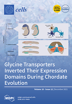
Glycine is an important neurotransmitter in vertebrates, and its synaptic levels are controlled by the action of glycine transporters. Albeit well studied in vertebrates, the evolution of glycinergic neurotransmission is poorly understood because only limited information is available for invertebrates. We show that amphioxus, a key invertebrate model to study the evolution of chordates, has three GlyT genes. Two of these, GlyT and GlyT2.1, are widely expressed in the amphioxus nervous system and are differentially expressed, respectively, in neurons and glia. Vertebrate glycinergic neurons express GlyT2 and glia GlyT1, suggesting that the evolution of the chordate glycinergic system was accompanied by a paralog-specific inversion of gene expression. Despite this genetic divergence, our results suggest a conserved role of glycinergic neurotransmission in the control of larval swimming.View this paper
  • Issues are regarded as officially published after their release is announced to the table of contents alert mailing list.
  • You may sign up for e-mail alerts to receive table of contents of newly released issues.
  • PDF is the official format for papers published in both, html and pdf forms. To view the papers in pdf format, click on the "PDF Full-text" link, and use the free Adobe Reader to open them.
Order results
Result details
Section
Select all
Export citation of selected articles as:
22 pages, 1040 KiB  
Review
Metabolic Reprogramming of Liver Fibrosis
by M. Eugenia Delgado, Beatriz I. Cárdenas, Núria Farran and Mercedes Fernandez
Cells 2021, 10(12), 3604; https://doi.org/10.3390/cells10123604 - 20 Dec 2021
Cited by 20 | Viewed by 4949
Abstract
Liver fibrosis is an excessive and imbalanced deposition of fibrous extracellular matrix (ECM) that is associated with the hepatic wound-healing response. It is also the common mechanism that contributes to the impairment of the liver function that is observed in many chronic liver [...] Read more.
Liver fibrosis is an excessive and imbalanced deposition of fibrous extracellular matrix (ECM) that is associated with the hepatic wound-healing response. It is also the common mechanism that contributes to the impairment of the liver function that is observed in many chronic liver diseases (CLD). Despite the efforts, no effective therapy against fibrosis exists yet. Worryingly, due to the growing obesity pandemic, fibrosis incidence is on the rise. Here, we aim to summarize the main components and mechanisms involved in the progression of liver fibrosis, with special focus on the metabolic regulation of key effectors of fibrogenesis, hepatic stellate cells (HSCs), and their role in the disease progression. Hepatic cells that undergo metabolic reprogramming require a tightly controlled, fine-tuned cellular response, allowing them to meet their energetic demands without affecting cellular integrity. Here, we aim to discuss the role of ribonucleic acid (RNA)-binding proteins (RBPs), whose dynamic nature being context- and stimuli-dependent make them very suitable for the fibrotic situation. Thus, we will not only summarize the up-to-date literature on the metabolic regulation of HSCs in liver fibrosis, but also on the RBP-dependent post-transcriptional regulation of this metabolic switch that results in such important consequences for the progression of fibrosis and CLD. Full article
Show Figures

Graphical abstract

15 pages, 1608 KiB  
Review
Immunoglobulin A Mucosal Immunity and Altered Respiratory Epithelium in Cystic Fibrosis
by Sophie Gohy, Alexandra Moeremans, Charles Pilette and Amandine Collin
Cells 2021, 10(12), 3603; https://doi.org/10.3390/cells10123603 - 20 Dec 2021
Cited by 8 | Viewed by 3765
Abstract
The respiratory epithelium represents the first chemical, immune, and physical barrier against inhaled noxious materials, particularly pathogens in cystic fibrosis. Local mucus thickening, altered mucociliary clearance, and reduced pH due to CFTR protein dysfunction favor bacterial overgrowth and excessive inflammation. We aimed in [...] Read more.
The respiratory epithelium represents the first chemical, immune, and physical barrier against inhaled noxious materials, particularly pathogens in cystic fibrosis. Local mucus thickening, altered mucociliary clearance, and reduced pH due to CFTR protein dysfunction favor bacterial overgrowth and excessive inflammation. We aimed in this review to summarize respiratory mucosal alterations within the epithelium and current knowledge on local immunity linked to immunoglobulin A in patients with cystic fibrosis. Full article
(This article belongs to the Collection Cystic Fibrosis: Cells, Physiopathology and Emerging Therapies)
Show Figures

Figure 1

19 pages, 2203 KiB  
Review
Aurora A and AKT Kinase Signaling Associated with Primary Cilia
by Yuhei Nishimura, Daishi Yamakawa, Takashi Shiromizu and Masaki Inagaki
Cells 2021, 10(12), 3602; https://doi.org/10.3390/cells10123602 - 20 Dec 2021
Cited by 7 | Viewed by 4494
Abstract
Dysregulation of kinase signaling is associated with various pathological conditions, including cancer, inflammation, and autoimmunity; consequently, the kinases involved have become major therapeutic targets. While kinase signaling pathways play crucial roles in multiple cellular processes, the precise manner in which their dysregulation contributes [...] Read more.
Dysregulation of kinase signaling is associated with various pathological conditions, including cancer, inflammation, and autoimmunity; consequently, the kinases involved have become major therapeutic targets. While kinase signaling pathways play crucial roles in multiple cellular processes, the precise manner in which their dysregulation contributes to disease is dependent on the context; for example, the cell/tissue type or subcellular localization of the kinase or substrate. Thus, context-selective targeting of dysregulated kinases may serve to increase the therapeutic specificity while reducing off-target adverse effects. Primary cilia are antenna-like structures that extend from the plasma membrane and function by detecting extracellular cues and transducing signals into the cell. Cilia formation and signaling are dynamically regulated through context-dependent mechanisms; as such, dysregulation of primary cilia contributes to disease in a variety of ways. Here, we review the involvement of primary cilia-associated signaling through aurora A and AKT kinases with respect to cancer, obesity, and other ciliopathies. Full article
(This article belongs to the Special Issue Cell Biology: State-of-the-Art and Perspectives in Japan)
Show Figures

Figure 1

17 pages, 2776 KiB  
Review
Dunking into the Lipid Bilayer: How Direct Membrane Binding of Nucleoporins Can Contribute to Nuclear Pore Complex Structure and Assembly
by Mohamed Hamed and Wolfram Antonin
Cells 2021, 10(12), 3601; https://doi.org/10.3390/cells10123601 - 20 Dec 2021
Cited by 11 | Viewed by 5324
Abstract
Nuclear pore complexes (NPCs) mediate the selective and highly efficient transport between the cytoplasm and the nucleus. They are embedded in the two membrane structure of the nuclear envelope at sites where these two membranes are fused to pores. A few transmembrane proteins [...] Read more.
Nuclear pore complexes (NPCs) mediate the selective and highly efficient transport between the cytoplasm and the nucleus. They are embedded in the two membrane structure of the nuclear envelope at sites where these two membranes are fused to pores. A few transmembrane proteins are an integral part of NPCs and thought to anchor these complexes in the nuclear envelope. In addition, a number of nucleoporins without membrane spanning domains interact with the pore membrane. Here we review our current knowledge of how these proteins interact with the membrane and how this interaction can contribute to NPC assembly, stability and function as well as shaping of the pore membrane. Full article
Show Figures

Figure 1

13 pages, 2943 KiB  
Article
Microtubule Integrity Is Associated with the Functional Activity of Mitochondria in HEK293
by Min Jeong Cho, Yu Jin Kim, Won Dong Yu, You Shin Kim and Jae Ho Lee
Cells 2021, 10(12), 3600; https://doi.org/10.3390/cells10123600 - 20 Dec 2021
Cited by 3 | Viewed by 3005
Abstract
Mitochondria move along the microtubule network and produce bioenergy in the cell. However, there is no report of a relationship between bioenergetic activity of mitochondria and microtubule stability in mammalian cells. This study aimed to investigate this relationship. We treated HEK293 cells with [...] Read more.
Mitochondria move along the microtubule network and produce bioenergy in the cell. However, there is no report of a relationship between bioenergetic activity of mitochondria and microtubule stability in mammalian cells. This study aimed to investigate this relationship. We treated HEK293 cells with microtubule stabilizers (Taxol and Epothilone D) or a microtubule disturber (vinorelbine), and performed live-cell imaging to determine whether mitochondrial morphology and bioenergetic activity depend on the microtubule status. Treatment with microtubule stabilizers enhanced the staining intensity of microtubules, significantly increased ATP production and the spare respiratory capacity, dramatically increased mitochondrial fusion, and promoted dynamic movement of mitochondria. By contrast, bioenergetic activity of mitochondria was significantly decreased in cells treated with the microtubule disturber. Our data suggest that microtubule stability promotes mitochondrial functional activity. In conclusion, a microtubule stabilizer can possibly recover mitochondrial functional activity in cells with unstable microtubules. Full article
(This article belongs to the Section Mitochondria)
Show Figures

Figure 1

16 pages, 3648 KiB  
Article
Defucosylated Mouse–Dog Chimeric Anti-EGFR Antibody Exerts Antitumor Activities in Mouse Xenograft Models of Canine Tumors
by Guanjie Li, Tomokazu Ohishi, Mika K. Kaneko, Junko Takei, Takuya Mizuno, Manabu Kawada, Masaki Saito, Hiroyuki Suzuki and Yukinari Kato
Cells 2021, 10(12), 3599; https://doi.org/10.3390/cells10123599 - 20 Dec 2021
Cited by 7 | Viewed by 3134
Abstract
The epidermal growth factor receptor (EGFR) contributes to tumor malignancy via gene amplification and protein overexpression. Previously, we developed an anti-human EGFR (hEGFR) monoclonal antibody, namely EMab-134, which detects hEGFR and dog EGFR (dEGFR) with high sensitivity and specificity. In this study, we [...] Read more.
The epidermal growth factor receptor (EGFR) contributes to tumor malignancy via gene amplification and protein overexpression. Previously, we developed an anti-human EGFR (hEGFR) monoclonal antibody, namely EMab-134, which detects hEGFR and dog EGFR (dEGFR) with high sensitivity and specificity. In this study, we produced a defucosylated mouse–dog chimeric anti-EGFR monoclonal antibody, namely E134Bf. In vitro analysis revealed that E134Bf highly exerted antibody-dependent cellular cytotoxicity and complement-dependent cytotoxicity against a canine osteosarcoma cell line (D-17) and a canine fibroblastic cell line (A-72), both of which express endogenous dEGFR. Moreover, in vivo administration of E134Bf significantly suppressed the development of D-17 and A-72 compared with the control dog IgG in mouse xenografts. These results indicate that E134Bf exerts antitumor effects against dEGFR-expressing canine cancers and could be valuable as part of an antibody treatment regimen for dogs. Full article
Show Figures

Figure 1

16 pages, 5146 KiB  
Article
Cyclophilin A Impairs Efferocytosis and Accelerates Atherosclerosis by Overexpressing CD 47 and Down-Regulating Calreticulin
by Vinitha Anandan, Thushara Thulaseedharan, Aishwarya Suresh Kumar, Karthika Chandran Latha, Amjesh Revikumar, Ajit Mullasari, Chandrasekharan C. Kartha, Abdul Jaleel and Surya Ramachandran
Cells 2021, 10(12), 3598; https://doi.org/10.3390/cells10123598 - 20 Dec 2021
Cited by 7 | Viewed by 4369
Abstract
Impairment of efferocytosis in apoptotic macrophages is a known determinant of the severity of atherosclerosis and the vulnerability of plaques to rupture. The precise mechanisms involved in impaired efferocytosis are unclear. Given the well-recognized role of the inflammatory cytokine cyclophilin A (Cyp A) [...] Read more.
Impairment of efferocytosis in apoptotic macrophages is a known determinant of the severity of atherosclerosis and the vulnerability of plaques to rupture. The precise mechanisms involved in impaired efferocytosis are unclear. Given the well-recognized role of the inflammatory cytokine cyclophilin A (Cyp A) in modulating several atherogenic mechanisms in high-glucose primed monocytes, we investigated the role of Cyp A in macrophage efferocytosis. The efficiency of efferocytosis in RAW 264.7 macrophages grown in vitro and primed with cyclophilin A was assessed using flow cytometry and confocal assays. Cholesterol content in cells was measured using cell-based cholesterol efflux assay. Proteomic analysis and bioinformatics tools were employed to decipher the link between cyclophilin A and the known ligand receptors involved in efferocytosis. Cyclophilin A was found to impair efferocytosis in apoptotic macrophages by reducing ABCA1-mediated cholesterol efflux in foam cells derived from macrophages. Cyclophilin A-primed macrophages showed an increase in expression of the don’t-eat-me signal CD 47 and a decrease in the expression of the eat-me signal, calreticulin. Phagocytosis was restored upon silencing of cyclophilin A. New Zealand white rabbits were fed a high-fat diet, and lesions in their aortae were analyzed histologically for evidence of atherosclerosis and the expression of Cyp A, CD 47 and calreticulin, the ligand receptor involved in efferocytosis. Gene and protein expressions in aortae and macrophages were analyzed by real-time PCR and Western blotting. Cyclophilin A, via its effects on the expression of CD 47 and calreticulin, impairs efferocytosis in apoptotic macrophages. Together with its impact on cholesterol efflux from macrophages, these effects can amplify other mechanisms of Cyp A in accelerating the progression of atherosclerosis. Full article
Show Figures

Graphical abstract

17 pages, 2382 KiB  
Article
7-Ketocholesterol Induces Lipid Metabolic Reprogramming and Enhances Cholesterol Ester Accumulation in Cardiac Cells
by Mei-Ling Cheng, Hsiang-Yu Tang, Pei-Ting Wu, Cheng-Hung Yang, Chi-Jen Lo, Jui-Fen Lin and Hung-Yao Ho
Cells 2021, 10(12), 3597; https://doi.org/10.3390/cells10123597 - 20 Dec 2021
Cited by 6 | Viewed by 3056
Abstract
7-Ketocholesterol (7KCh) is a major oxidized cholesterol product abundant in lipoprotein deposits and atherosclerotic plaques. Our previous study has shown that 7KCh accumulates in erythrocytes of heart failure patients, and further investigation centered on how 7KCh may affect metabolism in cardiomyocytes. We applied [...] Read more.
7-Ketocholesterol (7KCh) is a major oxidized cholesterol product abundant in lipoprotein deposits and atherosclerotic plaques. Our previous study has shown that 7KCh accumulates in erythrocytes of heart failure patients, and further investigation centered on how 7KCh may affect metabolism in cardiomyocytes. We applied metabolomics to study the metabolic changes in cardiac cell line HL-1 after treatment with 7KCh. Mevalonic acid (MVA) pathway-derived metabolites, such as farnesyl-pyrophosphate and geranylgeranyl-pyrophosphate, phospholipids, and triacylglycerols levels significantly declined, while the levels of lysophospholipids, such as lysophosphatidylcholines (lysoPCs) and lysophosphatidylethanolamines (lysoPEs), considerably increased in 7KCh-treated cells. Furthermore, the cholesterol content showed no significant change, but the production of cholesteryl esters was enhanced in the treated cells. To explore the possible mechanisms, we applied mRNA-sequencing (mRNA-seq) to study genes differentially expressed in 7KCh-treated cells. The transcriptomic analysis revealed that genes involved in lipid metabolic processes, including MVA biosynthesis and cholesterol transport and esterification, were differentially expressed in treated cells. Integrated analysis of both metabolomic and transcriptomic data suggests that 7KCh induces cholesteryl ester accumulation and reprogramming of lipid metabolism through altered transcription of such genes as sterol O-acyltransferase- and phospholipase A2-encoding genes. The 7KCh-induced reprogramming of lipid metabolism in cardiac cells may be implicated in the pathogenesis of cardiovascular diseases. Full article
Show Figures

Graphical abstract

20 pages, 2218 KiB  
Review
Early Life Inflammation and the Developing Hematopoietic and Immune Systems: The Cochlea as a Sensitive Indicator of Disruption
by Kelly S. Otsuka, Christopher Nielson, Matthew A. Firpo, Albert H. Park and Anna E. Beaudin
Cells 2021, 10(12), 3596; https://doi.org/10.3390/cells10123596 - 20 Dec 2021
Cited by 7 | Viewed by 3235
Abstract
Emerging evidence indicates that perinatal infection and inflammation can influence the developing immune system and may ultimately affect long-term health and disease outcomes in offspring by perturbing tissue and immune homeostasis. We posit that perinatal inflammation influences immune outcomes in offspring by perturbing [...] Read more.
Emerging evidence indicates that perinatal infection and inflammation can influence the developing immune system and may ultimately affect long-term health and disease outcomes in offspring by perturbing tissue and immune homeostasis. We posit that perinatal inflammation influences immune outcomes in offspring by perturbing (1) the development and function of fetal-derived immune cells that regulate tissue development and homeostasis, and (2) the establishment and function of developing hematopoietic stem cells (HSCs) that continually generate immune cells across the lifespan. To disentangle the complexities of these interlinked systems, we propose the cochlea as an ideal model tissue to investigate how perinatal infection affects immune, tissue, and stem cell development. The cochlea contains complex tissue architecture and a rich immune milieu that is established during early life. A wide range of congenital infections cause cochlea dysfunction and sensorineural hearing loss (SNHL), likely attributable to early life inflammation. Furthermore, we show that both immune cells and bone marrow hematopoietic progenitors can be simultaneously analyzed within neonatal cochlear samples. Future work investigating the pathogenesis of SNHL in the context of congenital infection will therefore provide critical information on how perinatal inflammation drives disease susceptibility in offspring. Full article
(This article belongs to the Special Issue The Impact of Immune Activation on Hematopoiesis)
Show Figures

Figure 1

19 pages, 6499 KiB  
Article
Development of a 3-Dimensional Model to Study Right Heart Dysfunction in Pulmonary Arterial Hypertension: First Observations
by Aida Llucià-Valldeperas, Rowan Smal, Fjodor T. Bekedam, Margaux Cé, Xiaoke Pan, Xue D. Manz, Paul J. M. Wijnker, Anton Vonk-Noordegraaf, Harm J. Bogaard, Marie-Jose Goumans and Frances S. de Man
Cells 2021, 10(12), 3595; https://doi.org/10.3390/cells10123595 - 20 Dec 2021
Cited by 1 | Viewed by 3364
Abstract
Pulmonary arterial hypertension (PAH) patients eventually die of right heart failure (RHF). Currently, there is no suitable pre-clinical model to study PAH. Therefore, we aim to develop a right heart dysfunction (RHD) model using the 3-dimensional engineered heart tissue (EHT) approach and cardiomyocytes [...] Read more.
Pulmonary arterial hypertension (PAH) patients eventually die of right heart failure (RHF). Currently, there is no suitable pre-clinical model to study PAH. Therefore, we aim to develop a right heart dysfunction (RHD) model using the 3-dimensional engineered heart tissue (EHT) approach and cardiomyocytes derived from patient-induced pluripotent stem cells (iPSCs) to unravel the mechanisms that determine the fate of a pressure-overloaded right ventricle. iPSCs from PAH and healthy control subjects were differentiated into cardiomyocytes (iPSC-CMs), incorporated into the EHT, and maintained for 28 days. In comparison with control iPSC-CMs, PAH-derived iPSC-CMs exhibited decreased beating frequency and increased contraction and relaxation times. iPSC-CM alignment within the EHT was observed. PAH-derived EHTs exhibited higher force, and contraction and relaxation times compared with control EHTs. Increased afterload was induced using 2× stiffer posts from day 0. Due to high variability, there were no functional differences between normal and stiffer EHTs, and no differences in the hypertrophic gene expression. In conclusion, under baseline spontaneous conditions, PAH-derived iPSC-CMs and EHTs show prolonged contraction compared with controls, as observed clinically in PAH patients. Further optimization of the hypertrophic model and profound characterization may provide a platform for disease modelling and drug screening. Full article
(This article belongs to the Special Issue Cellular Pathogenesis of Pulmonary Arterial Hypertension)
Show Figures

Graphical abstract

17 pages, 6920 KiB  
Article
Targeting Chondroitin Sulfate Reduces Invasiveness of Glioma Cells by Suppressing CD44 and Integrin β1 Expression
by Yin-Hung Chu, Wen-Chieh Liao, Ying-Jui Ho, Chih-Hsien Huang, To-Jung Tseng and Chiung-Hui Liu
Cells 2021, 10(12), 3594; https://doi.org/10.3390/cells10123594 - 20 Dec 2021
Cited by 12 | Viewed by 3976
Abstract
Chondroitin sulfate (CS) is a major component of the extracellular matrix found to be abnormally accumulated in several types of cancer tissues. Previous studies have indicated that CS synthases and modification enzymes are frequently elevated in human gliomas and are associated with poor [...] Read more.
Chondroitin sulfate (CS) is a major component of the extracellular matrix found to be abnormally accumulated in several types of cancer tissues. Previous studies have indicated that CS synthases and modification enzymes are frequently elevated in human gliomas and are associated with poor prognosis. However, the underlying mechanisms of CS in cancer progression and approaches for interrupting its functions in cancer cells remain largely unexplored. Here, we have found that CS was significantly enriched surrounding the vasculature in a subset of glioma tissues, which was akin to the perivascular niche for cancer-initiating cells. Silencing or overexpression of the major CS synthase, chondroitin sulfate synthase 1 (CHSY1), significantly regulated the glioma cell invasive phenotypes and modulated integrin expression. Furthermore, we identified CD44 as a crucial chondroitin sulfate proteoglycan (CSPG) that was modified by CHSY1 on glioma cells, and the suppression of CS formation on CD44 by silencing the CHSY1-inhibited interaction between CD44 and integrin β1 on the adhesion complex. Moreover, we tested the CS-specific binding peptide, resulting in the suppression of glioma cell mobility in a fashion similar to that observed upon the silencing of CHSY1. In addition, the peptide demonstrated significant affinity to CD44, promoted CD44 degradation, and suppressed integrin β1 expression in glioma cells. Overall, this study proposes a potential regulatory loop between CS, CD44, and integrin β1 in glioma cells, and highlights the importance of CS in CD44 stability. Furthermore, the targeting of CS by specific binding peptides has potential as a novel therapeutic strategy for glioma. Full article
Show Figures

Figure 1

17 pages, 4544 KiB  
Article
p66α Suppresses Breast Cancer Cell Growth and Migration by Acting as Co-Activator of p53
by Qun Zhang, Yihong Zhang, Jie Zhang, Dan Zhang, Mengying Li, Han Yan, Hui Zhang, Liwei Song, Jiamin Wang, Zhaoyuan Hou, Yunhai Yang and Xiuqun Zou
Cells 2021, 10(12), 3593; https://doi.org/10.3390/cells10123593 - 20 Dec 2021
Cited by 3 | Viewed by 2850
Abstract
p66α is a GATA zinc finger domain-containing transcription factor that has been shown to be essential for gene silencing by participating in the NuRD complex. Several studies have suggested that p66α is a risk gene for a wide spectrum of diseases such as [...] Read more.
p66α is a GATA zinc finger domain-containing transcription factor that has been shown to be essential for gene silencing by participating in the NuRD complex. Several studies have suggested that p66α is a risk gene for a wide spectrum of diseases such as diabetes, schizophrenia, and breast cancer; however, its biological role has not been defined. Here, we report that p66α functions as a tumor suppressor to inhibit breast cancer cell growth and migration, evidenced by the fact that the depletion of p66α results in accelerated tumor growth and migration of breast cancer cells. Mechanistically, immunoprecipitation assays identify p66α as a p53-interacting protein that binds the DNA-binding domain of p53 molecule predominantly via its CR2 domain. Depletion of p66α in multiple breast cells results in decreased expression of p53 target genes, while over-expression of p66α results in increased expression of these target genes. Moreover, p66α promotes the transactivity of p53 by enhancing p53 binding at target promoters. Together, these findings demonstrate that p66α is a tumor suppressor by functioning as a co-activator of p53. Full article
(This article belongs to the Collection Targeting Cancer Stem Cell)
Show Figures

Figure 1

19 pages, 1368 KiB  
Review
New Onset of Autoimmune Diseases Following COVID-19 Diagnosis
by Abraham Edgar Gracia-Ramos, Eduardo Martin-Nares and Gabriela Hernández-Molina
Cells 2021, 10(12), 3592; https://doi.org/10.3390/cells10123592 - 20 Dec 2021
Cited by 124 | Viewed by 24607
Abstract
There is growing evidence that coronavirus disease 2019 (COVID-19) can lead to a dysregulation of the immune system with the development of autoimmune phenomena. The consequence of this immune dysregulation ranges from the production of autoantibodies to the onset of rheumatic autoimmune disease. [...] Read more.
There is growing evidence that coronavirus disease 2019 (COVID-19) can lead to a dysregulation of the immune system with the development of autoimmune phenomena. The consequence of this immune dysregulation ranges from the production of autoantibodies to the onset of rheumatic autoimmune disease. In this context, we conducted a systematic review to analyze the current data regarding the new-onset systemic and rheumatic autoimmune diseases in COVID-19 patients. A literature search in PubMed and Scopus databases from December 2019 to September 2021 identified 99 patients that fulfilled the specific diagnostic/classification criteria and/or nomenclature for each rheumatic autoimmune disease. The main diseases reported were vasculitis and arthritis. Idiopathic inflammatory myopathies, systemic lupus erythematosus, and sarcoidosis were also reported in a limited number of patients, as well as isolated cases of systemic sclerosis and adult-onset Still’s disease. These findings highlight the potential spectrum of systemic and rheumatic autoimmune diseases that could be precipitated by SARS-CoV-2 infection. Complementary studies are needed to discern the link between the SARS-CoV-2 and new onset-rheumatic diseases so that this knowledge can be used in early diagnosis and the most suitable management. Full article
Show Figures

Graphical abstract

4 pages, 189 KiB  
Editorial
Cellular Immunology and COVID-19
by Isabella Quinti
Cells 2021, 10(12), 3591; https://doi.org/10.3390/cells10123591 - 20 Dec 2021
Viewed by 2122
Abstract
In “Cellular Immunology and COVID-19” (a Special Issue of Cells), a panel of leading scientists provides an exhaustive overview of the different aspects of the immune mechanisms underlying COVID-19 [...] Full article
(This article belongs to the Collection Cellular Immunology and COVID-19)
19 pages, 13462 KiB  
Review
Taming, Domestication and Exaptation: Trajectories of Transposable Elements in Genomes
by Pierre Capy
Cells 2021, 10(12), 3590; https://doi.org/10.3390/cells10123590 - 20 Dec 2021
Cited by 10 | Viewed by 4182
Abstract
During evolution, several types of sequences pass through genomes. Along with mutations and internal genetic tinkering, they are a useful source of genetic variability for adaptation and evolution. Most of these sequences are acquired by horizontal transfers (HT), but some of them may [...] Read more.
During evolution, several types of sequences pass through genomes. Along with mutations and internal genetic tinkering, they are a useful source of genetic variability for adaptation and evolution. Most of these sequences are acquired by horizontal transfers (HT), but some of them may come from the genomes themselves. If they are not lost or eliminated quickly, they can be tamed, domesticated, or even exapted. Each of these processes results from a series of events, depending on the interactions between these sequences and the host genomes, but also on environmental constraints, through their impact on individuals or population fitness. After a brief reminder of the characteristics of each of these states (taming, domestication, exaptation), the evolutionary trajectories of these new or acquired sequences will be presented and discussed, emphasizing that they are not totally independent insofar as the first can constitute a step towards the second, and the second is another step towards the third. Full article
Show Figures

Figure 1

13 pages, 4112 KiB  
Article
Unravelling the Impact of the Genetic Variant rs1042058 within the TPL2 Risk Gene Locus on Molecular and Clinical Disease Course Patients with Inflammatory Bowel Disease
by Yasser Morsy, Nathalie Brillant, Yannick Franc, Michael Scharl, Marcin Wawrzyniak and on behalf of the Swiss IBD Cohort Study Group
Cells 2021, 10(12), 3589; https://doi.org/10.3390/cells10123589 - 20 Dec 2021
Cited by 4 | Viewed by 2772
Abstract
Background: The single nucleotide polymorphism (SNP) rs1042058 within the gene locus encoding tumor progression locus 2 (TPL2) has been recently identified as a risk gene for inflammatory bowel disease (IBD). TPL2 has been shown to regulate pro-inflammatory signaling and cytokine secretion, while inhibition [...] Read more.
Background: The single nucleotide polymorphism (SNP) rs1042058 within the gene locus encoding tumor progression locus 2 (TPL2) has been recently identified as a risk gene for inflammatory bowel disease (IBD). TPL2 has been shown to regulate pro-inflammatory signaling and cytokine secretion, while inhibition of TPL2 decreases intestinal inflammation in vivo. However, the clinical and molecular implications of this disease-associated TPL2 variation in IBD patients have not yet been studied. Methods: We analyzed the impact of the IBD-associated TPL2 variation using clinical data of 2145 genotyped patients from the Swiss IBD Cohort Study (SIBDCS). Furthermore, we assessed the molecular consequences of the TPL2 variation in ulcerative colitis (UC) and Crohn’s disease (CD) patients by real-time PCR and multiplex ELISA of colon biopsies or serum, respectively. Results: We found that presence of the SNP rs1042058 within the TPL2 gene locus results in significantly higher numbers of CD patients suffering from peripheral arthritis. In contrast, UC patients carrying this variant feature a lower risk for intestinal surgery. On a molecular level, the presence of the rs1042058 (GG) IBD-risk polymorphism in TPL2 was associated with decreased mRNA levels of IL-10 in CD patients and decreased levels of IL-18 in the intestine of UC patients. Conclusions: Our data suggest that the presence of the IBD-associated TPL2 variation might indicate a more severe disease course in CD patients. These results reveal a potential therapeutic target and demonstrate the relevance of the IBD-associated TPL2 SNP as a predictive biomarker in IBD. Full article
Show Figures

Figure 1

14 pages, 2583 KiB  
Article
Potential Antagonistic Bacteria against Verticillium dahliae Isolated from Artificially Infested Nursery
by Xiaofeng Su, Siyuan Wu, Lu Liu, Guoqing Lu, Haiyang Liu, Xi Jin, Yi Wang, Huiming Guo, Chen Wang and Hongmei Cheng
Cells 2021, 10(12), 3588; https://doi.org/10.3390/cells10123588 - 20 Dec 2021
Cited by 6 | Viewed by 3201
Abstract
As an ecofriendly biocontrol agent, antagonistic bacteria are a crucial class of highly efficient fungicides in the field against Verticillium dahliae, the most virulent pathogen for cotton and other crops. Toward identifying urgently needed bacterial candidates, we screened bacteria isolated from the [...] Read more.
As an ecofriendly biocontrol agent, antagonistic bacteria are a crucial class of highly efficient fungicides in the field against Verticillium dahliae, the most virulent pathogen for cotton and other crops. Toward identifying urgently needed bacterial candidates, we screened bacteria isolated from the cotton rhizosphere soil for antagonisitic activity against V. dahliae in an artificially infested nursery. In preliminary tests of antagonistic candidates to characterize the mechanism of action of on culture medium, 88 strains that mainly belonged to Bacillus strongly inhibited the colony diameter of V. dahliae, with inhibiting efficacy up to 50% in 9 strains. Among the most-effective bacterial strains, Bacillus sp. ABLF-18, and ABLF-50 and Paenibacillus sp. ABLF-90 significantly reduced the disease index and fungal biomass of cotton to 40–70% that of the control. In further tests to elucidate the biocontrol mechanism (s), the strains secreted extracellular enzymes cellulase, glucanase, and protease, which can degrade the mycelium, and antimicrobial lipopeptides such as surfactin and iturin homologues. The expression of PAL, MAPK and PR10, genes related to disease resistance, was also elicited in cotton plants. Our results clearly show that three candidate bacterial strains can enhance cotton defense responses against V. dahliae; the secretion of fungal cell-wall-degrading enzymes, synthesis of nonribosomal antimicrobial peptides and induction of systemic resistance shows that the strains have great potential as biocontrol fungicides. Full article
Show Figures

Graphical abstract

22 pages, 1275 KiB  
Review
The Functional and Mechanistic Roles of Immunoproteasome Subunits in Cancer
by Satyendra Chandra Tripathi, Disha Vedpathak and Edwin Justin Ostrin
Cells 2021, 10(12), 3587; https://doi.org/10.3390/cells10123587 - 20 Dec 2021
Cited by 13 | Viewed by 5489
Abstract
Cell-mediated immunity is driven by antigenic peptide presentation on major histocompatibility complex (MHC) molecules. Specialized proteasome complexes called immunoproteasomes process viral, bacterial, and tumor antigens for presentation on MHC class I molecules, which can induce CD8 T cells to mount effective immune responses. [...] Read more.
Cell-mediated immunity is driven by antigenic peptide presentation on major histocompatibility complex (MHC) molecules. Specialized proteasome complexes called immunoproteasomes process viral, bacterial, and tumor antigens for presentation on MHC class I molecules, which can induce CD8 T cells to mount effective immune responses. Immunoproteasomes are distinguished by three subunits that alter the catalytic activity of the proteasome and are inducible by inflammatory stimuli such as interferon-γ (IFN-γ). This inducible activity places them in central roles in cancer, autoimmunity, and inflammation. While accelerated proteasomal degradation is an important tumorigenic mechanism deployed by several cancers, there is some ambiguity regarding the role of immunoproteasome induction in neoplastic transformation. Understanding the mechanistic and functional relevance of the immunoproteasome provides essential insights into developing targeted therapies, including overcoming resistance to standard proteasome inhibition and immunomodulation of the tumor microenvironment. In this review, we discuss the roles of the immunoproteasome in different cancers. Full article
(This article belongs to the Special Issue The Immunoproteasome in Health and Disease)
Show Figures

Figure 1

28 pages, 37763 KiB  
Article
Somatic Mutational Profile of High-Grade Serous Ovarian Carcinoma and Triple-Negative Breast Carcinoma in Young and Elderly Patients: Similarities and Divergences
by Pedro Adolpho de Menezes Pacheco Serio, Gláucia Fernanda de Lima Pereira, Maria Lucia Hirata Katayama, Rosimeire Aparecida Roela, Simone Maistro and Maria Aparecida Azevedo Koike Folgueira
Cells 2021, 10(12), 3586; https://doi.org/10.3390/cells10123586 - 20 Dec 2021
Cited by 7 | Viewed by 3449
Abstract
Background: Triple-negative breast cancer (TNBC) and High-Grade Serous Ovarian Cancer (HGSOC) are aggressive malignancies that share similarities; however, different ages of onset may reflect distinct tumor behaviors. Thus, our aim was to compare somatic mutations in potential driver genes in 109 TNBC and [...] Read more.
Background: Triple-negative breast cancer (TNBC) and High-Grade Serous Ovarian Cancer (HGSOC) are aggressive malignancies that share similarities; however, different ages of onset may reflect distinct tumor behaviors. Thus, our aim was to compare somatic mutations in potential driver genes in 109 TNBC and 81 HGSOC from young (Y ≤ 40 years) and elderly (E ≥ 75 years) patients. Methods: Open access mutational data (WGS or WES) were collected for TNBC and HGSOC patients. Potential driver genes were those that were present in the Cancer Gene Census—CGC, the Candidate Cancer Gene Database—CCGD, or OncoKB and those that were considered pathogenic in variant effect prediction tools. Results: Mutational signature 3 (homologous repair defects) was the only gene that was represented in all four subgroups. The median number of mutated CGCs per sample was similar in HGSOC (Y:3 vs. E:4), but it was higher in elderly TNBC than it was in young TNBC (Y:3 vs. E:6). At least 90% of the samples from TNBC and HGSOC from Y and E patients presented at least one known affected TSG. Besides TP53, which was mutated in 67–83% of the samples, the affected TSG in TP53 wild-type samples were NF1 (yHGSOC and yTNBC), PHF6 (eHGSOC and yTNBC), PTEN, PIK3R1 and ZHFX3 (yTNBC), KMT2C, ARID1B, TBX3, and ATM (eTNBC). A few samples only presented one affected oncogene (but no TSG): KRAS and TSHR in eHGSOC and RAC1 and PREX2 (a regulator of RAC1) in yTNBC. At least ⅔ of the tumors presented mutated oncogenes associated with tumor suppressor genes; the Ras and/or PIK3CA signaling pathways were altered in 15% HGSOC and 20–35% TNBC (Y vs. E); DNA repair genes were mutated in 19–33% of the HGSOC tumors but were more frequently mutated in E-TNBC (56%). However, in HGSOC, 9.5% and 3.3% of the young and elderly patients, respectively, did not present any tumors with an affected CGC nor did 4.65% and none of the young and elderly TNBC patients. Conclusion: Most HGSOC and TNBC from young and elderly patients present an affected TSG, mainly TP53, as well as mutational signature 3; however, a few tumors only present an affected oncogene or no affected cancer-causing genes. Full article
(This article belongs to the Collection Emerging Cancer Target Genes)
Show Figures

Figure 1

20 pages, 2914 KiB  
Article
Single-Cell Transcriptomics Links Loss of Human Pancreatic β-Cell Identity to ER Stress
by Nathalie Groen, Floris Leenders, Ahmed Mahfouz, Amadeo Munoz-Garcia, Mauro J. Muraro, Natascha de Graaf, Ton. J. Rabelink, Rob Hoeben, Alexander van Oudenaarden, Arnaud Zaldumbide, Marcel J. T. Reinders, Eelco J. P. de Koning and Françoise Carlotti
Cells 2021, 10(12), 3585; https://doi.org/10.3390/cells10123585 - 19 Dec 2021
Cited by 3 | Viewed by 3533
Abstract
The maintenance of pancreatic islet architecture is crucial for proper β-cell function. We previously reported that disruption of human islet integrity could result in altered β-cell identity. Here we combine β-cell lineage tracing and single-cell transcriptomics to investigate the mechanisms underlying this process [...] Read more.
The maintenance of pancreatic islet architecture is crucial for proper β-cell function. We previously reported that disruption of human islet integrity could result in altered β-cell identity. Here we combine β-cell lineage tracing and single-cell transcriptomics to investigate the mechanisms underlying this process in primary human islet cells. Using drug-induced ER stress and cytoskeleton modification models, we demonstrate that altering the islet structure triggers an unfolding protein response that causes the downregulation of β-cell maturity genes. Collectively, our findings illustrate the close relationship between endoplasmic reticulum homeostasis and β-cell phenotype, and strengthen the concept of altered β-cell identity as a mechanism underlying the loss of functional β-cell mass. Full article
(This article belongs to the Special Issue Cellular and Molecular Biology of the Beta Cell)
Show Figures

Figure 1

25 pages, 7614 KiB  
Article
Poincaré Maps and Aperiodic Oscillations in Leukemic Cell Proliferation Reveal Chaotic Dynamics
by Konstantinos Adamopoulos, Dimitis Koutsouris, Apostolos Zaravinos and George I. Lambrou
Cells 2021, 10(12), 3584; https://doi.org/10.3390/cells10123584 - 19 Dec 2021
Viewed by 2313
Abstract
Biological systems are dynamic systems featuring two very common characteristics; Initial conditions and progression over time. Conceptualizing this on tumour models it can lead to important conclusions about disease progression, as well as the disease’s “starting point”. In the present study we tried [...] Read more.
Biological systems are dynamic systems featuring two very common characteristics; Initial conditions and progression over time. Conceptualizing this on tumour models it can lead to important conclusions about disease progression, as well as the disease’s “starting point”. In the present study we tried to answer two questions: (a) which are the evolving properties of proliferating tumour cells that started from different initial conditions and (b) we have attempted to prove that cell proliferation follows chaotic orbits and it can be described by the use of Poincaré maps. As a model we have used the acute lymphoblastic leukemia cell line CCRF-CEM. Measurements of cell population were taken at certain time points every 24 h or 48 h. In addition to the population measurements flow cytometry studies have been conducted in order to examine the apoptotic and necrotic rate of the system and also the DNA content of the cells as they progress through. The cells exhibited a proliferation rate of nonlinear nature with aperiodic oscillatory behavior. In addition to that, the (positive) Lyapunov indices and the Poincaré representations in phase-space that we performed confirmed the presence of chaotic orbits. Several studies have dealt with the complex dynamic behaviour of animal populations, but few with cellular systems. This type of approach could prove useful towards the understanding of leukemia dynamics, with particular interest in the understanding of leukemia onset and progression. Full article
(This article belongs to the Section Cellular Biophysics)
Show Figures

Figure 1

16 pages, 609 KiB  
Review
Direct Current Stimulation in Cell Culture Systems and Brain Slices—New Approaches for Mechanistic Evaluation of Neuronal Plasticity and Neuromodulation: State of the Art
by Nadine Euskirchen, Michael A. Nitsche and Christoph van Thriel
Cells 2021, 10(12), 3583; https://doi.org/10.3390/cells10123583 - 19 Dec 2021
Cited by 2 | Viewed by 2745
Abstract
Non-invasive direct current stimulation (DCS) of the human brain induces neuronal plasticity and alters plasticity-related cognition and behavior. Numerous basic animal research studies focusing on molecular and cellular targets of DCS have been published. In vivo, ex vivo, and in vitro models enhanced [...] Read more.
Non-invasive direct current stimulation (DCS) of the human brain induces neuronal plasticity and alters plasticity-related cognition and behavior. Numerous basic animal research studies focusing on molecular and cellular targets of DCS have been published. In vivo, ex vivo, and in vitro models enhanced knowledge about mechanistic foundations of DCS effects. Our review identified 451 papers using a PRISMA-based search strategy. Only a minority of these papers used cell culture or brain slice experiments with DCS paradigms comparable to those applied in humans. Most of the studies were performed in brain slices (9 papers), whereas cell culture experiments (2 papers) were only rarely conducted. These ex vivo and in vitro approaches underline the importance of cell and electric field orientation, cell morphology, cell location within populations, stimulation duration (acute, prolonged, chronic), and molecular changes, such as Ca2+-dependent intracellular signaling pathways, for the effects of DC stimulation. The reviewed studies help to clarify and confirm basic mechanisms of this intervention. However, the potential of in vitro studies has not been fully exploited and a more systematic combination of rodent models, ex vivo, and cellular approaches might provide a better insight into the neurophysiological changes caused by tDCS. Full article
(This article belongs to the Special Issue The Molecular and Cellular Underpinning of Neurostimulation)
Show Figures

Figure 1

13 pages, 1273 KiB  
Review
T-Cell Receptor Repertoire Analysis with Computational Tools—An Immunologist’s Perspective
by Mahima Arunkumar and Christina E. Zielinski
Cells 2021, 10(12), 3582; https://doi.org/10.3390/cells10123582 - 18 Dec 2021
Cited by 10 | Viewed by 8869
Abstract
Over the last few years, there has been a rapid expansion in the application of information technology to biological data. Particularly the field of immunology has seen great strides in recent years. The development of next-generation sequencing (NGS) and single-cell technologies also brought [...] Read more.
Over the last few years, there has been a rapid expansion in the application of information technology to biological data. Particularly the field of immunology has seen great strides in recent years. The development of next-generation sequencing (NGS) and single-cell technologies also brought forth a revolution in the characterization of immune repertoires. T-cell receptor (TCR) repertoires carry comprehensive information on the history of an individual’s antigen exposure. They serve as correlates of host protection and tolerance, as well as biomarkers of immunological perturbation by natural infections, vaccines or immunotherapies. Their interrogation yields large amounts of data. This requires a suite of highly sophisticated bioinformatics tools to leverage the meaning and complexity of the large datasets. Many different tools and methods, specifically designed for various aspects of immunological research, have recently emerged. Thus, researchers are now confronted with the issue of having to choose the right kind of approach to analyze, visualize and ultimately solve their task at hand. In order to help immunologists to choose from the vastness of available tools for their data analysis, this review addresses and compares commonly used bioinformatics tools for TCR repertoire analysis and illustrates the advantages and limitations of these tools from an immunologist’s perspective. Full article
(This article belongs to the Special Issue Human T Cell Responses in Human Health and Disease)
Show Figures

Figure 1

17 pages, 2498 KiB  
Article
Neutrophil-Derived Extracellular Vesicles Activate Platelets after Pneumolysin Exposure
by Eleftheria Letsiou, Luiz Gustavo Teixeira Alves, Matthias Felten, Timothy J. Mitchell, Holger C. Müller-Redetzky, Steven M. Dudek and Martin Witzenrath
Cells 2021, 10(12), 3581; https://doi.org/10.3390/cells10123581 - 18 Dec 2021
Cited by 13 | Viewed by 3354
Abstract
Pneumolysin (PLY) is a pore-forming toxin of Streptococcus pneumoniae that contributes substantially to the inflammatory processes underlying pneumococcal pneumonia and lung injury. Host responses against S. pneumoniae are regulated in part by neutrophils and platelets, both individually and in cooperative interaction. Previous studies [...] Read more.
Pneumolysin (PLY) is a pore-forming toxin of Streptococcus pneumoniae that contributes substantially to the inflammatory processes underlying pneumococcal pneumonia and lung injury. Host responses against S. pneumoniae are regulated in part by neutrophils and platelets, both individually and in cooperative interaction. Previous studies have shown that PLY can target both neutrophils and platelets, however, the mechanisms by which PLY directly affects these cells and alters their interactions are not completely understood. In this study, we characterize the effects of PLY on neutrophils and platelets and explore the mechanisms by which PLY may induce neutrophil–platelet interactions. In vitro studies demonstrated that PLY causes the formation of neutrophil extracellular traps (NETs) and the release of extracellular vesicles (EVs) from both human and murine neutrophils. In vivo, neutrophil EV (nEV) levels were increased in mice infected with S. pneumoniae. In platelets, treatment with PLY induced the cell surface expression of P-selectin (CD62P) and binding to annexin V and caused a significant release of platelet EVs (pl-EVs). Moreover, PLY-induced nEVs but not NETs promoted platelet activation. The pretreatment of nEVs with proteinase K inhibited platelet activation, indicating that the surface proteins of nEVs play a role in this process. Our findings demonstrate that PLY activates neutrophils and platelets to release EVs and support an important role for neutrophil EVs in modulating platelet functions in pneumococcal infections. Full article
(This article belongs to the Special Issue Molecular and Cellular Mechanism of Airway Diseases)
Show Figures

Figure 1

21 pages, 2304 KiB  
Review
Pediatric Atypical Hemolytic Uremic Syndrome Advances
by Rupesh Raina, Nina Vijayvargiya, Amrit Khooblall, Manasa Melachuri, Shweta Deshpande, Divya Sharma, Kashin Mathur, Manav Arora, Sidharth Kumar Sethi and Sonia Sandhu
Cells 2021, 10(12), 3580; https://doi.org/10.3390/cells10123580 - 18 Dec 2021
Cited by 20 | Viewed by 7044
Abstract
Atypical hemolytic uremic syndrome (aHUS) is a rare disorder characterized by dysregulation of the alternate pathway. The diagnosis of aHUS is one of exclusion, which complicates its early detection and corresponding intervention to mitigate its high rate of mortality and associated morbidity. Heterozygous [...] Read more.
Atypical hemolytic uremic syndrome (aHUS) is a rare disorder characterized by dysregulation of the alternate pathway. The diagnosis of aHUS is one of exclusion, which complicates its early detection and corresponding intervention to mitigate its high rate of mortality and associated morbidity. Heterozygous mutations in complement regulatory proteins linked to aHUS are not always phenotypically active, and may require a particular trigger for the disease to manifest. This list of triggers continues to expand as more data is aggregated, particularly centered around COVID-19 and pediatric vaccinations. Novel genetic mutations continue to be identified though advancements in technology as well as greater access to cohorts of interest, as in diacylglycerol kinase epsilon (DGKE). DGKE mutations associated with aHUS are the first non-complement regulatory proteins associated with the disease, drastically changing the established framework. Additional markers that are less understood, but continue to be acknowledged, include the unique autoantibodies to complement factor H and complement factor I which are pathogenic drivers in aHUS. Interventional therapeutics have undergone the most advancements, as pharmacokinetic and pharmacodynamic properties are modified as needed in addition to their as biosimilar counterparts. As data continues to be gathered in this field, future advancements will optimally decrease the mortality and morbidity of this disease in children. Full article
(This article belongs to the Special Issue Immune Mechanisms in Glomerulonephritis)
Show Figures

Figure 1

17 pages, 2568 KiB  
Article
Adjunctive Thymosin Beta-4 Treatment Influences PMN Effector Cell Function during Pseudomonas aeruginosa-Induced Corneal Infection
by Yuxin Wang, Thomas W. Carion, Abdul Shukkur Ebrahim, Gabriel Sosne and Elizabeth A. Berger
Cells 2021, 10(12), 3579; https://doi.org/10.3390/cells10123579 - 18 Dec 2021
Cited by 6 | Viewed by 2677
Abstract
Previous work examining the therapeutic efficacy of adjunct thymosin beta 4 (Tβ4) to ciprofloxacin for ocular infectious disease has revealed markedly reduced inflammation (inflammatory mediators and innate immune cells) with increased activation of wound healing pathways. Understanding the therapeutic mechanisms of action have [...] Read more.
Previous work examining the therapeutic efficacy of adjunct thymosin beta 4 (Tβ4) to ciprofloxacin for ocular infectious disease has revealed markedly reduced inflammation (inflammatory mediators and innate immune cells) with increased activation of wound healing pathways. Understanding the therapeutic mechanisms of action have further revealed a synergistic effect with ciprofloxacin to enhance bacterial killing along with a regulatory influence over macrophage effector cell function. As a natural extension of the aforementioned work, the current study uses an experimental model of P. aeruginosa-induced keratitis to examine the influence of Tβ4 regarding polymorphonuclear leukocyte (PMN/neutrophil) cellular function, contributing to improved disease response. Flow cytometry was utilized to phenotypically profile infiltrating PMNs after infection. The generation of reactive oxygen species (ROS), neutrophil extracellular traps (NETs), and PMN apoptosis were investigated to assess the functional activities of PMNs in response to Tβ4 therapy. In vitro work using peritoneal-derived PMNs was similarly carried out to verify and extend our in vivo findings. The results indicate that the numbers of infiltrated PMNs into infected corneas were significantly reduced with adjunctive Tβ4 treatment. This was paired with the downregulated expression of proinflammatory markers on these cells, as well. Data generated from PMN functional studies suggested that the corneas of adjunctive Tβ4 treated B6 mice exhibit a well-regulated production of ROS, NETs, and limited PMN apoptosis. In addition to confirming the in vivo results, the in vitro findings also demonstrated that neutrophil elastase (NE) was unnecessary for NETosis. Collectively, these data provide additional evidence that adjunctive Tβ4 + ciprofloxacin treatment is a promising option for bacterial keratitis that addresses both the infectious pathogen and cellular-mediated immune response, as revealed by the current study. Full article
(This article belongs to the Special Issue Immunomodulatory Factors in Host Defense)
Show Figures

Figure 1

16 pages, 4245 KiB  
Article
Lactobacillus casei and Epidermal Growth Factor Prevent Osmotic Stress-Induced Tight Junction Disruption in Caco-2 Cell Monolayers
by Geetha Samak, Rupa Rao and Radhakrishna Rao
Cells 2021, 10(12), 3578; https://doi.org/10.3390/cells10123578 - 18 Dec 2021
Cited by 6 | Viewed by 2808
Abstract
Osmotic stress plays a crucial role in the pathogenesis of many gastrointestinal diseases. Lactobacillus casei and epidermal growth factor (EGF) effects on the osmotic stress-induced epithelial junctional disruption and barrier dysfunction were investigated. Caco-2 cell monolayers were exposed to osmotic stress in the [...] Read more.
Osmotic stress plays a crucial role in the pathogenesis of many gastrointestinal diseases. Lactobacillus casei and epidermal growth factor (EGF) effects on the osmotic stress-induced epithelial junctional disruption and barrier dysfunction were investigated. Caco-2 cell monolayers were exposed to osmotic stress in the presence or absence of L. casei or EGF, and the barrier function was evaluated by measuring inulin permeability. Tight junction (TJ) and adherens junction integrity were assessed by immunofluorescence confocal microscopy. The role of signaling molecules in the L. casei and EGF effects was determined by using selective inhibitors. Data show that pretreatment of cell monolayers with L. casei or EGF attenuates osmotic stress-induced TJ and adherens junction disruption and barrier dysfunction. EGF also blocked osmotic stress-induced actin cytoskeleton remodeling. U0126 (MEK1/2 inhibitor), the MAP kinase inhibitor, blocked EGF-mediated epithelial protection from osmotic stress. In contrast, the L. casei-mediated epithelial protection from osmotic stress was unaffected by U0126, AG1478 (EGFR tyrosine kinase inhibitor), SP600125 (JNK1/2 inhibitor), or SB202190 (P38 MAP kinase inhibitor). On the other hand, Ro-32-0432 (PKC inhibitor) blocked the L. casei-mediated prevention of osmotic stress-induced TJ disruption and barrier dysfunction. The combination of EGF and L. casei is more potent in protecting the barrier function from osmotic stress. These findings suggest that L. casei and EGF ameliorate osmotic stress-induced disruption of apical junctional complexes and barrier dysfunction in the intestinal epithelium by distinct signaling mechanisms. Full article
Show Figures

Figure 1

19 pages, 17811 KiB  
Article
Differential Dose- and Tissue-Dependent Effects of foxo on Aging, Metabolic and Proteostatic Pathways
by Maria S. Manola, Sentiljana Gumeni and Ioannis P. Trougakos
Cells 2021, 10(12), 3577; https://doi.org/10.3390/cells10123577 - 18 Dec 2021
Cited by 6 | Viewed by 3181
Abstract
Aging is the gradual deterioration of physiological functions that culminates in death. Several studies across a wide range of model organisms have revealed the involvement of FOXO (forkhead box, class O) transcription factors in orchestrating metabolic homeostasis, as well as in regulating longevity. [...] Read more.
Aging is the gradual deterioration of physiological functions that culminates in death. Several studies across a wide range of model organisms have revealed the involvement of FOXO (forkhead box, class O) transcription factors in orchestrating metabolic homeostasis, as well as in regulating longevity. To study possible dose- or tissue-dependent effects of sustained foxo overexpression, we utilized two different Drosophila transgenic lines expressing high and relatively low foxo levels and overexpressed foxo, either ubiquitously or in a tissue-specific manner. We found that ubiquitous foxo overexpression (OE) accelerated aging, induced the early onset of age-related phenotypes, increased sensitivity to thermal stress, and deregulated metabolic and proteostatic pathways; these phenotypes were more intense in transgenic flies expressing high levels of foxo. Interestingly, there is a defined dosage of foxo OE in muscles and cardiomyocytes that shifts energy resources into longevity pathways and thus ameliorates not only tissue but also organismal age-related defects. Further, we found that foxo OE stimulates in an Nrf2/cncC dependent-manner, counteracting proteostatic pathways, e.g., the ubiquitin-proteasome pathway, which is central in ameliorating the aberrant foxo OE-mediated toxicity. These findings highlight the differential dose- and tissue-dependent effects of foxo on aging, metabolic and proteostatic pathways, along with the foxo-Nrf2/cncC functional crosstalk. Full article
(This article belongs to the Special Issue Molecular-Cellular Basis of Ageing and Cancer)
Show Figures

Graphical abstract

22 pages, 2028 KiB  
Review
Pregnancy-Associated Plasma Protein (PAPP)-A2 in Physiology and Disease
by Vicente Barrios, Julie A. Chowen, Álvaro Martín-Rivada, Santiago Guerra-Cantera, Jesús Pozo, Shoshana Yakar, Ron G. Rosenfeld, Luis A. Pérez-Jurado, Juan Suárez and Jesús Argente
Cells 2021, 10(12), 3576; https://doi.org/10.3390/cells10123576 - 18 Dec 2021
Cited by 15 | Viewed by 3761
Abstract
The growth hormone (GH)/insulin-like growth factor (IGF) axis plays fundamental roles during development, maturation, and aging. Members of this axis, composed of various ligands, receptors, and binding proteins, are regulated in a tissue- and time-specific manner that requires precise control that is not [...] Read more.
The growth hormone (GH)/insulin-like growth factor (IGF) axis plays fundamental roles during development, maturation, and aging. Members of this axis, composed of various ligands, receptors, and binding proteins, are regulated in a tissue- and time-specific manner that requires precise control that is not completely understood. Some of the most recent advances in understanding the implications of this axis in human growth are derived from the identifications of new mutations in the gene encoding the pregnancy-associated plasma protein PAPP-A2 protease that liberates IGFs from their carrier proteins in a selective manner to allow binding to the IGF receptor 1. The identification of three nonrelated families with mutations in the PAPP-A2 gene has shed light on how this protease affects human physiology. This review summarizes our understanding of the implications of PAPP-A2 in growth physiology, obtained from studies in genetically modified animal models and the PAPP-A2 deficient patients known to date. Full article
Show Figures

Figure 1

24 pages, 5976 KiB  
Article
Hypoxic Processes Induce Complement Activation via Classical Pathway in Porcine Neuroretinas
by Ana M. Mueller-Buehl, Torsten Buehner, Christiane Pfarrer, Leonie Deppe, Laura Peters, Burkhard H. Dick and Stephanie C. Joachim
Cells 2021, 10(12), 3575; https://doi.org/10.3390/cells10123575 - 18 Dec 2021
Cited by 4 | Viewed by 2594
Abstract
Considering the fact that many retinal diseases are yet to be cured, the pathomechanisms of these multifactorial diseases need to be investigated in more detail. Among others, oxidative stress and hypoxia are pathomechanisms that take place in retinal diseases, such as glaucoma, age-related [...] Read more.
Considering the fact that many retinal diseases are yet to be cured, the pathomechanisms of these multifactorial diseases need to be investigated in more detail. Among others, oxidative stress and hypoxia are pathomechanisms that take place in retinal diseases, such as glaucoma, age-related macular degeneration, or diabetic retinopathy. In consideration of these diseases, it is also evidenced that the immune system, including the complement system and its activation, plays an important role. Suitable models to investigate neuroretinal diseases are organ cultures of porcine retina. Based on an established model, the role of the complement system was studied after the induction of oxidative stress or hypoxia. Both stressors led to a loss of retinal ganglion cells (RGCs) accompanied by apoptosis. Hypoxia activated the complement system as noted by higher C3+ and MAC+ cell numbers. In this model, activation of the complement cascade occurred via the classical pathway and the number of C1q+ microglia was increased. In oxidative stressed retinas, the complement system had no consideration, but strong inflammation took place, with elevated TNF, IL6, and IL8 mRNA expression levels. Together, this study shows that hypoxia and oxidative stress induce different mechanisms in the porcine retina inducing either the immune response or an inflammation. Our findings support the thesis that the immune system is involved in the development of retinal diseases. Furthermore, this study is evidence that both approaches seem suitable models to investigate undergoing pathomechanisms of several neuroretinal diseases. Full article
(This article belongs to the Collection Oxidative Stress in Human Health and Disease)
Show Figures

Figure 1

Previous Issue
Back to TopTop