Next Issue
Volume 10, December
Previous Issue
Volume 10, October


Cells, Volume 10, Issue 11 (November 2021) – 463 articles

Cover Story (view full-size image): 
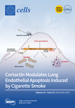
A key feature of cigarette smoke (CS)-induced lung disease is dysfunction of the vascular endothelium; however, the underlying mechanisms remain poorly described. Lung vascular endothelial cell (EC) responses to injurious stimuli such as CS or e-cigarettes are mediated by cytoskeleton changes. We have identified cortactin (CTTN), a central regulator of the actin cytoskeleton, as an important modulator of lung EC function. This study demonstrates that CS exposure induces lung EC injury by causing a series of events that include rapid phosphorylation of CTTN within minutes, subsequent cytoskeletal rearrangement, and associated ROS production, and then finally induction of apoptosis as a downstream functional effect. These results provide novel insights into CTTN function in lung EC and CS-induced lung injury. View this paper
  • Issues are regarded as officially published after their release is announced to the table of contents alert mailing list.
  • You may sign up for e-mail alerts to receive table of contents of newly released issues.
  • PDF is the official format for papers published in both, html and pdf forms. To view the papers in pdf format, click on the "PDF Full-text" link, and use the free Adobe Reader to open them.
Order results
Result details
Section
Select all
Export citation of selected articles as:
20 pages, 8050 KiB  
Article
Differential Responses of Neural Retina Progenitor Populations to Chronic Hyperglycemia
by Nicole Schmitner, Christina Recheis, Jakob Thönig and Robin A. Kimmel
Cells 2021, 10(11), 3265; https://doi.org/10.3390/cells10113265 - 22 Nov 2021
Cited by 4 | Viewed by 3084
Abstract
Diabetic retinopathy is a frequent complication of longstanding diabetes, which comprises a complex interplay of microvascular abnormalities and neurodegeneration. Zebrafish harboring a homozygous mutation in the pancreatic transcription factor pdx1 display a diabetic phenotype with survival into adulthood, and are therefore uniquely suitable [...] Read more.
Diabetic retinopathy is a frequent complication of longstanding diabetes, which comprises a complex interplay of microvascular abnormalities and neurodegeneration. Zebrafish harboring a homozygous mutation in the pancreatic transcription factor pdx1 display a diabetic phenotype with survival into adulthood, and are therefore uniquely suitable among zebrafish models for studying pathologies associated with persistent diabetic conditions. We have previously shown that, starting at three months of age, pdx1 mutants exhibit not only vascular but also neuro-retinal pathologies manifesting as photoreceptor dysfunction and loss, similar to human diabetic retinopathy. Here, we further characterize injury and regenerative responses and examine the effects on progenitor cell populations. Consistent with a negative impact of hyperglycemia on neurogenesis, stem cells of the ciliary marginal zone show an exacerbation of aging-related proliferative decline. In contrast to the robust Müller glial cell proliferation seen following acute retinal injury, the pdx1 mutant shows replenishment of both rod and cone photoreceptors from slow-cycling, neurod-expressing progenitors which first accumulate in the inner nuclear layer. Overall, we demonstrate a diabetic retinopathy model which shows pathological features of the human disease evolving alongside an ongoing restorative process that replaces lost photoreceptors, at the same time suggesting an unappreciated phenotypic continuum between multipotent and photoreceptor-committed progenitors. Full article
(This article belongs to the Special Issue The Retina in Health and Disease)
Show Figures

Figure 1

14 pages, 2694 KiB  
Article
Heat-Killed Lactobacilli Preparations Promote Healing in the Experimental Cutaneous Wounds
by Wan-Hua Tsai, Chia-Hsuan Chou, Tsuei-Yin Huang, Hui-Ling Wang, Peng-Ju Chien, Wen-Wei Chang and Hsueh-Te Lee
Cells 2021, 10(11), 3264; https://doi.org/10.3390/cells10113264 - 22 Nov 2021
Cited by 14 | Viewed by 3269
Abstract
Probiotics are defined as microorganisms with beneficial health effects when consumed by humans, being applied mainly to improve allergic or intestinal diseases. Due to the increasing resistance of pathogens to antibiotics, the abuse of antibiotics becomes inefficient in the skin and in systemic [...] Read more.
Probiotics are defined as microorganisms with beneficial health effects when consumed by humans, being applied mainly to improve allergic or intestinal diseases. Due to the increasing resistance of pathogens to antibiotics, the abuse of antibiotics becomes inefficient in the skin and in systemic infections, and probiotics may also provide the protective effect for repairing the healing of infected cutaneous wounds. Here we selected two Lactobacillus strains, L. plantarum GMNL-6 and L. paracasei GMNL-653, in heat-killed format to examine the beneficial effect in skin wound repair through the selection by promoting collagen synthesis in Hs68 fibroblast cells. The coverage of gels containing heat-killed GMNL-6 or GMNL-653 on the mouse tail with experimental wounds displayed healing promoting effects with promoting of metalloproteinase-1 expression at the early phase and reduced excessive fibrosis accumulation and deposition in the later tail-skin recovery stage. More importantly, lipoteichoic acid, the major component of Lactobacillus cell wall, from GMNL-6/GMNL-653 could achieve the anti-fibrogenic benefit similar to the heat-killed bacteria cells in the TGF-β stimulated Hs68 fibroblast cell model. Our study offers a new therapeutic potential of the heat-killed format of Lactobacillus as an alternative approach to treating skin healing disorders. Full article
(This article belongs to the Collection Molecular Determinants of Skin Integrity)
Show Figures

Graphical abstract

24 pages, 646 KiB  
Review
Treatable Traits in Chronic Respiratory Disease: A Comprehensive Review
by Yong Qin Lee, Asvin Selvakumar and Kay Choong See
Cells 2021, 10(11), 3263; https://doi.org/10.3390/cells10113263 - 22 Nov 2021
Cited by 3 | Viewed by 2615
Abstract
Chronic respiratory diseases are major contributors to the global burden of disease. While understanding of these diseases has improved, treatment guidelines have continued to rely on severity and exacerbation-based approaches. A new personalised approach, termed the “treatable traits” approach, has been suggested to [...] Read more.
Chronic respiratory diseases are major contributors to the global burden of disease. While understanding of these diseases has improved, treatment guidelines have continued to rely on severity and exacerbation-based approaches. A new personalised approach, termed the “treatable traits” approach, has been suggested to address the limitations of the existing treatment strategies. We aim to systematically review the current evidence regarding treatable traits in chronic respiratory diseases and to identify gaps in the current literature. We searched the PubMed and Embase databases and included studies on treatable traits and chronic respiratory diseases. We then extracted information on prevalence, prognostic implications, treatment options and benefits from these studies. A total of 58 papers was included for review. The traits identified were grouped into five broad themes: physiological, biochemical, psychosocial, microbiological, and comorbidity traits. Studies have shown advantages of the treatable traits paradigm in the clinical setting. However, few randomised controlled trials have been conducted. Findings from our review suggest that multidisciplinary management with therapies targeted at treatable traits has the potential to be efficacious when added to the best practices currently implemented. This paradigm has the potential to improve the holistic care of chronic respiratory diseases. Full article
(This article belongs to the Collection Pulmonary Fibrosis and Cell Therapy)
Show Figures

Graphical abstract

19 pages, 1362 KiB  
Review
The Interface between Cell Signaling Pathways and Pregnane X Receptor
by Robert S. Rogers, Annemarie Parker, Phill D. Vainer, Elijah Elliott, Dakota Sudbeck, Kaushal Parimi, Venkata P. Peddada, Parker G. Howe, Nick D’Ambrosio, Gregory Ruddy, Kaitlin Stackable, Megan Carney, Lauren Martin, Thomas Osterholt and Jeff L. Staudinger
Cells 2021, 10(11), 3262; https://doi.org/10.3390/cells10113262 - 22 Nov 2021
Cited by 6 | Viewed by 3612
Abstract
Highly expressed in the enterohepatic system, pregnane X receptor (PXR, NR1I2) is a well-characterized nuclear receptor (NR) that regulates the expression of genes in the liver and intestines that encode key drug metabolizing enzymes and drug transporter proteins in mammals. The net effect [...] Read more.
Highly expressed in the enterohepatic system, pregnane X receptor (PXR, NR1I2) is a well-characterized nuclear receptor (NR) that regulates the expression of genes in the liver and intestines that encode key drug metabolizing enzymes and drug transporter proteins in mammals. The net effect of PXR activation is to increase metabolism and clear drugs and xenobiotics from the body, producing a protective effect and mediating clinically significant drug interaction in patients on combination therapy. The complete understanding of PXR biology is thus important for the development of safe and effective therapeutic strategies. Furthermore, PXR activation is now known to specifically transrepress the inflammatory- and nutrient-signaling pathways of gene expression, thereby providing a mechanism for linking these signaling pathways together with enzymatic drug biotransformation pathways in the liver and intestines. Recent research efforts highlight numerous post-translational modifications (PTMs) which significantly influence the biological function of PXR. However, this thrust of research is still in its infancy. In the context of gene-environment interactions, we present a review of the recent literature that implicates PXR PTMs in regulating its clinically relevant biology. We also provide a discussion of how these PTMs likely interface with each other to respond to extracellular cues to appropriately modify PXR activity. Full article
Show Figures

Graphical abstract

20 pages, 52935 KiB  
Article
Microglia-Based Sex-Biased Neuropathology in Early-Stage Alzheimer’s Disease Model Mice and the Potential Pharmacologic Efficacy of Dioscin
by Xiao Liu, Qian Zhou, Jia-He Zhang, Ke-Yong Wang, Takashi Saito, Takaomi C. Saido, Xiaoying Wang, Xiumei Gao and Kagaku Azuma
Cells 2021, 10(11), 3261; https://doi.org/10.3390/cells10113261 - 22 Nov 2021
Cited by 5 | Viewed by 2905
Abstract
Alzheimer’s disease (AD), the most common form of dementia, is characterized by amyloid-β (Aβ) accumulation, microglia-associated neuroinflammation, and synaptic loss. The detailed neuropathologic characteristics in early-stage AD, however, are largely unclear. We evaluated the pathologic brain alterations in young adult App knock-in model [...] Read more.
Alzheimer’s disease (AD), the most common form of dementia, is characterized by amyloid-β (Aβ) accumulation, microglia-associated neuroinflammation, and synaptic loss. The detailed neuropathologic characteristics in early-stage AD, however, are largely unclear. We evaluated the pathologic brain alterations in young adult App knock-in model AppNL-G-F mice at 3 and 6 months of age, which corresponds to early-stage AD. At 3 months of age, microglia expression in the cortex and hippocampus was significantly decreased. By the age of 6 months, the number and function of the microglia increased, accompanied by progressive amyloid-β deposition, synaptic dysfunction, neuroinflammation, and dysregulation of β-catenin and NF-κB signaling pathways. The neuropathologic changes were more severe in female mice than in male mice. Oral administration of dioscin, a natural product, ameliorated the neuropathologic alterations in young AppNL-G-F mice. Our findings revealed microglia-based sex-differential neuropathologic changes in a mouse model of early-stage AD and therapeutic efficacy of dioscin on the brain lesions. Dioscin may represent a potential treatment for AD. Full article
Show Figures

Figure 1

12 pages, 1018 KiB  
Review
Impact of Airway Inflammation on the Efficacy of CFTR Modulators
by Carla M. P. Ribeiro and Martina Gentzsch
Cells 2021, 10(11), 3260; https://doi.org/10.3390/cells10113260 - 22 Nov 2021
Cited by 11 | Viewed by 2550
Abstract
Defective CFTR biogenesis and activity in cystic fibrosis airways leads to airway dehydration and impaired mucociliary clearance, resulting in chronic airway infection and inflammation. Most cystic fibrosis patients have at least one copy of the F508del CFTR mutation, which results in a protein [...] Read more.
Defective CFTR biogenesis and activity in cystic fibrosis airways leads to airway dehydration and impaired mucociliary clearance, resulting in chronic airway infection and inflammation. Most cystic fibrosis patients have at least one copy of the F508del CFTR mutation, which results in a protein retained in the endoplasmic reticulum and degraded by the proteosomal pathway. CFTR modulators, e.g., correctors, promote the transfer of F508del to the apical membrane, while potentiators increase CFTR activity. Corrector and potentiator double therapies modestly improve lung function, whereas triple therapies with two correctors and one potentiator indicate improved outcomes. Enhanced F508del rescue by CFTR modulators is achieved by exposing F508del/F508del primary cultures of human bronchial epithelia to relevant inflammatory stimuli, i.e., supernatant from mucopurulent material or bronchoalveolar lavage fluid from human cystic fibrosis airways. Inflammation enhances the biochemical and functional rescue of F508del by double or triple CFTR modulator therapy and overcomes abrogation of CFTR correction by chronic VX-770 treatment in vitro. Furthermore, the impact of inflammation on clinical outcomes linked to CFTR rescue has been recently suggested. This review discusses these data and possible mechanisms for airway inflammation-enhanced F508del rescue. Expanding the understanding of how airway inflammation improves CFTR rescue may benefit cystic fibrosis patients. Full article
(This article belongs to the Collection Cystic Fibrosis: Cells, Physiopathology and Emerging Therapies)
Show Figures

Graphical abstract

18 pages, 1499 KiB  
Review
The Contribution of Cardiac Fatty Acid Oxidation to Diabetic Cardiomyopathy Severity
by Qutuba G. Karwi, Qiuyu Sun and Gary D. Lopaschuk
Cells 2021, 10(11), 3259; https://doi.org/10.3390/cells10113259 - 21 Nov 2021
Cited by 19 | Viewed by 4061
Abstract
Diabetes is a major risk factor for the development of cardiovascular disease via contributing and/or triggering significant cellular signaling and metabolic and structural alterations at the level of the heart and the whole body. The main cause of mortality and morbidity in diabetic [...] Read more.
Diabetes is a major risk factor for the development of cardiovascular disease via contributing and/or triggering significant cellular signaling and metabolic and structural alterations at the level of the heart and the whole body. The main cause of mortality and morbidity in diabetic patients is cardiovascular disease including diabetic cardiomyopathy. Therefore, understanding how diabetes increases the incidence of diabetic cardiomyopathy and how it mediates the major perturbations in cell signaling and energy metabolism should help in the development of therapeutics to prevent these perturbations. One of the significant metabolic alterations in diabetes is a marked increase in cardiac fatty acid oxidation rates and the domination of fatty acids as the major energy source in the heart. This increased reliance of the heart on fatty acids in the diabetic has a negative impact on cardiac function and structure through a number of mechanisms. It also has a detrimental effect on cardiac efficiency and worsens the energy status in diabetes, mainly through inhibiting cardiac glucose oxidation. Furthermore, accelerated cardiac fatty acid oxidation rates in diabetes also make the heart more vulnerable to ischemic injury. In this review, we discuss how cardiac energy metabolism is altered in diabetic cardiomyopathy and the impact of cardiac insulin resistance on the contribution of glucose and fatty acid to overall cardiac ATP production and cardiac efficiency. Furthermore, how diabetes influences the susceptibility of the myocardium to ischemia/reperfusion injury and the role of the changes in glucose and fatty acid oxidation in mediating these effects are also discussed. Full article
(This article belongs to the Special Issue Free Fatty Acids and Pathogenesis of Diabetes Mellitus)
Show Figures

Figure 1

19 pages, 2482 KiB  
Article
Role of Endocytosis Proteins in Gefitinib-Mediated EGFR Internalisation in Glioma Cells
by Elisabete Cruz Da Silva, Laurence Choulier, Jessica Thevenard-Devy, Christophe Schneider, Philippe Carl, Philippe Rondé, Stéphane Dedieu and Maxime Lehmann
Cells 2021, 10(11), 3258; https://doi.org/10.3390/cells10113258 - 21 Nov 2021
Cited by 3 | Viewed by 2983
Abstract
EGFR (epidermal growth factor receptor), a member of the ErbB tyrosine kinase receptor family, is a clinical therapeutic target in numerous solid tumours. EGFR overexpression in glioblastoma (GBM) drives cell invasion and tumour progression. However, clinical trials were disappointing, and a molecular basis [...] Read more.
EGFR (epidermal growth factor receptor), a member of the ErbB tyrosine kinase receptor family, is a clinical therapeutic target in numerous solid tumours. EGFR overexpression in glioblastoma (GBM) drives cell invasion and tumour progression. However, clinical trials were disappointing, and a molecular basis to explain these poor results is still missing. EGFR endocytosis and membrane trafficking, which tightly regulate EGFR oncosignaling, are often dysregulated in glioma. In a previous work, we showed that EGFR tyrosine kinase inhibitors, such as gefitinib, lead to enhanced EGFR endocytosis into fused early endosomes. Here, using pharmacological inhibitors, siRNA-mediated silencing, or expression of mutant proteins, we showed that dynamin 2 (DNM2), the small GTPase Rab5 and the endocytosis receptor LDL receptor-related protein 1 (LRP-1), contribute significantly to gefitinib-mediated EGFR endocytosis in glioma cells. Importantly, we showed that inhibition of DNM2 or LRP-1 also decreased glioma cell responsiveness to gefitinib during cell evasion from tumour spheroids. By highlighting the contribution of endocytosis proteins in the activity of gefitinib on glioma cells, this study suggests that endocytosis and membrane trafficking might be an attractive therapeutic target to improve GBM treatment. Full article
(This article belongs to the Section Cellular Pathology)
Show Figures

Figure 1

10 pages, 1716 KiB  
Article
Accumulation of Genetic and Epigenetic Alterations in the Background Liver and Emergence of Hepatocellular Carcinoma in Patients with Non-Alcoholic Fatty Liver Disease
by Satoru Hagiwara, Naoshi Nishida, Kazuomi Ueshima, Yasunori Minami, Yoriaki Komeda, Tomoko Aoki, Masahiro Takita, Masahiro Morita, Hirokazu Chishina, Akihiro Yoshida, Hiroshi Ida and Masatoshi Kudo
Cells 2021, 10(11), 3257; https://doi.org/10.3390/cells10113257 - 21 Nov 2021
Cited by 6 | Viewed by 2121
Abstract
The incidence of hepatocellular carcinoma (HCC) related to non-alcoholic fatty liver disease (NAFLD) is increasing worldwide. We analyzed 16 surgically resected HCC cases in which the background liver was pathologically diagnosed as NAFLD. Specimens with Brunt classification grade 3 or higher were assigned [...] Read more.
The incidence of hepatocellular carcinoma (HCC) related to non-alcoholic fatty liver disease (NAFLD) is increasing worldwide. We analyzed 16 surgically resected HCC cases in which the background liver was pathologically diagnosed as NAFLD. Specimens with Brunt classification grade 3 or higher were assigned as the fibrotic progression group (n = 8), and those with grade 1 or lower were classified as the non-fibrosis progression group (n = 8). Comprehensive mutational and methylome analysis was performed in cancerous and noncancerous tissues. The target gene mutation analysis with deep sequencing revealed that CTNNB1 and TP53 mutation was observed in 37.5% and TERT promoter mutation was detected in 50% of cancerous samples. Furthermore, somatic mutations in non-cancerous samples were less frequent, but were observed regardless of the progression of fibrosis. Similarly, on cluster analysis of methylome data, status for methylation events involving non-cancerous liver was similar regardless of the progression of fibrosis. It was found that, even in cases of non-progressive fibrosis, accumulation of gene mutations and abnormal methylation within non-cancerous areas were observed. Patients with NAFLD require a rigorous liver cancer surveillance due to the high risk of HCC emergence based on the accumulation of genetic and epigenetic abnormalities, even when fibrosis is not advanced. Full article
(This article belongs to the Special Issue Molecular Mechanisms of Hepatocellular Carcinoma)
Show Figures

Figure 1

21 pages, 3929 KiB  
Article
Lentiviral Vectors Expressing Chimeric NEDD4 Ubiquitin Ligases: An Innovative Approach for Interfering with Alpha-Synuclein Accumulation
by Stefania Vogiatzis, Michele Celestino, Marta Trevisan, Gloria Magro, Claudia Del Vecchio, Deran Erdengiz, Giorgio Palù, Cristina Parolin, Kathleen Maguire-Zeiss and Arianna Calistri
Cells 2021, 10(11), 3256; https://doi.org/10.3390/cells10113256 - 21 Nov 2021
Cited by 3 | Viewed by 2205
Abstract
One of the main pathological features of Parkinson’s disease (PD) is a diffuse accumulation of alpha-synuclein (aS) aggregates in neurons. The NEDD4 E3 Ub ligase promotes aS degradation by the endosomal–lysosomal route. Interestingly, NEDD4, as well as being a small molecule able to [...] Read more.
One of the main pathological features of Parkinson’s disease (PD) is a diffuse accumulation of alpha-synuclein (aS) aggregates in neurons. The NEDD4 E3 Ub ligase promotes aS degradation by the endosomal–lysosomal route. Interestingly, NEDD4, as well as being a small molecule able to trigger its functions, is protective against human aS toxicity in evolutionary distant models. While pharmacological activation of E3 enzymes is not easy to achieve, their flexibility and the lack of “consensus” motifs for Ub-conjugation allow the development of engineered Ub-ligases, able to target proteins of interest. We developed lentiviral vectors, encoding well-characterized anti-human aS scFvs fused in frame to the NEDD4 catalytic domain (ubiquibodies), in order to target ubiquitinate aS. We demonstrate that, while all generated ubiquibodies bind to and ubiquitinate aS, the one directed against the non-amyloid component (NAC) of aS (Nac32HECT) affects aS’s intracellular levels. Furthermore, Nac32HECT expression partially rescues aS’s overexpression or mutation toxicity in neural stem cells. Overall, our data suggest that ubiquibodies, and Nac32HECT in particular, represent a valid platform for interfering with the effects of aS’s accumulation and aggregation in neurons. Full article
(This article belongs to the Section Cells of the Nervous System)
Show Figures

Figure 1

25 pages, 2359 KiB  
Review
Revisiting the Role of GSK3, A Modulator of Innate Immunity, in Idiopathic Inclusion Body Myositis
by Manuela Piazzi, Alberto Bavelloni, Vittoria Cenni, Irene Faenza and William L. Blalock
Cells 2021, 10(11), 3255; https://doi.org/10.3390/cells10113255 - 21 Nov 2021
Cited by 6 | Viewed by 3107
Abstract
Idiopathic or sporadic inclusion body myositis (IBM) is the leading age-related (onset >50 years of age) autoimmune muscular pathology, resulting in significant debilitation in affected individuals. Once viewed as primarily a degenerative disorder, it is now evident that much like several other neuro-muscular [...] Read more.
Idiopathic or sporadic inclusion body myositis (IBM) is the leading age-related (onset >50 years of age) autoimmune muscular pathology, resulting in significant debilitation in affected individuals. Once viewed as primarily a degenerative disorder, it is now evident that much like several other neuro-muscular degenerative disorders, IBM has a major autoinflammatory component resulting in chronic inflammation-induced muscle destruction. Thus, IBM is now considered primarily an inflammatory pathology. To date, there is no effective treatment for sporadic inclusion body myositis, and little is understood about the pathology at the molecular level, which would offer the best hopes of at least slowing down the degenerative process. Among the previously examined potential molecular players in IBM is glycogen synthase kinase (GSK)-3, whose role in promoting TAU phosphorylation and inclusion bodies in Alzheimer’s disease is well known. This review looks to re-examine the role of GSK3 in IBM, not strictly as a promoter of TAU and Abeta inclusions, but as a novel player in the innate immune system, discussing some of the recent roles discovered for this well-studied kinase in inflammatory-mediated pathology. Full article
Show Figures

Figure 1

15 pages, 1735 KiB  
Article
Effects of 5′,8′-Cyclo-2′-Deoxypurines on the Base Excision Repair of Clustered DNA Lesions in Nuclear Extracts of the XPC Cell Line
by Julia Kaźmierczak-Barańska, Karolina Boguszewska, Michał Szewczuk and Bolesław T. Karwowski
Cells 2021, 10(11), 3254; https://doi.org/10.3390/cells10113254 - 20 Nov 2021
Cited by 3 | Viewed by 1452
Abstract
Clustered DNA lesions (CDL) containing 5′,8-cyclo-2′-deoxypurines (cdPus) are an example of extensive abnormalities occurring in the DNA helix and may impede cellular repair processes. The changes in the efficiency of nuclear base excision repair (BER) were investigated using (a) two cell lines, one [...] Read more.
Clustered DNA lesions (CDL) containing 5′,8-cyclo-2′-deoxypurines (cdPus) are an example of extensive abnormalities occurring in the DNA helix and may impede cellular repair processes. The changes in the efficiency of nuclear base excision repair (BER) were investigated using (a) two cell lines, one of the normal skin fibroblasts as a reference (BJ) and the second from Xeroderma pigmentosum patients’ skin (XPC), and (b) synthetic oligonucleotides with single- and double-stranded CDL (containing 5′,8-cyclo-2′-deoxyadenosine (cdA) and the abasic (AP) site at various distances between lesions). The nuclear BER has been observed and the effect of both cdA isomers (5′R and 5′S) presence in the DNA was tested. CdPus affected the repair of the second lesion within the CDL. The BER system more efficiently processed damage in the vicinity of the ScdA isomer and changes located in the 3′-end direction for dsCDL and in the 5′-end direction for ssCDL. The presented study is the very first investigation of the repair processes of the CDL containing cdPu considering cells derived from a Xeroderma pigmentosum patient. Full article
Show Figures

Figure 1

18 pages, 9856 KiB  
Article
Hepatitis-D Virus Infection Is Not Impaired by Innate Immunity but Increases Cytotoxic T-Cell Activity
by Sebastian Maximilian Altstetter, Oliver Quitt, Francesca Pinci, Veit Hornung, Aaron Michael Lucko, Karin Wisskirchen, Stephanie Jung and Ulrike Protzer
Cells 2021, 10(11), 3253; https://doi.org/10.3390/cells10113253 - 20 Nov 2021
Cited by 2 | Viewed by 2609
Abstract
Approximately 70 million humans worldwide are affected by chronic hepatitis D, which rapidly leads to liver cirrhosis and hepatocellular carcinoma due to chronic inflammation. The triggers and consequences of this chronic inflammation, induced by co-infection with the hepatitis D virus (HDV) and the [...] Read more.
Approximately 70 million humans worldwide are affected by chronic hepatitis D, which rapidly leads to liver cirrhosis and hepatocellular carcinoma due to chronic inflammation. The triggers and consequences of this chronic inflammation, induced by co-infection with the hepatitis D virus (HDV) and the hepatitis B virus (HBV), are poorly understood. Using CRISPR technology, we characterized the recognition of HDV mono- and co-infection by intracellular innate immunity and determined its influence on the viral life cycle and effector T-cell responses using different HBV and HDV permissive hepatoma cell lines. We showed that HDV infection is detected by MDA5 and -after a lag phase -induces a profound type I interferon response in the infected cells. The type I interferon response, however, was not able to suppress HDV replication or spread, thus providing a persistent trigger. Using engineered T-cells directed against the envelope proteins commonly used by HBV and HDV, we found that HDV immune recognition enhanced T-cell cytotoxicity. Interestingly, the T-cell effector function was enhanced independently of antigen presentation. These findings help to explain immune mediated tissue damage in chronic hepatitis D patients and indicate that combining innate triggers with T-cell activating therapies might allow for a curative approach. Full article
(This article belongs to the Special Issue Signaling Pathways in Host Cell Antiviral Responses)
Show Figures

Graphical abstract

18 pages, 2230 KiB  
Review
LSD1: Expanding Functions in Stem Cells and Differentiation
by Carlos Martinez-Gamero, Sandhya Malla and Francesca Aguilo
Cells 2021, 10(11), 3252; https://doi.org/10.3390/cells10113252 - 20 Nov 2021
Cited by 22 | Viewed by 4573
Abstract
Embryonic stem cells (ESCs) and induced pluripotent stem cells (iPSC) provide a powerful model system to uncover fundamental mechanisms that control cellular identity during mammalian development. Histone methylation governs gene expression programs that play a key role in the regulation of the balance [...] Read more.
Embryonic stem cells (ESCs) and induced pluripotent stem cells (iPSC) provide a powerful model system to uncover fundamental mechanisms that control cellular identity during mammalian development. Histone methylation governs gene expression programs that play a key role in the regulation of the balance between self-renewal and differentiation of ESCs. Lysine-specific demethylase 1 (LSD1, also known as KDM1A), the first identified histone lysine demethylase, demethylates H3K4me1/2 and H3K9me1/2 at target loci in a context-dependent manner. Moreover, it has also been shown to demethylate non-histone substrates playing a central role in the regulation of numerous cellular processes. In this review, we summarize current knowledge about LSD1 and the molecular mechanism by which LSD1 influences the stem cells state, including the regulatory circuitry underlying self-renewal and pluripotency. Full article
Show Figures

Figure 1

16 pages, 2965 KiB  
Article
Dose and Dose Rate-Dependent Effects of Low-Dose Irradiation on Inflammatory Parameters in ApoE-Deficient and Wild Type Mice
by Annegret Glasow, Ina Patties, Nicholas D. Priest, Ronald E. J. Mitchel, Guido Hildebrandt and Katrin Manda
Cells 2021, 10(11), 3251; https://doi.org/10.3390/cells10113251 - 20 Nov 2021
Cited by 2 | Viewed by 2021
Abstract
Anti-inflammatory low-dose therapy is well established, whereas the immunomodulatory impact of doses below 0.1 Gy is much less clear. In this study, we investigated dose, dose rate and time-dependent effects in a dose range of 0.005 to 2 Gy on immune parameters after [...] Read more.
Anti-inflammatory low-dose therapy is well established, whereas the immunomodulatory impact of doses below 0.1 Gy is much less clear. In this study, we investigated dose, dose rate and time-dependent effects in a dose range of 0.005 to 2 Gy on immune parameters after whole body irradiation (IR) using a pro-inflammatory (ApoE−/−) and a wild type mouse model. Long-term effects on spleen function (proliferation, monocyte expression) were analyzed 3 months, and short-term effects on immune plasma parameters (IL6, IL10, IL12p70, KC, MCP1, INFγ, TGFβ, fibrinogen, sICAM, sVCAM, sE-selectin/CD62) were analyzed 1, 7 and 28 days after Co60 γ-irradiation (IR) at low dose rate (LDR, 0.001 Gy/day) and at high dose rate (HDR). In vitro measurements of murine monocyte (WEHI-274.1) adhesion and cytokine release (KC, MCP1, IL6, TGFβ) after low-dose IR (150 kV X-ray unit) of murine endothelial cell (EC) lines (H5V, mlEND1, bEND3) supplement the data. RT-PCR revealed significant reduction of Ki67 and CD68 expression in the spleen of ApoE−/− mice after 0.025 to 2 Gy exposure at HDR, but only after 2 Gy at LDR. Plasma levels in wild type mice, showed non-linear time-dependent induction of proinflammatory cytokines and reduction of TGFβ at doses as low as 0.005 Gy at both dose rates, whereas sICAM and fibrinogen levels changed in a dose rate-specific manner. In ApoE−/− mice, levels of sICAM increased and fibrinogen decreased at both dose rates, whereas TGFβ increased mainly at HDR. Non-irradiated plasma samples revealed significant age-related enhancement of cytokines and adhesion molecules except for sICAM. In vitro data indicate that endothelial cells may contribute to systemic IR effects and confirm changes of adhesion properties suggested by altered sICAM plasma levels. The differential immunomodulatory effects shown here provide insights in inflammatory changes occurring at doses far below standard anti-inflammatory therapy and are of particular importance after diagnostic and chronic environmental exposures. Full article
Show Figures

Figure 1

31 pages, 3023 KiB  
Review
Induced Pluripotent Stem Cells as a Tool for Modeling Hematologic Disorders and as a Potential Source for Cell-Based Therapies
by Ponthip Pratumkaew, Surapol Issaragrisil and Sudjit Luanpitpong
Cells 2021, 10(11), 3250; https://doi.org/10.3390/cells10113250 - 19 Nov 2021
Cited by 12 | Viewed by 5962
Abstract
The breakthrough in human induced pluripotent stem cells (hiPSCs) has revolutionized the field of biomedical and pharmaceutical research and opened up vast opportunities for drug discovery and regenerative medicine, especially when combined with gene-editing technology. Numerous healthy and patient-derived hiPSCs for human disease [...] Read more.
The breakthrough in human induced pluripotent stem cells (hiPSCs) has revolutionized the field of biomedical and pharmaceutical research and opened up vast opportunities for drug discovery and regenerative medicine, especially when combined with gene-editing technology. Numerous healthy and patient-derived hiPSCs for human disease modeling have been established, enabling mechanistic studies of pathogenesis, platforms for preclinical drug screening, and the development of novel therapeutic targets/approaches. Additionally, hiPSCs hold great promise for cell-based therapy, serving as an attractive cell source for generating stem/progenitor cells or functional differentiated cells for degenerative diseases, due to their unlimited proliferative capacity, pluripotency, and ethical acceptability. In this review, we provide an overview of hiPSCs and their utility in the study of hematologic disorders through hematopoietic differentiation. We highlight recent hereditary and acquired genetic hematologic disease modeling with patient-specific iPSCs, and discuss their applications as instrumental drug screening tools. The clinical applications of hiPSCs in cell-based therapy, including the next-generation cancer immunotherapy, are provided. Lastly, we discuss the current challenges that need to be addressed to fulfill the validity of hiPSC-based disease modeling and future perspectives of hiPSCs in the field of hematology. Full article
(This article belongs to the Special Issue iPS Cells (iPSCs) for Modelling and Treatment of Human Diseases)
Show Figures

Figure 1

14 pages, 1681 KiB  
Article
Butyrate and Class I Histone Deacetylase Inhibitors Promote Differentiation of Neonatal Porcine Islet Cells into Beta Cells
by Yichen Zhang, Yutian Lei, Mohsen Honarpisheh, Elisabeth Kemter, Eckhard Wolf and Jochen Seissler
Cells 2021, 10(11), 3249; https://doi.org/10.3390/cells10113249 - 19 Nov 2021
Cited by 10 | Viewed by 2427
Abstract
Neonatal porcine islets-like clusters (NPICCs) are a promising source for cell therapy of type 1 diabetes. Freshly isolated NPICCs are composed of progenitor cells and endocrine cells, which undergo a maturation process lasting several weeks until the normal beta cell function has developed. [...] Read more.
Neonatal porcine islets-like clusters (NPICCs) are a promising source for cell therapy of type 1 diabetes. Freshly isolated NPICCs are composed of progenitor cells and endocrine cells, which undergo a maturation process lasting several weeks until the normal beta cell function has developed. Here, we investigated the effects of short-chain fatty acids on the maturation of islet cells isolated from two to three day-old piglets. NPICCs were cultivated with acetate, butyrate and propionate (0–2000 µM) for one to eight days. Incubation with butyrate resulted in a significant upregulation of insulin gene expression and an increased beta cell number, whereas acetate or propionate had only marginal effects. Treatment with specific inhibitors of G-protein-coupled receptor GPR41 (β-hydroxybutyrate) and/or GPR43 (GPLG0974) did not abolish butyrate induced insulin expression. However, incubation of NPICCs with class I histone deacetylase inhibitors (HDACi) mocetinostat and MS275, but not selective class II HDACi (TMP269, MC1568) mimicked the butyrate effect on beta cell differentiation. Our study revealed that butyrate treatment has the capacity to increase the number of beta cells, which may be predominantly mediated through its HDAC inhibitory activity. Butyrate and specific class I HDAC inhibitors may represent beneficial supplements to promote differentiation of neonatal porcine islet cells towards beta cells for cell replacement therapies. Full article
(This article belongs to the Special Issue Signal Transduction in the Islets of Langerhans)
Show Figures

Figure 1

21 pages, 3320 KiB  
Article
Retinal Protection from LED-Backlit Screen Lights by Short Wavelength Absorption Filters
by Celia Sanchez-Ramos, Cristina Bonnin-Arias, Vanesa Blázquez-Sánchez, Victoria Aguirre-Vilacoro, Teresa Cobo, Olivia García-Suarez, María Jesús Perez-Carrasco, Cristina Alvarez-Peregrina and José A. Vega
Cells 2021, 10(11), 3248; https://doi.org/10.3390/cells10113248 - 19 Nov 2021
Cited by 4 | Viewed by 4180
Abstract
(1) Background: Ocular exposure to intense light or long-time exposure to low-intensity short-wavelength lights may cause eye injury. Excessive levels of blue light induce photochemical damage to the retinal pigment and degeneration of photoreceptors of the outer segments. Currently, people spend a lot [...] Read more.
(1) Background: Ocular exposure to intense light or long-time exposure to low-intensity short-wavelength lights may cause eye injury. Excessive levels of blue light induce photochemical damage to the retinal pigment and degeneration of photoreceptors of the outer segments. Currently, people spend a lot of time watching LED screens that emit high proportions of blue light. This study aims to assess the effects of light emitted by LED tablet screens on pigmented rat retinas with and without optical filters. (2) Methods: Commercially available tablets were used for exposure experiments on three groups of rats. One was exposed to tablet screens, the other was exposed to the tablet screens with a selective filter and the other was a control group. Structure, gene expression (including life/death, extracellular matrix degradation, growth factors, and oxidative stress related genes), and immunohistochemistry in the retina were compared among groups. (3) Results: There was a reduction of the thickness of the external nuclear layer and changes in the genes involved in cell survival and death, extracellular matrix turnover, growth factors, inflammation, and oxidative stress, leading decrease in cell density and retinal damage in the first group. Modulation of gene changes was observed when the LED light of screens was modified with an optical filter. (4) Conclusions: The use of short-wavelength selective filters on the screens contribute to reduce LED light-induced damage in the rat retina. Full article
(This article belongs to the Special Issue The Retina in Health and Disease)
Show Figures

Figure 1

13 pages, 968 KiB  
Review
The Importance of Exosomal PD-L1 in Cancer Progression and Its Potential as a Therapeutic Target
by Lingxiao Ye, Zhengxin Zhu, Xiaochuan Chen, Haoran Zhang, Jiaqi Huang, Shengxian Gu and Xiaoyin Zhao
Cells 2021, 10(11), 3247; https://doi.org/10.3390/cells10113247 - 19 Nov 2021
Cited by 26 | Viewed by 4789
Abstract
Binding of programmed cell death ligand 1 (PD-L1) to its receptor programmed cell death protein 1 (PD-1) can lead to the inactivation of cytotoxic T lymphocytes, which is one of the mechanisms for immune escape of tumors. Immunotherapy based on this mechanism has [...] Read more.
Binding of programmed cell death ligand 1 (PD-L1) to its receptor programmed cell death protein 1 (PD-1) can lead to the inactivation of cytotoxic T lymphocytes, which is one of the mechanisms for immune escape of tumors. Immunotherapy based on this mechanism has been applied in clinic with some remaining issues such as drug resistance. Exosomal PD-L1 derived from tumor cells is considered to play a key role in mediating drug resistance. Here, the effects of various tumor-derived exosomes and tumor-derived exosomal PD-L1 on tumor progression are summarized and discussed. Researchers have found that high expression of exosomal PD-L1 can inhibit T cell activation in in vitro experiments, but the function of exosomal PD-L1 in vivo remains controversial. In addition, the circulating exosomal PD-L1 has high potential to act as an indicator to evaluate the clinical effect. Moreover, therapeutic strategy targeting exosomal PD-L1 is discussed, such as inhibiting the biogenesis or secretion of exosomes. Besides, some specific methods based on the strategy of inhibiting exosomes are concluded. Further study of exosomal PD-L1 may provide an effective and safe approach for tumor treatment, and targeting exosomal PD-L1 by inhibiting exosomes may be a potential method for tumor treatment. Full article
Show Figures

Figure 1

14 pages, 824 KiB  
Review
The Impact of Acquired Genetic Abnormalities on the Clinical Translation of Human Pluripotent Stem Cells
by Alexander Keller and Claudia Spits
Cells 2021, 10(11), 3246; https://doi.org/10.3390/cells10113246 - 19 Nov 2021
Cited by 8 | Viewed by 3030
Abstract
Human pluripotent stem cells (hPSC) are known to acquire chromosomal abnormalities, which range from point mutations to large copy number changes, including full chromosome aneuploidy. These aberrations have a wide-ranging influence on the state of cells, in both the undifferentiated and differentiated state. [...] Read more.
Human pluripotent stem cells (hPSC) are known to acquire chromosomal abnormalities, which range from point mutations to large copy number changes, including full chromosome aneuploidy. These aberrations have a wide-ranging influence on the state of cells, in both the undifferentiated and differentiated state. Currently, very little is known on how these abnormalities will impact the clinical translation of hPSC, and particularly their potential to prime cells for oncogenic transformation. A further complication is that many of these abnormalities exist in a mosaic state in culture, which complicates their detection with conventional karyotyping methods. In this review we discuss current knowledge on how these aberrations influence the cell state and how this may impact the future of research and the cells’ clinical potential. Full article
(This article belongs to the Special Issue Pluripotent Stem Cells for Regenerative Medicine)
Show Figures

Figure 1

13 pages, 2473 KiB  
Review
Retinoid Agonists in the Targeting of Heterotopic Ossification
by Robert J. Pignolo and Maurizio Pacifici
Cells 2021, 10(11), 3245; https://doi.org/10.3390/cells10113245 - 19 Nov 2021
Cited by 7 | Viewed by 3305
Abstract
Retinoids are metabolic derivatives of vitamin A and regulate the function of many tissues and organs both prenatally and postnatally. Active retinoids, such as all trans-retinoic acid, are produced in the cytoplasm and then interact with nuclear retinoic acid receptors (RARs) to [...] Read more.
Retinoids are metabolic derivatives of vitamin A and regulate the function of many tissues and organs both prenatally and postnatally. Active retinoids, such as all trans-retinoic acid, are produced in the cytoplasm and then interact with nuclear retinoic acid receptors (RARs) to up-regulate the transcription of target genes. The RARs can also interact with target gene response elements in the absence of retinoids and exert a transcriptional repression function. Studies from several labs, including ours, showed that chondrogenic cell differentiation and cartilage maturation require (i) the absence of retinoid signaling and (ii) the repression function by unliganded RARs. These and related insights led to the proposition that synthetic retinoid agonists could thus represent pharmacological agents to inhibit heterotopic ossification (HO), a process that recapitulates developmental skeletogenesis and involves chondrogenesis, cartilage maturation, and endochondral ossification. One form of HO is acquired and is caused by injury, and another severe and often fatal form of it is genetic and occurs in patients with fibrodysplasia ossificans progressiva (FOP). Mouse models of FOP bearing mutant ACVR1R206H, characteristic of most FOP patients, were used to test the ability of the retinoid agonists selective for RARα and RARγ against spontaneous and injury-induced HO. The RARγ agonists were found to be most effective, and one such compound, palovarotene, was selected for testing in FOP patients. The safety and effectiveness data from recent and ongoing phase II and phase III clinical trials support the notion that palovarotene may represent a disease-modifying treatment for patients with FOP. The post hoc analyses showed substantial efficacy but also revealed side effects and complications, including premature growth plate closure in some patients. Skeletally immature patients will need to be carefully weighed in any future regulatory indications of palovarotene as an important therapeutic option in FOP. Full article
(This article belongs to the Special Issue Retinoic Acid and Retinoid X Receptors)
Show Figures

Figure 1

19 pages, 5283 KiB  
Article
Quantification of EGFR-HER2 Heterodimers in HER2-Overexpressing Breast Cancer Cells Using Liquid-Phase Electron Microscopy
by Diana B. Peckys, Daniel Gaa and Niels de Jonge
Cells 2021, 10(11), 3244; https://doi.org/10.3390/cells10113244 - 19 Nov 2021
Cited by 4 | Viewed by 2800
Abstract
Currently, breast cancer patients are classified uniquely according to the expression level of hormone receptors, and human epidermal growth factor receptor 2 (HER2). This coarse classification is insufficient to capture the phenotypic complexity and heterogeneity of the disease. A methodology was developed for [...] Read more.
Currently, breast cancer patients are classified uniquely according to the expression level of hormone receptors, and human epidermal growth factor receptor 2 (HER2). This coarse classification is insufficient to capture the phenotypic complexity and heterogeneity of the disease. A methodology was developed for absolute quantification of receptor surface density ρR, and molecular interaction (dimerization), as well as the associated heterogeneities, of HER2 and its family member, the epidermal growth factor receptor (EGFR) in the plasma membrane of HER2 overexpressing breast cancer cells. Quantitative, correlative light microscopy (LM) and liquid-phase electron microscopy (LPEM) were combined with quantum dot (QD) labeling. Single-molecule position data of receptors were obtained from scanning transmission electron microscopy (STEM) images of intact cancer cells. Over 280,000 receptor positions were detected and statistically analyzed. An important finding was the subcellular heterogeneity in heterodimer shares with respect to plasma membrane regions with different dynamic properties. Deriving quantitative information about EGFR and HER2 ρR, as well as their dimer percentages, and the heterogeneities thereof, in single cancer cells, is potentially relevant for early identification of patients with HER2 overexpressing tumors comprising an enhanced share of EGFR dimers, likely increasing the risk for drug resistance, and thus requiring additional targeted therapeutic strategies. Full article
(This article belongs to the Special Issue Emerging Targets and Therapeutic Strategies in Cancer)
Show Figures

Graphical abstract

16 pages, 2361 KiB  
Article
Microbiome Analysis of Mucosal Ileoanal Pouch in Ulcerative Colitis Patients Revealed Impairment of the Pouches Immunometabolites
by Orazio Palmieri, Stefano Castellana, Giuseppe Biscaglia, Anna Panza, Anna Latiano, Rosanna Fontana, Maria Guerra, Giuseppe Corritore, Tiziana Latiano, Giuseppina Martino, Tommaso Mazza, Angelo Andriulli, Francesco Perri and Fabrizio Bossa
Cells 2021, 10(11), 3243; https://doi.org/10.3390/cells10113243 - 19 Nov 2021
Cited by 8 | Viewed by 3458
Abstract
The pathogenesis of ulcerative colitis (UC) is unknown, although genetic loci and altered gut microbiota have been implicated. Up to a third of patients with moderate to severe UC require proctocolectomy with ileal pouch ano-anastomosis (IPAA). We aimed to explore the mucosal microbiota [...] Read more.
The pathogenesis of ulcerative colitis (UC) is unknown, although genetic loci and altered gut microbiota have been implicated. Up to a third of patients with moderate to severe UC require proctocolectomy with ileal pouch ano-anastomosis (IPAA). We aimed to explore the mucosal microbiota of UC patients who underwent IPAA. Methods: For microbiome analysis, mucosal specimens were collected from 34 IPAA individuals. Endoscopic and histological examinations of IPAA were normal in 21 cases, while pouchitis was in 13 patients. 19 specimens from the healthy control (10 from colonic and 9 from ileum) were also analyzed. Data were analyzed using an ensemble of software packages: QIIME2, coda-lasso, clr-lasso, PICRUSt2, and ALDEx2. Results: IPAA specimens had significantly lower bacterial diversity as compared to normal. The microbial composition of the normal pouch was also decreased also when compared to pouchitis. Faecalibacterium prausnitzii, Gemmiger formicilis, Blautia obeum, Ruminococcus torques, Dorea formicigenerans, and an unknown species from Roseburia were the most uncommon in pouch/pouchitis, while an unknown species from Enterobacteriaceae was over-represented. Propionibacterium acnes and Enterobacteriaceae were the species most abundant in the pouchitis and in the normal pouch, respectively. Predicted metabolic pathways among the IPAA bacterial communities revealed an important role of immunometabolites such as SCFA, butyrate, and amino acids. Conclusions: Our findings showed specific bacterial signature hallmarks of dysbiosis and could represent bacterial biomarkers in IPAA patients useful to develop novel treatments in the future by modulating the gut microbiota through the administration of probiotic immunometabolites-producing bacterial strains and the addition of specific prebiotics and the faecal microbiota transplantation. Full article
(This article belongs to the Special Issue Microbiota, Inflammation and Systemic Diseases)
Show Figures

Figure 1

20 pages, 9912 KiB  
Review
The Role of Fibroblast Growth Factor (FGF) Signaling in Tissue Repair and Regeneration
by Mariya Farooq, Abdul Waheed Khan, Moon Suk Kim and Sangdun Choi
Cells 2021, 10(11), 3242; https://doi.org/10.3390/cells10113242 - 19 Nov 2021
Cited by 55 | Viewed by 9977
Abstract
Fibroblast growth factors (FGFs) are a large family of secretory molecules that act through tyrosine kinase receptors known as FGF receptors. They play crucial roles in a wide variety of cellular functions, including cell proliferation, survival, metabolism, morphogenesis, and differentiation, as well as [...] Read more.
Fibroblast growth factors (FGFs) are a large family of secretory molecules that act through tyrosine kinase receptors known as FGF receptors. They play crucial roles in a wide variety of cellular functions, including cell proliferation, survival, metabolism, morphogenesis, and differentiation, as well as in tissue repair and regeneration. The signaling pathways regulated by FGFs include RAS/mitogen-activated protein kinase (MAPK), phosphatidylinositol-4,5-bisphosphate 3-kinase (PI3K)–protein kinase B (AKT), phospholipase C gamma (PLCγ), and signal transducer and activator of transcription (STAT). To date, 22 FGFs have been discovered, involved in different functions in the body. Several FGFs directly or indirectly interfere with repair during tissue regeneration, in addition to their critical functions in the maintenance of pluripotency and dedifferentiation of stem cells. In this review, we summarize the roles of FGFs in diverse cellular processes and shed light on the importance of FGF signaling in mechanisms of tissue repair and regeneration. Full article
(This article belongs to the Collection Fibroblast Growth Factors: Pathophysiology and Therapeutics)
Show Figures

Figure 1

15 pages, 914 KiB  
Review
The Secrets of Alternative Autophagy
by Kaja Urbańska and Arkadiusz Orzechowski
Cells 2021, 10(11), 3241; https://doi.org/10.3390/cells10113241 - 19 Nov 2021
Cited by 9 | Viewed by 3546
Abstract
For many years, it was thought that ATG5 and ATG7 played a pivotal role in autophagy, and that the knockdown of one of these genes would result in its inhibition. However, cells with ATG5 or ATG7 depletion still generate autophagic vacuoles with mainly [...] Read more.
For many years, it was thought that ATG5 and ATG7 played a pivotal role in autophagy, and that the knockdown of one of these genes would result in its inhibition. However, cells with ATG5 or ATG7 depletion still generate autophagic vacuoles with mainly trans-Golgi-originated isolation membranes and do not die. This indicates that autophagy can occur via ATG5/ATG7-independent alternative autophagy. Its molecular mechanism differs from that of the canonical pathway, including inter alia the phosphorylation of ULK1, and lack of LC3 modifications. As the alternative autophagy pathway has only recently been described, little is known of its precise role; however, a considerable body of evidence suggests that alternative autophagy participates in mitochondrion removal. This review summarizes the latest progress made in research on alternative autophagy and describes its possible molecular mechanism, roles and methods of detection, and possible modulators. There is a need for further research focused on types of autophagy, as this can elucidate the functioning of various cell types and the pathogenesis of human and animal diseases. Full article
(This article belongs to the Special Issue 10th Anniversary of Cells—Advances in Autophagy)
Show Figures

Figure 1

9 pages, 1004 KiB  
Brief Report
Enduring Effects of Conditional Brain Serotonin Knockdown, Followed by Recovery, on Adult Rat Neurogenesis and Behavior
by Maria Sidorova, Golo Kronenberg, Susann Matthes, Markus Petermann, Rainer Hellweg, Oksana Tuchina, Michael Bader, Natalia Alenina and Friederike Klempin
Cells 2021, 10(11), 3240; https://doi.org/10.3390/cells10113240 - 19 Nov 2021
Cited by 2 | Viewed by 2176
Abstract
Serotonin (5-hydroxytryptamine, 5-HT) is a crucial signal in the neurogenic niche of the hippocampus, where it is involved in antidepressant action. Here, we utilized a new transgenic rat model (TetO-shTPH2), where brain 5-HT levels can be acutely altered based on doxycycline (Dox)-inducible shRNA-expression. [...] Read more.
Serotonin (5-hydroxytryptamine, 5-HT) is a crucial signal in the neurogenic niche of the hippocampus, where it is involved in antidepressant action. Here, we utilized a new transgenic rat model (TetO-shTPH2), where brain 5-HT levels can be acutely altered based on doxycycline (Dox)-inducible shRNA-expression. On/off stimulations of 5-HT concentrations might uniquely mirror the clinical course of major depression (e.g., relapse after discontinuation of antidepressants) in humans. Specifically, we measured 5-HT levels, and 5-HT metabolite 5-HIAA, in various brain areas following acute tryptophan hydroxylase 2 (Tph2) knockdown, and replenishment, and examined behavior and proliferation and survival of newly generated cells in the dentate gyrus. We found that decreased 5-HT levels in the prefrontal cortex and raphe nuclei, but not in the hippocampus of TetO-shTPH2 rats, lead to an enduring anxious phenotype. Surprisingly, the reduction in 5-HT synthesis is associated with increased numbers of BrdU-labeled cells in the dentate gyrus. At 3 weeks of Tph2 replenishment, 5-HT levels return to baseline and survival of newly generated cells is unaffected. We speculate that the acutely induced decrease in 5-HT concentrations and increased neurogenesis might represent a compensatory mechanism. Full article
Show Figures

Graphical abstract

42 pages, 1233 KiB  
Review
Localization and Functional Roles of Components of the Translation Apparatus in the Eukaryotic Cell Nucleus
by Zaur M. Kachaev, Sergey D. Ivashchenko, Eugene N. Kozlov, Lyubov A. Lebedeva and Yulii V. Shidlovskii
Cells 2021, 10(11), 3239; https://doi.org/10.3390/cells10113239 - 19 Nov 2021
Cited by 6 | Viewed by 4851
Abstract
Components of the translation apparatus, including ribosomal proteins, have been found in cell nuclei in various organisms. Components of the translation apparatus are involved in various nuclear processes, particularly those associated with genome integrity control and the nuclear stages of gene expression, such [...] Read more.
Components of the translation apparatus, including ribosomal proteins, have been found in cell nuclei in various organisms. Components of the translation apparatus are involved in various nuclear processes, particularly those associated with genome integrity control and the nuclear stages of gene expression, such as transcription, mRNA processing, and mRNA export. Components of the translation apparatus control intranuclear trafficking; the nuclear import and export of RNA and proteins; and regulate the activity, stability, and functional recruitment of nuclear proteins. The nuclear translocation of these components is often involved in the cell response to stimulation and stress, in addition to playing critical roles in oncogenesis and viral infection. Many components of the translation apparatus are moonlighting proteins, involved in integral cell stress response and coupling of gene expression subprocesses. Thus, this phenomenon represents a significant interest for both basic and applied molecular biology. Here, we provide an overview of the current data regarding the molecular functions of translation factors and ribosomal proteins in the cell nucleus. Full article
Show Figures

Figure 1

23 pages, 7578 KiB  
Article
Transcriptional Profiles of Cell Fate Transitions Reveal Early Drivers of Neuronal Apoptosis and Survival
by Giovanna Morello, Ambra Villari, Antonio Gianmaria Spampinato, Valentina La Cognata, Maria Guarnaccia, Giulia Gentile, Maria Teresa Ciotti, Pietro Calissano, Velia D’Agata, Cinzia Severini and Sebastiano Cavallaro
Cells 2021, 10(11), 3238; https://doi.org/10.3390/cells10113238 - 19 Nov 2021
Cited by 3 | Viewed by 3066
Abstract
Neuronal apoptosis and survival are regulated at the transcriptional level. To identify key genes and upstream regulators primarily responsible for these processes, we overlayed the temporal transcriptome of cerebellar granule neurons following induction of apoptosis and their rescue by three different neurotrophic factors. [...] Read more.
Neuronal apoptosis and survival are regulated at the transcriptional level. To identify key genes and upstream regulators primarily responsible for these processes, we overlayed the temporal transcriptome of cerebellar granule neurons following induction of apoptosis and their rescue by three different neurotrophic factors. We identified a core set of 175 genes showing opposite expression trends at the intersection of apoptosis and survival. Their functional annotations and expression signatures significantly correlated to neurological, psychiatric and oncological disorders. Transcription regulatory network analysis revealed the action of nine upstream transcription factors, converging pro-apoptosis and pro-survival-inducing signals in a highly interconnected functionally and temporally ordered manner. Five of these transcription factors are potential drug targets. Transcriptome-based computational drug repurposing produced a list of drug candidates that may revert the apoptotic core set signature. Besides elucidating early drivers of neuronal apoptosis and survival, our systems biology-based perspective paves the way to innovative pharmacology focused on upstream targets and regulatory networks. Full article
(This article belongs to the Special Issue Neurotrophic Factors in Health and Disease)
Show Figures

Figure 1

15 pages, 4598 KiB  
Article
Gliogenic Potential of Single Pallial Radial Glial Cells in Lower Cortical Layers
by Ana Cristina Ojalvo-Sanz and Laura López-Mascaraque
Cells 2021, 10(11), 3237; https://doi.org/10.3390/cells10113237 - 19 Nov 2021
Cited by 6 | Viewed by 1946
Abstract
During embryonic development, progenitor cells are progressively restricted in their potential to generate different neural cells. A specific progenitor cell type, the radial glial cells, divides symmetrically and then asymmetrically to produce neurons, astrocytes, oligodendrocytes, and NG2-glia in the cerebral cortex. However, the [...] Read more.
During embryonic development, progenitor cells are progressively restricted in their potential to generate different neural cells. A specific progenitor cell type, the radial glial cells, divides symmetrically and then asymmetrically to produce neurons, astrocytes, oligodendrocytes, and NG2-glia in the cerebral cortex. However, the potential of individual progenitors to form glial lineages remains poorly understood. To further investigate the cell progeny of single pallial GFAP-expressing progenitors, we used the in vivo genetic lineage-tracing method, the UbC-(GFAP-PB)-StarTrack. After targeting those progenitors in embryonic mice brains, we tracked their adult glial progeny in lower cortical layers. Clonal analyses revealed the presence of clones containing sibling cells of either a glial cell type (uniform clones) or two different glial cell types (mixed clones). Further, the clonal size and rostro-caudal cell dispersion of sibling cells differed depending on the cell type. We concluded that pallial E14 neural progenitors are a heterogeneous cell population with respect to which glial cell type they produce, as well as the clonal size of their cell progeny. Full article
(This article belongs to the Special Issue Single Cell Analysis 2.0)
Show Figures

Figure 1

17 pages, 3902 KiB  
Article
Matrix Metalloproteinases Inhibition by Doxycycline Rescues Extracellular Matrix Organization and Partly Reverts Myofibroblast Differentiation in Hypermobile Ehlers-Danlos Syndrome Dermal Fibroblasts: A Potential Therapeutic Target?
by Nicola Chiarelli, Nicoletta Zoppi, Marina Venturini, Daniele Capitanio, Cecilia Gelfi, Marco Ritelli and Marina Colombi
Cells 2021, 10(11), 3236; https://doi.org/10.3390/cells10113236 - 19 Nov 2021
Cited by 7 | Viewed by 5764
Abstract
Hypermobile Ehlers-Danlos syndrome (hEDS) is the most frequent type of EDS and is characterized by generalized joint hypermobility and musculoskeletal manifestations which are associated with chronic pain, and mild skin involvement along with the presence of more than a few comorbid conditions. Despite [...] Read more.
Hypermobile Ehlers-Danlos syndrome (hEDS) is the most frequent type of EDS and is characterized by generalized joint hypermobility and musculoskeletal manifestations which are associated with chronic pain, and mild skin involvement along with the presence of more than a few comorbid conditions. Despite numerous research efforts, no causative gene(s) or validated biomarkers have been identified and insights into the disease-causing mechanisms remain scarce. Variability in the spectrum and severity of symptoms and progression of hEDS patients’ phenotype likely depend on a combination of age, gender, lifestyle, and the probable multitude of genes involved in hEDS. However, considering the clinical overlap with other EDS forms, which lead to abnormalities in extracellular matrix (ECM), it is plausible that the mechanisms underlying hEDS pathogenesis also affect the ECM to a certain extent. Herein, we performed a series of in vitro studies on the secretome of hEDS dermal fibroblasts that revealed a matrix metalloproteinases (MMPs) dysfunction as one of the major disease drivers by causing a detrimental feedback loop of excessive ECM degradation coupled with myofibroblast differentiation. We demonstrated that doxycycline-mediated inhibition of MMPs rescues in hEDS cells a control-like ECM organization and induces a partial reversal of their myofibroblast-like features, thus offering encouraging clues for translational studies confirming MMPs as a potential therapeutic target in hEDS with the expectation to improve patients’ quality of life and alleviate their disabilities. Full article
Show Figures

Figure 1

Previous Issue
Next Issue
Back to TopTop