Article

Rhizosphere Microbiomes of Amaranthus spp. Grown in Soils with Anthropogenic Polyelemental Anomalies

1
Institute of Biochemistry and Physiology of Plants and Microorganisms, Saratov Scientific Centre of the Russian Academy of Sciences (IBPPM RAS), Saratov 410049, Russia
2
Natural Science Institute, Tula State University, Tula 300012, Russia
3
Institute of Fundamental Medicine and Biology, Kazan (Volga Region) Federal University, Kazan 420021, Russia
4
Agrarian and Technological Institute, Peoples’ Friendship University of Russia, Moscow 117198, Russia
5
Federal State Budgetary Scientific Institution “Federal Scientific Vegetable Center”, Odintsovo 143080, Russia
*
Author to whom correspondence should be addressed.
Agronomy 2023, 13(3), 759; https://doi.org/10.3390/agronomy13030759
Submission received: 7 February 2023 / Revised: 27 February 2023 / Accepted: 3 March 2023 / Published: 6 March 2023
(This article belongs to the Special Issue Metagenomic Analysis for Unveiling Agricultural Microbiome)

Abstract

:
Study of rhizospheric microbial communities of plants growing under different environmental conditions is important for understanding the habitat-dependent formation of rhizosphere microbiomes. The rhizosphere bacterial communities of four amaranth cultivars were investigated in a laboratory pot experiment. Amaranthus tricolor cv. Valentina, A. cruentus cv. Dyuimovochka, and A. caudatus cvs. Bulava and Zelenaya Sosulka were grown for six months in three soils with different anthropogenic polyelemental anomalies and in a background control soil. After the plant cultivation, the rhizosphere soils were sampled and subjected to metagenomic analysis for the 16S rRNA gene. The results showed that the taxonomic structure of the amaranth rhizosphere microbiomes was represented by the dominant bacterial phyla Actinobacteriota and Proteobacteria. A feature of the taxonomic profile of the rhizobiomes of A. tricolor cv. Valentina and A. cruentus cv. Dyuimovochka was a large abundance of sequences related to Cyanobacteria. The formation of the amaranth rhizosphere microbiomes was largely unaffected by soils, but cultivar differences in the formation of the amaranth rhizosphere microbial structure were revealed. Bacterial taxa were identified that are possibly selected by amaranths and that may be important for plant adaptation to various habitat conditions. The targeted enrichment of the amaranth rhizosphere with members of these taxa could be useful for improving the efficacy of amaranth use for agricultural and remediation purposes.

1. Introduction

In the past decade, research in rhizosphere biology has enjoyed an increased interest [1,2,3,4]. This interest is because the plant root zone is a unique niche that is saturated with physical, chemical, and biological interactions between macro- and microorganisms and between organisms and their environment. Studies on the functions of rhizosphere microorganisms have led researchers to understand their important role in plant life. Specifically, microbes improve plant nutrition through atmospheric nitrogen fixation, mobilization of hard-to-reach phosphorus, increased availability of trace elements, and siderophore production. Furthermore, microbes participate in plant growth regulation through the production of phytohormones and other phytoactive substances, and they increase plant adaptability through stimulation of the antioxidant defense, induction of systemic resistance, and protection from pathogens and organic/inorganic toxicants. Rhizosphere microbial communities are studied by both culture-based [2,5] and culture-independent methods [3,6,7,8], which helps to protect plants against diseases, improve yields, and increase the efficacy of plant use to restore disturbed soils (phytoremediation). Modern molecular technologies make it possible to develop tools for the artificial modeling of rhizosphere microbiomes, which is of great importance for improving agricultural biotechnologies and predicting their results [9].
The main factors affecting the formation of plant-microbial complexes are the plant species [10] and the soil type [11]. Plant-root-associated microbiomes are considered an important extension of the plant genome itself [12]. The endosphere is the most plant-species-dependent and conserved compartment of rhizobacteria, which determines the so-called plant core microbiome [13]. Components of the core microbiome can be present in and can largely determine the composition of the microbiomes of other root zones, including the rhizoplane and the rhizosphere [10,14]. By contrast, the root-free edaphosphere (bulk soil) is the most plant-independent niche, whose microbiome is characterized by greater variability and is determined by the soil type. The rhizosphere microbiome is crucial for connecting plant and soil microbiomes [12,15]. Plants control the composition of their rhizosphere microbiome via root exudates and can modify it by selecting beneficial microorganisms, thereby contributing largely to the effectiveness of agricultural biotechnologies [1,14,16].
The organisms present in the rhizosphere include bacteria, fungi, oomycetes, nematodes, protozoa, algae, viruses, archaea, and arthropods [15], with bacteria being the most numerous. Bacterial diversity in the rhizosphere can be heavily influenced by abiotic and environmental conditions and can differ depending on the soil type and the plant genotype [7]. Studies on the metagenomes of plant rhizosphere communities are extremely relevant in modern science, because they carry specific information on the rhizobiomes of economically important plants and lead researchers to develop methods for their modification so that the efficiency of a particular agricultural biotechnology can be increased [7,15,17,18]. The rhizosphere microbiome composition has already been characterized for many plant species (e.g., [15,17,19,20]).
Amaranth is a widespread plant genus, many species of which are of great economic importance. A. caudatus, A. cruentus, and some other species are ancient grain and oilseed crops, which are grown in many countries. A. tricolor is also grown as a vegetable plant rich in essential amino acids and biologically active substances [21]. Many cultivars of A. caudatus, A. hypochondriacus, A. tricolor, and other species have richly colored leaves and hanging inflorescences and are used as ornamental crops. A number of Amaranthus plants are able to accumulate heavy metals [22,23,24,25,26] and radionuclides [27] and are, therefore, regarded as promising remediators. Soil phytoremediation from both organic and inorganic pollutants is known based on plant-microbial interactions [28], and the presence in the plant rhizosphere of microorganisms resistant to pollutants and able to promote plant growth is critical to the phytoremediation of polluted soils. In this context, the study of rhizosphere microbial communities may contribute to the characterization of the remediation potential of amaranth plants. Information on amaranth-associated rhizosphere microorganisms is extremely limited, but interesting. It was reported that rhizosphere microbiomes of a number of amaranth species are characterized by a pronounced abundance of representatives of Cyanobacteria [29].
The purpose of the study was to characterize the microbial communities of four Amaranthus cultivars grown on soils with anthropogenic polyelemental anomalies. Comparison of the rhizosphere microbiomes of plants growing under different environmental conditions will make it possible to better understand the habitat-dependent and species-(or cultivar)-dependent formation of microbiomes of these plants.

2. Materials and Methods

2.1. Soil Sampling

The soil samples used in the pot experiment were those of urban soils based on gray forest soils (WRB, 2006: Greyic Phaeozems), which were characterized by polyelemental anomalies [30]. The soils were collected in the sanitary protection zones near Kosaya Gora Iron Works (KGIW; Tula, Russia) and Tulachermet Co. (Tula, Russia) and on Tula’s central avenue, Lenin Avenue. The background soil was collected near the Yasnaya Polyana museum estate of Leo Tolstoy (Tula Region, Russia). The content of toxic substances in the background soil did not exceed the maximum permissible concentration (MPC) and approximate permissible concentration (APC); however, the iron content was quite high as compared with the world values, but minimal as compared with that of the urban soils used. Soil was sampled and prepared for the determination of toxic elements in accordance with the Russian State Standard [31]. Soil samples (~2 kg) from each location were taken from a depth of 0–25 cm from several sampling points using the envelope method. In total, at least 15 samples taken from each location were mixed and used for the pot experiment and analyses.

2.2. Soil Characterization

Soil used in our experiment was characterized on several parameters, such as type (on the basis of particle size distribution); pH; total carbon, water-soluble carbon, humus, N-NO3, N-NH4, and P2O5 contents; oil products; and metal(loid)s content. In the potassium chloride extract of soil, the potentiometric determination of pH using a glass electrode and a Mettler Toledo Delta 320 pH meter (Mettler-Toledo Instruments Shanghai Ltd., Shanghai, China) was carried out in accordance with the Russian State Standard [32]. Total organic carbon was determined according to [33]. The technique included the dichromate-wet combustion of soil organic matter by concentrated sulfuric acid and the quantitative colorimetric determination (at 590 nm) of the amount of Cr2+ generated by dichromate oxidation of soil organic matter. To determine the content of water-soluble carbon, aqueous (distilled water) extracts from the soil samples were obtained, and after drying, were subjected to the same procedure as the dried soil samples. Based on the data obtained, the total carbon, water-soluble carbon, and humus content were calculated. The content of mobile (available) phosphorus (P2O5) in mg/100 g was measured photocolorimetrically according to [34]. The method is based on the extraction of mobile compounds of phosphorus from the soil with a solution of ammonium carbonate and the subsequent photocolorimetric determination (at 710 nm) of phosphorus in the form of a blue phosphorus–molybdenum complex. Nitrates and water-soluble ammonium were measured by standard photocolorimetric methods [35,36]. The determination of nitrates included the extraction of nitrates from soil with a potassium chloride solution, reduction of nitrates to nitrites with hydrazine in the presence of copper as a catalyst, and photometric measurement (at 545 nm) of the colored diazo compound formed. The determination of exchangeable ammonium included the extraction of exchangeable ammonium from soil with a potassium chloride solution, generation of a colored indophenol compound formed by the interaction of ammonium with hypochlorite and sodium salicylate in an alkaline medium, and photometry of the colored solution (at 655 nm). All photocolorimetric measurements were carried out using an Evolution 60 UV-Vis Spectrophotometer (Thermo Scientific, Madison, WI USA). Oil products were measured gravimetrically [37]. The method is based on extraction of oil products with chloroform from air-dried soil, separation from polar compounds by liquid chromatography after replacing the solvent with hexane, and quantitative determination by gravimetric analysis. The determination of the elemental composition of soils was carried out using X-ray fluorescence analysis in the certified Laboratory of Chemical-analytical Research of GIN RAS. Soil preparation for analysis and quality control was carried out following certified methods and recommendations [30,38]. The concentrations of Mn, Fe, V, Cr, Ni, Cu, Zn, Pb, and As were determined using a serial wave XRF spectrometer “S4 Pioneer” (Bruker AXS GmbH, Karlsruhe, Germany) with a rhodium tube (capacity 4 kW). The obtained data were processed using the S4 Spectra Plus program using coefficients for correction of the routine samples matrix effects. Standard samples of the composition IAEA Soil-7, SChT-1.2 (soil), GBW-07404, 07405 (soil) were used as reference samples for soil analysis. The concentration of potentially toxic trace elements in the soil was compared with the MPCs and APCs of the metals by hygienic standards that meet international standards [39,40]. All chemical analyses of soil were performed in at least triplicate.

2.3. Experimental Design

We used four Amaranthus cultivars bred by the All-Russian Research Institute of Vegetable Breeding and Seed Production of the Federal Scientific Vegetable Center (VNIISSOK; Odintsovo District, Moscow Region, Russia). The specific cultivars were Valentina (A. tricolor L.), Dyuimovochka (A. cruentus L.), Bulava (A. caudatus L.), and Zelenaya Sosulka (A. caudatus L.). The description of the Amaranthus cultivars used is given in Table S1.
Plant seeds (~50 mg) were sown in soil-filled 2 L plastic pots. After one month, the seedlings were thinned out, leaving 10 plants per pot. Plants were cultivated under controlled conditions (day/night light cycle, 14/10 h; temperature, 22/24 °C) for 6 months. There were three replicates for each combination of Amaranthus cultivar and soil. At the end of growth, the plants were removed from the pots, and the soil was vigorously shaken off the roots. The rhizosphere soil adhering to young roots from 5 plants from each pot were combined from three replicates of one variant, and mixed samples were used for metagenomic analysis. The remaining soil adhering to the root surface as rhizosphere soil was carefully scraped off with sterilized tweezers.

2.4. A 16S rRNA Gene-Based Metagenomic Analysis of Rhizosphere Soil

Extraction and purification of soil DNA for metagenomic analysis was carried out using the Fast DNA®SPIN Kit for Soil (MP Biomedicals, Santa Ana, CA, USA) and a Fast Prep®24 homogenizer (MP Biomedicals, Santa Ana, CA, USA), according to the manufacturer’s instructions.
A 16S rRNA sequencing library was constructed, according to the 16S metagenomics sequencing library preparation protocol (Illumina, San Diego, CA, USA), targeting the V3 and V4 hypervariable regions of the 16S rRNA gene. The initial PCR was performed with template DNA using region-specific adapters shown to have compatibility with the Illumina index and sequencing primers (forward primer: 5′-TCGTCGGCAGCGTCAGATGTGTATAAGAGACAGCCTACGGGNGGCWGCAG-3′; reverse primer: 5′-GTCTCGTGGGCTCGGAGATGTGTATAAGAGACAGGACTACHVGGGTATCTAATCC-3′) [41]. Amplification was performed using the Veriti™ Thermal Cycle, 96-Well (Applied Biosystems™, Foster City, CA, USA), according to the Illumina protocol. After the first round of amplification, PCR products were visualized using gel electrophoresis. Then, the PCR products were purified with AMPure XT magnetic beads, and the second PCR was performed using primers from a Nextera XT Index Kit (Illumina). Subsequently, purified PCR products were quantified with a Qubit dsDNA HS Assay Kit (Thermo Scientific) on a Qubit 2.0 fluorometer. The sample pool (4 nM) was denatured with 0.2 N NaOH, diluted further to 10 pM, and combined with 20% (v/v) denatured 4 pM PhiX, prepared following Illumina guidelines. Sequencing of 16S rRNA gene V3–V4 variable regions was performed on the Illumina MiSeq platform in 2 × 300 bp mode.

2.5. Bioinformatics and Statistical Analysis

Reads were analyzed using QIIME2 software, version 2022.8 (http://qiime2.org/, accessed on 25 February 2023) [42]. Before filtering, there were 118,339 read pairs per sample on average. Raw reads were processed using the DADA2 algorithm implemented in QIIME [43]. After quality filtering, chimera and phiX sequences removal, we analyzed 17,870 joined read pairs per sample on average. The taxonomy was assigned to the sequences using the Naive Bayes classifier pre-trained on the latest SILVA 138 database 99% OTUs [44]. The number of observed features varied from 601 to 1030. To characterize the richness and evenness of the bacterial community, alpha diversity indices were calculated using Chao1, Shannon, and Simpson metrics. Similarities between microbial compositions of the samples were evaluated using the beta diversity characteristics, which were estimated using weighted and unweighted Unifrac measures with further non-metric multidimensional scaling (nMDS) visualization.
Venn diagrams were constructed with Creately software (https://creately.com/lp/venn-diagram-maker/, accessed on 5 February 2023; Cinergix Pty Ltd., Melbourne, VIC, Australia). Spearman’s rank correlation coefficients were calculated with Statistica software 13 (TIBCO Software Inc. 2017, Statsoft Russia, Moscow, Russia). Other calculations were done with Excel 2019 software (Microsoft, Redmond, WA, USA).

3. Results

3.1. Soil Characteristics

For all soils used in the experiment, the main characteristics were determined: type; pH; and the total content of (including water-soluble) carbon, biogenic forms of nitrogen (NH4 and NO3) and phosphorus (P2O5), heavy metals and metalloids (Fe, Mn, V, Ni, Cu, Zn, Pb, and As), and hydrocarbons. The results are given in Table 1.
All soil samples were represented by gray forest soils, had neutral or close to slightly acidic pH values, and were also characterized by a high content of iron. The soils of the sanitary protection zones near Kosaya Gora Iron Works (KGIW; Tula, Russia) and Tulachermet Co. (Tula, Russia) and the urban soil of Tula’s central avenue, Lenin Avenue, all have an excessive content of heavy metals and oil products, indicating human-caused pollution. The KGIW soil had a high content of Mn, Zn, and petroleum products, which exceeded MPCs by 380, 140, and 260%, respectively. The Tulachermet soil had a high content of V, Ni, Cu, Zn, As, and oil products, exceeding MPCs and APCs by 36, 275, 127, 290, 300, and 410%, respectively. The Lenin Avenue soil had a high content of Mn, Cu, and oil products, exceeding the permissible concentrations by 690, 107, and 250%, respectively. The background soil was that collected near the Yasnaya Polyana museum estate of Leo Tolstoy (Tula Region). In it, the content of environmentally regulated elements did not exceed the MPCs or APCs.

3.2. Metagenomic Analysis of Rhizosphere Microbial Communities of Amaranthus spp.

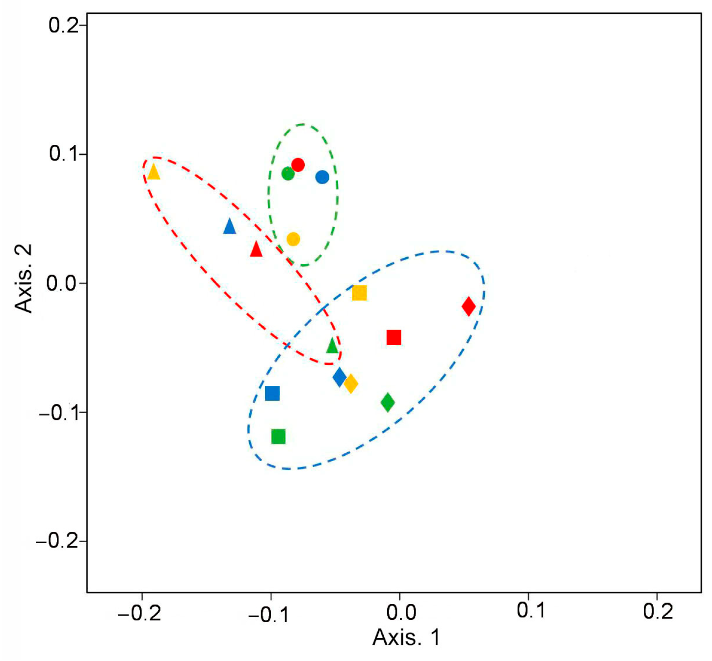
3.2.1. Diversity of Rhizosphere Communities

Sequencing of the 16s rRNA gene from 16 rhizosphere samples resulted in 1,915,453 raw reads. After data denoising and chimera screening, 17,870 joined read pairs per sample on average were used for further identification. Rarefaction curves obtained with the normalized OTU number almost reached saturation levels for all samples, indicating that the bacterial communities were covered well by the sequence analysis (Figure S1). The sequences with >97% similarity were combined into operational taxonomic units (OTUs). The OTUs were assigned to 38, 124, 288, 449, and 804 taxa at the phylum, class, order, family, and genus levels, respectively.
To characterize the bacterial diversity and the richness of the microbial communities, we calculated the α- and β-diversity (Table 2 and Figure 1).
The α-diversity was measured by using the species richness indices (Chao1, Shannon, and Simpson indices; Faith’s phylogenetic diversity [PD], Table 2). Traditional (Shannon’s and Simpson’s) and phylogenetic (Faith’s PD) indices of bacterial alpha-diversity in the rhizosphere communities of amaranths yielded similar conclusions: all communities were quite diverse and differed between plant species (cultivars) grown on the same soil.
A comparison of the rhizosphere microbiomes of different samples showed that the influence of soils on the formation of rhizosphere communities was not pronounced: the samples were not grouped according to soils. Yet, there were pronounced differences in rhizospheric samples according to the plant species studied (Figure 1). A. caudatus cv. Bulava and A. caudatus cv. Zelenaya Sosulka formed one cluster, which distinctly distanced itself from A. tricolor cv. Valentina, whereas the cluster of A. cruentus cv. Dyuimovochka had intersections with both A. caudatus cultivars.

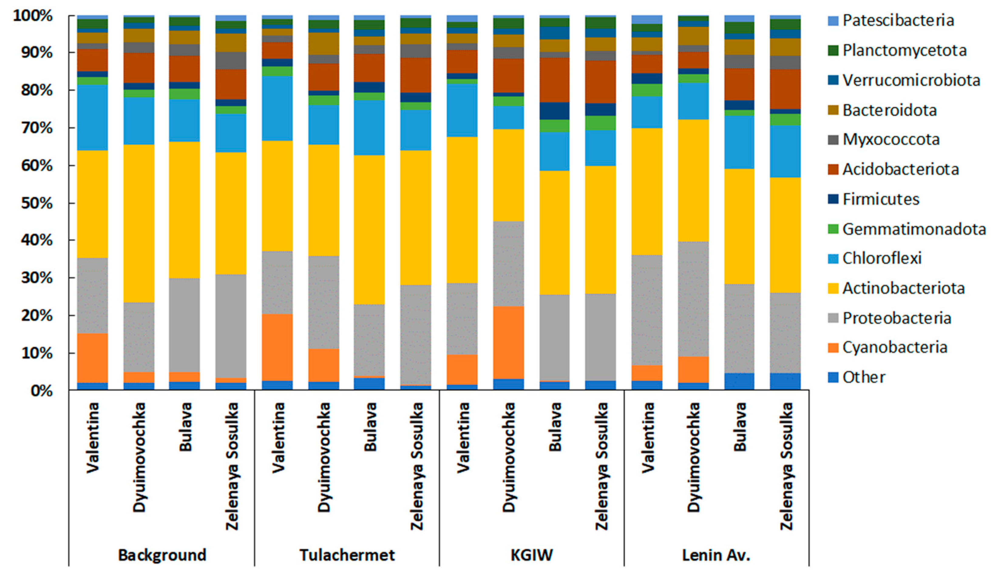
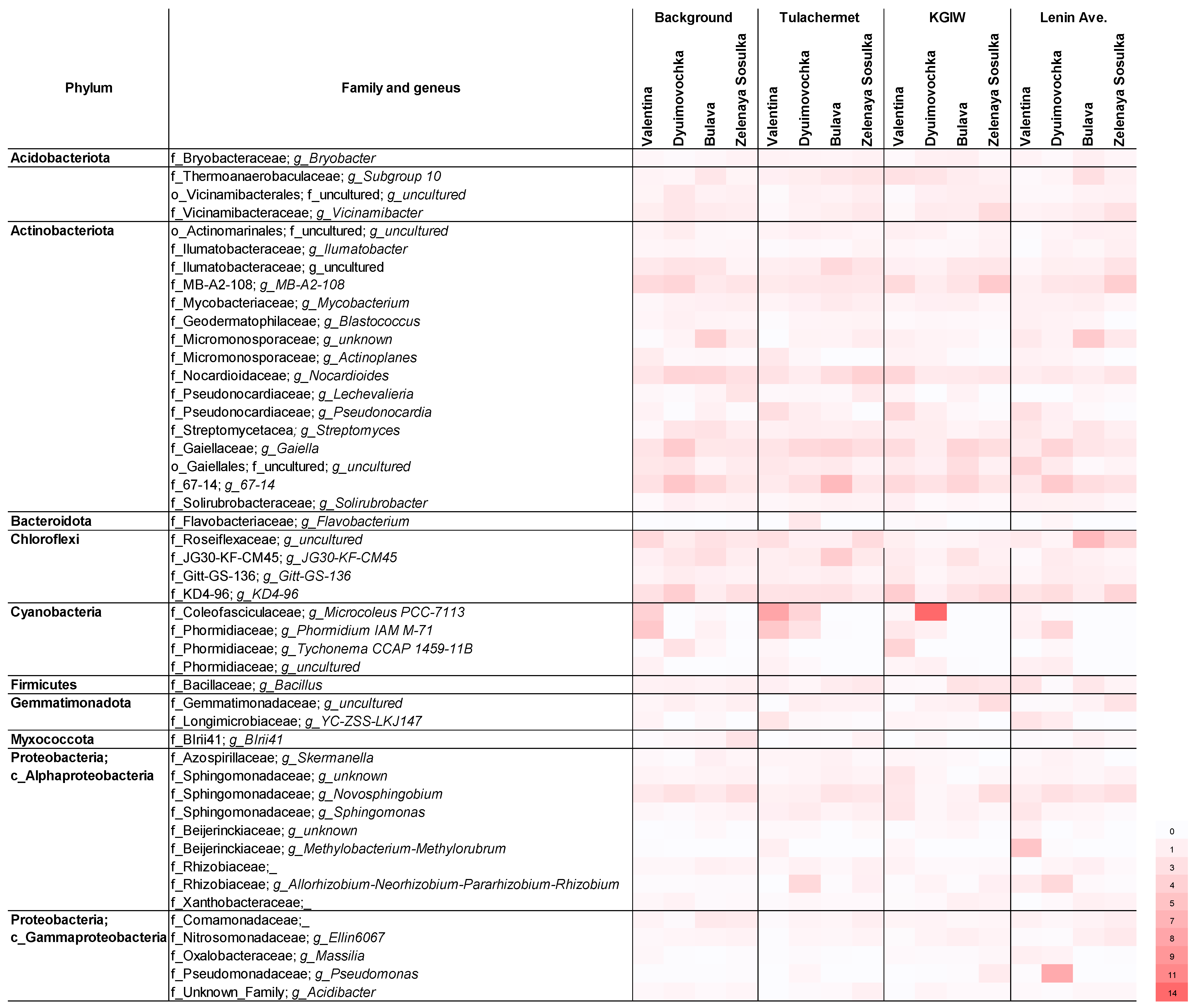
3.2.2. Taxonomic Structure of Rhizosphere Communities

MiSeq sequencing showed that the amaranth rhizosphere communities included 804 genera of bacteria belonging to 449 families of 38 phyla.
Figure 2 illustrates the relative abundances of OTUs associated at the phylum level in the rhizosphere of the amaranth cultivars studied. In different soils in the rhizosphere communities of cv. Valentina, most OTUs were assigned to Actinobacteriota (29–39%), Proteobacteria (17–29%), Chloroflexi (8–18%), Cyanobacteria (4–18%), and Acidobacteriota (4–6%). In the rhizosphere cv. Dyuimovochka, the dominant phyla were also Actinobacteriota (25–42%), Proteobacteria (19–31%), Chloroflexi (6–12%), Cyanobacteria (3–19%), and Acidobacteriota (4–9%). In the rhizosphere cv. Bulava, the dominant phyla were Actinobacteriota (31–40%), Proteobacteria (19–25%), Chloroflexi (10–15%), and Acidobacteriota (7–12%). In the rhizosphere cv. Zelenaya Sosulka, the dominant phyla were Actinobacteriota (31–36%), Proteobacteria (21–27%), Chloroflexi (9–14%), and Acidobacteriota (8–11%). The percentages of OTUs assigned to other phyla were much smaller.
The list of taxa at the family level, for which the share of amaranths in the rhizosphere was ≥1%, is given in Table S2. Of the dominant Actinobacteriota phylum, 51 families were identified, of which 13 were most abundant (Table S2, Figure 3), making up 80 to 86% of all detected actinobacterial families in the amaranth rhizosphere. The family Gaiellaceae had the maximum share in the rhizospheric population of actinobacteria. Its abundance reached 1.8–3.0% in the rhizosphere of cv. Valentina, 1.5–4.6% in the rhizosphere of cv. Dyuimovochka, 2.1–3.7% in the rhizosphere of cv. Bulava, and 1.9–2.9% in the rhizosphere of cv. Zelenaya Sosulka. Other notable actinobacteria were members of the families Nocardioidaceae (1.9–4.8% of all actinobacteria) and Micromonosporaceae (0.8–5.4%) and members of the orders Solirubrobacterales (67-14 family) and MB-A2-108 (MB-A2-108 family).
In another dominant phylum, Proteobacteria, 93 bacterial families were identified. In total, 10 of them (Table S2, Figure 3) made the largest contribution to the structure of the amaranth rhizosphere microbiomes (54–76% of all detected families). Alphaproteobacteria was the major class of the phylum, accounting for 64–84% of all OTUs assigned to the Proteobacteria. Among the Alphaproteobacteria, the dominant position was occupied by the families Sphingomonadaceae (3–7%) and Beijerinckiaceae (1–6%); a large proportion of OTUs also belonged to the Rhizobiaceae family (1–5%). The Gammaproteobacteria class accounted for 17 to 45% of all OTUs assigned to the Proteobacteria phylum, and the Nitrosomonadaceae and Comamonadaceae families were its predominant representatives. The contribution of the Betaproteobacteria and Deltaproteobacteria classes to the overall taxonomic structure of the amaranth microbiomes was not noticeable.
The rhizosphere microbiomes of two species of Amaranthus (A. tricolor cv. Valentina and A. cruentus cv. Dyuimovochka) were clearly enriched with members of the Cyanobacteria phylum, among which the Coleofasciculaceae and Phormidiaceae families dominated on all soils (Table S2, Figure 3), but were poorly represented in the rhizosphere of A. caudatus cv. Bulava and cv. Zelenaya Sosulka. The Phormidiaceae family was also abundant in the rhizosphere of A. tricolor cv. Valentina and A. cruentus cv. Dyuimovochka. This family was poorly represented in the rhizosphere of A. caudatus cv. Bulava and was not in the rhizosphere of cv. Zelenaya Sosulka. This may indicate that the maintenance of these bacterial taxa by host plants is species specific.
The species-specific changes in the taxonomic profile of the amaranth rhizosphere communities, as induced by the soil characteristics, can be seen in Figure 2 and Figure 3. Spearman’s rank correlation and principal component analysis did not reveal significant correlations between the kinds of soil used and the dominant taxa in the amaranth rhizosphere microbiomes. However, a close correlation was established between the cultivars (rs = 0.64, p < 0.05) and the abundance of OTUs assigned to the Cyanobacteria phylum. In addition, moderate correlations were found between the cultivars and the abundance of the Acidobacteriota (rs = 0.55, p < 0.05), Bacteroidota (rs = 0.52, p < 0.05), and Planctomycetota (rs = 0.52, p < 0.05) phyla in the rhizosphere communities.

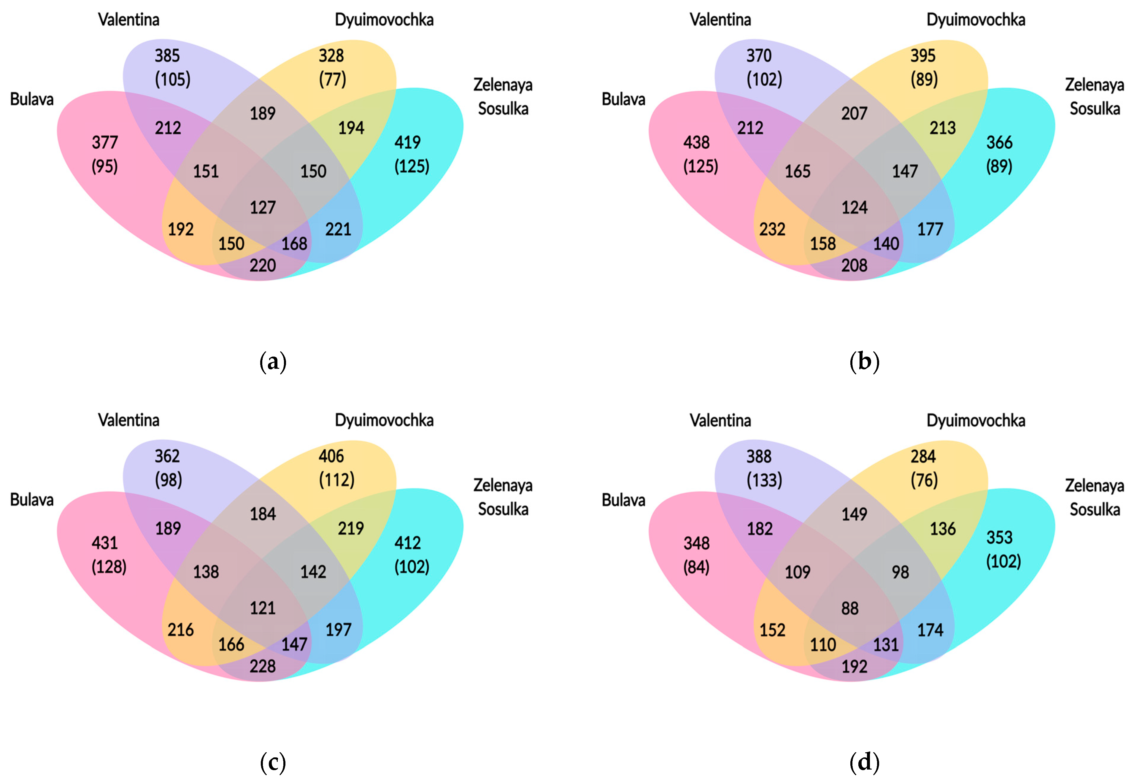
3.2.3. Shared and Unique Taxa among Rhizosphere Microbial Communities

To determine which OTUs were shared by or were specific to the rhizosphere of each cultivar on the four soils, we did several comparative analyses (Figure 4).
In the background soil, the largest number of OTUs at the bacterial species level was found in the rhizosphere of A. caudatus cv. Zelenaya Sosulka, followed by A. tricolor cv. Valentina, A. caudatus cv. Bulava, and A. cruentus cv. Dyuimovochka. A total of 127 species were shared and accounted for from 30 to 39% of the rhizosphere communities of the 4 plant cultivars. The percentage of unique taxa ranged from 24 to 30%. The maximal number of unique species (125) was found for the rhizosphere of A. caudatus cv. Zelenaya Sosulka.
In the Tulachermet soil, the largest number of OTUs at the bacterial species level was found in the rhizosphere of A. caudatus cv. Bulava, followed by A. cruentus cv. Dyuimovochka, A. tricolor cv. Valentina, and A. caudatus cv. Zelenaya Sosulka. A total of 124 species were shared and accounted for from 28 to 34% of the rhizosphere communities of all plant cultivars. The percentage of unique taxa ranged from 23 to 29%. The maximal number of unique species (125) was found for the rhizosphere of A. caudatus cv. Bulava.
In the KGIW soil, the largest number of OTUs at the bacterial species level was found in the rhizosphere of A. caudatus cv. Bulava, followed by A. cruentus cv. Dyuimovochka, A. caudatus cv. Zelenaya Sosulka, and A. tricolor cv. Valentina. A total of 121 species were shared and accounted for from 28 to 33% of the rhizosphere communities of all plant cultivars. The percentage of unique taxa ranged from 25 to 30%. The maximal number of unique species (128) was found for the rhizosphere of A. caudatus cv. Bulava.
In the Lenin Ave. soil, the largest number of OTUs at the bacterial species level was found in the rhizospheres of A. tricolor cv. Valentina, followed by A. caudatus cv. Zelenaya Sosulka, A. caudatus cv. Bulava, and A. cruentus cv. Dyuimovochka. Only 88 species were shared and accounted for from 23 to 25% of the rhizosphere communities of the 4 plant cultivars. The percentage of unique taxa ranged from 24 to 29%. The maximal number of unique species (133) was found for the rhizosphere of A. tricolor cv. Valentina.
Among the unique taxa identified in the rhizosphere microbiome of each amaranth variety, those taxa that occur in at least two different soils were determined (Tables S3–S6). Overall, 53, 39, 50, and 52 such taxa were revealed in the rhizosphere of A. tricolor cv. Valentina, A. cruentus cv. Dyuimovochka, and A. caudatus cvs. Bulava and Zelenaya Sosulka, respectively. In addition, seven, two, eight, and six such unique taxa were revealed in the rhizosphere of cvs. Valentina, Dyuimovochka, Bulava, and Zelenaya Sosulka, respectively, grown in three different soils studied (Table 3). Most of those taxa were represented by uncultured bacteria.
The bacterial taxa listed in Table 3 are unique to each of the four plant cultivars studied and can be considered as plant-specific.

4. Discussion

Studies characterizing the rhizosphere microbiomes of Amaranthus plants are extremely scarce. Nambisan et al. [29] found that the Cyanobacteria phylum is distinctly enriched in the roots and the rhizosphere soil of the three grain amaranths—A. hupochondriacus, A. cruentus, and A. caudatus. Our study confirmed that the peculiarity of the rhizosphere microbial communities of two of the three Amaranthus species (A. tricolor cv. Valentina and A. cruentus cv. Dyuimovochka) is the distinct presence of cyanobacteria along with the dominant bacterial phyla, such as Proteobacteria, Actinobacteriota, and Chloroflexi. In addition, we obtained the first data on the rhizosphere microbiome of another amaranth species, A. tricolor. In Nambisan et al.’s research [29], the negative controls were other plant species (Beta vulgaris, Cicer arietinum, and Solanum lycopersicum), and in their rhizobiomes, no predominance of cyanobacteria was observed. In our study, besides the amaranth rhizosphere, we also analyzed the rhizosphere of other plants under the same experimental conditions—Sorghum bicolor cv. Sucro and Sorghum bicolor cv. Biomass. The rhizosphere of these plants showed no predominance of cyanobacteria either [20]. An analysis of the taxonomic profile of the cyanobacteria found in the amaranth rhizobiomes made it possible to identify members of the dominant Microcoleus and Phormidium genera. The presence of these genera in the rhizosphere of various plants was also noted earlier [4,45]. The accumulated data indicate that the amaranths may have specifically selected cyanobacteria from the surrounding soil microflora. In turn, cyanobacteria favor the growth of the amaranth plants, possibly through their plant-beneficial characteristics. It is known that cyanobacteria can produce plant-growth-promoting substances, including auxins, gibberellins, cytokinins, abscisic acids, vitamins, and amino acids [4,46,47,48]. Cyanobacteria can also add organic matter, synthesize and liberate amino acids and vitamins, reduce the content of soil oxidizable matter, provide oxygen to the submerged rhizosphere, ameliorate salinity, buffer the pH, solubilize phosphates, and increase the efficiency of fertilizer use in crop plants [46]. Cyanobacteria such as Nostoc and Microcoleus can form associations with cycads [49] and Gunnera [50] and can fix nitrogen [45], either as free-living organisms or in association with host plants, in which they reside in specific tissues. Cyanobacteria such as Calothrix and Anabena can be used as biofertilizers [51]. Rhizospheric cyanobacteria are still insufficiently studied, although interest in their use for soil fertilization and plant-growth promotion is increasing steadily [4]. The established abundance of cyanobacteria in the amaranth rhizosphere requires further and deeper research on the part they play in plant vital activity.
To determine the contribution of different plant species (or cultivars) to the formation of their rhizosphere community in a particular soil, we compared their taxonomic composition, identifying shared and unique taxa (Figure 4). It was revealed that only about a third of taxa were shared between all four amaranths studied, and also about a third of taxa were unique. The contribution of A. tricolor cv. Valentina and A. cruentus cv. Dyuimovochka to the selection of cyanobacteria in their rhizosphere was confirmed. In addition, we revealed that two cultivars of A. caudatus selected different unique taxa in their rhizosphere, which suggests the cultivar-specific formation of rhizosphere microbiomes by amaranths (Tables S2–S5). No debated that plant root exudates are the principal connecting link between plant and plant-root-associated microbial communities [1,14,16]. In turn, the composition of root exudates is determined by plant physiology, which is different among plant species and even cultivars.
Current views hold that the relative content of individual taxa in the microbial community is a biological indicator of the soil status—for example, heavy metal pollution [52]. The urban soils used in our experiment were polluted by heavy metals. Many taxa identified in the amaranth rhizosphere microbiomes had previously been described as resistant to heavy metals or as oil-degraders, and they may be important for the resistance of amaranth to the human-caused pollution of soil. These taxa include (1) members of Proteobacteria, such as the genera Pseudomonas [18,53,54], Novosphingobium [55,56], Sphingomonas [57], Rhizobium [58,59], and Massilia [18,60]; (2) members of Actinobacteriota, such as Mycobacterium, Nocardiodes, Streptomyces [61], and the family Gaiellaceae [62]; (3) members of the Cyanobacteria phylum, such as the genus Microcoleus [63]; and (4) members of Firmicutes, such as the genus Bacillus [18]. In the rhizosphere of the amaranths used in this study, some of these taxa were present in large numbers (the genera Gaiella, Nocardiodes, Microcoleus, Novosphingobium, and Pseudomonas), whereas others were found in minor or single samples. Of note, increased numbers of members of Gaiellaceae had previously been found by Sun et al. [62] in the metal-polluted rhizosphere of crops. Those authors found a significant correlation between the increased abundance of Gaiellaceae-related bacteria and various metals and metalloids, and they concluded that these bacteria play a potentially active ecological part in the interaction with soil metals.
Rhizospheric microbial communities exert a great effect on the phytoremediation of metal-contaminated soils, not only by changing the bioavailability of metals [64], but also by promoting plant growth under pollutant stress through the fixation of nitrogen; production of phytohormones (indole-3-acetic acid, cytokinins, and gibberellins), siderophores, and enzymes (1-aminocyclopropane-1-carboxylate deaminase); and transformation of nutrients [65,66,67]. However, only 2–5% of rhizosphere microorganisms contribute to plant growth, and plants naturally select these beneficial microorganisms, which help them to grow and survive, especially under unfavorable conditions [3]. In the rhizospheric microbial communities of the amaranths used in this study, alongside cyanobacteria, we identified other groups of rhizobacteria with plant-growth-promoting potential (Figure 3). These included Bacillus (Bacillaceae, [68,69]), Sphingomonas (Sphingomonadaceae, [70]), Streptomyces (Streptomycetaceae, [69,71]), Pseudomonas (Pseudomonadaceae, [72]), and Rhizobium (Rhizobiaceae, [73]). Although the percentages of these taxa varied between cultivars and soils, their mere presence indicates that the rhizosphere microbiomes have the potential to promote amaranth growth on all urban soils tested.
We were unable to reveal any significant influence of the soils on the taxonomic structure of the amaranth rhizospheric microbiomes. Only about one third of the taxa identified in the cultivar rhizosphere microbiomes were common to all soil samples (Figure 4), which may indicate the maintenance of a specific plant microbiome, regardless of the kind of soil. We supposed that peculiarities and differences in the plant root exudate composition among plant cultivars had stronger differences than among soils, which resulted in the formation of different rhizosphere microbiomes depending more on the plants.
It is known that the composition of rhizosphere microbial communities is determined not only (and possibly not so much) by the soil type, but also by the plants. In our case, all soils studied were Greyic Phaeozems with slight differences, and all plants tested were Amaranthus belonging to different species. The compositions of the studied rhizosphere communities were quite similar, but the analysis of the obtained results revealed a greater influence of plants than soils.
For the final characterization of the amaranth core rhizobiomes, additional studies of the endosphere microbiome of each plant are required.

5. Conclusions

Limited data are available on the structure of the rhizosphere microbial communities of Amaranthus spp. We conducted a comparative study on the rhizosphere microbiomes of four Amaranthus cultivars (A. tricolor cv. Valentina, A. cruentus cv. Dyuimovochka, A. caudatus cv. Bulava, and A. caudatus cv. Zelenaya Sosulka). The cultivars were grown in an unpolluted (background) soil and in three polluted soils with polyelemental anomalies. The A. tricolor rhizosphere microbiome was characterized for the first time. The taxonomic structure of the amaranth rhizosphere microbiomes was represented by the dominant bacterial phyla Actinobacteriota, Proteobacteria, and Chloroflexi and by the phyla Cyanobacteria, Acidobacteriota, Planctomycetota, and Bacteroidota. In the taxonomic profile of the rhizobiomes of two Amaranthus species (A. tricolor cv. Valentina and A. cruentus cv. Dyuimovochka), there was a significant abundance of OTUs associated with the Cyanobacteria phylum. Bacterial taxa were identified that are possibly selected by amaranth plants during their coexistence and that may be important for plant adaptation to various habitat conditions, including polluted soils. The targeted enrichment of the amaranth rhizosphere microbiomes with members of these taxa could be useful for improving the efficacy of amaranth use for agricultural and remediation purposes.

Supplementary Materials

The following supporting information can be downloaded at: https://www.mdpi.com/article/10.3390/agronomy13030759/s1, Table S1: Characteristics of Amaranthus cultivars used in the research; Table S2: Taxonomic structure of the rhizosphere microbial communities of the amaranths studied; Tables S3–S6: Unique rhizosphere taxa occurred in at least two different soils planted with amaranths studied; Figure S1: Dependence of the number of detected taxa (OTUs) on the number of sequences obtained for amaranth rhizosphere soil samples.

Author Contributions

Conceptualization, S.G. (Svetlana Gorelova) and A.M.; methodology, A.M. and D.K.; software, A.M.; validation, S.G. (Sergey Golubev), S.G. (Svetlana Gorelova), and M.G.; formal analysis, D.K. and A.M.; investigation, A.M. and S.G. (Sergey Golubev); resources, S.G. (Svetlana Gorelova); data curation, A.M.; writing—original draft preparation, A.M.; writing—review and editing, S.G. (Svetlana Gorelova) and S.G. (Sergey Golubev); visualization, A.M.; supervision, A.M. and M.G.; project administration, S.G. (Svetlana Gorelova) and M.G.; funding acquisition, S.G. (Svetlana Gorelova). All authors have read and agreed to the published version of the manuscript.

Funding

This research was funded by the Russian Foundation for Basic Research, project no. 19-29-05257.

Institutional Review Board Statement

Not applicable.

Informed Consent Statement

Not applicable.

Data Availability Statement

Raw reads are deposited in the NCBI under BioProject ID PRJNA917498 in the fastq format (https://www.ncbi.nlm.nih.gov/bioproject/PRJNA917498, accessed on 1 March 2023).

Acknowledgments

We are grateful to Anatoliy V. Gorbunov, Senior Researcher of the Laboratory of Chemical-analytical Research of GIN RAS, for X-ray fluorescence analysis of soils. We are also grateful to Dmitry N. Tychinin for his assistance in the preparation of the English text of this paper.

Conflicts of Interest

The authors declare no conflict of interest. The funders had no role in the design of the study; in the collection, analyses, or interpretation of data; in the writing of the manuscript; or in the decision to publish the results.

References

  1. Bais, H.P.; Broeckling, C.D.; Vivanco, J.M. Root exudates modulate plant–microbe interactions in the rhizosphere. In Secondary Metabolites in Soil Ecology. Soil Biology; Karlovsky, P., Ed.; Springer: Berlin/Heidelberg, Germany, 2008; Volume 14, pp. 241–252. [Google Scholar]
  2. Fan, D.; Schwinghamer, T.; Smith, D.L. Isolation and diversity of culturable rhizobacteria associated with economically important crops and uncultivated plants in Québec, Canada. Syst. Appl. Microbiol. 2018, 41, 629–640. [Google Scholar] [CrossRef] [PubMed]
  3. Mohanram, S.; Kumar, P. Rhizosphere microbiome: Revisiting the synergy of plant-microbe interactions. Ann. Microbiol. 2019, 69, 307–320. [Google Scholar] [CrossRef]
  4. Srivastava, R.; Kanda, T.; Yadav, S.; Mishra, R.; Atri, N. Cyanobacteria in rhizosphere: Dynamics, diversity, and symbiosis. In Plant, Soil and Microbes in Tropical Ecosystems. Rhizosphere Biology; Dubey, S.K., Verma, S.K., Eds.; Springer: Singapore, 2021; pp. 51–69. [Google Scholar]
  5. Moreno-Espíndola, I.P.; Ferrara-Guerrero, M.J.; de León-González, F.; Rivera-Becerril, F.; Mayorga-Reyes, L.; Pérez, N.O. Enzymatic activity and culturable bacteria diversity in rhizosphere of amaranth, as indicators of crop phenological changes. Bot. Sci. 2018, 96, 640–649. [Google Scholar] [CrossRef] [Green Version]
  6. Kumar, A.; Dubey, A. Rhizosphere microbiome: Engineering bacterial competitiveness for enhancing crop production. J. Adv. Res. 2020, 24, 337–352. [Google Scholar] [CrossRef] [PubMed]
  7. Castellano-Hinojosa, A.; Strauss, S.L. Insights into the taxonomic and functional characterization of agricultural crop core rhizobiomes and their potential microbial drivers. Sci. Rep. 2021, 11, 10068. [Google Scholar] [CrossRef]
  8. Moorthy, A.; Balasundaram, U. Rhizosphere metagenomics: Methods and challenges. In Omics Science for Rhizosphere Biology, Rhizosphere Biology; Pudake, R.N., Sahu, B.B., Kumari, M., Sharma, A.K., Eds.; Springer Nature Singapore Pte Ltd.: Singapore, 2021; pp. 1–20. [Google Scholar]
  9. Ke, J.; Wang, B.; Yoshikuni, Y. Microbiome engineering: Synthetic biology of plant-associated microbiomes in sustainable agriculture. Trends Biotechnol. 2021, 39, 244–261. [Google Scholar] [CrossRef]
  10. Ofek-Lalzar, M.; Sela, N.; Goldman-Voronov, M.; Green, S.J.; Hadar, Y.; Minz, D. Niche and host-associated functional signatures of the root surface microbiome. Nat. Commun. 2014, 5, 4950. [Google Scholar] [CrossRef] [Green Version]
  11. Schreiter, S.; Ding, G.-C.; Heuer, H.; Neumann, G.; Sandmann, M.; Grosch, R.; Kropf, S.; Smalla, K. Effect of the soil type on the microbiome in the rhizosphere of field-grown lettuce. Front. Microbiol. 2014, 5, 144. [Google Scholar] [CrossRef]
  12. Sun, X.; Song, B.; Xu, R.; Zhang, M.; Gao, P.; Lin, H.; Sun, W. Root-associated (rhizosphere and endosphere) microbiomes of the Miscanthus sinensis and their response to the heavy metal contamination. J. Environ. Sci. 2021, 104, 387–398. [Google Scholar] [CrossRef]
  13. Neu, A.T.; Allen, E.E.; Roy, K. Defining and quantifying the core microbiome: Challenges and prospects. Proc. Natl. Acad. Sci. USA 2021, 118, e2104429118. [Google Scholar] [CrossRef]
  14. Chen, Y.; Ding, Q.; Chao, Y.; Wei, X.; Wang, S.; Qiu, R. Structural development and assembly patterns of the root-associated microbiomes during phytoremediation. Sci. Tot. Environ. 2018, 644, 1591–1601. [Google Scholar] [CrossRef]
  15. Mendes, R.; Garbeva, P.; Raaijmakers, J.M. The rhizosphere microbiome: Significance of plant beneficial, plant pathogenic, and human pathogenic microorganisms. FEMS Microbiol. Rev. 2013, 37, 634–663. [Google Scholar] [CrossRef] [PubMed] [Green Version]
  16. Bell, T.H.; Cloutier-Hurteau, B.; Al-Otaibi, F.; Turmel, M.-C.; Yergeau, E.; Courchesne, F.; St-Arnaud, M. Early rhizosphere microbiome composition is related to the growth and Zn uptake of willows introduced to a former landfill. Environ. Microbiol. 2015, 17, 3025–3038. [Google Scholar] [CrossRef] [PubMed]
  17. Kour, D.; Rana, K.L.; Yadav, N.; Yadav, A.N.; Kumar, A.; Meena, V.S.; Singh, B.; Chauhan, V.S.; Dhaliwal, H.S.; Saxena, A.K. Rhizospheric microbiomes: Biodiversity, mechanisms of plant growth promotion, and biotechnological applications for sustainable agriculture. In Plant Growth Promoting Rhizobacteria for Agricultural Sustainability (From Theory to Practices); Kumar, A., Meena, V.S., Eds.; Springer Nature Singapore Pte Ltd.: Singapore, 2019; pp. 19–65. [Google Scholar]
  18. Barra Caracciolo, A.; Terenzi, V. Rhizosphere microbial communities and heavy metals. Microorganisms 2021, 9, 1462. [Google Scholar] [CrossRef]
  19. Lundberg, D.S.; Lebeis, S.L.; Paredes, S.H.; Yourstone, S.; Gehring, J.; Malfatti, S.; Tremblay, J.; Engelbrektson, A.; Kunin, V.; del Rio, T.G.; et al. Defining the core Arabidopsis thaliana root microbiome. Nature 2012, 488, 86–90. [Google Scholar] [CrossRef] [Green Version]
  20. Muratova, A.Y.; Gorelova, S.V.; Sungurtseva, I.Y.; Zelenova, N.A. Rhizospheric microbiomes of Sorghum bicolor grown on soils with anthropogenic polyelement anomalies. In BIO Web of Conferences PLAMIC2020; EDP Sciences: Les Ulis, France, 2020; Volume 23, p. 03008. [Google Scholar]
  21. Gins, M.S. Biologicheski Aktivnyye Veshchestva Amaranta Amarantin: Svoystva, Mekhanizmy Deystviya i Prakticheskoye Ispol’zovaniye (Biologically Active Substances of Amaranth Amaranthine: Properties, Mechanisms of Action and Practical Use); RUDN: Moscow, Russia, 2002; 183p. (In Russian) [Google Scholar]
  22. Shevyakova, N.I.; Cheremisina, A.I.; Kuznetsov, V.V. Phytoremediation potential of Amaranthus hybrids: Antagonism between nickel and iron and chelating role of polyamines. Russ. J. Plant Physiol. 2011, 58, 634–642. [Google Scholar] [CrossRef]
  23. Chinmayee, M.D.; Mahesh, B.; Pradesh, S.; Mini, I.; Swapna, T.S. The assessment of phytoremediation potential of invasive weed Amaranthus spinosus L. Appl. Biochem. Biotechnol. 2012, 167, 1550–1559. [Google Scholar] [CrossRef]
  24. Ziarati, P.; Alaedini, S. The phytoremediation technique for cleaning up contaminated soil by Amaranthus sp. J. Environ. Anal. Toxicol. 2014, 4, 1000208. [Google Scholar]
  25. Odiyi, B.; Ologundudu, F.A.; Adegbite, T. Phytoremediation potential of Amaranthus hybridus L. (Caryophyllales: Amaranthaceae) on soil amended with brewery effluent. Braz. J. Biol. Sci. 2019, 6, 401–411. [Google Scholar] [CrossRef] [Green Version]
  26. Gorelova, S.V.; Gins, M.S.; Frontasyeva, M.V. Phytoextraction of toxic elements by Amaranthus tricolor grown on technogenically polluted soils in open ground conditions. Chim. Techno Acta 2022, 9, 202292S8. [Google Scholar] [CrossRef]
  27. Song, N.; Zhang, X.; Wang, F.; Zhang, C.; Tang, S. Elevated CO2 increases Cs uptake and alters microbial communities and biomass in the rhizosphere of Phytolacca americana Linn (pokeweed) and Amaranthus cruentus L. (purple amaranth) grown on soils spiked with various levels of Cs. J. Environ. Radioact. 2012, 112, 29–37. [Google Scholar] [CrossRef] [PubMed]
  28. Mishra, J.; Singh, R.; Arora, N.K. Alleviation of heavy metal stress in plants and remediation of soil by rhizosphere microorganisms. Front. Microbiol. 2017, 8, 1706. [Google Scholar] [CrossRef] [Green Version]
  29. Nambisan, S.; Sunil, M.; Choudhary, B.; Srinivasan, S. Cyanobacteria is uniquely enriched in the roots of grain amaranths. bioRxiv 2019, 540484. [Google Scholar] [CrossRef]
  30. Gorelova, S.V.; Gorbunov, A.V.; Frontasyeva, M.V.; Sylina, A.K. Toxic elements in the soils of urban ecosystems and technogenic sources of pollution. WSEAS Trans. Environ. Dev. 2020, 16, 608–618. [Google Scholar] [CrossRef]
  31. GOST 53123-2008; Soil Quality. Sampling. Part 5. Guidance on the Procedure for the Investigation of Urban and Industrial Sites with Regard to Soil Contamination. Russian Gost: Moscow, Russian, 2008. Available online: https://docs.cntd.ru/document/1200074384 (accessed on 1 October 2022).
  32. GOST 26483-85; Soils. Preparation of Salt Extract and Determination of Its pH by CINAO Method. Russian Gost: Moscow, Russian, 1985. Available online: https://docs.cntd.ru/document/1200023490 (accessed on 21 February 2023).
  33. Malahov, S.G. Interim Guidelines on Soil Pollution Control. 1984. Available online: https://files.stroyinf.ru/Data2/1/4294847/4294847412.htm (accessed on 21 February 2023).
  34. GOST 26205-91; Soils. Determination of Mobile Compounds of Phosphorus and Potassium by Machigin Method Modified by CINAO. Russian Gost: Moscow, Russian, 1991. Available online: https://docs.cntd.ru/document/1200023449 (accessed on 21 February 2023).
  35. GOST 26488-85; Soils. Determination of Nitrates by CINAO Method. Russian Gost: Moscow, Russian, 1985. Available online: https://docs.cntd.ru/document/1200023495 (accessed on 21 February 2023).
  36. GOST 26489-85; Soils. Determination of Exchangeable Ammonium by CINAO Method. Russian Gost: Moscow, Russian, 1986. Available online: https://docs.cntd.ru/document/1200023496 (accessed on 21 February 2023).
  37. PND F 16.1:2:2.2:2.3:3.64-10; Quantitative Chemical Analysis of Soils. Method for Measuring the Mass Fraction of Oil Products in Samples of Soils, Soils, Bottom Sediments, Silts, Sewage Sludge, Production and Consumption Wastes by the Gravimetric Method. Federal Service for Environmental, Technological and Nuclear Supervision: Moscow, Russia, 2010. Available online: https://files.stroyinf.ru/Data2/1/4293807/4293807051.htm (accessed on 21 February 2023).
  38. PND F 16.1.42-04; Measurement Method for Measuring the Mass Fraction of Metals and Metal Oxides in Soil Powder Samples by X-Ray Fluorescence Analysis. Federal Service for Environmental, Technological and Nuclear Supervision: Moscow, Russia, 2004. (In Russian)
  39. GN 2.1.7.2041-06; Predel’no Dopustimye Koncentracii (PDK) Himicheskih Veshchestv v Pochve: Gigienicheskie Normativy.—M.: Federal’nyj Centr Gigieny i Epidemiologii Rospotrebnadzora. Maximum Permissible Concentrations (MPCs) of Chemicals in the Soil: Moscow, Russia, 2006; 15p. Available online: https://files.stroyinf.ru/Data2/1/4293850/4293850511.pdf (accessed on 21 February 2023). (In Russian).
  40. GN 2.1.7.2511-09; Orientirovochno Dopustimye Koncentracii (ODK) Himicheskih Veshchestv v Pochve: Gigienicheskie Normativy.—M.: Federal’nyj Centr Gigieny i Epidemiologii Rospotrebnadzora. Approximate allowable concentrations (AAC) of Chemical Substances in Soils: Moscow, Russia, 2009; 10p. Available online: https://files.stroyinf.ru/Data2/1/4293828/4293828439.pdf (accessed on 21 February 2023). (In Russian).
  41. Klindworth, A.; Pruesse, E.; Schweer, T.; Peplies, J.; Quast, C.; Horn, M.; Glöckner, F.O. Evaluation of general 16S ribosomal RNA gene PCR primers for classical and next-generation sequencing-based diversity studies. Nucleic Acids Res. 2013, 41, e1. [Google Scholar] [CrossRef] [PubMed]
  42. Bolyen, E.; Rideout, J.R.; Dillon, M.R.; Bokulich, N.A.; Abnet, C.C.; Al-Ghalith, G.A.; Alexander, H.; Alm, E.J.; Arumugam, M.; Asnicar, F.; et al. Reproducible, interactive, scalable and extensible microbiome data science using QIIME 2. Nat. Biotechnol. 2019, 37, 852–857. [Google Scholar] [CrossRef]
  43. Callahan, B.J.; McMurdie, P.J.; Rosen, M.J.; Han, A.W.; Johnson, A.J.A.; Holmes, S.P. DADA2: High-resolution sample inference from Illumina amplicon data. Nat. Methods 2016, 13, 581–583. [Google Scholar] [CrossRef] [Green Version]
  44. Yilmaz, P.; Parfrey, L.W.; Yarza, P.; Gerken, J.; Pruesse, E.; Quast, C.; Schweer, T.; Peplies, J.; Ludwig, W.; Glockner, F.O. The SILVA and "All-species Living Tree Project (LTP)" taxonomic frameworks. Nucleic Acids Res. 2014, 42, D643–D648. [Google Scholar] [CrossRef] [Green Version]
  45. Jing, H.; Xia, X.; Liu, H.; Zhou, Z.; Wu, C.; Nagarajan, S. Anthropogenic impact on diazotrophic diversity in the mangrove rhizosphere revealed by nifH pyrosequencing. Front. Microbiol. 2015, 6, 1172. [Google Scholar] [CrossRef] [Green Version]
  46. Prasanna, R.; Nain, L.; Ancha, R.; Srikrishna, J.; Joshi, M.; Kaushik, B.D. Rhizosphere dynamics of inoculated cyanobacteria and their growth-promoting role in rice crop. Egypt. J. Biol. 2009, 11, 26–36. [Google Scholar]
  47. Suresh, A.; Soundararajan, S.; Elavarasi, S.; Oscara, F.L.; Thajuddin, N. Evaluation and characterization of the plant growth promoting potentials of two heterocystous cyanobacteria for improving food grains growth. Biocatal. Agric. Biotechnol. 2019, 17, 647–652. [Google Scholar] [CrossRef]
  48. Toribio, A.J.; Suárez-Estrella, F.; Jurado, M.M.; López, M.J.; López-González, J.A.; Moreno, J. Prospection of cyanobacteria producing bioactive substances and their application as potential phytostimulating agents. Biotechnol. Rep. 2020, 26, e00449. [Google Scholar] [CrossRef] [PubMed]
  49. Lindblad, P. Cyanobacteria in symbiosis with cycads. In Prokaryotic Symbionts in Plants. Microbiology Monographs; Pawlowski, K., Ed.; Springer: Heidelberg, Germany, 2008; Volume 8, pp. 225–233. [Google Scholar]
  50. Bergman, B.; Johansson, C.; Söderbäck, E. The Nostoc-Gunnera symbiosis. New Phytol. 1992, 122, 379–400. [Google Scholar] [CrossRef]
  51. Chittora, D.; Meena, M.; Barupal, T.; Swapnil, P.; Sharma, K. Cyanobacteria as a source of biofertilizers for sustainable agriculture. Biochem. Biophys. Rep. 2020, 22, 100737. [Google Scholar] [CrossRef]
  52. Hermans, S.M.; Buckley, H.L.; Case, B.S.; Curran-Cournane, F.; Taylor, M.; Lear, G. Bacteria as emerging indicators of soil condition. Appl. Environ. Microbiol. 2017, 83, e02826-16. [Google Scholar] [CrossRef] [PubMed] [Green Version]
  53. Dasgupta, D.; Ghosh, R.; Sengupta, T.K. Biofilm-mediated enhanced crude oil degradation by newly isolated Pseudomonas species. Int. Sch. Res. Notices 2013, 2013, 250749. [Google Scholar] [CrossRef] [Green Version]
  54. Ueshima, M.; Ginn, B.R.; Haack, E.A.; Szymanowski, J.E.S.; Fein, J.B. Cd adsorption onto Pseudomonas putida in the presence and absence of extracellular polymeric substances. Geochim. Cosmochim. Acta 2008, 72, 5885–5895. [Google Scholar] [CrossRef]
  55. Chettri, B.; Singh, A.K. Kinetics of hydrocarbon degradation by a newly isolated heavy metal tolerant bacterium Novosphingobium panipatense P5:ABC. Bioresour. Technol. 2019, 294, 122190. [Google Scholar] [CrossRef]
  56. Lee, S.Y.; Lee, Y.Y.; Cho, K.S. Effect of Novosphingobium sp. CuT1 inoculation on the rhizoremediation of heavy metal- and diesel-contaminated soil planted with tall fescue. Environ. Sci. Pollut. Res. 2022, 30, 16612–16625. [Google Scholar] [CrossRef]
  57. Asaf, S.; Numan, M.; Khan, A.L.; Al-Harrasi, A. Sphingomonas: From diversity and genomics to functional role in environmental remediation and plant growth. Crit. Rev. Biotechnol. 2020, 40, 138–152. [Google Scholar] [CrossRef]
  58. Nocelli, N.; Bogino, P.C.; Banchio, E.; Giordano, W. Roles of extracellular polysaccharides and biofilm formation in heavy metal resistance of rhizobia. Materials 2011, 9, 418. [Google Scholar] [CrossRef] [PubMed] [Green Version]
  59. Gupta, P.; Diwan, B. Bacterial exopolysaccharide mediated heavy metal removal: A review on biosynthesis, mechanism and remediation strategies. Biotechnol. Rep. 2017, 13, 58–71. [Google Scholar] [CrossRef] [PubMed]
  60. Feng, G.D.; Yang, S.Z.; Li, H.P.; Zhu, H.H. Massilia putida sp. nov., a dimethyl disulfide-producing bacterium isolated from wolfram mine tailing. Int. J. Syst. Evol. Microbiol. 2016, 66, 50–55. [Google Scholar] [CrossRef]
  61. Chikere, C.B.; Okpokwasili, G.C.; Chikere, B.O. Monitoring of microbial hydrocarbon remediation in the soil. 3 Biotech 2011, 1, 117–138. [Google Scholar] [CrossRef] [PubMed] [Green Version]
  62. Sun, W.; Xiao, E.; Krumins, V.; Häggblom, M.M.; Dong, Y.; Pu, Z.; Li, B.; Wang, Q.; Xiao, T.; Li, F. Rhizosphere microbial response to multiple metal(loid)s in different contaminated arable soils indicates crop-specific metal-microbe interactions. Appl. Environ. Microbiol. 2018, 84, e00701–e00718. [Google Scholar] [CrossRef] [PubMed] [Green Version]
  63. Diestra, E.; Esteve, I.; Burnat, M.; Maldonado, J.; Solé, A. Isolation and characterization of a heterotrophic bacterium able to grow in different environmental stress conditions, including crude oil and heavy metals. Commun. Curr. Res. Educ. Top. Trends Appl. Microbiol. 2007, 1, 90–99. [Google Scholar]
  64. Jing, Y.; He, Z.; Yang, X. Role of soil rhizobacteria in phytoremediation of heavy metal contaminated soils. J. Zhejiang Univ. Sci. B (Biomed. Biotechnol.) 2007, 8, 192–207. [Google Scholar] [CrossRef] [Green Version]
  65. Santoyo, G.; Moreno-Hagelsieb, G.; Orozco-Mosqueda, M.C.; Glick, B.R. Plant growth-promoting bacterial endophytes. Microbiol. Res. 2016, 183, 92–99. [Google Scholar] [CrossRef]
  66. Ojuederie, O.; Babalola, O. Microbial and plant-assisted bioremediation of heavy metal polluted environments: A review. Int. J. Environ. Res. Public Health 2017, 14, 1504. [Google Scholar] [CrossRef] [Green Version]
  67. Hussain, S.S.; Mehnaz, S.; Siddique, K.H.M. Harnessing the plant microbiome for improved abiotic stress tolerance. In Plant Microbiome: Stress Response; Egamberdieva, D., Ahmad, P., Eds.; Springer Nature Singapore Pte Ltd: Singapore, 2018; pp. 21–43. [Google Scholar]
  68. Babu, A.G.; Kim, J.-D.; Oh, B.-T. Enhancement of heavy metal phytoremediation by Alnus firma with endophytic Bacillus thuringiensis GDB-1. J. Hazard. Mater. 2013, 250–251, 477–483. [Google Scholar] [CrossRef]
  69. Chen, Z.-J.; Tian, W.; Li, Y.-J.; Sun, L.-N.; Chen, Y.; Zhang, H.; Li, Y.Y.; Han, H. Responses of rhizosphere bacterial communities, their functions and their network interactions to Cd stress under phytostabilization by Miscanthus spp. Environ. Pollut. 2021, 287, 117663. [Google Scholar] [CrossRef] [PubMed]
  70. Chen, B.; Zhang, Y.; Rafiq, M.T.; Khan, K.Y.; Pan, F.; Yang, X.; Feng, Y. Improvement of cadmium uptake and accumulation in Sedum alfredii by endophytic bacteria Sphingomonas SaMR12: Effects on plant growth and root exudates. Chemosphere 2014, 17, 367–373. [Google Scholar] [CrossRef] [PubMed]
  71. Ali, A.; Guo, D.; Mahar, A.; Wang, Z.; Muhammad, D.; Li, R.; Wang, P.; Shen, F.; Xue, Q.; Zhang, Z. Role of Streptomyces pactum in phytoremediation of trace elements by Brassica juncea in mine polluted soils. Ecotoxicol. Environ. Saf. 2017, 144, 387–395. [Google Scholar] [CrossRef] [PubMed]
  72. Preston, G.M. Plant perceptions of plant growth-promoting Pseudomonas. Philos. Trans. R. Soc. Lond. B Biol. Sci. 2004, 359, 907–918. [Google Scholar] [CrossRef] [PubMed] [Green Version]
  73. Deshwal, V.K.; Singh, S.B.; Kumar, P.; Chubey, A. Rhizobia unique plant growth promoting rhizobacteria: A review. Int. J. Life Sci. 2013, 2, 74–86. [Google Scholar]
Figure 1. Principal coordinate analysis for the rhizosphere microbial communities of the Amaranthus cvs.: triangles (▲), A. tricolor cv. Valentina; circles (●), A. cruentus cv. Dyuimovochka; squares (■), A. caudatus cv. Bulava; diamonds (♦), A. caudatus cv. Zelenaya Sosulka; blue, background soil; yellow, Tulachermet soil; red, KGIW soil; green, Lenin Ave. soil.
Agronomy 13 00759 g001
Figure 2. Relative abundances of OTUs associated at the phylum level of the microbial communities in the rhizospheres of four amaranth cultivars grown on different soils.
Agronomy 13 00759 g002
Figure 3. Heat map illustrating the relative abundances of OTUs associated at the genus level of the microbial communities in the rhizospheres of four amaranth cultivars grown on different soils.
Agronomy 13 00759 g003
Figure 4. Venn diagram depicting shared and unique OTUs among the rhizosphere microbial communities of the amaranths grown on the background and polluted soils. The number of unique taxa is given in brackets. (a) Background; (b) Tulachermet; (c) KGIW; (d) Lenin Ave.
Agronomy 13 00759 g004
Table 1. Characteristics of the soils used in the experiment.
PropertiesBackground Soil
(Yasnaya Polyana)
KGIWTulachermetLenin Ave.
Soil typeClay loamClay loamSandy loamClay loam
pH6.207.267.357.29
Total carbon (% of air-dried soil)4.87 ± 0.054.41 ± 0.114.84 ± 0.333.83 ± 0.06
Water-soluble carbon (% of total carbon)0.14 ± 0.050.13 ± 0.020.14 ± 0.060.12 ± 0.03
Humus (% of air-dried soil)8.12 ± 0.167.14 ± 0.147.62 ± 0.156.16 ± 0.12
Humus carbon (%)4.72 ± 0.094.15 ± 0.084.43 ± 0.093.58 ± 0.07
N-NO3 (mg/kg dw)20.5 ± 1.228.2 ± 1.638.6 ± 3.541.1 ± 2.4
N-NH4 (mg/kg dw)12.9 ±1.26.9 ± 0.79.5 ± 0.32.3 ± 0.2
P2O5 (mg/kg dw)51.0 ± 4.8159.5 ± 3.698.4 ± 7.6218.0 ± 6.7
Oil products (g/kg dw)1.5 ± 0.62.6 ± 0.44.1 ± 1.02.5 ± 0.7
Metal(loid)s (mg/kg dw):
Fe15,600 ± 186078,100 ± 1280120,600 ± 583037,400 ± 2160
Mn1300 ± 675700 ± 1801100 ± 871600 ± 58
V57 ± 3.041 ± 4.1136 ± 7.261 ± 2.9
Ni25 ± 3.131 ± 4.255 ± 3.335 ± 2.8
Cu29 ± 2.352 ± 4.175 ± 0.8378 ± 0.9
Zn47 ± 2.0310 ± 9.4161 ± 3.6186 ± 5.3
Pb18 ± 2.172 ± 6.026 ± 0.759 ± 1.1
As5.1 ± 0.45.9 ± 0.56.4 ± 0.27.3 ± 0.3
Note: Data expressed as mean ± standard deviation (n ≥ 3). Bold type means the permissible concentrations [29,30] were exceeded for: Fe (MPCtotal 1500 mg/kg), Mn (MPCtotal, 1500 mg/kg), V (MPCtotal, 100 mg/kg), Ni (APCtotal, sandy loam, 20 mg/kg; APCtotal, clay loam, 80 mg/kg), Cu (MPCtotal 55 mg/kg; APCtotal, sandy loam, 33 mg/kg), Zn (APCtotal, clay loam, 220 mg/kg; APCtotal, sandy loam, 55 mg/kg), As (APCtotal, sandy loam, 2 mg/kg; APCtotal, clay loam, 10 mg/kg), and oil products (1.0 g/kg). The oil product content in soil is not regulated at the regional level; the recommended value is 1.0 g/kg.
Table 2. The α-diversity indices for the rhizospheric microbial communities of amaranths grown on different soils.
SoilPlantObserved
Features
Chao1Shannon
Index
Simpson
Index
Faith PD
BackgroundA. tricolor cv. Valentina743748.0008.65600.996481.03
A. cruentus cv. Dyuimovochka716718.1118.63090.995773.42
A. caudatus cv. Bulava890898.3478.57010.993086.78
A. caudatus cv. Zelenaya Sosulka910916.9498.74660.995597.51
TulachermetA. tricolor cv. Valentina741743.2588.60190.995687.83
A. cruentus cv. Dyuimovochka823829.3438.87150.997087.65
A. caudatus cv. Bulava10101014.6149.27490.997892.29
A. caudatus cv. Zelenaya Sosulka819831.4708.84020.996781.70
KGIWA. tricolor cv. Valentina807807.7868.90550.997082.02
A. cruentus cv. Dyuimovochka806806.9928.60810.994088.70
A. caudatus cv. Bulava990994.8449.12060.997097.06
A. caudatus cv. Zelenaya Sosulka964974.3509.15070.997488.53
Lenin Ave.A. tricolor cv. Valentina832836.8338.92320.996985.43
A. cruentus cv. Dyuimovochka601602.6008.44010.995963.57
A. caudatus cv. Bulava758766.9288.37500.992681.42
A. caudatus cv. Zelenaya Sosulka707716.7848.58880.995774.67
Table 3. Unique bacterial taxa revealed in the rhizosphere of Amaranthus plants and occurred in three different soils.
Plant CultivarsBacterial Taxa
A. tricolor
cv. Valentina
g_Chloronema; s_Scytonema tolypothrichoidesBackground; Tulachermet; KGIW
g_Leptolyngbya_VRUC_135;s_uncultured bacteriumBackground; Tulachermet; Lenin Ave.
g_Alsobacter; s_Alsobacter_metalliduransTulachermet; KGIW; Lenin Ave.
g_C0119; s_uncultured_bacteriumBackground; Tulachermet; Lenin Ave.
g_Rhodocytophaga; s_uncultured BacteroidetesBackground; Tulachermet; KGIW
f_Rhodanobacteraceae; g_uncultured; s_uncultured bacteriumBackground; Tulachermet; KGIW
g__DS-100; s_unknownTulachermet; KGIW; Lenin Ave.
A. cruentus
cv. Dyuimovochka
g_Methylophilus; s_uncultured bacteriumTulachermet; KGIW; Lenin Ave.
g_CENA518; s_uncultured_bacteriumTulachermet; KGIW; Lenin Ave.
A. caudatus
cv. Bulava
g_Nakamurella; g_unknown; s_unknownBackground; Tulachermet; KGIW
g_Pir4_lineage; s_uncultured PirellulaBackground; Tulachermet; Lenin Ave.
f_Moraxellaceae; g_uncultured; s_uncultured gammaBackground; KGIW; Lenin Ave.
f_0319-7L14; g_0319-7L14; s_uncultured bacteriumBackground; Tulachermet; Lenin Ave.
o_Planctomycetales; f_uncultured; g_unculturedBackground; Tulachermet; KGIW
f_Phycisphaeraceae; g_unculturedTulachermet; KGIW; Lenin Ave.
f_Kapabacteriales; g_KapabacterialesTulachermet; KGIW; Lenin Ave.
f_Sericytochromatia; g_SericytochromatiaBackground; Tulachermet; KGIW
A. caudatus
cv. Zelenaya Sosulka
f_Vicinamibacteraceae; g_Vicinamibacteraceae;s_uncultured GeothrixTulachermet; KGIW; Lenin Ave.
f_Xanthomonadaceae; g_unknown; s_unknownBackground; Tulachermet; KGIW
f_Rhodanobacteraceae; g_Ahniella; s_uncultured bacteriumBackground; Tulachermet; Lenin Ave.
f_Latescibacterota; g_Latescibacterota; s_uncultured soilBackground; Tulachermet; Lenin Ave.
f_Blastocatellaceae; g_Aridibacter; s_uncultured bacteriumBackground; KGIW; Lenin Ave.
f_Subgroup 22; g_Subgroup 22Tulachermet; KGIW; Lenin Ave.
Disclaimer/Publisher’s Note: The statements, opinions and data contained in all publications are solely those of the individual author(s) and contributor(s) and not of MDPI and/or the editor(s). MDPI and/or the editor(s) disclaim responsibility for any injury to people or property resulting from any ideas, methods, instructions or products referred to in the content.

