Article

Fungicidal Activity of Caproate Produced by Clostridium sp. strain E801, a Bacterium Isolated from Cocopeat Medium Subjected to Anaerobic Soil Disinfestation

1
Graduate School of Horticulture, Chiba University, 648 Matsudo, Chiba 271-8510, Japan
2
Institute for Horticultural Plant Breeding, 2-5-1 Kamishiki, Chiba 270-2221, Japan
3
Environmental Management Research Institute, National Institute of Advanced Industrial Science and Technology, Onogawa 16-1, Tsukuba 305-8569, Japan
*
Author to whom correspondence should be addressed.
Agronomy 2023, 13(3), 747; https://doi.org/10.3390/agronomy13030747
Submission received: 30 January 2023 / Revised: 28 February 2023 / Accepted: 1 March 2023 / Published: 3 March 2023

Abstract

:
Anaerobic soil disinfestation (ASD) consists of the application of labile organic materials to soil, flooding, and covering the soil surface with plastic film. Anaerobic soil disinfestation is a widely used ecofriendly alternative to chemical fumigation for eliminating soil-borne plant pathogens. However, the exact mode of action of ASD has not been elucidated. In particular, the potential role of anaerobic soil bacteria in disinfestation is unclear. In this study, we isolated a predominant bacterium designated as strain E801 from cocopeat medium after laboratory-scale ASD with ethanol as the carbon source. The strain was closely related with Clostridium kluyveri, and fermentatively produced butyrate and caproate from ethanol and acetate. Interestingly, the culture supernatant of strain E801 strongly suppressed the growth of Fusarium oxysporum f. sp. lycopersici (Fol) in a pH-dependent manner. Among the volatile fatty acids produced by E801, only caproate showed significant growth suppression at pHs below 5.5. In addition, caproate eliminated Fol conidia completely at pHs 5.5 and 5.0 and suppressed Fol growth even at a low temperature (15 °C). Furthermore, cocopeat medium amended with caproate eliminated Fol conidia completely within 6 days. These results suggest that caproate is one of the key disinfestation factors in ethanol-based ASD and that the direct application of caproate to soil could be a promising strategy for rapid and stable soil disinfestation.

1. Introduction

The continuous cultivation of a single crop causes soil deterioration through acidification, salinization, and the accumulation of soil-borne plant pathogens. Among these, soil-borne diseases are particularly difficult to control because most of the pathogens are filamentous fungi that survive under harsh environments by forming resting propagules [1,2]. For many years, chemical fumigants have been used to disinfect soil-borne pathogens. Although these fumigants are very powerful and have a broad-spectrum, there are concerns about their negative impact on human health and the environment. Methyl bromide was an ideal fumigant, but its use was banned under the Montreal Protocol due to its high potential for damaging the stratospheric ozone layer [3,4]. Other fumigants such as chloropicrin, 1,3-dichrolopropene, and dazomet are widely used as alternatives to methyl bromide, but their use is restricted due to their irritancy and toxicity [5]. Thus, ecofriendly disinfestation alternatives are urgently needed for the development of sustainable agriculture.
Anaerobic soil disinfestation (ASD), also known as biological soil disinfestation or reductive soil disinfestation, was developed independently in Japan and the Netherlands [6,7]. It consists of incorporating labile organic materials such as rice bran, wheat bran, molasses or diluted ethanol into soil as the carbon source, flooding or irrigation to saturation, and the covering of the soil surface with plastic film for 3 to 6 weeks [8,9]. During ASD, carbon sources are degraded and assimilated by aerobic microorganisms, leading to a rapid decrease in soil redox potential (Eh) [10]. The subsequent reducing (anoxic or anaerobic) condition stimulates the growth of anaerobic microorganisms such as fermentative bacteria and metal-reducing bacteria. The former bacteria produce various volatile fatty acids such as acetate and butyrate, and the latter bacteria release Fe2+ and Mn2+ ions into the soil. Finally, ASD significantly alters the composition of the soil bacterial community, which is commonly dominated by the phylum Firmicutes [11]. To date, considerable evidence has revealed that ASD is effective against soil-borne plant pathogens, plant-parasitic nematodes, and weeds. Currently, ASD is implemented in Japan, the USA, the Netherlands, China, and some South American countries [12].
Despite its increasing application in different countries, the exact mode of action of ASD remains unclear. Although Fusarium oxysporum f. sp. lycopersici (Fol) cannot grow under anaerobic conditions, low soil Eh itself is not lethal to Fol [8,9]. Additionally, both Fol and Ralstonia solanacearum were destroyed completely by acetate and butyrate in vitro, but the lethality was significantly lower in soil amended with these volatile fatty acids [13]. Other volatile organic compounds such as isothiocyanate, alcohols, organic sulfides, and esters are thought to contribute to ASD-mediated pathogen suppression [14,15]. Momma et al. [16] reported that the survival of Fol was significantly reduced in the presence of Fe2+ and Mn2+ ions in vitro. Even though anaerobic soil bacteria are supposed to be involved in the suppression of pathogens, their key role in ASD-treated soil is still unclear. Ueki et al. [17] isolated two Clostridium strains from ASD-treated soil and found that both strains eliminated F. oxysporum f. sp. spinaciae when co-incubated with the pathogen in a liquid medium. They suggested that fungal cell wall-degrading enzymes such as β-1,3-glucanase and chitosanase produced by the Clostridium strains play a significant role in the elimination of the pathogen [18]. Though the mechanism of ASD is not fully understood, researchers consider that it is a combination of changes in the soil microbial community composition, the production of volatile organic compounds, and the generation of lethal anaerobic conditions [9].
The purpose of this study was to isolate bacteria that predominate after ASD, and to determine if such bacteria produce certain compounds that suppress soil-borne pathogens. To this end, we performed laboratory-scale ASD that mimics field-scale ASD by using ethanol as the carbon source [19]. Cocopeat medium was used instead of soil, since the medium is expected to have a simpler microbial community structure compared with soil. Cocopeat medium has been widely used in Japan for the cultivation of strawberries and tomatoes, and ethanol-based ASD is actually applied to these cultivation sites. After laboratory-scale ASD, the high-throughput sequencing of the 16S rRNA gene was performed to identify predominant bacteria, and one such bacteria (strain E801) was successfully isolated by means of a microbiological technique. In vitro analyzes were then performed to determine if strain E801 produces certain compounds that suppress the growth of Fol, a representative fungal plant pathogen. In addition, the compound (caproate) was tested to determine if it could eliminate Fol conidia and the cells of R. solanacearum. The latter pathogen is well known to cause bacterial wilt on a wide range of plants and is used as a representative bacterial plant pathogen. Finally, caproate was inoculated to cocopeat medium together with Fol conidia to determine if this organic acid alone can suppress and eliminate the soil-borne pathogen. The potential mechanism of caproate in the suppression and elimination of Fol and R. solanacearum, as well as the direct application of caproate to soil as a promising disinfestation method, were also discussed.

2. Materials and Methods

2.1. Preparation of Conidial Suspension

Fol strain CU1 (race 1) [20] was grown on a potato dextrose agar (PDA) medium at 30 °C for 4 days. Sterilized distilled water was poured onto the agar, and conidia were collected using a scraper. The conidia were washed twice and resuspended in sterilized distilled water to reach approximately 5 × 10⁸ spores mL−1.

2.2. Laboratory-Scale ASD

To mimic field-scale ASD in the laboratory, small-scale ASD was performed in glass bottles under anaerobic conditions. We used cocopeat medium instead of soil in this study, since the medium is expected to have a simpler microbial community structure compared with soil. Cocopeat medium (Cocoblock; KANEKO SEEDS, Gunma, Japan) that had been used for strawberry cultivation was dried and passed through a 2 mm sieve. Five grams of dried cocopeat medium, 20 mL of sterile distilled water, and 5 mL of 1% (v/v) ethanol were dispensed into 60 mL serum bottles under N2 atmosphere. The bottles were sealed with thick butyl rubber stoppers and aluminum caps. Conidial suspension was then inoculated into each bottle at 106 spores g medium−1. The bottles without ethanol were also prepared as carbon source-free controls. The bottles were incubated at 30 °C in the dark without shaking. At days 3, 6, 8, 10, 14, and 17, the bottles were opened and the aqueous phase was spread on a Fo-G1 agar, a selective medium for F. oxysporum [21], after appropriate dilution. The agar was then incubated at 30 °C for a week, and viable number of Fol conidia was counted.

2.3. High-Throughput Sequencing of 16S rRNA Gene Amplicons

Total DNA was extracted from the cocopeat medium using the ISOIL for Beads Beating kit (Nippon Gene, Toyama, Japan) and DNeasy PowerClean Pro Cleanup Kit (Qiagen, Hilden, Germany). The V3 and V4 hypervariable region of 16S rRNA gene was amplified using the prokaryote universal primers Pro341F and Pro805R as described previously [22]. Barcoded amplicons were sequenced using the paired-ends method and a 600-cycle MiSeq Reagent Kit (Illumina, San Diego, CA, USA) with a MiSeq sequencer (Illumina). After alignment, the overlapping regions between the paired-end reads were merged and primer regions were omitted, which resulted in a 430 bp sequence. Paired-end reads were then concatenated using the FASTQ-join software with default options [23]. Only joined reads with quality value (QV) score of >20 for more than 99 % of the sequence were extracted using FASTX-Toolkit (http://hannonlab.cshl.edu/fastx_toolkit/index.html (accessed on 7 March 2022). Chimeric sequences were removed using QIIME1.8.0 software [24]. The determined 16S rRNA gene sequences were subjected to homology search using Metagenome@KIN software (World Fusion, Tokyo, Japan) and the TechnoSuruga Lab Microbial Identification database (NGS-DB-BA ver. 16.0; TechnoSuruga Laboratory, Shizuoka, Japan) with a homology threshold > 97%.

2.4. Isolation of the Predominant Bacterium from ASD-Treated Soil

From Section 2.3, Clostridium kluyveri-like bacterium was found to predominate in the cocopeat medium after ASD (see the Section 3). Since C. kluyveri is known to form endospores [25] and thus is expected to be heat resistant, we attempted to isolate this bacterium after heat shock of cocopeat medium to eliminate non-spore-forming bacteria. The aqueous phase of ASD-treated cocopeat medium was heated at 80 °C for 10 min and inoculated into a minimal medium [26]. The medium contained the following (per liter): NH4Cl (0.535 g), KH2PO4 (0.136 g), MgCl2·6H2O (0.204 g), CaCl2·2H2O (0.147 g), trace mineral element solution (1 mL), vitamin solution (1 mL), Se/W solution (1 mL), and NaHCO3 (2.52 g). The medium was dispensed into 60 mL serum bottles under the N2/CO2 (80:20) atmosphere and autoclaved. Ethanol (electron donor) and acetate (electron acceptor) were added separately from sterile anaerobic stock solutions to achieve final concentrations of 25 mM and 10 mM, respectively. Cysteine-HCl (1.5 mM) was also added as a reducing agent. The inoculated culture was incubated at 30 °C in the dark. After turbidity was observed; the culture was serially diluted and inoculated into anaerobic shake tubes containing the minimal medium and 0.4% Bacto agar (Difco, Sparks, MD, USA). A single colony was picked by a sterile Pasteur pipette and inoculated into a new tube. The culture purity was determined by microscopy, and the isolated bacterium was designated as strain E801.

2.5. Phylogenetic Analysis of Strain E801

Genomic DNA of strain E801 was extracted as described previously [27]. The PCR amplification of the 16S rRNA gene was performed with the primers 8F (5′-AGAGTTTGATCCTGGCTCAG-3′) and 1491R (5′-GGTTACCTTGTTACGACTT-3′). The PCR products were purified using a QIAquick PCR Purification kit (Qiagen) and sequenced using a BigDye Terminator Cycle Sequencing kit (Applied Biosystems, Foster City, CA) and an ABI Prism 3100 Genetic Analyzer (Applied Biosystems, Foster City, CA) using appropriate sequencing primers [28]. The 16S rRNA gene sequence was compared with reference sequences using BLAST similarity searches. The phylogenetic tree was constructed using the neighbor-joining method with the MEGA11 software package [29].

2.6. Effect of Culture Supernatant of Strain E801 on the Growth of Fol

Glucose-yeast extract-peptone (GYP) medium containing 20 g L−1 glucose, 5 g L−1 yeast extract, and 10 g L−1 peptone was prepared. The medium pH was adjusted to 5.5, 5.0, 4.5, and 4.0 with 1 N HCl. After autoclaving, 4 mL of GYP medium was dispensed into 18 mL sterile test tubes. Conidia of Fol were inoculated into each test tube at 103 spores mL−1. Finally, 500 µL of culture supernatant of strain E801 was added to the test tube. To prepare the culture supernatant, strain E801 was grown anaerobically in the minimal medium for 1 week. Heat-treated supernatant was also prepared by treating the supernatant at 100 °C for 20 min. Test tubes were incubated at 30 °C for 3 days with shaking at 180 rpm, and the growth of Fol was determined on potato dextrose agar (PDA) medium after appropriate dilution. A similar experiment was performed with sodium acetate (Fujifilm Wako Pure Chemical Corporation, Osaka, Japan), sodium butyrate (Fujifilm Wako Pure Chemical Corporation), or sodium caproate (Sigma-Aldrich, St. Louis, MO, USA) instead of the culture supernatant. In such cases, these volatile fatty acids were added at a final concentration of 1 mM.

2.7. Effect of Volatile Fatty Acids on the Viability of Fol Conidia and R. solanacearum Cells

The pH of 100 mM MES buffer was adjusted to 5.5 and 5.0 with 1 N NaOH, and 4 mL of buffer was dispensed into 18 mL test tubes. Conidia of Fol were inoculated at 103 spores mL−1. After sodium caproate was added at different concentrations, the tubes were incubated at 30 °C without shaking. Viable number of Fol conidia was measured on PDA medium as described above. A similar experiment was performed with cells of R. solanacearum EK1-2 (biovar 4, race 1) [20] instead of Fol. In this case, R. solanacearum was pre-grown in a CPG medium [30], washed twice with sterile water, and resuspended to MES buffer at 103 cells mL−1.

2.8. Effect of Volatile Fatty Acids on the Growth of Fol at a Low Temperature

Glucose-yeast extract-peptone medium supplemented with 5 mM sodium acetate, sodium butyrate, or sodium caproate was prepared, and its pH was adjusted to 5.5, 5.0, 4.5, and 4.0 with 1 N HCl. After autoclaving, the medium was mixed with separately autoclaved Bacto agar and solidified. On the GYP agar medium, 15 µL of Fol culture pre-grown in GYP liquid medium was placed. The agar medium was incubated at 15 °C for 20 days, and colony sizes were observed.

2.9. Evaluation of Fungicidal Activity of Caproate in Cocopeat Medium

Three grams of dried cocopeat medium and 30 mL of sterile distilled water were mixed in 60 mL serum bottles under N2 atmosphere. Bottles were sealed with butyl rubber stoppers and aluminum caps. Conidia of Fol were inoculated at 106 spores g medium−1. After sodium acetate, sodium butyrate, or sodium caproate was added at a concentration of 3 mM, the bottles were incubated at 30 °C without shaking. At days 2, 4, and 6, the bottles were opened and viable number of Fol conidia was measured on Fo-G1 agar medium as described above.

2.10. Analytical Techniques

Volatile fatty acids were determined by high-performance liquid chromatography (HPLC; L-7000; Hitachi, Tokyo, Japan) with an Aminex HPX-87H ion exclusion column (Bio-Rad Laboratories, Hercules, CA, USA) under UV detection at 210 nm. The mobile phase was 0.01 N H2SO4 at a flow rate of 0.6 mL min−1, and the column was maintained at 35 °C. Growth of strain E801 was monitored with optical density (OD) at 600 nm by a BioSpectrometer basic (Eppendorf, Hamburg, Germany).

2.11. Nucleotide Sequence Accession Numbers

The 16S rRNA gene sequence of strain E801 was deposited in the DDBJ/GenBank databases under an accession number of LC744592. The results of high-throughput sequencing were uploaded to DDBJ under run accession numbers DRR436616 to DRR436620.

3. Results

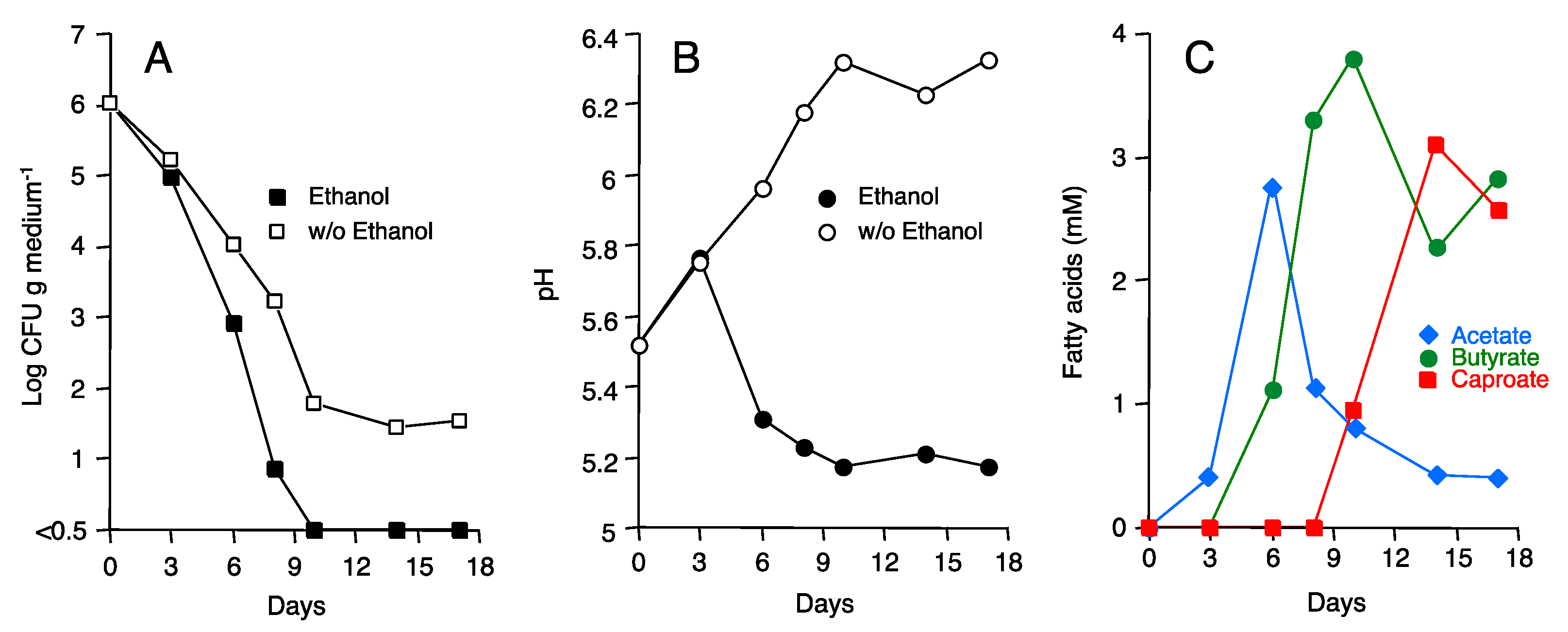
3.1. Laboratory-Scale ASD with Ethanol as Carbon Source

To mimic field-scale ASD in the laboratory, we first performed small-scale ASD in glass bottles under anaerobic conditions. Cocopeat medium used for strawberry cultivation was mixed with water, Fol conidia, and 0.2 % (v/v) ethanol and incubated for 17 days at 30 °C. In the presence of ethanol, Fol was eliminated within 10 days, while approximately 30 CFU g medium−1 of Fol survived after 17 days in medium unamended with ethanol (Figure 1A). The pH of medium amended with ethanol gradually decreased from 5.5 to 5.2, whereas pH of unamended medium increased to 6.3 (Figure 1B). The decrease in medium pH was due to volatile fatty acids produced in the ethanol-amended medium (Figure 1C). In this type of medium, approximately 3 mM of acetate was produced during the early stage of ASD (3 to 6 days). During the mid-stage of ASD (6 to 10 days), approximately 4 mM of butyrate was produced at the expense of acetate consumption. Finally, during the late stage of ASD (10 to 14 days), approximately 3 mM of caproate was produced at the expense of acetate and butyrate consumption. In contrast, only 0.6 mM of acetate was produced in medium unamended with ethanol, but neither butyrate nor caproate was produced (data not shown).

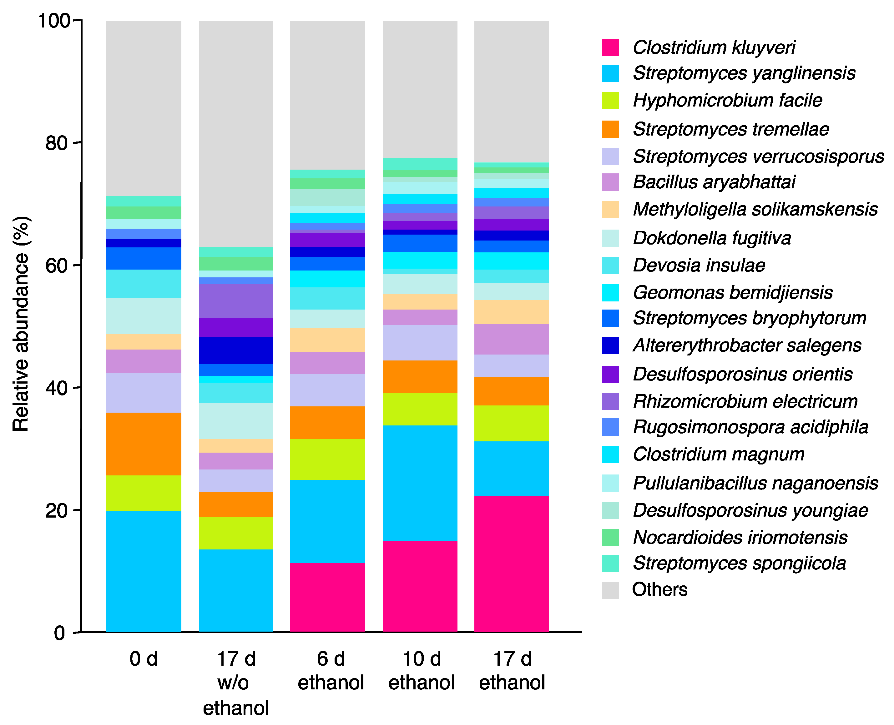
3.2. Microbial Community Shift during ASD

Microbial community composition was determined before and after ASD by means of 16S rRNA gene amplicon analysis. On the phylum level, original cocopeat medium consisted mainly of the phyla Proteobacteria and Actinobacteria, which accounted for 90% of the total community (Figure S1). However, ethanol-based ASD changed the community composition significantly, in which the phylum Firmicutes accounted for 42% of the total community. After ethanol-free ASD, community composition changed moderately, in which the phylum Firmicutes accounted for only 17%. Species level community analysis revealed that Streptomyces spp. and Hyphomicrobium facile were predominant in the original cocopeat medium (Figure 2). However, during ethanol-amended ASD, the relative abundance of a bacterium closely related to Clostridium kluyveri in the phylum Firmicutes increased gradually, with the abundance estimated to be 11%, 15%, and 22% at days 6, 10, and 17, respectively. In ethanol-free ASD, in contrast, the C. kluyveri-like bacterium was absent, and the species-level community composition was relatively similar with that in the original cocopeat medium. We repeated the laboratory-scale ASD experiment but for only 14 days with another cocopeat medium. In this case, the relative abundance of the phylum Firmicutes increased to 44% after ethanol-based ASD (Figure S2), while the C. kluyveri-like bacterium accounted for 22% (Figure S3). Thus, our two independent laboratory-scale experiments showed that relative abundance of Firmicutes bacteria increased to approximately 40% in ethanol-based ASD, of which about half was constituted by the C. kluyveri-like bacterium.

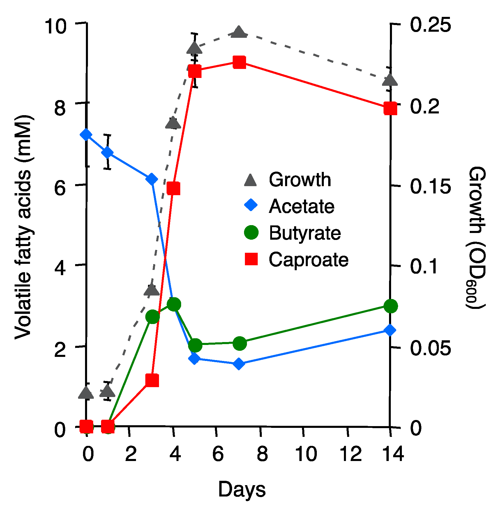
3.3. Isolation of Clostridium sp. E801 from ASD-Treated Cocopeat Medium

The C. kluyveri-like bacterium that predominated the ASD-treated cocopeat medium was isolated by means of the anaerobic shake tube technique and designated as strain E801. Cells of strain E801 were gram-positive rods that formed endospores. The 16S rRNA gene of strain E801 was 100% identical to that of the bacterium predominating in ethanol-based ASD-treated cocopeat medium. It also showed 98.2 to 98.5% similarity with those of C. kluyveri strains in the DDBJ/GenBank databases. As shown in Figure S4, phylogenetic analysis also indicated that strain E801 is closely related to C. kluyveri.

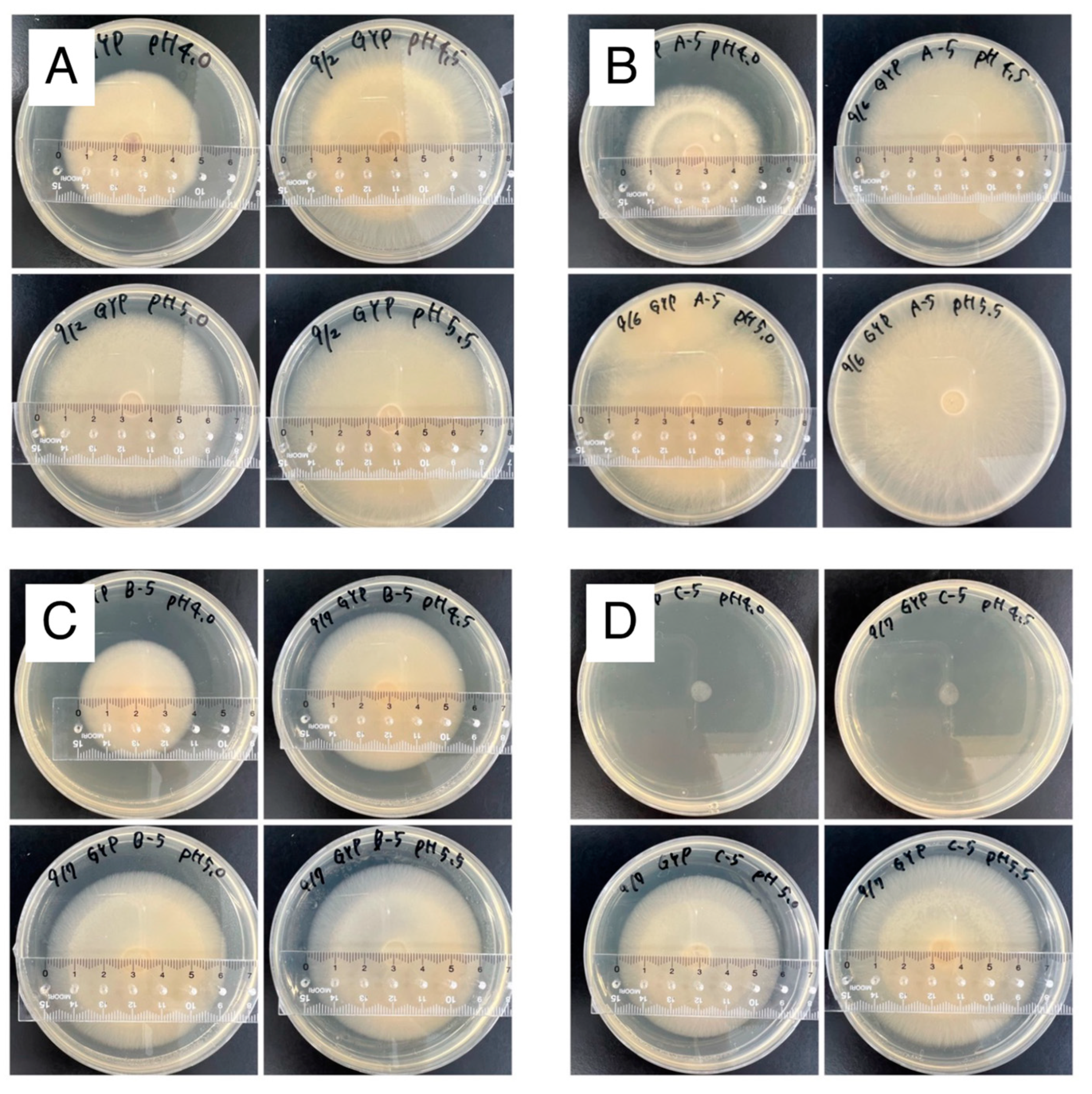
3.4. Effect of Culture Supernatant of Strain E801 on the Growth of Fol

To determine if strain E801 produced certain fungicidal substances, Fol was grown in a liquid medium in the presence or absence of the culture supernatant of strain E801. In the absence of the culture supernatant, Fol grew from 3.0 Log CFU mL−1 to approximately 5.0 to 6.0 Log CFU mL−1 regardless of the medium pH (Table 1). In the presence of the culture supernatant, however, colony-forming units of Fol were less than the detection limit of 0.50 Log CFU mL−1 at pH 4.5 and pH 4.0, while those at pH 5.5 and pH 5.0 were 6.0 and 4.5 Log CFU mL−1, respectively. Such pH-dependent suppression of Fol growth was still observed after the supernatant was treated at 100 °C for 20 min.
To determine volatile fatty acids produced by strain E801, the strain was grown in the minimal medium containing ethanol and acetate (Figure 3). Although ethanol was not quantified in this study, acetate was consumed probably with ethanol to form butyrate. Subsequently, butyrate was consumed with acetate to form caproate. Thus, it was found that strain E801 was capable of producing caproate, with the intermediate production of butyrate, at the expense of ethanol and acetate through a chain elongation reaction, as reported in C. kluyveri [25].
Fol was then grown in the presence of 1 mM each of acetate, butyrate, or caproate to determine which volatile fatty acids could suppress Fol growth. As shown in Table 2, Fol grew from 3.0 Log CFU mL−1 to approximately 5.0 to 6.0 Log CFU mL−1 in the presence of acetate and butyrate regardless of the medium pH. However, in the presence of caproate, Fol growth was strongly suppressed at pH 5.5 and pH 5.0, and no Fol conidia were detected at pH 4.5 and pH 4.0.

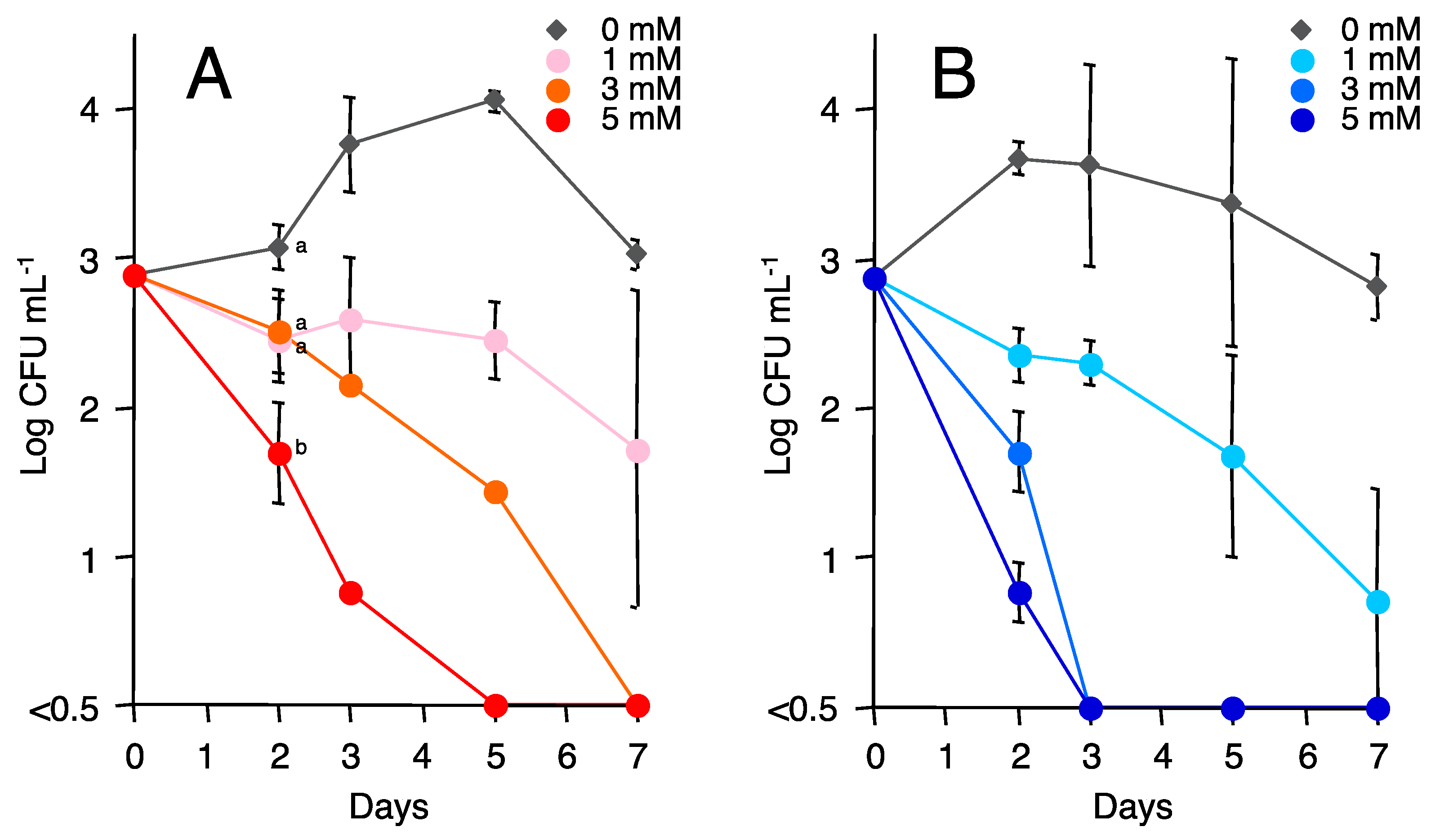
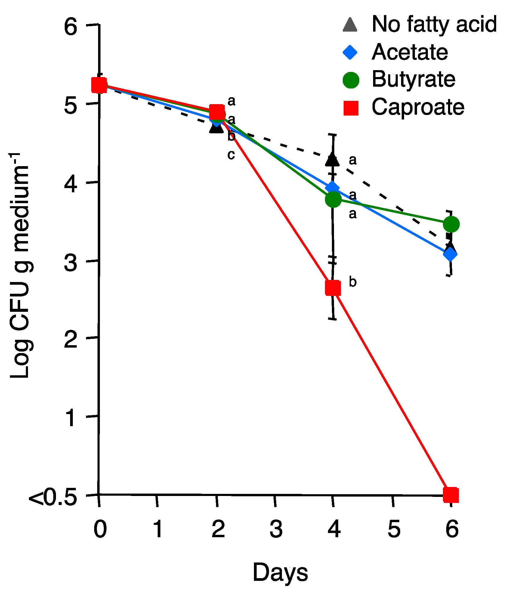
3.5. Effect of Caproate on the Viability of Fol Conidia and R. solanacearum Cells

To determine if caproate not only suppresses the growth of Fol but also eliminates Fol, conidia of Fol were incubated in MES buffer containing different concentrations of caproate. As shown in Figure 4, colony-forming units of Fol conidia did not decrease in the absence of caproate. However, Fol conidia were eliminated in the presence of 3 mM of caproate within 7 days at pH 5.5. At pH 5.0, the elimination of Fol conidia occurred much faster than that at pH 5.5, and no viable Fol conidia were observed by the third day in the presence of 3 mM caproate. Similar experiments were performed using cells of R. solanacearum, a representative bacterial plant pathogen (Figure S5). At pH 5.5, neither acetate nor butyrate eliminated R. solanacearum cells significantly, but 3 mM of caproate eliminated this pathogenic bacterium within 7 days.

3.6. Effect of Caproate on the Growth of Fol at Low Temperature

To determine if caproate was able to suppress the growth of Fol even under cold conditions, Fol was inoculated on the GYP agar medium containing 5 mM of acetate, butyrate, or caproate, and the media were incubated at 15 °C. As shown in Figure 5, acetate- and butyrate-amended media did not show significant suppressive effects compared with fatty acid-free controls regardless of the media pH. In contrast, caproate inhibited the growth of Fol completely at pH 4.5 and 4.0, while no suppression was observed at pH 5.5 and pH 5.0. The complete inhibition of Fol growth by caproate persisted for more than two months. Essentially the same experiments were performed with 1 mM volatile fatty acids, but no significant suppression was observed (data not shown).

3.7. Evaluation of Fungicidal Activity of Caproate in Cocopeat Medium

Finally, the fungicidal activity of caproate was evaluated by applying 3 mM of caproate directly to the cocopeat medium that was inoculated with Fol conidia. As shown in Figure 6, acetate- and butyrate-amended cocopeat medium did not show significant fungicidal activity compared with the fatty acid-free control. In contrast, caproate decreased colony-forming units of Fol conidia in cocopeat medium and eliminated Fol conidia within 6 days. In this experiment, the pH of cocopeat medium was 5.0 to 5.2 throughout the experimental period regardless of organic acid addition (data not shown).

4. Discussion

The proliferation and domination of the phylum Firmicutes have been reported in soil after ASD [31,32,33,34,35]. Interestingly, such dominance of the phylum Firmicutes was observed regardless of the carbon sources incorporated into soil, i.e., wheat bran [31,32], rice bran [35], maize straw [33,34], rice straw [34], tomato pomace [35], red grape pomace [35], or various grasses [31,34]. In addition, bacteria belonging to the genus Clostridium were repeatedly reported as predominant microorganisms in ASD-treated soils through various molecular techniques including denaturing gradient gel electrophoresis (DGGE) [36,37], clone library analysis [31,32], and high-throughput sequencing [33,34,35,38]. Nevertheless, very few attempts were conducted to isolate and characterize such predominant Clostridium-related bacteria in ASD-treated soils. In this study, we isolated Clostridium sp. strain E801 from cocopeat medium after laboratory-scale ASD and demonstrated that caproate produced by this strain has a strong biocidal activity against Fol and R. solanacearum. Such trials, together with comprehensive microbial community composition analyses, are of great importance to clarify the potential functions of these strictly anaerobic spore-forming bacteria in ASD and to fully understand the mechanism of the ASD-mediated suppression of soil-borne plant pathogens.
Strain E801 is a bacterium closely related to C. kluyveri, which is known to produce butyrate and caproate from ethanol and acetate [25]. Since its first discovery in 1937 [39], C. kluyveri has gained attention due to its unique feature of metabolizing and producing volatile fatty acids. C. kluyveri produces butyrate through a chain elongation reaction from ethanol and acetate [25].
1 ethanol + 1 acetate → 1 butyrate + 1 H2O
C. kluyveri then produces caproate through a chain elongation reaction from ethanol and butyrate [25].
1 ethanol + 1 butyrate → 1 caproate + 1 H2O
The profile of fatty acid production in our laboratory-scale ASD showed good agreement with the stepwise reaction mechanism of butyrate and caproate production by C. kluyveri (Figure 1C), suggesting that these volatile fatty acids were produced mainly by strain E801. Acetate required for butyrate production might have been generated from ethanol with the help of certain homoacetogenic bacteria such as the genera Acetobacterium [40] and Sporomusa [41], or by the strain E801 itself [25].
Our results suggest that the growth suppression of Fol was not due to heat-labile substances such as enzyme proteins but due to volatile fatty acids produced by strain E801 (Table 1). The antimicrobial activities of acetate and butyrate have been reported. Momma et al. [13] found that both Fol and R. solanacearum were eliminated in 13 mM of acetate (pH 3.1) and 9 mM of butyrate (pH 3.2) solutions. Katase et al. [42] reported that 10 mM of acetate and 10 mM of butyrate showed nearly equal nematicidic activity below pH 4.5. Huang et al. [43] observed a significant reduction of F. oxysporum, R. solanacearum, and Rhizoctonia solani populations in soils (pH 4.7) amended with 30 mM of acetate or 30 mM of butyrate. In contrast, in this study, we did not observe the significant growth suppression or elimination of Fol or R. solanacearum by acetate and butyrate (Table 2, Figure 5, Figure 6, and Figure S5). This was probably due to the low concentrations of acetate and butyrate used in this study (1 to 5 mM) as well as to some experiments conducted under relatively high pH conditions (Figure 5, Figure 6, and Figure S5).
As shown in Table 2, our results suggest that only caproate played a key role in the growth suppression of Fol among the volatile fatty acids produced by strain E801. It is noteworthy that caproate suppressed or eliminated Fol and R. solanacearum at low concentrations under relatively high pH conditions, wherein neither acetate nor butyrate demonstrated any antimicrobial effect (Table 2, Figure 4, Figure 5, Figure 6, and Figure S5). Generally, weak acids show antimicrobial activity in their undissociated forms as they are hydrophobic and can penetrate the cytoplasmic membrane of microbial cells [44]. Once in the cells, weak acids dissociate because the intracellular pH is higher than the external pH and disrupts the proton gradient across the membrane, which is essential for cell metabolism. Thus, weak acids with a relatively high dissociation constant (pKa) are expected to be promising antimicrobial compounds [44]. Unexpectedly, however, the dissociation constants of acetate, butyrate, and caproate are very similar, i.e., 4.76, 4.83, and 4.85, respectively, at 25 °C. These results suggest that the antimicrobial activity of caproate observed in this study cannot be fully explained based on its property of being a weak acid, and hence, it must be presumed that caproate has a distinct antimicrobial mode of action.
Caproate is present naturally in fermented foods such as butter, cream, and cheese and is used as a food additive and flavoring agent. The Joint FAO/WHO Expert Committee on Food Additives (JECFA) has declared that there is no safety concern for caproate at current levels of intake when used as a flavoring agent. Due to its natural occurrence and no apparent toxicity at low concentrations, caproate has been tested for its antimicrobial activity against several phytopathogens [45,46,47]. Leyva et al. [46] demonstrated the antifungal activity of caproate against Botrytis cinerea and showed that caproate increased fungal membrane permeability. Caccalano et al. [47] also reported the antimicrobial activity of caproate toward Xanthomonas citri, a bacterial pathogen of citrus canker, but speculated that the cytoplasmic membrane and cell division are not the primary targets. Interestingly, caproate decreased the infected lesion in both tomato and citrus leaves, when sprayed prior to fungal and bacterial infection [46,47]. Further studies are needed to understand the antimicrobial mechanism of caproate that is supposedly different from those of common weak acids such as acetate and butyrate.
Our results demonstrate that caproate possesses antimicrobial activity even at a low temperature of 15 °C (Figure 5). Temperature is a key factor in determining the effectiveness of ASD [48,49,50]. According to a meta-analysis performed by Shrestha et al. [49], more than 80% of pathogen suppression was achieved within three weeks when the temperature ranged from 16 to 30 °C, while pathogens are not suppressed when temperature is lower than 16 °C. At low temperatures, the higher amendment of carbon sources and longer incubation periods of 10 to 25 weeks are required for successful ASD [49]. Hewavitharana et al. [51] reported that volatile organic compounds including acetate, butyrate, and caproate were produced more abundantly under high incubation temperatures than under low temperatures. Considering that a high incubation temperature is required for the proliferation and domination of specific bacteria with the capacity to produce volatile fatty acids, the direct amendment of biocidal caproate to soil could be an option to suppress plant pathogens. Previous trials for the direct application of volatile fatty acids such as acetate and butyrate to soil did not always show a sufficient antimicrobial effect against soil-borne plant pathogens [13,43,52]. However, as discussed above, caproate has a superior antimicrobial effect compared to acetate or butyrate, and caproate eliminated Fol when applied directly to cocopeat medium (Figure 6). Thus, the direct application of caproate to soil could be a promising strategy to stabilize the effectiveness of ASD at low temperatures or to shorten the incubation periods of ASD even at high temperatures. Furthermore, the direct application of caproate might enable “aerobic” soil disinfestation in the future, as caproate does not require anaerobic conditions for suppressing and eliminating plant pathogens (Table 2, Figure 4, Figure 5 and Figure S5).
As caproate is generally more expensive than ethanol, it would not be practical to apply commercially available caproate to soil. Hence, it seems to be better to produce caproate by the fermentation of strain E801 and apply it after appropriate dilution. Caproate production by C. kluyveri is well studied, and a maximum production of approximately 200 mM has been reported [53,54]. If high concentrations of caproate can be produced from cheap sources such as ethanol and acetate, ASD using caproate could be made less expensive, and ASD cheaper than ethanol-based ASD may become feasible. The optimization of caproate production by strain E801 and its application to Andosols (Andisols) are underway in our laboratory.

Supplementary Materials

The following are available online at https://www.mdpi.com/article/10.3390/agronomy13030747/s1, Figure S1: Phylum-level bacterial community structures before (0 d) and after (17 d) the laboratory-scale anaerobic soil disinfestation as determined by high-throughput sequencing of 16S rRNA genes. Figure S2: Phylum-level bacterial community structures before (0 d) and after (14 d) another laboratory-scale anaerobic soil disinfestation. Figure S3: Species-level bacterial community structures before (0 d) and after (14 d) another laboratory-scale anaerobic soil disinfestation. Figure S4: Phylogenetic tree showing the relationship between strain E801 and related bacteria within Clostridium sensu stricto (Clostridium cluster I). Figure S5: Viable number of Ralstonia solanacearum cells incubated in MES buffer adjusted at pH 5.5. Ref. [55] is cited in the Supplementary Materials.

Author Contributions

Conceptualization, S.A. and N.M.; methodology, N.M., T.U., T.H. and T.A.; validation, S.A.; formal analysis, N.M.; investigation, S.S. and C.S.; resources, N.M. and T.U.; data curation, T.H. and T.A.; writing—original draft preparation, S.S. and S.A.; writing—review and editing, S.A. and N.M.; visualization, S.S., N.M. and S.A.; supervision, S.A.; project administration, S.A.; funding acquisition, S.A. All authors have read and agreed to the published version of the manuscript.

Funding

This research was financially supported by JSPS KAKENHI grant numbers 17H03955 and 21K19106.

Institutional Review Board Statement

Not applicable.

Informed Consent Statement

Not Applicable.

Data Availability Statement

Data are included in the manuscript or will be available upon request.

Acknowledgments

The authors are grateful to M. Shishido (Chiba University) for kindly providing us with R. solanacearum EK1-2.

Conflicts of Interest

The authors declare no conflict of interest.

References

  1. Mihajlovic, M.; Rekanovic, E.; Hrustic, J.; Grahovac, M.; Tanovic, B. Methods for management of soil-borne plant pathogens. Pestic. Fitomed. 2017, 32, 9–24. [Google Scholar] [CrossRef] [Green Version]
  2. Panth, M.; Hassler, S.C.; Baysal-Gurel, F. Methods for management of soil-borne diseases in crop production. Agriculture 2020, 10, 16. [Google Scholar] [CrossRef] [Green Version]
  3. Thomas, W.B. Methyl bromide: Effective pest management tool and environmental threat. J. Nematol. 1996, 28, 586–589. [Google Scholar] [PubMed]
  4. Velders, G.J.M.; Andersen, S.O.; Daniel, J.S.; Fahey, D.W.; McFarland, M. The importance of the Montreal Protocol in protecting climate. Proc. Natl. Acad. Sci. USA 2007, 104, 4814–4819. [Google Scholar] [CrossRef] [PubMed] [Green Version]
  5. Desaeger, J.; Dickson, D.W.; Locascio, S.J. Methyl bromide alternatives for control of root-knot nematode (Meloidogyne spp.) in tomato production in Florida. J. Nematol. 2017, 49, 140–149. [Google Scholar]
  6. Shinmura, A. Causal agent and control of root rot of welsh onion. PSJ Soilborne Dis. Workshop Rep. 2000, 20, 133–143. [Google Scholar]
  7. Blok, W.J.; Lamers, J.G.; Termorshuizen, A.J.; Bollen, G.J. Control of soil-borne plant pathogens by incorporating fresh organic amendments followed by tarping. Phytopathology 2000, 90, 253–259. [Google Scholar] [CrossRef] [Green Version]
  8. Momma, N.; Kobara, Y.; Uematsu, S.; Kita, N.; Shinmura, A. Development of biological soil disinfestations in Japan. Appl. Microbiol. Biotechnol. 2013, 97, 3801–3809. [Google Scholar] [CrossRef]
  9. Strauss, S.L.; Kluepfel, D.A. Anaerobic soil disinfestation: A chemical-independent approach to pre-plant control of plant pathogens. J. Integr. Agric. 2015, 14, 2309–2318. [Google Scholar] [CrossRef]
  10. Priyashantha, A.K.H.; Attanayake, R.N. Can anaerobic soil disinfestation (ASD) be a game changer in tropical agriculture? Pathogens 2021, 10, 133. [Google Scholar] [CrossRef]
  11. Ueki, A.; Kaku, N.; Ueki, K. Role of anaerobic bacteria in biological soil disinfestation for elimination of soil-borne plant pathogens in agriculture. Appl. Microbiol. Biotechnol. 2018, 102, 6309–6318. [Google Scholar] [CrossRef]
  12. Shennan, C.; Muramoto, J.; Lamers, J.; Mazzola, M.; Rosskopf, E.N.; Kokalis-Burelle, N.; Momma, N.; Butler, D.M.; Kobara, Y. Anaerobic soil disinfestation for soil borne disease control in strawberry and vegetable systems: Current knowledge and future directions. Acta Hortic. 2014, 1044, 165–175. [Google Scholar] [CrossRef]
  13. Momma, N.; Yamamoto, K.; Simandi, P.; Shishido, M. Role of organic acids in the mechanism of biological soil disinfestation (BSD). J. Gen. Plant Pathol. 2006, 72, 247–252. [Google Scholar] [CrossRef]
  14. Hewavitharana, S.S.; Ruddell, D.; Mazzola, M. Carbon source-dependent antifungal and nematicidal volatiles derived during anaerobic soil disinfestation. Eur. J. Plant Pathol. 2014, 140, 39–52. [Google Scholar] [CrossRef]
  15. van Agtmaal, M.; van Os, G.J.; Gera Hol, W.H.; Hundscheid, M.P.J.; Runia, W.T.; Hordijk, C.A.; de Boer, W. Legacy effects of anaerobic soil disinfestation on soil bacterial community composition and production of pathogen-suppressing volatiles. Front. Microbiol. 2015, 6, 701. [Google Scholar] [CrossRef] [PubMed] [Green Version]
  16. Momma, N.; Kobara, Y.; Momma, M. Fe2+ and Mn2+, potential agents to induce suppression of Fusarium oxysporum for biological soil disinfestation. J. Gen. Plant Pathol. 2011, 77, 331–335. [Google Scholar] [CrossRef]
  17. Ueki, A.; Takeshita, T.; Ishioka, G.; Kaku, N.; Ueki, K. Degradation of the fungal cell wall by clostridial strains isolated from soil subjected to biological soil disinfestation and biocontrol of Fusarium wilt disease of spinach. Appl. Microbiol. Biotechnol. 2017, 101, 8267–8277. [Google Scholar] [CrossRef]
  18. Ueki, A.; Takeshita, T.; Ishioka, G.; Kaku, N.; Ueki, K. Production of β-1,3-glucanase and chitosanase from clostridial strains isolated from the soil subjected to biological soil disinfestation. AMB Express 2019, 9, 114. [Google Scholar] [CrossRef] [Green Version]
  19. Momma, N.; Momma, M.; Kobara, Y. Biological soil disinfestation using ethanol: Effect on Fusarium oxysporum f. sp. lycopersici and soil microorganisms. J. Gen. Plant Pathol. 2010, 76, 336–344. [Google Scholar] [CrossRef]
  20. Momma, N. Biological soil disinfestation (BSD) of soilborne pathogens and its possible mechanisms. Jpn. Agric. Res. Q. 2008, 42, 7–12. [Google Scholar] [CrossRef] [Green Version]
  21. Nishimura, N. Selective media for Fusarium oxysporum. J. Gen. Plant Pathol. 2007, 73, 342–348. [Google Scholar] [CrossRef]
  22. Takahashi, S.; Tomita, J.; Nishioka, K.; Hisada, T.; Nishijima, M. Development of a prokaryotic universal primer for simultaneous analysis of Bacteria and Archaea using next-generation sequencing. PLoS ONE 2014, 9, e105592. [Google Scholar] [CrossRef] [PubMed] [Green Version]
  23. Aronesty, E. Comparison of sequencing utility programs. Open Bioinform. J. 2013, 7, 1–8. [Google Scholar] [CrossRef] [Green Version]
  24. Caporaso, J.G.; Kuczynski, J.; Stombaugh, J.; Bittinger, K.; Bushman, F.D.; Costello, E.K.; Fierer, N.; Gonzalez Peña, A.; Goodrich, J.K.; Gordon, J.I.; et al. QIIME allows analysis of high-throughput community sequencing data. Nat. Methods 2010, 7, 335336. [Google Scholar] [CrossRef] [Green Version]
  25. Seedorf, H.; Fricke, W.F.; Veith, B.; Brüggemann, H.; Liesegang, H.; Strittmatter, A.; Miethke, M.; Buckel, W.; Hinderberger, J.; Li, F.; et al. The genome of Clostridium kluyveri, a strict anaerobe with unique metabolic features. Proc. Natl. Acad. Sci. USA 2008, 105, 2128–2133. [Google Scholar] [CrossRef] [Green Version]
  26. Yamamura, S.; Kurasawa, H.; Kashiwabara, Y.; Hori, T.; Aoyagi, T.; Nakajima, N.; Amachi, S. Soil microbial communities involved in reductive dissolution of arsenic from arsenate-laden minerals with different carbon sources. Environ. Sci. Technol. 2019, 53, 12398–12406. [Google Scholar] [CrossRef] [PubMed]
  27. Hiraishi, A. Direct automated sequencing of 16S rDNA amplified by polymerase chain reaction from bacterial cultures without DNA purification. Lett. Appl. Microbiol. 1992, 15, 210–213. [Google Scholar] [CrossRef] [PubMed]
  28. Weisburg, W.G.; Barns, S.M.; Pelletier, D.A.; Lane, D.J. 16S ribosomal DNA amplification for phylogenetic study. J. Bacteriol. 1991, 173, 697–703. [Google Scholar] [CrossRef] [Green Version]
  29. Saito, N.; Nei, M. The neighbor-joining method: A new method for reconstructing phylogenetic trees. Mol. Biol. Evol. 1987, 4, 406–425. [Google Scholar]
  30. Kelman, A. The relationship of pathogenicity in Pseudomonas solanacearum to colony appearance on a tetrazolium medium. Phytopathology 1954, 44, 693–695. [Google Scholar]
  31. Mowlick, S.; Takehara, T.; Kaku, N.; Ueki, K.; Ueki, A. Proliferation of diversified clostridial species during biological soil disinfestation incorporated with plant biomass under various conditions. Appl. Microbiol. Biotechnol. 2013, 97, 8365–8379. [Google Scholar] [CrossRef] [Green Version]
  32. Mowlick, S.; Inoue, T.; Takehara, T.; Kaku, N.; Ueki, K.; Ueki, A. Changes and recovery of soil bacterial communities influenced by biological soil disinfestation as compared with chloropicrin-treatment. AMB Express 2013, 3, 46. [Google Scholar] [CrossRef] [PubMed] [Green Version]
  33. Huang, X.; Liu, L.; Wen, T.; Zhu, R.; Zhang, J.; Cai, Z. Illumina MiSeq investigations on the changes of microbial community in the Fusarium oxysporum f.sp. cubense infected soil during and after reductive soil disinfestation. Microbiol. Res. 2015, 181, 33–42. [Google Scholar] [PubMed]
  34. Tan, X.; Liao, H.; Shu, L.; Yao, H. Effect of different substrates on soil microbial community structure and the mechanism of reductive soil disinfestation. Front. Microbiol. 2019, 10, 2851. [Google Scholar] [CrossRef] [PubMed]
  35. Poret-Peterson, A.T.; Sayed, N.; Glyzewski, N.; Forbes, H.; González-Orta, E.T.; Kluepfel, D.A. Temporal responses of microbial communities to anaerobic soil disinfestation. Microb. Ecol. 2020, 80, 191–201. [Google Scholar] [CrossRef] [Green Version]
  36. Momma, N.; Usami, T.; Shishido, M. Detection of Clostridium sp. inducing biological soil disinfestation (BSD) and suppression of pathogens causing fusarium wilt and bacterial wilt of tomato by gases evolved during BSD. Soil Microorg. 2007, 61, 3–9, (In Japanese with English Abstract). [Google Scholar]
  37. Huang, X.Q.; Wen, T.; Zhang, J.B.; Meng, L.; Zhu, T.B.; Liu, L.L.; Cai, Z.C. Control of soil-borne pathogen Fusarium oxysporum by biological soil disinfestation with incorporation of various organic matters. Eur. J. Plant Pathol. 2015, 143, 223–235. [Google Scholar] [CrossRef]
  38. Hewavitharana, S.S.; Klarer, E.; Reed, A.J.; Leisso, R.; Poirier, B.; Honaas, L.; Rudell, D.R.; Mazzola, M. Temporal dynamics of the soil metabolome and microbiome during simulated anaerobic soil disinfestation. Front. Microbiol. 2019, 10, 2365. [Google Scholar] [CrossRef] [Green Version]
  39. Barker, H.A. The production of caproic and butyric acids by the methane fermentation of ethyl alcohol. Arch. Mikrobiol. 1937, 8, 415–421. [Google Scholar] [CrossRef]
  40. Bertsch, J.; Siemund, A.L.; Kremp, F.; Müller, V. A novel route for ethanol oxidation in the acetogenic bacterium, Acetobacterium woodii: The acetoaldehyde/ethanol dehydrogenase pathway. Environ. Microbiol. 2016, 18, 2913–2922. [Google Scholar] [CrossRef]
  41. Igarashi, K.; Kato, S. Reductive transformation of Fe(III) (oxyhydr)oxides by mesophilic homoacetogens in the genus Sporomusa. Front. Microbiol. 2021, 12, 600808. [Google Scholar] [CrossRef]
  42. Katase, M.; Kubo, C.; Ushio, S.; Ootsuka, E.; Takeuchi, T.; Mizukubo, T. Nematicidal activity of volatile fatty acids generated from wheat bran in reductive soil disinfection. Nematol. Res. 2009, 39, 53–62. [Google Scholar] [CrossRef] [Green Version]
  43. Huang, X.; Wen, T.; Zhang, J.; Meng, L.; Zhu, T.; Cai, Z. Toxic organic acids produced in biological soil disinfestation mainly caused the suppression of Fusarium oxysporum f. sp. cubense. BioControl 2015, 60, 113–124. [Google Scholar] [CrossRef]
  44. Jarboe, L.R.; Royce, L.A.; Liu, P. Understanding biocatalyst inhibition by carboxylic acids. Front. Microbiol. 2013, 4, 272. [Google Scholar] [CrossRef] [PubMed] [Green Version]
  45. Liu, S.; Ruan, W.; Li, J.; Xu, H.; Wang, J.; Gao, Y.; Wang, J. Biological control of phytopathogenic fungi by fatty acids. Mycopathologia 2008, 166, 93–102. [Google Scholar] [CrossRef] [PubMed]
  46. Leyva, M.O.; Vicedo, B.; Finiti, I.; Flors, V.; Del Amo, G.; Real, M.D.; García-Agustín, P.; González-Bosch, C. Preventive and post-infection control of Botrytis cinerea in tomato plants by hexanoic acid. Plant Pathol. 2008, 57, 1038–1046. [Google Scholar] [CrossRef]
  47. Caccalano, M.N.; Dilarri, G.; Domingues, D.S.; Ferreira, H. Hexanoic acid: A new potential substitute for copper-based agrochemicals against citrus canker. J. Appl. Microbiol. 2021, 131, 2488–2499. [Google Scholar] [CrossRef]
  48. Horita, M.; Kitamoto, H.K. Biological soil disinfestation using bioethanol fermentation products: Role of residual organic substances. J. Gen. Plant Pathol. 2015, 81, 304–314. [Google Scholar] [CrossRef]
  49. Shrestha, U.; Augé, R.M.; Butler, D.M. A meta-analysis of the impact of anaerobic soil disinfestation on pest suppression and yield of horticultural crops. Front. Plant Sci. 2016, 7, 1254. [Google Scholar] [CrossRef] [Green Version]
  50. Shrestha, U.; Ownley, B.H.; Bruce, A.; Rosskopf, E.N.; Butler, D.M. Anaerobic soil disinfestation efficacy against Fosarium oxysporum is affected by soil temperature, amendment type, rate, and C:N ratio. Phytopathology 2021, 111, 1380–1392. [Google Scholar] [CrossRef]
  51. Hewavitharana, S.S.; Klarer, E.; Muramoto, J.; Shennan, C.; Mazzola, M. Analysis of environmental variables and carbon input on soil microbiome, metabolome and disease control efficacy in strawberry attributable to anaerobic soil disinfestation. Microorganisms 2021, 9, 1638. [Google Scholar] [CrossRef]
  52. Rosskopf, E.N.; Burelle, N.; Hong, J.; Butler, D.M.; Noling, J.W.; He, Z.; Booker, B.; Sances, F. Comparison of anaerobic soil disinfestation and drip-applied organic acids for raised-bed specialty crop production in Florida. Acta Hortic. 2014, 1044, 221–228. [Google Scholar] [CrossRef]
  53. Yin, Y.; Zhang, Y.; Karakashev, D.B.; Wang, J.; Angelidaki, I. Biological caproate production by Clostridium kluyveri from ethanol and acetate as carbon sources. Bioresour. Technol. 2017, 241, 638–644. [Google Scholar] [CrossRef] [Green Version]
  54. Otten, J.K.; Zou, Y.; Papoutsakis, E.T. The potential of caproate (hexanoate) production using Clostridium kluyveri syntrophic cocultures with Clostridium acetobutylicum or Clostridium saccharolyticum. Front. Bioeng. Biotechnol. 2022, 10, 965614. [Google Scholar] [CrossRef] [PubMed]
  55. Aoyagi, T.; Hanada, S.; Itoh, H.; Sato, Y.; Ogata, A.; Friedrich, M.W.; Kikuchi, Y.; Hori, T. Ultra-high-sensitivity stable-isotope probing of rRNA by high-throughput sequencing of isopycnic centrifugation gradients. Environ. Microbiol. Rep. 2015, 7, 282–287. [Google Scholar] [CrossRef] [PubMed]
Figure 1. Viable number of Fusarium oxysporum f. sp. lycopersici conidia (A), cocopeat medium pH (B), and volatile fatty acid production (C) during laboratory-scale anaerobic soil disinfestation (ASD).
Agronomy 13 00747 g001
Figure 2. Species-level bacterial community structures before (0 d) and after (17 d) the laboratory-scale ASD performed at 30 °C as determined by high-throughput sequencing of 16S rRNA genes. Bacterial community shifts during ASD (6 d and 10 d) are also shown.
Agronomy 13 00747 g002
Figure 3. Growth and volatile fatty acid production by Clostridium sp. strain E801. The strain was grown anaerobically in the minimal medium containing ethanol and acetate. Symbols represent the mean values obtained for triplicate determinations, and bars indicate standard deviations.
Agronomy 13 00747 g003
Figure 4. Viable number of Fusarium oxysporum f. sp. lycopersici conidia incubated at 30 °C in MES buffer adjusted at pH 5.5 (A) and pH 5.0 (B). The buffer contained 0 to 5 mM of caproate. Values are expressed as means of Log colony forming unit (CFU) of Fol conidia mL−1 ±1.96 × standard error, when a normal distribution is assumed. Symbols with different alphabets indicate statistically significant differences based on Tukey’s HSD (α = 0.05).
Agronomy 13 00747 g004
Figure 5. Effect of volatile fatty acids on the growth of Fusarium oxysporum f. sp. lycopersici at 15 °C. The GYP agar medium was adjusted at pH 5.5 (lower right), pH 5.0 (lower left), pH 4.5 (upper right) or pH 4.0 (upper left). The medium contained 5 mM each of acetate (B), butyrate (C), or caproate (D). Fatty acid-free controls (A) are also shown. Pictures were taken after incubating for 20 days.
Agronomy 13 00747 g005
Figure 6. Viable number of Fusarium oxysporum f. sp. lycopersici conidia inoculated into cocopeat medium containing 3 mM each of acetate, butyrate, or caproate. A fatty acid-free control is also shown. Values are expressed as means of Log colony forming unit (CFU) of Fol conidia g medium−1 ± 1.96 × standard error, when a normal distribution is assumed. Symbols with different alphabets indicate statistically significant differences based on Tukey’s HSD (α = 0.05).
Agronomy 13 00747 g006
Table 1. Effect of culture supernatant of strain E801 on Fol growth.
AmendmentMedium pH
5.55.04.54.0
No addition5.66 ± 0.09 A5.36 ± 0.73 A5.10 ± 0.305.58 ± 0.29
Culture supernatant6.04 ± 0.20 B4.49 ± 0.23 AB<0.50 *<0.50 *
Heat-treated sup.5.64 ± 0.07 A5.73 ± 0.26 B<0.50 *<0.50 *
Values are expressed as means of Log colony forming unit (CFU) mL−1 ±1.96 × standard error, when a normal distribution is assumed. Different alphabets indicate statistically significant differences based on Tukey’s HSD (α = 0.05). * Less than the detection limit of 0.50 Log CFU mL−1.
Table 2. Effect of volatile fatty acids on Fol growth.
AmendmentMedium pH
5.55.04.54.0
Acetate6.18 ± 0.17A5.84 ± 0.06 A5.60 ± 0.125.53 ± 0.20
Butyrate5.87 ± 0.19A5.61 ± 0.10 B5.40 ± 0.045.00 ± 0.13
Caproate3.39 ± 0.01B2.39 ± 0.11 C<0.50 *<0.50 *
Values are expressed as means of Log colony forming unit (CFU) mL−1 ±1.96 × standard error, when a normal distribution is assumed. Different alphabets indicate statistically significant differences based on Tukey’s HSD (α = 0.05). * Less than the detection limit of 0.50 Log CFU mL−1.
Disclaimer/Publisher’s Note: The statements, opinions and data contained in all publications are solely those of the individual author(s) and contributor(s) and not of MDPI and/or the editor(s). MDPI and/or the editor(s) disclaim responsibility for any injury to people or property resulting from any ideas, methods, instructions or products referred to in the content.

