Article

Characterization and Antimicrobial Properties of Essential Oils from Four Wild Taxa of Lamiaceae Family Growing in Apulia

by
Francesca Valerio
1,*,
Giuseppe N. Mezzapesa
2,
Ahmed Ghannouchi
2,
Donato Mondelli
2,
Antonio F. Logrieco
1 and
Enrico V. Perrino
2
1
Institute of Sciences of Food Production, National Research Council of Italy, Via Amendola 122/O, 70126 Bari, Italy
2
CIHEAM, Mediterranean Agronomic Institute of Bari, Via Ceglie 9, 70010 Valenzano, Italy
*
Author to whom correspondence should be addressed.
Agronomy 2021, 11(7), 1431; https://doi.org/10.3390/agronomy11071431
Submission received: 3 June 2021 / Revised: 9 July 2021 / Accepted: 17 July 2021 / Published: 18 July 2021

Abstract

:
Four taxa of the Lamiaceae family growing in Apulia (Clinopodium suaveolens, Satureja montana subsp. montana, Thymbra capitata, and Salvia fruticosa subsp. thomasii) that had not been previously studied for their potential use in the food sector, were analyzed for their essential oils (EOs) composition and antioxidant and antimicrobial properties against some microorganisms, isolated from bread and bakery products, including molds (Aspergillus niger, Penicillium roqueforti) and spore-forming bacteria (Bacillus amyloliquefaciens and Bacillus subtilis). Two different sites were considered for each plant species, and the strongest antimicrobial EOs, which were active against all of the microorganisms tested, were those from one S. montana subsp. montana sample (Sm2) and both T. capitata EOs (Tc1 and Tc2) with Minimal Inhibitory Concentration (MIC) values ranging between 0.093% and 0.375% (v/v) against molds, while higher values were registered for bacteria (0.75–1%). In particular, the biological activity of EOs from T. capitata and S. montana subsp. montana was maybe due to the high amount of thymol and carvacrol, which were also responsible for the highest antioxidant activity. S. fruticosa subsp. thomasii EOs had different chemical profiles but showed only a slight antibacterial effect and no antifungal activity. C. suaveolens showed no significant changes between EOs with an interesting antifungal activity (MIC 0.093%÷0.187% v/v), which may be due to the presence of pulegone. These plant species can be considered as promising sources of bioactive compounds to be exploited as biopreservatives in bread and bakery products mainly considering the low concentration needed to inhibit microorganism’s growth.

Graphical Abstract

1. Introduction

Bread and bakery products represent fundamental foods in the daily diet. Although they are considered as staple foods, the microbiological spoilage occurs a few days after production if no preservative systems have been used, then causing economic losses for industries and consumers due to the wastes. The most common contaminants are molds, which grow on the product surface and originate from the bakery environment [1,2], but the presence of bacterial sporeformers belonging to the Bacillus genus is also a relevant issue for the bakery industries, since they can cause the “rope” spoilage often confused as insufficient bake [3,4,5]. While spores from fungi contaminate the product after baking [2], Bacillus spores originate from grains and flours but survive to the cooking process, germinating during storage into the bread crumb [4,6]. The most common solutions for shelf-life extension are those including the use of chemical preservatives (mainly propionate and sorbate salts), packaging (active or MAP), biopreservatives (including sourdough or essential oils from plants) [7,8,9,10]. The last trend in food manufacture is toward the application of biopreserving systems: the fermentation by lactic acid bacteria in sourdough is a well-known biotechnological strategy appreciated by consumers since the pleasant flavor given to the final products [11,12,13]. The use of essential oils from medicinal plants in food manufacturing is an ancient technology mainly used to add specific flavor to foods. These substances contain a complex pattern of chemical compounds (essential oils, hereafter EOs) characterized by several biological activities as antioxidants, antimicrobial, anticancer, etc. [14,15,16]. The EOs influence the multiplication and development of microorganisms interfering in physiological and biochemical processes. These activities are different for bacteria and fungi; in fact, some bacteria are more susceptible to essential oils because there are no hydrophilic lipopolysaccharides in the outer membrane that ensure protection to antimicrobial compounds [17], whereas the antifungal activities of essential oils are manifold: they have an action on the disintegration of fungal hyphae operated by the terpene compounds or increase the membrane permeability and influence the enzymes involved in the synthesis of the cell wall [18,19].
Generally, EOs are synthesized in the aromatic vascular plants as a defense mechanism to exert antifungal, anti-parasitic, antiviral, and antibacterial activities [20]. The use of medicinal and aromatic herbs in the treatment of infectious diseases dates back thousands of years. Many of the secondary metabolites of these plants have been shown to have important biological activities that are desperately needed even now [16]. Plant-derived antimicrobial compounds capable of killing bacteria with a different mechanism than antibiotics may represent a valid approach especially for the treatment of infections caused by resistant microbe strains [21].
The use of wild medicinal plants has long since been lost, but the process of speciation and extinction has continued. This has led to a lack of knowledge, as these “recent” medicinal plants have been described by taxonomists as new species, never used by man for medicinal or food purposes. Furthermore, it must be added that some ancient medicinal species have never been studied from a biochemical point of view, because they are very rare, with limited distribution or endemic.
The Lamiaceae (formerly known as Labiatae) constitute the family that best represents officinal plants, including the Lamiales order with 258 genera and more 6000 species [22]. It can inhabit different natural ecosystems and can be cultivated. Lamiaceae are distributed globally with a particularly high concentration in the Mediterranean region, including Italy, which provides a great level of biodiversity [23]. Most of the species belonging to the family are aromatic and possess essential oils (EOs) making them valuable in cosmetics, perfumery, food, agriculture, and medicine [24,25]. The use of Lamiaceae species as antimicrobial agents for bread and bakery products preservation has also been reported [7,26,27,28,29].
For their interesting properties, investigation into the mode of action, biological activities, and potential uses of essential oils has regained momentum. Moreover, as the public is becoming more informed about issues of food safety, nutrition, and health, a strong demand has appeared for reducing chemicals in agriculture and food commodities and to produce healthier and more natural alternatives. Therefore, it becomes worthy to develop a better understanding of the chemical complexity and the biological properties of these extracts in order to invent new and valuable applications in fields related to human health, agriculture, and environment without inducing the same secondary effects that chemicals may cause. The strong variation in the chemical composition confers to EOs different biological properties. However, the major constituents are not the sole agents responsible for these properties [30]; the inactive and minor compounds can influence the rate of reactions and the bioactivity of active compounds. Therefore, the synergy between all phytochemicals can play a significant role in enhancing or reducing the power of the biological effects that EOs can induce [30]. Hence, EOs are a possible alternative providing natural compounds with antimicrobial properties. These natural extracts are considered by the Food and Drug Administration as GRAS (Generally Recognized as Safe) (U.S code of Federal Regulations, 2016), making them a good option for food preservation and packaging.
Particularly, active food packaging can be applied on different food matrices, and the system has gained increasing attention mainly due to its ability to reduce fungal contaminations occurring on the food surface. In the case of bacterial contaminations occurring inside the product, it would be more convenient to add EOs as ingredients into the food formulation [7,8].
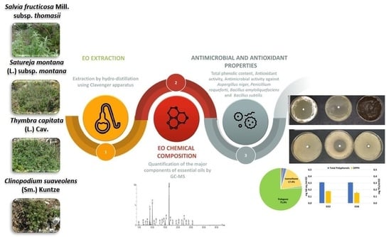
The objective of the present study was to evaluate the antimicrobial properties of EOs from two common officinal wild species Thymbra capitata (L.) Cav., Satureja montana L. subsp. montana, and two threatened taxa, which are both listed in the Regional Red List [31], the rare Clinopodium suaveolens (Sm.) Kuntze and the endemic Salvia fruticosa Mill. subsp. thomasii (Lacaita) Brullo, Guglielmo, Pavone & Terrasi, against common fungal and bacterial contaminants of bakery products, and to correlate the chemical composition of EOs with their biological activity (antimicrobial and antioxidant properties). In particular, the EOs were investigated for their ability to inhibit the growth of two fungal strains, Aspergillus niger and Penicillium roqueforti, and two bacterial strains causing the rope spoilage (Bacillus amyloliquefaciens and Bacillus subtilis) to have a preliminary understanding on the suitability of the plant species as sources of potential biopreservatives for bakery products.

2. Materials and Methods

2.1. Plant Material

The following plant species were considered for this study: Clinopodium suaveolens, Salvia fruticosa subsp. thomasii, Satureja montana subsp. montana, and Thymbra capitata. Germplasm was taken from two sites for each species, for a total of 8 sites. The ecological and plant community characteristics of sites were previously studied in Perrino et al. [32]. The number of individuals collected for the laboratory analyses and the correspondence between species and site collection are reported in Table 1. For each plant, a specific identification code (IC) was attributed.

2.2. Extraction and Chemical Characterization of EOs

The essential oils (EOs) of the wild plant species were extracted by hydrodistillation as described by Perrino et al. [32], using a Clevenger-type apparatus. Briefly, the dry plant material (100 g) was distilled for 4 h in 500 mL H2O in a 1000 mL flask (extraction ratio 1:5 w/vol). Each EO was collected in an amber glass vial, weighted, and stored at 4 °C. The identification and relative abundance of EOs components was performed, as reported in Perrino et al. [32], by gas chromatography coupled with Mass Spectrometer (GC-MS) using a Clarus 680 GC equipped with an Elite-5 MS fused silica capillary column (30 m × 0.25 mm and 0.25 μm film thickness) and interfaced with a single quadrupole Clarus SQ8C (Perkin Elmer). The analytic instrumental conditions were as follows: ionization energy: 70 eV; mass transfer line and injector temperatures: 280 °C; Oven temperature: (a) 50–160 °C at 5 °C/min; (b) 250 °C at 10 °C/min for 5 min; split mode with a split ration: 1:20; solvent delay: 4 min. Mass spectra of target compounds were obtained by an electron impact ionization system with ionization energy of 70 eV. Pure helium was used as a carrier gas at a constant flow rate of 1 mL/min. Mass transfer line and injector temperatures were set at 280 °C, and the oven temperature was programmed from 50 to 160 °C at 5 °C/min; then, it was finally raised to 250 °C at 10 °C/min for 5 min. Diluted samples (1:10 v/v in hexane) were injected in split mode at a ratio of 1:20. Data were collected in full-scan mode in the range 40–300 Da. The relative abundance of each identified compound was expressed as percentage of the total peak areas in chromatographs using a three-step approach: (a) comparison of the obtained mass spectra with a reference mass spectra database (NIST and Wiley); (b) calculation of Retention Indexes (RI) through the injection of a n−alkanes mixture C8−C20 under the same analytic conditions [33]; (c) comparison of components RIs with published data [34] and subsequent identification.
The most abundant compounds detected were identified and quantified by using commercially available standards in the current study. Calibration curves were obtained by injecting standard solutions of each compound at 10, 25, 50, 100, and 200 ng/mL. After plotting the peaks of the different compounds and concentrations, the linearity of the curve was checked by the determination of R2 that exceeds 0.99.
The limit of detection (LOD) and limit of quantification (LOQ) for each compound were calculated and were respectively 0.541 ng/mL and 1.802 ng/mL for α-pinene; 0.574 ng/mL and 1.915 ng/mL for myrcene; 0.461 ng/mL and 1.536 ng/mL for p-cymene; 0.485 ng/mL and 1.616 ng/mL for eucalyptol; 0.566 ng/mL and 1.886 ng/mL for γ-terpinene; 0.685 ng/mL and 2.284 ng/mL for linalol; 0.531 ng/mL and 1.771 ng/mL for camphor; 0.601 ng/mL and 2.003 ng/mL for iso-menthone; 0.690 ng/mL and 2.299 ng/mL for pulegone; 0.681 ng/mL and 2.270 ng/mL for thymol; 0.560 ng/mL and 1.868 ng/mL for carvacrol; 0.623 ng/mL and 2.075 ng/mL for caryophyllene (E).

2.3. Total Phenols Content and In Vitro Antioxidant Activity

The total phenolic content (TPC) was determined by Folin–Ciocalteau via colorimetric method according to Waterhouse [35]. The final concentration was expressed in mg of gallic acid equivalent per ml of EO. The radical scavenging activity was investigated by 2,2-diphenyl-1-picrylhydrazyl (DPPH) assay according to the method reported in Kedare and Singh [36]. Results were expressed in mg Trolox equivalent per ml of EO. All samples were analyzed in triplicate.

2.4. Biological Assays

2.4.1. Microbial Strains and Growth Conditions

Fungal strains used in this study were isolated from bakery products. Aspergillus niger ITEM5132 belongs to the ITEM Culture Collection of the CNR, Institute of Sciences of Food Production, Bari, Italy (http://server.ispa.cnr.it/ITEM/Collection/). Penicillium roqueforti IBT18687 was obtained from the Culture Collection of the Technical University of Denmark, Lyngby, Denmark and deposited into the ITEM Culture Collection with the number ITEM5139. Fungal conidia were collected from 7-day-old cultures on potato dextrose agar (PDA, Biolife Italiana, Milan, Italy) using Triton X 100 0.05% (v/v), counted in the Thoma chamber, and used in antifungal activity tests. Among bacterial contaminants, Bacillus subtilis ATCC8473 and Bacillus amyloliquefaciens DSM7 strains, isolated from bread and soil, respectively, were used. Bacterial strains are stored in the ITEM collection and were cultured in Brain–Heart Infusion broth (BHI; Difco, Becton Dickinson Co., Sparks, MD, USA) for 24 h at 37 °C. For long-term storage, stock cultures were prepared by mixing 0.8 mL of a culture with 0.2 mL of Bacto glycerol (Difco) and freezing mixtures at −80 °C. Strains were subcultured (1% vol/vol) twice in BHI for 24 h at 37 °C using an inoculation before use in experiments.

2.4.2. Disc Diffusion Test

The agar disc diffusion method was employed to determine the antimicrobial activity of EOs. A suspension of 0.1 mL of the tested microorganism (105 fungal spores/mL or 107 bacterial cells/mL) was spread on PDA for fungi and BHI agar plates for bacteria. Filter paper discs (6 mm diameter, Biolife Italiana) were impregnated with 10 μL of the oil, or of a calcium propionate 0.3% (w/v) solution (control), placed on the inoculated plates and then incubated at 25 °C for 5 days for the fungal strains and at 37 °C for 24 h for the bacterial strains. The diameters of the inhibition zones were measured in centimeters, and all tests were performed in duplicate.

2.4.3. Antimicrobial Microdilution Test

Based on the disc diffusion results, specific EOs were selected and tested with a spectrophotometric assay to determine the Minimal Inhibitory Concentration (MIC) using a Bioscreen C (Bioscreen C Oy‘growth curves AB Ltd.; Helsinki, Finland) spectrophotometer, following the method reported by Valerio et al. [36] with slight modifications. Twofold serial dilutions (v/v) of EOs (10%, 7.5%, 5%, 3.75%, 2.5%, 1.87%, 1.25%, 0.93%, 0.62%) were prepared in the growth medium containing Tween 80 (0.5% v/v). PDB and BHI broths were used as media for fungal and bacterial strains, respectively.
After shaking, 20 μL of each EO dilution were inserted in each well of sterile and disposable, multiwell plates (“honeycomb” plates 10×10 wells; Lab systems, Helsinki, Finland). Then, 160 μL of BHI broth or PDB, both containing Tween 80 (0.05%) and 20 μL of the microbial suspension (bacteria ca.1×107 CFU/mL, fungi ca. 0.5−1×10⁵ spores/mL) were added in each well. The final concentration of each EO into the wells was decimally diluted (from 1% to 0.062%). A well containing 180 μL of broth and 20 μL of microbial suspension was used as a growth control. Three replicates for each EO dilution were considered. As blank wells (not inoculated wells), two replicates were considered for each EO dilution.
Then, microplates were incubated at 25 or 37 °C for about 120 h or 48 h in the Bioscreen C, and the growth of microorganisms was automatically measured through determining the optical density at 600 nm every 30 min (for B. subtilis and B. amyloliquefaciens) and at 580 nm every 3 h (for A. niger and P. roqueforti). The microplate was shaken every 10 min to ensure the solubilization of the EO.
The efficacy of different concentrations of EOs was expressed as inhibition percentage (inhibition %), which was calculated basing on the fungal growth reached after 72 h and bacterial growth after 24 h compared to the growth control (PDB−Tween or BHI−Tween), and as minimum inhibitory concentration (MIC), which was defined as the minimal concentration able to cause at least 90% of growth inhibition after 24 h for bacteria and 72 h for fungi [37]. Additionally, optical density measurements recorded every 6 h from zero time to 162 h were used to generate growth curves for each fungal strain.
The inhibitory efficacy of the tested EOs was expressed as growth inhibition percentage calculated with respect to the fungal growth in the inoculated PDB-Tween medium or to the bacterial growth in the inoculated BHI-Tween medium not containing the EO, and it was obtained using the OD values (adjusted by subtracting the value relevant to the blank well) after 72 and 24 h growth, respectively.
Percent Inhibition = 1 − (OD test well/OD of corresponding control well) × 100.

2.5. Statistical Analysis

All data are presented as mean values ± standard error of the mean. Data relevant to the chemical composition of EOs (factors: plant species and chemical compound) and to the disk diffusion test (factors: plant species and target microorganism) were compared by two-way ANOVA followed by Fisher’s LSD post hoc test. The one-way ANOVA followed by Fisher’s LSD post hoc test was used for data from TPC, antioxidant activity, and percentage of inhibition. Results were considered as statistically significant when the p-value was less than 0.05. Data (EO composition, antioxidant activity, TPC, antimicrobial activity by disk diffusion test) were analyzed by principal component analysis (PCA) to discriminate EOs basing on chemical composition and biological activity. The relationships between variables (EOs chemical compounds and biological activities) were tested by Pearson correlation analyses.
Multivariate analysis (PCA) was performed by the Unscrambler (version 10.1, CAMO, Oslo, Norway). All statistical analyses were performed by Statistica 13 software (Dell Inc., 2015).

3. Results

3.1. Essential Oil Composition and Yield

Among plant samples analyzed, some species were more productive in terms of quantity as observed by the different percentages of EO yield (Table 1).
Hence, S. montana subsp. montana samples Sm1 and Sm2 had the highest yields followed by Sf2, Tc1, Sf1, Tc2, Cs2, and Cs1. A difference in yield within species was also apparent for all sampled individuals except for S. montana subsp. montana, which exhibited a minimal variation. The highest variation was observed within the samples of S. fruticosa subsp. thomasii (Sf1 and Sf2) and T. capitata (Tc1 and Tc2). EOs samples were first subjected to a qualitative evaluation to study the chemical composition (data reported in Perrino et al. [32]) and then, the identified compounds were quantified.
In Table 2, the concentration (µg/mL) of the major compounds investigated in the EOs is shown. Both S. fruticosa subsp. thomasii samples have high content of eucalyptol. Moreover, Salvia samples differed regarding the camphor content that was higher in Sf1 (232.7 ± 17.2 µg/mL) with respect to Sf2 (14.9 ± 0.6 µg/mL). Although thymol and carvacrol were not observed in Sf1, it contained a higher concentration of almost each compound. The chemical composition of T. capitata EOs samples was quite similar. The EOs extracted from this plant species were those most rich in chemical compounds. In the case of Clinopodium L., only four out of twelve compounds have been found in the two EOs. Both Cs1 and Cs2 have a high content of pulegone and iso-pulegone. Santureja montana subsp. montana EOs samples showed a different chemical compound profile. Indeed, Sm1 was characterized by the highest amount of α-pinene, α-myrcene, and linalool, while Sm2 EOs was rich in thymol, γ-terpinene, and carvacrol.

3.2. Total Phenolic Content and Antioxidant Activity

The results in Table 3 indicated that T. capitata had the highest phenolic amounts in both Tc1 and Tc2 followed by Sm2 of S. montana subsp. montana. All samples of S. fruticosa subsp. thomasii, C. suaveolens, and sample Sm1 of S. montana subsp. montana presented much lower amounts ranging from 0.25 to 0.77 mg GA eq/mL EO. Significant differences within species were only observed for S. montana subsp. montana, whereas the rest of the analyzed samples had similar antioxidant properties and TPC amounts.
As expected, the antioxidant activity of EOs was linked to the TPC value (Table 2 and Table 3). Both samples of T. capitata and Sm2 of S. montana subsp. montana had high antioxidant activities as a result of the phenolic content. The rest of EOs were drastically weaker and ranging from 0.12 to 0.18 mg TEs/mL EO. S. fruticosa subsp. thomasii and C. suaveolens had the lowest radical scavenging activity.
Despite the absence of differences in the phenolic content of T. capitata samples, significant differences in their antioxidant activity were observed, with Tc1 having a higher antioxidant potential compared to Tc2. Additionally, a big difference was observed between the two samples of S. montana subsp. montana with Sm1 being drastically lower than Sm2. As for C. suaveolens and S. fruticosa subsp. thomasii, the antioxidant activity was almost undetectable, and no significant differences were registered among samples.
Similarly, the strong presence of thymol was responsible for the antioxidant activity of Sm2 of S. montana subsp. montana. Sm1 was deprived of these phenolic compounds and was richer in α-pinene, α-terpineol, and linalol, which lack a phenolic group, making them unable to induce a strong activity (Table 2 and Table 3).
Pulegone was the predominant compound for both samples of C. suaveolens. Therefore, lower antioxidant activities were observed. This weak effect was also apparent in both S. fruticosa subsp. thomasii samples, which were rich in eucalyptol and β-pinene.

3.3. Antimicrobial Activity

3.3.1. Disk Diffusion Assay

Results from the disk diffusion assay are reported in Table 4 and showed differences in the antimicrobial effect among the EOs and microorganisms. Generally, EOs were more effective against the fungal strains than the Bacillus species. In particular, the most active EOs were those from T. capitata samples and from one S. montana subsp. montana sample (Sm2), which inhibited both fungi and bacteria. EOs from C. suaveolens were also active against both fungi and bacteria but to a lesser extent. Finally, EOs from the two samples of S. fruticosa subsp. thomasii were almost ineffective. The higher inhibitory activity was registered against the P. roqueforti strain by Sm2.
In order to assess the strength of the effect, intervals of inhibition were selected. Inhibition zones lower than 1 cm were translated to very low antimicrobial activity, those from 2 to 4 cm were considered as medium antimicrobial activity, those from 4 to 6 cm were considered as high antimicrobial activity, whereas EOs with inhibition zones higher than 6 cm were classified as very high antimicrobial activity. Based on this classification, an effective antifungal activity was observed in EOs Sm2 (S. montana subsp. montana), Tc1, and Tc2 (T. capitata), which were very strong against A. niger and P. roqueforti (Table 4). These samples were followed by EOs Cm1 and Cm2 (C. suaveolons), which had a medium inhibitory activity on the same fungal strains. No observable effect on fungal strains was present for both samples of S. fruticosa subsp. thomasii. Similarly, to S. fruticosa subsp. thomasii, the EO Sm1 from S. montana subsp. montana has no effect on fungal strains. It showed a completely opposite behavior to EO Sm2.
The most effective samples against Bacillus strains were Tc1, Tc2, and Sm2. The same differences in the antibacterial potential were observed within the S. montana subsp. montana samples, and the inhibition properties of Sm1 were extremely lower than those of EO Sm2.

3.3.2. Antimicrobial Activity of EOs by Microdilution Test

The microdilution experiments were performed to study the antimicrobial activity of the most effective EOs. EOs Tc1 and Sm2 were tested against B. subtilis and B. amyloliquefaciens, whereas EOs Cs1 and Cs2, Tc1 and Tc2, and Sm2 were evaluated against A. niger and P. roqueforti at different concentrations. The results confirmed the higher susceptibility of the fungal strains to EOs as indicated by the percentage of growth inhibition even high at the low concentrations tested (Table 5 and Table 6).
In particular, P. roqueforti was more susceptible than A. niger to the EO samples, which caused almost total growth inhibition until levels of 0.093% (Cs1, Sm2) or 0.125% (Tc1, Tc2, Cs2). Values higher than 50% were observed starting from 0.187% for Cs1 and Cs2, 0.375% for Tc1, and 0.25% for Tc2 and Sm2.
Looking to the growth curves (Figure S1), it was clear that A. niger grew better, reaching OD values higher than P. roqueforti. By comparing the effect of EO from C. suaveolons against the two fungal strains, it was observed that all dilutions of Cs1 induced an inhibition or a delay in the growth kinetics with respect to the control but, while a complete inhibition of growth was observed for P. roqueforti using an EO concentration up to 0.93% until at least 135 h incubation, a level of 0.375% was needed to obtain the same effect on A. niger. A delay in the development of A. niger was obtained using Cs1 concentrations lower than 0.375%. However, the lowest concentration tested was still able to delay the growth of both fungal strains, mainly P. roqueforti.
Regarding Cs2, although the same percentage of inhibition of Cs1 was registered after 72 h incubation, the concentrations lower than 0.19% were shown to be less active against the two fungal strains. In the case of A. niger, a significant reduction of the antifungal activity of Cs2 was observed at concentration lower than 0.187%, while for P. roqueforti, it was observed at concentration lower than 0.125%.
The growth kinetics of A. niger treated with Tc1 (T. capitata) indicated that the lowest concentrations (0.125 to 0.0625%) did not show an effect and behaved similarly to the control. Concentrations from 0.187% to 0.25% were slightly more effective in the short term. Whereas dilution ranging from 0.375% to 1% caused a significant increase in the strength of inhibition and induced a complete inhibition of the strain’s growth. Hence, the choice of the lowest dilution between effective ones is sufficient to ensure the same effect and a better management of biological compounds to avoid any risk of toxicity in very high amounts. In the case of P. roqueforti, a complete inhibition was observed until the level 0.125% for the whole incubation time, while the concentration lower than this level had no effect on the fungal growth similarly to the control, as also demonstrated by the high percentage of inhibition (Table 5).
Similarly to EO Tc1, sample Tc2 showed three separate reaction groups for A. niger; the one of lower concentrations (0.063%, 0.093%, and 0.125%) induced similar results to the control, the second group (0.187% and 0.25%) caused a better inhibition, while the third batch almost completely blocked the development of the fungi (0.375%, 0.5%, 0.75%, and 1%). Instead, a slight difference was observed for EO Tc2 against P. roqueforti, since a level of 0.25% was needed to completely inhibit the fungal growth throughout the experimental time. However, looking to the percentage of growth inhibition after 72 h incubation reported in Table 5, Tc2 was slightly more efficacious on A. niger than Tc1 at 0.187% and 0.125% concentration, while a lower effect was observed against P. roqueforti that was partially inhibited at 0.125%.
Different dilutions of EO Sm2 from of S. montana subsp. montana exhibited more homogeneous growth curves of A. niger. The strength of inhibition decreased with the decrease of the EO’s concentration (Figure S1, Table 5). With the exception of the lowest dilution (0.062%) that did not differ from the control, the rest of the concentrations presented an antifungal activity that ranged from a slight inhibition to a complete one. However, a concentration of 0.25% of Sm1, causing 80% of the growth inhibition after 72 h, can be used to reduce and delay the fungal growth. Similarly to the other EOs tested, a higher inhibitory activity was observed against P. roqueforti: Sm2 0.125% completely inhibited the fungal growth during the entire experimental time, thus suggesting a fungicidal effect, and a very low concentration (0.093%) was able to inhibit the fungal growth at 72 h incubation.
The growth kinetic experiments on B. subtilis and B. amyloliquefaciens were performed using only Tc2 and Sm2, which resulted in the most active EOs from the disk diffusion test (Table 4). As reported in Table 6 and Figure S2, EO Tc2 was completely active against both strains at concentrations higher than 0.75%, while a complete loss of efficacy was observed at the lower concentrations. For Sm2, the degree of strength of the antibacterial effect was proportional to the concentration of the EO. The lower dilutions (0.062% to 0.187%) had similar kinetics to the control at the beginning of growth but later on induced an effect on the development of the bacteria.
A big increase in the antibacterial activity appeared with dilutions 0.5% and 0.75%, which almost caused a complete inhibition of bacterial strains.
The percentage of inhibition observed at 24 h showed variability in the activity among dilutions, and values above 50% of inhibition were observed at concentrations higher than 0.5% for EO Tc2 and 0.75% for EO Sm2 (Table 6).

3.3.3. Minimum Inhibitory Concentration (MIC)

The Minimum Inhibitory Concentration (MIC) was defined as the minimal concentration able to cause at least 90% of growth inhibition after 24 h for bacteria and 72 h for fungi. Results extrapolated from Table 5 and Table 6 are reported in Table 7.
C. suaveolens had the lowest MIC values for both of its samples against A. niger (0.187%) and P. roqueforti (0.093% and 0.187%), while EO Tc1 and 2 showed a different response on the fungal strains with MIC values higher for A. niger with respect to P. roqueforti, which was more inhibited by Tc1. Finally, only one sample of S. montana subsp. montana (Sm2) exerted an inhibitory activity against microorganisms. Hence, T. capitata and S. montana subsp. montana remain more effective even if they required higher concentrations, while C. suaveolens produced the most active antifungal EOs. Interestingly, although the MIC shown by Cs2 against A. niger was the same than Cs1, the effect of the two EOs on the fungal growth was slightly different, as also resulted from the percentage of growth inhibition, which was higher for Cs1 (Table 5 and Table 6).
The two bacterial strains tested were considerably more resistant to the EOs: the two EOs tested (Tc2 and Sm2) resulted in completely inhibiting the bacterial growth after 24 h at concentrations higher than 0.75%.

3.4. Principal Component Analysis

In order to discriminate the EOs basing on their biological properties and chemical composition, a PCA was performed. Results are shown in Figure 1 and demonstrated that all EOs were clearly discriminated on the basis of the plant species except for Sm EOs, which were located separately in the plot due to their different composition and biological activity (Figure 1a). On the left part of the plot can be distinguished the most active EOs (Tm1, Tm2, Sm2). Of the remaining samples located in the right part, Sm1 was more similar in chemical composition and biological activity to EOs from S. fruticosa subsp. thomasii, while Sm2 was located in the left high part of the plot, and it was characterized by the higher antimicrobial activity mainly against fungi. Figure 1b shows the loading plot that was utilized to identify the compounds and biological activities accountable for the separation among EOs. The compounds pulegone and iso-menthone mainly characterized the Cs EOs, while linalol, α-pinene, camphor, eucalyptol, and myrcene accounted for Sm1, Sf1, and Sf2. The Tc EOs were mainly characterized by the presence of antioxidant compounds and antibacterial activity and the Sm2 EOs were mainly characterized by the antifungal properties. As shown in Table S1, the antioxidant properties and antibacterial activity were mainly correlated to the presence of thymol, γ-terpinene, carvacrol, and p-cymene. No significant correlations were found between the chemical compounds and the antifungal activity, suggesting a synergistic action of all compounds.

4. Discussion

In the current study, the EOs from four Lamiaceae species from Southern Italy have been chemically and biologically characterized. Results demonstrated the high variability and complexity of EOs composition and biological effect mainly in relation to the antimicrobial activity. The compound composition is characteristic for each plant depending on the species, specific site, temperature, rainfall, altitude, sun radiation, and soil features [38,39,40]. In the current research, four wild plant species belonging to the Lamiaceae family (C. suaveolens, S. fruticosa subsp. thomasii, S. montana subsp. montana, and T. capitata), recently characterized for the chemical composition of their EOs in Perrino et al. [32], were further studied to obtain an exact quantification of the most abundant compounds occurring in the EOs and the relevant biological activity. In particular, the antimicrobial activity of EOs was tested against fungal and bacterial contaminants of bakery products to evaluate their potential as bio-preservatives in these food products.
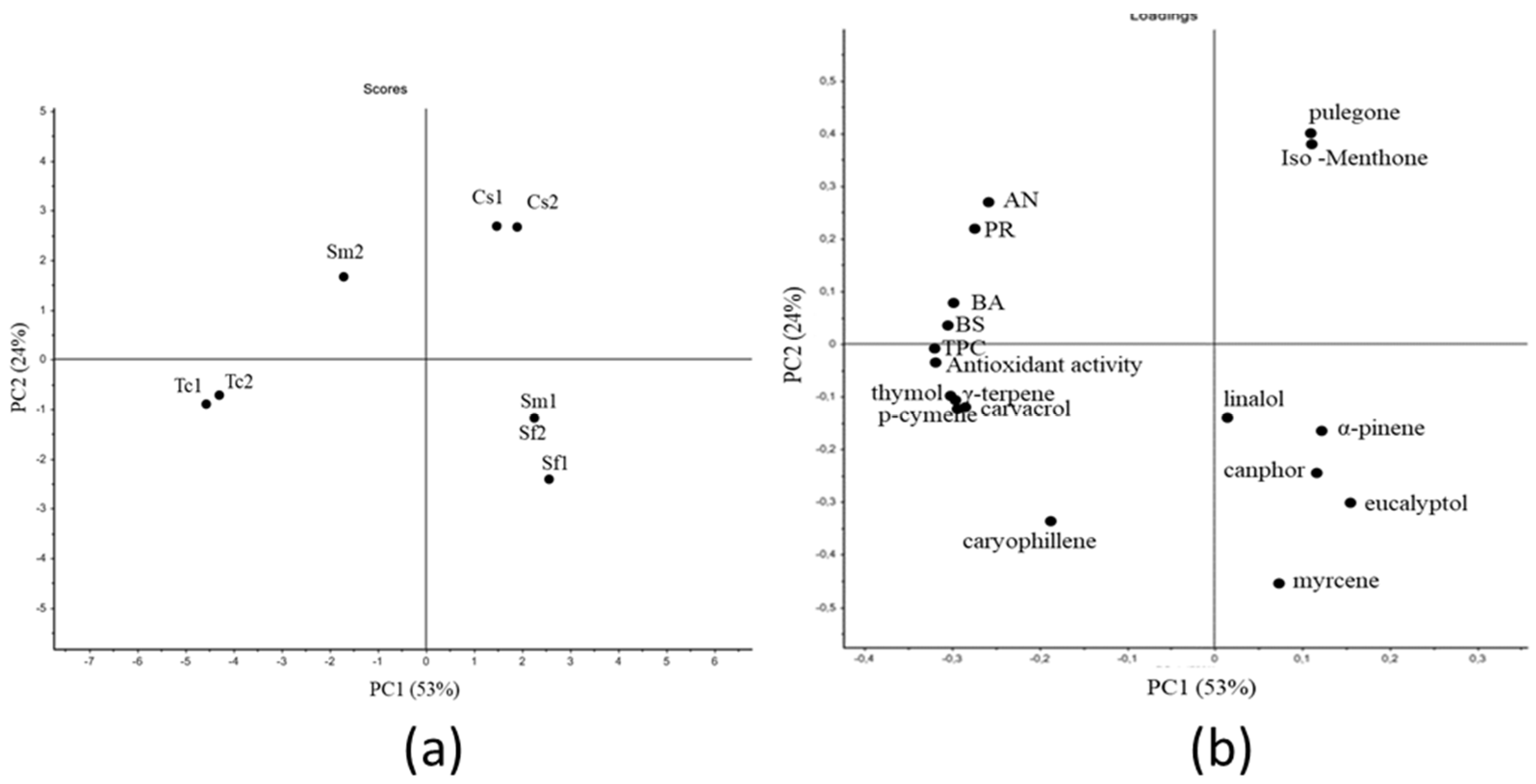
The strong variability between the plant species and within the same species observed in our preliminary study [32] reflects the antioxidant activity registered for EOs and was the result of the differences in EOs compositions and abundances of the major compounds, since the most active EOs were those showing also the higher antioxidant activity. In order to better understand the relationships between the biological properties (results from the disk diffusion assay, antioxidant properties, and total phenol content) and the amounts of the most abundant compounds in EOs, a PCA analysis was performed. As a result, the chemical composition and quantification of the specific compounds in EOs were correlated with the biological activity. The analysis allowed better understanding the significant factors responsible for the antioxidant and antibacterial properties (thymol, carvacrol, γ-terpinene, p-cymene) activity and for the antifungal effect (thymol, carvacrol, γ-terpinene, p-cymene, pulegone, and iso-menthone). Although the presence of compounds with known antimicrobial properties, such as camphor and eucalyptol [41,42] EOs from S. fruticosa subsp. thomasii samples (Sf1 and Sf2), did not show significant effect against the tested microorganisms. Differently from Chaturvedi et al. [42], in this study, the EO containing camphor did not show an antifungal effect despite it being present in combination with eucalyptol in Sf1 and Sf. Similarly to S. fruticosa subsp. thomasii, the Sm1 from S. montana subsp. montana has no effect on fungal strains and showed a completely opposite behavior to Sm2. This difference in effect for the S. montana subsp. montana samples could be explained by the impact of environmental and plant community conditions on the chemical composition of EOs [32]. Indeed, Sm2 had a higher content of thymol and carvacrol than Sm1, which was ineffective against microorganisms, but lower than Tc1 and Tc2 and even showing comparable MIC values, which may be due to a synergistic effect of all compounds detected even if occurring at low concentrations. It has been reported that most of the antioxidant activity from plant sources are derived from phenolic phytochemicals [43] such as thymol and carvacrol, which were proven to be potent antioxidants [44]. In contrast, other usually abundant phytochemicals such as γ-terpinene, α-pinene, β-pinene, limonene, linalol, eucalyptol, pulegone, and menthone have extremely low and almost nonexistent antioxidant activities [44,45]. However, other authors found a strong association between the monoterpene γ-terpinene and the antimicrobial and antioxidant properties of Satureja thymbra [46]. In our study, EOs Tc1 and Tc2, characterized by the highest thymol (413.2 and 484.5 µg/mL, respectively) and carvacrol (261.3 and 163.5 µg/mL, respectively) amounts, were characterized by the highest antioxidant activity as EO Sm2, although this sample contained a significantly lower concentration of thymol (53.4 µg/mL). This result can be explained by the different composition of Sm2 with respect to Tc1 and Tc2: as reported in Perrino et al. [32], thymol represented 46.1% of the total compounds in Sm2, while in Tc1 and Tc2, thymol and carvacrol accounted for 31–35% and 26–17%, respectively, thus not representing the predominant compounds. Moreover, Sm2 was characterized by the presence of γ-terpinene to the same extent (14.57%) of Tc1 and Tc2 (14.95% and 17.71%, respectively) although lower amounts were quantified among the three EOs (14.9, 178.7, and 202.9 µg/mL). We can suppose that the strong antimicrobial activity of T. capitata samples was the result of the dominance of phenolic compounds thymol and carvacrol and the monoterpene γ-terpinene, which are all known for their strong bioactivity [46,47,48].
In contrast, the EOs from S. fruticosa subsp. thomasii did not show biological activity. Sf1 was rich in eucalyptol (541.2 µg/mL), camphor (232.7 µg/mL), α-myrcene (50 µg/m), α-pinene (36.2 µg/m), and caryophyllene (E) (15.5 µg/mL). Sf2 mainly differed from Sf1 in the higher eucalyptol (671.9 µg/mL) content and the presence of a low amount of carvacrol (6.6 µg/mL), but no differences in the biological activity between the two samples were observed. Moreover, EOs from S. fruticosa subsp. thomasii had no effect on A. niger and had a weak activity against B. subtilis, which was mainly due to the predominance of eucalyptol and camphor, which are only active in extremely high amounts.
In the case of C. suaveolens, pulegone was the predominant compound in both samples: it is a monoterpene ketone, typical of Lamiaceae EOs and with commercial value because of its important antimicrobial activities [49,50]. The most known sources of pulegone are Mentha pulegium L., Hedeoma pulegioides L. Pers., and genus Calamintha Mill. and Acinos Mill., which show a high content of pulegone (59.9–96.9%) [51,52]. Both EO samples did not show antioxidant properties due to the lack of phenolic compounds.
Therefore, it can be supposed that the biological activity of EOs is not only due to the higher concentration of specific compounds, but a different combination of compounds can act in a synergistic way, thus affecting the biological properties of EOs.
Regarding the antimicrobial activity of EOs, a comparison with MIC values reported in the literature is not possible, since the antimicrobial susceptibility mainly depends on the test used other than the target strains. However, some similarities were found with published data. In particular, Moukhles et al. [53] demonstrated that T. capitata was active against B. subtilis. Additional articles showcased the strength of Thymbra L. species against A. niger and numerous fungal and bacterial strains such as Aspergillus fumigatus, Penicillium funiculosum, Bacillus cereus, Listeria monocytogenes, Salmonella enterica, and Escherichia coli [54,55,56]. S. montana subsp. montana had lower MIC results in the literature against B. subtilis (0.25%) and A. niger (from 0.06 to 0.5%) [57]. The antimicrobial effect was apparent on a wide range of microorganisms such as Bacillus cereus, Staphylococcus aureus, Streptococcus faecalis, Escherichia coli, Salmonella typhi, Aspergillus fumigates, Pseudomonas aeruginosa, Enterecoccus feacalis, and Saccharomyces cerevisiae [57,58]. However, it is important to indicate a deficiency in comparable data; with the exception of T. capitata, the tested species are rare and/or endemic and are found in specific places around the Mediterranean basin. Rather few data are available for S. fruticosa subsp. thomasii and C. suaveolens. Additionally, it is necessary to consider the differences in the methodological approaches, making the comparison between findings rather inaccurate.
Several studies have been performed to extend bread shelf-life using essential oils, as recently reviewed by some authors [7,10,59]. EOs can be applied as ingredients in the bread formulation, as additives in the packaging or in combination with other technologies (hurdle technology) to prolong the product shelf-life. Recently, Skendi et al. [28] tested the effect of three aromatic plants belonging to the Lamiaceae family (oregano, thyme, and Satureja) as EOs or in dry form directly in the bread formulation. According to our results, the authors detected carvacrol, α-pinene, p-cymene, and γ-terpinene as main components and observed that both forms of addition (essential oil and dry material) resulted in a higher efficacy against the Penicillium spp. strain compared to Aspergillus niger. The use of EOs at a concentration of 50 µl/100 g flour allowed reducing the fungal growth on the bread surface with respect to the control. Even if a comparison with our results cannot provide a realistic prevision of the actual efficacy of the EOs, we suppose that the combination of different preserving technologies, such as the use of sourdough, metabolites from lactic acid bacteria, and/or packaging can induce a delay in the microbial growth on bread. As an example, Debonne et al. [8] tested different concentrations of EO from Thymus zygis in sourdough bread to inhibit the growth of A. niger and P. paneum, but the promising performance observed in vitro were not confirmed in bread. This result was attributed to the food matrix effect, which caused a not homogeneous distribution of the oil. However, other studies [59] demonstrated that large phenolic compounds such as thymol and eugenol (thyme, cinnamon, and clove) show a higher antifungal effect if they are applied directly to the medium, whereas small molecules such as allyl isothiocyanate and citral (mustard and lemongrass) were most efficient when added as volatiles.
S. montana subsp. montana and T. capitata, which are generally used as spices and food preservatives, mainly in meat and fruit products, for their antimicrobial properties [60,61,62,63,64,65] were shown in the current study to be the most active against fungi and bacteria and have the highest the antioxidant properties. The low MIC values observed for the studied EOs, mainly against fungal strains, can be exploited in the preservation of bakery products, which are frequently contaminated by strains belonging to the species tested in the current study. Moreover, the results of the present study highlight the antifungal efficacy of a scarcely studied species, C. suaveolens, which was rich in pulegone, which is a monoterpene ketone associated to the inhibition of wood-decay fungi [66].
The most actives were those from two species, S. montana subsp. montana and T. capitata, which can be exploited as bio-preservatives for bakery products. Moreover, the research provides additional scientific evidence on the potential uses of some common Lamiaceae species for food uses, in order to obtain effective conservation, protection, and valorization of these species. Data relevant to C. suaveolens and S. fruticosa subsp. thomasii, both species of conservation interest and limited geographic distribution, provide new information on the chemical composition of their essential oils and on the potential of C. suaveolens as a source of antifungal compounds.
Factors as temperature, rainfall, altitude, soil features, and habitat conditions have a qualitative and quantitative impact on Lamiaceae species essential oils. This variability does not permit a comparison between essential oils composition. Nevertheless, exploring the essential oils composition is possible to understand how environmental and plant communities condition affect the second metabolism of plants. In turns, this can allow recreating the optimal conditions for domesticating plants that have to produce specific essential oils.
The results suggest that using very low concentrations of EOs in different antifungal approaches such as the hurdle technology can result in the efficacious fungal inhibition. The hurdle technology (or combination technology) is a concept that relies mainly on the simultaneous manipulation and optimization of a series of preservation factors (or hurdles) and their interaction (hurdle effect) in food.
Further studies are necessary to demonstrate the efficacy of the EOs deriving from aromatic plants of the Lamiaceae family when applied to bakery products.

5. Conclusions

In the current study, the EOs from four Lamiaceae species from the Southern Italy have been chemically and biologically characterized. The most active were those from two species, S. montana subsp. montana and T. capitata, which can be exploited as bio-preservatives for bakery products. Moreover, the research provides additional scientific evidence on the potential uses of some common Lamiaceae species for food uses, in order to obtain effective conservation, protection, and valorization of these species. Data relevant to C. suaveolens and S. fruticosa subsp. thomasii, both species of conservation interest and limited geographic distribution, provide new information on the chemical composition of their essential oils and on the potential of C. suaveolens as a source of antifungal compounds.
As reported in the recent study by Perrino et al. [32], some factors as temperature, rainfall, altitude, soil features, and habitat conditions have a qualitative and quantitative impact on Lamiaceae species essential oils. This variability does not permit a comparison between essential oils composition. Nevertheless, exploring the essential oils composition makes it possible to understand how environmental and plant communities’ conditions affect the second metabolism of plants. In turns, this can allow recreating the optimal conditions for domesticating plants that have to produce specific essential oils.
The results suggest that using very low concentrations of EOs in different antifungal approaches such as the hurdle technology can result in efficacious fungal inhibition. The hurdle technology (or combination technology) is a concept that relies mainly on the simultaneous manipulation and optimization of a series of preservation factors (or hurdles) and their interaction (hurdle effect) in food.

Supplementary Materials

The following are available online at https://www.mdpi.com/article/10.3390/agronomy11071431/s1. Figure S1: Growth curves (optical density at 580 nm) of Penicillium roqueforti ITEM5139 (PR) and Aspergillus niger ITEM5132 (AN) registered during incubation at 25 °C for 135 h in the presence of different concentrations of the selected EOs Cs1, Cs2, Tc1, Tc2, and Sm2 in comparison to the control medium (PDB−Tween); Figure S2: Growth curves (optical density at 600 nm) of Bacillus amyloliquefaciens DSM7 and Bacillus subtilis ATCC8473 registered during incubation at 37 °C for 45 h in the presence of different concentrations of the selected EOs Tc2 and Sm2 in comparison to the control medium (BHI−Tween), Table S1: Pearson correlation coefficients between chemical compounds and biological properties of EOs. Total phenol content (TPC); antioxidant activity; antimicrobial activity against: B. amyloliquefaciens (BA), B. subtilis (BS); A. niger (AN), P. roqueforti (PR).

Author Contributions

Conceptualization, F.V. and E.V.P.; Data curation, F.V., G.N.M., A.G. and D.M.; Methodology, G.N.M., A.G. and D.M.; Supervision, F.V., A.F.L. and E.V.P.; Writing—original draft, F.V., G.N.M. and E.V.P.; Writing—review and editing, F.V. and E.V.P. All authors interpreted the results, contributed to the manuscript revision, and read and approved the submitted draft. All authors have read and agreed to the published version of the manuscript.

Funding

This research did not received funding, but it was performed within the Master’s in “Mediterranean Organic Agriculture” assigned to Ahmed Ghannouchi at the CIHEAM, Mediterranean Agronomic Institute of Bari.

Institutional Review Board Statement

Not applicable.

Informed Consent Statement

Not applicable.

Data Availability Statement

The data that support the findings of this study are available from the corresponding author upon reasonable request.

Acknowledgments

This research did not received funding but it was performed within the Master’s in “Mediterranean Organic Agriculture” assigned to Ahmed Ghannouchi at the CIHEAM, Mediterranean Agronomic Institute of Bari. We thank Simonetta Martena (ISPA-CNR) and Lina Al Bitar (CIHEAM) and Ramez Mohamad (CIHEAM) for the administrative support in the management of the Master.

Conflicts of Interest

The authors declare no conflict of interest.

References

  1. Saranraj, P.; Sivasakthivelan, P. Microorganisms involved in spoilage of bread and its control measures. In Bread and Its Fortification Nutrition and Health Benefits; Rossel, C.M., Sheikha, A.F.L., Eds.; CRC Press: Boca Raton, FL, USA, 2015; pp. 132–149. [Google Scholar]
  2. Garcia, M.V.; Bregão, A.S.; Parussolo, G.; Bernardi, A.O.; Stefanello, A.; Copetti, M.V. Incidence of spoilage fungi in the air of bakeries with different hygienic status. Int. J. Food Microbiol. 2019, 290, 254–261. [Google Scholar] [CrossRef]
  3. Valerio, F.; De Bellis, P.; Di Biase, M.; Lonigro, S.L.; Giussani, B.; Visconti, A.; Lavermicocca, P.; Sisto, A. Diversity of spore-forming bacteria and identification of Bacillus amyloliquefaciens as a species frequently associated with the ropy spoilage of bread. Int. J. Food Microbiol. 2012, 156, 278–285. [Google Scholar] [CrossRef]
  4. Valerio, F.; Di Biase, M.; Huchet, V.; Desriac, N.; Lonigro, S.L.; Lavermicocca, P.; Sohier, D.; Postollec, F. Comparison of three Bacillus amyloliquefaciens strains growth behaviour and evaluation of the spoilage risk during bread shelf-life. Food Microbiol. 2015, 45, 2–9. [Google Scholar] [CrossRef] [PubMed]
  5. Pereira, A.P.M.; Stradiotto, G.C.; Freire, L.; Alvarenga, V.O.; Crucello, A.; Morassi, L.L.; Silva, F.P.; Sant’Ana, A.S. Occurrence and enumeration of rope-producing spore forming bacteria in flour and their spoilage potential in different bread formulations. LWT 2020, 133, 110108. [Google Scholar] [CrossRef]
  6. De Bellis, P.; Minervini, F.; Di Biase, M.; Valerio, F.; Lavermicocca, P.; Sisto, A. Toxigenic potential and heat survival of spore-forming bacteria isolated from bread and ingredients. Int. J. Food Microbiol. 2015, 197, 30–39. [Google Scholar] [CrossRef] [PubMed]
  7. Debonne, E.; Van Bockstaele, F.; Samapundo, S.; Eeckhout, M.; Devlieghere, F. The use of essential oils as natural antifungal preservatives in bread products. J. Essent. Oil Res. 2018, 30, 309–318. [Google Scholar] [CrossRef]
  8. Debonne, E.; Van Bockstaele, F.; De Leyn, I.; Devlieghere, F.; Eeckhout, M. Validation of in-vitro antifungal activity of thyme essential oil on Aspergillus niger and Penicillium paneum through application in par-baked wheat and sourdough bread. LWT 2018, 87, 368–378. [Google Scholar] [CrossRef]
  9. Garcia, M.V.; Copetti, M.V. Alternative methods for mould spoilage control in bread and bakery products. Int. Food Res. J. 2019, 26, 737–749. [Google Scholar]
  10. Gavahian, M.; Chu, Y.H.; Lorenzo, J.M.; Mousavi Khaneghah, A.; Barba, F.J. Essential oils as natural preservatives for bakery products: Understanding the mechanisms of action, recent findings, and applications. Crit. Rev. Food Sci. Nutr. 2020, 60, 310–321. [Google Scholar] [CrossRef] [PubMed]
  11. Lavermicocca, P.; Valerio, F.; Evidente, A.; Lazzaroni, S.; Corsetti, A.; Gobbetti, M. Purification and characterization of novel antifungal compounds from the sourdough Lactobacillus plantarum strain 21B. Appl. Environ. Microbiol. 2000, 66, 4084–4090. [Google Scholar] [CrossRef] [Green Version]
  12. Di Biase, M.; Lavermicocca, P.; Lonigro, S.L.; Valerio, F. Lactobacillus brevis-based bioingredient inhibits Aspergillus niger growth on pan bread. Ital. J. Agron. 2014, 9, 146–151. [Google Scholar] [CrossRef] [Green Version]
  13. Valerio, F.; Di Biase, M.; Caputo, L.; Creanza, T.M.; Ancona, N.; Visconti, A.; Lavermicocca, P. Effect of Lactobacillus brevis-based bioingredient and bran on microbiological, physico-chemical and textural quality of yeast-leavened bread during storage. Innov. Food Sci. Emerg. Technol. 2014, 25, 2–8. [Google Scholar] [CrossRef]
  14. Amorati, R.; Foti, M.C.; Valgimigli, L. Antioxidant activity of essential oils. J. Agric. Food Chem. 2013, 61, 10835–10847. [Google Scholar] [CrossRef]
  15. Adorjan, B.; Buchbauer, G. Biological properties of essential oils: An updated review. Flavour Fragr. J. 2010, 25, 407–426. [Google Scholar] [CrossRef]
  16. Brooks, B.D.; Brooks, A.E. Therapeutic strategies to combat antibiotic resistance. Adv. Drug Deliv. Rev. 2014, 2, 446–475. [Google Scholar] [CrossRef]
  17. Trombetta, D.; Castelli, F.; Sarpietro, M.G.; Venuti, V.; Cristani, M.; Daniele, C.; Saija, A.; Mazzanti, G.; Bisignano, G. Mechanisms of antibacterial action of three monoterpenes. Antimicrob. Agents Chemother. 2005, 49, 2474–2478. [Google Scholar] [CrossRef] [PubMed] [Green Version]
  18. Cox, S.D.; Mann, C.M.; Markham, J.L.; Bell, H.C.; Gustafson, J.E.; Warmington, J.R.; Wyllie, S.G. The mode of antimicrobial action of the essential oil of Melaleuca alternifolia (tea tree oil). J. Appl. Microbiol. 2000, 88, 170–175. [Google Scholar] [CrossRef] [PubMed]
  19. Dorman, H.D.; Deans, S.G. Antimicrobial agents from plants: Antibacterial activity of plant volatile oils. J. Appl. Microbiol. 2000, 88, 308–316. [Google Scholar] [CrossRef]
  20. Raut, J.S.; Karuppayil, S.M. A status review on the medicinal properties of essential oils. Ind. Crop. Prod. 2014, 62, 250–264. [Google Scholar] [CrossRef]
  21. Eloff, J.N. It is possible to use herbarium specimens to screen for antibacterial components in some plants. J. Ethnopharmacol. 1999, 67, 355–360. [Google Scholar] [CrossRef]
  22. Abedini, A. Evaluation Biologique et Phytochimique des Substances Naturelles d’Hyptis Atrorubens Poit. (Lamiaceae), Sélectionnée par un Criblage D’extraits de 42 Plantes. Ph.D. Thesis, Université du Droit et de la Santé-Lille II, Lille, France, 2013. Available online: https://tel.archives-ouvertes.fr/tel-01016507 (accessed on 17 July 2021).
  23. Kokkini, S. Herbs of the Labiatae. In Encyclopaedia of Food Science, Food Technology and Nutrition; Macrae, R., Robinson, R., Sadler, M., Fullerlove, G., Eds.; Academic Press: London, UK, 1994; p. 2342. [Google Scholar]
  24. Özkan, M. Glandular and eglandular hairs of Salvia Recognita Fisch. and Mey. (Lamiaceae) in Turkey. Bangladesh J. Bot. 2008, 37, 93–95. [Google Scholar] [CrossRef] [Green Version]
  25. Carović-Stanko, K.; Petek, M.; Grdisa, M.; Pintar, J.; Bedekovic, D.; Herak Custic, M.; Satovic, Z. Medicinal plants of the family of Lamiaceae as functional foods—A Review. Czech J. Food Sci. 2016, 34, 377–390. [Google Scholar] [CrossRef] [Green Version]
  26. Gonçalves, N.D.; Pena, F.L.; Sartoratto, A.; Derlamelina, C.; Duarte, M.C.T.; Antunes, A.E.C.; Prata, A.S. Encapsulated thyme (Thymus vulgaris) essential oil used as a natural preservative in bakery product. Food Res. Int. 2017, 96, 154–160. [Google Scholar] [CrossRef]
  27. Císarová, M.; Hleba, L.; Tančinová, D.; Florková, M.; Foltinová, D.; Charousová, I.; Vrbová, K.; Božik, M.; Klouček, P. Inhibitory effect of essential oils from some Lamiaceae species on growth of Eurotium spp. isolated from bread. J. Microbiol. Biotechnol. Food Sci. 2018, 8, 857–862. [Google Scholar] [CrossRef]
  28. Skendi, A.; Katsantonis, D.Ν.; Chatzopoulou, P.; Irakli, M.; Papageorgiou, M. Antifungal Activity of Aromatic Plants of the Lamiaceae Family in Bread. Foods 2020, 9, 1642. [Google Scholar] [CrossRef]
  29. Tančinová, D.; Foltínová, D.; Mašková, Z.; Štefániková, J.; Árvay, J. Effect of essential oils of Myrtaceae plants on the Penicillium commune. Potravinarstvo 2019, 13, 604–613. [Google Scholar] [CrossRef] [Green Version]
  30. Bakkali, F.; Averbeck, S.; Averbeck, D.; Idaomar, M. Biological effects of essential oils—A review. Food Chem. Toxicol. 2008, 46, 446–475. [Google Scholar] [CrossRef]
  31. Conti, F.; Manzi, A.; Pedrotti, F. Liste Rosse Regionali delle Piante d’Italia. In WWF Italia, Società Botanica Italiana; CIAS: Camerino, Italy, 1997. [Google Scholar]
  32. Perrino, E.V.; Valerio, F.; Gannouchi, A.; Trani, A.; Mezzapesa, G. Ecological and Plant Community Implication on Essential Oils Composition in Useful Wild Officinal Species: A Pilot Case Study in Apulia (Italy). Plants 2021, 10, 574. [Google Scholar] [CrossRef] [PubMed]
  33. Kováts, E. Gas chromatographische charakterisierung organischer verbindungen. Teil 1: Retentions indices aliphatischer halogenide, alkohole, aldehyde und ketone. Helv. Chim. Acta 1958, 41, 1915–1932. [Google Scholar] [CrossRef]
  34. Babushok, V.I.; Linstrom, P.J.; Zenkevich, I.G. Retention indices for frequently reported compounds of plant essential oils. J. Phys. Chem. Ref. Data 2011, 40. [Google Scholar] [CrossRef] [Green Version]
  35. Waterhouse, A.L. Determination of total phenolics. Curr. Protoc. Food Anal. Chem. 2002, 6, I1-1. [Google Scholar] [CrossRef]
  36. Kedare, S.B.; Singh, R.P. Genesis and development of DPPH method of antioxidant assay. J. Food Sci. Technol. 2011, 48, 412–422. [Google Scholar] [CrossRef] [PubMed] [Green Version]
  37. Valerio, F.; Masi, M.; Cimmino, A.; Moeini, S.A.; Lavermicocca, P.; Evidente, A. Antimould microbial and plant metabolites with potential use in intelligent food packaging. Nat. Prod. Res. 2018, 32, 1605–1610. [Google Scholar] [CrossRef]
  38. Sangwan, N.S.; Farooqi, A.H.A.; Shabih, F.; Sangwan, R.S. Regulation of essential oil production in plants. Plant Growth Regul. 2001, 34, 3–21. [Google Scholar] [CrossRef]
  39. Sotomayor, J.A.; Martínez, R.M.; García, A.J.; Jordán, M.J. Thymus zygis subsp. Gracilis: Watering level effect on phytomass production and essential oil quality. J. Agric. Food Chem. 2004, 52, 5418–5424. [Google Scholar] [CrossRef] [PubMed]
  40. Nezhadali, A.; Nabavi, M.; Rajabian, M.; Akbarpour, M.; Pourali, P.; Amini, F. Chemical variation of leaf essential oil at different stages of plant growth and in vitro antibacterial activity of Thymus vulgaris Lamiaceae, from Iran. Beni-Suef Univ. J. Basic Appl. Sci. 2014, 3, 87–92. [Google Scholar] [CrossRef] [Green Version]
  41. Özek, T.; Tabanca, N.; Demirci, F.; Wedge, D.E.; Baser, K.H.C. Enantiomeric distribution of some linalool containing essential oils and their biological activities. Rec. Nat. Prod. 2010, 4, 180–192. [Google Scholar]
  42. Chaturvedi, T.; Kumar, A.; Kumar, A.; Verma, R.S.; Padalia, R.C.; Sundaresan, V.; Chauhan, A.; Saikia, D.; Venkatesha, K.T. Chemical composition, genetic diversity, antibacterial, antifungal and antioxidant activities of camphor-basil (Ocimum kilimandscharicum Guerke). Ind. Crop. Prod. 2018, 118, 246–258. [Google Scholar] [CrossRef]
  43. Mansouri, A.; Embarek, G.; Kokkalou, E.; Kefalas, P. Phenolic profile and antioxidant activity of the Algerian ripe date palm fruit (Phoenix dactylifera). Food Chem. 2005, 89, 411–420. [Google Scholar] [CrossRef]
  44. Ündeğer, Ü.; Başaran, A.R.İ.F.; Degen, G.H.; Başaran, N. Antioxidant activities of major thyme ingredients and lack of (oxidative) DNA damage in V79 Chinese hamster lung fibroblast cells at low levels of carvacrol and thymol. Food Chem. Toxicol. 2009, 47, 2037–2043. [Google Scholar] [CrossRef]
  45. Jabri-Karoui, I.; Bettaieb, I.; Msaada, K.; Hammami, M.; Marzouk, B. Research on the phenolic compounds and antioxidant activities of Tunisian Thymus capitatus. J. Funct. Foods 2012, 4, 661–669. [Google Scholar] [CrossRef]
  46. Giweli, A.; Džamić, A.M.; Soković, M.; Ristić, M.S.; Marin, P.D. Antimicrobial and antioxidant activities of essential oils of Satureja thymbra growing wild in Libya. Molecules 2012, 17, 4836–4850. [Google Scholar] [CrossRef] [Green Version]
  47. Oyedemi, S.O.; Okoh, A.I.; Mabinya, L.V.; Pirochenva, G.; Afolayan, A.J. The proposed mechanism of bactericidal action of eugenol, α-terpineol and γ-terpinene against Listeria monocytogenes, Streptococcus pyogenes, Proteus vulgaris and Escherichia coli. Afr. J. Biotechnol. 2009, 8, 1280–1286. [Google Scholar] [CrossRef]
  48. Memar, M.Y.; Raei, P.; Alizadeh, N.; Aghdam, M.A.; Kafil, H.S. Carvacrol and thymol: Strong antimicrobial agents against resistant isolates. Rev. Med. Microbiol. 2017, 28, 63–68. [Google Scholar] [CrossRef]
  49. Reichling, J.; Schnitzler, P.; Suschke, U.; Saller, R. Essential oils of aromatic plants with antibacterial, antifungal, antiviral, and cytotoxic properties—An overview. Forsch. Komplement. Res. Complement. Med. 2009, 16, 79–90. [Google Scholar] [CrossRef] [Green Version]
  50. Božović, M.; Ragno, R. Calamintha nepeta (L.) Savi and its main essential oil constituent pulegone: Biological activities and chemistry. Molecules 2017, 22, 290. [Google Scholar] [CrossRef] [PubMed]
  51. Muñoz, S.; Collin, G.J.; Gagnon, M.; Ferrufino, J.S. The essential oils of Hedeoma mandoniana Wedd and of Minthostachys andina (Brett) Epling. J. Essent. Oil Res. 1990, 2, 61–66. [Google Scholar] [CrossRef]
  52. Rocha, D.; Novo, M.; Matos, O.; Figueiredo, A.C.; Delgado, M.; Cabral, M.D.; Liberato, M.; Moiteiro, C. Potential of Mentha pulegium for mosquito control. Rev. Ciênc. Agrár. 2015, 38, 155–165. [Google Scholar] [CrossRef]
  53. Moukhles, A.; Ibn Mansour, A.; Ellaghdach, A.; Abrini, J. Chemical composition and in vitro antibacterial activity of the pure essential oils and essential oils extracted from their corresponding hydrolats from different wild varieties of Moroccan thyme. J. Mater. Environ. Sci. 2018, 9, 235–244. [Google Scholar] [CrossRef]
  54. Salgueiro, L.R.; Pinto, E.; Goncalves, M.J.; Pina-Vaz, C.; Cavaleiro, C.; Rodrigues, A.G.; Palmeira, A.; Tavares, C.; Costa-de-Oliveira, S.; Martinez-de-Oliveira, J. Chemical composition and antifungal activity of the essential oil of Thymbra capitata. Planta Med. 2004, 70, 572–575. [Google Scholar] [CrossRef]
  55. Mith, H.; Dure, R.; Delcenserie, V.; Zhiri, A.; Daube, G.; Clinquart, A. Antimicrobial activities of commercial essential oils and their components against food borne pathogens and food spoilage bacteria. Food Sci. Nutr. 2014, 2, 403–416. [Google Scholar] [CrossRef] [Green Version]
  56. Džamić, A.M.; Nikolić, B.J.; Giweli, A.A.; Mitić-Ćulafić, D.S.; Soković, M.D.; Ristić, M.S.; Knežević-Vukčević, J.B.; Marin, P.D. Libyan Thymus capitatus essential oil: Antioxidant, antimicrobial, cytotoxic and colon pathogen adhesion-inhibition properties. J. Appl. Microbiol. 2015, 119, 389–399. [Google Scholar] [CrossRef] [PubMed]
  57. Skočibušić, M.; Bezić, N. Phytochemical analysis and in vitro antimicrobial activity of two Satureja species essential oils. Phytother Res. 2004, 18, 967–970. [Google Scholar] [CrossRef]
  58. Chouhan, S.; Sharma, K.; Guleria, S. Antimicrobial activity of some essential oils—Present status and future perspectives. Medicines 2017, 4, 58. [Google Scholar] [CrossRef] [PubMed] [Green Version]
  59. Suhr, K.I.; Nielsen, P.V. Antifungal activity of essential oils evaluated by two different application techniques against rye bread spoilage fungi. J. Appl. Microbiol. 2003, 94, 665–674. [Google Scholar] [CrossRef] [PubMed]
  60. Marin, S.; Sanchis, V.; Ramos, A.J. Plant products in the control of mycotoxins and mycotoxigenic fungi on food commodities. In Natural Products in Plant Pest Management; Dubey, N.K., Ed.; CAB International: Wallingford, UK, 2011; pp. 31–35. [Google Scholar]
  61. Jayari, A.; El Abed, N.; Jouini, A.; Mohammed Saed Abdul-Wahab, O.; Maaroufi, A.; Ben Hadj Ahmed, S. Antibacterial activity of Thymus capitatus and Thymus algeriensis essential oils against four food-borne pathogens inoculated in minced beef meat. J. Food Saf. 2018, 38, e12409. [Google Scholar] [CrossRef]
  62. Aminzare, M.; Hashemi, M.; Ansarian, E.; Bimkar, M.; Azar, H.H.; Mehrasbi, M.R.; Daneshamooz, S.; Raeisi, M.; Jannat, B.; Afshari, A. Using natural antioxidants in meat and meat products as preservatives: A review. Adv. Anim. Vet. Sci. 2019, 7, 417–426. [Google Scholar] [CrossRef] [Green Version]
  63. Santos, J.D.; Coelho, E.; Silva, R.; Passos, C.P.; Teixeira, P.; Henriques, I.; Coimbra, M.A. Chemical composition and antimicrobial activity of Satureja montana byproducts essential oils. Ind. Crops Prod. 2019, 137, 541–548. [Google Scholar] [CrossRef]
  64. Charfi, S.; Boujida, N.; Abrini, J.; Senhaji, N.S. Study of chemical composition and antibacterial activity of Moroccan Thymbra capitata essential oil and its possible use in orange juice conservation. Mater. Today Proc. 2019, 13, 706–712. [Google Scholar] [CrossRef]
  65. Kontaxakis, E.; Filippidi, E.; Stavropoulou, A.; Daferera, D.; Tarantilis, P.A.; Lydakis, D. Evaluation of eight essential oils for postharvest control of Aspergillus carbonarius in grapes. J. Food Prot. 2020, 83, 1632–1640. [Google Scholar] [CrossRef]
  66. Pinheiro, C.G.; Bianchini, N.H.; Batista, B.F.; Pavlack, A.S.; Brião Muniz, M.F.; Pavanato, M.A.; Gouveia, F.N.; Reiniger, L.R.S.; Heinzmann, B.M. Hesperozygis ringens (Benth.) Epling essential oil: Antifungal activity and effect on ergosterol content of wood-decay fungi. J. Essent. Oil Res. 2020, 1–11. [Google Scholar] [CrossRef]
Figure 1. Principal component analysis (PCA) of data from the biological activity of EOs and their chemical characterization. (a) Score plot indicating sample distribution (EOs) basing on the biological activity and EO composition. (b) Loading plot indicating the correlation between variables: antimicrobial activity against A. niger (AN), P. roqueforti (PR), B. amyloliquefaciens (BA), and B. subtilis (BS); total phenol content (TPC); antioxidant activity; concentration of the single compounds (thymol, carvacrol, caryophillene, eucalyptol, canphor, α-pinene, γ-terpene, linalool, myrcene, p-cymene, pulegone, and iso-menthone) for each EO.
Agronomy 11 01431 g001
Table 1. Plant germoplasm location, code, number of individuals detected, and yield of the relevant essential oils extracted by hydrodistillation.
SpeciesDetected LocationRelief and Data ReferenceYield (% w/w)Individual DetectedIC
Salvia fruticosa subsp. thomasiiGravina Petruscio (Mottola–TA)11.03.19.030.567Sf1
Gaudella (Laterza−TA)15.03.19.010.6810Sf2
Clinopodium suaveolensScannapecora (Altamura–BA)17.04.19.010.37150Cs1
Jazzo Filieri (BA)17.04.19.020.42120Cs2
Thymbra capitataGravina Capo di Gavito (Mottola–TA)11.03.19.020.6030Tc1
Torre Santa Sabina (Carovigno–BR)22.03.19.010.4850Tc2
Satureja montana subsp. montanaAzienda Sabatelli (Fasano–BR)22.03.19.020.9040Sm1
Monte Castiglione (Altamura–BA)1.04.19.010.9250Sm2
Table 2. Concentrations (µg/mL) of the most abundant compounds occurring in the EOs, identified and quantified by GC-MS.
Salvia fruticosa subsp. thomasiiThymbra capitataClinopodium suaveolensSatureja montana subsp. montana
Compound NameSf1Sf2Tc1Tc2Cs1Cs2Sm1Sm2
µg/mL
α-Pinene36.2 ± 0.1 b,c18.7 ± 0.8 a,b1.9 ± 0.3 aa270.4 ± 24.7 e
α-Myrcene50 ± 1.1 c,d58.5 ± 0.9 d35.3 ± 1.7 e26.8 ± 0.1 c,d6.4 ± 0.4 a7.3 ± 0.1 a42.8 ± 5.1 c5 ± 0.1 a,b
p-Cymene3.2 ± 0.2 a83.8 ± 0.1 f82.2 ± 4.6 f4.3 ± 0.2 a,b
Eucalyptol541.2 ± 39.2 f671.9 ± 2.6 g
γ-Terpinene3.3 ± 0.3 a0.99 ± 0.1 a178.7 ± 4.4 h202.9 ± 8.6 j14.9 ± 0.2 b
Linalool4.06 ± 0.4 a4.1 ± 0.1 a16.4 ± 0.6 b29.5 ± 0.7 d,e7.7 ± 0.3 a5.2 ± 0.3 a82 ± 5.3 d4.7 ± 0.1 a,b
Camphor232.7 ± 17.2 e14.9 ± 0.6 a
Thymol10.2 ± 0.1 a413.2 ± 5.1 i484.5 ± 13.3 l7.1 ± 0.2 a,b53.4 ± 1.6 c
Carvacrol6.6 ± 0.1 a261.3 ± 4.2 k163.5 ± 6.2 g5.8 ± 0.1 a,b6.7 ± 0.2 a,b
Caryophyllene, (E)15.5 ± 1.4 a10.7 ± 0.1 a19.2 ± 1.4 b,c20.3 ± 0.3 b,c1.1 ± 1.5 a0.6 ± 0.9 a
Isopulegone, trans-116.8 ± 0.1 b172.3 ± 7.3 c
Pulegone1146.9 ± 8.5 e1062.7 ± 41.6 d
For each plant species, means followed by different letters differ significantly (p < 0.05) by the LSD post hoc test.; a values ≤ LOD.
Table 3. Total phenolic content (TPC) and antioxidant activity of EOs.
EO CodeTPCAntioxidant Activity
mg GA eq./mL EOmg TEs/mL EO
Sf10.25 ± 0.03 a0.12 ± 0.003 a
Sf20.33 ± 0.02 a0.17 ± 0.004 a
Cs10.31 ± 0.03 a0.18 ± 0.001 a
Cs20.31 ± 0.04 a0.16 ± 0.01 a
Tc1279 ± 8.3 c9.61 ± 0.19 d
Tc2286 ± 19.8 c8.39 ± 0.11 c
Sm10.77 ± 0.03 a0.19 ± 0.004 a
Sm2231 ± 9.4 b6.16 ± 0.25 b
In each column, means followed by different letters differ significantly (p < 0.05) by the LSD post hoc test.
Table 4. Screening of the antimicrobial activity of EOs against A. niger ITEM5132, P. roqueforti ITEM5139, B. amyloliquefaciens DSM7, and B. subtilis ATCC8473 using the disk diffusion assay.
EOA. niger ITEM5132P. roqueforti ITEM5139B. subtilis ATCC 8473B. amyloliquefaciens DSM7
Sf1–a1 ± 0.17 c
Sf20.97 ± 0.11 c1 ± 0.10 c,d
Cs14.9 ± 0.13 d4.05 ± 0.13 d1.23 ± 0.11 c1.25 ± 0.13 c
Cs23.05 ± 0.12 e3.1 ± 0.14 e1.13 ± 0.10 c1.23 ± 0.10 c
Tc16.05 ± 0.13 b6.95 ± 0.12 c4.33 ± 0.11 a4.5 ± 0.13 b
Tc25.55 ± 0.12 c7.25 ± 0.13 b3.5 ± 0.10 b4.76 ± 0.11 a,b
Sm11.13 ± 0.11 c1.1 ± 0.13 c,d
Sm26.25 ± 0.13 a8.95 ± 0.13 a4.03 ± 0.11 a5.2 ± 0.12 a
Halo of inhibition expressed in cm.; Means followed by different letters differs significantly (p < 0.05) by the LSD post hoc test.; a Halo of inhibition not visible around the disc.
Table 5. Percent of inhibition (%) of fungal growth after 72 h incubation at 25 °C by the EOs.
A. niger ITEM5132P. roqueforti ITEM5139A. niger ITEM5132P. roqueforti ITEM5139A. niger ITEM5132P. roqueforti ITEM5139A. niger ITEM5132P. roqueforti ITEM5139A. niger ITEM5132P. roqueforti ITEM5139
EO ConcentrationCs1Cs2Tc1Tc2Sm2
1%100 ± 0.4 a95.5±0.8 b98.8 ± 0.4 a,b100.9 ± 0.7 a100 ± 3.2 a,b91.5 ± 13.9 a100 ± 2.0 a100 ± 21.2 a100 ± 2.2 a100 ± 0.2 a
0.75%99.8 ± 0.1 a100.3 ± 0.1 a98.5 ± 0.5 a,b100.0 ± 0.1 a100 ± 2.1 a100 ± 3.2 a100 ± 3.4 a100 ± 1.9 a,b100 ± 6.4 a100 ± 0.6 a
0.5%99.7 ± 0.04 a99.7 ± 0.1 a99.7 ± 0.1 a,b98.9 ± 1.7 a,b98.8 ± 0.1 a,b100 ± 3.4 a100 ± 1.4 a97.8 ± 2.0 b97.5 ± 1.7 a100 ± 0.4 a
0.375%99.7 ± 0.1 a99.4 ± 0.3 a99.4 ± 0.3 a,b99.0 ± 0.3 a,b95.3 ± 2.4 b99.9 ± 0.2 a97.5 ± 0.6 a100 ± 0.1 b92.3 ± 3.1 a,b100 ± 1.02 a
0.25%98.04 ± 0.7 a99.9 ± 0.1 a100 ± 0.3 a95.9±1.5 a,b41.1 ± 13.3 c99.0 ± 0.5 a60.4 ± 1.5 b100 ± 0.06 b79.9 ± 8.6 b100 ± 0.08 a
0.187%90.1 ± 2.6 a100.3 ± 0.1 a89.1 ± 6.2 b98.3 ± 0.7 a,b30.0 ± 1.9 c100 ± 0.1 a48.7 ± 0.5 b100 ± 0.2 b27.8 ± 4.8 c99.6 ± 0.4 a
0.125%66.1 ± 4.2 b99.9 ± 0.5 a27.7 ± 7.7 c82.0 ± 9.6 b7.6 ± 4.3 d100 ± 23.2 a10.04 ± 1.7 c87.5 ± 10.1 b17.8 ± 5.7 c,d100 ± 0.06 a
0.093%46.7 ± 8.8 c98.9 ± 2.4 a13.2 ± 3.4 d59.0 ± 13.8 c9.2 ± 1.5 d23.4 ± 3.9 b5.3 ± 1.6 c21.2 ± 4.2 c5.8 ± 1.6 c100 ± 5.1 a
0.062%10.2 ± 5.4 d35.9 ± 0.08 c1.9 ± 2.1 e23.0 ± 5.3 d-33.1 ± 12.6 b-22.02 ± 3.02 c-20.6 ± 2.6 b
In each column, means followed by different letters differ significantly (p < 0.05) by the LSD post hoc test.
Table 6. Percent of inhibition (%) of bacterial growth (B. amyloliquefaciens DSM7 and B. subtilis ATCC8473) after 24 h incubation at 37 °C.
B. subtilis ATCC8473B. amyloliquefaciens DSM7B. subtilis ATCC8473B. amyloliquefaciens DSM7
EO ConcentrationTc2Sm2
1%100 ± 5.68 a100 ± 10.6 a96.8 ± 2.3 a96.2 ± 1.0 a
0.75%100 ± 2.62 a100 ± 4.17 a88.2 ± 1.8 b93.9 ± 0.8 a
0.5%34.4 ± 1.13 b17.0 ± 1.77 c,d61.6 ± 2.9 c65.7 ± 1.0 b
0.375%24.0±1.47 c0.8 ± 2.13 e27.1 ± 0.6 d8.8 ± 1.0 e,f
0.25%34.0 ± 0.34 b28.2 ± 0.1 b,c23.8 ± 3.0 d7.2 ± 0.9 f
0.187%37.6 ± 1.29 b23.65 ± 1.3 b,c,d23.4 ± 0.4 d30.2 ± 0.7 c
0.125%4.56 ± 0.17 d22.8 ± 2.5 b,c,d22.2 ± 0.2 d27.7 ± 0.3 c
0.093%19.8 ± 5.45 c29.6 ± 0.5 b2.6 ± 1.0 e13.7 ± 1.3 d
0.062%1.8 ± 1.82 d13.9 ± 0.06 d-11.5 ± 2.3 d,e
In each column, means followed by different letters differ significantly (p < 0.05) by the LSD post hoc test.
Table 7. MIC values of EOs against fungal (A. niger ITEM5132 and P. roqueforti ITEM5139) and bacterial (B. subtilis ATCC 8473 and B. amyloliquefaciens DSM7).
MIC90 72 hMIC90 24 h
EOA. niger ITEM5132P. roqueforti ITEM5139B. subtilis ATCC 8473B. amyloliquefaciens DSM7
Sf1NTNTNTNT
Sf2NTNTNTNT
Cs10.187%0.093%NTNT
Cs20.187%0.187%NTNT
Tc10.375%0.125%NTNT
Tc20.375%0.187%0.75%0.75%
Sm1NTNTNTNT
Sm20.375%0.093%1%0.75%
NT: not tested.
Publisher’s Note: MDPI stays neutral with regard to jurisdictional claims in published maps and institutional affiliations.

