Review

Influence of Environmental Factors Light, CO2, Temperature, and Relative Humidity on Stomatal Opening and Development: A Review

1
Department of Biosystems-MeBioS, KU Leuven, Willem De Croylaan 42, 3001 Heverlee, Belgium
2
Department of Biology-Laboratory of Molecular Plant Biology, KU Leuven, Kasteelpark Arenberg 31, 3001 Heverlee, Belgium
3
Department of Biosystems-Crop Biotechnics, KU Leuven, Willem De Croylaan 42, 3001 Heverlee, Belgium
4
Department of Biosystems-MeBioS, KU Leuven, Kasteelpark Arenberg 30, 3001 Heverlee, Belgium
*
Author to whom correspondence should be addressed.
Agronomy 2020, 10(12), 1975; https://doi.org/10.3390/agronomy10121975
Submission received: 14 November 2020 / Revised: 11 December 2020 / Accepted: 11 December 2020 / Published: 15 December 2020
(This article belongs to the Special Issue Stomata, Photosynthesis and Plant Water Use Efficiency)

Abstract

:
Stomata, the microscopic pores surrounded by a pair of guard cells on the surfaces of leaves and stems, play an essential role in regulating the gas exchange between a plant and the surrounding atmosphere. Stomatal development and opening are significantly influenced by environmental conditions, both in the short and long term. The rapid rate of current climate change has been affecting stomatal responses, as a new balance between photosynthesis and water-use efficiency has to be found. Understanding the mechanisms involved in stomatal regulation and adjustment provides us with new insights into the ability of stomata to process information and evolve over time. In this review, we summarize the recent advances in research on the underlying mechanisms of the interaction between environmental factors and stomatal development and opening. Specific emphasis is placed on the environmental factors including light, CO2 concentration, ambient temperature, and relative humidity, as these factors play a significant role in understanding the impact of global climate change on plant development.

1. Introduction

Stomata are microscopic pores on the surfaces of plant leaves and stems. This term is derived from the Greek word “stoma” (στομα) which is translated as “mouth”. They can be present on both leaf surfaces (amphistomatic) or on a single surface (hypostomatic) [1]. They consist of specialized guard cells surrounding the central pore which can open and close. Stomata play a fundamental role in the regulation of water evaporation and carbon dioxide assimilation. By adjusting the turgor pressure of their guard cells, plants actively alter their stomatal aperture, thereby mediating gas exchange rates between the surrounding atmosphere and the leaf interior. The regulation of diffusion is dependent on both stomatal dimensions and stomatal density per leaf area. Darcy′s law is used to model flow rates through a porous medium in response to a potential gradient. The flux is determined by the concentration difference (leaf-air mole fraction difference) and leaf-specific stomatal conductivity to water vapour diffusion. Stomata regulate the water uptake via changes in stomatal conductance, opening, and density. By reducing or increasing the stomatal opening and the potential flow area, diffusion is controlled [2,3]. Stomata can vary in size, shape, and number depending on the plant species.
Given the central role that stomata play in regulating carbon uptake and transpiration of plants, changes in stomatal development and opening can show adaptive relationships to the environmental conditions surrounding the plant [4]. Stomatal development and opening have been identified as adaptive, including changes in stomatal density, size, and shape [4]. Both genetic factors and differences in growing conditions have been reported to cause variation in the number, distribution, and density of stomata [5]. Since plants are sessile, it is required that they are able to adapt their physiology to external factors. These adaptations include the alteration of stomatal distribution and function. Stomatal guard cells perceive various environmental cues to which they have to respond to optimize the plant gas exchange to maximize the photosynthetic rate, while avoiding drought stress. In the short term, plants adjust to changing environmental conditions by adjusting stomatal closure to minimize water loss and moderate CO2 uptake [6]. In the long term, plants adapt to the environmental conditions by regulating the stomatal density on developing leaves, thus defining the minimal and maximal rates of gas exchange [1,7,8]. Therefore, the maximum stomatal conductance (gs) is set by the long-term changes in stomatal density. These changes in stomatal density can be expressed as stomatal density per unit of area (SD) or as the stomatal index (SI), i.e., the ratio of stomata to epidermal cells plus stomata, multiplied by 100 [5,9].
Due to global climate change, extreme temperatures and CO2 enrichment will occur [9,10,11]. This will affect the stomatal responses, because a new balance between water-use efficiency and photosynthesis will have to be found [10,12]. To provide a comprehensive overview of all factors influencing stomatal development and movement in the short and long term, the aim of this study was to review the available reports on the effects of the following main environmental factors: light, CO2 concentration, ambient temperature, and relative humidity. As Arabidopsis thaliana (L.) Heynh. is widely studied as a model organism in fundamental plant molecular genetics research, it is also dominant in this review. However, to broaden the scope, the findings for Arabidopsis thaliana are complemented with examples of other plants. We provide an overview of the current knowledge on stomatal development and opening before exploring how these main environmental factors regulate these processes and how they may change in the context of global warming.

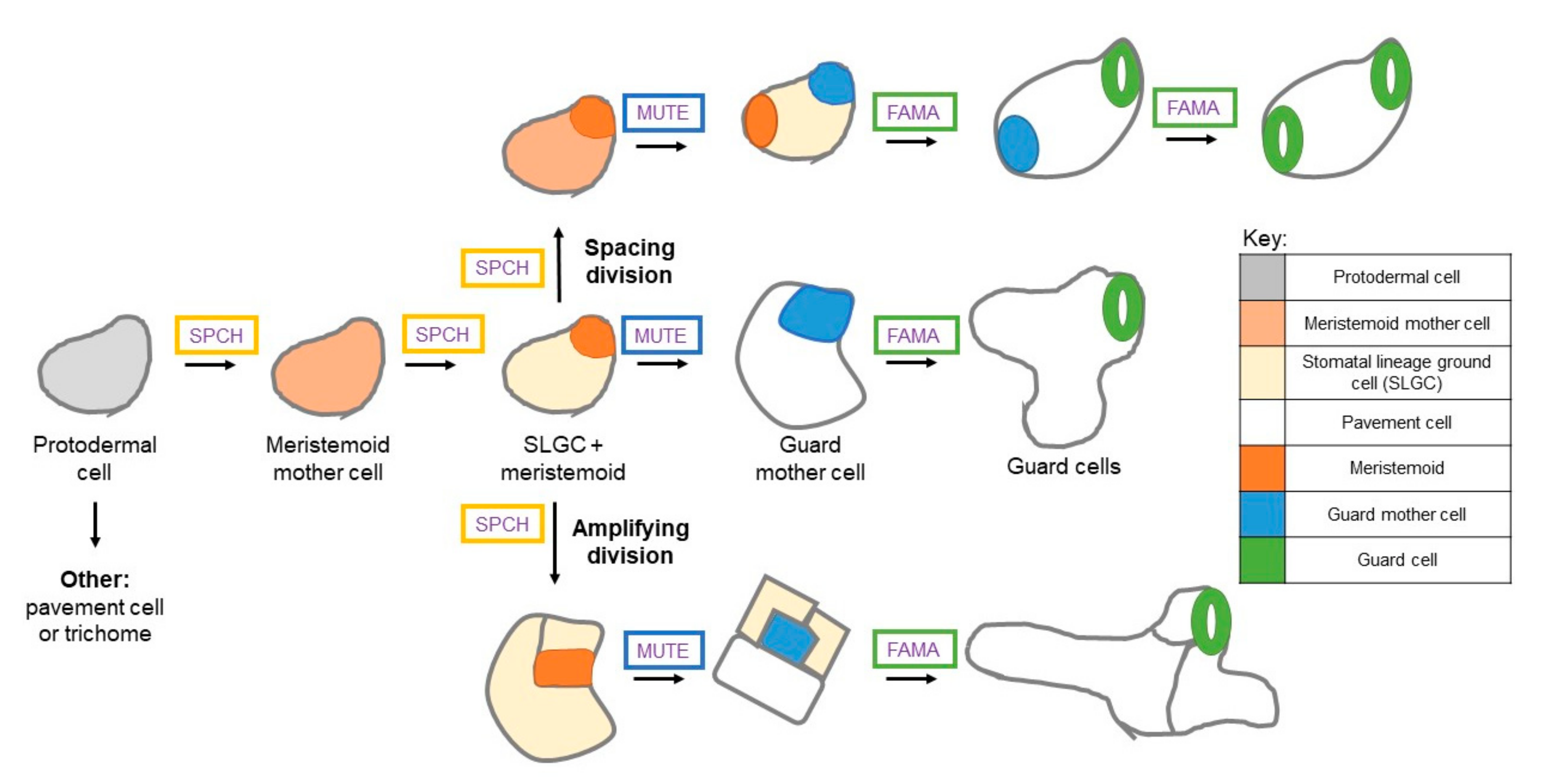
1.1. Stomatal Development

Stomatal development is a process in which promeristem cells are subjected to symmetric and asymmetric divisions to form the leaf epidermis [1]. Undifferentiated protodermal cells construct the epidermis of developing leaves. These undifferentiated cells develop into the following three main cell types: leaf hairs (trichomes), pavement or epidermal cells, and stomatal guard cells [1]. There are intermediary steps in the development of mature stomata, starting from the protodermal cells. This series of steps is called the stomatal lineage and there are many steps in which cells may exit from this stomatal lineage or have their development stopped for a period of time. This means that entry into the stomatal lineage does not impose that a stoma is formed. This provides the developing leaf with flexibility to respond to environmental conditions.
Development of a protodermal cell to a meristemoid mother cell (MMC) is the first step in the stomatal lineage (Figure 1). This step is followed by a symmetric division to from a stomata lineage ground cell (SLGC) and a meristemoid [1]. The meristemoid may undergo amplifying divisions or develop into a guard mother cell (GMC). Symmetrical divisions of a guard mother cell form a set of guard cells (GCs) [1,13,14]. SLGCs can differentiate into pavement cells (white) or they can undergo a spacing division to create a new meristemoid that is separated by a pavement cell with the second meristemoid (Figure 1) [6]. Amplifying divisions increases the epidermal cell total, while spacing divisions establish the one-cell-spacing pattern [12]. This means that stomata do not make direct contact with each other. They are always separated by a non-GC [1,15].
The formation of stomata is regulated by master transcription factors. The number and placement of stomata in the epidermal layer are partially determined by the number of entries in the stomatal lineage and by the type of asymmetric divisions (amplifying and spacing divisions) that occur in this stomatal lineage [12,14]. Three basic-helix-loop-helix (bHLH) transcription factors are necessary and sufficient for driving the production of protodermal cells into stomata. These master transcription factors are SPEECHLESS (SPCH), MUTE, and FAMA [16] (Figure 1). These bHLH transcription factors are critical for the development of stomata, while mutation leads to the loss of correctly formed stomata [1,6,14].
Protodermal cells differentiate into MMCs and consecutively divide into SLGCs and meristemoids, a process promoted by SPCH [1]. SPCH mutants are not able to enter the stomatal lineage and form an epidermis made out of pavement cells. Depending on SPCH levels and activity, the meristemoid can undergo amplifying or spacing divisions, or proceed down the stomatal lineage (Figure 1) [1,6]. The transcription factor MUTE regulates the transition from meristemoid to GMC [5]. In the absence of MUTE, meristemoids fail to differentiate stomata after arresting due to excessive asymmetric divisions. Initial spacing and patterning does not require MUTE, because amplifying and spacing divisions still occurs even when MUTE is mutated. Initial spacing and patterning are under the control of SPCH [1]. When MUTE is constitutively overexpressed in the wild-type plant, the epidermis created is practically entirely constructed of stomata, because the entire epidermis adopts the guard cell identity [1,17]. The final cell division, which is the symmetric division into the two guard cells, is regulated by FAMA. FAMA mutants are unable to produce stomata. They produce fama tumours after a series of uncontrolled symmetrical divisions of GMCs [1,6,14,17,18]. Therefore, stomata are formed after a series of cell fate changes where each precursor cell is subjected to a specific cell division [15].
Two more bHLH transcription factors, ICE1/SCREAM and SCREAM2 play a role in the stomatal lineage initiation, through direct interactions with SPCH, MUTE, and FAMA [19,20]. They regulate progress through the successive stages of stomatal development. The leucine-rich repeat receptor-like protein TMM (TOO MANY MOUTHS) partly moderates entry into the stomatal lineage. TMM forms an active complex with members of the ERECTA family [8]. ERECTA is required for diverse processes, including organ growth and stomatal development. It is also important in responses to different biotic and abiotic stresses. This TMM-ERECTA complex is believed to signal through a MAPK signaling pathway to negatively regulate stomatal development by targeting the bHLH transcription factors that moderate the steps in stomatal differentiation [8]. Mutations in TMM result in alteration in spacing and amplifying divisions in the stomatal lineage and leads to the formation of excess stomata in leaves [15]. This indicates that the function of TMM is the repression of divisions. Mutants are unsuccessful to inhibit asymmetric divisions in cells adjacent to two or more stomata or their precursor cells. The premature transformation of meristemoids into GMCs is a result of the mutants displaying a reduced number of amplifying divisions [15].
Recently, Jiao et al. [21] found that N-glycosylation, the reaction in which a glycan is attached to a nitrogen atom of another molecule, was involved in stomatal development. They showed that stomatal development was modulated by N-glycosylation through the regulation of the release of abscisic acid (ABA) and auxin by β-glucosidase activity [21,22,23]. Using the Arabidopsis gene AtBG1, which encodes a β-glucosidase of the glycoside hydrolase family 1, they showed that AtBG1 is a glycoprotein with three N-glycosylation sites [21,24]. In the stt3a-2 mutant, which was inadequate in the transfer of N-glycan to peptides, the stability of the glycoprotein AtBG1 significantly decreased, leading to reduced ABA and auxin contents. ABA and auxin regulate downstream transcription factors of the stomatal lineage [24,25]. Reduced ABA and auxin contents associate with upregulation of SPCH, MUTE, and FAMA, leading to abnormal stomatal development in this mutant [21]. Another hypothesis in this research is that N-glycosylation also plays a role in the biological function of TMM. TMM mutants display severe stomatal clustering on the plant leaves, which is possibly one of the reasons that stt3a-2 mutants form stomatal clusters. However, additional research is required to further unravel the mechanisms of N-glycosylation in modulating stomatal development [21].

1.2. Stomatal Opening

Stomatal opening is rapidly and reversibly regulated by guard cells. Dicots and non-graminaceous monocots have kidney-shaped stomata, while grasses have dumbbell-shaped stomata (Figure 2).
To protect the plant from excessive water loss or starvation for CO2, stomatal opening is finely controlled. The guard cell′s sensitivity to environmental and endogenous plant signals, including light, CO2, temperature, humidity, water status, hormones, and sugars plays an important role in achieving its fine control. Ion channels, transporters, and pumps located in the plasma membrane of the guard cells, regulate the influx and efflux of solutes. During stomatal opening, the H+-ATPase pumps H+ out of the guard cells and hyperpolarizes the guard cell membrane. Subsequently, this triggers the activation of inward K+ channels, such as KAT1, KAT2, and AKT1 (potassium channels found in Arabidopsis) [26]. K+ uptake is balanced by Cl, acquired from the apoplast, and malate2− derived from starch breakdown and NO3. These anionic molecules contribute to the intracellular solute concentration, which mediates the sugar import. Ions that enter the guard cells together with water, transported via aquaporins, generate turgor pressure necessary for stomatal opening [26].
Recently, Flütsch et al. [27] reported that glucose, not malate, was the major starch-derived metabolite in Arabidopsis guard cells. Contrary to previous research where malate was reported to increase in the guard cells, Flütsch et al. [27] reported a decrease in glucose levels of the amy3bam1 guard cells during a blue-light treatment, demonstrating that glucose was derived from guard cell starch degradation. α-AMYLASE3 (AMY3) and β-AMYLASE1 (BAM1) are glucan hydrolases, degrading starch in Arabidopsis guard cells to promote stomatal opening. After the blue light treatment, isolated WT guard cells contained high amounts of glucose, while glucose levels in the isolated amy3bam1 guard cells were almost undetectable [27]. They concluded that previous research did not exclude the possibility of malate transport from the mesophyll. For this reason, they have worked with isolated guard cells. Furthermore, they hypothesized that it was unlikely that malate was synthesized from starch degradation, but proposed that malate was presumably produced from anaplerotic CO2 fixation in the guard cells or imported from the apoplast, fulfilling its function as an osmotically active solute [27,28,29].
During stomatal closure, H+-ATPase inhibition and the activation of S-type and R-type anion channels result in membrane depolarization. S-type and R-type channels facilitate the efflux of Cl, malate2−, and NO3. Membrane depolarization is caused by an efflux of K+ through outward K+ channels, such as GORK (guard cell outwardly rectifying K+ channel found in Arabidopsis). Ca2+ release via channels located on the plasma membrane and the tonoplast results in an increase in cytoplasmic Ca2+ concentration, which accompanies stomatal closure. The efflux of solutes from the guard cells leads to a decreased turgor pressure and stomatal closure [26,30]. The plant hormone abscisic acid is a very important signal molecule for stomatal closure. ABA is produced in roots and leaves of plants. Guard cell ABA signal transduction involves ion channel activation, membrane potential depolarization, cytosolic free Ca2+ elevation, and cytosolic pH alkalinization, closing the stomata [31,32]. The ABA signaling pathway consists of cytosolic PYR1/PYL/RCAR receptors (pyrabactin resistance 1/pyr1-like/regulatory components of ABA receptors) that sequester type 2 protein phosphatases (PP2Cs) in the presence of ABA. This subsequently leads to the activation of protein kinase OST1 (Open Stomatal 1), activating a slow-type anion channel (SLAC1). The stomata close as a result of anion efflux and the subsequent decrease in turgor pressure inside the guard cells [33].

2. Light Regulation of Stomatal Development and Opening

Light is a very important environmental factor which influences plant development and growth. Plants need light to do photosynthesis (= growth light) and to regulate their development (= control light) [34]. Several classes of photoreceptors act to control photomorphogenesis or light-mediated development. Phytochromes (phy) absorb red light, with wavelengths around 660 nm, and far-red light with wavelengths around 730 nm. Phototropins and cryptochromes mediate the effects of UV-A (315–400 nm) and blue light (380–500 nm), respectively [35].

2.1. Stomatal Development and Light

Generally, an increase in light intensity results in an increase in stomatal index [5,36]. In tomato (Solanum lycopersicum L.), stomatal frequency increased as light intensity increased. With increasing light intensity (from 50 to 550 µmol/m2s) a significant increase in stomatal frequency (resp. 1108.53 stomata/mm2 and 1603.96 stomata/mm2) was recorded, while light intensity did not cause any significant effect on the pore area of a single stoma [37]. Casson et al. [8] investigated if phytochromes were involved in the light-mediated control of stomatal development, by measuring the stomatal index of Arabidopsis plants grown under different photon irradiances of monochromatic red light, only considering phytochromes. The stomatal index of plants grown under higher photon irradiances of red light was significantly higher as compared with the one obtained from plants grown under a lower irradiance [8]. PhyB mutants (phyB, phyAB, and phyBC) had a significant lower stomatal index as compared with the wild-type plant at higher photon irradiance. These responses did not show in the other phytochrome mutants, phyA and phyC. From these results, it was concluded that changes in stomatal index induced by light quantity are mediated by phytochrome, with a dominant role for phyB [8].
Translocation of phyB from the cytoplasm to the nucleus followed photoactivation. Within the nucleus, active phytochromes interact with the phytochrome-interacting factors (PIFs). PIFs consist of several related bHLH transcription factors and are involved in regulating phytochrome signaling. It was found that PIF4 mediated the response of the stomatal index to light. When pif4 Arabidopsis plants were grown under red light (130 µmol/m2s), pif4 mutants showed a weakened response in red light [8,38]. This confirmed that PIF4 was required for light quantity induced changes in stomatal development.
Casson et al. [5] found that phyB is required for the systemic response among the mature leaves modulating stomatal development in developing leaves. By using tissue-specific promoters to stimulate the expression of the fusion protein PHYB-YFP, they tried to determine in which tissues and cells phyB was necessary for stomatal development. They used three different promotors, including a promoter that drove the expression within the stomatal lineage, a promoter controlling expression within non-epidermal leaf tissues, and a constitutive promoter. They demonstrated that phyB regulate cell fate changed during stomatal development both in the stomatal lineage, as well as in non-epidermal tissue. According to reports, phyB acted upstream of the MAPK signaling cascade that targeted the SPCH protein and potentially MUTE and FAMA [5,39].
Thomas et al. [40] investigated the effect of a systemic irradiance signal from mature leaves on the shaded developing leaves in tobacco (Nicotiana tabaccum L.) and found that stomatal pore length and stomatal index were affected. The responses were reversible by modifying irradiance, with the exception of stomatal pore length. Irradiance was detected by the mature leaves and converted into a systemic signal, which exerted a significant impact on the developing leaves. It has been reported that the light environment of mature leaves affected the stomatal index of developing leaves systemically [36]. In this way, external information of environmental conditions was transmitted to new leaves of the same plant, producing an appropriate reaction of stomatal development in the new leaves. However, the signals that are transmitted by the mature leaves to developing leaves are largely unknown. Coupe et al. [41] investigated the response through transcriptomic analysis. In this experiment, mature leaves of Arabidopsis plants (four weeks old) were inserted, and thus shaded within a cuvette system, while developing leaves were growing out of the top of the cuvette, exposed to light. The plants were grown until full expansion of the developing leaves, which was after two weeks. The shade treatment caused a significant decline in the stomatal index of the unshaded developing leaves as compared with a control treatment. A significant reduction in the level of sugar (sucrose, glucose, and fructose) was measured in the mature leaves in response to the shade treatment after 2 h [41]. As altered sucrose concentrations influence the rate of sugar export, this may partially explain why the developing leaves grew at different rates in the two treatments. A clear decrease in the sugar content of the developing leaves after 4 h could be seen. Therefore, there was a direct effect of the treatment to mature leaves on the sugar content of the developing leaves, associated with an altered rate of phloem transport [41]. To identify the genes that respond to a specific stimulus, the transcriptomic response was characterized by Devlin, Yanovsky, and Key [42]. They found an upregulation of a number of auxin-related genes, such as PIN3, PIN7, and IAA1. Components of the gibberellin and brassinosteroid signaling pathways were also affected. These genes have previously been determined to be regulated by shade [41,42].
In summary, these findings suggest that phyB, sugars, and hormones are involved in systemic signaling. Within the genes affected in the untreated developing leaves, the presence of EIN3 and SDD1 is very interesting. These genes are closely involved in guard cell specifications and cell fate. SDD1 encodes a putative subtilisin-like serine protease. Mutations in SDD1 cause an elevation in stomatal density and violations in the 1-cell spacing rule, which prevents the development of stomata next to each other [41,43]. While the full role of SDD1 is still unknown, it has been identified as an actor in the signaling pathway, as well as TMM and ERECTA proteins, to cleave a ligand perceived by TMM and a co-receptor kinase. Shade treatment of mature leaves results in a downregulation of SDD1 and SDD1-related genes. These results suggest that SDD1 expression is affected in the developing leaves by the signaling received from the shaded mature leaves. However, further research is required to uncover the precise mechanism [40,41,43].

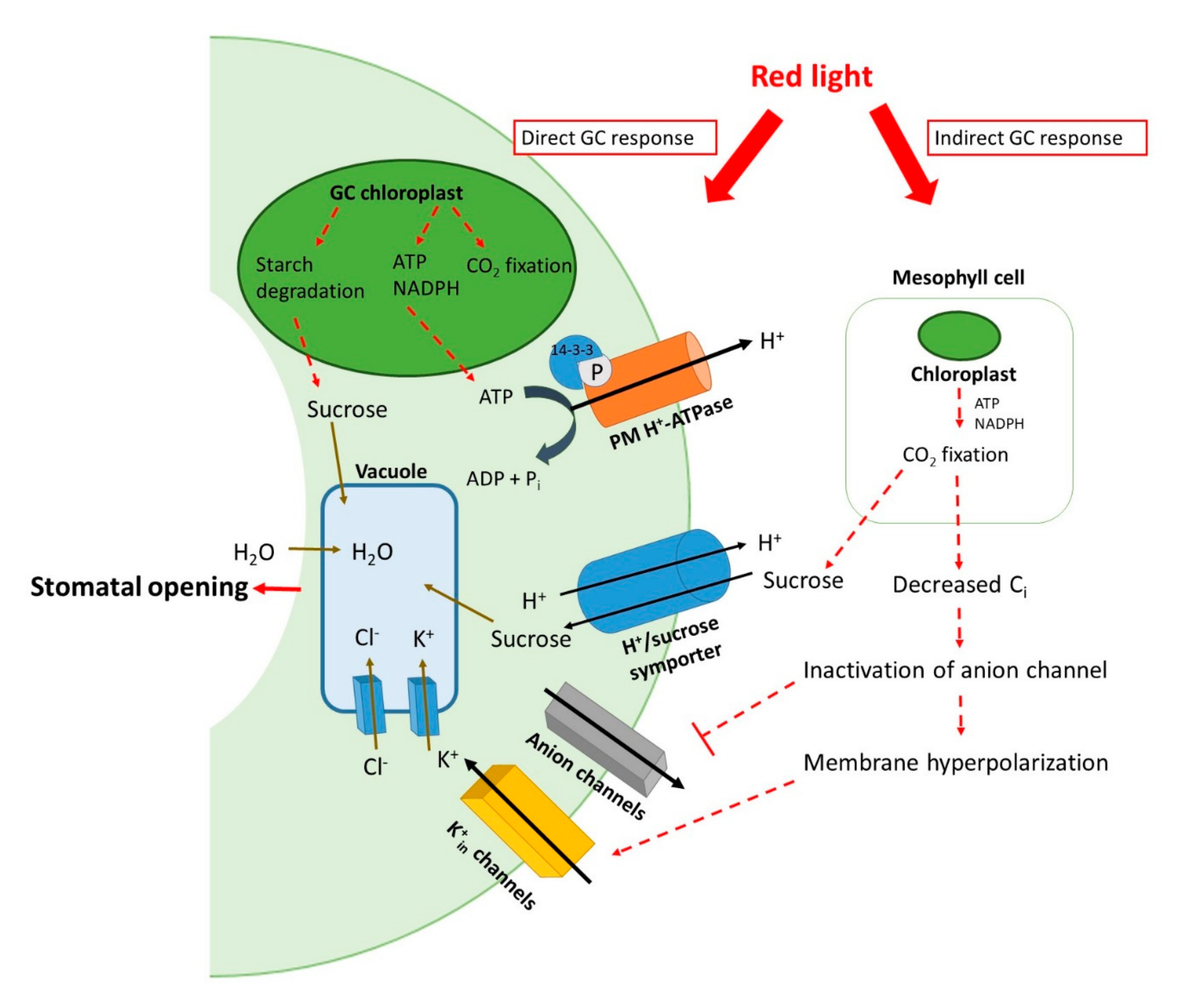
2.2. Stomatal Opening and Light

The fine regulation of stomatal opening in response to light is crucial for crop production. Changes in plasma membrane potential induced by light, which alters the K+ transport across this plasma membrane, have been observed in the stomatal guard cells of leaves [30,44,45,46]. Blue and red light both stimulate stomatal opening by two distinct pathways. Red light induces stomatal opening via photosynthesis in the mesophyll and guard cell chloroplasts and decreases the intercellular CO2 concentration, therefore, red light acts both as a signal and an energy source [47]. The red-light-induced stomatal opening may result from a combination of both guard cell responses to the reduction in intercellular CO2 and a direct response of the chloroplasts located in the guard cells to red light [46,48,49]. It has been thought to be the main mechanism that coordinates stomatal behaviour in relation to photosynthesis [50]. Contrarily, the guard cell-specific blue light response is often considered to be independent of photosynthesis [45,50].
Fluctuations in light spectra have an impact on stomatal behaviour. These fluctuations can be the result of the diurnal pattern of light spectra, clouds, and shading due to the canopy or overlapping leaves [50]. During sunrise and sunset, when the sun is near or below the horizon, and thus the solar angle is smaller, the rays must travel a greater distance through air. This longer path through the atmosphere enhances atmospheric light absorption and scattering, resulting in a reduction in light intensity and an increase in the B:R ratio [50,51,52]. This B:R ratio follows a diurnal pattern, changing the light spectrum during the day, and therefore the stomatal dynamic response. As previously stated, stomata open in response to increasing light intensity and to blue and red light. While red light responses occur at high fluence rates, the blue light response is already saturated at a low fluence rate (5 to 10 µmol m−2 s−1) [47,50,53].
Blue light induces stomatal opening by activating the plasma membrane H+-ATPase through phosphorylation of its second last threonine residue, resulting in hyperpolarization of the membrane potential. This drives K+ uptake through the K+ channels. On a quantum basis, blue light is 20 times more effective than red light in opening stomata, which suggests that the guard cells contain a specific blue light photoreceptor [30,45,47,54]. Blue light-specific stomatal opening, even with a short period of blue light, has been observed in a number of C3 and C4 plants. Iino et al. [55] reported that giving a single pulse of blue light (1–100 s, 470 nm, 250 µmole/m2s) on a red background (500 µmole/m2s) to the adaxial side of Commelina communis L. leaves induced stomatal conductance (gs) peaking after 15 min and returning to the initial steady state within 60 min after the pulse was given [55]. Red light pulses (500 µmole/m2s) on a red background did not cause a similar reaction. In line with these findings, Zhu et al. [48] found that blue light-induced stomatal opening required low light intensity and a short duration of illumination as compared with red light-induced stomatal opening [48].
Blue-light irradiation of guard cells activates phototropins, which are blue light photoreceptor protein kinases, through autophosphorylation (Figure 3). Blue light primarily is perceived by photoreceptors PHOT1 and PHOT2, which activate the guard cell PM H+-ATPase and CBC (CONVERGENCE OF BLUE LIGHT (BL) AND CO2) kinases, inhibiting S-type anion channels [48,56,57]. Phototropins are light-activated serine/threonine protein kinases. These light receptors can be divided into the following two parts: a N-terminal light sensor domain and a C-terminal serine/threonine kinase domain. The N-terminal light sensor domain contains the following two similar domains of approximately 110 amino acids: LOV1 and LOV2. Light-oxygen-voltage (LOV) domains bind the cofactor flavin mononucleotide (FMN) and function as a light sensor for blue wavelengths. Blue light-induced photo-excitation of the LOV domain leads to the autophosphorylation of the phototropin receptor, and thus the initiation of the phototropin signal transduction [58]. In the dark, the LOV domain and the FMN chromophore are non-covalently linked to form LOV447. LOV447 stands for the non-active form of LOV, with a maximum absorption peak at 447 nm. Blue light absorption by the FMN chromophore leads to the formation of a highly reactive LOV660 intermediate. This leads to the formation of a covalent bond between FMN and the conserved cysteine residue of the LOV domain. This light-driven formation of the FMN-cysteinyl bond occurs in a microsecond and forms LOV390, the active form of LOV with a maximum absorption at a wavelength of 390 nm. This response is reversible. If the plant returns to the dark, LOV390 returns to the inactive LOV447 stage. Therefore, depending on the light conditions and light composition, the LOV domain switches between the active (LOV390) and inactive (LOV447) stages [30,47,58,59]. This activation of phototropins triggers the H+-ATPase proton pump, activated by phosphorylation. This H+ extrusion to the apoplast leads to a guard cell plasma membrane hyperpolarization, which in turn generates an electrical gradient providing the incentive for K+ uptake via activation of K+ selective ion channels in the guard cell membrane [45]. K+ and counter-ion Cl are transported into the vacuole. In addition to K+ accumulation, blue light activates a rise in intracellular malate2− and sucrose concentrations from starch breakdown and biosynthesis. The activity of PEP carboxylase, which produces the precursor of malate (oxaloacetic acid) by carboxylation of PEP (phosphoenolpyruvate) is elevated by blue light [30,45,48]. Accumulation of these ions results in a reduction in the water potential inside the guard cells, which leads to water uptake into the vacuoles (Figure 3). This increases the turgor inside the guard cells, leading to stomatal opening [47,56,58].
Blue light dependent stomatal opening is inhibited by the plant hormone ABA (Figure 3). Little is known about the mechanisms by which ABA inhibits the blue-light activation of H+-ATPase. Takemiya and Shimazaki [60] reported that phosphatidic acid (PA) inhibited the protein phosphatase 1 (PP1) in Vicia faba L. PA is a phospholipid second messenger which is produced by ABA in guard cells, while PP1 is a positive regulator of blue light signaling [60]. More specifically, they found that the phosphatase activity of PP1c was inhibited by PA in vitro. Additionally, blue light-dependent H+ pumping and phosphorylation of H+-ATPase was suppressed by PA. However, the autophosphorylation of phototropins in guard cells was not affected by PA [60]. These results suggested that PA affected PP1c, suppressing the signaling between phototropins and H+-ATPase, which resulted in inhibiting the stomatal opening. Takemiya and Shimazaki [60] provided the first evidence for a protein molecule, PP1c, involved in crosstalk between ABA signaling and blue light in guard cells. Further genetic studies are needed to provide direct evidence for the regulation of PP1 by PA in guard cells. Recently, Shen et al. [61] found that, in rice, PA directly bound to two adjacent arginine residues in the ANK domain of OsAKT2, a K+ rectifying channel preventing H+/sucrose-symport-induced membrane depolarization. PA mediated inhibition of OsAKT2 K+ currents by physically binding with the OsAKT2 ANK domain, restraining the guard cell K+ uptake and stomatal opening. This indicates that there is a direct link between phospholipid signaling and K+ channel modulation [61].
Red light causes stomatal opening in intact leaves (Figure 4). This red light-induced stomatal opening is believed to be dependent on photosynthesis in mesophyll cells and guard cell chloroplasts rather than direct guard cell sensing [49,50]. Red light illumination leads to the accumulation of sugars in guard cells through photosynthesis and starch degradation. Sucrose synthesized in the mesophyll cells is imported via H+/sucrose symporters from the apoplast to the guard cells. Guard cells react to intercellular CO2 concentrations (Ci). A high Ci concentration activates anion channels and depolarizes the plasma membrane of guard cells, closing the stomata (Figure 4). Red light illumination of a large leaf area decreases Ci concentrations [49]. Roelfsema et al. [46] found that red light response in intact leaves was mediated through a decline of Ci, which changed the direction of the K+ fluxes across the plasma membrane of the guard cells, normally favouring outward K+ fluxes, and thus stomatal closure in the presence of a high ambient CO2 concentration (700 µL/L). After illumination with red light, an inward K+ flux across the plasma membrane occurred due to inactivation of anion channels. Red light projection on a large leaf area decreased the Ci concentration by 250 µL/L, while illumination with a small beam of red light or illumination of a small leaf area was not sufficient to induce a change in the PM potential. It was hypothesized that the guard cell response to red light may be mediated through a signal generated by mesophyll cells, namely the CO2 concentration of the sub-stomatal cavity [46,49]. Additionally, suppression of the red light response of guard cells by DMCU (3-(3,4-dichlorophenyl)-1,1-dimethylurea), a photosystem II inhibitor, has been reported to inhibit red light-induced stomatal opening in isolated epidermal peels of Vicia faba [48,62,63]. This suggested a direct response of guard cells to red light, which was also found when Ci was experimentally held constant. These findings further suggest that stomatal guard cells are direct sensors of red light, which means that such responses are not solely a result of the decreased Ci resulting from photosynthesis in the mesophyll cells [48].
Activation of PM H+-ATPase induces the hyperpolarization of the plasma membrane and drives the K+ uptake through K+ inward channels. Red light leads to the accumulation of K+ in guard cells, which means PM H+-ATPase may be required for this response. Ando and Kinoshita [49] have found that red light induced PM H+-ATPase phosphorylation in whole leaves of Arabidopsis, detected by an immune-histochemical technique to visualize this phosphorylation level. This PM H+-ATPase phosphorylation induced by red light illumination in whole leaves was found to correlate with stomatal opening under red light and was inhibited by ABA [49]. One year later, Ando and Kinoshita [49] reported that red light illumination of whole leaves induced photosynthesis-dependent phosphorylation of threonine in guard cells. Threonine is the C-terminal penultimate residue of PM H+-ATPase. Threonine is regarded to have significant importance for activation of PM H+-ATPase in guard cells in whole leaves. They investigated the effect of red-light fluence rate on phosphorylation of PM H+-ATPase, with red light at 10, 50, 100, 300, and 600 µmol m−2 s−1 for 30 min, while examining the phosphorylation levels. Starting from levels at 50 µmol m−2 s−1, red light-induced phosphorylation of PM H+-ATPase was saturated. They found that faint red light (10 m−2 s−1) was insufficient to induce phosphorylation of PM H+-ATPase [64].
In summary, it can be concluded that a stomatal red-light response is the result of a combination of a mesophyll-independent direct response of the guard cells and an indirect response from the guard cells′ receptivity to the decreased intercellular CO2 concentration, caused by mesophyll photosynthesis [46,48,65]. The red and blue wavelengths are considered to be the main drivers of stomatal opening in plants [50]. However, it is also important to acknowledge the impact of green and ultraviolet light on stomatal behaviour. Green light (500–560 nm) has been reported to inhibit stomatal opening induced by blue light [50,66,67]. The exact mechanism at the basis of the changes in stomatal opening induced by green light has not yet been identified. Green light is known to deactivate the blue light cryptochrome photoreceptors, which can contribute to shade avoidance responses under a canopy [50]. Thereby, it can be of adaptive significance [68]. However, further research is required to elucidate a green light photoreceptor. Furthermore, little research has been published on the effects of ultraviolet (UV) light (100–400 nm) on stomatal aperture. UV radiation can be divided into UV-A (315–400 nm), UV-B (280–315 nm), and UV-C (100–280 nm). UV-C radiation is completely absorbed in the stratospheric ozone layer and in the atmosphere. Therefore, only UV-A and UV-B radiation reach the plants. Contrary to green light, photoreceptors have already been found for UV-B and UV-A light. UV-B light is detected by the protein UV resistance locus 8 (UVR8), while UV-A light is detected by cryptochromes, which also act as a photoreceptor for blue light [69]. Several researchers have reported on the stomatal responses to UV-B light [69,70,71]. However, a UV action spectrum for stomatal aperture and the mechanisms by which UV light regulates stomatal aperture have not yet been elucidated.

3. Carbon Dioxide Regulation of Stomatal Development and Opening

Plants must balance the CO2 influx for photosynthesis with the loss of water vapour through their stomatal pores [72]. An increase in atmospheric CO2 concentration levels induces stomatal closure, which impacts plant growth, leaf temperature, and water-use efficiency. CO2 has both an influence on the regulation of stomatal development and opening [36,73,74]. Since the industrial revolution, atmospheric CO2 concentration has increased to 409 ppm [75]. Yearly, 40% of the atmospheric CO2 enters plant leaves through stomata [76]. A change in stomatal index in response to atmospheric CO2 can influence photosynthesis, resulting in a change in atmospheric CO2 concentration. The ongoing increase in atmospheric CO2 concentration causes global warming, but also affects the physiology and development of terrestrial plants [72]. A long-term effect of the continuing CO2-concentration rise is the downregulation of stomatal development in the leaves. The decrease in stomatal index in parallel with an increasing atmospheric CO2 concentration confirms that plants responses to changes in CO2 concentration levels [73]. Because CO2-regulated stomatal development and movements together regulate stomatal conductance (gs), and thus also CO2 exchange in plants, the recent literature (from 2000 until 2020) on these mechanisms is reviewed in this section.

3.1. Stomatal Development and Carbon Dioxide

The impact of an elevated CO2 concentration on the stomatal index is a very important topic concerning the evolutionary change with rising global CO2 levels. Stomatal densities have decreased in the last 200 years in response to the rising CO2 levels [77,78]. Carbonic anhydrase (CA) enzymes, which bind CO2 and catalyze its conversion into HCO3 and H+, present in the guard cells, are important for the stomatal development response to changes in CO2 concentration levels. Research has shown that the isolated Arabidopsis β-carbonic anhydrase double mutants, ca1 and ca4, exhibit an increase in stomatal development at elevated CO2 levels. This indicates that the catalytic activity of the CA enzymes is part of the signaling pathway of CO2-controlled stomatal development [72,79,80,81]. These carbonic anhydrase genes were also found to influence stomatal function [6]. The βca2 and βca4 plants were found to be insensitive to CO2-induced stomatal closing. The mutant plants showed an elevated stomatal conductance (gs) and stomatal index at higher ambient CO2 levels. These mutants were also found to display insensitivity to ABA [80].
The Arabidopsis gene HIC (HIGH CARBON DIOXIDE) which encodes a negative regulator for stomatal development in response to CO2 concentration, has a role in regulating changes in stomatal index in response to increased CO2 levels [5,73,80]. Contrary to the wild type, hic mutants develop a significantly higher stomatal index when exposed to increased CO2 levels. The rise in stomatal index observed for hic plant grown at increased CO2 levels is generated by a disruption in the HIC gene [73]. HIC and the Arabidopsis KCS1 gene, which encodes a 3-keto acyl CoA synthase involved in cuticular wax biosynthesis, were found to be similar. A mutation in a KCS gene was carried by hic plants, which resulted in a disruption in the signaling pathway responsible for the control of stomatal patterning as a result of increased CO2 levels [73]. The ERECTA receptor kinase and the epidermal patterning factor gene EPF2 have also been shown to be involved in determining transpiration via regulation of stomatal index [82,83]. Several signaling components that are responsible for regulating stomatal development have been characterized, including the putative secretory peptide EPF1 (epidermal patterning factor 1), LRR (leucine-rich repeat domain) receptor components TMM and ER (ERECTA), and a peptidase SDD1 [83]. EPF2 is a peptide related to EPF1. EPF2 is expressed in meristemoids and in guard mother cells (GMC), which affects the stomatal index on the mature leaf. EPF2 expression during leaf development has an effect on the stomatal index of the mature leaf in Arabidopsis [83]. Hunt and Gray [83] found that in the absence of EPF2, extreme amounts of cells entered the stomatal lineage and, subsequently, produced small epidermal cells expressing stomatal lineage reporter genes. This indicates that EPF2 regulates a different aspect of stomatal development as EPF1. EPF2 is involved the determination of the cell count that enters the stomatal lineage [83]. Because Epf2 mutants show an inverted stomatal development at elevated CO2, EPF2 is essential for CO2-induced control of stomatal development [79].
The key mechanisms mediating the perception and communication of the CO2 signal to the stomatal development are elusive. Because new leaves sheathed by antecedent leaf primordia may not accurately detect the ambient CO2 concentration, the CO2 concentration is detected by mature leaves in ambient conditions, which transmit signals to new leaves to induce an appropriate developmental response [36]. It has been reported that the total soluble sugar content (sucrose, glucose, and fructose) of mature leaves increased in response to elevated CO2 treatment, which partially explained why the developing leaves grew at a faster rate [41].
A recent study by Higaki et al. [84] showed that the DNA replication licensing factor CDC6, which is a positive regulator of SLGC asymmetric cell division, played a role in the production of satellite stomata. As indicated in the section about “stomatal development”, the SLGC can separate into a pavement cell or a meristemoid mother cell. Then, the meristemoid mother cell can undergo asymmetric cell divisions to produce a meristemoid (Figure 1). In this instance, the newly formed meristemoid is located near a pair of guard cells already formed. This meristemoid is called a satellite meristemoid [5,84]. Elevated CO2 concentration in young and expanded cotyledons of A. thaliana resulted in a decreased stomatal index, mainly due to pavement cells increasing in size, contributing to decreased stomatal density. Spacing between stomata, which makes up the stomatal pattern, was troubled by elevated CO2 satellite stomata production, decreasing from approximately 37 µm (at 380 ppm) to 31 µm (at 1000 ppm). However, the underlying molecular mechanisms have not yet been identified. Higaki et al. [84] found that overexpression of CDC6 resulted in the overproduction of satellite stomata, while decreasing cotyledon expansion [84]. This suggests that the mechanisms promoting satellite stomata production is different from the mechanisms responsible for enhancement of cotyledon expansion. Future research, focusing on the CDC6-related factors, is needed, as these could be the targets for the elevated CO2 signaling pathways that promote satellite stomata production.

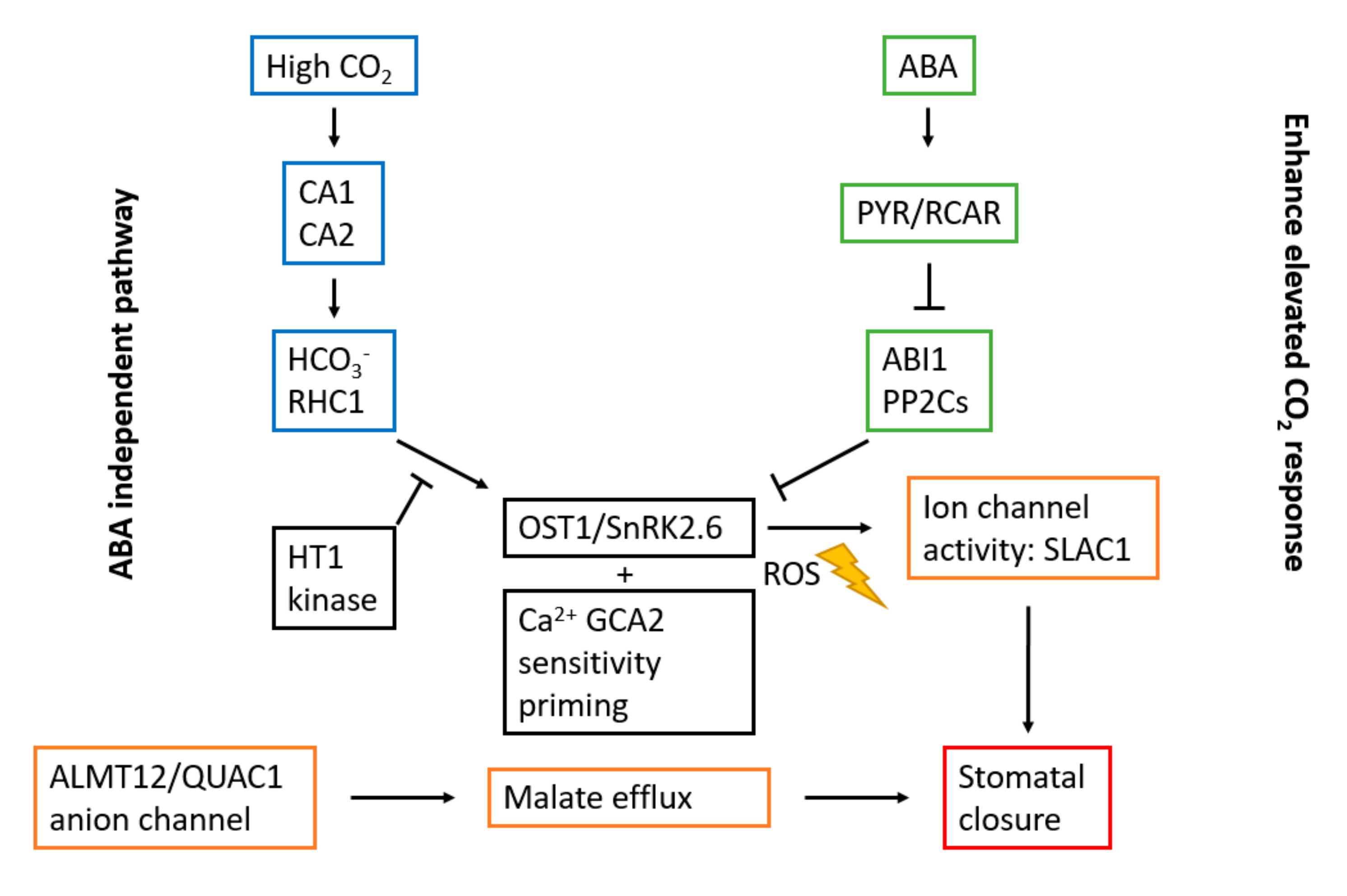
3.2. Stomatal Opening and Carbon Dioxide

Stomatal opening is moderated by an influx of water and solutes such as K+ into the guard cells surrounding the stomatal pore. Stomatal closing is regulated by an efflux of water and solutes from guard cells. Guard cells have developed a sophisticated signaling mechanism that enables appropriate control of stomatal movement [85]. CO2 concentrations in the intercellular space (Ci) of leaves higher than ambient CO2 concentrations mediate closure of stomatal pores in plants. Contrarily, low CO2 concentrations trigger stomatal aperture. A high concentration of CO2 activates anion channels and depolarizes the plasma membrane of guard cells. An increase in CO2 also inactivates inward K+ channels and activates outward K+ channels. When the leaves are exposed to light, photosynthesis causes a reduction in the Ci. In contrast, respiration in plant leaves in darkness triggers closure of stomatal pores as a result of a rapid rise in the Ci. Global levels of CO2 have risen, which causes a rise in leaf Ci resulting in a reaction of stomatal aperture in Ci [49,72,80].
Elevated CO2 concentration and ABA both induce stomatal closure. The signal mechanism of ABA-induced stomatal closure has been well studied [86,87]. ABA receptors and core signaling cascades have been identified. These include PYR/RCAR ABA receptors (pyrabactine resistance/PYR1-like/regulatory component of ABA receptor), SnRK2-type protein kinases, and type 2C protein phosphatases (PP2Cs). However, the sensing and signal transduction mechanisms caused by elevated CO2 concentrations remain less well understood. Molecular components, including β-carbonic anhydrase, βCA1 and βCA2, High Leaf Temperature 1 (HT1), OST1/SnRK2.6, SLAC 1 (Slow Anion Channel-associated 1), ALMT12/QUAC1 (aluminium-activated malate transporter 12/quickly activating anion channel 1), anion channel, RHC1 (Resistant to High CO2), GCA2 (Growth Controlled by ABA2), MPK12 (Mitogen-Activated Protein Kinase 12), and GHR1 (Guard cell Hydrogen peroxide-Resistant 1) are involved in stomatal CO2 signaling [74,80,81,88,89]. However, the detailed interaction and regulation among these components remains to be identified.
A simplified model on how CO2 and ABA signaling pathways mediate stomatal opening is displayed in Figure 5. An elevated CO2-induced closure and reduction in stomatal index requires the generation of Reactive Oxygen Species (ROS) [80,88,90]. Moreover, ABA and the PYR/RCAR family of ABA receptors are also required in both responses. This indicates that the CO2-induced reductions in stomatal aperture operate through the intermediacy of ABA by accessing the guard cell ABA signaling pathway (Figure 5). It implies that there is a requirement for ABA receptors and ABA for the CO2-dependent stomatal responses, which explains why guard cell ABA and CO2 signaling have many components in common [90]. This suggests that some of the effects of CO2-induced stomatal movement result from the ability to access the guard cell ABA signaling pathway through the intermediacy of ABA. However, the precise signaling convergence point remains to be identified [72,80,88,91,92]. The following two models could be considered: (i) Increased CO2 concentration causes an elevation in the ABA concentration or enhances early ABA signaling, thus, mediating stomatal closure, and (ii) CO2 signaling merges with ABA signaling at OST1/SnRK2.6 protein kinase activation, while synergistically amplifying the common downstream signaling mechanisms, as depicted in Figure 5 [80,88]. Genetic studies have shown that the protein kinase OST1/SnRK2.6 plays an important role in both CO2- and ABA-induced stomatal movement [88,93,94,95]. The activation of the OST1/SnRK2.6 protein kinase, phosphorylating SLAC1 anion channels, mediates stomatal closure induced by ABA. (Figure 5) [88].
Recent studies have investigated how CO2 and ABA signaling pathways converge and whether an increased CO2 concentration rapidly induces ABA concentration elevation in the guard cells of the stomata triggering stomatal closure [88]. Two pathways have been suggested, i.e., an ABA independent pathway and a CO2-mediated enhanced response [88,90]. Chater et al. [90] found that ROS were required for the stomatal density and opening response as a result of elevated CO2 concentration, which added a new component to the signaling pathway. Furthermore, they showed that elevated CO2-mediated control of stomatal opening and stomatal index also require the presence of ABA itself and PYR/RCAR ABA receptors. The data showed that increases in guard cell ABA were induced by CO2 elevation. The requirement of ABA in stomatal closure as a result of CO2 concentration elevation suggests that ABA and CO2 signaling have many components in common [90]. This suggested pathway is illustrated in Figure 5 as the “enhance elevated CO2 response”. Hsu et al. [88] investigated the stomatal CO2 responses in ABA signal transduction mutants and found that ABA signaling amplified the CO2-induced stomatal closure. This indicates that elevated CO2-induced stomatal closure is reduced, but not overturned in higher-order ABA biosynthesis and receptor mutants. These results and previous reports show that stomata in ABA receptor mutants are still responsive to CO2 elevation [88,93,94]. They also found that S-type anion channels were activated as a result of extracellular CO2 concentration increasement in ABA biosynthesis nced3/nced5 double mutants and ABA receptor pyr1/pyl1/pyl2/pyl4/pyl5/pyl8 hextuple mutants [88]. This suggests that ABA signaling is not directly needed for this rapid response due to an increase in CO2 concentration. Additionally, ABA nanoreporter imaging, using the ABA-FRET reporter ABAleon2.15, indicated that an increase in CO2 concentration did not influence [ABA] in guard cells, as opposed to previous studies [88]. It was found that OST1/SnRK2 kinase activities in guard cells were activated by ABA, but unexpectedly not by an increase in CO2 concentration. These findings point to a model where elevated CO2 also triggers rapid stomatal closing via an ABA-independent pathway, as shown in Figure 5.
In summary, the latest studies show that there is strong evidence for a model where CO2 and ABA collaborate to induce stomatal closure downstream of the OST1/SnRK2.6 protein kinase. However, a basal level of OST1/SnRK2.6 protein kinase activity is still required for stomatal closing in response to elevated CO2 concentration (Figure 5). Further research is required on the underlying mechanisms by which CO2 regulates stomatal aperture, boosting new approaches to adapt our crop plants to climate change.

4. Effect of Temperature on Stomatal Development and Opening

Climate models predict more extreme weather-related events, such as heat waves and extended drought periods over the next decades. Instrumental observations of the global and hemispheric temperatures have revealed a pronounced warming during the past 150 years. The observed increase in heat waves is one result of this warming [96,97]. Heat influences plants in multiple ways including an impact on stomatal development and opening.

4.1. Stomatal Development and Temperature

High ambient temperature inhibits stomatal production. Lau et al. [98] found that elevated temperatures suppress the expression of SPCH, the bHLH transcription factor that acts as the master regulator for initiation of the stomatal lineage (Figure 1) [1,98]. The bHLH transcription factor PIF4 is an essential element in high temperature signaling and mediates the suppression of SPCH, and thus stomatal development. Lau et al. [98] demonstrated that upon exposure to high temperature, PIF4 accumulated in the stomatal precursors (meristemoids) and subsequently bound to the promoter of SPCH, thus, restricting stomatal production at elevated temperatures. The reduced numbers and activity of the stomatal precursors are the result of this transcriptional repression of SPCH. Suppression of the expression of SPCH eventually results in a lowered production of the guard cells, as it is a critical bHLH transcription factor in the stomatal lineage [98].
Furthermore, a negative feedback loop of the PIF4 gene by SPCH has been uncovered [98]. In this negative feedback, SPCH directly binds and represses PIF4 expression (Figure 6). SPCH represses PIF4 via the direct binding of PIF4’s promoter [16,98]. This enables SPCH to accumulate sufficiently to ensure stomatal cell fate, as SPCH also plays a role in the spacing and amplifying division branches (Figure 1). This mechanism generates a switch-like behaviour to the stomatal precursors. If the stomatal lineage is repressed by high ambient temperature, the cells that already accumulated a certain threshold level of SPCH can continue to accumulate SPCH, in turn downregulating PIF4, to complete the stomatal lineage [98].
High temperature poses a risk to plants of heat damage and water shortage. Plants grown at high ambient temperature display an array of responses, including thermonasty or the elevation of leaves and elongation of the petioles, known to enhance plant cooling [99,100]. As transpiration also contributes to leaf cooling, reducing the stomatal development in response to high temperature is expected to decrease plant cooling. Plants need to make a trade-off when exposed to a hot environment. The increased leaf cooling by transpiration has to be balanced against water loss. Mutants of PIF4 do not display elongation responses or leaf hyponasty upon high temperature. PIF4 controls both thermonasty and stomatal density, clearly emphasizing the important role of this transcription factor in managing these plant responses [98,99,100]. Koini et al. [100] suggested that PIF4 could be acting as a mediator in the auxin signaling pathway at high temperatures. The auxin-responsive gene IAA29 increased transcription when transferred to high temperature. This response was not present in pif4 mutants. This auxin-responsive gene has been shown to be a component of auxin-mediated elongation in shade-avoidance responses. This strengthens the idea that PIF4 has a prominent role as a point of crosstalk between light and temperature signaling [100,101].
In summary, the response of stomatal development to ambient temperature is complex and influenced by the local environment. PIF family genes are known to act as a repressor in light signaling. Among the PIF genes, however, only PIF4 is involved in the high temperature response [38]. More research is required to clarify whether the regulation of stomatal development in response to light and high temperature by PIF4 follows similar strategies.

4.2. Stomatal Opening and Temperature

An elevated temperature negatively affects photosynthesis. Rubisco (ribulose-1,5-bisphosphate carboxylase/oxygenase) activase plays an important role in photosynthesis and has been found to be sensitive to heat. This influences the activity of the Calvin cycle in the following two ways: (i) a faster inactivation of active Rubisco and (ii) a slower reactivation by the heat-sensitive enzyme Rubisco activase [96,102]. Feller [96] found that leaf segments of bean (Phaseolus vulgaris L.) incubated at temperatures from 20 to 50 °C had a different stomatal opening. Leaf segments of primary leaves were incubated floating on water in darkness at the indicated temperature. After incubation for 30 min, the stomatal aperture at incubation temperatures of 23, 30, and 35 °C were, respectively, 0.66, 2.76, and 4.28 µm. This effect was found to be reversible (within 30 min) as the stomata closed again after transferring the samples from the highest temperature to the lowest temperature (23 °C). Adding 0.1 µM ABA to the incubation medium changed the temperature for stomatal opening to higher values. This suggests an antagonistic interaction between heat (stimulating stomatal opening) and drought (stimulating stomatal closure). High temperatures stimulate stomatal opening, hence, a trade-off between leaf cooling and water use efficiency has to be made [96]. This is of importance in extreme weather situations, for example, heat waves, which are characterized by high ambient temperatures and drought. Feller [96] also found that the illumination of a (part of a) leaf may change rapidly as a result of shadowing by other plant parts or clouds. When mimicking this situation, they found a difference of 10 °C between permanently sun-exposed bean leaves and permanently shadowed bean leaves. Transferring the leaves from sun to shadow (and vice versa) led to a fast temperature change during the first minute after altering the illumination. On the one hand, air convection and transpiration help the cooling of the previously illuminated leaves, while on the other hand, the energy absorbed from the sunlight leads to a rapid rise in temperature of a previously shadowed leaf. They concluded that the opening of stomata was an important response to heat and allowed an efficient evaporative cooling of the leaf to protect the photosynthetic apparatus [96].
Kostaki et al. [103] found that guard cell movement induced by high temperatures called for components involved in blue light-mediated stomatal opening. These results suggest the possibility of crosstalk between temperature and light signaling pathways. Firstly, they investigated stomatal responses to high temperature by using epidermal peel bioassays, without confounding alterations in humidity. At higher temperature, epidermal peels of Arabidopsis sp., Hordeum vulgare L., and Commelina communis clearly showed increased stomatal opening. Guard cells have the molecular machinery necessary for perceiving temperature changes, as the epidermis could no longer receive signals from the mesophyll layer in this experimental setup [103]. Secondly, the involvement of already know high temperature signaling components was inspected through stomatal bioassays, using cngc, arp6, pif4, and ft null mutants [103]. The wild-type plants of all of these mutants had stomatal apertures at 35 °C. The ft mutants showed reduced stomatal opening when transferred from the dark to (red + blue) light, in contrast to the stomatal apertures similar to the wild-type plant, when maintained in white light. These results led Kostaki et al. [103] to investigate the role of phototropins and their downstream target, BLUS1 (blue light signaling 1), in high temperature-mediated stomatal opening (Figure 7). They found that phototropins were of great importance for the temperature-induced stomatal opening. Impaired stomatal aperture was most severe in the phot1/2 mutants. Their results suggest that guard cell movement can still occur in response to a temperature of 35 °C independently of phototropin, but in order to achieve full stomatal opening, phototropin activation is required. Additionally, blus1-3 mutants also displayed significantly smaller stomatal openings as compared with wild-type plants at 35 °C, suggesting involvement in this response [103]. Some stomatal opening, as a result of high temperature, was also observed in the dark. These findings may be explained by the existence of an additional phototropin-independent pathway. Devireddy, Arbogast, and Mittler [104] suggested that RBOHD-mediated (respiratory burst oxidase protein D) ROS production could mediate this response (Figure 7). They found that rapid alteration in stomatal opening and closing, induced by different environmental stimuli, triggered a ROS-dependent systemic signal that altered the stomatal aperture within minutes, even in systemic untreated leaves [104]. Thirdly, the role of H+-ATPase channels in the temperature-regulated stomatal response was observed. Kostaki et al. [103] found genetic evidence suggesting that AHA1 and AHA2, isoforms of the Arabidopsis plasma membrane H+-ATPase family, were required for full stomatal opening as a response to high temperature in epidermal peels [105]. Recruitment of AHA1 to the plasma membrane in guard cells is mediated by the protein PATROL1 (proton ATPase translocation control 1), which is essential for stomatal opening in response to high temperature, low CO2 concentration and blue light. The potential role of AHA-regulating 14-3-3 proteins was investigated. In response to blue light, 14-3-3 proteins bind to the auto-inhibitory C-terminal region of the H+-ATPase in the plasma membrane, which promotes pumping. Genetic analysis of 14-3-3 mutants has shown that they contribute to high-temperature mediated stomatal opening with potentially antagonistic interactions between isoforms [103].
In summary, there are multiple possible sites of high temperature signal integration during stomatal opening [103]. Transfer of isolated guard cells from the dark to blue light results in stomatal opening involving phototropin-mediated phosphorylation of BLUS1, which in turn activates PM H+-ATPase channels. The protein PATROL1 is required for AHA1 insertion into the plasma membrane. To achieve complete stomatal opening in guard cells at high temperature under white light phototropins, PM H+-ATPase activity and redundant activities of 14-3-3 proteins are required [103]. It has been demonstrated that guard cells integrate both light and temperature signals to control stomatal opening [103]. However, more research is required to find the point at which these signals converge.

5. Effect of Relative Humidity on Stomatal Development and Opening

Relative humidity (RH) is the ratio of the water vapour pressure in the air to the water vapour pressure at saturation. RH is closely related to ambient temperature, because the vapour pressure at saturation increases as the air temperature rises. The vapour pressure deficit (VPD) is the difference between saturation vapour pressure and actual air vapour pressure, which combines the effect of RH and temperature. Higher VPD equals drier air [106]. Stomata will close in response to a larger vapour pressure deficit or lower relative humidity. This response was an important step in the evolution of plants evolving from water to land, since it enabled plants to control their water loss when exposed to a dry atmosphere [33]. The stomatal closure in response to low RH serves to prevent desiccation under high evaporative demand. To predict future impacts of the rising VPD on plant functioning, such as reduced photosynthesis and growth, Grossiord et al. [107] provided an overview of plant responses to high VPD [107]. Yuan et al. [108] examined whether increased VPD was one of the drivers of the widespread drought-related forest mortality over the past decades [108]. Their results support the hypothesis that increased VPD triggers stomatal closure to avoid excess water loss, leading to a negative carbon balance depleting the carbohydrate reserves and resulting in tissue-level carbohydrate starvation [108]. In addition, higher evaporative demand coupled with reduced soil water supply leads to cavitation in the xylem, stopping water flow and eventually leading to plant death [108]. Therefore, insight into key processes driving plant responses to VPD are very important for making predictions regarding future VPD impacts on plant development, both on a global and local scale.
While the stomatal response to VPD has been heavily researched, the driving mechanism remains unclear. A passive response would result from the reduced leaf water content and turgor loss due to a stronger evaporative demand in dryer air. However, the stomatal response to VPD could also be the result of an active ABA-mediated response [33,109].

5.1. Stomatal Development and Humidity

A high ambient RH during plant growth influences the stomatal closing ability, but also alters the stomatal density and size [110]. Fanourakis et al. [110] investigated the stomatal features (density, size, and pore dimension) of four rose cultivars, grown under moderate (60%) and high (95%) relative humidity. Stomatal density was significantly altered by the interaction between RH and the rose cultivars. Plants grown under high RH for a long term showed a higher stomatal density (8–22%) in three of the cultivars as compared with those grown under moderate RH. In addition to stomatal density, stomatal length also increased in all four cultivars when cultivated under high RH [110]. This positive response of stomatal size to elevated RH levels has been observed in many different species [111,112,113]. Guard cells in Tradescantia virginiana L. grown at high RH were 29% longer than in plants grown under moderate RH [111]. Bakker [114] found similar results when investigating the stomatal density in cucumber (Cucumis sativus L.), tomato (Lycopersicon esculentum L. Mill.), sweet pepper (Capsicum annuum L.), and eggplant (Solanum melongena L.) grown under humidity ranging from 0.2 to 1.6 kPa VPD. The stomatal densities observed in tomato, eggplant, and sweet pepper were significantly higher at high humidity [114]. Stomatal density has also been investigated in relation to plant diseases. Stomata expose the internal leaf tissue to pathogens [114]. By increasing stomatal density, the risk of exposing internal leaf tissues to pathogens is increased by increasing the number of potential infection sites, i.e., the stomatal pores. A high humidity is also a more favourable condition for germination of spores [114,115]. This may explain why plants grown in a commercial setting under high humidity are considered to be “weak′ in terms of their resistance to plant pathogens.

5.2. Stomatal Opening and Humidity

Stomata respond rapidly to air humidity, resulting in a lower stomatal conductance (gs) at high VPD (low RH). It has been a matter of debate whether stomata respond actively or passively to VPD [116]. While active stomatal movements have been supported by findings that changes in VPD associate with some elements of the signal cascades engaged by ABA, there have also been arguments for passive stomatal movements. Passive stomatal movements are also called hydropassive movements. This is the closing of the stomata as a result of reduced water content and turgor pressure due to stronger evaporative demand in dry air [33,116]. In basal vascular land plants, lycophytes, ferns, and gymnosperms included, it has been shown that stomatal closure was a passive-hydraulic response to VPD that did not require foliar ABA [109]. It is still debatable whether these basal vascular land plants show a functional ABA response to VPD, but, in some fern species, the stomatal response to VPD is thought to be more than a passive process [117,118]. ABA-driven stomatal regulation would have evolved approximately 400 million years ago, during the early evolution of seed plants. This hypothesis is based on the observation of passive stomatal control, unresponsive to ABA in certain fern species, while ABA-induced stomatal closure has been detected in some mosses and lycophytes. Cai et al. [117] found that some ABA signaling and membrane transporter protein families have diversified over the evolution of land plants [117,119]. It is clear that the debate between passive and active stomatal responses to VPD is connected with the evolution of their stomatal physiology. Kübarsepp et al. [118] studied the stomatal structural characteristics and stomatal responses to environmental changes, specifically humidity, CO2, and light, across 29 fern species originating from contrasting environments. Increasing the air humidity from 60 to 75% resulted in increased stomatal conductance in all fern species. Stomatal reactions to CO2 and light changes appeared more comparable in lag time for stomatal opening or closing, while the lag times for opening and closing in ferns in response to VPD were shorter. This difference in reaction to VPD as compared with the other environmental stimuli could imply different regulatory mechanisms, participating in the continuing debate whether ABA regulates the stomatal reactions in ferns [118].
In angiosperms, there is substantial evidence that ABA would be involved in the VPD-induced stomatal closure. McAdams et al. [120] found that, in angiosperms, VPD-induced stomatal responses were regulated by ABA, associated with a rapid upregulation of NCED genes (ABA biosynthesis genes), which indicated that an ABA-mediated component was involved. After a 20 min exposure of plants from three angiosperm species to a doubling in VPD, stomata closed, while foliar ABA levels increased and NCED genes were significantly upregulated. This suggested that VPD-induced stomatal closure would be accompanied by a rapid biosynthesis of ABA, mediated by a single gene [120]. ABA evokes stomatal closure via a complex signaling cascade, with secondary messengers as key elements of this signaling pathway. ABA-induced stomatal closure has been associated with an increase in cytosolic free Ca2+, preceded by H2O2 and NO. That is why H2O2, NO, and Ca2+ have been put forward to act as secondary messengers in the stomatal response pathways for ABA as a stomatal closing signal. When leaves were exposed to downstream elements of the ABA signaling network (H2O2, NO, and Ca2+), stomata formed under high RH conditions closed less as compared with stomata on leaves developed at moderate RH [121].
It has also been found that OST1 is required for RH-induced stomatal responses. Merilo et al. [33] used ABA mutants to analyze the role of ABA biosynthesis and signaling in RH-induced stomatal closure. They monitored the stomatal closure of Arabidopsis mutants (ost1-3) as well as Pisum sativum L. wilty and Solanum lycopersicum L. flacca in response to a rapid decrease in RH from 65–70% to 30–35% [33]. The ost1-3 mutants showed no induced stomatal opening when the air humidity went back to the initial level, which further illustrated how essential OST1 protein kinase was in stomatal movements induced by relative humidity. The stomatal conductance in the whole plant is controlled by the [ABA] and PYR/RCAR receptors rather than by signaling through OST1. OST1 is a central kinase in the stomatal regulation in response to ABA and environmental factors such as CO2, ozone and darkness [33,80,88,93,122]. Plants carrying a mutation in OST1 have lower steady-state gs than ABA-insensitive mutants (f.e., 112458, aba2-11, and nced3). This result indicates that ABA has involvement in the regulation of other plant processes related to stomatal signaling or leaf hydraulics which affects gs, independently of OST1. The higher stomatal conductance values of ABA-insensitive (or ABA-deficient) plants as compared with non-OST1-functional plants could be explained by the role of ABA in reducing leaf hydraulic conductance, independently of OST1. One option could be the guard cell plasma membrane H+-ATPases, that initiates stomatal opening. ABA inhibits the phosphorylation and activation of H+-ATPase, directly or through secondary messengers [33]. However, inward K+ channels, which are required for K+ accumulation during stomatal opening, are also an option, because they are inhibited by ABAs secondary messengers. Merilo et al. [33] proposed a hypothetical model for RH-induced stomatal closure (Figure 8). The impaired RH response of ost1-3 highlights that signals via OST1, which may be partially dependent on ABA, are very important for RH-induced stomatal closure. Xie et al. [123] also reported the involvement of OST1 and ABA2 in guard cell ABA signaling as a response to reduced RH. ABA2 encodes a dehydrogenase involved in ABA biosynthesis, while OST1 encodes a protein kinase that affects the elevation of ROS and cytosolic-free CA2+ and activates the guard cell SLAC1 Cl channels in response to ABA. They found that these two gene products are involved in the guard cell′s response to a reduction in relative humidity (65% to 25%). This gives credit to the hypothesis that ABA would be involved in the stomatal response to a reduced RH [123].
Plants produced at high RH also show poor control of transpiration when they are transferred to low RH [111]. To find the impact of RH on functioning stomata, stomatal morphology and leaf anatomy, Aliniaeifard et al. [112] grew fava bean plants at low (0.23 kPa) or moderate (1.17 kPa) VPD. Plants grown at low VPD showed larger stomata, larger pore area, and thinner leaves asvcompared with plants grown under moderate VPD. The plants translocated from moderate VPD to low VPD showed stomatal morphology (except opening) and leaf anatomy similar to plants grown under moderate VPD, while the transpiration rate and stomatal conductance (gs) were the same as for plants grown under low VPD [112]. The mechanisms involved in the stomatal movement could be disturbed under certain environmental conditions. This could result in reduced closing capacity of stomata, although they were stimulated with environmental conditions that usually would induce a closing response. Reduced closing ability of stomata has been observed in plants grown at low VPD [111,112]. Nejad and van Meeteren [111] investigated the stomatal size and response at moderate (55%) and high (90%) RH in Tradescantia virginiana plants to treatments as desiccation, ABA, and exposure to darkness, which are treatments that normally induce stomatal closure. As compared with plants grown at moderate RH, the stomatal size was larger for plants grown under high RH (guard cell length of 56.7 and 73.3 mm, respectively). In addition to the differences in guard cell length, there was also a clear difference in stomatal behaviour. Both stomata in moderate and high RH grown plants reacted to desiccation, ABA, and darkness, but the stomatal closure in high RH grown plants showed a high variability. Some stomata developed at high RH, closed in response to the treatments, whereas others closed partly or not at all. None of the treatments resulted in total closure of all the stomata in plants grown under high RH [111]. Plants grown under high RH expressed a lower leaf ABA concentration during growth [106,112,121]. This resulted in a plant with stomata with a reduced closing ability. Stomata of plants grown under high RH were found to have lost their responsiveness to ABA [112,121]. Giday et al. [121] found that stomatal responsiveness was positively related to ABA concentration during growth in cultivars of Rosa hybrid L. grown at different RHs.

6. Conclusions and Future Perspectives

Stomata are important regulatory structures in plant leaves that can increase CO2 uptake for photosynthesis by opening and can reduce water loss through transpiration by closing, and thus play a vital role in balancing water loss with photosynthetic performance. In the past decade, our insight in the influence of environmental factors on stomatal development and opening has been increased substantially. In this review, we focused on the alteration of stomatal behaviour in response to environmental signals, focusing on light, CO2, temperature, and humidity. Currently, global temperature, CO2 concentration, and water demand for agricultural use are increasing, emphasizing the potential of manipulating stomatal opening and development. This could lead to maximization of drought tolerance, a higher WUE, and an increased yield. Table 1 provides an overview of the influence of light, CO2, temperature, and relative humidity on stomatal development and opening, including the molecular components involved in the stomatal response, as described in this paper.
Stomatal opening is induced by light, with blue and red light stimulating stomatal opening by two distinct pathways. While blue light acts a signal, red light also acts as an energy source, driving photosynthesis in chloroplasts located in guard cells and mesophyll cells. Activation of PM H+-ATPase by blue light induces the hyperpolarization of the plasma membrane and drives the K+ uptake through K+ inward channels. The accumulation of positively charged K+ ions in guard cells is compensated for by anions such as malate2− and Cl*. Accumulation of these ions decreases the water potential inside the guard cells, resulting in water uptake into the vacuoles. This increase in the turgor pressure inside the guard cells leads to stomatal opening. Red light-induced stomatal opening also results from a decrease in Ci. While red and blue light are considered to be the main drivers of stomatal opening, green light should also be considered. Green light has been reported to inhibit the blue light response, but the exact mechanism has not yet been identified. The stomatal index generally increases with increasing light intensity, mediated by phyB. This is also required for the systemic response between mature and developing leaves, modulating stomatal development in these developing leaves.
CO2 has an influence on both the regulation of stomatal development and opening. The atmospheric CO2 concentration has increased since the industrial revolution to values of 409 ppm. Increasing levels of CO2 concentration induce stomatal closure and a downregulation of stomatal development in leaves, changing CO2 exchange in plants, and thus changing the physiology and development of terrestrial plants. The key mechanisms that mediate the perception and signaling of CO2 to stomatal development are still elusive. However, the carbonic anhydrase enzyme, the Arabidopsis HIC gene, the ERECTA receptor kinase, and the EPF2 gene have been proven to be important for the stomatal development response to changes in CO2 concentration. While several signaling components have been characterized, more research is required to elucidate the full signal transduction pathway. Elevated CO2 concentrations induce stomatal closure. The following two models could be considered for the CO2 signaling pathway in guard cells that mediates stomatal movement: an ABA-dependent pathway, where increased CO2 concentration causes an elevation in ABA concentration mediating stomatal closure and an ABA-independent pathway. The models both explain the stomatal closing downstream of the OST1/SnRK2.6 protein kinase, as a basal level of OST1/SnRK2.6 protein kinase activity is necessary for stomatal closing in response to higherCO2 concentration.
High ambient temperature inhibits stomatal development, by suppressing the expression of SPCH in the stomatal lineage. This suppression is mediated by PIF4, a core component in high temperature signaling. PIF4 accumulates in the meristemoids and binds to the promotor of SPCH, restricting stomatal production at elevated temperatures. A negative feedback regulation, where SPCH binds directly to PIF4, and thus represses PIF4 expression is also uncovered, generating a switch-like behaviour. This enables SPCH to still accumulate sufficiently to ensure stomatal cell fate in cells that already accumulated a threshold level of SPCH, in order to complete the stomatal lineage. Temperature-induced stomatal opening depends on phototropins, required for stomatal opening in response to high temperature. The phototropin-dependent pathway involves BLUS1, activating PM H+-ATPases. AHA1, isoform of the Arabidopsis PM H+-ATPases family, is required for full stomatal opening, mediated by the protein PATROL1. There is also a phototropin-independent pathway, involving ROS production resulting in stomatal opening as a reaction to elevated temperatures.
Stomata close in response to a lower relative humidity. Whether stomata respond passively or actively to RH has been a matter of debate. The passive response comes from the reduced leaf water content, resulting in turgor loss due to stronger evaporative demand in drier air. The active response involves an active ABA-mediated response, with involvement of OST1 and ABA2 in guard cell ABA signaling as a response to low RH levels. The stomatal closing is influenced by RH, and also stomatal density and size. Plants grown under high RH showed a significantly higher stomatal density and index. Stomatal length also increased when cultivated under high RH.
The work presented in this review has focused on the underlying mechanisms of the influence of light intensity and wavelengths, CO2 concentrations, temperature, and relative humidity, separately. These factors play a significant role in understanding the repercussions of global climate change on plant development. However, these effects have been studied rather unidimensional, while the interaction between these different factors should be evaluated as well to obtain a full understanding. Plants integrate these separate signals, coming from different environmental stimuli, to form a developmental response. Therefore, further research is recommended to unravel how plants perceive these different environmental changes and integrate these separate signals in their responses.

Author Contributions

E.D. performed the literature review and wrote the manuscript; W.V.d.E., W.S., and M.D.P. each contributed to the design and review of the final manuscript. All authors have read and agreed to the published version of the manuscript.

Funding

This research was funded by Flanders Innovation & Entrepreneurship (Belgium) (project number: HBC.2018.2070).

Acknowledgments

The authors acknowledge the financial support of Flanders Innovation & Entrepreneurship (Belgium) (project number HBC.2018.2070).

Conflicts of Interest

The authors declare no conflict of interest.

References

  1. Zoulias, N.; Harrison, E.L.; Casson, S.A.; Gray, J.E. Molecular control of stomatal development. Biochem. J. 2018, 475, 441–454. [Google Scholar] [CrossRef] [PubMed] [Green Version]
  2. Wolf, A.; Anderegg, W.R.L.; Pacala, S.W. Optimal stomatal behavior with competition for water and risk of hydraulic impairment. Proc. Natl. Acad. Sci. USA 2016, 113, E7222–E7230. [Google Scholar] [CrossRef] [PubMed] [Green Version]
  3. Waisel, Y.; Eshel, A.; Beeckman, T.; Kafkafi, U. Plant Roots: The Hidden Half. Ann. Bot. 2002, 90, 775–776. [Google Scholar]
  4. Richardson, F.; Brodribb, T.J.; Jordan, G.J. Amphistomatic leaf surfaces independently regulate gas exchange in response to variations in evaporative demand. Tree Physiol. 2017, 37, 869–878. [Google Scholar] [CrossRef] [Green Version]
  5. Casson, S.; Gray, J.E. Influence of environmental factors on stomatal development. New Phytol. 2008, 178, 9–23. [Google Scholar] [CrossRef]
  6. Pillitteri, L.J.; Torii, K.U. Mechanisms of Stomatal Development. Annu. Rev. Plant Biol. 2012, 63, 591–614. [Google Scholar] [CrossRef] [Green Version]
  7. Haworth, M.; Elliott-Kingston, C.; McElwain, J.C. Stomatal control as a driver of plant evolution. J. Exp. Bot. 2011, 62, 2419–2423. [Google Scholar] [CrossRef] [Green Version]
  8. Casson, S.A.; Franklin, K.A.; Gray, J.E.; Grierson, C.S.; Whitelam, G.C.; Hetherington, A.M. Phytochrome B and PIF4 Regulate Stomatal Development in Response to Light Quantity. Curr. Biol. 2009, 19, 229–234. [Google Scholar] [CrossRef] [Green Version]
  9. Bertolino, L.T.; Caine, R.S.; Gray, J.E. Impact of Stomatal Density and Morphology on Water-Use Efficiency in a Changing World. Front. Plant Sci. 2019, 10, 225. [Google Scholar] [CrossRef] [Green Version]
  10. Qu, M.; Hamdani, S.; Bunce, J.A. The physiology and genetics of stomatal adjustment under fluctuating and stressed environments. Appl. Photosynth. New Prog. 2016. [Google Scholar] [CrossRef] [Green Version]
  11. KBecklin, K.M.; Anderson, J.T.; Gerhart, L.M.; Wadgymar, S.M.; Wessinger, C.A.; Ward, J.K. Examining plant physiological responses to climate change through an evolutionary lens. Plant Physiol. 2016, 172, 635–649. [Google Scholar]
  12. Lee, L.R.; Bergmann, D.C. The plant stomatal lineage at a glance. J. Cell Sci. 2019, 132, jcs228551. [Google Scholar] [CrossRef] [PubMed] [Green Version]
  13. Le, J.; Zou, J.; Yang, K.; Wang, M. Signaling to stomatal initiation and cell division. Front. Plant Sci. 2014, 5, 1–6. [Google Scholar] [CrossRef] [Green Version]
  14. Lau, O.S.; Bergmann, D.C. Stomatal development: A plant’s perspective on cell polarity, cell fate transitions and intercellular communication. Development 2012, 139, 3683–3692. [Google Scholar] [CrossRef] [PubMed] [Green Version]
  15. Bergmann, D.C.; Sack, F.D. Stomatal Development. Annu. Rev. Plant Biol. 2007, 58, 163–181. [Google Scholar] [CrossRef]
  16. Chen, L.; Wu, Z.; Hou, S. SPEECHLESS Speaks Loudly in Stomatal Development. Front. Plant Sci. 2020, 11, 1–7. [Google Scholar] [CrossRef]
  17. Pillitteri, L.J.; Sloan, D.B.; Bogenschutz, N.L.; Torii, K.U. Termination of asymmetric cell division and differentiation of stomata. Nature 2006, 445, 501–505. [Google Scholar] [CrossRef]
  18. Lampard, G.R.; MacAlister, C.A.; Bergmann, D.C. Arabidopsis Stomatal Initiation Is Controlled by MAPK-Mediated Regulation of the bHLH SPEECHLESS. Science 2008, 322, 1113–1116. [Google Scholar] [CrossRef] [Green Version]
  19. LaRue, H.; Zhang, S. SCREAM in the making of stomata. Nat. Plants 2019, 5, 648–649. [Google Scholar] [CrossRef]
  20. Kanaoka, M.M.; Pillitteri, L.J.; Fujii, H.; Yoshida, Y.; Bogenschutz, N.L.; Takabayashi, J.; Zhu, J.-K.; Torii, K.U. SCREAM/ICE1 and SCREAM2 Specify Three Cell-State Transitional Steps Leading to Arabidopsis Stomatal Differentiation. Plant Cell 2008, 20, 1775–1785. [Google Scholar] [CrossRef] [Green Version]
  21. Jiao, Q.; Chen, T.; Niu, G.; Zhang, H.; Zhou, C.; Hong, Z. N-glycosylation is involved in stomatal development by modulating the release of active abscisic acid and auxin in Arabidopsis. J. Exp. Bot. 2020, 71, 5865–5879. [Google Scholar] [CrossRef]
  22. Han, Y.; Watanabe, S.; Shimada, H.; Sakamoto, A. Dynamics of the leaf endoplasmic reticulum modulate β-glucosidase-mediated stress-activated ABA production from its glucosyl ester. J. Exp. Bot. 2020, 71, 2058–2071. [Google Scholar] [CrossRef] [Green Version]
  23. Nagashima, Y.; Von Schaewen, A.; Koiwa, H. Function of N-glycosylation in plants. Plant Sci. 2018, 274, 70–79. [Google Scholar] [CrossRef] [PubMed]
  24. Allen, J.; Guo, K.; Zhang, D.; Ince, M.; Jammes, F. ABA-glucose ester hydrolyzing enzyme ATBG1 and PHYB antagonistically regulate stomatal development. PLoS ONE 2019, 14, e0218605. [Google Scholar] [CrossRef] [PubMed]
  25. Balcerowicz, M.; Hoecker, U. Auxin—A novel regulator of stomata differentiation. Trends Plant Sci. 2014, 19, 747–749. [Google Scholar] [CrossRef] [PubMed]
  26. Daszkowska-Golec, A.; Szarejko, I. Open or Close the Gate—Stomata Action under the Control of Phytohormones in Drought Stress Conditions. Front. Plant Sci. 2013, 4, 138. [Google Scholar] [CrossRef] [PubMed] [Green Version]
  27. Flütsch, S.; Wang, Y.; Takemiya, A.; Vialet-Chabrand, S.; Klejchová, M.; Nigro, A.; Hills, A.; Lawson, T.; Blatt, M.R.; Santelia, D. Guard Cell Starch Degradation Yields Glucose for Rapid Stomatal Opening in Arabidopsis. Plant Cell 2020, 32, 2325–2344. [Google Scholar] [CrossRef] [PubMed]
  28. Robaina-Estévez, S.; Daloso, D.M.; Zhang, Y.; Fernie, A.R.; Nikoloski, Z. Resolving the central metabolism of Arabidopsis guard cells. Sci. Rep. 2017, 7, 1–13. [Google Scholar] [CrossRef] [PubMed] [Green Version]
  29. Lee, M.; Choi, Y.; Burla, B.; Kim, Y.-Y.; Jeon, B.; Maeshima, M.; Yoo, J.-Y.; Martinoia, E.; Lee, Y. The ABC transporter AtABCB14 is a malate importer and modulates stomatal response to CO2. Nat. Cell Biol. 2008, 10, 1217–1223. [Google Scholar] [CrossRef] [Green Version]
  30. Inoue, S.-I.; Kinoshita, T. Blue Light Regulation of Stomatal Opening and the Plasma Membrane H+-ATPase. Plant Physiol. 2017, 174, 531–538. [Google Scholar] [CrossRef] [Green Version]
  31. Pei, Z.-M.; Kuchitsu, K. Early ABA Signaling Events in Guard Cells. J. Plant Growth Regul. 2005, 24, 296–307. [Google Scholar] [CrossRef]
  32. Arve, L.E.; Kruse, O.M.O.; Tanino, K.K.; Olsen, J.E.; Futsaether, C.M.; Torre, S. Growth in continuous high air humidity increases the expression of CYP707A-genes and inhibits stomatal closure. Environ. Exp. Bot. 2015, 115, 11–19. [Google Scholar] [CrossRef]
  33. Merilo, E.; Yarmolinsky, D.; Jalakas, P.; Parik, H.; Tulva, I.; Rasulov, B.; Kilk, K.; Kollist, H. Stomatal VPD Response: There Is More to the Story Than ABA. Plant Physiol. 2018, 176, 851–864. [Google Scholar] [CrossRef] [Green Version]
  34. Bögre, L.; Beemster, G.T. Plant Growth Signaling; Springer: Berlin/Heidelberg, Germany, 2008. [Google Scholar] [CrossRef]
  35. Sager, J.C.; Farlane, J.C.M. Chapter 1. Radiation, Growth Chamber Handbook; Iowa State University of Science and Technology: Ames, IA, USA, 2003. [Google Scholar] [CrossRef]
  36. Lake, J.A.; Quick, W.P.; Beerling, D.J.; Woodward, F.I. Signals from mature to new leaves. Nature 2001, 411, 154. [Google Scholar] [CrossRef]
  37. Fan, X.-X.; Xu, Z.-G.; Liu, X.-Y.; Tang, C.-M.; Wang, L.-W.; Han, X.-L. Effects of light intensity on the growth and leaf development of young tomato plants grown under a combination of red and blue light. Sci. Hortic. 2013, 153, 50–55. [Google Scholar] [CrossRef]
  38. Endo, H.; Torii, K.U. Stomatal Development and Perspectives toward Agricultural Improvement. Cold Spring Harb. Perspect. Biol. 2019, 11, a034660. [Google Scholar] [CrossRef] [PubMed] [Green Version]
  39. Casson, S.; Hetherington, A.M. Phytochrome B Is Required for Light-Mediated Systemic Control of Stomatal Development. Curr. Biol. 2014, 24, 1216–1221. [Google Scholar] [CrossRef] [PubMed] [Green Version]
  40. Thomas, P.; Woodward, F.I.; Quick, W.P. Systemic irradiance signalling in tobacco. New Phytol. 2003, 161, 193–198. [Google Scholar] [CrossRef]
  41. Coupe, S.A.; Palmer, B.G.; Lake, J.A.; Overy, S.A.; Oxborough, K.; Woodward, F.I.; Gray, J.E.; Quick, W.P. Systemic signalling of environmental cues in Arabidopsis leaves. J. Exp. Bot. 2006, 57, 329–341. [Google Scholar] [CrossRef] [Green Version]
  42. Devlin, P.F.; Yanovsky, M.J.; Kay, S.A. A Genomic Analysis of the Shade Avoidance Response in Arabidopsis. Plant Physiol. 2003, 133, 1617–1629. [Google Scholar] [CrossRef] [Green Version]
  43. Bergmann, D.C. Integrating signals in stomatal development. Curr. Opin. Plant Biol. 2004, 7, 26–32. [Google Scholar] [CrossRef] [PubMed]
  44. O’Carrigan, A.; Hinde, E.; Lu, N.; Xu, X.-Q.; Duan, H.; Huang, G.; Mak, M.; Bellotti, B.; Chen, Z.-H. Effects of light irradiance on stomatal regulation and growth of tomato. Environ. Exp. Bot. 2014, 98, 65–73. [Google Scholar] [CrossRef]
  45. Assmann, S.M.; Shimazaki, K.-I. The Multisensory Guard Cell. Stomatal Responses to Blue Light and Abscisic Acid. Plant Physiol. 1999, 119, 809–816. [Google Scholar] [CrossRef] [PubMed] [Green Version]
  46. Roelfsema, M.R.G.; Hanstein, S.; Felle, H.H.; Hedrich, R. CO2 provides an intermediate link in the red light response of guard cells. Plant J. 2002, 32, 65–75. [Google Scholar] [CrossRef] [PubMed]
  47. Shimazaki, K.-I.; Doi, M.; Assmann, S.M.; Kinoshita, T. Light Regulation of Stomatal Movement. Annu. Rev. Plant Biol. 2007, 58, 219–247. [Google Scholar] [CrossRef] [PubMed] [Green Version]
  48. Zhu, M.; Geng, S.; Chakravorty, D.; Guan, Q.; Chen, S.; Assmann, S.M. Metabolomics of red-light-induced stomatal opening in Arabidopsis thaliana: Coupling with abscisic acid and jasmonic acid metabolism. Plant J. 2019, 101, 1331–1348. [Google Scholar] [CrossRef]
  49. Ando, E.; Kinoshita, T. Red Light-Induced Phosphorylation of Plasma Membrane H+-ATPase in Stomatal Guard Cells. Plant Physiol. 2018, 178, 838–849. [Google Scholar] [CrossRef] [Green Version]
  50. Matthews, J.S.A.; Vialet-Chabrand, S.; Lawson, T. Role of blue and red light in stomatal dynamic behaviour. J. Exp. Bot. 2020, 71, 2253–2269. [Google Scholar] [CrossRef]
  51. Kotilainen, T.; Aphalo, P.; Brelsford, C.; Böök, H.; Devraj, S.; Heikkilä, A.; Hernández, R.; Kylling, A.; Lindfors, A.; Robson, T.M. Patterns in the spectral composition of sunlight and biologically meaningful spectral photon ratios as affected by atmospheric factors. Agric. For. Meteorol. 2020, 291, 108041. [Google Scholar] [CrossRef]
  52. Brelsford, C.; Nybakken, L.; Kotilainen, T.; Robson, T.M. The influence of spectral composition on spring and autumn phenology in trees. Tree Physiol. 2019, 39, 925–950. [Google Scholar] [CrossRef]
  53. Assman, S.H. Enhancement of the Stomatal Response to Blue Light by Red Light, Reduced Intercellular Concentrations of CO2, and Low Vapor Pressure Differences. Plant. Physiol. 1988, 87, 226–231. [Google Scholar] [CrossRef] [PubMed] [Green Version]
  54. Hsiao, T.C.; Allaway, W.G.; Evans, L.T. Action Spectra for Guard Cell Rb+ Uptake and Stomatal Opening in Vivia faba. Plant Physiol. 1973, 51, 82–88. [Google Scholar] [CrossRef] [PubMed] [Green Version]
  55. Iino, M.; Ogawa, T.; Zeiger, E. Kinetic properties of the blue-light response of stomata. Proc. Natl. Acad. Sci. USA 1985, 82, 8019–8023. [Google Scholar] [CrossRef] [PubMed] [Green Version]
  56. Hiyama, A.; Takemiya, A.; Munemasa, S.; Okuma, E.; Sugiyama, N.; Tada, Y.; Murata, Y.; Shimazaki, K.-I. Blue light and CO2 signals converge to regulate light-induced stomatal opening. Nat. Commun. 2017, 8, 1–13. [Google Scholar] [CrossRef]
  57. Kinoshita, T.; Doi, M.; Suetsugu, N.; Kagawa, T.; Wada, M.; Shimazaki, K.-I. Phot1 and phot2 mediate blue light regulation of stomatal opening. Nature 2001, 414, 656–660. [Google Scholar] [CrossRef]
  58. Christie, J.M. Phototropin Blue-Light Receptors. Annu. Rev. Plant Biol. 2007, 58, 21–45. [Google Scholar] [CrossRef] [Green Version]
  59. Chen, C.; Xiao, Y.-G.; Li, X.; Ni, M. Light-Regulated Stomatal Aperture in Arabidopsis. Mol. Plant 2012, 5, 566–572. [Google Scholar] [CrossRef] [Green Version]
  60. Takemiya, A.; Shimazaki, K.-I. Phosphatidic Acid Inhibits Blue Light-Induced Stomatal Opening via Inhibition of Protein Phosphatase 1. Plant Physiol. 2010, 153, 1555–1562. [Google Scholar] [CrossRef]
  61. Shen, L.; Tian, Q.; Yang, L.; Zhang, H.; Shi, Y.; Shen, Y.; Zhou, Z.; Wu, Q.; Zhang, Q.; Zhang, W. Phosphatidic acid directly binds with rice potassium channel OsAKT2 to inhibit its activity. Plant J. 2020, 102, 649–665. [Google Scholar] [CrossRef]
  62. Schwartz, A.; Zeiger, E. Metabolic energy for stomatal opening. Roles of photophosphorylation and oxidative phosphorylation. Planta 1984, 161, 129–136. [Google Scholar] [CrossRef]
  63. Tominaga, M.; Kinoshita, T.; Shimazaki, K.-I. Guard-Cell Chloroplasts Provide ATP Required for H+ Pumping in the Plasma Membrane and Stomatal Opening. Plant Cell Physiol. 2001, 42, 795–802. [Google Scholar] [CrossRef] [PubMed] [Green Version]
  64. Ando, E.; Kinoshita, T. Fluence rate dependence of red light-induced phosphorylation of plasma membrane H+-ATPase in stomatal guard cells. Plant Signal. Behav. 2019, 14, 1–3. [Google Scholar] [CrossRef] [PubMed] [Green Version]
  65. Baroli, I.; Price, G.D.; Badger, M.R.; Von Caemmerer, S. The Contribution of Photosynthesis to the Red Light Response of Stomatal Conductance. Plant Physiol. 2007, 146, 737–747. [Google Scholar] [CrossRef] [Green Version]
  66. Talbott, L.D.; Hammad, J.W.; Harn, L.C.; Nguyen, V.H.; Patel, J.; Zeiger, E. Reversal by Green Light of Blue Light-stimulated Stomatal Opening in Intact, Attached Leaves of Arabidopsis Operates Only in the Potassium-dependent, Morning Phase of Movement. Plant Cell Physiol. 2006, 47, 332–339. [Google Scholar] [CrossRef] [PubMed]
  67. Talbott, L.D.; Nikolova, G.; Ortiz, A.; Shmayevich, I.; Zeiger, E. Green light reversal of blue-light-stimulated stomatal opening is found in a diversity of plant species. Am. J. Bot. 2002, 89, 366–368. [Google Scholar] [CrossRef] [PubMed] [Green Version]
  68. Bouly, J.-P.; Schleicher, E.; Dionisio-Sese, M.; Vandenbussche, F.; Van Der Straeten, D.; Bakrim, N.; Meier, S.; Batschauer, A.; Galland, P.; Bittl, R.; et al. Cryptochrome Blue Light Photoreceptors Are Activated through Interconversion of Flavin Redox States. J. Biol. Chem. 2007, 282, 9383–9391. [Google Scholar]
  69. Huche-Thelier, L.; Crespel, L.; Gourrierec, J.G.-L.; Morel, P.; Sakr, S.; LeDuc, N. Light signaling and plant responses to blue and UV radiations—Perspectives for applications in horticulture. Environ. Exp. Bot. 2016, 121, 22–38. [Google Scholar] [CrossRef]
  70. Eisinger, W.; Swartz, T.E.; Bogomolni, R.A.; Taiz, L. The Ultraviolet Action Spectrum for Stomatal Opening in Broad Bean. Plant Physiol. 2000, 122, 99–106. [Google Scholar] [CrossRef] [Green Version]
  71. Teramura, A.H.; Tevini, M.; Iwanzik, W. Effects of ultraviolet-B irradiation on plants during mild water stress. I. Effects on diurnal stomatal resistance. Physiol. Plant. 1983, 57, 175–180. [Google Scholar] [CrossRef]
  72. Zhang, J.; De-Oliveira-Ceciliato, P.; Takahashi, Y.; Schulze, S.; Dubeaux, G.; Hauser, F.; Azoulay-Shemer, T.; Tõldsepp, K.; Kollist, H.; Rappel, W.-J.; et al. Insights into the Molecular Mechanisms of CO2-Mediated Regulation of Stomatal Movements. Curr. Biol. 2018, 28, R1356–R1363. [Google Scholar] [CrossRef] [Green Version]
  73. Gray, J.E.; Holroyd, G.H.; Van Der Lee, F.M.; Bahrami, A.R.; Sijmons, P.C.; Woodward, F.I.; Schuch, W.; Hetherington, A.M. The HIC signalling pathway links CO2 perception to stomatal development. Nature 2000, 408, 713–716. [Google Scholar] [CrossRef] [PubMed]
  74. Hashimoto, M.; Negi, J.; Young, J.; Israelsson, M.; Schroeder, J.I.; Iba, K. Arabidopsis HT1 kinase controls stomatal movements in response to CO2. Nat. Cell Biol. 2006, 8, 391–397. [Google Scholar] [CrossRef] [PubMed]
  75. Scripps. The Keeling Curve. 2019. Available online: https://scripps.ucsd.edu/programs/keelingcurve/ (accessed on 23 October 2019).
  76. Ciais, P.; Denning, A.S.; Tans, P.P.; Berry, J.A.; Randall, D.; Collatz, G.J.; Sellers, P.J.; White, J.W.C.; Trolier, M.; Meijer, H.A.J.; et al. A three-dimensional synthesis study of δ18O in atmospheric CO2: 1. Surface fluxes. J. Geophys. Res. Space Phys. 1997, 102, 5857–5872. [Google Scholar] [CrossRef]
  77. Woodward, F.I.; Kelly, C.K. The influence of CO2 concentration on stomatal density. New Phytol. 1995, 131, 311–327. [Google Scholar] [CrossRef]
  78. Beerling, D.J.; Chaloner, W.G. Evolutionary responses of stomatal density to global CO2 change. Biol. J. Linn. Soc. 1993, 48, 343–353. [Google Scholar] [CrossRef]
  79. Engineer, C.B.; Ghassemian, M.; Anderson, J.C.; Peck, S.C.; Hu, H.; Schroeder, J.I. Carbonic anhydrases, EPF2 and a novel protease mediate CO2 control of stomatal development. Nature 2014, 513, 246–250. [Google Scholar] [CrossRef] [Green Version]
  80. Engineer, C.B.; Hashimoto-Sugimoto, M.; Negi, J.; Israelsson-Nordström, M.; Azoulay-Shemer, T.; Rappel, W.-J.; Iba, K.; Schroeder, J.I. CO 2 Sensing and CO 2 Regulation of Stomatal Conductance: Advances and Open Questions. Trends Plant Sci. 2016, 21, 16–30. [Google Scholar] [CrossRef] [Green Version]
  81. Hu, H.; Boisson-Dernier, A.; Israelsson-Nordström, M.; Böhmer, M.; Xue, S.; Ries, A.; Godoski, J.; Kuhn, J.M.; Schroeder, J.I. Carbonic anhydrases are upstream regulators of CO2-controlled stomatal movements in guard cells. Nat. Cell Biol. 2010, 12, 87–93. [Google Scholar] [CrossRef] [Green Version]
  82. Hara, K.; Yokoo, T.; Kajita, R.; Onishi, T.; Yahata, S.; Peterson, K.M.; Torii, K.U.; Kakimoto, T. Epidermal Cell Density is Autoregulated via a Secretory Peptide, EPIDERMAL PATTERNING FACTOR 2 in Arabidopsis Leaves. Plant Cell Physiol. 2009, 50, 1019–1031. [Google Scholar] [CrossRef] [Green Version]
  83. Hunt, L.; Gray, J.E. The Signaling Peptide EPF2 Controls Asymmetric Cell Divisions during Stomatal Development. Curr. Biol. 2009, 19, 864–869. [Google Scholar] [CrossRef] [Green Version]
  84. Higaki, T.; Akita, K.; Hasezawa, S. Elevated CO2 promotes satellite stomata production in young cotyledons of Arabidopsis thaliana. Genes Cells 2020, 25, 475–482. [Google Scholar] [CrossRef] [PubMed]
  85. Murata, Y.; Mori, I.C.; Munemasa, S. Diverse Stomatal Signaling and the Signal Integration Mechanism. Annu. Rev. Plant Biol. 2015, 66, 369–392. [Google Scholar] [CrossRef] [PubMed]
  86. Hubbard, K.E.; Nishimura, N.; Hitomi, K.; Getzoff, E.D.; Schroeder, J.I. Early abscisic acid signal transduction mechanisms: Newly discovered components and newly emerging questions. Genes Dev. 2010, 24, 1695–1708. [Google Scholar] [CrossRef] [PubMed] [Green Version]
  87. Kim, T.-H.; Böhmer, M.; Hu, H.; Nishimura, N.; Schroeder, J.I. Guard Cell Signal Transduction Network: Advances in Understanding Abscisic Acid, CO2, and Ca2+ Signaling. Annu. Rev. Plant Biol. 2010, 61, 561–591. [Google Scholar] [CrossRef] [Green Version]
  88. Hsu, P.-K.; Takahashi, Y.; Munemasa, S.; Merilo, E.; Laanemets, K.; Waadt, R.; Pater, D.; Kollist, H.; Schroeder, J.I. Abscisic acid-independent stomatal CO2 signal transduction pathway and convergence of CO2 and ABA signaling downstream of OST1 kinase. Proc. Natl. Acad. Sci. USA 2018, 115, E9971–E9980. [Google Scholar] [CrossRef] [Green Version]
  89. Zhang, J.; Wang, N.; Miao, Y.; Hauser, F.; McCammon, J.A.; Rappel, W.-J.; Schroeder, J.I. Identification of SLAC1 anion channel residues required for CO2/bicarbonate sensing and regulation of stomatal movements. Proc. Natl. Acad. Sci. USA 2018, 115, 11129–11137. [Google Scholar] [CrossRef] [Green Version]
  90. Chater, C.; Peng, K.; Movahedi, M.; Dunn, J.A.; Walker, H.J.; Liang, Y.-K.; McLachlan, D.H.; Casson, S.A.; Isner, J.C.; Wilson, I.D.; et al. Elevated CO2 -Induced Responses in Stomata Require ABA and ABA Signaling. Curr. Biol. 2015, 25, 2709–2716. [Google Scholar] [CrossRef] [Green Version]
  91. Leymarie, J.; Vavasseur, A.; Lasceve, G. CO2 sensing in stomata of abi1-1 and abi2-1 mutants of Arabidopsis thaliana. Plant Physiol. Biochem. 1998, 36, 539–543. [Google Scholar] [CrossRef]
  92. Webb, A.A.R.; Hetherington, A.M. Convergence of the Abscisic Acid, CO2, and Extracellular Calcium Signal Transduction Pathways in Stomatal Guard Cells. Plant Physiol. 1997, 114, 1557–1560. [Google Scholar] [CrossRef] [Green Version]
  93. Xue, S.; Hu, H.; Ries, A.; Merilo, E.; Kollist, H.; Schroeder, J.I. Central functions of bicarbonate in S-type anion channel activation and OST1 protein kinase in CO2 signal transduction in guard cell. EMBO J. 2011, 30, 1645–1658. [Google Scholar] [CrossRef] [Green Version]
  94. Merilo, E.; Laanemets, K.; Hu, H.; Xue, S.; Jakobson, L.; Tulva, I.; Gonzalez-Guzman, M.; Rodriguez, P.L.; Schroeder, J.I.; Broschè, M.; et al. PYR/RCAR Receptors Contribute to Ozone-, Reduced Air Humidity-, Darkness-, and CO2-Induced Stomatal Regulation. Plant Physiol. 2013, 162, 1652–1668. [Google Scholar] [CrossRef] [PubMed] [Green Version]
  95. Yoshida, R.; Umezawa, T.; Mizoguchi, T.; Takahashi, S.; Takahashi, F.; Shinozaki, K. The Regulatory Domain of SRK2E/OST1/SnRK2.6 Interacts with ABI1 and Integrates Abscisic Acid (ABA) and Osmotic Stress Signals Controlling Stomatal Closure inArabidopsis. J. Biol. Chem. 2006, 281, 5310–5318. [Google Scholar] [CrossRef] [PubMed] [Green Version]
  96. Feller, U. Stomatal opening at elevated temperature: An underestimated regulatory mechanism. Gen. Appl. Plant Physiol. Spec. Issue 2006, 19–31. [Google Scholar] [CrossRef]
  97. Schär, C.; Vidale, P.L.; Lüthi, D.; Frei, C.; Häberli, C.; Liniger, M.A.; Appenzeller, C. The role of increasing temperature variability in European summer heatwaves. Nature 2004, 427, 332–336. [Google Scholar] [CrossRef]
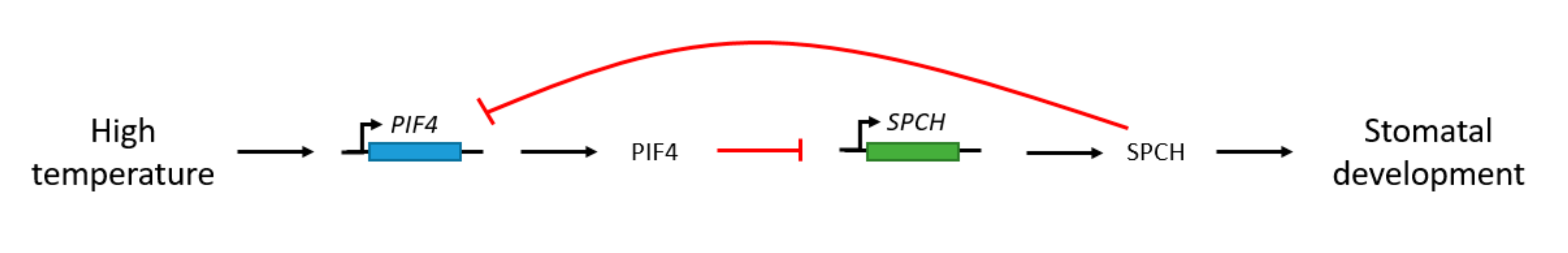
  98. Lau, O.S.; Song, Z.; Zhou, Z.; Davies, K.A.; Chang, J.; Yang, X.; Wang, S.; Lucyshyn, D.; Tay, I.H.Z.; Wigge, P.A.; et al. Direct Control of SPEECHLESS by PIF4 in the High-Temperature Response of Stomatal Development. Curr. Biol. 2018, 28, 1273–1280.e3. [Google Scholar] [CrossRef] [Green Version]
  99. Crawford, A.J.; McLachlan, D.H.; Hetherington, A.M.; Franklin, K.A. High temperature exposure increases plant cooling capacity. Curr. Biol. 2012, 22, R396–R397. [Google Scholar] [CrossRef] [Green Version]
  100. Koini, M.A.; Alvey, L.; Allen, T.; Tilley, C.A.; Harberd, N.P.; Whitelam, G.C.; Franklin, K.A. High Temperature-Mediated Adaptations in Plant Architecture Require the bHLH Transcription Factor PIF4. Curr. Biol. 2009, 19, 408–413. [Google Scholar] [CrossRef] [Green Version]
  101. Lucyshyn, D.; Wigge, P.A. Plant development: PIF4 integrates diverse environmental signals. Curr. Biol. 2009, 19, R265–R266. [Google Scholar] [CrossRef] [Green Version]
  102. Salvucci, M.E.; Crafts-Brandner, S.J. Mechanism for deactivation of Rubisco under moderate heat stress. Physiol. Plant. 2004, 122, 513–519. [Google Scholar] [CrossRef]
  103. Kostaki, K.-I.; Coupel-Ledru, A.; Bonnell, V.C.; Gustavsson, M.; Sun, P.; McLaughlin, F.J.; Fraser, D.P.; McLachlan, D.H.; Hetherington, A.M.; Dodd, A.N.; et al. Guard Cells Integrate Light and Temperature Signals to Control Stomatal Aperture. Plant Physiol. 2020, 182, 1404–1419. [Google Scholar] [CrossRef] [Green Version]
  104. Devireddy, A.R.; Arbogast, J.; Mittler, R. Coordinated and rapid whole-plant systemic stomatal responses. New Phytol. 2020, 225, 21–25. [Google Scholar] [CrossRef] [PubMed] [Green Version]
  105. Hashimoto-Sugimoto, M.; Higaki, T.; Yaeno, T.; Nagami, A.; Irie, M.; Fujimi, M.; Miyamoto, M.; Akita, K.; Negi, J.; Shirasu, K.; et al. A Munc13-like protein in Arabidopsis mediates H+-ATPase translocation that is essential for stomatal responses. Nat. Commun. 2013, 4, 2215. [Google Scholar] [CrossRef] [PubMed] [Green Version]
  106. Fanourakis, D.; Bouranis, D.; Giday, H.; Carvalho, D.R.; Nejad, A.R.; Ottosen, C.-O. Improving stomatal functioning at elevated growth air humidity: A review. J. Plant Physiol. 2016, 207, 51–60. [Google Scholar] [CrossRef] [PubMed]
  107. Grossiord, C.; Buckley, T.N.; Cernusak, L.A.; Novick, K.A.; Poulter, B.; Siegwolf, R.T.W.; Sperry, J.S.; McDowell, N.G. Plant responses to rising vapor pressure deficit. New Phytol. 2020, 226, 1550–1566. [Google Scholar] [CrossRef] [PubMed] [Green Version]
  108. Yuan, W.; Zheng, Y.; Piao, S.; Ciais, P.; Lombardozzi, D.L.; Wang, Y.; Ryu, Y.; Chen, G.; Dong, W.; Hu, Z.; et al. Increased atmospheric vapor pressure deficit reduces global vegetation growth. Sci. Adv. 2019, 5, eaax1396. [Google Scholar] [CrossRef] [Green Version]
  109. McAdam, S.A.; Brodribb, T.J. The Evolution of Mechanisms Driving the Stomatal Response to Vapor Pressure Deficit. Plant Physiol. 2015, 167, 833–843. [Google Scholar] [CrossRef] [Green Version]
  110. Fanourakis, D.; Heuvelink, E.; Carvalho, S.M. A comprehensive analysis of the physiological and anatomical components involved in higher water loss rates after leaf development at high humidity. J. Plant Physiol. 2013, 170, 890–898. [Google Scholar] [CrossRef] [PubMed]
  111. Nejad, A.R.; Van Meeteren, U. Stomatal response characteristics of Tradescantia virginiana grown at high relative air humidity. Physiol. Plant. 2005, 125, 324–332. [Google Scholar] [CrossRef]
  112. Aliniaeifard, S.; Matamoros, P.M.; Van Meeteren, U. Stomatal malfunctioning under low VPD conditions: Induced by alterations in stomatal morphology and leaf anatomy or in the ABA signaling? Physiol. Plant. 2014, 152, 688–699. [Google Scholar] [CrossRef] [PubMed]
  113. Aliniaeifard, S.; Van Meeteren, U. Stomatal characteristics and desiccation response of leaves of cut chrysanthemum (Chrysanthemum morifolium) flowers grown at high air humidity. Sci. Hortic. 2016, 205, 84–89. [Google Scholar] [CrossRef]
  114. Bakker, J. Effects of humidity on stomatal density and its relation to leaf conductance. Sci. Hortic. 1991, 48, 205–212. [Google Scholar] [CrossRef]
  115. McKown, A.D.; Guy, R.D.; Quamme, L.; Klápště, J.; La Mantia, J.; Constabel, C.P.; El-Kassaby, Y.A.; Hamelin, R.C.; Zifkin, M.; Azam, M.S. Association genetics, geography and ecophysiology link stomatal patterning inPopulus trichocarpawith carbon gain and disease resistance trade-offs. Mol. Ecol. 2014, 23, 5771–5790. [Google Scholar] [CrossRef] [PubMed]
  116. Pantin, F.; Blatt, M.R. Stomatal Response to Humidity: Blurring the Boundary between Active and Passive Movement. Plant Physiol. 2018, 176, 485–488. [Google Scholar] [CrossRef] [Green Version]
  117. Cai, S.; Chen, G.; Wang, Y.; Huang, Y.; Marchant, B.; Wang, Y.; Yang, Q.; Dai, F.; Hills, A.; Franks, P.J.; et al. Evolutionary Conservation of ABA Signaling for Stomatal Closure. Plant Physiol. 2017, 174, 732–747. [Google Scholar] [CrossRef] [PubMed]
  118. Kübarsepp, L.; Laanisto, L.; Niinemets, Ü.; Talts, E.; Tosens, T. Are stomata in ferns and allies sluggish? Stomatal responses to CO2, humidity and light and their scaling with size and density. New Phytol. 2019, 225, 183–195. [Google Scholar] [CrossRef] [PubMed]
  119. Chater, C.; Kamisugi, Y.; Movahedi, M.; Fleming, A.; Cuming, A.C.; Gray, J.E.; Beerling, D.J. Regulatory Mechanism Controlling Stomatal Behavior Conserved across 400 Million Years of Land Plant Evolution. Curr. Biol. 2011, 21, 1025–1029. [Google Scholar] [CrossRef] [PubMed] [Green Version]
  120. McAdam, S.A.; Sussmilch, F.C.; Brodribb, T.J. Stomatal responses to vapour pressure deficit are regulated by high speed gene expression in angiosperms. Plant Cell Environ. 2016, 39, 485–491. [Google Scholar] [CrossRef]
  121. Giday, H.; Fanourakis, D.; Kjaer, K.H.; Fomsgaard, I.S.; Ottosen, C.-O. Foliar abscisic acid content underlies genotypic variation in stomatal responsiveness after growth at high relative air humidity. Ann. Bot. 2013, 112, 1857–1867. [Google Scholar] [CrossRef]
  122. Vahisalu, T.; Puzõrjova, I.; Brosché, M.; Valk, E.; Lepiku, M.; Moldau, H.; Pechter, P.; Wang, Y.-S.; Lindgren, O.; Salojärvi, J.; et al. Ozone-triggered rapid stomatal response involves the production of reactive oxygen species, and is controlled by SLAC1 and OST1. Plant J. 2010, 62, 442–453. [Google Scholar] [CrossRef]
  123. Xie, X.D.; Wang, Y.B.; Williamson, L.; Holroyd, G.H.; Tagliavia, C.; Murchie, E.; Theobald, J.C.; Knight, M.R.; Davies, W.J.; Leyser, H.M.O.; et al. The Identification of Genes Involved in the Stomatal Response to Reduced Atmospheric Relative Humidity. Curr. Biol. 2006, 16, 882–887. [Google Scholar] [CrossRef]
Figure 1. Cells of the stomatal lineage. A protodermal cell (grey) enters the stomatal lineage when it becomes a meristemoid mother cell (MMC). A symmetric division of the MMC produces a meristemoid (orange) and a stomatal lineage ground cell (SLGC). Meristemoids undergo self-renewing amplifying divisions or develop into a guard mother cell (blue). This guard mother cell (GMC) symmetrically splits once to shape a pair of guard cells (green). SLGCs can develop into pavement cells (white) or they can be subjected to a spacing division to create a new meristemoid (orange). The basic-helix-loop-helix (bHLH) master transcription factors SPEECHLESS (SPCH), MUTE and FAMA, critical for the development of stomata, are displayed in the figure.
Agronomy 10 01975 g001
Figure 2. Kidney-shaped stoma of Ocimum basilicum L. (left) and dumbbell-shaped stoma of grasses (right).
Agronomy 10 01975 g002
Figure 3. Blue light signaling pathway in stomatal guard cells. Blue light is perceived by phot1 and phot2, activating plasma membrane (PM) H+-ATPase, inducing K+ uptake through K+ inward channels by PM hyperpolarization. Blue light drives the formation of a covalent adduct between flavin mononucleotide (FMN) chromophore and the light-oxygen-voltage (LOV) domain. Starch degradation and CO2 fixation in chloroplasts of guard cell (GC) and mesophyll cells raise sucrose, glucose, and malate intracellular concentrations. Accumulation of these ions leads to water uptake into the vacuoles, resulting in stomatal opening. Abscisic acid (ABA) inhibits the blue-light dependent stomatal opening through the production of PA, inhibiting PP1, thereby suppressing the activation of the H+-ATPase. LOV, Light-Oxygen-Voltage; PP1, protein phosphatase 1; PA, phosphatidic acid; ABA, abscisic acid; PM, plasma membrane; FMN, flavin mononucleotide.
Agronomy 10 01975 g003
Figure 4. Stomatal opening in guard cells as a result of red light signaling. Red light is absorbed by guard cells (direct GC response) and mesophyll chloroplasts (indirect GC response). ATP and NADPH are provided in the cytosol by guard cell chloroplasts. Starch degradation elevates sucrose levels. Sucrose synthesized in the mesophyll cells is imported via H+/sucrose symporters from the apoplast to the guard cells. CO2 fixation in mesophyll cells decreases Ci concentrations, resulting in membrane hyperpolarization. GC, guard cell; Ci, intercellular CO2 concentration.
Agronomy 10 01975 g004
Figure 5. Schematic illustration of the interaction of elevated CO2 concentration and ABA mediating stomatal movement. CO2 and ABA signaling unite downstream of OST1/SnRK2.6 regulating stomatal closure. An increase in CO2 concentration triggers stomatal closure via an ABA-independent pathway. CA, carbonic anhydrase; PYR/RCAR, pyrabactin resistance/PYR1-like/regulatory component of ABA receptor; PP2C, protein phosphatase 2C; OST1/SnRK2.6, Open Stomatal 1/Snf1-related protein kinase 1; SLAC1, Slow Anion Channel-associated 1; RHC1, Resistant to High CO2; GCA2, Growth Controlled by ABA2; HT1, High leaf Temperature 1; and ALMT12/QUAC1, aluminium-activated malate transporter 12/quickly activating anion channel 1.
Agronomy 10 01975 g005
Figure 6. Model of the PIF4 and SPCH regulation of stomatal development when exposed to high temperatures. At high temperatures, the PIF4 gene is induced and PIF4 binds and represses the SPCH gene. The negative feedback loop of SPCH on the PIF4 promoter is illustrated.
Agronomy 10 01975 g006
Figure 7. Temperature-induced stomatal opening. The phototropin-dependent pathway involves phototropin-mediated phosphorylation of BLUS1, activating PM H+-ATPases. AHA1 insertion into the PM is mediated by PATROL1. AHA1 regulates 14-3-3, promoting H+ pumping. The phototropin-independent pathway involves RBOHD-mediated ROS production, resulting in stomatal opening. Phot, phototropin; PP1, protein phosphatase 1; BLUS1, blue light signaling 1; PATROL1, proton ATPase translocation control 1; AHA, Arabidopsis plasma membrane H+-ATPase; RBOHD, respiratory burst oxidase protein D; ROS, Reactive Oxygen Species; GC PM, guard cell plasma membrane.
Agronomy 10 01975 g007
Figure 8. Hypothetical model for relative humidity (RH)-induced stomatal closure. The protein kinase OST1 is required for RH-induced stomatal closure, with either an ABA-dependent or ABA-independent activation. A passive hydraulic response to a reduced leaf turgor pressure is also possible under a low ambient RH. Adapted from Merilo et al. [33].
Agronomy 10 01975 g008
Table 1. Summary of the influence of the environmental factors light, CO2, temperature, and relative humidity on stomatal development and opening, including the molecular components involved in this stomatal response.
Stomatal Development
Environmental FactorStomatal ResponseMolecular Components Involved in Stomatal Response
Light↑ light intensity: ↑stomatal indexphyB, PIF4
CO2↑ CO2-concentration: downregulation of stomatal development: ↓ stomatal indexCA enzymes, HIC gene, ERECTA receptor kinase, EPF1, EPF2, LRR, TMM, ER, SDD1, CDC6
Temperature↑ ambient temperature: ↓stomatal developmentPIF4, SPCH
Relative humidity↑ RH: ↑ stomatal density and ↑ stomatal length
Stomatal Opening
Environmental FactorStomatal ResponseMolecular Components Involved in Stomatal Response
LightLight induces stomatal opening
Blue and red light induces stomatal opening by two distinct pathways
phot1, phot2
PM H+-ATPase
K+ inward channels
Malate2+, Cl, glucose, sucrose
PP1, 14-3-3
CO2↑ CO2-concentration induces stomatal closureβCA1, βCA2, HT1, OST1/SnRK2.6, SLAC1, ALMT12/QUAC1, RHC1, GCA2, MPK12, GHR1, AHA1
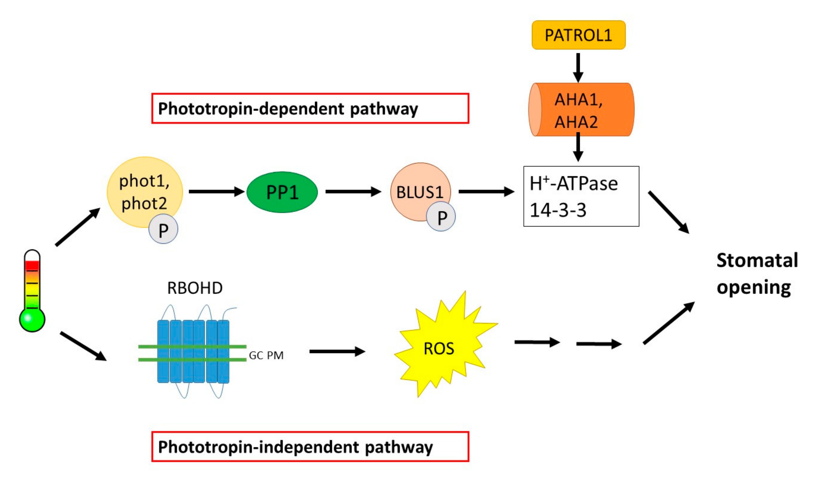
Temperature↑ temperature induces stomatal openingPhototropin-dependent pathway: BLUS1, 14-3-3 proteins, AHA1, PATROL1
Phototropin-independent pathway: RBOHD-mediated ROS production
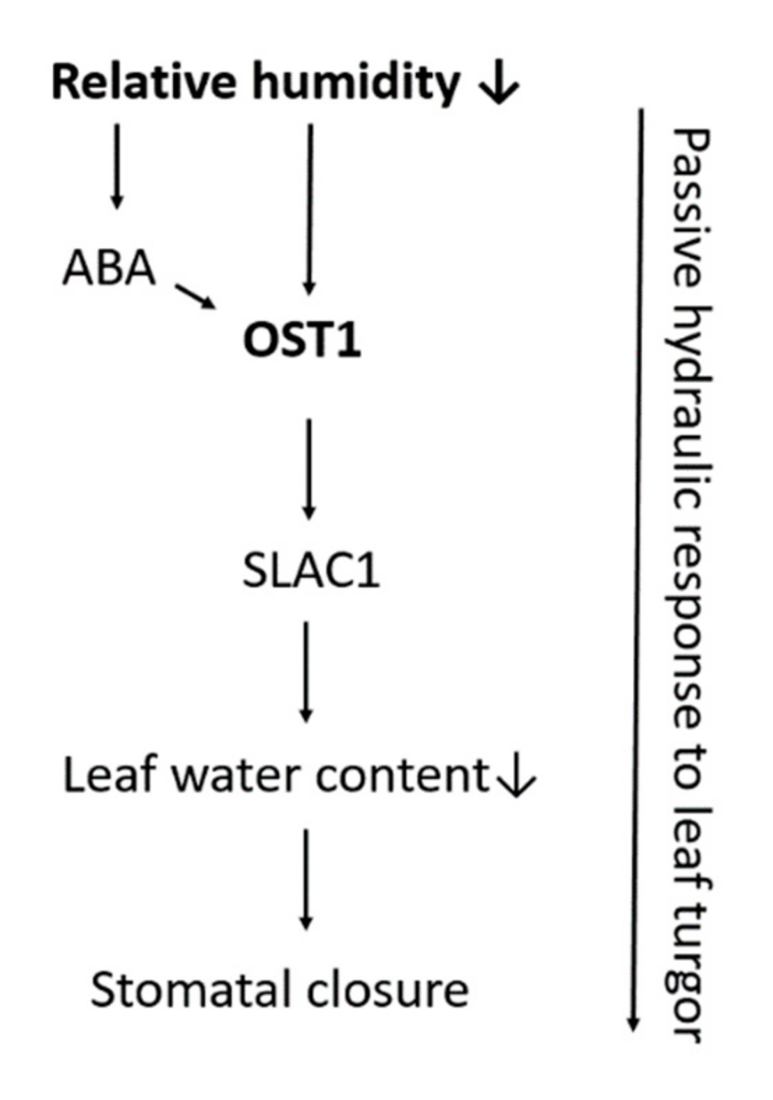
Relative humidity↓ relative humidity induces stomatal closurePassive hydraulic response
Active/ABA driven response: NCED genes, OST1, SLAC1, ABA2
“↑” refers to an elevated, increased or high value or concentration; “↓” refers to a decreased, low value or concentration.
Publisher’s Note: MDPI stays neutral with regard to jurisdictional claims in published maps and institutional affiliations.

