Article

The Effect of Accelerated Aging on the Molecular Weight and Thermal and Mechanical Properties of Polyester Yarns Containing Ceramic Particles

by
Gabriela Mijas
1,2,
Marta Riba-Moliner
1,3 and
Diana Cayuela
1,3,*
1
Terrassa Institute of Textile Research and Industrial Cooperation (INTEXTER), Universitat Politècnica de Catalunya (UPC), 08222 Terrassa, Spain
2
Fundación Asociación de Becarios Retornados EC (ABREC), Quito 170518, Ecuador
3
Department of Materials Science and Engineering (CEM), Universitat Politècnica de Catalunya (UPC-ESEIAAT), 08222 Terrassa, Spain
*
Author to whom correspondence should be addressed.
Polymers 2023, 15(6), 1348; https://doi.org/10.3390/polym15061348
Submission received: 5 February 2023 / Revised: 24 February 2023 / Accepted: 28 February 2023 / Published: 8 March 2023
(This article belongs to the Special Issue Durability and Degradation of Polymeric Materials II)

Abstract

:
The accelerated aging of polyethylene terephthalate (PET) multifilament yarns containing nano or microparticles of titanium dioxide (TiO2), silicon carbide (SiC), or fluorite (CaF2) at a maximum percentage of 2% has been studied. For this, the yarn samples were introduced into a climatic chamber at 50 °C, 50% relative humidity, and an ultraviolet A (UVA) irradiance of 1.4 W/m2. They were then removed from the chamber after periods of between 21 and 170 days of exposure. Subsequently, the variation in weight average molecular weight, number molecular weight, and polydispersity was evaluated by gel permeation chromatography (GPC), the surface appearance was evaluated using scanning electron microscopy (SEM), the thermal properties were evaluated using differential scanning calorimetry (DSC), and the mechanical properties were evaluated using dynamometry. The results showed that, at the test conditions, there was degradation in all of the exposed substrates, possibly due to the excision of the chains that make up the polymeric matrix, which resulted in the variation in the mechanical and thermal properties depending on the type and size of the particle used. This study provides insight into the evolution of the properties of PET-based nano- and microcomposites and might be helpful when selecting materials for specific applications, which is of great interest from an industrial point of view.

1. Introduction

The incorporation of a small amount of ceramics into a polymer yields a composite material with properties such as hardness, stiffness, heat resistance, UV resistance, water absorption, gas permeability, etc., at a low cost. For this reason, the development of functional fibers will be a key factor for the textile industry and, of particular interest, those containing ceramic powders. The use of nanotechnology can enhance the use of multifunctional textiles and provide them with new features, such as antibacterial, fungicidal, or water-repellent properties [1,2,3].
Some ceramic particles have been used from a textile point of view due to the multiple advantages they could bring to a substrate thanks to their intrinsic properties. Titanium dioxide (TiO2), because of its photocatalytic properties, provides self-cleaning properties and UV protection as well as an antibacterial barrier [4,5,6,7,8]. Calcium fluoride (CaF2) is of interest for its high stability, nonhygroscopic behavior, and transparency [9,10] and silicon carbide (SiC) has low density, high melting and decomposition temperatures, good mechanical and thermal properties, and excellent chemical and oxidation stabilities that have led to its study as a UV-blocking agent [11,12,13].
In this context, in previous studies carried out by the research team, the effect of the inclusion of nano and microparticles of TiO2, SiC, and CaF2 on the physical and chemical properties of polyethylene terephthalate (PET) has been analyzed. These results have made it possible to determine the most suitable processing conditions and dispersant for obtaining the PET multifilament yarns used in this work [14,15,16,17,18,19,20].
Polyethylene terephthalate, as well as other polymeric substrates, is susceptible to being affected by climatic conditions, which have destructive effects on the durability and performance of these materials. Among the factors that can be mentioned are solar radiation, humidity, and temperature [21,22]. The degradation process is executed in two simultaneous ways: yellowish discoloration and the loss of mechanical properties. Damage occurs when photons of light interact with the molecular chains that constitute the polymer structures. Shorter wavelengths possessing higher photon energies are more strongly absorbed in most polymeric materials and are more likely to break their chemical bonds. Because ultraviolet radiation has more photons of energy, it can break more types of bonds [23,24,25].
The objective of this research was to study the influence of the presence of nano or microparticles of TiO2, SiC, or CaF2 in the accelerated aging (50 °C, 50% RH, 1.4 W/m2 UVA) of PET multifilament yarns. For this, molecular weight and polydispersity variation were evaluated by gel permeation chromatography (GPC), the surface appearance by scanning electron microscopy (SEM), the thermal properties by differential scanning calorimetry (DSC), and the mechanical properties by dynamometry.

2. Materials and Methods

2.1. Materials

The study was carried out on POY multifilament yarns fabricated and supplied by IQAP Masterbatch Group S.L. (Masies de Roda, Spain). One hundred percent polyester (extra-bright) textile quality pellets were provided by ANTEX, S.L. (Anglès, Spain). The type and quantity of particles present in each yarn as well as the amount of dispersant Licowax-E (montane acid ester with multifunctional alcohols) supplied by Clariant (Sant Joan Despí, Spain) used, are presented in Table 1.
TiO2 particles were supplied by Zeus Química S.A. in rutile and anatase crystalline forms of nano and micrometer sizes, and their characteristics are shown in Table 2. CaF2 nanoparticles were synthesized with the methodology described in previous work [20] and their mean particle size obtained by dynamic light scattering (DLS) was 110 nm. β-SiC spherical nanoparticles, with a purity of 97.5%, an average particle size of 45–55 nm, and a specific surface area (BET) of 35–40 m2/g were purchased from NanoAmor (Houston, United States). TEM micrographs of raw materials are presented in Figure 1.

2.2. Post-Spinning Stretching

All substrates were subjected to a 1:2 stretching to provide them with optimum textile properties. The material infeed and outfeed speeds were 10 m/min and 20 m/min, respectively, on the corresponding drafting rolls.

2.3. Accelerated Aging

Thirty multifilament yarns of each substrate were placed parallel to each other on a sample holder grid. The substrates thus arranged were treated in a climatic chamber Binder GmbH 240 KBF ICH (E5) at conditions inside the equipment of 50 °C, 50% relative humidity, and 1.4 W/m2 ultraviolet A (UVA) exposure. To establish the treatment conditions, the “humidity–temperature with light” diagram available in the equipment specification sheet was used. Both temperature and humidity were within the standard range. The yarns were taken out of the chamber after 21, 35, 77, 91, 105, 119, 133, 148, and 170 days of exposure.

2.4. Washing

Once removed from the climatic chamber, the substrates were washed with a 1 g/L Hostapal UH liquid surfactant solution (Archroma, Sant Joan Despí, Spain) at 35 ± 2 °C for 30 min with a 1:30 liquor ratio to remove surface impurities. Afterward, the substrates were rinsed with distilled water at 35 ± 2 °C for 5 min and then rinsed a further three times with distilled water at room temperature for 5 min each time. Once the yarns were washed, they were left to dry at room temperature.

2.5. Molecular Weight

The procedure described in Gacén [31] was followed. Approximately, 3 mg of washed yarns were placed in a test tube with 0.2 mL of o-chlorophenol >99% (Sigma-Aldrich, Steinheim, Germany). The tubes were placed in a polyethylene glycol bath at 80 °C for 30 min. Once the yarn was dissolved and the tubes cooled, 1.8 mL of chloroform (HPLC grade, stabilized with amylene, approx. 150 ppm, Scharlau, Barcelona, Spain) was added. The mixture was stirred and then filtered (PTFE filter, 0.45 µm) and transferred to a vial. Analysis was carried out in a high-performance liquid chromatograph (Perkin Elmer, Norwalk, CN, USA) provided with an ISS200 injector, 200lc series pump, an oven for series 200 columns, and a series 200a refractive index detector. The test was performed at 40 °C with HPLC grade chloroform (Scharlau, Barcelona, Spain) at a flow rate of 1 mL/min on a PLgel 5 µm 300 × 7.5 mm Mixed-D column (Agilent, Stockport, UK) and proceeded using an Oligopore Column 6 µm 300 × 7.5 mm (Agilent, Stockport, UK). The system was calibrated with polystyrene standards with weight average molecular weight in the range of 2350–200,000 g/mol (Waters, Milford, CN, USA). TotalChrom Navigator—TurboSEC v. 6.3.1.0504 software (Perkin Elmer, Norwalk, CN, USA) was used to calculate molecular weight in number, molecular weight in weight, and polydispersity.

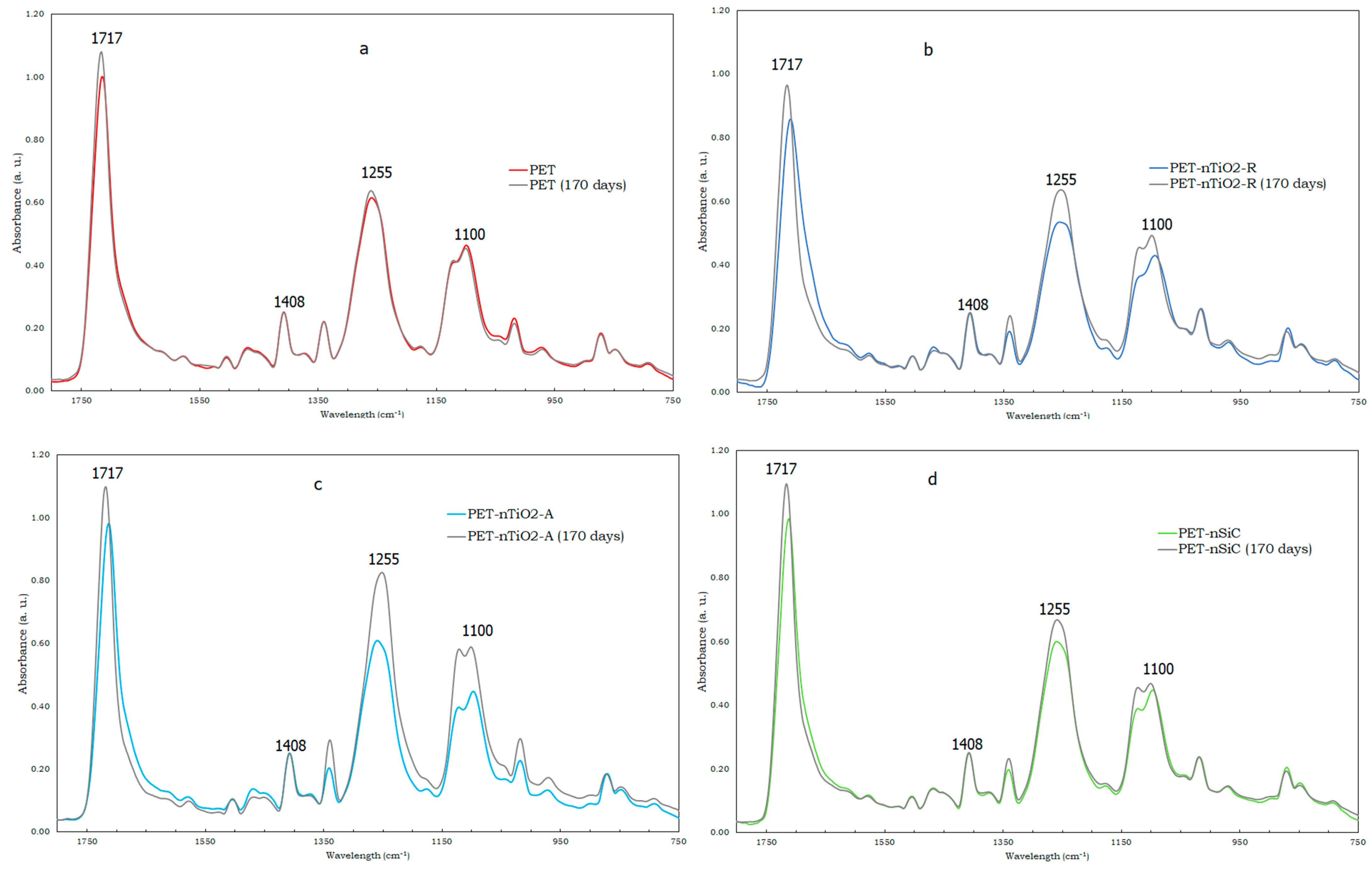
2.6. Fourier Transform Infrared Analysis-Attenuated Total Reflection (FTIR–ATR)

A Nicolet 6700 FT–IR spectrometer (Thermo Scientific, Waltham, MA, USA) with a SmartOrbit Diamond ATR accessory was used to determine the functional group changes during aging. Thirty-two scans were completed in the 400–4000 cm−1 range, at a spectral resolution of 2 cm−1.

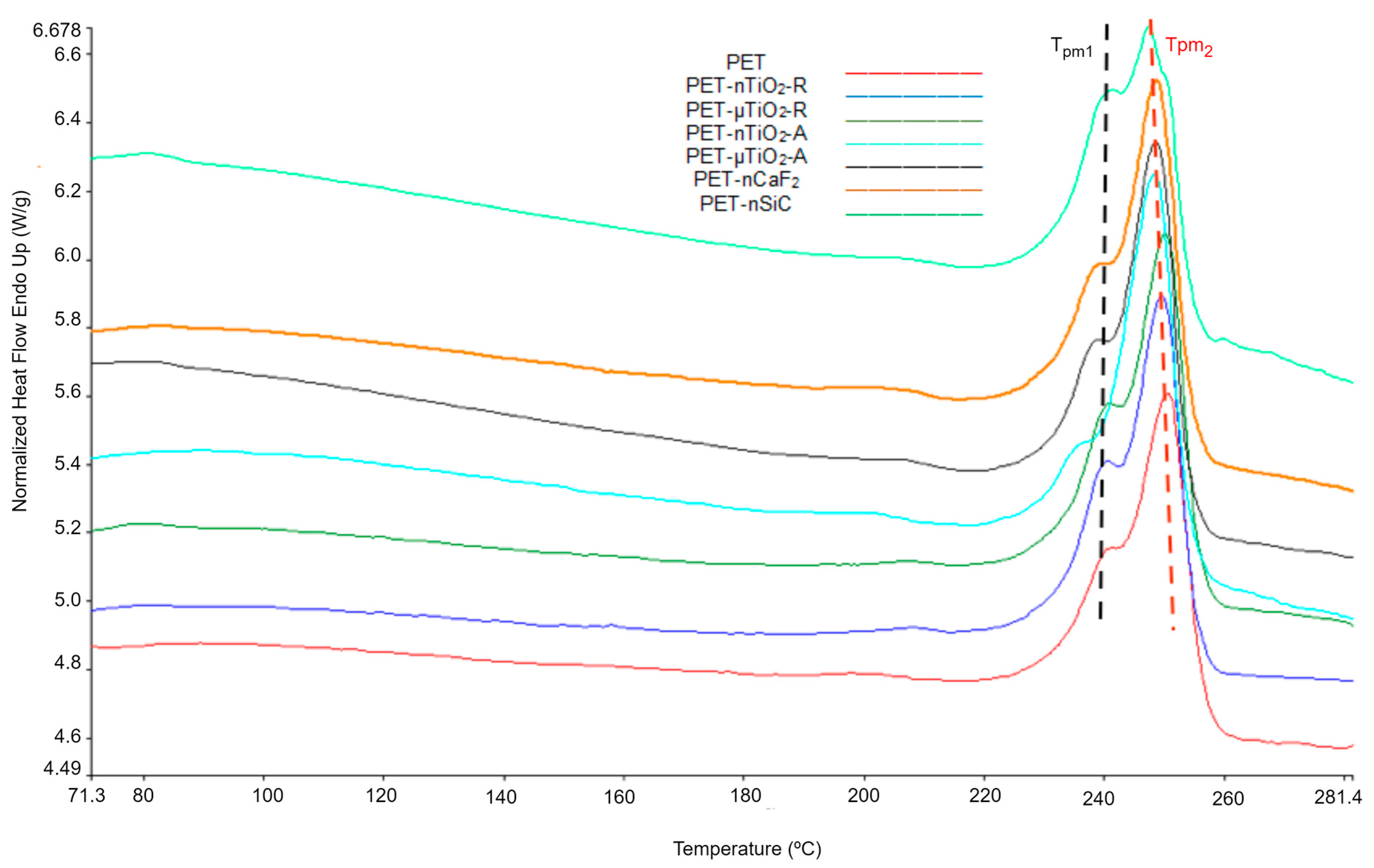
2.7. Thermal Analysis (Differential Scanning Calorimetry)

Samples of 4–5 mg sealed in an aluminum crucible were analyzed in a DSC 7 differential scanning calorimeter (Perkin Elmer, Norwalk, CN, USA). The studies were carried out under the following conditions: (i) holding at 40 °C for 5 min, (ii) heating from 40 °C to 300 °C at 20 °C/min, (iii) holding at 300 °C for 5 min, (iv) cooling from 300 °C to 40°C at 20 °C/min, (v) holding at 40 °C for 5 min, and (vi) heating from 40 °C to 300 °C at 20 °C/min. Nitrogen (N2) was used as purge gas at a flow rate of 35 mL/min. Samples were analyzed with Pyris 5.00.02 software (Perkin Elmer, Norwalk, CN, USA) and the results are the average of two determinations.

2.8. Lineal Density (Count)

Prior to testing, the yarns were conditioned at 20 ± 2 °C and 65 ± 4% RH for 24 h according to UNE-EN 13392:2001 [32]. For count determination, 100 m of each yarn was collected in a wrap reel Aspe (JBA, Cabrils, Spain) and then weighed on an analytical balance (±0.1 mg). Subsequently, the mass-to-length ratio in tex was calculated. This procedure was carried out in triplicate.

2.9. Tenacity and Elongation
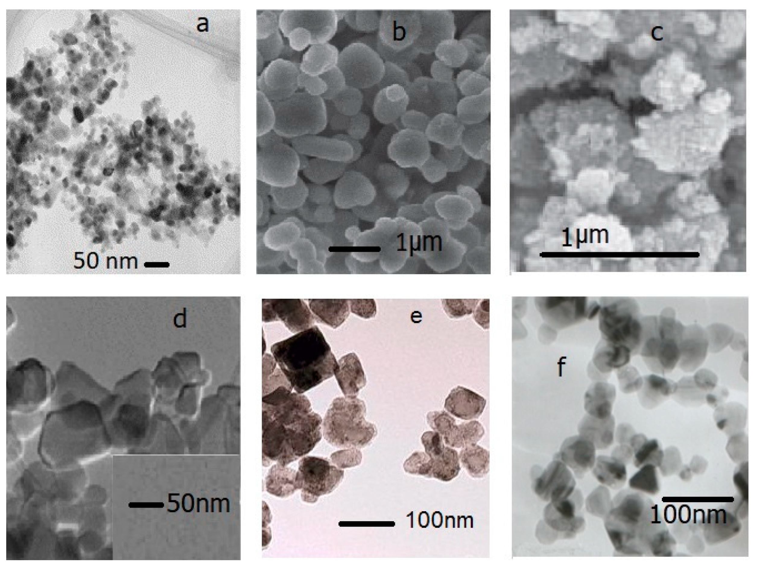
Tensile tests were performed based on the UNE-EN ISO 2062:2010 standard [33]. A Uster Tensokid automatic PE 4056 dynamometer was used with a grip separation of 500 mm, a test speed of 500 mm/min, and an elongation of 85% of the initial specimen. TestXpert Standard v 6.01 software (ZwickRoell S.L., Barcelona, Spain) was employed to analyze the tensile properties of the yarn.

2.10. Micrographs

Surface changes to the yarns during the exposure in the climatic chamber were analyzed using a FEI Phenom FP3950/00 scanning electron microscope (Phenom-World, Eindhoven, The Netherlands). Previously, the samples were coated with Au/Pd in an inert Ar atmosphere for 135 s and a current of 18 mA. The distance between the samples and the coating source was 35 mm.

3. Result and Discussion

The variations in the average molecular weight, weight average molecular weight, and polydispersity of the yarns at different exposure times are shown in Figure 2.
In all cases, there was a decrease in the number average molecular weight ( M n ¯ ) (Figure 2a), which could be the result of the scission of the molecular chains that constitute the polymeric matrix leading to the formation of oligomers [34]. Nevertheless, depending on the type of particle, the effect was different on the substrate when compared to pure PET. The reduction in the yarns with anatase TiO2 particles was greater than that with the rutile type, which could be because the rutile crystalline form has a higher ultraviolet scattering effect in comparison to the anatase form [35,36]. Similarly, a decrease in the weight average molecular weight ( M w ¯ ) (Figure 2b) values were observed, to a greater degree, in PET-nSiC. The final reduction percentages after 170 days of exposure are shown in Figure 3. In the case of PET-nSiC, degradation was observed to have stopped after 133 days of exposure in the chamber. The greatest decrease in molecular weight in number was found in PET-nTiO2-A, very similar to PET-nSiC, and both higher than that found for pure PET. The higher specific surface area of the nanoparticles could have caused the ultraviolet radiation to produce a larger degradation in the polymeric matrix [4,37].
Regarding the polydispersity (Figure 2c), an increase was observed over time that could be due to the scission of the molecular chains resulting from the degradation undergone by the polymer. This led to a wider molecular weight distribution compared to that of the initial substrates [38]. The yarn with the highest polydispersity was PET-nTiO2-A with a value of 3.2.
The rate of polymer degradation can be defined as the rate of scission of the molecular chains and is represented by Equation (1) [39]:
1 M ¯ nt = C + k n t
where C = 1 / M ¯ n 0 , M ¯ n 0 , and y M ¯ nt are the number of average molecular weights at time t = 0 and t = t, respectively, kn is the rate constant based on the average molecular weight, and t is the thermal degradation time.
From the plots of 1 / M ¯ nt ) vs. exposure time, it was observed that values obtained from correlation coefficients (R2) are higher than 0.94 for all cases, which would validate the assumption that the degradation of the polymer is mainly due to a scission of the molecular chains. Table 3 summarizes the regression coefficients of the degradation kinetics plots. The increasing order of the degradation kinetic constant Kn according to its magnitude is PET-nTiO2-R < PET-µTiO2-R < PET-µTiO2-A < PET = PET-nCaF2 < PET-nTiO2-A < PET-nSiC.
In order to relate the decrease in the molecular weight of the substrates to changes in their chemical composition, FTIR–ATR was carried out. The infrared spectra of pure PET, PET-nTiO2-R, PET-nTiO2-A, and PET-nSiC are shown in Figure 4. The intensity of the spectra was normalized with a reference peak, the IR band at 1408 cm−1, since it is assigned to the benzene ring of PET, which is not expected to be affected by aging [40,41,42] This normalization method allows a proper comparison of the absorption intensity of characteristic bands showing some structural changes during accelerated aging in the 750–1800 cm−1 range. An increase in the intensity of characteristic peaks of PET was noted, that is, in the aromatic ester C=O stretch (1717 cm−1), aromatic ester C-C-O stretch (1255 cm−1), and aromatic ester O-C-C stretch (1100 cm−1) [43]. According to a previous study [42], these peaks are due not only to the original PET substrates by themselves but are also the result of the presence of the carbonyl groups in the degradation products. In the case of PET (Figure 4a), the highest increase was observed in the C=O stretch in PET-nTiO2-R (Figure 4b), which had the lowest reduction in the molecular weight and a greater raise noted in the three mentioned peaks, and finally, in PET-nTiO2-A (Figure 4c) and PET-nSiC (Figure 4d), which were the samples with the highest reduction in the molecular weight, the increase was more evident in the one containing TiO2.
For the study of the variation in the yarn microstructure during the exposure time, DSC melting endotherms of the first heating were analyzed. Figure 5 shows the thermograms corresponding to the study of the yarns after 170 days of exposure. It was observed that the onset of the melting occurred at different temperatures and that, in all cases, there were two crystal distributions. The “shoulder” Tpm1 corresponds to lower melting temperatures with smaller crystal sizes and Tpm2 corresponds to high melting temperatures and larger crystal sizes. For all temperatures, the standard deviation (SD) corresponds to the average of the 10 values obtained from each of the extractions, from 0 to 170 days.
As for the melting onset temperature (Figure 6a), a decrease was observed during the exposure time for all cases. This could be due to the breakage of the molecular chains causing a diminution in the size of the crystals, which melted at a lower temperature [44]. For PET-nCaF2 and PET-nSiC, the decrease in the melting onset temperature was more noticeable compared to the yarns containing TiO2. This was seen at a greater level in PET-nSiC yarns.
Regarding the temperature of the shoulder Tpm1 (Figure 6b) and main melting peak Tpm2 (Figure 6c), for pure PET they remained constant since the standard deviation (SD) of the average values did not exceed ± 0.8 °C. This behavior was also observed in the four yarns with TiO2. For the case of PET-nCaF2 and PET-nSiC, it could be seen that the temperatures of the main melting peak Tpm2 remained constant throughout the exposure time since the standard deviation of the average values was not greater than ± 0.9 °C. Nevertheless, a decrease in the shoulder temperature Tpm1 next to the main melting peak was observed in both cases. For PET-nCaF2, there was an overlapping of the shoulder Tpm1 and main melting peak temperature Tpm2 from 133 days of exposure.
In terms of crystallinity (Figure 7a), an increasing trend was observed in the case of pure PET, which could be due to a decrease in the amorphous part of the material. The temperature at which the aging of the samples was carried out (50 °C) was lower than the glass transition of PET (70–80 °C) [44,45] and as the water in the medium was not a plasticizer of the polyester [46], no intense heat treatment that enhanced the crystallinity of the substrates would have taken place at that temperature. An increase in crystallinity was also noted for the TiO2 yarns, which could be due to the decrease in the amorphous part of the material for the reasons discussed above. The crystallinity of PET-nCaF2 at the end of the exposure time was higher than that of PET-nSiC, and no significant increase in crystallinity was observed compared to the unexposed substrates.
The average percent of crystallinity after 170 days of exposure for the yarns was 51.5 ± 2.7% for PET, 52.5 ± 1.8% for PET-nTiO2-R, 51.4 ± 2.2% for PET-µTiO2-R, 52.7 ± 2.7% PET-nTiO2-A, and 52.7 ± 2.6% for PET-µTiO2-A, which, in this case, indicates that the particle size and crystalline form of the particles have not greatly influenced the crystallinity of the substrate at aging conditions, 54.0 ± 1.5% PET-nCaF2 and 52.5 ± 1.7% PET-nSiC.
Regarding the nonisothermal crystallization peak (Tc) (Figure 7b), no significant variations were observed in pure PET because of the exposure in the climatic chamber, and the differences were attributable to random experimental errors. For the blends with particles, an increment of Tc in the unexposed yarns with nanoparticles was seen, which meant that they acted as nucleating agents [44,47] In previous studies [14,15,19], it was found that the use of Licowax-E (montane acid ester with multifunctional alcohols) as a dispersant influenced such behavior of the ceramic particles. In the case of exposed yarns, a similar response was found in all of them. A tendency for the temperature of crystallization (Tc) to decrease at higher exposure times was observed. This may be a consequence of a diminution in the concentration of the nucleating agents in the molten polymer, resulting in a lower crystallization temperature upon cooling compared to the untreated substrates. This reduction in concentration could be due to the scission of the chains during the aging process, which would make it possible for the ceramic particles to rise to the surface of the yarns, or for a possible migration of the particles to the surface in analogous mode to disperse dyes [48]. In both cases, the particles would be more easily removed with the airflow circulating inside the climatic chamber or removed in the washing before the DSC test.
The melting endotherms of the second heating were analyzed to determine the variation in the properties of polymer blends during the exposure time. The thermograms after 170 days of exposure are shown in Figure 8. Two melting peaks Tpm1 and Tpm2 are observed.
As for the melting onset temperature Ton (Figure 9a), a slight increase was observed with respect to the value for pure PET. Nevertheless, the standard deviation was ± 0.4 °C, which was within the experimental error of the test. For TiO2-containing polymer blends, no clear trend was observed during the exposure time; however, there was a tendency for this value to increase. For the PET-nCaF2 and PET-nSiC blends, the changes were more evident. In the first case, an increase in onset temperature was noted of about 4 °C, and in the second case, a reduction of about 4 °C was seen. This would indicate that, for these two cases, the presence of the nanoparticles has influenced the size of the polymer crystals.
A slight decrease in the shoulder temperature (Tpm1) (Figure 9b) was seen with respect to the original PET during the course of exposure time, though it was within the experimental error of the test. The temperature of the main peak (Tpm2) (Figure 9c) remained practically constant (SD ± 0.4 °C). In addition, an increase in crystallinity (Figure 9d) of PET was observed, possibly due to the decrease in the amorphous part of the material [47] because of the influence of UV radiation, temperature, and humidity.
For TiO2 blends, the temperature of the main peak (Tpm2) was similar during the exposure and was around 250 °C. For all four cases, an increase in crystallinity was observed with respect to the unexposed substrates as a result of the nucleating effect of the ceramic particles used. After 170 days of exposure, the most crystalline was PET-nTiO2-R with an average value of 48.2 ± 2.7%, followed by PET-nTiO2-A with 46.7 ± 2.4%, PET-µTiO2-R, 46.4 ± 1.8%, and PET-µTiO2-A with 46.0 ± 1.1%, which indicates that, for this case, the nanoparticles, due to their size, have allowed the formation of a greater quantity of crystals as there are more crystallization nuclei available [49].
Concerning the blends PET-nCaF2 and PET-nSiC, the results showed that the main peak (Tpm2) remained constant throughout the treatment time (SD = 0.7). As for crystallinity, in both cases, the values at the end of the treatment remain similar to those of the unexposed blends with 43.7 ± 2.6% and 45.8 ± 2.0%, respectively.
Comparing the DSC melting endotherms of the first and second heating, some differences were noted. Regarding the presence of two melting peaks, which are observed in Figure 5 and Figure 8 (or a main peak and a shoulder), it was observed that in the first heating, the proportion of smaller crystals (lower melting temperature) was higher than the larger ones (higher melting temperature), while in the second heating, the proportion of larger crystals was higher than the smaller ones. These differences could be due to the cooling rate. In the case of spinning, the yarn collection speed on the winding machine was 3000 m/min, which implies fast cooling and, therefore, the formation of a greater quantity of smaller crystals. On the contrary, in the calorimetric test, the cooling speed was 20 °C/min, considerably lower than in the first case, which enabled the formation of a larger quantity of crystals of larger size.
Regarding the onset melting temperatures (Ton) obtained in the first second heating (Figure 6a and Figure 9a), a decrease in the onset temperature in the second heating was noted, which could be a consequence of the existence of smaller crystals that could have formed in the nonisothermal crystallization, which began to melt at lower temperatures.
The percentages of crystallinity of the mixtures in the first and second heating are shown in Figure 7a and Figure 9d. It was observed that the crystallinity in the second heating was lower, which could be because the substrate, once melted, has neither been cooled with stretching (not oriented) nor subjected to other conditions that allow the increase in its crystallinity, unlike the first heating, in which a previous stretching of the yarns had been performed at 190 °C at a 1:2 ratio.
The average values obtained from the tensile tests of the yarns before their exposure in the climatic chamber are shown in Table 4. It can be observed that PET-nTiO2-A was the yarn with the highest count (18.5 ± 0.6 tex) and the lowest count corresponded to PET-µTiO2-R (13.3 ± 0.1 tex). Although the yarns with ceramic particles should have a higher count compared to the original PET (16.0 ± 0.2 tex), the conditions of preparation of the blends, their subsequent spinning, and the loss of filaments in the drawing could have influenced the weight of the yarns and, therefore, the final count.
Regarding tenacity, in all cases, the values obtained were lower than those of pure PET (35.86 cN/tex), which could be a consequence of the decrease in molecular weight caused by the inclusion of ceramic particles and their high concentration in the substrates. Previous studies have found that increasing the number of ceramic particles, specifically TiO2, to a value higher than 0.33 % in Nylon yarns results in a reduction in tensile strength. High particle concentrations, which, for this case, are in the range of 1.8 wt.% to 2 wt.%, cause interactions and agglomerations, leading to a reduction in the contact surface between the particles and the matrix. In addition, these particles could also act as stress concentration sites and, therefore, when subjected to tensile deformation, the larger particles induce higher stress concentrations in the matrix and induce a reduction in impact energy, which results in the initial breakage of the material appearing earlier [50,51,52,53,54].
The reductions in tenacity after 135 days of exposure in the climatic chamber are shown in Figure 10. As can be seen, all of the yarns experienced a reduction in their tenacity greater than that of pure PET, which was a consequence of the high number of ceramic particles in the composite materials and the breakage of the molecular chains that constituted the substrate [50]. The yarns in descending order according to their tenacity would be as follows: PET > PET-nTiO2-R > PET-µTiO2-R > PET-nCaF2> PET-nSiC > PET-µTiO2-A > PET-nTiO2-A.
In the case of the PET-nTiO2-A yarn, it was not possible to perform the dynamometric test due to its high brittleness, which is why a 100% reduction in its resistance was considered after 35 days of exposure in the climatic chamber. In particular, the brittleness of the yarn could be the result of exposure to the climatic chamber and/or the high content of particles. On one hand, for an estimated 13% reduction in molecular weight, the yarns with microparticles showed a 20% reduction in strength, less than the reduction in pure PET, which was approximately 25%. On the other hand, PET-nTiO2-R lost 35% and PET-nTiO2-A lost 100% of its tensile strength. It could be observed that, over time, PET-µTiO2-A significantly decreased in strength compared to PET-µTiO2-R because of the brittleness, as observed in Figure 11.
When PET-nTiO2-A yarn had lost 100% of its tensile strength with a molecular weight reduction of 13%, PET-nTiO2-R lost 35% of its strength, PET-nSiC and pure PET lost 25 %, and PET-nCaF2 had lost 20%. Subsequently, the loss of the PET-nSiC yarn strength increased significantly. For a 30% reduction in molecular weight, a 33% reduction in PET tensile strength was observed, 50% for PET-nCaF2, 53% for PET-nSiC, and 100% for PET-nTiO2-A.
The micrographs corresponding to the samples observed after 0, 35, 77, 105, and 133 days of exposure are presented in Figure 11. In the case of pure PET yarns, oligomers could be seen on the surface. As for the yarns with ceramic particles, some agglomerations were observed on the surface, which, at first sight, could not be differentiated as oligomers or particles. Nevertheless, the use of the dispersant, the shear forces applied for blending, and the melt spinning technique made it possible to obtain a composite with a good dispersive and distributive blending so that it would not be possible that the agglomerates were particles and would rather be oligomers. Moreover, as mentioned before, this effect was also observed in pure PET that did not contain any charge, which indicated that they were not particles.
In the yarns with nano and microparticles of anatase TiO2, fractures caused by brittleness due to their exposure in the climatic chamber were observed, which could lead to the loss of their mechanical properties.

4. Conclusions

The presence of ceramic nano and microparticles in the degradation of PET yarns subjected to accelerated aging in a climatic chamber (1.4 W/m2 UVA, 50 °C 50% RH) for 170 days was assessed. The following aspects can be highlighted:
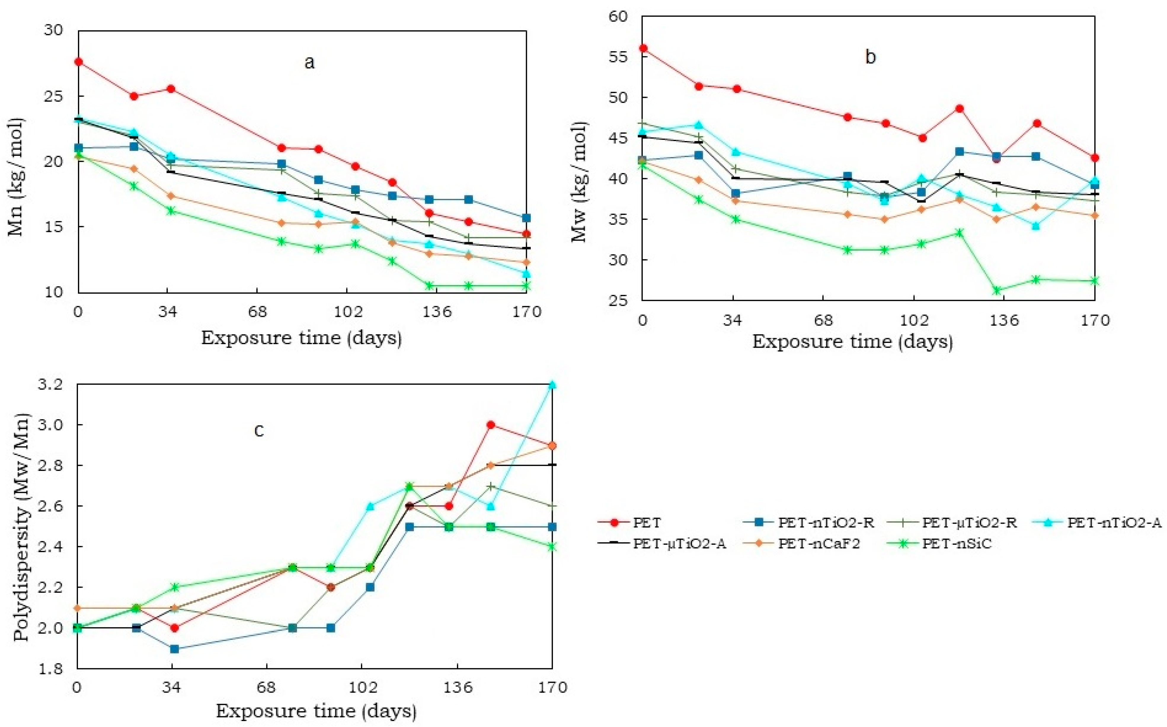
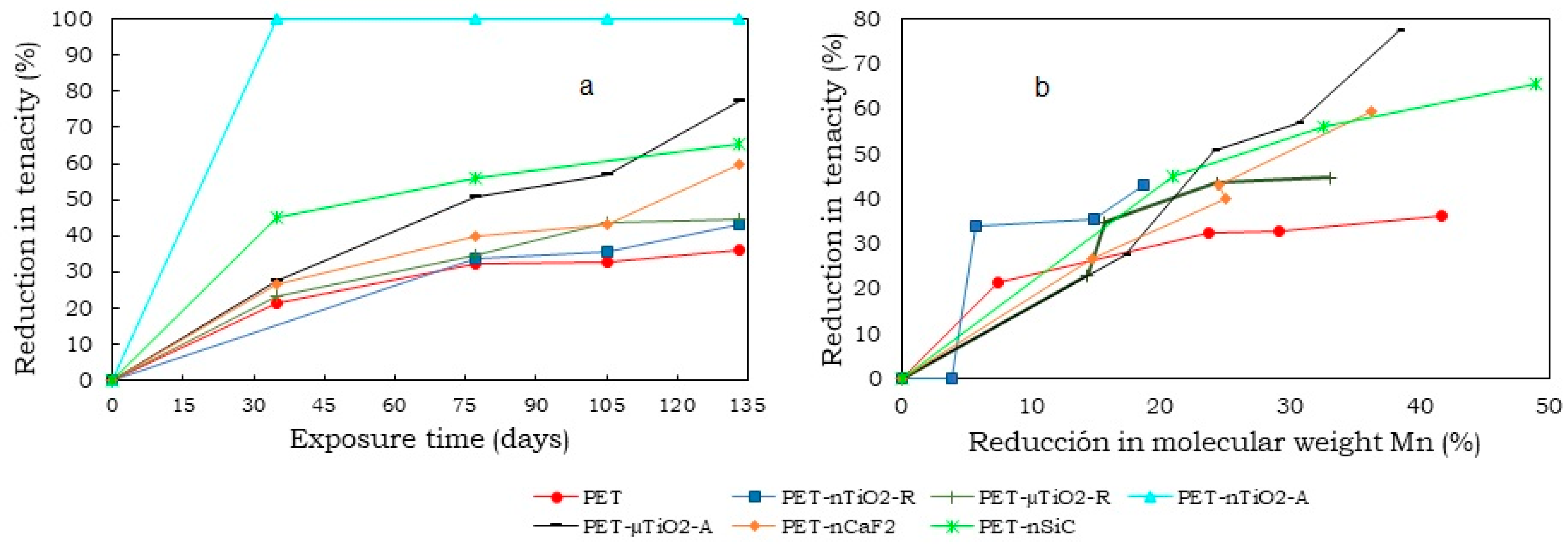
  •  A decrease in the molecular weight of all yarns that have been subjected to exposure in the climatic chamber was noted. This could be due to the excision of the molecular chains that formed the polymeric matrix. On one hand, the reduction was more evident in yarns with anatase TiO2 and SiC nanoparticles (about 50%), possibly due to their higher specific surface area that could have caused the ultraviolet radiation to produce a larger degradation. On the other hand, the rutile TiO2 nanometric had only a reduction of 25.3%.
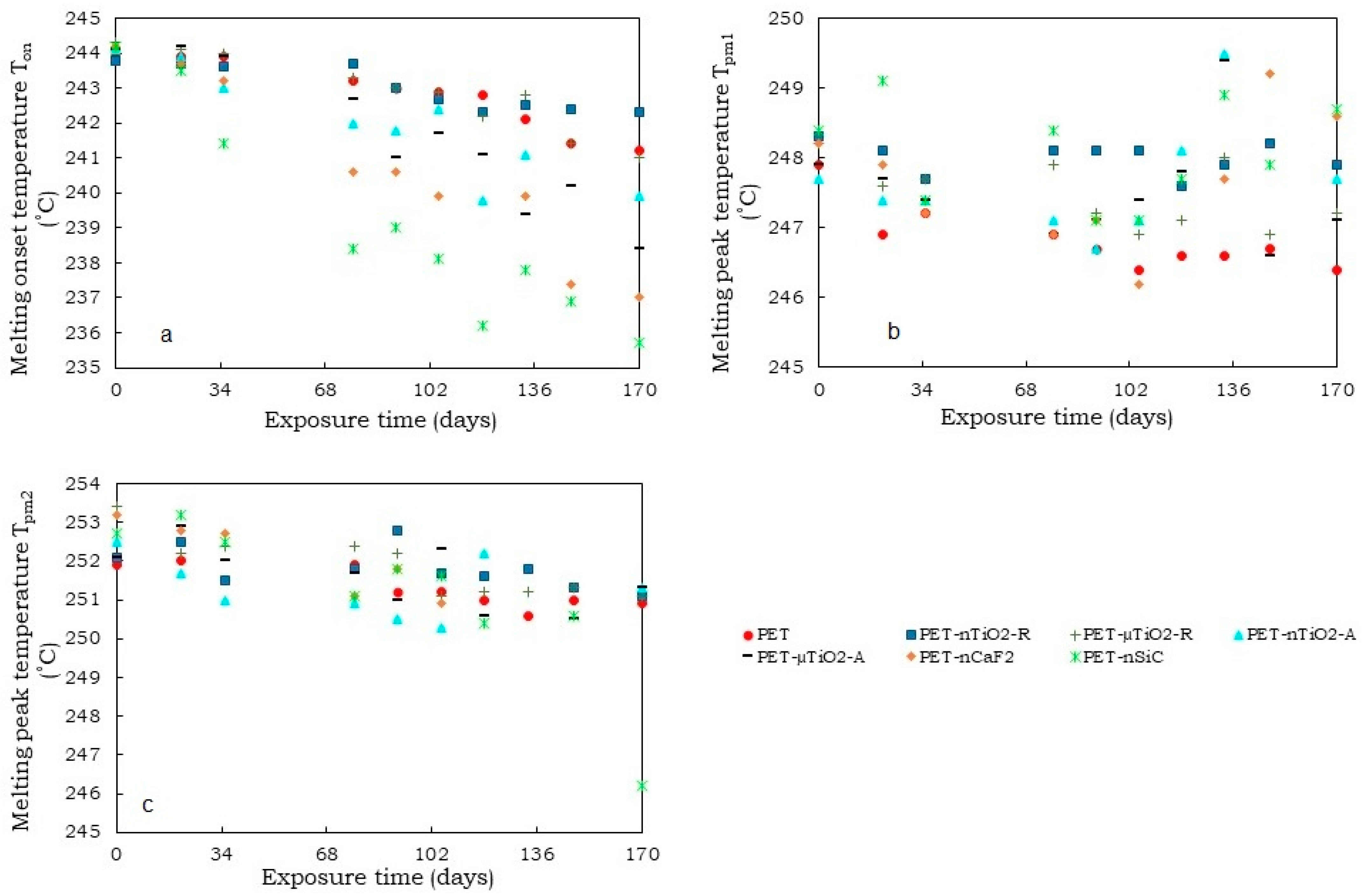
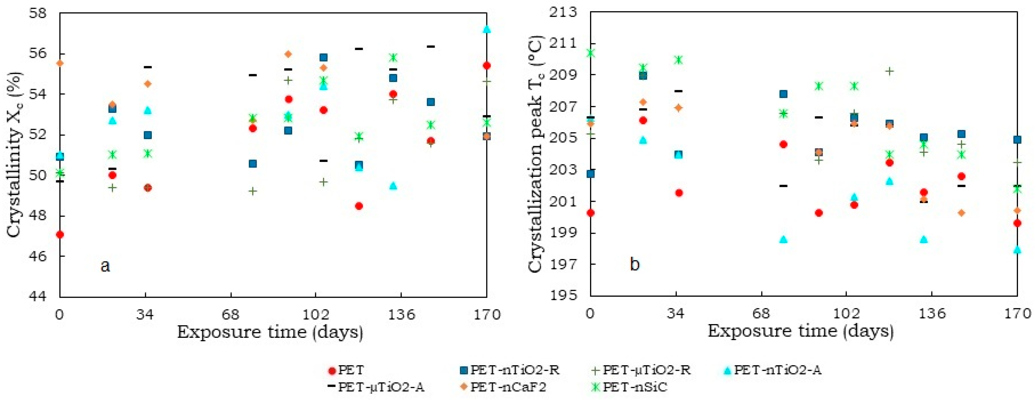
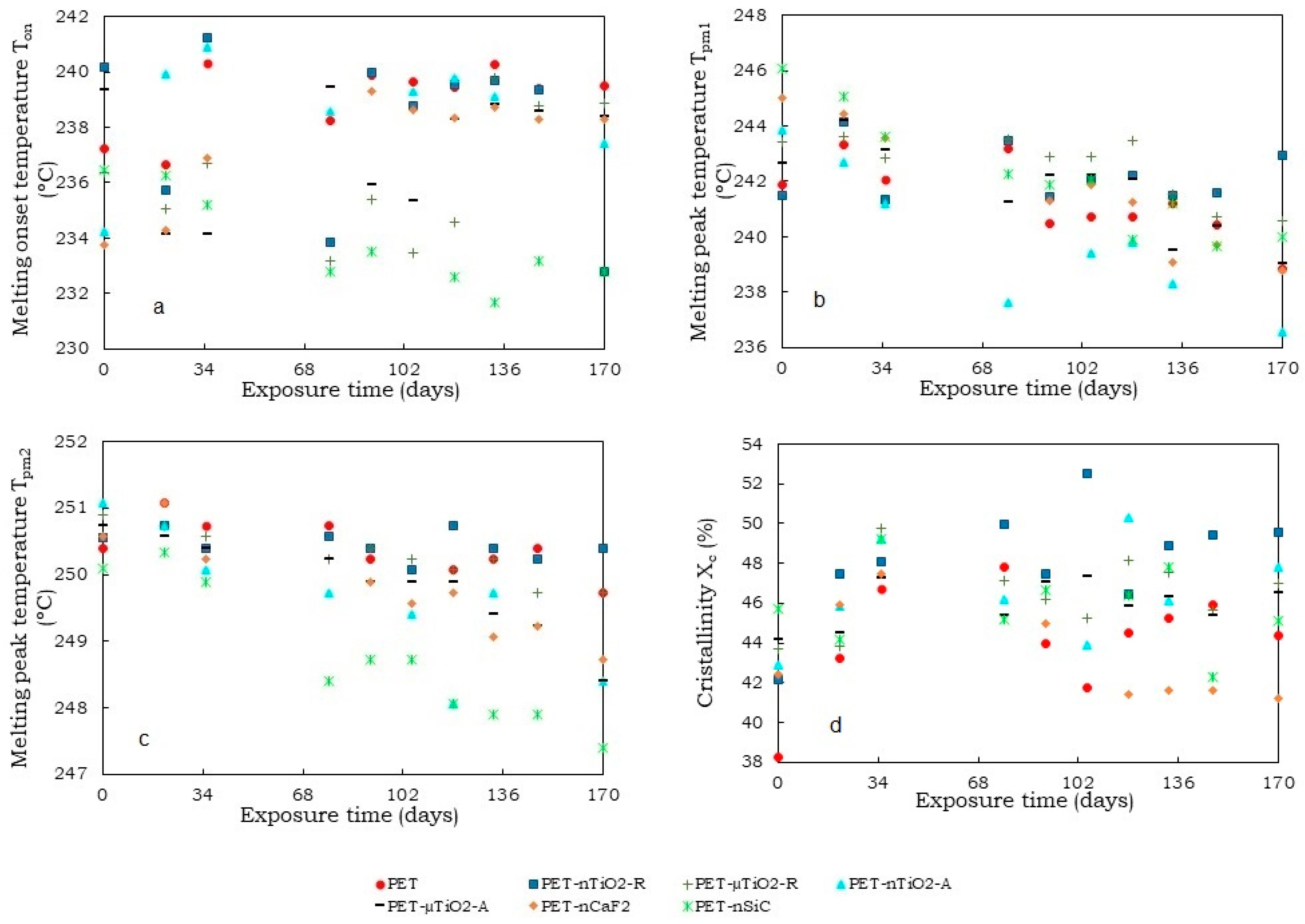
  •  A trend of increasing crystallinity in the pure PET yarns was observed over time, which could be due to a decrease in the amorphous part of the material. Moreover, the particle size and crystalline form of the ceramic particles did not increase the crystallinity compared to yarns that had not been exposed. Contrarily, in the case of polymer blends containing TiO2, unlike CaF2 and SiC, there was an increase in crystallinity because of the nucleating effect of the ceramic particles used. In this case, the nanoparticles, due to their size, allowed the formation of a greater number of crystals.
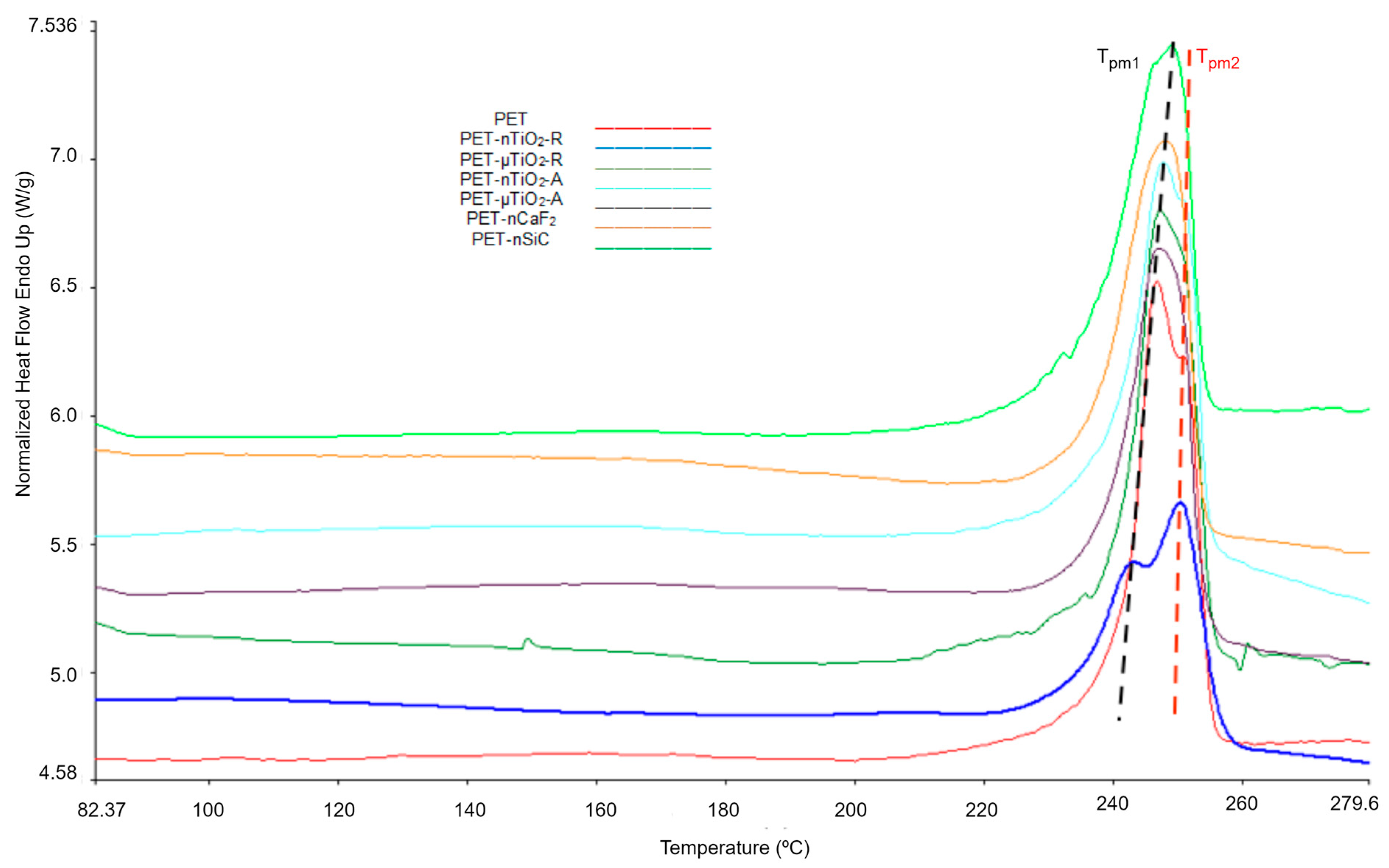
  •  Although an increase in nonisothermal crystallization temperature was observed in yarns with ceramic particles compared to pure PET, a tendency was found for it to decrease at higher exposure times because of a possible decrease in the concentration of the nucleating agents in the polymer melt because of the exposure in the climatic chamber.
  •  Finally, a reduction in tensile strength greater than that of the original PET was observed for all of the yarns that were subjected to the aging treatment. This could be a consequence of the high number of ceramic particles present in the composite materials and the breakage of the molecular chains of the substrate. The yarn with the anatase TiO2 micrometric showed the highest brittleness, which also experienced the greatest decrease in its molecular weight.
This study provides insight into the evolution of the properties of PET-based nano- and microcomposites and might be helpful when selecting materials for specific applications that require continuous exposure to UV radiation, humidity, and temperature, which is of great interest from an industrial point of view.

Author Contributions

Investigation, writing—original draft, G.M.; investigation, supervision, writing—Review and editing, M.R.-M.; funding acquisition, methodology, supervision, writing—review and editing, D.C. All authors have read and agreed to the published version of the manuscript.

Funding

The authors gratefully acknowledge the financial support for this research from the Ministry of Science and Innovation of Spain (Project MAT2010-20324-CO2-01) and the National Secretary of the Higher Education Science Technology and Innovation of Ecuador (SENESCYT) for Fellowship No. CZ02-000926-2018.

Data Availability Statement

The data presented in this study are available on request from the corresponding author. The data are not publicly available due to privacy.

Acknowledgments

The authors would like to thank the companies ANTEX, S.L. for providing the different polyester pellets, Clariant for providing the dispersing agents, and IQAP Masterbatch Group S.L. for producing the hybrid multifilaments. Finally, the authors acknowledge Carmen Escamilla and Montserrat García for their support in the experimental work.

Conflicts of Interest

The authors declare that they have no known competing financial interests or personal relationships that could have appeared to influence the work reported in this paper.

References

  1. Yokoyama, T.; Masuda, H.; Suzuki, M.; Ehara, K.; Nogi, K.; Fuji, M.; Fukui, T.; Suzuki, H.; Tatami, J.; Hayashi, K.; et al. Basic Properties and Measuring Methods of Nanoparticles. In Nanoparticle Technology Handbook; Nogi, K., Hosokawa, M., Naito, M., Yokoyama, T., Eds.; Elsevier: Amsterdam, The Netherlands, 2012; pp. 3–48. ISBN 9780444563361. [Google Scholar]
  2. Mishra, R.; Militky, J.; Baheti, V.; Huang, J.; Kale, B.; Venkataraman, M.; Bele, V.; Arumugam, V.; Zhu, G.; Wang, Y. The Production, Characterization and Applications of Nanoparticles in the Textile Industry. Text. Prog. 2014, 46, 133–226. [Google Scholar] [CrossRef]
  3. Sankauskaite, A.; Rubežiene, V.; Kubiliene, D.; Abraitiene, A.; Baltušnikaite-Guzaitiene, J.; Dubinskaite, K. Investigation of Thermal Behavior of 3D PET Knits with Different Bioceramic Additives. Polymers 2020, 12, 1319. [Google Scholar] [CrossRef]
  4. Montazer, M.; Pakdel, E. Functionality of Nano Titanium Dioxide on Textiles with Future Aspects: Focus on Wool. J. Photochem. Photobiol. C Photochem. Rev. 2011, 12, 293–303. [Google Scholar] [CrossRef]
  5. Almaieli, L.M.A.; Khalaf, M.M.; Gouda, M.; Alhayyani, S.; Abou Taleb, M.F.; Abd El-Lateef, H.M. Titanium Dioxide/Chromium Oxide/Graphene Oxide Doped into Cellulose Acetate for Medical Applications. Polymers 2023, 15, 485. [Google Scholar] [CrossRef]
  6. Taga, Y. Titanium Oxide Based Visible Light Photocatalysts: Materials Design and Applications. Thin Solid Films 2009, 517, 3167–3172. [Google Scholar] [CrossRef]
  7. Pakdel, E.; Daoud, W.A.; Wang, X. Assimilating the Photo-Induced Functions of TiO2-Based Compounds in Textiles: Emphasis on the Sol-Gel Process. Text. Res. J. 2015, 85, 1404–1428. [Google Scholar] [CrossRef]
  8. Akhsin Muflikhun, M.; Kuncoro Adi, R.; Nonato, C.; Santos, G. Nanocomposite Material Synthesized Via Horizontal Vapor Phase Growth Technique: Evaluation and Application Perspective. In 21st Century Nanostructured Materials—Physics, Chemistry, Classification, and Emerging Applications in Industry, Biomedicine, and Agriculture; Phuong, P., Ed.; IntechOpen: London, UK, 2022. [Google Scholar]
  9. Doualan, J.L.; Camy, P.; Moncorgé, R.; Daran, E.; Couchaud, M.; Ferrand, B. Latest Developments of Bulk Crystals and Thin Films of Rare-Earth Doped CaF2 for Laser Applications. J. Fluor. Chem. 2007, 128, 459–464. [Google Scholar] [CrossRef]
  10. Pandurangappa, C.; Lakshminarasappa, B.N.; Nagabhushana, B.M. Synthesis and Characterization of CaF2 Nanocrystals. J. Alloys Compd. 2010, 489, 592–595. [Google Scholar] [CrossRef]
  11. Park, S.W.; Choi, H.M.; Shin, D.S.; Oh, T.H. Effect of SiC and ZnO Nanoparticles on UV Absorbance and Heat Transfer of PET Composite Film. Fibers Polym. 2018, 19, 188–194. [Google Scholar] [CrossRef]
  12. He, R.; Zhou, N.; Zhang, K.; Zhang, X.; Zhang, L.; Wang, W.; Fang, D. Progress and Challenges towards Additive Manufacturing of SiC Ceramic. J. Adv. Ceram. 2021, 10, 637–674. [Google Scholar] [CrossRef]
  13. Chen, X.; Cheng, G.; Zhang, J.; Guo, F.; Zhou, H.; Liao, C.; Wang, H.; Zhang, X.; Dong, S. Residual Stress Variation in SiCf/SiC Composite during Heat Treatment and Its Effects on Mechanical Behavior. J. Adv. Ceram. 2020, 9, 567–575. [Google Scholar] [CrossRef]
  14. Riba-Moliner, M.; Mijas, G.; Sánchez-Loredo, M.G.; Cayuela, D. Obtaining of a PET-CaF2 Hybrid Multifilament: Non-Isothermal Crystallization Studies. Polym. Test. 2020, 86, 106449. [Google Scholar] [CrossRef]
  15. Riba-Moliner, M.; Mijas, G.; Roig, D.; Cayuela, D. Obtaining of a PET/SiC Hybrid Multifilament: Non-Isothermal Crystallization Studies. J. Text. Inst. 2021, 112, 1779–1787. [Google Scholar] [CrossRef]
  16. Cayuela, D.; Cot, M.; Riva, M.; Sanchez, R.J.; Sánchez-Loredo, M.G.; Algaba, I.; Manich, A.M. Effect of Surface Treatment of Titanium Dioxide Nanoparticles on Non-Isothermal Crystallization Behavior, Viscoelastic Transitions and Cold Crystallization of Poly(Ethylene Terephthalate) Nanocomposites. J. Macromol. Sci. Part A 2014, 51, 831–841. [Google Scholar] [CrossRef]
  17. Manich, A.M.; Cot, M.A.; Algaba, I.; Cayuela, D. Effect of the Presence of an Ester of Montanic Acids with Multifunctional Alcohols in the Composites of Titanium Dioxide Nanoparticles with Poly(Ethylene Terephthalate) in Their Non-Isothermal Crystallization. J. Macromol. Sci. Part A Pure Appl. Chem. 2015, 52, 770–777. [Google Scholar] [CrossRef] [Green Version]
  18. Manich, A.M.; Cot, M.A.; Algaba, I.; Cayuela, D. Effect of the Titania Polymorph and Concentration of Dispersant on Thermal Transitions of PET/Titania Composites, Determined by Thermomechanical Analysis (TMA) and Differential Scanning Calorimetry (DSC). In Proceedings of the XXXVI Biennial Meeting of the Royal Spanish Society of Chemistry, Sitges, Spain, 25–29 June 2017. [Google Scholar]
  19. Cayuela, D.; Cot, M.; Algaba, I.; Manich, A.M. Effect of Different Dispersing Agents in the Non-Isothermal Kinetics and Thermomechanical Behavior of PET/TiO2 Composites. J. Macromol. Sci. Part A 2016, 53, 237–244. [Google Scholar] [CrossRef] [Green Version]
  20. Sánchez-Leija, R.J.; Riba-Moliner, M.; Cayuela-Marín, D.; Domínguez-Espinós, O.; Sánchez-Loredo, M.G. Surface Effect of Two Different Calcium Fluoride Fillers on the Non-Isothermal Crystallization Behavior of Poly(Ethylene Terephthalate). J. Macromol. Sci. Part B 2014, 53, 173–190. [Google Scholar] [CrossRef]
  21. Searle, N. Weathering. In Encyclopedia of Polymer Science and Technology; Mark, H., Kroschwitz, J., Eds.; John Wiley & Sons Inc.: Hoboken, NJ, USA, 2002; pp. 629–659. [Google Scholar]
  22. Jones, M.S. Effects of UV Radiation on Building Materials. In Proceedings of the UV Workshop, Christchurch (Published in Current Proceedings); Building Research Association of New Zealand (BRANZ): Judgeford, New Zealand, 2002; pp. 26–28. [Google Scholar]
  23. Fotopoulou, K.N.; Karapanagioti, H.K. Degradation of Various Plastics in the Environment. In Handbook of Environmental Chemistry; Springer: Berlin/Heidelberg, Germany, 2019; Volume 78, pp. 71–92. [Google Scholar]
  24. Shokrieh, M.M.; Bayat, A. Effects of Ultraviolet Radiation on Mechanical Properties of Glass/Polyester Composites. J. Compos. Mater. 2007, 41, 2443–2455. [Google Scholar] [CrossRef]
  25. Jacques, L.F.E. Accelerated and Outdoor/Natural Exposure Testing of Coatings. Prog. Polym. Sci. 2000, 25, 1337–1362. [Google Scholar] [CrossRef]
  26. Bae, H.; Lee, M.; Kim, W.; Rhee, C. Dispersion Properties of TiO2 Nano-Powder Synthesized by Homogeneous Precipitation Process at Low Temperatures. Colloids Surfaces A Physicochem. Eng. Asp. 2003, 220, 169–177. [Google Scholar] [CrossRef]
  27. Nguyen, T.-C.; Nguyen, T.-D.; Vu, D.-T.; Dinh, D.-P.; Nguyen, A.-H.; Ly, T.-N.-L.; Dao, P.-H.; Nguyen, T.-L.; Bach, L.-G.; Thai, H. Modification of Titanium Dioxide Nanoparticles with 3-(Trimethoxysilyl)Propyl Methacrylate Silane Coupling Agent. J. Chem. 2020, 2020, 1381407. [Google Scholar] [CrossRef]
  28. Prastomo, N.; Ayad, M.; Kawamura, G.; Matsuda, A. Synthesis and Characterization of Polyaniline Nanofiber/TiO2 Nanoparticles Hybrids. J. Ceram. Soc. Japan 2011, 119, 342–345. [Google Scholar] [CrossRef] [Green Version]
  29. Gatto, S. Photocatalytic Activity Assessment of Micro-Sized TiO2 Used as Powders and as Starting Material for Porcelain Gres Tiles Production. Ph.D. Thesis, Università Degli Studi Di Milano, Milan, Italy, 2014. [Google Scholar]
  30. Nanostructured & Amorphous Materials Silicon Carbide Nanopowder. Available online: https://www.nanoamor.com/inc/sdetail/621 (accessed on 18 February 2023).
  31. Gacén Esbec, I. Modificación de La Estructura Fina de La Fibras de PET En El Termofijado y En Su Tintura Posterior. Tintura Competitiva de Sustratos Termofijados a Temperaturas Vecinas. Ph.D. Thesis, Universidad Politécnica de Cataluña, Barcelona, Spain, 2004. [Google Scholar]
  32. UNE-EN 13392:2001; Textiles. Monofilamentos. Determinación de La Densidad Lineal. AENOR: Madrid, Spain, 2001.
  33. ISO 2062:2009; Determinación de La Fuerza o Carga de Rotura y Del Alargamiento En La Rotura de Hilos Individuales Con Un Equipo de Velocidad Constante de Alargamiento (CRE). AENOR: Madrid, Spain, 2010.
  34. Aflori, M.; Drobota, M. Modification of Polyethylene Terephthalate. In Poly(Ethylene Terephthalate) Based Blends, Composites and Nanocomposites; Visakh, P.M., Liang, M., Eds.; Elsevier: Oxford, UK, 2015; pp. 15–39. ISBN 9780323266987. [Google Scholar]
  35. Skandan, G.; Chen, Y.J.; Glumac, N.; Kear, B.H. Synthesis of Oxide Nanoparticles in Low Pressure Flames. Nanostructured Mater. 1999, 11, 149–158. [Google Scholar] [CrossRef]
  36. Luttrell, T.; Halpegamage, S.; Tao, J.; Kramer, A.; Sutter, E.; Batzill, M. Why Is Anatase a Better Photocatalyst than Rutile?—Model Studies on Epitaxial TiO2 Films. Sci. Rep. 2014, 4, 4043. [Google Scholar] [CrossRef] [PubMed] [Green Version]
  37. Li, D.; Song, H.; Meng, X.; Shen, T.; Sun, J.; Han, W.; Wang, X. Effects of Particle Size on the Structure and Photocatalytic Performance by Alkali-Treated TiO2. Nanomaterials 2020, 10, 546. [Google Scholar] [CrossRef] [Green Version]
  38. Hagihara, H.; Oishi, A.; Funabashi, M.; Kunioka, M.; Suda, H. Free-Volume Hole Size Evaluated by Positron Annihilation Lifetime Spectroscopy in the Amorphous Part of Poly(Ethylene Terephthalate) Degraded by a Weathering Test. Polym. Degrad. Stab. 2014, 110, 389–394. [Google Scholar] [CrossRef]
  39. Seo, K.S.; Cloyd, J.D. Kinetics of Hydrolysis and Thermal Degradation of Polyester Melts. J. Appl. Polym. Sci. 1991, 42, 845–850. [Google Scholar] [CrossRef]
  40. Awad, S.A.; Khalaf, E.M. Improvement of the Chemical, Thermal, Mechanical and Morphological Properties of Polyethylene Terephthalate–Graphene Particle Composites. Bull. Mater. Sci. 2018, 41, 67. [Google Scholar] [CrossRef] [Green Version]
  41. Pires, H.M.; Mendes, L.C.; Cestari, S.P.; Pita, V.J.R.R. Effect of Weathering and Accelerated Photoaging on PET/PC (80/20 Wt/Wt%) Melt Extruded Blend. Mater. Res. 2015, 18, 763–768. [Google Scholar] [CrossRef] [Green Version]
  42. Nguyen-Tri, P.; Prud’homme, R.E. Nanoscale Analysis of the Photodegradation of Polyester Fibers by AFM-IR. J. Photochem. Photobiol. A Chem. 2019, 371, 196–204. [Google Scholar] [CrossRef]
  43. Smith, B.C. Infrared Spectroscopy of Polymers, VIII: Polyesters and the Rule of Three. Spectroscopy 2022, 37, 25–28. [Google Scholar] [CrossRef]
  44. Ehrenstein, G.; Riedel, G.; Trawiel, P. Thermal Analysis of Plastics; Hanser: Munich, Germany, 2004; ISBN 1-56990-362-X. [Google Scholar]
  45. De Clerck, K.; Rahier, H.; Van Mele, B.; Kiekens, P. Thermal Properties Relevant to the Processing of PET Fibers. J. Appl. Polym. Sci. 2003, 89, 3840–3849. [Google Scholar] [CrossRef]
  46. Wadey, B.L. Plasticizers. In Encyclopedia of Physical Science and Technology; Meyers, R.A., Ed.; Academic Press: Millbrae, CA, USA, 2003; pp. 441–456. ISBN 978-0-12-227410-7. [Google Scholar]
  47. Todorov, L.V.; Martins, C.I.; Viana, J.C. Characterization of PET Nanocomposites with Different Nanofillers. Solid State Phenom. 2009, 151, 113–117. [Google Scholar] [CrossRef]
  48. Gacen Guillen, J. Fibras de Poliéster, 2nd ed.; Universidad Politécnica de Catalunya: Terrassa, Spain, 1991; ISBN 84-7653-080-3. [Google Scholar]
  49. Peacock, A.J.; Calhoun, A.R. Polymer Chemistry: Properties and Applications; Hanser Gardner Publications: Cincinnati, OH, USA, 2006; ISBN 9781569903971. [Google Scholar]
  50. Shayestehfar, S.; Yazdanshenas, M.E.; Khajavi, R.; Rashidi, A.S. Physical and Mechanical Properties of Nylon 6/Titanium Dioxide Micro and Nano-Composite Multifilament Yarns. J. Eng. Fiber. Fabr. 2014, 9, 158–167. [Google Scholar]
  51. Selvin, T.P.; Kuruvilla, J.; Sabu, T. Mechanical Properties of Titanium Dioxide-Filled Polystyrene Microcomposites. Mater. Lett. 2004, 58, 281–289. [Google Scholar] [CrossRef]
  52. Rusu, M.; Sofian, N.; Rusu, D. Mechanical and Thermal Properties of Zinc Powder Filled High Density Polyethylene Composites. Polym. Test. 2001, 20, 409–417. [Google Scholar] [CrossRef]
  53. Burke, M.; Young, R.J.; Stanford, J.L. The Relationship between Structure and Properties in Titanium Dioxide Filled Polypropylene. Polym. Bull. 1993, 30, 361–368. [Google Scholar] [CrossRef]
  54. Haupert, F.; Wetzel, B. Reinforcement of Thermosetting Polymers by the Incorporation of Micro- and Nanoparticles. In Polymer Composites: From Nano- to Macro-Scale; Springer: Boston, MA, USA, 2005; pp. 45–62. ISBN 978-0-387-26213-0. [Google Scholar]
Figure 1. TEM micrographs of raw materials: (a) TiO2 rutile nm [26], (b) TiO2 rutile µm [27], (c) TiO2 anatase nm [28], (d) TiO2 anatase µm [29], (e) CaF2 nm, and (f) SiC nm [30].
Polymers 15 01348 g001
Figure 2. Variation in (a) number average molecular weight, (b) weight average molecular weight, and (c) polydispersity of yarns at different exposure times.
Polymers 15 01348 g002
Figure 3. Molecular weight reduction after 170 days of exposure.
Polymers 15 01348 g003
Figure 4. Normalized FTIR–ATR spectra of unexposed and exposed samples after 170 days of accelerated exposure: (a) pure PET, (b) PET-nTiO2-R, (c) PET-nTiO2-A, and (d) PET-nSiC. The 1408 cm−1 band was used for normalization.
Polymers 15 01348 g004
Figure 5. Thermograms corresponding to the first heating of the yarns after 170 days of exposure.
Polymers 15 01348 g005
Figure 6. Variation in temperatures as a function of the exposure time (1st heating): (a) melting onset temperature (Ton), (b) melting peak temperature (Tpm1), and (c) melting peak temperature (Tpm2).
Polymers 15 01348 g006
Figure 7. (a) Crystallinity of yarns (1st heating) and (b) Crystallization peak of yarns as a function of the exposure time.
Polymers 15 01348 g007
Figure 8. Thermograms of the second heating of the polymer blends after 170 days of exposure.
Polymers 15 01348 g008
Figure 9. (a) Variation in temperatures as a function of the exposure time (2nd heating): (a) melting onset temperature (Ton), (b) melting peak temperature (Tpm1), (c) melting peak temperature (Tpm2), and (d) crystallinity (Xc).
Polymers 15 01348 g009
Figure 10. (a) Reduction in tenacity after 135 days in the climatic chamber and (b) Reduction in tenacity as a function of reduction in molecular weight.
Polymers 15 01348 g010
Figure 11. Micrographs of yarns.
Polymers 15 01348 g011
Table 1. Multifilament yarns used in this study.
SubstrateCeramic ParticleCeramic Particle (%)Dispersant
(%)
Name of the Sample
PET-00PET
TiO2 rutile nm1.81.8PET-nTiO2-R
TiO2 rutile µm2.02.0PET-µTiO2-R
TiO2 anatase nm2.02.0PET-nTiO2-A
TiO2 anatase µm2.02.0PET-µTiO2-A
CaF2 nm2.02.0PET-nCaF2
SiC nm2.02.0PET-nSiC
Table 2. TiO2 particle characteristics provided by the manufacturer.
Particle typeRutile NanometricRutile MicrometricAnatase NanometricAnatase Micrometric
Commercial referenceTiO2 MT-500HD
Tayca Corporation
TiO2 KRONOS 2360TiO2 AMT-600
Tayca Corporation
TiO2 KRONOS 1071
Particle size (nm)3019030220
Specific surface area (m2/g)4813–17529–11
Composition
(%)
TiO285TiO2≥ 92TiO2 80–98TiO2≥96
Al2O31–15Al2O33–3.8Al2O31–1.2
ZrO21–10SiO22.4–3SiO20.5–0.7
C0.18–0.2P2O50.3–0.4
C0.15–0.25
Melting temperature (°C)1843
Table 3. Coefficients of yarn degradation kinetics.
SampleKn (mol/kg·day)
(10−4)
C (mol/kg) (10−2)R2
PET2.0 ± 0.23.4 ± 0.20.946
PET-nTiO2-R0.9 ± 0.14.6 ± 0.10.941
PET-µTiO2-R1.7 ± 0.14.2 ± 0.10.949
PET-nTiO2-A2.6 ± 0.14.0 ± 0.10.984
PET-µTiO2-A1.9 ± 0.14.3 ± 0.10.982
PET-nCaF21.9 ± 0.14.9 ± 0.10.970
PET-nSiC 2.9 ± 0.14.9 ± 0.30.947
Table 4. Yarns count, tenacity, and elongation before exposure to the climatic chamber.
SampleCount (tex)TenacityElongation
Value
(cN/tex)
CV
(%)
Value
(%)
CV
(%)
PET16.0 ± 0.235.94.423.19.6
PET-nTiO2-R17.2 ± 0.217.912.224.520.6
PET-µTiO2-R13.3 ± 0.133.15.520.412.7
PET-nTiO2-A18.5 ± 0.621.48.027.114.4
PET-µTiO2-A16.9 ± 0.431.04.521.59.7
PET-nCaF215.3 ± 0.128.57.521.913.8
PET-nSiC16.4 ± 0.924.34.723.07.3
Disclaimer/Publisher’s Note: The statements, opinions and data contained in all publications are solely those of the individual author(s) and contributor(s) and not of MDPI and/or the editor(s). MDPI and/or the editor(s) disclaim responsibility for any injury to people or property resulting from any ideas, methods, instructions or products referred to in the content.

