
Green Synthesis and Potential Antibacterial Applications of Bioactive Silver Nanoparticles: A Review

 ,
,  , and
, and
Abstract
:1. Introduction
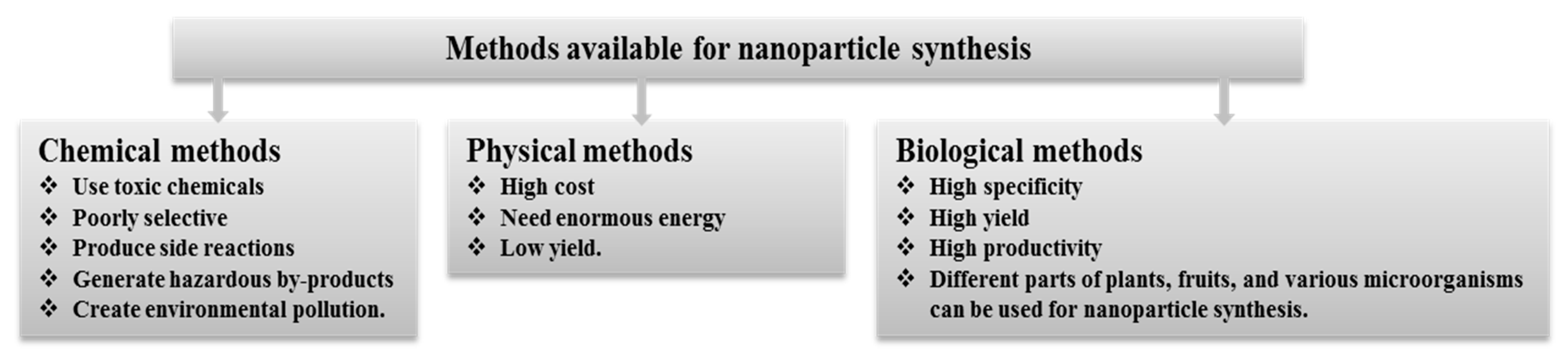
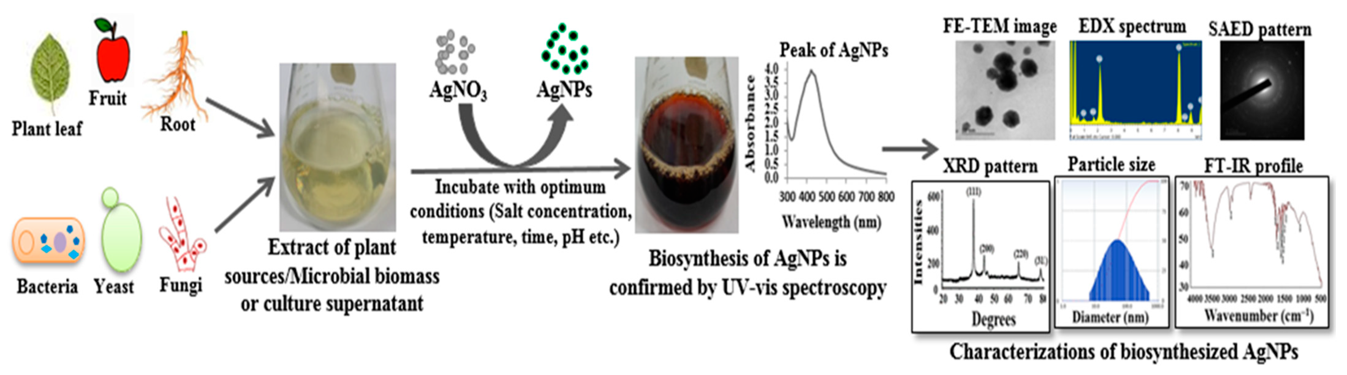
2. Green Synthesis of AgNPs
3. Plant Mediated Synthesis of AgNPs
4. Microbe Mediated Synthesis of AgNPs
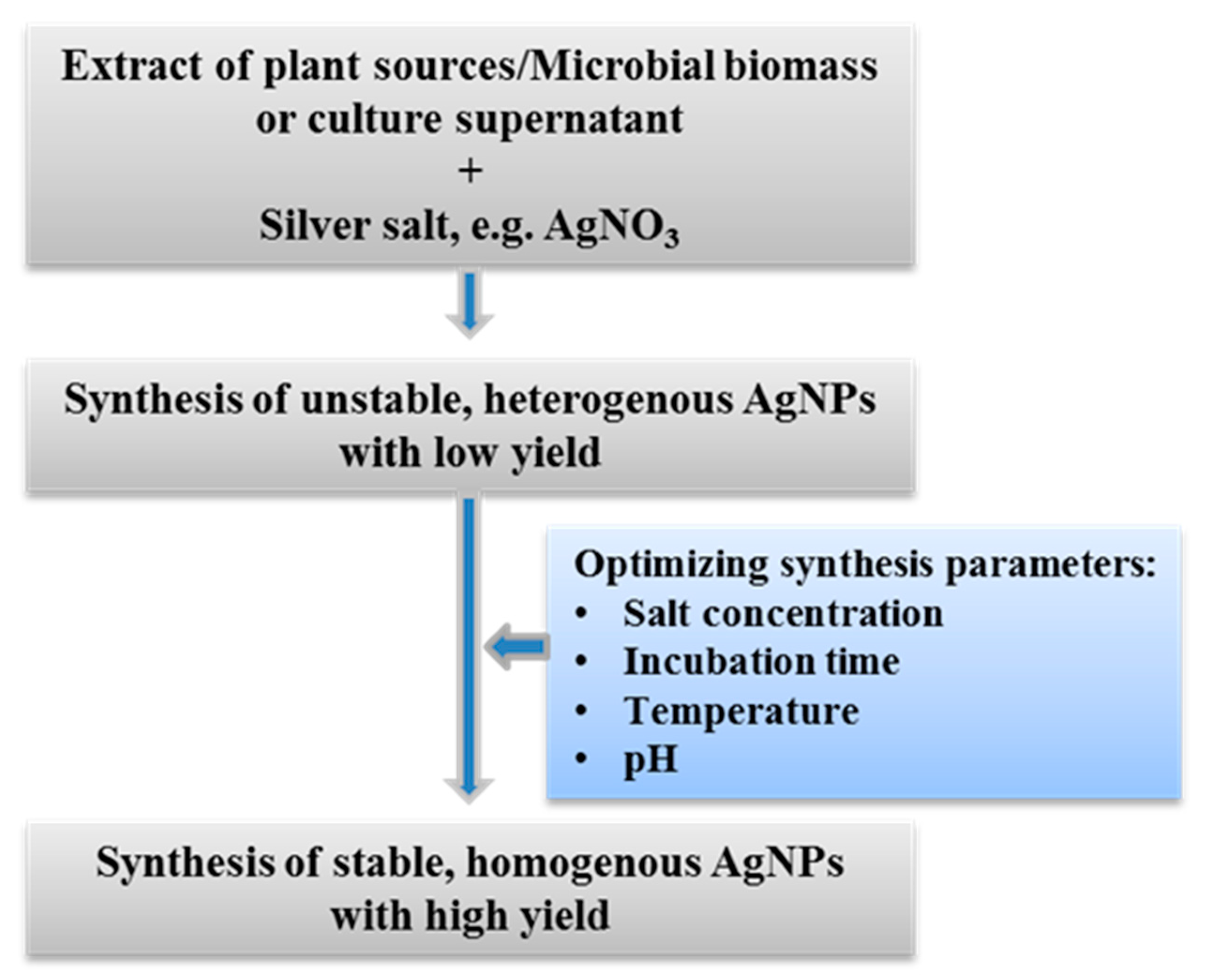
5. Critical Parameters for Rapid, Facile, and Stable Synthesis of AgNPs
6. Characterization of Green Synthesized AgNPs
7. Antibacterial Application of Green Synthesized AgNPs
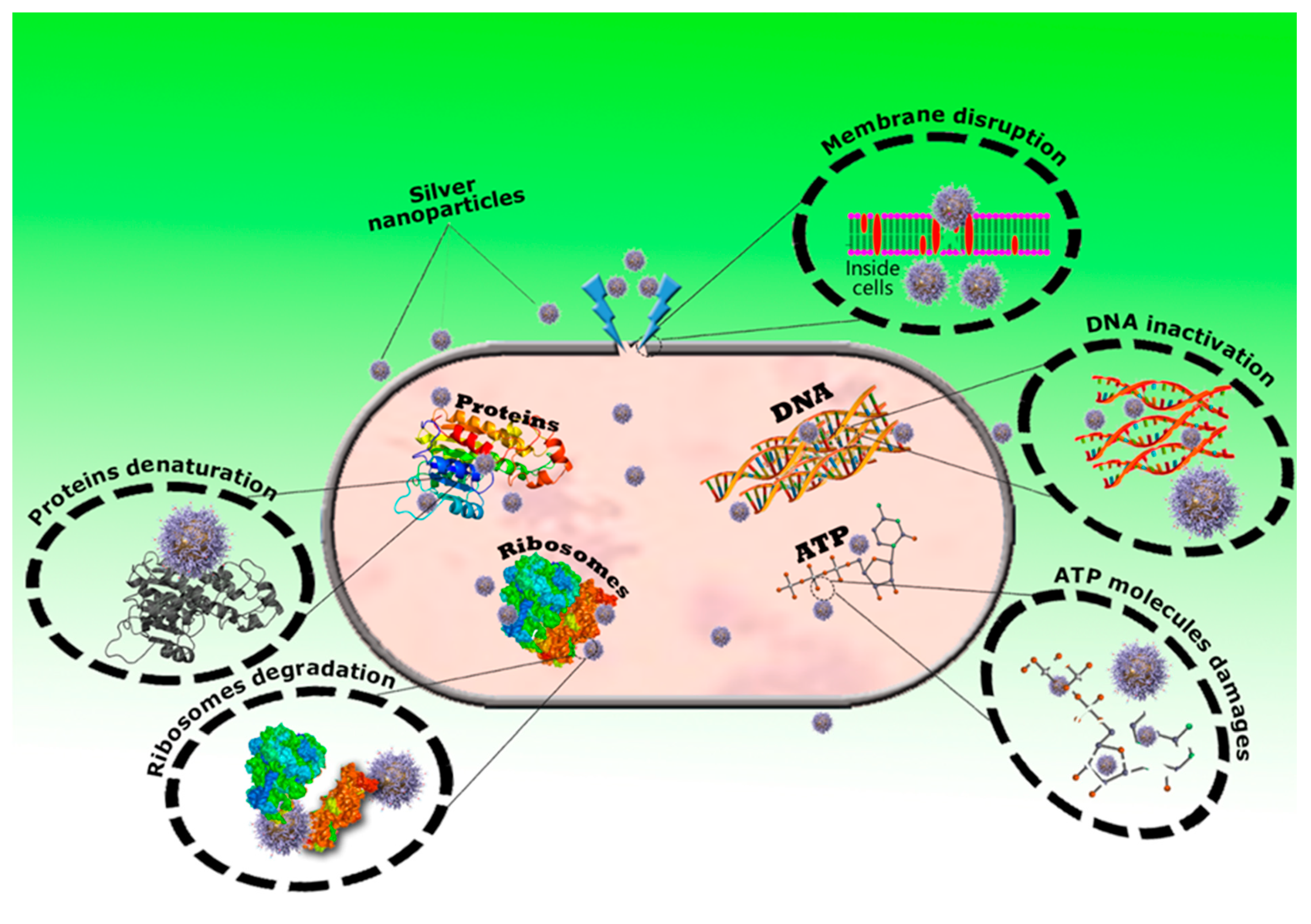
8. Antibacterial Mechanisms of AgNPs
9. Conclusions and Future Prospects
Author Contributions
Funding
Institutional Review Board Statement
Informed Consent Statement
Data Availability Statement
Conflicts of Interest
References
- Bachheti, R.K.; Fikadu, A.; Bachheti, A.; Husen, A. Biogenic fabrication of nanomaterials from flower-based chemical compounds, characterization and their various applications: A review. Saudi J. Biol. Sci. 2020, 27, 2551–2562. [Google Scholar] [CrossRef] [PubMed]
- Akter, S.; Huq, M.A. Biologically rapid synthesis of silver nanoparticles by Sphingobium sp. MAH-11 T and their antibacterial activity and mechanisms investigation against drug-resistant pathogenic microbes. Artif. Cells Nanomed. Biotechnol. 2020, 48, 672–682. [Google Scholar] [CrossRef] [PubMed] [Green Version]
- Hamouda, R.A.; Hussein, M.H.; Abo-Elmagd, R.A.; Bawazir, S.S. Synthesis and biological characterization of silver nanoparticles derived from the cyanobacterium Oscillatoria limnetica. Sci. Rep. 2019, 9, 13071. [Google Scholar] [CrossRef]
- Kulkarni, N.; Muddapur, U. Biosynthesis of metal nanoparticles: A review. J. Nanotechnol. 2014, 2014, 510246. [Google Scholar] [CrossRef] [Green Version]
- Jamkhande, P.G.; Ghule, N.W.; Bamer, A.H.; Kalaskar, M.G. Metal nanoparticles synthesis: An overview on methods of preparation, advantages and disadvantages, and applications. J. Drug Deliv. Sci. Technol. 2019, 53, 101174. [Google Scholar] [CrossRef]
- Huq, M.A. Green synthesis of silver nanoparticles using Pseudoduganella eburnea MAHUQ-39 and their antimicrobial mechanisms investigation against drug resistant human pathogens. Int. J. Mol. Sci. 2020, 21, 1510. [Google Scholar] [CrossRef] [Green Version]
- Kedi, P.B.E.; Meva, F.E.; Kotsedi, L. Eco-friendly synthesis, characterization, in vitro and in vivo anti-inflammatory activity of silver nanoparticle-mediated Selaginella myosurus aqueous extract. Int. J. Nanomed. 2018, 13, 8537–8548. [Google Scholar] [CrossRef] [PubMed] [Green Version]
- El-Naggar, N.E.; Hussein, M.H.; El-Sawah, A.A. Bio-fabrication of silver nanoparticles by phycocyanin, characterization, in vitro anticancer activity against breast cancer cell line and in vivo cytotoxicity. Sci. Rep. 2017, 7, 10844. [Google Scholar] [CrossRef] [Green Version]
- Fouda, A.; Abdel-Maksoud, G.; Abdel-Rahman, M.A.; Eid, A.M.; Barghoth, M.G.; El-Sadany, M.A.H. Monitoring the effect of biosynthesized nanoparticles against biodeterioration of cellulose-based materials by Aspergillus niger. Cellulose 2019, 26, 6583–6597. [Google Scholar] [CrossRef]
- Burdusel, A.C.; Gherasim, O.; Grumezescu, A.M.; Mogoanta, L.; Ficai, A.; Andronescu, E. Biomedical applications of silver nanoparticles: An up-to-date overview. Nanomaterials 2018, 8, 681. [Google Scholar] [CrossRef] [Green Version]
- Rafique, M.; Sadaf, I.; Rafique, M.S.; Tahir, M.B. A review on green synthesis of silver nanoparticles and their applications. Artif. Cells Nanomed. Biotechnol. 2017, 45, 1272–1291. [Google Scholar] [CrossRef] [PubMed]
- Huq, M.A.; Akter, S. Bacterial mediated rapid and facile synthesis of silver nanoparticles and their antimicrobial efficacy against pathogenic microorganisms. Materials 2021, 14, 2615. [Google Scholar] [CrossRef] [PubMed]
- Salem, S.S.; Fouda, A. Green synthesis of metallic nanoparticles and their prospective biotechnological applications: An overview. Biol. Trace Elem. Res. 2021, 199, 344–370. [Google Scholar] [CrossRef] [PubMed]
- Sharma, A.K.; Kaith, B.S.; Shanker, U.; Gupta, B. γ-radiation induced synthesis of antibacterial silver nanocomposite scaffolds derived from natural gum Boswellia serrata. J. Drug Deliv. Sci. Technol. 2020, 56, 101550. [Google Scholar] [CrossRef]
- Elemike, E.E.; Onwudiwe, D.C.; Fayemi, O.E.; Botha, T.L. Green synthesis and electrochemistry of Ag, Au, and Ag–Au bimetallic nanoparticles using golden rod (Solidago canadensis) leaf extract. Appl. Phys. A Mater. Sci. Process. 2019, 125, 42. [Google Scholar] [CrossRef]
- Dos Santos, M.A.; Paterno, L.G.; Moreira, S.G.C.; Sales, M.J.A. Original photochemical synthesis of Ag nanoparticles mediated by potato starch. SN Appl. Sci. 2019, 1, 554. [Google Scholar] [CrossRef] [Green Version]
- Guzman, M.G.; Dille, J.; Godet, S. Synthesis of silver nanoparticles by chemical reduction method and their antibacterial activity. Int. J. Chem. Biomol. Eng. 2009, 2, 104–111. [Google Scholar]
- Pauzi, N.; Zain, N.M.; Yusof, N.A.A. Microwave-assisted synthesis of ZnO nanoparticles stabilized with Gum Arabic: Effect of microwave irradiation time on ZnO nanoparticles size and morphology. Bull. Chem. React. Eng. Catal. 2019, 14, 182. [Google Scholar] [CrossRef] [Green Version]
- Iravani, S.; Korbekandi, H.; Mirmohammadi, S.V.; Zolfaghari, B. Synthesis of silver nanoparticles: Chemical, physical and biological methods. Res. Pharm. Sci. 2014, 9, 385–406. [Google Scholar]
- Akter, S.; Lee, S.-Y.; Siddiqi, M.Z.; Balusamy, S.R.; Ashrafudoulla, M.; Rupa, E.J.; Huq, M.A. Ecofriendly synthesis of silver nanoparticles by Terrabacter humi sp. nov. and their antibacterial application against antibiotic-resistant pathogens. Int. J. Mol. Sci. 2020, 21, 9746. [Google Scholar] [CrossRef]
- Du, J.; Sing, H.; Yi, T.H. Antibacterial, anti-biofilm and anticancer potentials of green synthesized silver nanoparticles using benzoin gum (Styrax benzoin) extract. Bioprocess Biosyst. Eng. 2016, 39, 1923–1931. [Google Scholar] [CrossRef] [PubMed]
- Vigneshwaran, N.; Kathe, A.A.; Varadarajan, P.V. Silve-protein (core-shell) nanoparticle production using spent mushroom substrate. Langmuir 2007, 23, 7113–7117. [Google Scholar] [CrossRef] [PubMed]
- Huq, M.A. Paenibacillus anseongense sp. nov. a silver nanoparticle producing bacterium isolated from rhizospheric soil. Curr. Microbiol. 2020, 77, 2023–2030. [Google Scholar] [CrossRef] [PubMed]
- Singh, P.; Kim, Y.J.; Zhang, D.; Yang, D.C. Biological synthesis of nanoparticles from plants and microorganisms. Trends Biotechnol. 2016, 34, 588–599. [Google Scholar] [CrossRef] [PubMed]
- Sukweenadhi, J.; Setiawan, K.I.; Avanti, C.; Kartini, K.; Rupa, E.J.; Yang, D.C. Scale-up of green synthesis and characterization of silver nanoparticles using ethanol extract of Plantago major L. leaf and its antibacterial potential. S. Afr. J. Chem. Eng. 2021, 38, 1–8. [Google Scholar] [CrossRef]
- Nahar, K.; Yang, D.C.; Rupa, E.J.; Khatun, M.K.; Al-Reza, S.M. Eco-friendly synthesis of silver nanoparticles from Clerodendrum viscosum leaf extract and its antibacterial potential. Nanomed. Res. J. 2020, 5, 276–287. [Google Scholar]
- Pawar, J.S.; Patil, R.H. Green synthesis of silver nanoparticles using Eulophia herbacea (Lindl.) tuber extract and evaluation of its biological and catalytic activity. SN Appl. Sci. 2020, 2, 52. [Google Scholar] [CrossRef] [Green Version]
- Soshnikova, V.; Kim, Y.J.; Singh, P.; Huo, Y.; Markus, J.; Ahn, S.; Castro-Aceituno, V.; Kang, J.; Chokkalingam, M.; Mathiyalagan, R.; et al. Cardamom fruits as a green resource for facile synthesis of gold and silver nanoparticles and their biological applications. Artif. Cells Nanomed. Biotechnol. 2018, 46, 108–117. [Google Scholar] [CrossRef] [Green Version]
- Sumitha, S.; Vasanthi, S.; Shalini, S.; Chinni, S.V.; Gopinath, S.C.B.; Anbu, P.; Bahari, M.B.B.; Harish, R.; Kathiresan, S.; Ravichandran, V. Phyto-mediated photo catalysed green synthesis of silver nanoparticles using Durio Zibethinus seed extract: Antimicrobial and cytotoxic activity and photocatalytic applications. Molecules 2018, 23, 3311. [Google Scholar] [CrossRef] [Green Version]
- Arokiyaraj, S.; Vincent, S.; Saravanan, M.; Lee, Y.; Oh, Y.K.; Kim, K.H. Green synthesis of silver nanoparticles using Rheum palmatum root extract and their antibacterial activity against Staphylococcus aureus and Pseudomonas aeruginosa. Artif. Cells Nanomed. Biotechnol. 2017, 45, 372–379. [Google Scholar] [CrossRef] [Green Version]
- Sharma, K.; Kaushik, S.; Jyoti, A. Green Synthesis of Silver Nanoparticles by Using Waste Vegetable Peel and its Antibacterial Activities. J. Pharm. Sci. Res. 2016, 8, 313–316. [Google Scholar]
- Singh, P.; Kim, Y.J.; Wang, C.; Mathiyalagan, R.; Yang, D.C. The development of a green approach for the biosynthesis of silver and gold nanoparticles by using Panax ginseng root extract, and their biological applications. Artif. Cells Nanomed. Biotechnol. 2015, 44, 1150–1157. [Google Scholar] [PubMed]
- Singh, P.; Kim, Y.J.; Yang, D.C. A strategic approach for rapid synthesis of gold and silver nanoparticles by Panax ginseng leaves. Artif. Cells Nanomed. Biotechnol. 2016, 44, 1949–1957. [Google Scholar] [CrossRef] [PubMed] [Green Version]
- Adeyemi, D.K.; Adeluola, A.O.; Akinbile, M.J.; Johnson, O.O.; Ayoola, G.A. Green synthesis of Ag, Zn and Cu nanoparticles from aqueous extract of Spondias mombin leaves and evaluation of their antibacterial activity. Afr. J. Clin. Exper. Microbiol. 2020, 21, 106–113. [Google Scholar] [CrossRef]
- Ssekatawa, K.; Byarugaba, D.K.; Kato, C.D.; Wampande, E.M.; Ejobi, F.; Nakavuma, J.L.; Maaza, M.; Sackey, J.; Nxumalo, E.; Kirabira, J.B. Green strategy-based synthesis of silver nanoparticles for antibacterial applications. Front. Nanotechnol. 2021, 3, 697303. [Google Scholar] [CrossRef]
- Ahmed, S.R.; Anwar, H.; Ahmed, S.W.; Shah, M.R.; Ahmed, A.; Abid Ali, S.A. Green synthesis of silver nanoparticles: Antimicrobial potential and chemosensing of a mutagenic drug nitrofurazone in real samples. Measurement 2021, 180, 109489. [Google Scholar] [CrossRef]
- Urnukhsaikhan, E.; Bold, B.E.; Gunbileg, A.; Sukhbaatar, N.; Mishig-Ochir, T. Antibacterial activity and characteristics of silver nanoparticles biosynthesized from Carduus crispus. Sci. Rep. 2021, 26, 21047. [Google Scholar] [CrossRef]
- Aabed, K.; Mohammed, A.E. Synergistic and antagonistic effects of biogenic silver nanoparticles in combination with antibiotics against some pathogenic microbes. Front. Bioeng. Biotechnol. 2021, 9, 249. [Google Scholar] [CrossRef]
- Madivoli, E.S.; Kareru, P.G.; Gachanja, A.N.; Mugo, S.M.; Makhanu, D.S.; Wanakai, S.I.; Gavamukulya, Y. Facile synthesis of silver nanoparticles using Lantana trifolia aqueous extracts and their antibacterial activity. J. Inorg. Organomet. Polym. 2020, 30, 2842–2850. [Google Scholar] [CrossRef]
- Chavan, R.R.; Bhinge, S.D.; Bhutkar, M.A.; Randive, D.S.; Wadkar, G.H.; Todkar, S.S. Characterization, antioxidant, antimicrobial and cytotoxic activities of green synthesized silver and iron nanoparticles using alcoholic Blumea eriantha DC plant extract. Urade. Mater. Today Commun. 2020, 24, 101320. [Google Scholar] [CrossRef]
- Hemlata; Meena, P.R.; Singh, A.P.; Tejavath, K.K. Biosynthesis of silver nanoparticles using Cucumis prophetarum aqueous leaf extract and their antibacterial and antiproliferative activity against cancer cell lines. ACS Omega 2020, 5, 5520–5528. [Google Scholar] [CrossRef] [Green Version]
- Shejawal, K.P.; Randive, D.S.; Bhinge, S.D.; Bhutkar, M.A.; Wadkar, G.H.; Jadhav, N.R. Green synthesis of silver and iron nanoparticles of isolated proanthocyanidin: Its characterization, antioxidant, antimicrobial, and cytotoxic activities against COLO320DM and HT. J. Genet. Eng. Biotechnol. 2020, 18, 43. [Google Scholar] [CrossRef] [PubMed]
- Kalpana, D.; Han, J.H.; Park, W.S.; Lee, S.M.; Wahab, R.; Lee, Y.S. Green biosynthesis of silver nanoparticles using Torreya nucifera and their antibacterial activity. Arab. J. Chem. 2019, 12, 1722–1732. [Google Scholar] [CrossRef] [Green Version]
- Huang, F.; Long, Y.; Liang, Q.; Purushotham, B.; Swamy, M.K.; Duan, Y. Safed musli (Chlorophytum borivilianum L.) callus-mediated biosynthesis of silver nanoparticles and evaluation of their antimicrobial activity and cytotoxicity against human colon cancer cells. J. Nanomater. 2019, 2019, 2418785. [Google Scholar] [CrossRef]
- Hasnain, M.S.; Javed, N.; Alam, S.; Rishishwar, P.; Rishishwar, S.; Ali, S.; Nayak, A.K.; Beg, S. Purple heart plant leaves extract-mediated silver nanoparticle synthesis: Optimization by Box-Behnken design. Mater. Sci. Eng. C 2019, 99, 1105–1114. [Google Scholar] [CrossRef]
- Oves, M.; Aslam, M.; Rauf, M.A.; Qayyum, S.; Qari, H.A.; Khan, M.S. Antimicrobial and anticancer activities of silver nanoparticles synthesized from the root hair extract of Phoenix dactylifera. Mater. Sci. Eng. C 2018, 89, 429–443. [Google Scholar] [CrossRef] [PubMed]
- Saratale, R.G.; Benelli, G.; Kumar, G.; Kim, D.S.; Saratale, G.D. Bio-Fabrication of silver nanoparticles using the leaf extract of an ancient herbal medicine, dandelion (Taraxacum officinale), evaluation of their antioxidant, anticancer potential, and antimicrobial activity against phytopathogens. Environ. Sci. Pollut. Res. 2017, 25, 10392–10406. [Google Scholar] [CrossRef] [PubMed]
- Khatami, M.; Zafarnia, N.; Heydarpoor Bami, M.; Sharifi, I.; Singh, H. Antifungal and antibacterial activity of densely dispersed silver nanospheres with homogeneity size which synthesized using chicory: An in vitro study. J. Mycol. Med. 2018, 28, 637–644. [Google Scholar] [CrossRef]
- Devanesan, S.; AlSalhi, M.S.; Balaji, R.V.; Ranjitsingh, A.J.A.; Ahamed, A.; Alfuraydi, A.A.; AlQahtani, F.Y.; Aleanizy, F.S.; Othman, A.H. Antimicrobial and cytotoxicity effects of synthesized silver nanoparticles from Punica granatum peel extract. Nanoscale Res. Lett. 2018, 13, 315. [Google Scholar] [CrossRef] [Green Version]
- Mythili, R.; Selvankumar, T.; Kamala-Kannan, S.; Sudhakar, C.; Ameen, F.; Al-Sabri, A.; Selvam, K.; Govarthanan, M.; Kim, H. Utilization of market vegetable waste for silver nanoparticle synthesis and its antibacterial activity. Mater. Lett. 2018, 225, 101–104. [Google Scholar] [CrossRef]
- Markus, J.; Wang, D.; Kim, Y.J.; Ahn, S.; Mathiyalagan, R.; Wang, C.; Yang, D.C. Biosynthesis, characterization, and bioactivities evaluation of silver and gold nanoparticles mediated by the roots of Chinese Herbal Angelica pubescens maxim. Nanoscale Res. Lett. 2017, 12, 46. [Google Scholar] [CrossRef] [PubMed] [Green Version]
- Mohanta, Y.K.; Panda, S.K.; Bastia, A.K.; Mohanta, T.K. Biosynthesis of silver nanoparticles from Protium serratum and investigation of their potential impacts on food safety and control. Front. Microbiol. 2017, 8, 626. [Google Scholar] [CrossRef] [Green Version]
- Huo, Y.; Singh, P.; Kim, Y.J.; Soshnikova, V.; Kang, J.; Markus, J.; Ahn, S.; Castro-Aceituno, V.; Mathiyalagan, R.; Chokkalingam, M.; et al. Biological synthesis of gold and silver chloride nanoparticles by Glycyrrhiza uralensis and in vitro applications. Artif. Cells Nanomed. Biotechnol. 2018, 46, 303–312. [Google Scholar] [CrossRef] [PubMed] [Green Version]
- Nasar, S.; Murtaza, G.; Mehmood, A.; Bhatti, T.M. Green approach to synthesis of silver nanoparticles using Ficus Palmata leaf extract and their antibacterial profile. Pharm. Chem. J. 2017, 51, 811–817. [Google Scholar] [CrossRef]
- Rajkuberan, C.; Prabukumar, S.; Sathishkumar, G.; Wilson, A.; Ravindran, K.; Sivaramakrishnan, S. Facile synthesis of silver nanoparticles using Euphorbia antiquorum L. latex extract and evaluation of their biomedical perspectives as anticancer agents. J. Saudi Chem. Soc. 2017, 21, 911–919. [Google Scholar] [CrossRef] [Green Version]
- Jain, S.; Mehata, M.S. Medicinal plant leaf extract and pure flavonoid mediated green synthesis of silver nanoparticles and their enhanced antibacterial property. Sci. Rep. 2017, 7, 15867. [Google Scholar] [CrossRef]
- Mitiku, A.A.; Yilma, B. Antibacterial and antioxidant activity of silver nanoparticles synthesized using aqueous extract of Moringa stenopetala leaves. Afr. J. Biotechnol. 2017, 16, 1705–1716. [Google Scholar]
- Singh, H.; Du, J.; Singh, P.; Yi, T.H. Ecofriendly synthesis of silver and gold nanoparticles by Euphrasia officinalis leaf extract and its biomedical applications. Artif. Cells Nanomed. Biotechnol. 2017, 46, 1163–1170. [Google Scholar] [CrossRef] [Green Version]
- Abbai, R.; Ramya Mathiyalagan, J.M.; Kim, Y.J.; Wang, C.; Singh, P.; Ahn, S.; Farh, M.E.A.; Yang, D.C. Green synthesis of multifunctional silver and gold nanoparticles from the oriental herbal adaptogen: Siberian ginseng. Int. J. Nanomed. 2016, 11, 3131–3143. [Google Scholar]
- Singh, H.; Du, J.; Yi, T.H. Green and rapid synthesis of silver nanoparticles using Borago officinalis leaf extract: Anticancer and antibacterial activities. Artif. Cells Nanomed. Biotechnol. 2017, 45, 1310–1316. [Google Scholar] [CrossRef] [Green Version]
- Lateef, A.; Azeez, M.A.; Asafa, T.B.; Yekeen, T.A.; Akinboro, A.; Oladipo, I.C.; Azeez, L.; Ojo, S.A.; Gueguim-Kana, E.B.; Beukes, L.S. Cocoa pod husk extract-mediated biosynthesis of silver nanoparticles: Its antimicrobial, antioxidant and larvicidal activities. J. Nanostruct. Chem. 2016, 6, 159–169. [Google Scholar] [CrossRef] [Green Version]
- Ahmed, S.; Saifullah; Ahmad, M.; Swami, B.L.; Ikram, S. Green synthesis of silver nanoparticles using Azadirachta indica aqueous leaf extract. J. Radiat. Res. Appl. Sci. 2016, 9, 1–7. [Google Scholar] [CrossRef] [Green Version]
- Anandalakshmi, K.; Venugobal, J.; Ramasamy, V. Characterization of silver nanoparticles by green synthesis method using Pedalium murex leaf extract and their antibacterial activity. Appl. Nanosci. 2016, 6, 399–408. [Google Scholar] [CrossRef] [Green Version]
- Mohanta, Y.K.; Panda, S.K.; Biswas, K.; Tamang, A.; Bandyopadhyay, J.; Bastia, A.K. Biogenic synthesis of silver nanoparticles from Cassia fistula (Linn.): In vitro assessment of their antioxidant, antimicrobial and cytotoxic activities. IET Nanobiotechnol. 2016, 10, 438–444. [Google Scholar] [CrossRef] [PubMed]
- Bose, D.; Chatterjee, S. Biogenic synthesis of silver nanoparticles using guava (Psidium guajava) leaf extract and its antibacterial activity against Pseudomonas aeruginosa. Appl. Nanosci. 2016, 6, 895–901. [Google Scholar] [CrossRef] [Green Version]
- Dhand, V.; Soumya, L.; Bharadwaj, S.; Chakra, S.; Bhatt, D.; Sreedhar, B. Green synthesis of silver nanoparticles using Coffea arabica seed extract and its antibacterial activity. Mater. Sci. Eng. C 2016, 58, 36–43. [Google Scholar] [CrossRef]
- Sundararajan, B.; Mahendran, G.; Thamaraiselvi, R.; Kumari, B.D. Biological activities of synthesized silver nanoparticles from Cardiospermum halicacabum L. Bull. Mater. Sci. 2016, 39, 423–431. [Google Scholar]
- Ravichandran, V.; Vasanthi, S.; Shalini, S.; Ali-Shah, S.A.; Harish, R. Green synthesis of silver nanoparticles using Atrocarpus altilis leaf extract and the study of their antimicrobial and antioxidant activity. Mater. Lett. 2016, 180, 264–267. [Google Scholar] [CrossRef]
- Nayak, D.; Ashe, S.; Rauta, P.R.; Kumari, M.; Nayak, B. Bark extract mediated green synthesis of silver nanoparticles: Evaluation of antimicrobial activity and antiproliferative response against osteosarcoma. Mater. Sci. Eng. C 2016, 58, 44–52. [Google Scholar] [CrossRef]
- Oluwaniyi, O.O.; Adegoke, H.I.; Adesuji, E.T.; Alabi, A.B.; Bodede, S.O.; Labulo, A.H.; Oseghale, C.O. Biosynthesis of silver nanoparticles using aqueous leaf extract of Thevetia peruviana Juss and its antimicrobial activities. Appl. Nanosci. 2016, 6, 903–912. [Google Scholar] [CrossRef]
- Benakashania, F.; Allafchian, A.R.; Jalali, S.A.H. Biosynthesis of silver nanoparticles using Capparis spinosa L. leaf extract and their antibacterial activity. Karbala Int. J. Mod. Sci. 2016, 2, 251–258. [Google Scholar] [CrossRef] [Green Version]
- Mittal, A.K.; Tripathy, D.; Choudhary, A.; Aili, P.K.; Chatterjee, A.; Singh, I.P.; Banerjee, U.C. Bio-synthesis of silver nanoparticles using potentilla fulgens wall. ex Hook. and its therapeutic evaluation as anticancer and antimicrobial agent. Mater. Sci. Eng. C 2015, 53, 120–127. [Google Scholar] [CrossRef] [PubMed]
- Roy, K.; Sarkar, C.; Ghosh, C. Plant-mediated synthesis of silver nanoparticles using parsley (Petroselinum crispum) leaf extract: Spectral analysis of the particles and antibacterial study. Appl. Nanosci. 2015, 5, 945–951. [Google Scholar] [CrossRef] [Green Version]
- Ali, K.; Ahmed, B.; Dwivedi, S.; Saquib, Q.; Al-Khedhairy, A.A.; Musarrat, J. Microwave accelerated green synthesis of stable silver nanoparticles with Eucalyptus globulus leaf extract and their antibacterial and antibiofilm activity on clinical isolates. PLoS ONE 2015, 10, e0131178. [Google Scholar] [CrossRef]
- Ibrahim, H.M. Green synthesis and characterization of silver nanoparticles using banana peel extract and their antimicrobial activity against representative microorganisms. J. Radiat. Res. Appl. Sc. 2015, 8, 265–275. [Google Scholar] [CrossRef] [Green Version]
- Shalaby, T.I.; Mahmoud, O.A.; Batouti, G.A.E.; Ibrahim, E.E. Green synthesis of silver nanoparticles: Synthesis, characterization and antibacterial activity. Nanosci. Nanotechnol. 2015, 5, 23–29. [Google Scholar]
- Sre, P.R.; Reka, M.; Poovazhagi, R.; Kumar, M.A.; Murugesan, K. Antibacterial and cytotoxic effect of biologically synthesized silver nanoparticles using aqueous root extract of Erythrina indica lam. Spectrochim. Acta Part A Mol. Biomol. Spectrosc. 2015, 135, 1137–1144. [Google Scholar]
- Miri, A.; Sarani, M.; Bazaz, M.R.; Darroudi, M. Plant-mediated biosynthesis of silver nanoparticles using Prosopis farcta extract and its antibacterial properties. Spectrochim. Acta A Mol. Biomol. Spectrosc. 2015, 141, 287–291. [Google Scholar] [CrossRef]
- Balashanmugam, P.; Kalaichelvan, P.T. Biosynthesis characterization of silver nanoparticles using Cassia roxburghii DC. aqueous extract, and coated on cotton cloth for effective antibacterial activity. Int. J. Nanomed. 2015, 10, 87. [Google Scholar] [CrossRef] [Green Version]
- Rajakannu, S.; Shankar, S.; Perumal, S.; Subramanian, S.; Dhakshinamoorthy, G.P. Biosynthesis of silver nanoparticles using Garcinia mangostana fruit extract and their antibacterial, antioxidant activity. Int. J. Curr. Microbiol. Appl. Sci. 2015, 4, 944–952. [Google Scholar]
- Krithiga, N.; Rajalakshmi, A.; Jayachitra, A. Green synthesis of silver nanoparticles using leaf extracts of Clitoria ternatea and Solanum nigrum and study of its antibacterial effect against common nosocomial pathogens. J. Nanosci. 2015, 8, 928204. [Google Scholar] [CrossRef] [Green Version]
- Harshiny, M.; Matheswaran, M.; Arthanareeswaran, G.; Kumaran, S.; Rajasree, S. Enhancement of antibacterial properties of silver nanoparticles–ceftriaxone conjugate through Mukia maderaspatana leaf extract mediated synthesis. Ecotoxicol. Environ. Saf. 2015, 121, 135–141. [Google Scholar] [CrossRef] [PubMed]
- Ahmed, S.; Ikram, S. Silver nanoparticles: One pot green synthesis using Terminalia arjuna extract for biological application. Nano Res. Appl. 2015, 1, 309. [Google Scholar]
- Premasudha, P.; Venkataramana, M.; Abirami, M.; Vanathi, P.; Krishna, K.; Rajendran, R. Biological synthesis and characterization of silver nanoparticles using Eclipta alba leaf extract and evaluation of its cytotoxic and antimicrobial Potential. Bull. Mater. Sci. 2015, 38, 965–973. [Google Scholar] [CrossRef] [Green Version]
- Kumar, D.A.; Palanichamy, V.; Roopan, S.M. Green synthesis of silver nanoparticles using Alternanthera dentata leaf extract at room temperature and their antimicrobial activity. Spectrochim. Acta A Mol. Biomol. Spectrosc. 2014, 127, 168–171. [Google Scholar] [CrossRef]
- Muniyappan, N.; Nagarajan, N.S. Green synthesis of silver nanoparticles with Dalbergia spinosa leaves and their applications in biological and catalytic activities. Process Biochem. 2014, 49, 1054–1061. [Google Scholar] [CrossRef]
- Khan, M.; Khan, S.T.; Khan, M.; Adil, S.F.; Musarrat, J.; Al-Khedhairy, A.A.; Al-Warthan, A.; Siddiqui, M.; Alkhathlan, H.Z. Antibacterial properties of silver nanoparticles synthesized using Pulicaria glutinosa plant extract as a green bioreductant. Int. J. Nanomed. 2014, 9, 3551–3565. [Google Scholar]
- Singh, K.; Panghal, M.; Kadyan, S.; Chaudhary, U.; Yadav, J.P. Green silver nanoparticles of Phyllanthus amarus: As an antibacterial agent against multi drug resistant clinical isolates of Pseudomonas aeruginosa. J. Nanobiotechnol. 2014, 12, 40. [Google Scholar] [CrossRef] [Green Version]
- Raut, R.W.; Mendhulkar, V.D.; Kashid, S.B. Photosensitized synthesis of silver nanoparticles using Withania somnifera leaf powder and silver nitrate, J. Photochem. Photobiol. B 2014, 132, 45–55. [Google Scholar] [CrossRef]
- Nakkala, J.R.; Mata, R.; Gupta, A.K.; Sadras, S.R. Biological activities of green silver nanoparticles synthesized with Acorous calamus rhizome extract, Eur. J. Med. Chem. 2014, 85, 784–794. [Google Scholar] [CrossRef]
- Mariselvam, R.; Ranjitsingh, A.; Nanthini, A.U.R.; Kalirajan, K.; Padmalatha, C.; Selvakumar, P.M. Green synthesis of silver nanoparticles from the extract of the inflorescence of Cocos nucifera (Family: Arecaceae) for enhanced antibacterial activity. Spectrochim. Acta Part A Mol. Biomol. Spectrosc. 2014, 129, 537–541. [Google Scholar] [CrossRef] [PubMed]
- Kumar, P.V.; Pammi, S.; Kollu, P.; Satyanarayana, K.; Shameem, U. Green synthesis and characterization of silver nanoparticles using Boerhaavia diffusa plant extract and their anti bacterial activity. Ind. Crops Prod. 2014, 52, 562–566. [Google Scholar] [CrossRef]
- Nazeruddin, G.M.; Prasad, N.R.; Waghmare, S.R.; Garadkar, K.M.; Mulla, I.S. Extracellular biosynthesis of silver nanoparticle using Azadirachta indica leaf extract and its anti-microbial activity. J. Alloy. Compd. 2014, 583, 272–277. [Google Scholar] [CrossRef]
- Nazeruddin, G.M.; Prasad, N.R.; Prasadd, S.R.; Shaikha, Y.I.; Waghmare, S.R.; Adhyapak, P. Coriandrum sativum seed extract assisted in situ green synthesis ofsilver nanoparticle and its anti-microbial activity. Ind. Crops Prod. 2014, 60, 212–216. [Google Scholar] [CrossRef]
- Bindhu, M.R.; Umadevi, M. Synthesis of monodispersed silver nanoparticles using Hibiscus cannabinus leaf extract and its antimicrobial activity. Spectrochim. Acta A Mol. Biomol. Spectrosc. 2013, 101, 184–190. [Google Scholar] [CrossRef]
- Patil, R.S.; Kokate, M.R.; Kolekar, S.S. Bioinspired synthesis of highly stabilized silver nanoparticles using Ocimum tenuiflorum leaf extract and their antibacterial activity. Spectrochim. Acta A 2012, 91, 234–238. [Google Scholar] [CrossRef]
- Gopinath, V.; MubarakAli, D.; Priyadarshini, S.; Priyadharsshini, N.M.; Thajuddin, N.; Velusamy, P. Biosynthesis of silver nanoparticles from Tribulus terrestris and its antimicrobial activity: A novel biological approach. Colloids Surf. B Biointerfaces 2012, 96, 69–74. [Google Scholar] [CrossRef]
- Sivakumar, P.; Nethradevi, C.; Renganathan, S. Synthesis of silver nanoparticles using Lantana camara fruit extract and its effect on pathogens, Asian J. Pharm. Clin. Res. 2012, 5, 97–101. [Google Scholar]
- Sathishkumar, G.; Gobinatha, C.; Karpagama, K.; Hemamalini, V.; Premkumar, K.; Sivaramakrishnan, S. Phyto-synthesis of silver nanoscale particles using Morinda citrifolia L. and its inhibitory activity against human pathogens. Colloids Surf. B Biointerfaces 2012, 95, 235–240. [Google Scholar] [CrossRef]
- Kumar, K.M.; Sinha, M.; Mandal, B.K.; Ghosh, A.R.; Siva Kumar, K.; Sreedhara Reddy, P. Green synthesis of silver nanoparticles using Terminalia chebula extract at room temperature and their antimicrobial studies. Spectrochim. Acta A Mol. Biomol. Spectrosc. 2012, 91, 228–233. [Google Scholar] [CrossRef]
- Amin, M.; Anwar, F.; Janjua, M.R.S.A.; Iqbal, M.A.; Rashid, U. Green Synthesis of Silver Nanoparticles through Reduction with Solanum xanthocarpum L. Berry Extract: Characterization, Antimicrobial and Urease Inhibitory Activities against Helicobacter pylori. Int. J. Mol. Sci. 2012, 13, 9923–9941. [Google Scholar] [CrossRef] [PubMed]
- Ghosh, S.; Patil, S.; Ahire, M.; Kitture, R.; Jabgunde, A.; Kale, S.; Pardesi, K.; Cameotra, S.S.; Bellare, J.; Dhavale, D.D.; et al. Synthesis of silver nanoparticles using Dioscorea bulbifera tuber extract and evaluation of its synergistic potential in combination with antimicrobial agents. Int. J. Nanomed. 2012, 7, 483–496. [Google Scholar]
- Veerasamy, R.; Xin, T.Z.; Gunasagaran, S.; Xiang, T.F.W.; Yang, E.F.C.; Jeyakumar, N.; Dhanaraj, S.A. Biosynthesis of silver nanoparticles using mangosteen leaf extract and evaluation of their antimicrobial activities, J. Saudi Chem. Society. 2011, 15, 113–120. [Google Scholar] [CrossRef] [Green Version]
- Masurkar, S.A.; Chaudhari, P.R.; Shidore, V.B.; Kamble, S.P. Rapid biosynthesis of silver nanoparticles using Cymbopogan Citratus (Lemongrass) and its antimicrobial activity. Nano-Micro Lett. 2011, 3, 189–194. [Google Scholar] [CrossRef] [Green Version]
- Nabikhan, A.; Kandasamy, K.; Raj, A.; Alikunhi, N.M. Synthesis of antimicrobial silver nanoparticles by callus and leaf extracts from saltmarsh plant, Sesuvium portulacastrum L. Colloids Surf. B 2010, 79, 488–493. [Google Scholar] [CrossRef]
- Huq, M.A.; Akter, S. Biosynthesis, characterization and antibacterial application of novel silver nanoparticles against drug resistant pathogenic Klebsiella pneumoniae and Salmonella enteritidis. Molecules 2021, 26, 5996. [Google Scholar] [CrossRef] [PubMed]
- Eltarahony, M.; Ibrahim, A.; El-shall, H.; Ibrahim, E.; Althobaiti, F.; Fayad, E. Antibacterial, antifungal and antibiofilm activities of silver nanoparticles supported by crude bioactive metabolites of bionanofactories isolated from lake mariout. Molecules 2021, 26, 3027. [Google Scholar] [CrossRef]
- Singh, P.; Pandit, S.; Jers, C.; Abhayraj, S.; Garnæs, J.; Mijakovic, I. Silver nanoparticles produced from Cedecea sp. exhibit antibiofilm activity and remarkable stability. Sci. Rep. 2021, 11, 12619. [Google Scholar] [CrossRef]
- Huq, M.A.; Akter, S. Characterization and genome analysis of Arthrobacter bangladeshi sp. nov., applied for the green synthesis of silver nanoparticles and their antibacterial efficacy against drug-resistant human pathogens. Pharmaceutics 2021, 13, 1691. [Google Scholar] [CrossRef]
- Vellingiri, M.M.; Ashwin, J.K.M.; Soundari, A.J.P.G.; Sathiskumar, S.; Priyadharshini, U.; Paramasivam, D.; Liu, W.C.; Balasubramanian, B. Mycofabrication of AgONPs derived from Aspergillus terreus FC36AY1 and its potent antimicrobial, antioxidant, and anti-angiogenesis activities. Mol. Biol. Rep. 2021, 48, 7933–7946. [Google Scholar] [CrossRef]
- Soliman, A.M.; Abdel-Latif, W.; Shehata, I.H.; Fouda, A.; Abdo, A.M.; Ahmed, Y.M. Green approach to overcome the resistance pattern of Candida spp. using biosynthesized silver nanoparticles fabricated by Penicillium chrysogenum F9. Biol. Trace Elem. Res. 2021, 199, 800–811. [Google Scholar] [CrossRef] [PubMed]
- Raza, S.; Ansari, A.; Siddiqui, N.N.; Ibrahim, F.; Abro, M.I.; Aman, A. Biosynthesis of silver nanoparticles for the fabrication of non cytotoxic and antibacterial metallic polymer based nanocomposite system. Sci. Rep. 2021, 11, 10500. [Google Scholar] [CrossRef] [PubMed]
- Sreenivasa, N.; Meghashyama, B.P.; Pallavi, S.S.; Bidhayak, C.; Dattatraya, A.; Muthuraj, R.; Shashiraj, K.N.; Halaswamy, H.; Dhanyakumara, S.B.; Vaishnavi, M.D. Biogenic synthesis of silver nanoparticles using Paenibacillus sp. in-vitro and their antibacterial, anticancer activity assessment against human colon tumour cell line. J. Environ. Biol. 2021, 42, 118–127. [Google Scholar] [CrossRef]
- Huq, M.A. Biogenic silver nanoparticles synthesized by Lysinibacillus xylanilyticus MAHUQ-40 to control antibiotic-resistant human pathogens vibrio parahaemolyticus and salmonella typhimurium. Front. Bioeng. Biotechnol. 2020, 8, 1407. [Google Scholar] [CrossRef] [PubMed]
- Hamida, R.S.; Abdelmeguid, N.E.; Ali, M.A.; Bin-Meferij, M.M.; Khalil, M.I. Synthesis of silver nanoparticles using a novel cyanobacteria Desertifilum sp. extract: Their antibacterial and cytotoxicity effects. Int. J. Nanomed. 2020, 15, 49–63. [Google Scholar] [CrossRef] [Green Version]
- Borah, D.; Das, N.; Das, N.; Bhattacharjee, A.; Sarmah, P.; Ghosh, K.; Chandel, M.; Rout, J.; Pandey, P.; Ghosh, N.N.; et al. Alga-mediated facile green synthesis of silver nanoparticles: Photophysical, catalytic and antibacterial activity. Appl. Organ. Chem. 2020, 34, e5597. [Google Scholar] [CrossRef]
- Mondal, A.H.; Yadav, D.; Ali, A.; Khan, N.; Jin, J.O.; Haq, Q.M.R. Anti-bacterial and anti-candidal activity of silver nanoparticles biosynthesized using Citrobacter spp. Ms5 culture supernatant. Biomolecules 2020, 10, 944. [Google Scholar] [CrossRef]
- Bhuyar, P.; Rahim, M.H.A.; Sundararaju, S.; Ramaraj, R.; Maniam, G.P.; Govindan, N. Synthesis of silver nanoparticles using marine macroalgae Padina sp. and its antibacterial activity towards pathogenic bacteria. Beni-Suef Univ. J. Basic Appl. Sci. 2020, 9, 3. [Google Scholar] [CrossRef] [Green Version]
- Mishra, B.; Saxena, A.; Tiwari, A. Biosynthesis of silver nanoparticles from marine diatoms Chaetoceros sp., Skeletonema sp., Thalassiosira sp., and their antibacterial study. Biotechnol. Rep. 2020, 28, e00571. [Google Scholar] [CrossRef] [PubMed]
- Feroze, N.; Arshad, B.; Younas, M.; Afridi, M.I.; Saqib, S.; Ayaz, A. Fungal mediated synthesis of silver nanoparticles and evaluation of antibacterial activity. Microsc. Res. Tech. 2020, 83, 72–80. [Google Scholar] [CrossRef]
- Mohd-Yusof, H.; Rahman, A.; Mohamad, R.; Zaidan, U.H. Microbial mediated synthesis of silver nanoparticles by Lactobacillus Plantarum TA4 and its antibacterial and antioxidant activity. Appl. Sci. 2020, 10, 6973. [Google Scholar] [CrossRef]
- Saeed, S.; Iqbal, A.; Ashraf, M.A. Bacterial-mediated synthesis of silver nanoparticles and their significant effect against pathogens. Environ. Sci. Pollut. Res. 2020, 27, 37347–37356. [Google Scholar] [CrossRef] [PubMed]
- Alsamhary, K.I. Eco-friendly synthesis of silver nanoparticles by Bacillus subtilis and their antibacterial activity. Saudi J. Biol. Sci. 2020, 27, 2185–2191. [Google Scholar] [CrossRef] [PubMed]
- Shaker, M.A.; Shaaban, M.I. Synthesis of silver nanoparticles with antimicrobial and anti-adherence activities against multidrug-resistant isolates from Acinetobacter Baumannii. J. Taibah Univ. Med. Sci. 2017, 12, 291–297. [Google Scholar] [CrossRef]
- Singh, H.; Du, J.; Singh, P.; Yi, T.H. Extracellular synthesis of silver nanoparticles by Pseudomonas sp. THG-LS1.4 and their antimicrobial application. J. Pharm. Anal. 2018, 8, 258–264. [Google Scholar] [CrossRef]
- Du, J.; Sing, H.; Yi, T.H. Biosynthesis of silver nanoparticles by Novosphingobium sp. THG-C3 and their antimicrobial potential. Artif. Cells Nanomed. Biotechnol. 2017, 45, 211–217. [Google Scholar] [CrossRef] [Green Version]
- Singh, P.; Singh, H.; Kim, Y.J. Extracellular synthesis of silver and gold nanoparticles by Sporosarcina koreensis DC4 and their biological applications. Enzym. Microb. Technol. 2016, 86, 75–83. [Google Scholar] [CrossRef]
- Deljou, A.; Goudarzi, S. Green extracellular synthesis of the silver nanoparticles using thermophilic Bacillus Sp. AZ1 and its antimicrobial activity against several human pathogenetic bacteria. Iran. J. Biotechnol. 2016, 14, 25–32. [Google Scholar] [CrossRef]
- Singh, H.; Du, J.; Yi, T.H. Biosynthesis of silver nanoparticles using Aeromonas sp. THG-FG1.2 and its antibacterial activity against pathogenic microbes. Artif. Cells Nanomed. Biotechnol. 2017, 45, 584–590. [Google Scholar] [CrossRef] [Green Version]
- Singh, H.; Du, J.; Yi, T.H. Kinneretia THG-SQI4 mediated biosynthesis of silver nanoparticles and its antimicrobial efficacy. Artif. Cells Nanomed. Biotechnol. 2017, 45, 602–608. [Google Scholar] [CrossRef] [Green Version]
- Lateef, A.; Adelere, I.A.; Gueguim-Kana, E.B.; Asafa, T.B.; Beukes, L.S. Green synthesis of silver nanoparticles using keratinase obtained from a strain of Bacillus safensis LAU 13. Int. Nano Lett. 2015, 5, 29–35. [Google Scholar] [CrossRef] [Green Version]
- Rajeshkumar, S.; Paulkumar, K.; Gnanajobitha, G.; Chitra, K.; Malarkodi, C.; Annadurai, G. Fungal assisted intracellular and enzyme based synthesis of silver nanoparticles and its bactericidal efficiency. Int. Res. J. Pharm. Biosci. 2015, 2, 8–19. [Google Scholar]
- Singh, P.; Kim, Y.J.; Wang, C.; Mathiyalagan, R.; Yang, D.C. Weissella oryzae DC6-facilitated green synthesis of silver nanoparticles and their antimicrobial potential. Artif. Cells Nanomed. Biotechnol. 2015, 44, 1569–1575. [Google Scholar] [CrossRef] [PubMed] [Green Version]
- Wang, C.; Singh, P.; Kim, Y.J.; Mathiyalagan, R.; Myagmarjav, D.; Wang, D.; Jin, C.-G.; Yang, D.C. Characterization and antimicrobial application of biosynthesized gold and silver nanoparticles by using Microbacterium resistens. Artif. Cells Nanomed. Biotechnol. 2016, 44, 1714–1721. [Google Scholar] [CrossRef] [PubMed]
- Wang, C.; Kim, Y.J.; Singh, P.; Mathiyalagan, R.; Jin, Y.; Yang, D.C. Green synthesis of silver nanoparticles by Bacillus methylotrophicus, and their antimicrobial activity. Artif. Cells Nanomed. Biotechnol. 2015, 44, 1127–1132. [Google Scholar] [PubMed]
- Jo, J.H.; Singh, P.; Kim, Y.J.; Wang, C.; Mathiyalagan, R.; Jin, C.-G.; Yang, D.C. Pseudomonas deceptionensis DC5-mediated synthesis of extracellular silver nanoparticles. Artif. Cells Nanomed. Biotechnol. 2015, 44, 1576–1581. [Google Scholar] [CrossRef] [Green Version]
- Singh, P.; Kim, Y.J.; Singh, H.; Mathiyalagan, R.; Wang, C.; Yang, D.C. Biosynthesis of anisotropic silver nanoparticles by Bhargavaea indica and their synergistic effect with antibiotics against pathogenic microorganisms. J. Nanomater. 2015, 2, 4. [Google Scholar]
- Abdeen, S.; Geo, S.; Praseetha, P.K.; Dhanya, R.P. Biosynthesis of silver nanoparticles from Actinomycetes for therapeutic applications. Int. J. Nano Dimen. 2014, 5, 155–162. [Google Scholar]
- Priyadarshini, S.; Gopinath, V.; Priyadharsshini, N.M.; Mubarak, A.D.; Velusamy, P. Synthesis of anisotropic silver nanoparticles using novel strain, Bacillus flexus and its biomedical application. Coll. Surf. B Biointerface 2013, 102, 232–237. [Google Scholar] [CrossRef]
- Tomaszewska, E.; Soliwoda, K.; Kadziola, K.; Tkacz-Szczesna, B.; Celichowski, G.; Cichomski, M.; Szmaja, W.; Grobelny, J. Detection limits of DLS and UV-Vis spectroscopy in characterization of polydisperse nanoparticles colloids. J. Nanomater. 2013, 2013, 313081. [Google Scholar] [CrossRef] [Green Version]
- Rajeshkumar, S.; Bharath, L.V. Mechanism of plant-mediated synthesis of silver nanoparticles e A review on biomolecules involved, characterisation and antibacterial activity. Chem.-Biol. Interact. 2017, 273, 219–227. [Google Scholar] [CrossRef] [PubMed]
- Anees-Ahmad, S.; Sachi-Das, S.; Khatoon, A.; Ansari, M.T.; Afzal, M.; Saquib-Hasnain, M.; Kumar-Nayak, A. Bactericidal activity of silver nanoparticles: A mechanistic review. Mater. Sci. Energy Technol. 2020, 3, 756–769. [Google Scholar] [CrossRef]
- Mahendra, R.; Yadav, A.; Gade, A. Silver nanoparticles as a new generation of antimicrobials. Biotechnol. Adv. 2009, 27, 76–83. [Google Scholar]
- Kim, J.S.; Kuk, E.; Yu, K.N.; Kim, J.-H.; Park, S.J.; Lee, H.J.; Kim, S.H.; Park, Y.K.; Park, Y.H.; Hwang, C.-Y.; et al. Antimicrobial effects of silver nanoparticles. Nanomed. Nanotechnol. Biol. Med. 2007, 3, 95–101. [Google Scholar] [CrossRef] [PubMed]
- Raffi, M.; Hussain, F.; Bhatti, T.M.; Akhter, J.I.; Hameed, A.; Hasan, M.M. Antibacterial characterization of silver nanoparticles against E. coli ATCC-15224. J. Mater. Sci. Technol. 2008, 24, 192–196. [Google Scholar]
- Goswami, S.R.; Sahareen, T.; Singh, M.; Kumar, S. Role of biogenic silver nanoparticles in disruption of cell-cell adhesion in Staphylococcus aureus and Escherichia coli biofilm. J. Ind. Eng. Chem. 2015, 26, 73–80. [Google Scholar] [CrossRef]
- Allahverdiyev, A.M.; Kon, K.V.; Abamor, E.S.; Bagirova, M.; Rafailovich, M. Coping with antibiotic resistance: Combining nanoparticles with antibiotics and other antimicrobial agents. Expert Rev. Anti. Infect. Ther. 2011, 9, 1035–1052. [Google Scholar] [CrossRef]
- Sekhon, B.S. Metalloantibiotics and antibiotic mimics-an overview. J. Pharm. Educ. Res. 2010, 1, 1. [Google Scholar]
- Das, S.S.; Neelam; Hussain, K.S.; Singh, S.; Hussain, A.; Faruk, A.; Tebyetekerwa, M. Laponite-based nanomaterials for biomedical applications: A review. Curr. Pharm. Des. 2019, 25, 424–443. [Google Scholar] [CrossRef]
- Shetti, N.P.; Nayak, D.S.; Malode, S.J.; Reddy, K.R.; Shukla, S.S.; Aminabhavi, T.M. Electrochemical behavior of flufenamic acid at amberlite XAD-4 resin and silver-doped titanium dioxide/amberlite XAD-4 resin modified carbon electrodes. Colloids Surf. B Biointerfaces 2019, 177, 407–415. [Google Scholar] [CrossRef]
- Bapat, R.A.; Chaubal, T.V.; Joshi, C.P.; Bapat, P.R.; Choudhury, H.; Pandey, M.; Gorain, B.; Kesharwani, P. An overview of application of silver nanoparticles for biomaterials in dentistry. Mater. Sci. Eng. C 2018, 91, 881–898. [Google Scholar] [CrossRef] [PubMed]
- Chouhan, S.; Guleria, S. Green synthesis of AgNPs using Cannabis sativa leaf extract: Characterization, antibacterial, anti-yeast and α-amylase inhibitory activity. Mater. Sci. Energy Technol. 2020, 3, 536–544. [Google Scholar] [CrossRef]
- Khorrami, S.; Zarrabi, A.; Khaleghi, M.; Danaei, M.; Mozafari, M. Selective cytotoxicity of green synthesized silver nanoparticles against the MCF-7 tumor cell line and their enhanced antioxidant and antimicrobial properties. Int. J. Nanomed. 2018, 13, 8013–8024. [Google Scholar] [CrossRef] [PubMed] [Green Version]
- Das, S.S.; Alkahtani, S.; Bharadwaj, P.; Ansari, M.T.; ALKahtani, M.D.F.; Pang, Z.; Hasnain, M.S.; Nayak, A.K.; Aminabhavi, T.M. Molecular insights and novel approaches for targeting tumor metastasis. Int. J. Pharm. 2020, 585, 119556. [Google Scholar] [CrossRef]
- Pareek, V.; Gupta, R.; Panwar, J. Do physico-chemical properties of silver nanoparticles decide their interaction with biological media and bactericidal action? A Review. Mater. Sci. Eng. C Mater. Biol. Appl. 2018, 90, 739–749. [Google Scholar] [CrossRef] [PubMed]
- Liao, C.; Li, Y.; Tjong, S.C. Bactericidal and cytotoxic properties of silver nanoparticles. Int. J. Mol. Sci. 2019, 20, 449. [Google Scholar] [CrossRef] [PubMed] [Green Version]
- Ahmed, K.B.R.; Nagy, A.M.; Brown, R.P.; Zhang, Q.; Malghan, S.G.; Goering, P.L. Silver nanoparticles: Significance of physicochemical properties and assay interference on the interpretation of in vitro cytotoxicity studies. Toxicol. Vitr. 2017, 38, 179–192. [Google Scholar] [CrossRef] [PubMed]
- Noronha, V.T.; Paula, A.J.; Duran, G.; Galembeck, A.; Cogo-Mueller, K.; Franz-Montan, M.; Duran, N. Silver nanoparticles in dentistry. Dent. Mater. 2017, 33, 1110–1126. [Google Scholar] [CrossRef]
- Soumya, E.A.; Saad, I.K.; Hassan, L.; Ghizlane, Z.; Hind, M.; Adnane, R. Carvacrol and thymol components inhibiting Pseudomonas aeruginosa adherence and biofilm formation. Afr. J. Microbiol. Res. 2011, 5, 3229–3232. [Google Scholar]
- Park, J.; Lim, D.H.; Lim, H.J.; Kwon, T.; Choi, J.S.; Jeong, S.; Choi, I.H.; Cheon, J. Size dependent macrophage responses and toxicological effects of Ag nanoparticles. Chem. Commun. 2011, 47, 4382–4384. [Google Scholar] [CrossRef]
- Jacob, J.M.; John, M.S.; Jacob, A.; Abitha, P.; Kumar, S.S.; Rajan, R.; Natarajan, S.; Pugazhendhi, A. Bactericidal coating of paper towels via sustainable biosynthesis of silver nanoparticles using Ocimum sanctum leaf extract. Mater. Res. Express 2019, 6, 45401. [Google Scholar] [CrossRef]
- Saravanan, M.; Arokiyaraj, S.; Lakshmi, T.; Pugazhendhi, A. Synthesis of silver nanoparticles from Phenerochaete chrysosporium (MTCC-787) and their antibacterial activity Antibacterial Activity against Human Pathogenic Bacteria. Microb. Pathog. 2018, 117, 68–72. [Google Scholar] [CrossRef] [PubMed]


| Plants | Used Parts | Size (nm) | Shape | Optimum Synthesis Parameters | Target Pathogens | References |
|---|---|---|---|---|---|---|
| Plantago major | Leaf extract | 10−20 | Spherical | 1 mM, 70 °C, 60 min | S. aureus, E. coli, P. aeruginosa | [25] |
| Prunus africana, Camellia sinensis | Plant extract | 10−19 | Spherical | 0.5 mM, 25 °C, 24 h | E. coli, K. pneumoniae | [35] |
| Tasmanian flax-lily | Dried leaves extract | Av. 70 | Spherical | 0.1 mM, 60 °C, 25 min | S. aureus, S. epidermidis, P. aeruginosa, C. albican | [36] |
| Carduus crispus | Plant extract | 33–131 | NA | 1 mM, room temperature, 24 h | E. coli, M. luteus | [37] |
| Anastatica hierochuntica, Artemisia absinthium | Plant and seed extracts | Av. 114, 125.5 | Spherical | 1 mM, room temperature, 48 h | P. aeruginosa, E. coli, S. aureus, C. albicans | [38] |
| Lantana trifolia | Leaf extract | 5–70 | Spherical | 1.5 M, 35 °C, 2 h | S. aureus, C. albicans, E. coli, P. aeruginosa, B. subtilis | [39] |
| Blumea eriantha | Plant extract | 10–60 | Spherical | 1%, ambient temperature, 24 h | S.aureus, B. subtilis, B. cereus, E. coli | [40] |
| Cucumis prophetarum | Leaf extract | 30−50 | Polymorphic | 1 mM, 80 °C, 3 h | S. typhi, S. aureus | [41] |
| Clerodendrum viscosum | Leaf extract | 36−74 | Spherical | 1 mM, 60 °C, 60 min | E. coli, P. aeruginosa, B. subtilis, S. aureus | [26] |
| Grape | Proanthocyanidin from seed | 100−120 | Aggregated | Ambient temperature, 2–3 h | S. aureus, P. aeruginosa, E. coli | [42] |
| Spondias mombin | Leaf extract | Rod or triangular | 1 mM, room temperature | S. aureus, P. aeruginosa, E. coli | [34] | |
| Eulophia herbacea | Tuber extract | Av. 11.7 | NA | 1 mM, room temperature, 5 h | E. coli, S. aureus, P. aruginosa, B. subtilis | [27] |
| Torreya nucifera | Leaf extract | 10–125 | Spherical | 1 M, 20 °C, 24 h | S. typhimurium | [43] |
| Chlorophytum borivilianum | Callus extracts | 35–168 | Spherical | 1 mM, room temperature, 5 h | B. subtilis, S. aureus, P. aeruginosa, E. coli | [44] |
| Purple heart plant | Leaves extract | Av. 104.6 | NA | 50 mM, 65 °C | E. coli, S. aureus | [45] |
| Phoenix dactylifera | Root hair extract | 21–41 | Spherical | 0.1 mM, 50 °C, 48 h | C. albicans, E. coli | [46] |
| Taraxacum officinale | Leaf extract | 5–30 | Spherical | 1 mM, room temperature, 15 min | X. axonopodis, P. syringae | [47] |
| Chicory | Seed exudates | ≤25 | Spherical | 5 mM, 30 °C | P. aeruginosa, K. pneumoniae, A. baumannii, F. solani | [48] |
| Punica granatum | Peel extract | 20–40 | Spherical | 0.1 mM, room temperature, 72 h | E. coli, S. epidermidis, P. aeruginosa, S. typhi, P. vulgaris, S. aureus, K. pneumonia | [49] |
| Durio Zibethinus | Seed extract | 20–75 | Spherical, rod | 1.5 mM, in sunlight, 30 min | E. coli, B. subtilis, S. typhimurium, S. typhi | [29] |
| Market vegetable | Vegetable waste extract | 10–90 | Spherical | 1 mM, 37 °C, 5 h | Klebsiella sp., Staphylococcus sp. | [50] |
| Rheum palmatum | Root extract | 44–113 | Hexagonal, spherical | 2 mM, room temperature, 24 h | S. aureus, P. aeruginosa | [30] |
| Angelica pubescens | Root extract | 20–50 | Quasi-spherical | 5 mM, 80 °C, 50 min | S. aureus, P. aeruginosa, E. coli, S. enterica | [51] |
| Protium serratum | Leaf extract | Av. 74.5 | Spherical | 1 mM, 25 °C, 4 h | P. aeruginosa, E. coli, B. subtilis | [52] |
| Amomum villosum | Dried fruit extract | 5–15 | Spherical | 1 mM, room temperature, 3 s | S. aureus, E. coli | [28] |
| Glycyrrhiza uralensis | Root extract | 5–15 | Spherical | 1 mM, 80 °C, 40 min | E. coli, S. aureus, P. aeruginosa, S. enterica | [53] |
| Ficus palmata | Leaf extract | 28–33 | Spherical | 2 mM, room temperature, 6 h | S. pneumonia, E. coli, P. aeruginosa, K. pneumonia, P. vulgaris | [54] |
| Euphorbia antiquorum | Latex extract | 10–50 | Spherical | 1 mM, room temperature, 24 h | K. Pneumoniae, P. mirabilis, V. cholerae, E. faecalis | [55] |
| Ocimum Sanctum | Leaf extract | Av. 14.6 | Spherical | 2 mM, 35 °C, 4 h | E. coli | [56] |
| Moringa stenopetala | Leaf extract | Av. 11.4 | NA | 1 mM, 60 °C, 15 min | S. aureus, E. coli | [57] |
| Euphrasia officinalis | Leaf extract | Av. 40.3 | Quasi-spherical | 1 mM, 65 °C, 19 min | P. aeruginosa, E. coli, S. aureus, V. parahaemolyticus. | [58] |
| Siberian ginseng | Dried stem | Av. 14.6 | Spherical | 1 mM, 80 °C, 1.5 h | S. aureus, B. anthracis, V. parahaemolyticus. E. coli | [59] |
| Borago officinalis | Leaf extract | 30–80 | Spherical, hexagonal, irregular | 1 mM, 65 °C, 68 s | P. aeruginosa, E. coli, V. parahaemolyticus, S. aureus | [60] |
| Cocoa pod | Husk extract | 4–32 | Spherical | 1 mM, 30°C, few minutes | E. coli, K. pneumoniae, S. pyogenes, S. aureus, P. aeruginosa | [61] |
| Lagenaria siceraria, Luffa cylindrica, Solanum lycopersicum, Solanum melongena, Cucumis sativus | Vegetable peel extract | up to 20 | Spherical | 2 mM, 80 °C, 10 min | E. coli, K. pneumoniae | [31] |
| Azadirachta indica | Leaf extract | Av. 34 | Spherical | 1 mM, room temperature, 24 h | S. aureus, E. coli | [62] |
| Pedalium murex | Leaf extract | 10–50 | Spherical | 10 mM, 20 min | B. subtilis, S. aureus, E. coli, M. flavus, P. aeruginosa, B. pumilus, K. pheumoniae | [63] |
| Cassia fistula | Leaf extract | 40–50 | Spherical | 1 mM, room temperature, overnight | B. subtilis, S. aureus, C. kruseii, T. mentagrophytes | [64] |
| Psidium guajava | Leaf extract | 10–90 | Spherical | 1 mM, 30 °C, 10 min | P. aeruginosa | [65] |
| Coffea arabica | Seed extract | 10–150 | Spherical, ellipsoidal | 20 mM, room temperature, 2 h | E. coli, S. aureus | [66] |
| Styrax benzoin | Benzoin gum extract | 12–38 | Spherical | 1 mM, 60 °C, 5 h | E. coli, P. aeruginosa, S. aureus, C. tropicalis | [21] |
| Cardiospermum halicacabum | Leaf extract | Av. 23 | Cubic | 1 mM, room temperature, 16 h | P. vulgaris, P. aeruginosa, S. aureus, B. subtilis, S. paratyphi, A. solani, F. oxysporum | [67] |
| Atrocarpus altilis | Leaf extract | 20–50 | Spherical | 1 mM, 25 °C, 24 h | S. aureus, P. aeruginosa, E. coli, A. vesicolor | [68] |
| Ficus benghalensis, Azadirachta indica | Bark extracts | Av. 60 | Spherical | 1 M, 80 °C, 30 min | E. coli, P. aeruginosa, V. cholera, B. subtilis | [69] |
| Thevetia peruviana | Leaf extract | Av. 18.1 | Spherical | 1 mM, 30 °C, 4 h | E. coli, K. pneumonia, P. aeruginosa, S. aureus, B. subtilis, S. typhi | [70] |
| Capparis spinosa | Leaf extract | 5–30 | Spherical | 10 mM, room temperature, 15 min | E. coli, S. typhimurium, S. aureus, B. cereus | [71] |
| Potentilla fulgens | Root extract | 10–15 | Spherical | 1 mM, 35 °C, 18 h | E. coli, B. subtilis | [72] |
| Petroselinum crispum | Leaf extract | 30–32 | Spherical | 10 mM, room temperature, 24 h | K. pneumoniae, E. coli, S. aureus | [73] |
| Eucalyptus globulus | Leaf extract | 5–25 | Spherical, oval | 1 mM, 37 °C, 60 min | P. aeruginosa, E. coli, S. aureus | [74] |
| Banana plant | Banana peel extract | 23.7 | Spherical | 1.75 mM, 30 °C, 72 h | E. coli, P. aeruginosa, B. subtilis, S. aureus | [75] |
| Zingiber officinale | Rhizome | 1.4–5.7 | Spherical | 1 mM, room temperature, 1 h | S. aureus, E. coli | [76] |
| Erythrina indica | Root extract | 20–118 | Spherical | 1 mM, room temperature, overnight | S. aureus, M. luteus, E. coli, B. subtilis, S. typhi, S. paratyphi | [77] |
| Prosopis farcta | Plant extract | Av. 10.8 | Spherical | 1 mM, room temperature, 1 h | S. aureus, B. subtilis, E. coli, P. aeruginosa | [78] |
| Cassia roxburghii | Aqueous extract | 10–30 | Spherical | 1 mM, room temperature, overnight | B. subtilis, S. aureus, M. luteus, P. aeruginosa, E. coli, E. aerogenes | [79] |
| Garcinia mangostana | Fruit extract | 30–50 | Various | 1 mM, 80 °C, 15 min | E. coli, P. aeruginosa, S. aureus | [80] |
| Panax ginseng | Root extract | 10–30 | Spherical | 1 mM, 80 °C, 2 h | B. anthracis, E. coli, V. parahaemolyticus, S. aureus, B. cereus | [32] |
| Panax ginseng | Leaf extract | 5–15 | Spherical | 1 mM, 80 °C, 45 min | E. coli, S. enterica, V. parahaemolyticus, S. aureus, B. anthracis, B. cereus | [33] |
| Clitoria ternatea, Solanum nigrum | Leaf extract | 20–28 | Spherical | 100 mM, room temperature, 60 min | B. subtilis, S. aureus, S. pyogenes, E. coli, P. aeruginosa, K. aerogenes | [81] |
| Mukia maderaspatana | Leaf extract | Av. 158 | Spherical | 1 mM, room temperature, 15–20 min | B. subtilis, K. pneumoniae, S. aureus, S. typhi | [82] |
| Terminalia arjuna | Plant extract | 8–16 | Spherical | 1 mM, room temperature, 15 min | S. aureus, E. coli | [83] |
| Eclipta alba | Leaf extract | 310–400 | Cubic | 1 mM, 32 °C, 24 h | E. coli, S. aureus, P. aeruginosa | [84] |
| Alternanthera dentata | Leaf extract | 50–100 | Spherical | 1 mM, 60 °C, 45 min | E. coli, P. aeruginosa, K. pneumonia, E. faecalis | [85] |
| Dalbergia spinosa | Leaf extract | Av. 18 | Spherical | 100 mM, room temperature, 30 min | B. subtilis, P. aeruginosa, S. aureus, E. coli, | [86] |
| Pulicaria glutinosa | Plant extract | 40–60 | Spherical | 1 mM, 90 °C, 2 h | E. coli, P. aeruginosa, S. aureus, M. luteus | [87] |
| Phyllanthus amarus | Aqueous extract | 15.7–29.9 | Spherical | 1 mM, 70 °C, 20 min | P. aeruginosa | [88] |
| Withania somnifera | Leaf powder | 5–30 | Spherical | 1 mM, room temperature, 12 h | S. aureus, E. coli | [89] |
| Acorous calamus | Rhizome extract | Av. 31.8 | Spherical | 1 mM, room temperature, 12 h | B. subtilis, B. cereus, S. aureus | [90] |
| Cocos nucifera | Plant extract | Av. 22 | Spherical | 0.9 mM, 36 °C, 24 h | K. pneumoniae, B. subtilis, P. aeruginosa, S. paratyphi | [91] |
| Boerhaavia diffusa | Plant extract | Av. 25 | Spherical | 100 mM, 24 h | A. hydrophila, P. fluorescens, F. branchiophilum | [92] |
| Azadirachta indica | Leaf extract | 4.7–18.9 | Spherical | 0.1 N, room temperature, 2 h | B. subtilis, S. typhimorium | [93] |
| Coriandrum sativum | Seed extract | 9.9–12.6 | Spherical | 0.1 N, room temperature, 2 h | B. subtilis | [94] |
| Hibiscus cannabinus | Leaf extract | 7 –25 | Spherical | 5 mM, 30°C, 40 min | E. coli, P. mirabilis, S. flexneri. | [95] |
| Ocimum tenuiflorum | Leaf extract | 25–40 | NA | 1 mM, room temperature, 10 min | E. coli, Corney bacterium, B. substilus | [96] |
| Tribulus terrestris | Fruit bodies | 16–28 | Spherical | 1 mM, room temperature, 36 h | S. pyogens, P. aeruginosa, E. coli, B. subtilis, S. aureus | [97] |
| Lantana camara | Fruit extract | 12.5–13.0 | Spherical | 1 mM, room temperature, 1 h | M. luteus, B. subtilis, S. aureus, V. cholerae, K. pneumoniae, S. typhi | [98] |
| Morinda citrifolia | Leaf extract | 10–60 | Spherical | 1 mM, 90 °C, 60 min | E. coli, P. aeroginosa, K. pneumoniae, E. aerogenes, B. cereus, Enterococci sp. | [99] |
| Terminalia chebula | Plant extract | less than 100 | Pentagons, spherical, triangular | 2 mM, room temperature, 15–20 min | S. aureus, E. coli | [100] |
| Solanum xanthocarpum | Berry extract | 4–18 | Spherical | 1 mM, 45 °C, 25 min | H. pylori | [101] |
| Dioscorea bulbifera | Tuber extract | 8–20 | Nanorods, triangles | 0.7 mM, 50 °C, 5 h | E. coli, P. aeruginosa, S. typhi, B. subtilis | [102] |
| Garcinia mangostana | Leaf extract | Av. 35 | Spherical | 1 mM, 75 °C, 60 min | E. coli, S. aureus | [103] |
| Cymbopogan citratus | Leaf extract | Av. 32 | Spherical | 1 mM, 37 °C, 24 h | E.coli, S. aureus, P. mirabilis, S. typhi, K. pnuemoniae | [104] |
| Sesuvium portulacastrum L. | Callus and leaf extracts | 5–20 | Spherical | 1 mM, room temperature, 24 h | P. aeruginosa, S. aureus, L. monocytogenes, M. luteu, K. pneumoniae, A. alternata, P. italicum, F. equisetii, C. albicans | [105] |
| Microorganisms | Method | Size (nm) | Shape | Optimum Synthesis Parameters | Target Pathogens | References |
|---|---|---|---|---|---|---|
| Massilia sp. MAHUQ-52 | Extracellular | 15–55 | Spherical | 1 mM, 30 °C, 48 h | K. pneumoniae, S. Enteritidis | [106] |
| Streptomyces strains | Intracellular | 1.17–13.3 | Spherical | 5 mM, 30 °C, 120 h | B. cereus, E. faecalis, S. aureus, E. coli, S. typhi, P. aeruginosa, K. pneumoniae, P. vulgaris | [107] |
| Cedecea sp. | Extracellular | 10–40 | Spherical | 2 mM, 37 °C, 48 h | E. coli, P. aeruginosa, S. epidermis, S. aureus | [108] |
| Arthrobacter bangladeshi | Extracellular | 12–50 | Spherical | 1 mM, 30 °C, 24 h | S. typhimurium, Y. enterocolitica | [109] |
| Aspergillus terreus | Extracellular | 60–100 | Spherical | 100 mM, 27 °C, 48 h | K. pneumoniae, S. aureus, S. typhi, P. aeruginosa, E. coli, S. epidermidis, E. faecalis, P. mirabilis, B. subtilis | [110] |
| Penicillium chrysogenum | Extracellular | 18–60 | Spherical | 1 mM, 28 °C, 24 h | C. albicans, C. krusei, C. tropicalis, C. parapsilosis, C. glabrata | [111] |
| Paenarthrobacter nicotinovorans | Extracellular | 13–27 | Spherical | 1 mM, 30 °C, 24 h | B. cereus, P. aeruginosa | [12] |
| Aspergillus fumigatus | Intracellular | <100 | Spherical | 3.5 mM, 25 °C, 72 h | 11 different pathogenic bacteria | [112] |
| Paenibacillus sp. | Extracellular | 17.4–52.8 | Spherical | 0.1 mM, room temperature, 120 h | S. aureus, E. faecalis, S. pneumoniae, E. coli | [113] |
| Lysinibacillus xylanilyticus | Extracellular | 8–30 | Spherical | 1 mM, 30 °C, 48 h | V. parahaemolyticus, S. Typhimurium | [114] |
| Cyanobacteria Desertifilum sp. | Intracellular | 4.5–26 | Spherical | 1 mM, room temperature, 24 h | B. cereus, P. aeruginosa, B. cercus, B. subtilis, S. flexneri, S enterica | [115] |
| Chlorella ellipsoidea | Intracellular | Av. 220 | Spherical, cubic, rod, triangular | 1 mM, room temperature, 24 h | S. aureus, E. coli, K. pneumoniae, P. aeruginosa | [116] |
| Citrobacter spp. MS5 | Extracellular | 5–15 | Spherical | 1 mM, 40 °C, 180 min | E. hormaechei, K. pneumoniae | [117] |
| Sphingobium sp. MAH-11 | Extracellular | 7–22 | Spherical | 1 mM, 30 °C, 48 h | E. coli, S. aureus | [2] |
| Padina sp. | Intracellular | 25–60 | Spherical | 10 mM, 60 °C, 48 h | S. aureus, B. subtilis, P. aeruginosa, S. typhi, E. coli | [118] |
| Chaetoceros sp., Skeletonema sp., Thalassiosira sp. | Biomass | 149–239 | Rectangular, square, regular | room temperature, 48 h | E. coli, B. subtilis, S. pneumonia, Aeromonas sp., S. aureus | [119] |
| Penicillium oxalicum | Extracellular | 60–80 | Spherical | 1 mM, 37 °C, 72 h | S. aureus, S. dysenteriae, S. typhi | [120] |
| Lactobacillus plantarum | Intracellular | Av. 14.0 | Spherical | 2 mM, 37 °C, 24 h | S. aureus, E. coli, S. epidermidis, Salmonella sp. | [121] |
| Escherichia coli | Extracellular | 5–50 | Spherical | 1 mM, 37 °C, 72 h | B. subtilis, S. aureus, B. cereus, P. aeruginosa, K. pneumoniae, E. coli, S. typhi, E. vermicularis | [122] |
| Terrabacter humi | Extracellular | 6–24 | Spherical | 1 mM, 30 °C, 48 h | E. coli, P. aeruginosa | [20] |
| Bacillus subtilis | Intracellular | 3–20 | Spherical | 1 mM, 30 °C, 24 h | S. aureus, E. coli, S. epidermidis, K. pneumoniae, C. albicans | [123] |
| Pseudoduganella eburnea MAHUQ-39 | Extracellular | 8–24 | Spherical | 1 mM, 30 °C, 24 h | S. aureus, P. aeruginosa | [6] |
| Oscillatoria limnetica | Extracellular | 3.3–17.9 | quasi-spherical | 10 mM, room temperature, 48 h | E. coli, B. cereus | [3] |
| Acinetobacter baumannii | Extracellular | 37–168 | Spherical | 1 mM, 37 °C | E. coli, P. aeruginosa, K. pneumoniae | [124] |
| Pseudomonas sp. THG-LS1.4 | Extracellular | 10–40 | Irregular | 1 mM, 28 °C, 48 h | B. cereus, S. aureus, C. tropicalis, V. parahaemolyticus, E. coli, P. aeruginosa | [125] |
| Novosphingobium sp. THG-C3 | Extracellular | 8–25 | Spherical | 1 mM, 25 °C, 48 h | S. aureus, C. tropicalis, P. aeruginosa, E. coli, V. parahaemolyticus, C. albicans, S. enterica, B. subtilis, B. cereus | [126] |
| Sporosarcina koreensis DC4 | Extracellular | 30–50 | Spherical | 1 mM, 25 °C, 48 h | V. parahaemolyticus, E. coli, S. enterica, B. anthracis, B. cereus, S. aureus | [127] |
| Bacillus sp. AZ1 | Extracellular | 7–31 | Spherical | 1 mM, 40 °C, 24 h | S. typhi, E. coli, S. epidermis, S. aureus | [128] |
| Aeromonas sp. THG-FG1.2 | Extracellular | 8–16 | Spherical | 1 mM, 28 °C, 48 h | S. enterica, E. coli, P. aeruginosa, V. parahaemolyticus, B. cereus, B. subtilis, S. aureus, C. albicans | [129] |
| Kinneretia THG-SQI4 | Extracellular | 15–20 | Spherical | 1 mM, 28 °C, 48 h | C. albicans, E. coli, C. tropicalis, B. cereus, B. subtilis, S. aureus, S. enterica, P. aeruginosa, V. parahaemolyticus | [130] |
| Bacillus safensis | Extracellular | 5–30 | Spherical | 1 mM, 30 °C, 2 h | E. coli | [131] |
| Aspergillus niger | Intracellular | 43–63 | Spherical | 1 mM, 35 °C, 48 h | K. planticola, E. coli, Pseudomonas sp., B. subtilis, B. cereus | [132] |
| Weissella oryzae | Extracellular | 10–30 | Spherical | 1 mM, 25 °C, 48 h | V. parahaemolyticus, B. cereus, B. anthracis, S. aureus, E. coli, C. albicans | [133] |
| Microbacterium resistens | Extracellular | 10–20 | Spherical | 1 mM, 30 °C, 48 h | S. enterica, S. aureus, B. anthracis, B. cereus, E. coli, C. albicans | [134] |
| Bacillus methylotrophicus | Extracellular | 10–30 | Spherical | 1 mM, 28 °C, 48 h | V. parahaemolyticus, S. enterica, E. coli, C. albicans | [135] |
| Pseudomonas deceptionensis | Extracellular | 10– 30 | Spherical | 1 mM, 25 °C, 48 h | S. aureus, S. enterica, V. parahaemolyticus, B. anthracis, C. albicans | [136] |
| Bhargavaea indica | Extracellular | 30–100 | Pentagon, spherical, hexagonal, triangle, nanobar | 1 mM, 25 °C, 48 h | V. parahaemolyticus, S. enterica, S. aureus, B. anthracis, B. cereus, E. coli, C. albicans | [137] |
| Actinomycetes | Extracellular, Intracellular | 65–80 | Spherical | 1 mM, 37 °C, 72 h | S. aureus, E. coli, K. pneumoniae, P. vulgaris, P. aeruginosa | [138] |
| Bacillus flexus | Extracellular | 12–61 | Spherical, triangular | 1 mM, room temperature, 8 h | S. pyogenes, B. subtilis, P. aeruginosa, E. coli | [139] |
Publisher’s Note: MDPI stays neutral with regard to jurisdictional claims in published maps and institutional affiliations. |
© 2022 by the authors. Licensee MDPI, Basel, Switzerland. This article is an open access article distributed under the terms and conditions of the Creative Commons Attribution (CC BY) license (https://creativecommons.org/licenses/by/4.0/).
Share and Cite
Huq, M.A.; Ashrafudoulla, M.; Rahman, M.M.; Balusamy, S.R.; Akter, S. Green Synthesis and Potential Antibacterial Applications of Bioactive Silver Nanoparticles: A Review. Polymers 2022, 14, 742. https://doi.org/10.3390/polym14040742
Huq MA, Ashrafudoulla M, Rahman MM, Balusamy SR, Akter S. Green Synthesis and Potential Antibacterial Applications of Bioactive Silver Nanoparticles: A Review. Polymers. 2022; 14(4):742. https://doi.org/10.3390/polym14040742
Chicago/Turabian StyleHuq, Md. Amdadul, Md. Ashrafudoulla, M. Mizanur Rahman, Sri Renukadevi Balusamy, and Shahina Akter. 2022. "Green Synthesis and Potential Antibacterial Applications of Bioactive Silver Nanoparticles: A Review" Polymers 14, no. 4: 742. https://doi.org/10.3390/polym14040742