Article

New Low-Dimensional Organic–Inorganic Lead Halide Hybrid Systems Directed by Imidazo[1,5-a]pyridinium-Based Cation or Imines: Synthesis, Structures, Non-Covalent Interactions and Optical Properties

by
Olga Yu. Vassilyeva
1,*,
Elena A. Buvaylo
1,
Oksana V. Nesterova
2,
Alexandre N. Sobolev
3 and
Dmytro S. Nesterov
2,*
1
Department of Chemistry, Taras Shevchenko National University of Kyiv, 64/13 Volodymyrska Str., 01601 Kyiv, Ukraine
2
Centro de Química Estrutural, Institute of Molecular Sciences, Instituto Superior Técnico, Universidade de Lisboa, Av. Rovisco Pais, 1049-001 Lisboa, Portugal
3
School of Molecular Sciences, M310, University of Western Australia, Perth, WA 6009, Australia
*
Authors to whom correspondence should be addressed.
Crystals 2023, 13(2), 307; https://doi.org/10.3390/cryst13020307
Submission received: 28 January 2023 / Revised: 6 February 2023 / Accepted: 10 February 2023 / Published: 13 February 2023

Abstract

:
The organic–inorganic lead halide hybrids comprising semiconducting perovskite components and organic modules have proven to be promising candidates for optoelectronic applications. The modulation of the inorganic components as optical centres by diverse organic cationic templates is under intense investigation. Herein, we successfully prepared new one-dimensional lead halide hybrid perovskites [L1]2n[Pb2Cl6]n∞·nH2O (1) and [PbBr2(L2)]n∞·0.5nH2O (2), and the dimeric complex [PbBr2(L3)]2 (3) in water media. In 1, 2-(2-hydroxyethyl)-2H-imidazo[1,5-a]pyridinium cation [L1]+ resulted from the oxidative condensation–cyclization between formaldehyde, ethanolamine and 2-pyridinecarbaldehyde (2-PCA); the polydentate Schiff base ligands L2 and L3 formed in the in situ condensation of 2-PCA and ethanolamine or ethylenediamine, respectively. The lead chloride hybrid 1 contains the previously unreported type of a [Pb2Cl6] double chain constructed from three-edge- and five-edge-sharing PbCl6 octahedra, and cations forming π-bonded stacks aligned along the inorganic wires. In the crystal of 2, pairs of the double-side organically decorated [PbBr2(L2)] chains built of corner-sharing PbBr3N2O octahedra arrange hydrophilic channels to host water molecules. In the solid state, the identically stacked dimers of 3 form columns parallel to the ab plane with the Pb2Br4 moieties in the column being strictly coplanar. Hirshfeld surface analysis was used to rationalize the packing patterns through hydrogen bonds of O−H···O/Cl and C−H···O/Cl types with the involvement of OH groups of [L1]+, L2 and water molecules in 1 and 2, as well as C–H∙∙∙Br hydrogen bonding in 2 and 3. The QTAIM analysis of non-covalent interactions in 13 was performed. According to the analysis of the solid-state UV–visible reflectance spectra by a Tauc plot, the optical band gap values of 1, 2 and 3 as direct gap semiconductors were estimated to be 3.36, 3.13 and 2.96 eV, respectively.

1. Introduction

The versatile optoelectronic properties of organic–inorganic hybrid metal halides make them an attractive alternative to photovoltaic devices utilizing a conventional crystalline silicon solar cell or emerging dye-sensitized solar cells, organic tandem cells and quantum dot cells [1]. In addition, due to their structural richness, semiconducting, electrical and optical properties, as well as processability in solution using low temperature techniques, perovskite-based hybrids are promising materials for use in other optoelectronic devices such as light-emitting diodes (LEDs), photodetectors, semiconductor optical amplifiers and lasers [2,3].
Considering the solid-state arrangement, lead halide hybrid perovskites are built of PbHal6 octahedra that share corners, edges or faces to form structures with dimensionality varying from zero (0D) to three (3D), templated by organic cations. The dimensionality and geometry of the inorganic framework determine the optical and electrical properties of the material, while the organic cation can alter/tune its optoelectronic characteristics by modifying this framework. The broadband emission of lead halide hybrid perovskites was proposed to originate from the intrinsic excited electron–hole pairs bound to a deformable lattice (self-trapped excitons), rather than from emissive extrinsic dopants or surface defect sites [4]. There is growing evidence that 1D systems—which often combine several modes of connectivity within a single lead(II) halide chain, depending on the number of bridging halides—create the strong quantum confinement to enable easier exciton self-trapping [5,6].
Recently, we proposed an advantageous synthetic procedure for preparing organic–inorganic hybrid halometalate salts with substituted imidazo[1,5-a]pyridinium cations [7,8,9,10,11,12]. Imidazo[1,5-a]pyridines are fused nitrogen-containing bicyclic systems of interest in many research areas, e.g., material science and pharmaceuticals [13,14]. They also demonstrate enhanced fluorescence intensity combined with high quantum yield [15,16]. The organic cation formation occurs in the interaction of equimolar amounts of two aldehydes—formaldehyde (FA) and 2-pyridinecarbaldehyde (2-PCA)—with amine in aqueous media [8,11,12]. The oxidative cyclocondensation is catalyzed by acid that is conveniently introduced as the amine adduct; the cation prepared in situ does not require isolation. The reaction of the preformed heterocyclic cation synthesized using methylamine hydrochloride and metal halides yielded hybrid compounds [L]n[PbCl3]n∞, [L]2[ZnCl4] [8] and [L]2[CdCl4] [11], as well as mixed-halide analogues of the latter [12], where L+ is 2-methyl-imidazo[1,5-a]pyridinium cation. The photophysical properties of the organic–inorganic 1D perovskite [L]n[PbCl3]n∞ and 0D pseudo-layered hybrid [L]2[ZnCl4] were presumed to originate from the synergistic effects of the electronic structure of the cation and the solid-state architectures.
In the present work, the developed approach was extended to replace CH3NH2·HCl with ethanolamine (Ea) hydrochloride, as summarized in Scheme 1, to obtain lead halide hybrid compounds with another electron-deficient cation, 2-(2-hydroxyethyl)-2H-imidazo[1,5-a]pyridinium [L1]+. Given that hydrogen bonds may impact important properties of the material due to their directionality and collective strength, Ea was chosen to examine the effect of introducing hydroxyl (–OH) functionality in the cation onto the resulting hybrid structure. The [L1]+ cation templated the formation of a 1D chloroplumbate(II) wire in [L1]2n[Pb2Cl6]n∞·nH2O (1), the hybrid perovskite structure of which was confirmed by X-ray crystal structure analysis. Further replacement of chloride with bromide anion was realized by the addition of the necessary acid component, HBr, directly to the reaction media. For comparison reasons, the study was supplemented with an analogous synthesis involving the ‘NH2’ alternative of Ea, ethylenediamine (En), to probe hydrogen bonding with amino group. In contrast, the isolated Pb(II) bromides appeared to bear Schiff-base ligands N-(2-pyridylmethylene)ethanolamine, L2, and N,N′-bis(2-pyridylmethylene)ethylenediamine, L3, produced in the amine-aldehyde condensation reactions between 2-PCA and Ea or En, respectively. Herein, we report the preparation, crystal structures, spectroscopic characterization and optical study of the new 1D hybrid lead halide perovskites [L1]2n[Pb2Cl6]n∞·nH2O (1) and [PbBr2(L2)]n∞·0.5nH2O (2), and the dimeric Schiff-base complex [PbBr2(L3)]2 (3). The Hirshfeld surface (HS) analysis was used to examine the packing patterns through non-covalent interactions. The quantum theory of atoms in molecules (QTAIM) was employed to perform the topological analysis of the electron density.

2. Experimental Section

2.1. Materials and General Methods

For the synthesis of lead compounds, 2-PCA (Merck) was used as received; all other chemicals were purchased from local suppliers and used without further purification. All solvents were of AP-grade; all the experiments were carried out in air. Elemental analyses for C, H and N were performed with a Perkin–Elmer 2400 analyzer. The 1H NMR spectra of 13 in DMSO-d6 were measured using a Mercury 400 Varian spectrometer at 400 MHz at r.t. The chemical shifts (δ) values are given in ppm downfield from internal Me4Si. J values are in hertz. The FT–IR spectra were recorded on a PerkinElmer 1600 FT–IR instrument from KBr pellets in the 400–4000 cm−1 region. Optical diffuse reflectance measurements were performed using a Shimadzu UV-2600i spectrophotometer equipped with the 60 mm integrating sphere operating in the 220–1400 nm region. Ground powder samples were placed in a powder sample holder; BaSO4 was used as the reference of 100% reflectance. The reflectance data were converted to absorption according to Kubelka–Munk function α/S = (1 – R)2 (2R)−1, where R represents the reflectance and α and S are the absorption and scattering coefficients, respectively, from which the band gap values were estimated by a Tauc plot [17,18]. HS and fingerprint plots were generated by CrystalExplorer 21.5 program (revision 608bb32) [19].

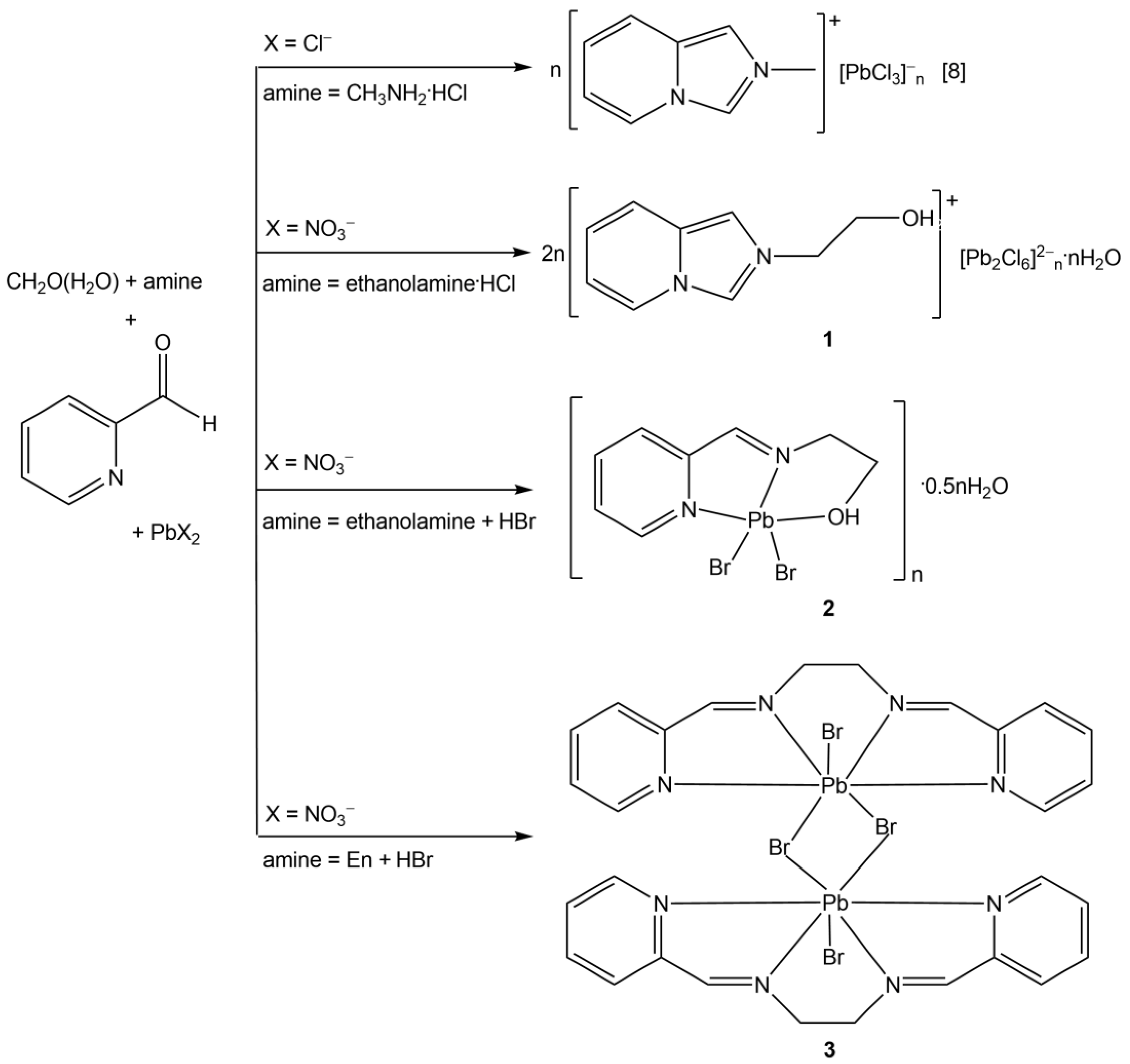
2.1.1. Synthesis of [L1]2n[Pb2Cl6]n∞·nH2O (1)

FA solution was prepared by dissolving paraform (0.13 g, 4.5 mmol) in 10 mL boiling deionized water in a 50 mL conic flask. After cooling at r.t., solid Ea·HCl (0.39 g, 4 mmol) was introduced into the FA solution, which was stirred vigorously at r.t. for half an hour. Then, the pale-yellow solution was filtered and left open overnight. The next day, 2-PCA (0.38 mL, 4 mmol) was added and the solution was kept stirring for 1 h while getting brownish. A subsequent dropwise addition of Pb(NO3)2 (0.33 g, 1 mmol) dissolved in DMF (5 mL) induced the precipitation of a small amount of a yellow residue. The mixture was kept stirring for another hour, then filtered and the brown solution was allowed to stand at r.t. Colourless needles of 1 suitable for X-ray crystallography were formed within a few days after addition of iPrOH (2 mL). The crystals were filtered off, washed with iPrOH and finally dried in air. Yield based on Pb(NO3)2: 47%. FT–IR (ν, cm−1): 3400vs, 3116s, 3072, 3060, 2954, 2928, 2880, 1654, 1560, 1544, 1508, 1448, 1384, 1350, 1328, 1258, 1224, 1150s, 1132, 1076, 946, 922, 880, 796s, 750, 666, 636, 428. 1H NMR (400 MHz, DMSO-d6): δ (ppm) 10.01 (s, 1H, =N–CH=N–), 8.79 (d, J = 7.0 Hz, py), 8.32 (s, 1H, =C=CH−N–), 7.87 (d, J = 9.2 Hz, 1H, py), 7.26 (dd, J = 9.1, 6.7 Hz, 1H, py), 7.16 (dd, J = 9.9, 3.8 Hz, 1H, py), 5.23 (s, 1H, OH), 4.67 (m, 2H, CH2), 3.92 (m, 2H, CH2). Analysis calculated for C18H24Cl6N4O3Pb2 (971.49): C, 22.25; H 2.49; N 5.77%. Found: C 22.39; H 2.28; N 5.52%.

2.1.2. Synthesis of [PbBr2(L2)]n∞·0.5nH2O (2)

Compound 2 was synthesized by adopting the procedure similar to that of 1 using Ea (0.24 mL, 4 mmol) and HBr (0.15 mL, 4 mmol) in place of solid Ea·HCl, and dry Pb(NO3)2 instead of its solution. Orange prisms of 2 suitable for X-ray crystallography were formed within a week after addition of iPrOH (3 mL) to the orange solution. Yield based on Pb(NO3)2: 31%. FT–IR (ν, cm−1): 3392br, 3180br, 3086, 3062, 3016, 2930, 2900, 2872, 1654, 1590, 1568, 1440, 1314, 1224, 1154, 1108, 1048vs, 1006, 988, 912, 874, 782s, 634, 570, 510, 408. 1H NMR (400 MHz, DMSO-d6): δ (ppm) 8.76 (m, 1H, py), 8.50 (s, 1H, −N=CH−py), 8.00 (m, 1H, py), 7.98 (m, 1H, py), 7.62 (m, 1H, py), 4.40 (m, 1H, OH), 3.76 (m, 2H, CH2), 3.71 (m, 2H, CH2). Analysis calculated for C8H11Br2N2O1.5Pb (526.20): C, 18.26; H 2.11; N 5.32%. Found: C 18.45; H 2.15; N 5.47%.

2.1.3. Synthesis of [PbBr2(L3)]2 (3)

Compound 3 was synthesized by adopting the procedure similar to that of 2 using En (0.26 mL, 4 mmol) in place of Ea. Yellow plate-like crystals of 3 suitable for X-ray crystallography were formed within a week after addition of iPrOH (3 mL) to the orange solution. Yield based on Pb(NO3)2: 38%. FT–IR (ν, cm−1): 3076, 3060, 3008, 2920, 2898, 1662, 1652, 1586s, 1564, 1474, 1434, 1374, 1306s, 1218, 1148, 1104, 1036, 1000s, 986, 942, 782, 748, 626, 506, 406. 1H NMR (400 MHz, DMSO-d6): δ (ppm) 8.99 (m, 2H, py), 8.86 (s, 2H, −N=CH−py), 8.01 (m, 4H, py), 7.60 (m, 2H, py), 4.17 (s, 4H, =NCH2CH2N=). Anal. Calcd for C14H14Br2N4Pb (605.30): C, 27.78; H 2.33; N 9.26%. Found: C 28.04; H 2.45; N 9.38%.

2.2. Single Crystal Structure Determination of 13

Crystallographic data for the structures were collected on an Oxford Diffraction Gemini (1) and Bruker D8 Quest diffractometers (2, 3) using Mo (λ = 0.71073 Å) radiation. Following analytical absorption corrections and solution by direct methods, the structures were refined against F2 with full-matrix least-squares using the program SHELXL-2019/2 [20]. Anisotropic displacement parameters were employed for the non-hydrogen atoms. The (N22, N23A) cation in 1 was modelled as being disordered over two sets of sites with site occupancies constrained to 0.701(5) and its complement. The hydroxyl and water molecule hydrogen atoms (1, 2) were located from the experimental data and refined with O−H distances restrained to their idealized values. Other hydrogen atoms were added at calculated positions and refined by the use of a riding model with isotropic displacement parameters based on those of the parent atom. Details of the data collection and processing, structure solution and refinement are summarized in Table 1, while the selected bond lengths and angles are presented in Table 2. CCDC 2217143, 2223148 and 2223147 contain the supplementary crystallographic data for this paper. These data can be obtained free of charge from the Cambridge Crystallographic Data Centre via www.ccdc.cam.ac.uk/data-request/cif.

2.3. Theoretical Calculations

The ORCA 5.0.3 package [21,22] was used for all DFT calculations. Range-separated ωB97M-V functional [23] with the ZORA-def2-TZVPP basis sets [24] and SARC/J auxiliary basis set [25,26] were used for all atoms except of Pb, for which the SARC-def2-TZVPP [25] basis set was applied. AutoAux keyword [27] was used to generate other auxiliary basis sets in all cases. The zero-order regular approximation (ZORA) [28] was used because of the presence of heavy elements. The crystal field was accounted for by means of the conductor-like polarizable continuum model (C-PCM) [29] with ε = infinity. The SCF optimization convergence criteria were settled with VeryTightSCF keywords, and integration grids of high density (Defgrid3 keyword) were employed. Analysis of bond critical points and non-covalent interactions indexes was performed using the Multiwfn 3.8 program [30]. Visualization of the reduced density gradient [31] isosurfaces was performed by means of VMD 1.9.4a53 program [32].

3. Results and Discussion

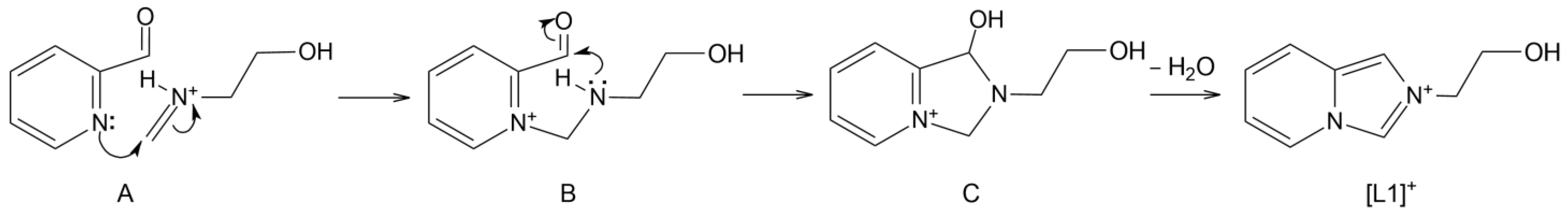
3.1. Synthesis and Characterization
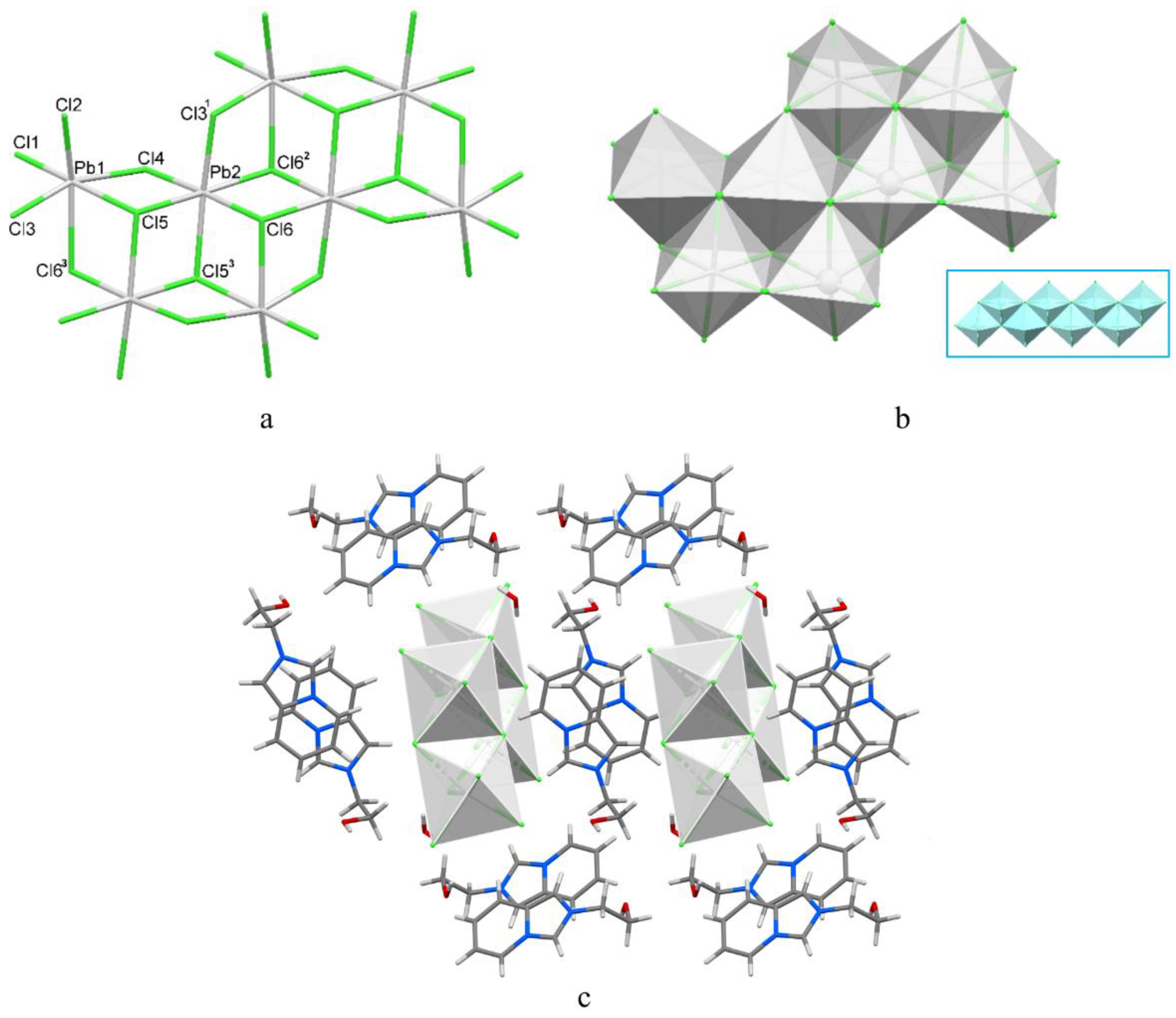
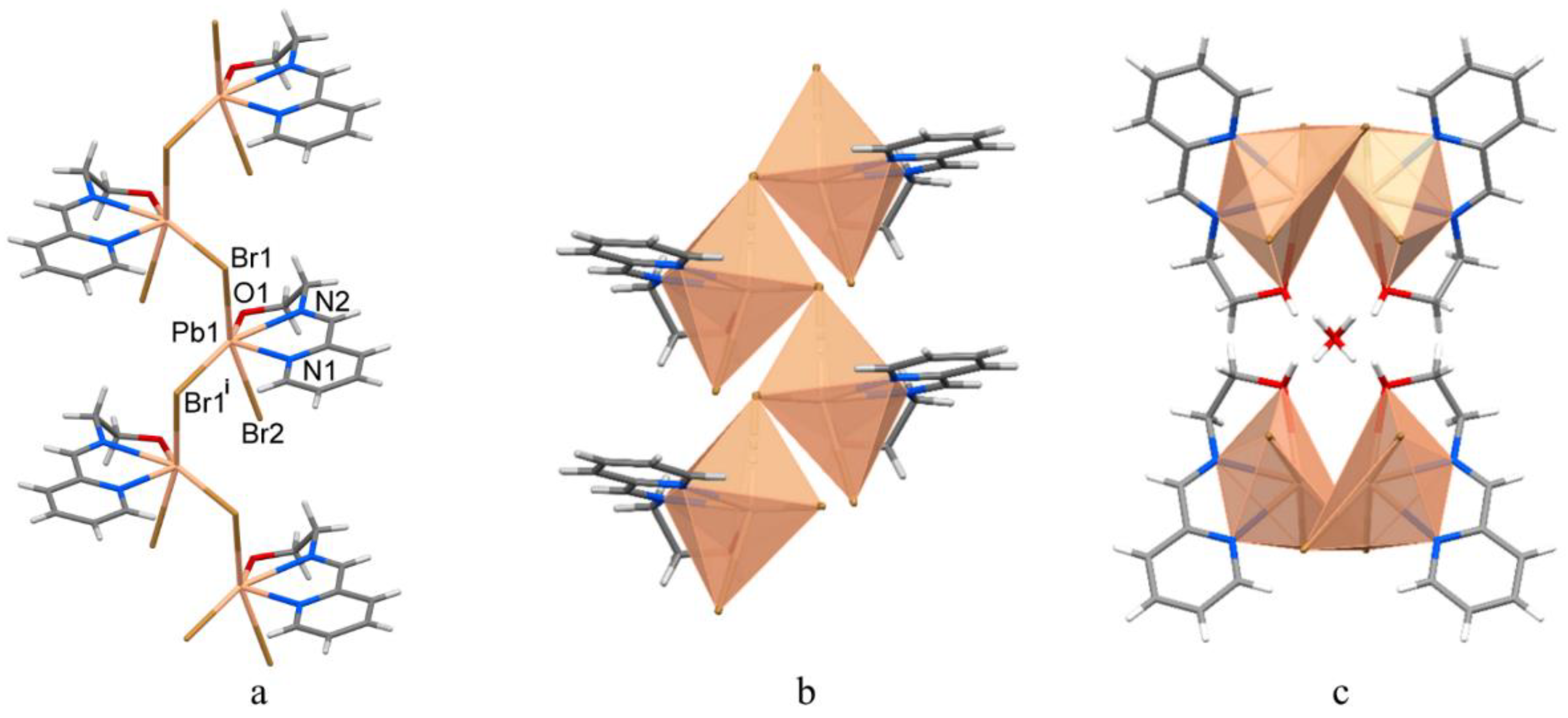
The mechanism of the formation of a substituted imidazo[1,5-a]pyridinium cation has been suggested by us before [8]. In the present study, the primary addition of Ea·HCl to FA aqueous solution yields 2-(methyleneamino)ethanol (Scheme 2). The subsequent 2-PCA attack on the protonated Schiff base A initiates condensation with the intermediate B. The intramolecular nucleophilic attack onto the aldehyde carbon atom by the amine nitrogen brings the product of cyclization C, which experiences an irrevocable dehydration reaction affording the aromatic heterocyclic cation [L1]+. The dehydration of the five-membered ring results in the formation of two double bonds and the delocalization of the positive charge on N atoms in a resonance structure. The reaction does not require the presence of metal ions.
The lead chloride hybrid perovskite 1 formed by self-assembly from the in situ prepared monovalent organic cations, Pb2+ and chloride ions at an overall 1PbCl2:4CH2O:4Ea·HCl:4(2-PCA) mole ratio. It is insoluble in alcohols and water and slightly soluble in N,N-dimethylformamide (DMF) and dimethylsulfoxide (DMSO); it is indefinitely stable in air. Similar reaction procedures that used Ea or En combined with HBr did not afford the respective imidazo[1,5-a]pyridinium cations, presumably due to the insufficient acid strength.
The IR spectra of 13 confirmed the presence of aromatic rings, alkyl and other functional groups (Figures S1–S3). The spectrum of the hybrid compound 1 demonstrates a distinctive pattern, which was suggested to characterize the imidazo[1,5-a]pyridinium skeleton [8,11,12]: the sharp very strong peaks attributed to aromatic C–H vibrations (3116–3060 cm−1), medium intensity sharp bands at 1654 and 1544 cm−1 associated with heterocyclic ring stretching, an intense absorption at 1150 cm−1 and three peaks in the region of out-of-plane C–H bending (796, 750 and 636 cm−1). The very intense band, due to ν(O–H) vibration at 3400 cm−1, dominated the spectrum; another intense absorption was ascribed to C–O stretching (1076 cm−1). In the case of compounds 2 and 3, the aromatic ν(C–H) bands above 3000 cm−1 are much weaker. The spectrum of compound 2 is dominated by a peak at 1048 cm−1 due to ν(C–O), while the absorption of moderate intensity due to O–H stretching (3392 cm−1) is broad. ν(C=N) + ν(C=C) stretching frequencies of the metal coordinated Schiff bases are observed at 1654, 1590 cm−1 (2) and 1652, 1586 cm−1 (3).
The compounds are easily distinguishable by their 1H NMR spectra that demonstrate the sets of signals, as well as the correct alkyl/aromatic proton ratios expected for [L1]+ cations and the Schiff base ligands. In the room temperature (r.t.) spectrum of compound 1 in DMSO-d6, CH protons in the imidazolium ring appear as singlets at δ = 10.01 and 8.32 ppm, the alkyl groups protons are observed as multiplets at 4.67 and 3.92 ppm. The imine protons in 2 and 3 are detected at 8.50 and 8.86 ppm, respectively, while the protons of CH2 groups appear as two multiplets at 3.76 and 3.71 (2) and one multiplet at 4.17 ppm (3). The hydroxyl protons in 1 and 2 are observed distinctly at 5.23 and 4.40 ppm, respectively.

3.2. Structural Description of 13

The asymmetric unit of the lead chloride hybrid 1 is comprised of two [L1]+ cations, [Pb2Cl6]2– anionic fragment and water molecule of crystallization (Figure 1). Two crystallographically non-equivalent cations configurations are very similar; the (N22, N23A) cation is disordered over two sites, with occupancies of 0.701(5) and 0.299(5)-. The pyridinium rings bond distances of their fused heterocyclic cores are uneventful; the N−C bond lengths in the imidazolium entities fall in the range 1.331(7)–1.414(6) Å. The N atoms are planar, showing a sum of three angles of 360°. In the cores, the five- and six-membered rings are nearly coplanar, with the respective dihedral angles of 1.35, 1.73 and 2.98°.
The distorted octahedral environment of Pb1 atom in the anionic motif consists of two terminal, two µ2-Cl and two µ3-Cl atoms, while the distorted octahedron around Pb2 is built of two µ2-Cl and four µ3-Cl ligands (Figure 2a). The metal–chloride distances vary in the range 2.6509(14)–3.1516(14) Å. The average value of 2.907 Å is reasonable for Pb(II) ions in chloroplumbates(II) with strong ionic Pb–Cl bonds [33,34]; the cis and trans angles at the metal atom fall in the ranges of 77.50(4)–107.33(5)° and 162.86(5)–177.20(4)°, respectively (Table 2), showing a strong degree of distortion of the metal polyhedra. The PbCl6 octahedra are joined by three-edge- and five-edge-sharing into a 1D twin lead(II) chloride wire of 10.344 Å width, lying in the a-axis direction (Figure 2a,b). The bulkier 2-(2-hydroxyethyl)-2H-imidazo[1,5-a]pyridinium cation in 1, if compared to its methyl analogue [8], expectedly templated the formation of a low-dimensional anionic perovskite frame, however, with a different type of a double chain. This set of connectivity modes within a double chain was not observed in the lead(II) chloride hybrid compounds in accordance with our survey of the Cambridge Structural Database [35]. The type of edge-sharing within a double haloplumbate(II) chain shown in the inset of Figure 2b is more frequent and found in, for example, 1D lead chloride perovskites with substituted diammonium and piperidinium dications [6,36].
Table 2. Selected bond lengths (Å) and angles (°) in 13 [a].
[L1]2n[Pb2Cl6]n∞·nH2O (1)
Pb1−Cl12.7587(16)Pb2−Cl42.7924(15)
Pb1−Cl22.6509(14)Pb2−Cl3 12.8177(14)
Pb1−Cl32.8223(13)Pb2−Cl6 22.8730(13)
Pb1−Cl43.0750(14)Pb2−Cl52.9355(13)
Pb1−Cl53.0472(14)Pb2−Cl62.9613(14)
Pb1−Cl6 33.1516(14)Pb2−Cl5 32.9957(14)
Cl2−Pb1−Cl193.17(5)Cl4−Pb2−Cl3 194.36(5)
Cl2−Pb1−Cl386.32(4)Cl4−Pb2−Cl6 296.36(4)
Cl1−Pb1−Cl394.13(5)Cl3 1−Pb2−Cl6 382.39(4)
Cl2−Pb1−Cl590.30(4)Cl4−Pb2−Cl587.03(4)
Cl1−Pb1−Cl5175.07(4)Cl3 1−Pb2−Cl594.79(4)
Cl3−Pb1−Cl589.58 (4)Cl6 2−Pb2−Cl5175.73(3)
Cl2−Pb1−Cl4107.33(5)Cl4−Pb2−Cl6177.20(4)
Cl1−Pb1−Cl495.37(4)Cl3 1−Pb2−Cl687.68(4)
Cl3−Pb1−Cl4162.86(5)Cl6 2−Pb2−Cl685.80(4)
Cl5−Pb1−Cl480.25(4)Cl5−Pb2−Cl690.89(4)
Cl2−Pb1−Cl6 3161.97(4)Cl4−Pb2−Cl5 392.01(5)
Cl1−Pb1−Cl6 395.85(5)Cl3 1−Pb2−Cl5 3173.17(4)
Cl3−Pb1−Cl6 377.50(4)Cl6 2−Pb2−Cl5 399.36(4)
Cl5−Pb1−Cl6 381.77(4)Cl5−Pb2−Cl5 383.07(4)
Cl4−Pb1−Cl6 387.35(4)Cl6−Pb2−Cl5 385.88(4)
[PbBr2(L2)]n·0.5nH2O (2)
Pb1−N1 2.584(7)Pb1−Br2 2.9777(10)
Pb1−N2 2.562(7)Pb1−Br1 43.0764(10)
Pb1−O1 2.839(7)Pb1−Br1 3.0293(11)
N2−Pb1−N1 64.4(3)O1−Pb1−Br1 4146.36(15)
N2−Pb1−O1 61.8(2)Br2−Pb1−Br1 4 89.35(3)
N1−Pb1−O1 123.3(2)N2−Pb1−Br1 84.41(17)
N2−Pb1−Br2 85.72(18)N1−Pb1−Br1 80.21(16)
N1−Pb1−Br2 80.57(16)O1−Pb1−Br1 110.23(16)
O1−Pb1−Br2 79.24(16)Br2−Pb1−Br1 160.70(3)
N2−Pb1−Br1 4149.39(18)Br1−Pb1−Br1 490.73(3)
N1−Pb1−Br1 485.02(18)
[PbBr2(L3)]2 (3)
Pb1−N1 2.745(6)Pb1−Br1 3.0632(9)
Pb1−N2 2.625(5)Pb1−Br2 2.9599(9)
Pb1−N3 2.621(6)Pb1−Br1 33.2525(8)
Pb1−N4 2.808(6)
N2−Pb1−N1 61.15(17)N2−Pb1−Br1 84.71(12)
N3−Pb1−N1 123.10(18)N1−Pb1−Br1 93.02(11)
N3−Pb1−N2 63.60(19)N4−Pb1−Br1 85.58(12)
N3−Pb1−N4 60.4(2)Br2−Pb1−Br1 174.31(2)
N2−Pb1−N4 121.93(19)N3−Pb1−Br1 3149.42(14)
N1−Pb1−N4 176.39(16)N2−Pb1−Br1 3145.63(13)
N3−Pb1−Br2 81.33(13)N1−Pb1−Br1 387.48(13)
N2−Pb1−Br2 89.91(12)N4−Pb1−Br1 389.05(15)
N1−Pb1−Br2 85.97(12)Br2−Pb1−Br1 3102.31(3)
N4−Pb1−Br2 95.75(12)Br1−Pb1−Br1 383.22(2)
N3−Pb1−Br1 94.60(13)
[a] Symmetry codes: 1 x − 1, y, z; 2x, −y + 1, −z + 1; 3x + 1, −y + 1, −z + 1; 4 1/2 − x, y, 1/2 + z.
In the crystal, non-equivalent [L1]+ cations of 1 form separate stacks parallel the a-axis with the fused heterocyclic cores of neighboring entities being strictly coplanar (Figure 2c). Centrosymmetically related trans-oriented [L1]+ cations are stacked with varying levels of offset, showing the ring centroid distances of 3.456, 3.651 Å for (N12, N13A) and 3.839, 5.258 Å for (N22, N23A) cations enabling the possibility of weak π-bonding [37]. Hydrogen bonds of O−H···O/Cl and C−H···O/Cl types with the involvement of the OH group of the organic cation and water molecule (Table 3) strengthen the hybrid salt structure. The introduction of OH group on the cation enabled a stronger interaction between cationic and anionic counterparts in 1 through additional conventional hydrogen bonding, which is absent in the lead chloride hybrid with methyl derivative of [L1]+ cation [8]. The new type of a double chain realized in 1 may also be considered a result of such a modification of the organic cation.
Lead halide hybrid 2, which crystallizes in the orthorhombic space group Pccn, consists of a 1D monobromo-bridged lead(II) polymer and water molecules of crystallization. The asymmetric unit of 2 is shown in Figure 1. In the double-sided, organically decorated chain structure, the nearest six-coordinate environment of each metal atom is formed by one tridentate Schiff base, one terminal and two µ2-Br ligands (Figure 3a,b). The neutral Schiff base ligand chelates the Pb1 ion by the N1 atom of the pyridyl ring, the imine N2 atom and O1 atom of the ethanol group with an average value of the Pb1−N distances of 2.57 Å, and the Pb−O1 bond [2.839(7) Å] being significantly elongated (Table 2). The metal distances to terminal and bridging bromide atoms are similar, with an average of 3.03 Å falling in the usual range for 1D lead(II) bromide systems [38,39]. The additional Pb···Br2{1/2 − x, y, −1/2 + z} contact of 3.378 Å is appreciably larger than the sum of the Shannon ionic radii of the octahedral lead(II) cation and bromide anion [r(Pb2+) + r(Br) = 3.15 Å] and is barely significant in the construction of the bromoplumbate(II) chain. The bond angles at the Pb1 atom vary from 61.8(2) to 123.3(2)° and from 146.36(15) to 160.70(3)° (Table 2), evidencing the severe distortion of the metal polyhedron. The closest Pb···Pb separation within the chain is 4.4144(6) Å. In the crystal, two bromoplumbate(II) chains related by a crystallographic two-fold axis form a hydrophilic channel to host water molecules held by O–H···O hydrogen bonds (Figure 3c, Table 3). Numerous C–H···Br contacts result in a 3D supramolecular architecture.
Complex 3 belongs to the triclinic space group Pī; the dimeric molecule is located on an inversion centre (Figure 4). The geometry of Pb1 atom is that of an irregular pentagonal bipyramid, the equatorial plane consisting of four nitrogens from the tetradentate chelate L3 and the centrosymmetrically related bromide ligand Br1{1 − x, 1 − y, 1 − z} of the dimer, with two other bromides—Br1 and Br2—occupying apical positions. The Pb–N bond distances within the range of 2.621(6)–2.804(6) Å (Table 2) agree well with those of two other known Pb(II) complexes with L3 [40,41]. The Pb1–Br1{1 − x, 1 − y, 1 − z} bond is elongated at 3.2525(8) Å, in contrast to the average Pb1–Br1/Br2 bond length of 3.014 Å. The cis angles at the lead atom vary from 60.4(2) to 95.75(12)°; the trans angles fall in the range 121.93(19)−176.39(16)°. In the dimer, the Pb···Pb’ separation is 4.7239(5) Å.
In the solid state, the dimeric molecules of 3 are stacked identically, forming columns parallel to the ab plane, with the Pb2Br4 moieties in the column being strictly coplanar (Figure 4c). The minimal Pb···Pb distance in the lattice is 8.1348(6) Å; no effective π-overlap is observed. C–H···Br hydrogen bonding (Table 3) consolidates an extended supramolecular 3D network structure (Figure 4c). The contacts strength depends on their direction in the crystal lattice. In the ab plane, the interactions are represented by four distinct crystallographically independent C–H···Br hydrogen bonds with d(C···Br) < 3.9 Å (Table 3). Every dimeric molecule of 3 forms 16 respective contacts with the four nearest molecules in the ab plane. The dimeric molecules along the c axis are bridged by weaker C–H···Br hydrogen bonds with d(C···Br) > 3.9 Å (Table 3).

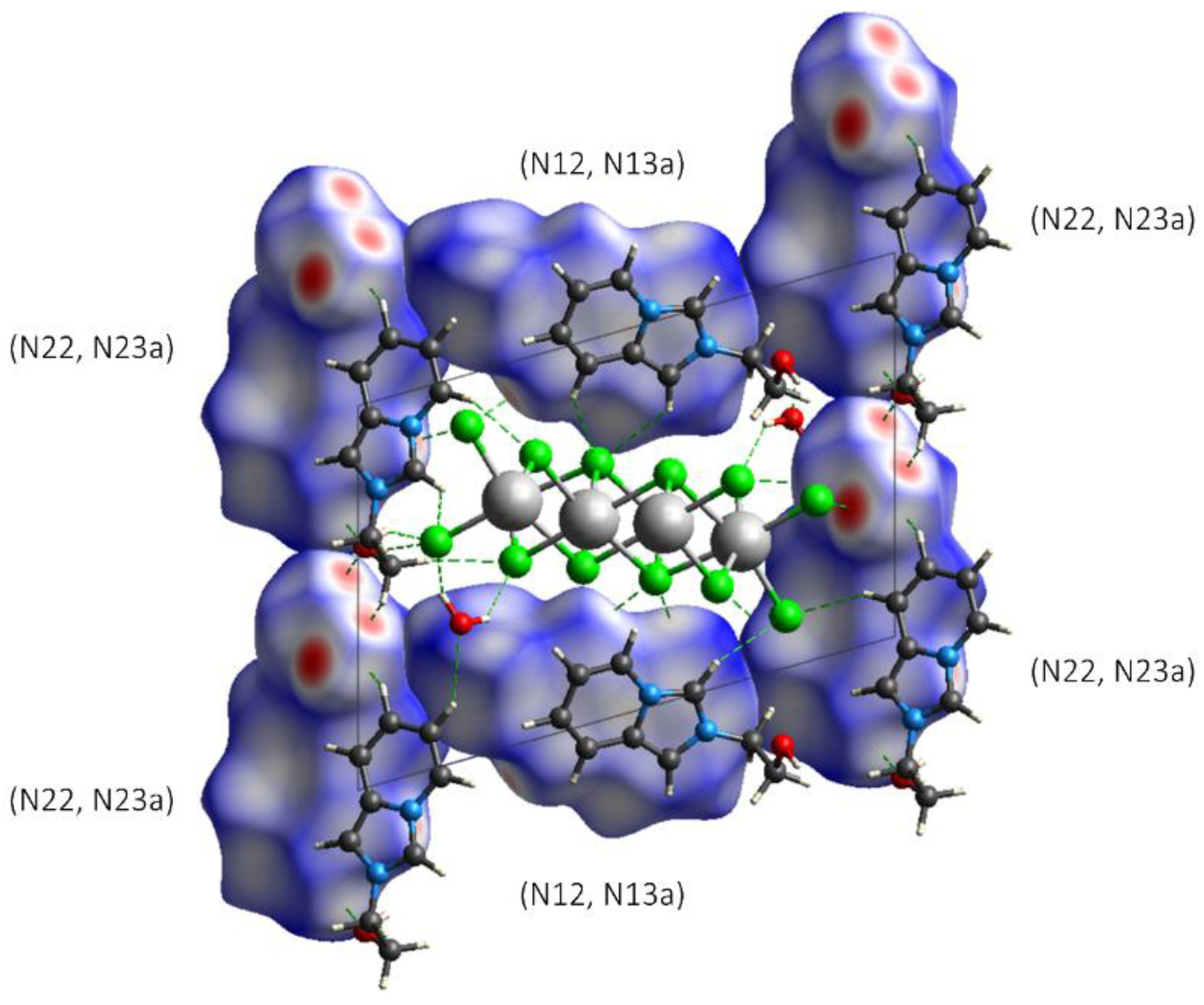
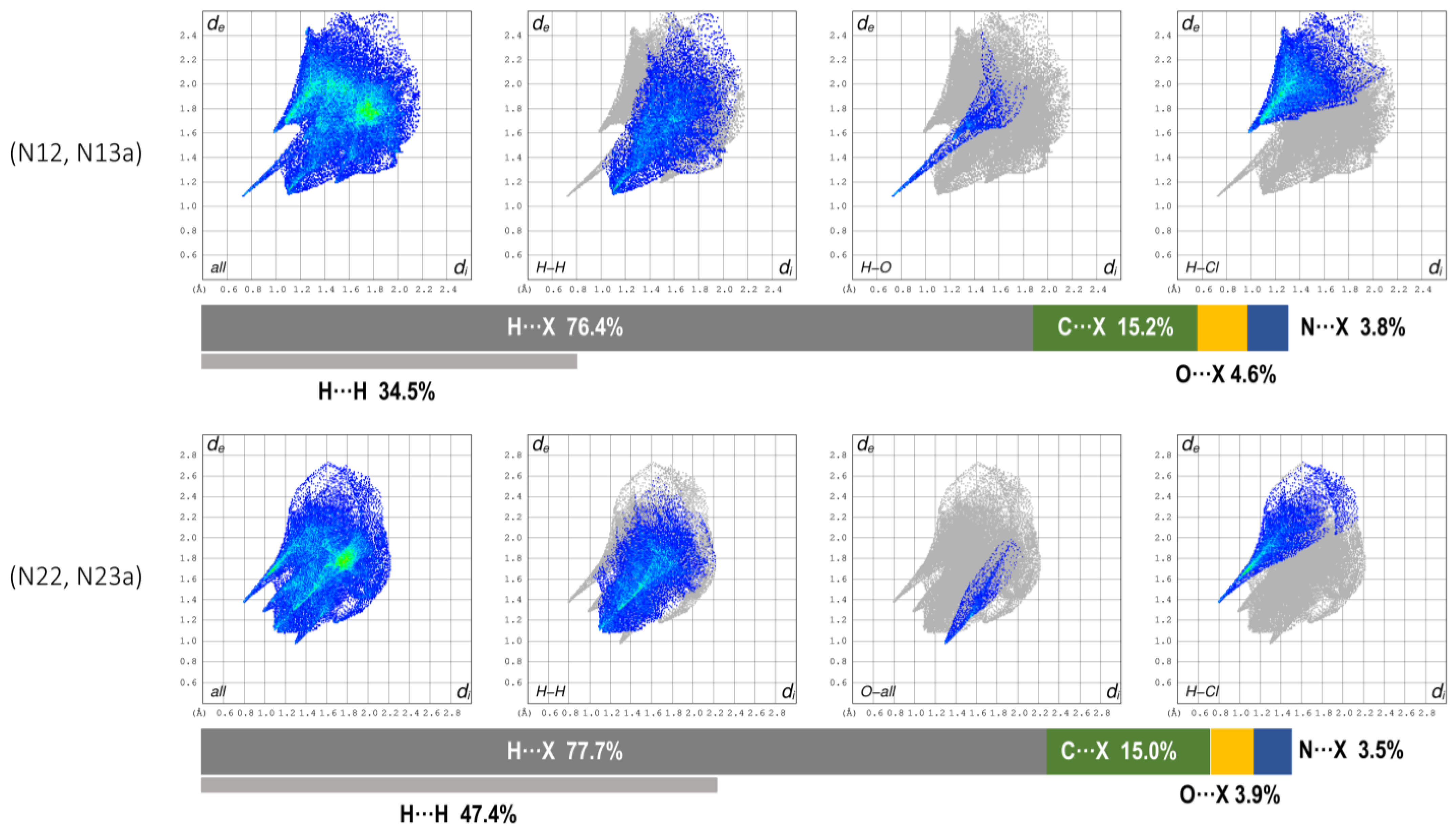
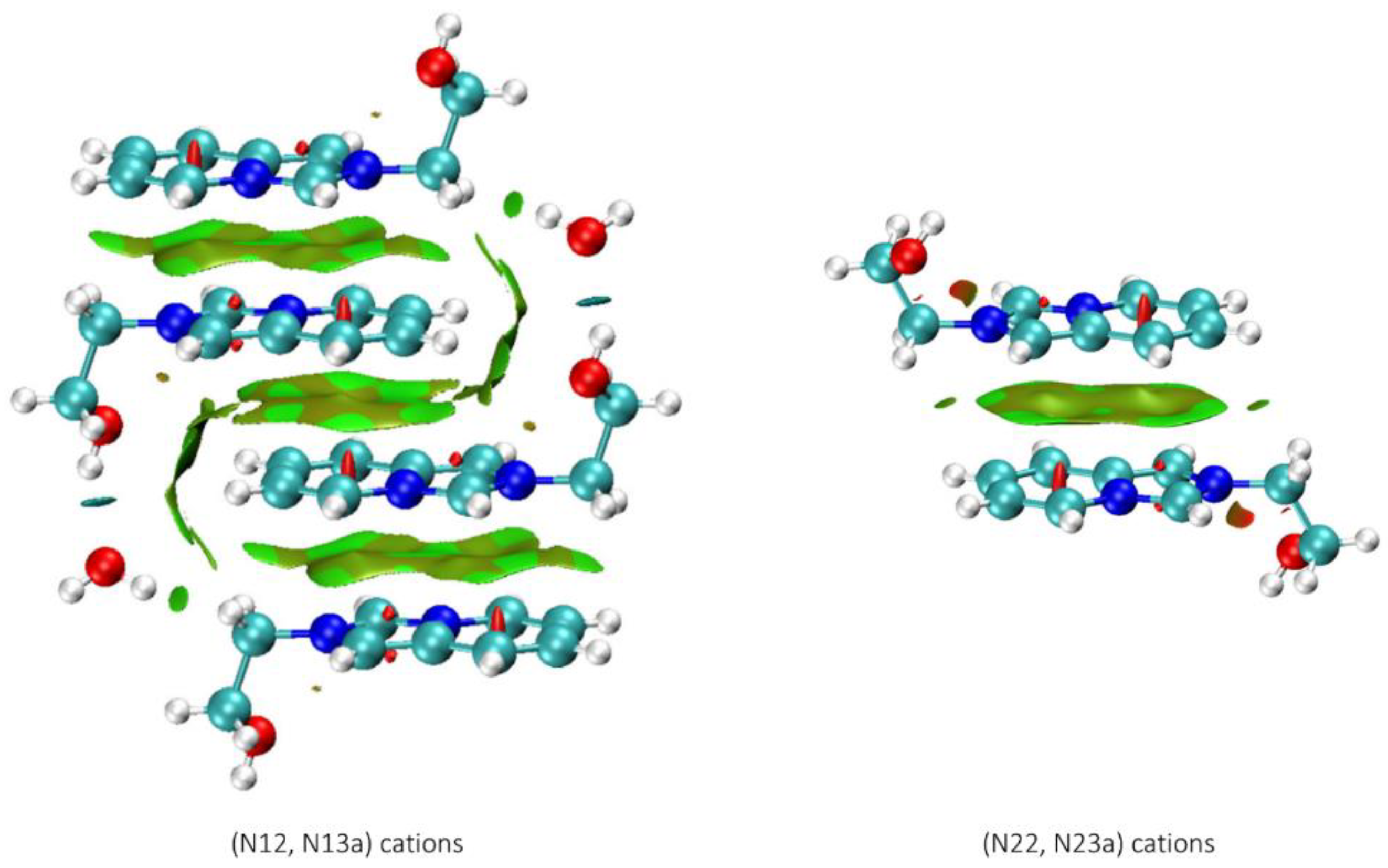
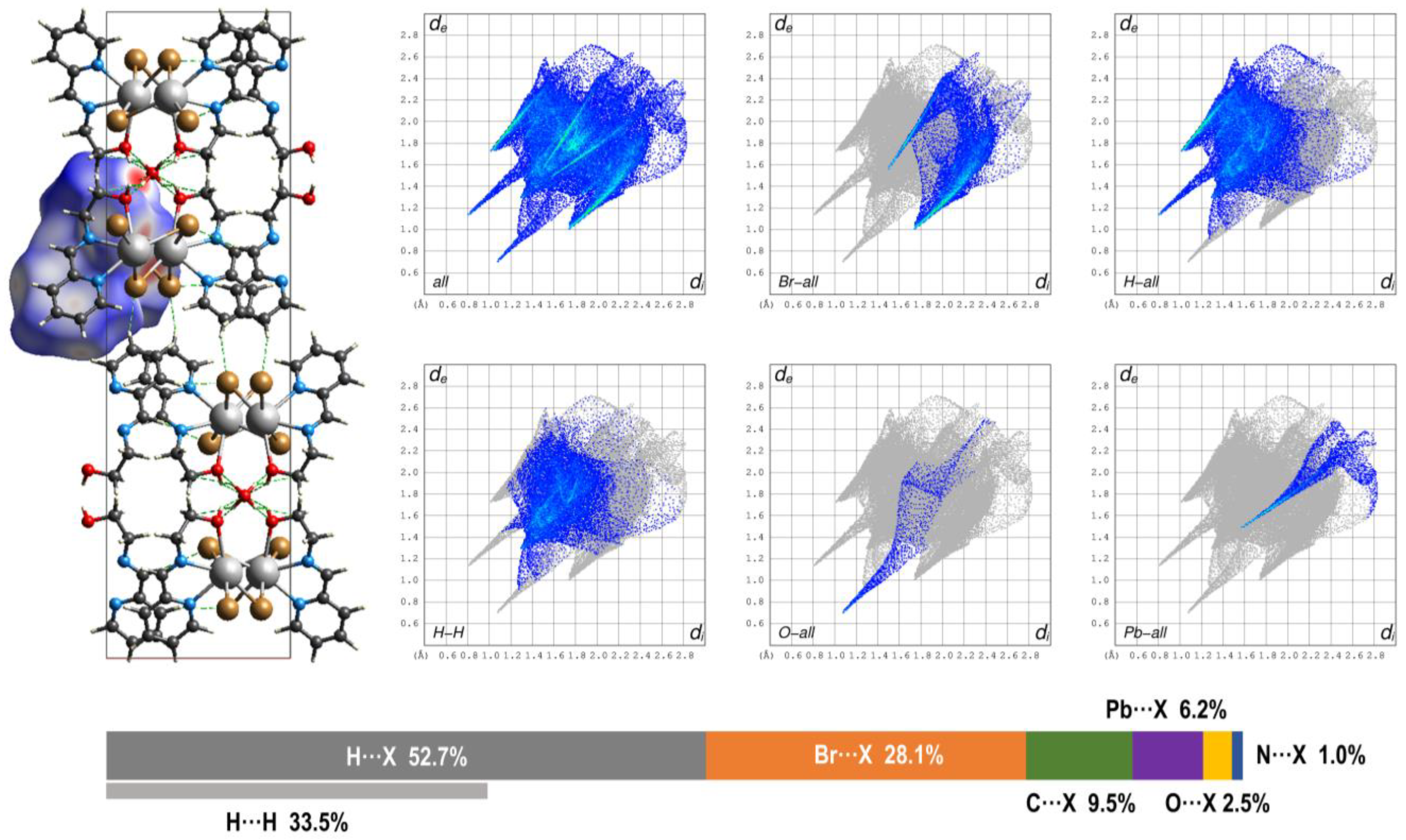
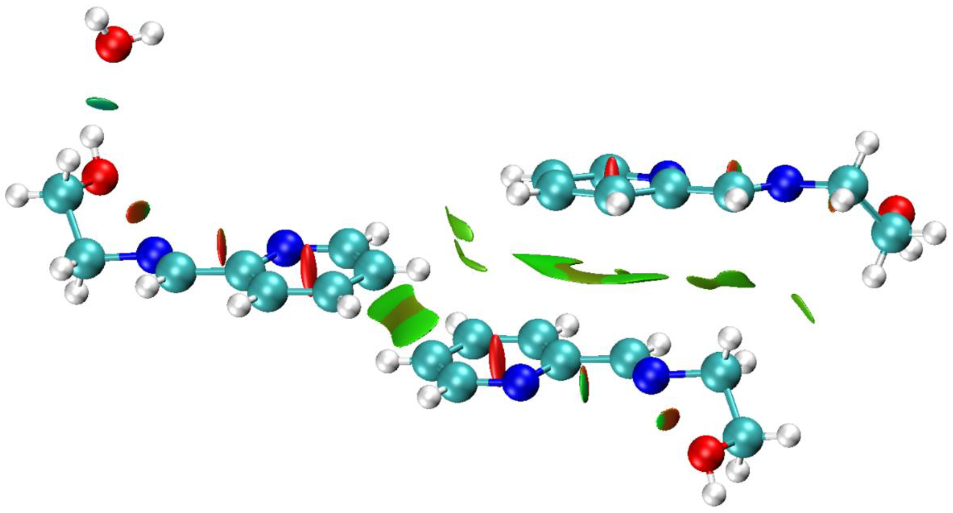
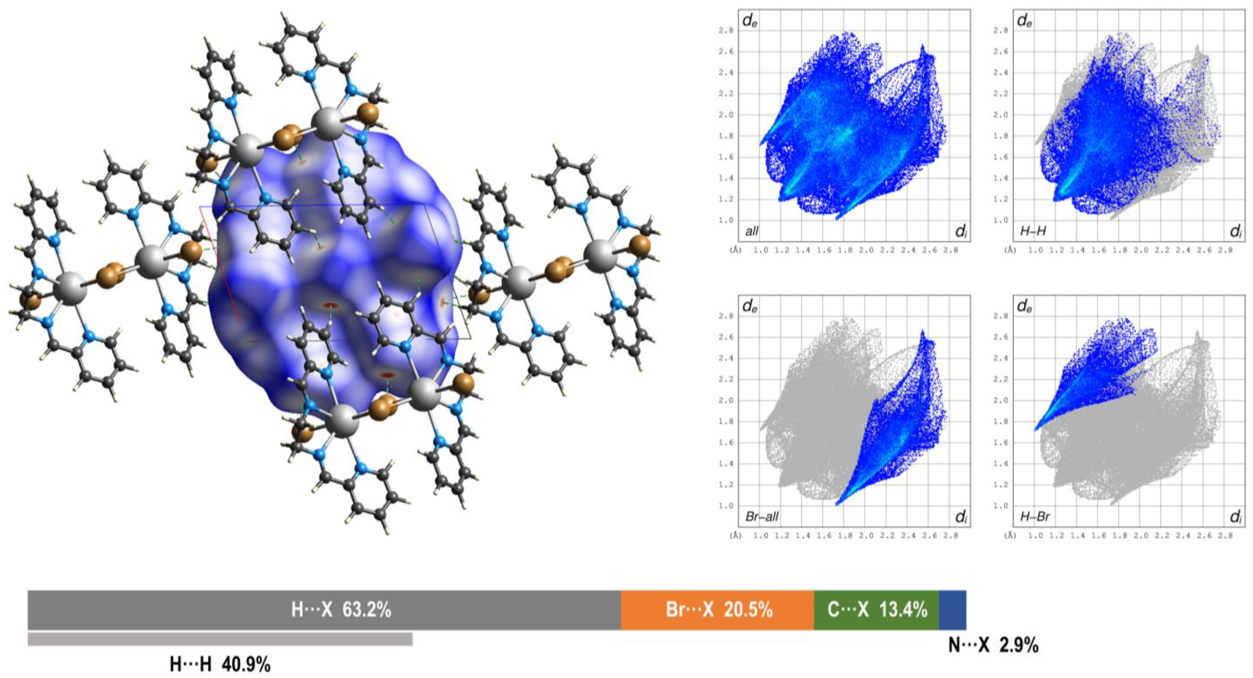
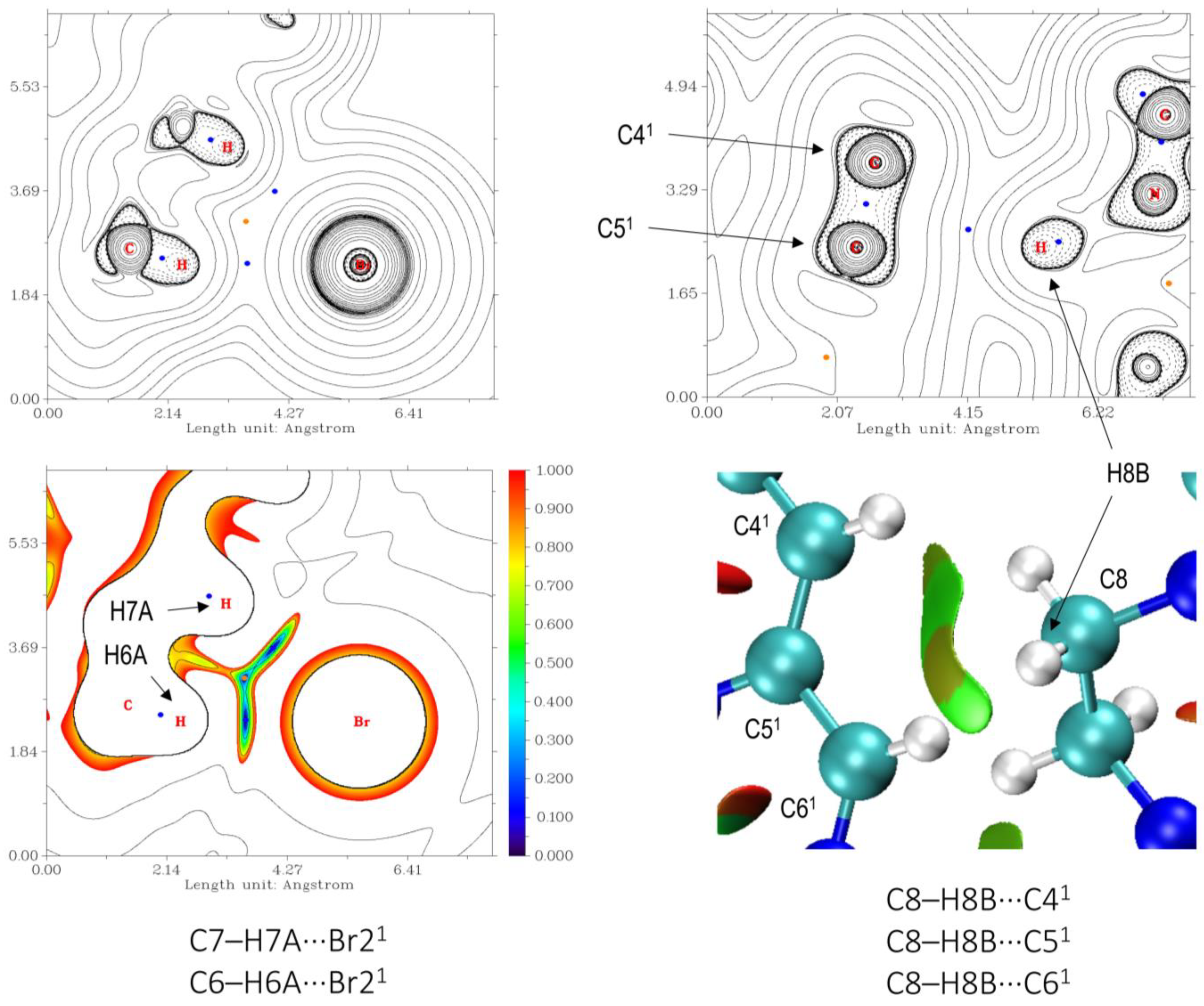
3.3. Hirshfeld Surface and QTAIM Analyses
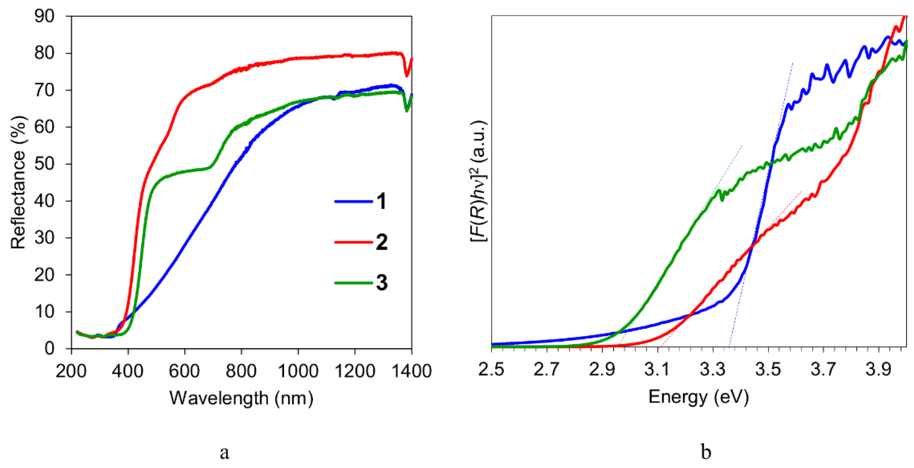
The set of intermolecular interactions in complex 1 can be viewed as a combination of contacts between crystallographically independent (N12, N13A) and (N22, N23A) [L1]+ cations, negatively charged [Pb2Cl6]n2n– chains and water molecules. The HS of both [L1]+ cations surrounding a fragment of the [Pb2Cl6]n2n– chain is shown in Figure 5. Here, and further on, the colours on the surface correspond to shortened contacts (red), van der Waals (vdW) interactions (white) and contacts with longer distances (blue).
Both [L1]+ cations reveal involvement in several distinct hydrogen bonds (Table 3), which are identifiable in the fingerprint plots (Figure 6). The plots are characterized by broad segments of H···H interactions (Figure 6), while the C···C ones are much less abundant (5.4 and 4.3% for (N12, N13A) and (N22, N23A) independent cations, respectively, Figure S4).
The weak interactions between the stacked cations were evaluated by the analysis of reduced density gradient (RDG) [31] of the electron density calculated at the ωB97M-V/ZORA-def2-TZVPP level using crystallographic atomic coordinates (Figure 7). In line with the HS analysis results, the non-covalent contacts in the tetramer {(N12, N13A)-[L1]4·2H2O}4+ are mostly characterized by the broad vdW areas between stacked imidazo[1,5-a]pyridine moieties. These findings indicate that [L1]+ cations in their stacks are too offset to engage in appreciable π-bonding. At the same time, the chain of (N12, N13A)-[L1]+ cations is supported by the strong attractive interactions involving water molecules (O121–H121···O01W), for which the electron density at the bond critical point—ρBCP(r) (3, −1) between the H121 and O01W atoms—is 0.02504 a.u. According to the dependence for charged assemblies reported in [42], the obtained value of ρBCP(r) corresponds to 39.3 kJ mol−1 of binding energy (BE) between the (N12, N13A)-[L1]+ cation and water molecule. However, as the BE vs. ρBCP(r) dependences [42] were developed for assemblies with a single overall charge (in contrast to 4+ charge of the tetramer), the obtained binding energy value has only indicative significance.
The monomeric fragment [PbBr2(L2)] of the 1D chain in complex 2 was used to construct the Hirshfeld surface (Figure 8). The fingerprint plots disclose several distinct non-covalent contacts mostly H···O and H···Br in nature (Figure 8 and Figure S5). The strongest hydrogen bonds between O1 and O1W atoms are reflected by sharp “peaks” having the shortest de and di distances in the fingerprint plots (Figure 8 and Figure S5). The ρBCP(r) electron density at BCP between H1 and O1W atoms was estimated as 0.01986 a.u., from which the binding energy of the respective contact can be evaluated as –15.4 kJ mol−1 (according to the equation for non-charged assemblies [42]). This energy strength can be classified as “weak-to-medium” [42]. The RGD isosurfaces of the vdW contacts between the aromatic groups in 2 are shown in Figure 9.
The dimeric molecules of 3 create a network of non-covalent interactions, of which the highest contribution (40.9%) to the HS is from numerous H···H contacts (Figure 10 and Figure S6). The latter form a broad surface with no distinct directions (Figure 11), except of the C8–H8A···H8A1−C81 (1 = 2 – x, –y, –z) interaction between methylene groups [d(C···C) = 4.379(15) Å] and a pair of weak interactions between pyridine rings C13–H13A···H14A–C141 and C14–H14A···H13A–C131 [1 = 2 – x, 1 – y, 1 – z; d(C···C) = 4.072(18) Å]. As can be seen from the particular Hirshfeld surface, the Br···H and H···Br contacts are mostly located in the ac plane (Figure 11). Since the molecules of 3 has no electrical charge, the strengths of these contacts can be estimated [42] from the ρBCP(r) electron density with sufficient precision (Table 4).
Inspection of the geometry and Hirshfeld surface suggests the existence of the contact C8–H8B···C61 [1 = 1 – x, 1 – y, –z; d(C···C) = 3.722(10) Å, ∠(C–H···C) = 161.1°] between methylene CH2 group and imine carbon atom of the neighboring molecules. However, despite the hydrogen atom being clearly oriented towards C6 one, the respective bond critical point is located rather between H8B and C51 atoms (Figure 12). Further analysis of the RDG isosurfaces disclosed the existence of a broad non-covalent interaction region between H8B donor and C41, C51 and C61 (1 = 1 – x, 1 – y, –z) atoms as acceptors containing 0.0054 electrons (Figure 12). The negative qbind index [43] of this domain (–0.00046383) accounts for a weak attractive interaction. The aromatic moieties of the ligand L3 in the molecule of 3 contact each other through vdW interactions (Figure S7).

3.4. Optical Properties of 13

The optical diffuse reflectance spectra of all the compounds were measured using powder samples at r.t. (Figure 13a). According to the analysis of the spectra by the Tauc plot based on the assumption of direct band gap, [F(R)hν]2 vs. hν (where F(R) denotes the Kubelka–Munk function, hν is photon energy in eV), the optical band gap values of 1, 2 and 3 were estimated to be 3.36, 3.13 and 2.96 eV, respectively (Figure 13b). The values of 1 and 2 are comparable to those of reported 1D hybrid lead perovskites, such as [H2bpp]Pb2Cl6 (3.55 eV), [H2bpp]Pb2Br6 (3.30 eV; bpp = 1,3-bis(4-pyridyl)-propane) [38], [2,6-dmpz]3Pb2Br10 (3.12 eV; 2,6-dmpz = 2,6-dimethylpiperazine), [hex]PbBr3 (3.41 eV; hex = hexamethyleneimine), [hep]PbBr3 (3.50 eV; hep = heptamethyleneimine) [39]. Using density functional theory calculations, [2,6-dmpz]3Pb2Br10, [hex]PbBr3 and [hep]PbBr3 were found to be direct band gap semiconductors [39]. Their valence band maximum (VBM) is composed of mixed Br-p and Pb-s orbitals, while empty Pb-p orbitals constitute conduction band minimum (CBM). In the case of two H2bpp hybrid lead halides, the VBM was derived from hybridization of Pb-6p and Cl-3p or Br-4p orbitals, and CBM mainly originated from the C-2p/N-2p states of the bicyclic organic cations [38].
Within the series of 1D and 2D lead bromide hybrids [39], the electronic structure was suggested to depend on the connectivity mode of PbBr6 octahedra, where the corner-connected compound [4-amp]PbBr4 [4-amp-4-(aminomethyl)piperidine] exhibits the smallest band gap value (2.93 eV). The face-sharing compounds [hex]PbBr3 and [hep]PbBr3 possess larger band gaps than [2,6-dmpz]3Pb2Br10 with the edge- and corner-connecting structure. The band gaps successively going up from “corner-sharing” to “edge-sharing” and further “face-sharing” have also been observed in the lead iodide perovskite-derived organic–inorganic hybrids [44]. The band gaps of edge-sharing 1 and corner-sharing 2 perovskites follow the general trend.

4. Conclusions

In this study, we aimed to explore the templating effect of imidazo[1,5-a]pyridinium-based cation with hydroxyl functionality on the dimensionality of the lead halide anionic framework and connectivity modes of the [PbHal6]4– octahedra, as well as H-bonding interactions in the resulting hybrid lead halide perovskite. To this aim, Ea·HCl was used in the developed protocol for the synthesis of substituted imidazo[1,5-a]pyridinium cations by the acid catalyzed oxidative cyclocondensation of FA, amine and 2-PCA. The obtained in situ [L1]+ cation directed the formation of the 1D chloroplumbate [L1]2n[Pb2Cl6]n∞·nH2O (1), revealing a new type of the [Pb2Cl6] twin chain constructed from three-edge- and five-edge-sharing PbCl6 octahedra. In the case of [PbBr2(L2)]n∞·0.5nH2O (2) and [PbBr2(L3)]2 (3), the introduction of HBr—the necessary acid component and source of bromide anions—directly to the reaction media subverted the expected oxidation cyclization and instead facilitated the amine-aldehyde condensation between 2-PCA and Ea or En, respectively. The isolated hybrid lead bromide 2 features a chain of corner-sharing PbBr3N2O octahedra, decorated with the tridenate Schiff base ligands on both sides. In the 0D dimer 3, two irregular PbBr3N4 pentagonal bipyramids are connected by edge sharing. The Hirshfeld surface and QTAIM analyses disclosed the presence of numerous weak interactions between molecular and polymeric fragments in the structures of 13. The band gaps of 1 (3.36 eV) and 2 (3.13 eV) with different connectivity follow the general trend of edge-sharing perovskites exhibiting larger band gaps than corner-sharing structures.

Supplementary Materials

The following supporting information can be downloaded at: https://www.mdpi.com/article/10.3390/cryst13020307/s1, Figure S1: IR spectrum of [L1]2n[Pb2Cl6]n∞·nH2O (1); Figure S2: IR spectrum of [PbBr2(L2)]n∞·0.5nH2O (2); Figure S3: IR spectrum of [PbBr2(L3)]2 (3); Figure S4: Selected fingerprint plots of the (N12, N13A) and (N22, N23A, major component) [L1]+ cations in the crystal structure of 1 showing Xi···Xe interactions (where X = Cl, O, C or H); Figure S5: Selected fingerprint plots of the fragment [PbBr2(L2)] in the crystal structure of 2 showing Xi···Xe interactions (where X = Br, O, C or H); Figure S6: Selected fingerprint plots of the dinuclear molecule of 3 showing Xi···Xe interactions (where X = Br, N, C or H); Figure S7: Reduced density gradient surfaces (isovalue of 0.5) showing weak vdW interactions (green area) between aromatic fragments of the ligands L3 in the molecular structure of 3 (lead and bromine atoms were excluded from calculations).

Author Contributions

Conceptualization, O.Y.V.; methodology, O.Y.V. and D.S.N.; investigation, O.Y.V., E.A.B., O.V.N. A.N.S. and D.S.N.; writing—original draft preparation, O.Y.V. and D.S.N.; writing—review and editing, O.Y.V. All authors have read and agreed to the published version of the manuscript.

Funding

The work was performed with the support of the Ministry of Education and Science of Ukraine (project 22BP037-13, grant for the perspective development of the scientific direction ‘Mathematical sciences and natural sciences’ at the Taras Shevchenko National University of Kyiv) and the Fundação para a Ciência e Tecnologia (FCT), Portugal (projects UIDB/00100/2020, UIDP/00100/2020, and LA/P/0056/2020 of Centro de Química Estrutural, contracts IST-ID/086/2018 and IST-ID/117/2018).

Data Availability Statement

Crystallographic data for the structures reported can be obtained free of charge from the Cambridge Crystallographic Data Centre via www.ccdc.cam.ac.uk/data-request/cif (accessed on 27 January 2023) quoting the deposition numbers CCDC 2217143 (1), 2223148 (2) and 2223147 (3).

Conflicts of Interest

The authors declare no conflict of interest.

References

  1. Green, M.A.; Hishikawa, Y.; Warta, W.; Dunlop, E.D.; Levi, D.H.; Hohl-Ebinger, J.; Ho-Baillie, A.W. Solar cell efficiency tables (version 50). Prog. Photovolt. Res. Appl. 2017, 25, 668–676. [Google Scholar] [CrossRef]
  2. Sutherland, B.R.; Sargent, E.H. Perovskite photonic sources. Nat. Photon. 2016, 10, 295–302. [Google Scholar] [CrossRef]
  3. Saparov, B.; Mitzi, D.B. Organic-Inorganic Perovskites: Structural Versatility for Functional Materials Design. Chem. Rev. 2016, 116, 4558–4596. [Google Scholar] [CrossRef] [PubMed]
  4. Smith, M.D.; Jaffe, A.; Dohner, E.R.; Lindenberg, A.M.; Karunadasa, H.I. Structural origins of broadband emission from layered Pb–Br hybrid perovskites. Chem. Sci. 2017, 8, 4497–4504. [Google Scholar] [CrossRef]
  5. Yuan, Z.; Zhou, C.; Tian, Y.; Shu, Y.; Messier, J.; Wang, J.C.; Van De Burgt, L.J.; Kountouriotis, K.; Xin, Y.; Holt, E.; et al. One-dimensional organic lead halide perovskites with efficient bluish white-light emission. Nat. Commun. 2017, 8, 14051. [Google Scholar] [CrossRef]
  6. Peng, Y.; Yao, Y.; Li, L.; Wu, Z.; Wang, S.; Luo, J. White-light emission in a chiral one-dimensional organic–inorganic hybrid perovskite. J. Mater. Chem. C 2018, 6, 6033–6037. [Google Scholar] [CrossRef]
  7. Buvaylo, E.A.; Kokozay, V.N.; Linnik, R.P.; Vassilyeva, O.Y.; Skelton, B.W. Hybrid organic–inorganic chlorozincate and a molecular zinc complex involving the in situ formed imidazo [1,5-a]pyridinium cation: Serendipitous oxidative cyclization, structures and photophysical properties. Dalton Trans. 2015, 44, 13735–13744. [Google Scholar] [CrossRef]
  8. Vassilyeva, O.Y.; Buvaylo, E.A.; Linnik, R.P.; Nesterov, D.S.; Trachevsky, V.V.; Skelton, B.W. Synthetic strategy towards halometallates with imidazo [1,5-a]pyridinium-based counterions. CrystEngComm 2020, 22, 5096–5105. [Google Scholar] [CrossRef]
  9. Vassilyeva, O.Y.; Buvaylo, E.A.; Kokozay, V.N.; Skelton, B.W.; Sobolev, A.N. Crystal structures of an imidazo[1,5-a] pyridinium-based ligand and its (C13H12N3)2[CdI4] hybrid salt. Acta Cryst. 2019, 75, 1209–1214. [Google Scholar] [CrossRef]
  10. Vassilyeva, O.; Buvaylo, E.; Kokozay, V.; Skelton, B.W.; Rajnák, C.; Titis, J.; Boca, R. Long magnetic relaxation time of tetracoordinate Co2+ in imidazo [1,5-a]pyridinium-based (C13H12N3)2[CoCl4] hybrid salt and [Co(C13H12N3)Cl3] molecular complex. Dalton Trans. 2019, 48, 11278–11284. [Google Scholar] [CrossRef] [PubMed]
  11. Vassilyeva, O.Y.; Buvaylo, E.A.; Lobko, Y.V.; Linnik, R.P.; Kokozay, V.N.; Skelton, B.W. Organic–inorganic hybrid tetrachlorocadmates as promising fluorescent agents for cross-linked polyurethanes: Synthesis, crystal structures and extended performance analysis. RSC Adv. 2021, 11, 7713–7722. [Google Scholar] [CrossRef] [PubMed]
  12. Vassilyeva, O.Y.; Buvaylo, E.A.; Kokozay, V.N.; Skelton, B.W. Organic-inorganic hybrid mixed-halide ZnII and CdII tetrahalometallates with the 2-methylimidazo[1,5-a]pyridinium cation. Acta Cryst. 2022, 78, 359–364. [Google Scholar] [CrossRef] [PubMed]
  13. Lyons, A.S.; Ingersoll, M.A.; Muniyan, S.; D’Cunha, N.; Robinson, T.; Hoelting, K.; Dwyer, J.G.; Biu, X.R.; Batra, S.K.; Lin, M.F. Novel imidazopyridine derivatives possess anti-tumor effect on human castration-resistant prostate cancer cells. PLoS ONE 2015, 10, e0131811. [Google Scholar]
  14. Song, G.J.; Bai, S.Y.; Dai, X.; Cao, X.Q.; Zhao, B.X. A ratiometric lysosomal pH probe based on the imidazo[1,5-a]pyridine–rhodamine FRET and ICT system. RSC Adv. 2016, 6, 41317–41322. [Google Scholar] [CrossRef]
  15. Volpi, G.; Garino, C.; Fresta, E.; Casamassa, E.; Giordano, M.; Barolo, C.; Viscardi, G. Strategies to increase the quantum yield: Luminescent methoxylated imidazo[1,5-a]pyridines. Dyes Pigment. 2021, 192, 109455. [Google Scholar] [CrossRef]
  16. Yagishita, F.; Nii, C.; Tezuka, Y.; Tabata, A.; Nagamune, H.; Uemura, N.; Yoshida, Y.; Mino, T.; Sakamoto, M.; Kawamura, Y. Fluorescent N-heteroarenes having large stokes shift and water solubility suitable for bioimaging. Asian J. Org. Chem. 2018, 7, 1614–1619. [Google Scholar] [CrossRef]
  17. Kortüm, G.; Braun, W.; Herzog, G. Principles and techniques of diffuse-reflectance spectroscopy. Angew. Chem. Int. Ed. 1963, 2, 333–341. [Google Scholar] [CrossRef]
  18. Landi, S., Jr.; Segundo, I.R.; Freitas, E.; Vasilevskiy, M.; Carneiro, J.; Tavares, C.J. Use and misuse of the Kubelka-Munk function to obtain the band gap energy from diffuse reflectance measurements. Solid State Commun. 2022, 341, 114573. [Google Scholar] [CrossRef]
  19. Spackman, P.R.; Turner, M.J.; McKinnon, J.J.; Wolff, S.K.; Grimwood, D.J.; Jayatilaka, D.; Spackman, M.A. CrystalExplorer: A program for Hirshfeld surface analysis, visualization and quantitative analysis of molecular crystals. J. Appl. Cryst. 2021, 54, 1006–1011. [Google Scholar] [CrossRef]
  20. Sheldrick, G.M. Crystal structure refinement with SHELXL. Acta Cryst. 2015, 71, 3–8. [Google Scholar]
  21. Neese, F. Software update: The ORCA program system—Version 5.0. Wil. Interdiscip. Rev. Comput. Mol. Sci. 2022, 12, e1606. [Google Scholar] [CrossRef]
  22. Neese, F. The ORCA program system. Wil. Interdiscip. Rev. Comput. Mol. Sci. 2012, 2, 73–78. [Google Scholar] [CrossRef]
  23. Mardirossian, N.; Head-Gordon, M. ωB97M-V: A combinatorially optimized, range-separated hybrid, meta-GGA density functional with VV10 nonlocal correlation. J. Chem. Phys. 2016, 144, 214110. [Google Scholar] [CrossRef] [PubMed]
  24. Weigend, F.; Ahlrichs, R. Balanced basis sets of split valence, triple zeta valence and quadruple zeta valence quality for H to Rn: Design and assessment of accuracy. Phys. Chem. Chem. Phys. 2005, 7, 3297–3305. [Google Scholar] [CrossRef]
  25. Pantazis, D.A.; Neese, F. All-electron scalar relativistic basis sets for the 6p elements. Theor. Chem. Acc. 2012, 131, 1292. [Google Scholar] [CrossRef]
  26. Weigend, F. Accurate Coulomb-fitting basis sets for H to Rn. Phys. Chem. Chem. Phys. 2006, 8, 1057–1065. [Google Scholar] [CrossRef] [PubMed]
  27. Stoychev, G.L.; Auer, A.A.; Neese, F. Automatic Generation of Auxiliary Basis Sets. J. Chem. Theor. Comput. 2017, 13, 554–562. [Google Scholar] [CrossRef]
  28. van Wüllen, C. Molecular density functional calculations in the regular relativistic approximation: Method, application to coinage metal diatomics, hydrides, fluorides and chlorides, and comparison with first-order relativistic calculations. J. Chem. Phys. 1998, 109, 392. [Google Scholar] [CrossRef]
  29. Barone, V.; Cossi, M. Quantum calculation of molecular energies and energy gradients in solution by a conductor solvent model. J. Phys. Chem. A 1998, 102, 1995–2001. [Google Scholar] [CrossRef]
  30. Lu, T.; Chen, F.W. Multiwfn: A multifunctional wavefunction analyzer. J. Comput. Chem. 2012, 33, 580–592. [Google Scholar] [CrossRef]
  31. Johnson, E.R.; Keinan, S.; Mori-Sanchez, P.; Contreras-Garcia, J.; Cohen, A.J.; Yang, W.T. Revealing Noncovalent Interactions. J. Am. Chem. Soc. 2010, 132, 6498–6506. [Google Scholar] [CrossRef] [PubMed]
  32. Humphrey, W.; Dalke, A.; Schulten, K. VMD—Visual Molecular Dynamics. J. Mol. Graph. 1996, 14, 33–38. [Google Scholar] [CrossRef] [PubMed]
  33. Dobrzycki, L.; Wozniak, K. Inorganic–organic hybrid salts of diaminobenzenes and related cations. CrystEngComm 2008, 10, 577–589. [Google Scholar] [CrossRef]
  34. Barkaoui, H.; Abid, H.; Yangui, A.; Triki, S.; Boukheddaden, K.; Abid, Y. Yellowish white-light emission involving resonant energy transfer in a new one-dimensional hybrid material: (C9H10N2)PbCl4. J. Phys. Chem. C 2018, 122, 24253–24261. [Google Scholar] [CrossRef]
  35. Groom, C.R.; Bruno, I.J.; Lightfoot, M.P.; Ward, S.C. The Cambridge Structural Database. Acta Cryst. 2016, 72, 171–179. [Google Scholar] [CrossRef]
  36. Wu, G.; Zhou, C.; Ming, W.; Han, D.; Chen, S.; Yang, D.; Besara, T.; Neu, J.; Siegrist, T.; Du, M.H.; et al. A one-dimensional organic lead chloride hybrid with excitation-dependent broadband emissions. ACS Energy Lett. 2018, 3, 1443–1449. [Google Scholar] [CrossRef]
  37. Martinez, C.R.; Iverson, B.L. Rethinking the term “pi-stacking”. Chem. Sci. 2012, 3, 2191–2201. [Google Scholar] [CrossRef]
  38. Sun, X.Y.; Yue, M.; Jiang, Y.X.; Zhao, C.H.; Liao, Y.Y.; Lei, X.W.; Yue, C.Y. Combining dual-light emissions to achieve efficient broadband yellowish-green luminescence in one-dimensional hybrid lead halides. Inorg. Chem. 2021, 60, 1491–1498. [Google Scholar] [CrossRef]
  39. Mao, L.; Guo, P.; Kepenekian, M.; Hadar, I.; Katan, C.; Even, J.; Schaller, R.D.; Stoumpos, C.C.; Kanatzidis, M.G. Structural diversity in white-light-emitting hybrid lead bromide perovskites. JACS 2018, 140, 13078–13088. [Google Scholar] [CrossRef]
  40. Roy, S.; Choubey, S.; Bhar, K.; Khan, S.; Mitra, P.; Ghosh, B.K. Syntheses, structures, and luminescence behavior of terephthalate bridged lead(II) complexes with tetradentate N-donor Schiff bases. J. Mol. Struct. 2013, 1051, 328–335. [Google Scholar] [CrossRef]
  41. Hashemi, L.; Morsali, A. Sonochemical synthesis of nano-structured lead(II) complex: Precursor for the preparation of PbO nano-structures. J. Coord. Chem. 2011, 64, 4088–4097. [Google Scholar] [CrossRef]
  42. Emamian, S.; Lu, T.; Kruse, H.; Emamian, H. Exploring Nature and Predicting Strength of Hydrogen Bonds: A Correlation Analysis between Atoms-in-Molecules Descriptors, Binding Energies, and Energy Components of Symmetry-Adapted Perturbation Theory. J. Comput. Chem. 2019, 40, 2868–2881. [Google Scholar] [CrossRef] [PubMed]
  43. Contreras-García, J.; Yang, W.; Johnson, E.R. Analysis of Hydrogen-Bond Interaction Potentials from the Electron Density: Integration of Noncovalent Interaction Regions. J. Phys. Chem. A 2011, 115, 12983–12990. [Google Scholar] [CrossRef] [PubMed]
  44. Kamminga, M.E.; de Wijs, G.A.; Havenith, R.W.; Blake, G.R.; Palstra, T.T. The role of connectivity on electronic properties of lead iodide perovskite-derived compounds. Inorg. Chem. 2017, 56, 8408–8414. [Google Scholar] [CrossRef] [PubMed] [Green Version]
Scheme 1. Reaction schemes and structural formulas of [L]n[PbCl3]n∞ [8], [L1]2n[Pb2Cl6]n∞·nH2O (1), [PbBr2(L2)]n∞·0.5nH2O (2) and [PbBr2(L3)]2 (3).
Crystals 13 00307 sch001
Scheme 2. Proposed mechanism of the formation of [L1]+cation.
Crystals 13 00307 sch002
Figure 1. Asymmetric units and principal atom labelling for [L1]2n[Pb2Cl6]n∞·nH2O (1) and [PbBr2(L2)]n∞·0.5nH2O (2) with the 50% probability ellipsoids. The major component of the disordered (N22, N23A) cation of 1 is shown.
Crystals 13 00307 g001
Figure 2. (a) Fragment of the 1D twin offset chloroplumbate(II) chain in 1. The symmetry codes have the same numbering as those in Table 2; (b) polyhedral representation of the same fragment; the inset shows a common type of edge-sharing within a double haloplumbate(II)wire; (c) fragment of the crystal packing of 1 demonstrating spatial arrangement of the organic and inorganic counterparts (the major component of the disordered (N22, N23A) cation is shown).
Crystals 13 00307 g002
Figure 3. (a) Fragment of the double-side organically decorated bromoplumbate(II) chain in 2 [symmetry code: i 1/2 − x, y, 1/2 + z]; (b) polyhedral representation of the same fragment; (c) fragment of the crystal packing viewed down the c-axis showing spatial arrangement of two bromoplumbate(II) chains to form a hydrophilic channel filled with water molecules.
Crystals 13 00307 g003
Figure 4. (a) Molecular structure and principal atom labelling of [PbBr2(L3)]2 (3), ellipsoids are shown at the 50% probability level [symmetry code: i 1 − x, 1 − y, 1 − z]; (b) the same molecule in a polyhedral form; (c) fragment of the 3D supramolecular network in 3 stabilized with C–H···Br hydrogen bonds (shown in blue, H atoms omitted).
Crystals 13 00307 g004
Figure 5. Hirshfeld surface mapped over dnorm of (N12, N13a) and (N22, N23A) [L1]+ cations for compound 1 (view along the a axis, the major component of the disordered (N22, N23A) cation is shown).
Crystals 13 00307 g005
Figure 6. Fingerprint plots of (N12, N13A) and (N22, N23A, major component) [L1]+ cations in the crystal structure of 1 showing the specific Xi···Xe interactions (where X = Cl, O or H) and their contributions (in %).
Crystals 13 00307 g006
Figure 7. Reduced density gradient surfaces (isovalue of 0.5) showing vdW interactions (green area) between [L1]+ cations and strong hydrogen bonds with participation of water molecules in the crystal structure of 1 (the major component of the disordered (N22, N23A) cation is shown). The colour scheme corresponds to the sign(λ2)ρ(r) function with the second largest eigenvalue of the Hessian of electron density, λ2, at respective points: negative (blue, strong attraction), nearly zero (green, vdW interaction), positive (red, strong repulsion).
Crystals 13 00307 g007
Figure 8. Top left: fragment of the crystal structure of 2 viewed down the c axis showing the HS of the fragment [PbBr2(L2)]. Top right: selected fingerprint plots. Bottom: contributions of the specific Xi···Xe contacts.
Crystals 13 00307 g008
Figure 9. Reduced density gradient surfaces (isovalue of 0.5) showing vdW interactions (green area) between aromatic fragments and strong hydrogen bonds involving water molecule for compound 2.
Crystals 13 00307 g009
Figure 10. Left: Hirshfeld surface of the dinuclear molecule of 3 surrounded by the closest molecules (view along the b axis). Right: selected fingerprint plots of the respective surface. Bottom: contributions (in %) of specific Xi···Xe interactions.
Crystals 13 00307 g010
Figure 11. Hirshfeld surface of the dinuclear molecule of 3 highlighting regions of specific interactions.
Crystals 13 00307 g011
Figure 12. Top: plots of the Laplacian of the electron density in the real space, ∇2ρ(r), for selected weak contacts. Bottom left: 2D cut plane of the RDG function for the same projection as in the top left plot. Bottom right: 3D isosurface of the RDG (isovalue of 0.5) illustrating non-covalent interactions in the H8B—C41/C51/C61 groups of atoms (1 = 1 – x, 1 – y, –z).
Crystals 13 00307 g012
Figure 13. Diffuse reflectance spectra measured using powder samples of 13 at r.t. (a) and the Tauc plots in the range 2.5–4 eV based on the assumption of direct band gap calculated from the reflectance spectra of 13 (b).
Crystals 13 00307 g013
Table 1. Crystallographic parameters and refinement data for 13.
Compound123
Empirical formulaC18H24Cl6N4O3Pb2C16H22Br4N4O3Pb2C28H28Br4N8Pb2
Formula weight971.491052.391210.60
Temperature/K100(2)296(2)296(2)
Crystal systemTriclinicOrthorhombicTriclinic
Space groupP 1 ¯ (No. 2)Pccn (No. 56)P 1 ¯ (No. 2)
a7.5232(3)9.5345(14)8.9423(5)
b11.3164(5)33.493(5)9.3263(8)
c16.5513(6)7.9626(12)12.3991(7)
α/°105.748(3)9082.812(3)
β/°96.367(3)9070.156(2)
γ/°90.885(3)9061.408(2)
V31346.32(10)2542.7(6)853.18(10)
Z241
Calculated density Dc/g cm−32.3962.7492.356
Absorption coefficient μ/mm−113.11219.53414.569
F0009001896556
θ range for data collection/°2.547 to 26.3722.809 to 28.3372.975 to 29.994
Completeness to θmax0.9990.9960.997
Reflections collected11,25777,38044,646
Independent reflections5508 [Rint = 0.0330]3175 [Rint = 0.0690]5257 [Rint = 0.0844]
Min. and max. transmission0.260 and 1.00.3359 and 0.74570.3640 and 0.7461
Data/restraints/parameters5508/237/3413175/3/1385257/0/190
Reflections (I > 2σ(I))455627834455
GoF1.0001.2641.052
R1, wR2 [I > 2σ(I)]0.0278, 0.05360.0469, 0.10130.0446, 0.1158
R1, wR2 (all data)0.0382, 0.05810.0547, 0.10440.0550, 0.1245
Largest diff. peak and hole/e Å−31.498 and −1.1591.420 and −2.1462.430 and −1.690
CCDC no.221714322231482223147
Table 3. Geometry of hydrogen bonds for 13 (Å and °) [a].
D–H∙∙∙Ad(D–H)d(H∙∙∙A)d(D∙∙∙A)∠(DHA)
[L1]2n[Pb2Cl6]n∞·nH2O (1) [b]    
C11–H11···Cl50.952.903.627(5)134
C12–H12B···Cl2 10.992.963.862(6)152
C13–H13···Cl2 10.952.713.558(6)148
C14–H14···Cl6 20.952.863.506(5)126
C15–H15···Cl6 20.952.863.509(5)127
C17–H17···Cl3 30.952.963.529(5)120
C17–H17···Cl50.952.883.649(5)139
C22–H22B···Cl1 30.992.813.705(11)150
C23–H23···Cl1 30.952.843.481(10)126
C24–H24···Cl3 30.952.753.597(12)149
C25–H25···O01W 20.952.483.325(12)148
C27–H27···Cl2 40.952.853.716(14)153
C221–H22C···O221 50.992.393.105(13)128
C221–H22D···Cl40.992.803.753(12)161
O01W–H01A···Cl1 60.852.343.179(7)172
O01W–H01B···Cl4 70.852.373.168(6)155
O121–H121···O01W0.841.942.710(8)151
O221–H221···Cl10.842.242.967(8)145
[PbBr2(L2)]n·0.5nH2O (2)    
O1–H1···O1W0.84(2)2.06(7)2.829(10)152(13)
O1W–H1W···O1 80.84(2)1.92(3)2.740(9)164(4)
C1–H1A···Br1 90.932.903.657(13)139.3
C2–H2A···Br1 100.932.943.845(13)164.3
C4–H4A···Br1 110.932.903.631(11)136.3
C6–H6A···Br2 120.932.893.819(9)172.8
C8–H8A···Br2 130.973.143.881(10)134.4
[PbBr2(L3)]2 (3)    
C2–H2A···Br1 140.932.883.690(8)147.0
C4–H4B···Br2 30.933.093.890(9)145.2
C6–H6A···Br2 50.933.153.977(7)149.7
C7–H7A···Br2 50.973.164.095(8)161.4
C11–H11A···Br2 150.933.073.869(10)145.1
C13–H13A···Br1 130.932.973.684(9)134.9
[a] Symmetry codes: 1x + 1, –y, –z + 1; 2x, –y, –z + 1; 3 x – 1, y, z; 4x + 1, –y, –z; 5x + 1, –y + 1, –z; 6x + 1, –y + 1, –z + 1; 7x, –y + 1, –z + 1; 8x + 1/2, y, z + 1/2; 9x + 1/2, y, z + 1/2; 10x,y + 1,z; 11x − 1/2, y, z + 1/2; 12x − 1/2, y, z − 1/2; 13 x, −y + 3/2, z − 1/2; 14 x – 1, y + 1, z; 15 x + 1, y – 1, z; 16 x + 1, y, z. [b] H bonds for the major component of the disordered (N22, N23A) cation are given.
Table 4. Electron density at the H···Br BCP and respective binding energies for selected intermolecular contacts in the structure 3 [a].
ContactρBCP(r), a.u.Binding energy, kJ mol−1
C2–H2A···Br1 14 [b]8.79 × 10⁻³–5.10
C4–H4B···Br2 36.01 × 10⁻³–2.50
C6–H6A···Br2 −55.32 × 10⁻³–1.86
C7–H7A···Br2 −55.38 × 10⁻³–1.92
C11–H11A···Br2 156.19 × 10⁻³–2.67
C13–H13A···Br1 137.71 × 10⁻³–4.09
[a] binding energies were estimated according to the equation for non-charged assemblies [42]; [b] symmetry operations have the same numbering as those in Table 3.
Disclaimer/Publisher’s Note: The statements, opinions and data contained in all publications are solely those of the individual author(s) and contributor(s) and not of MDPI and/or the editor(s). MDPI and/or the editor(s) disclaim responsibility for any injury to people or property resulting from any ideas, methods, instructions or products referred to in the content.

