
High-Pressure X-ray Diffraction and DFT Studies on Spinel FeV2O4
 , , , , , and
Abstract
:1. Introduction
2. Materials and Methods
2.1. Experimental Details
2.2. Computer Simulations
3. Results and Discussion
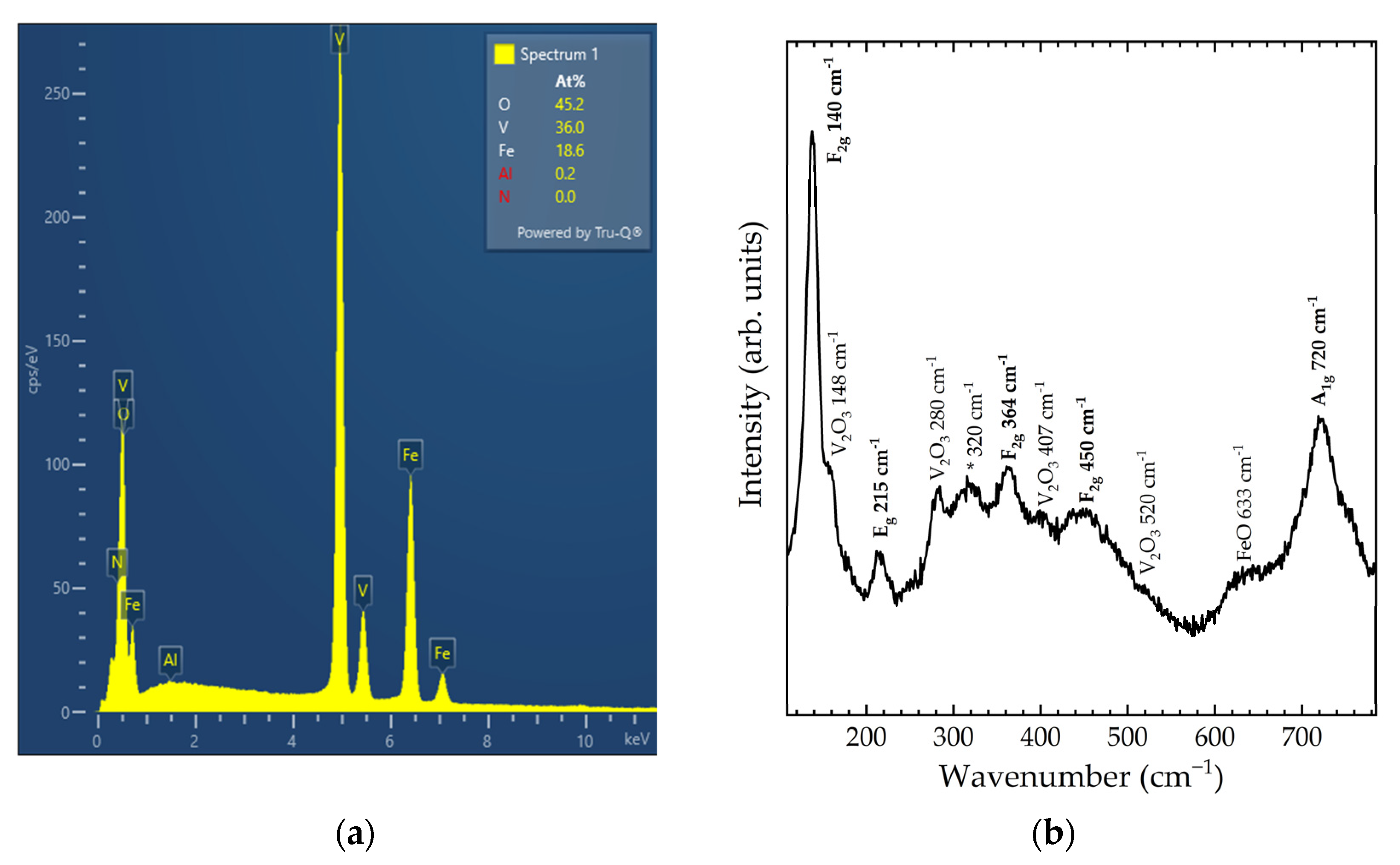
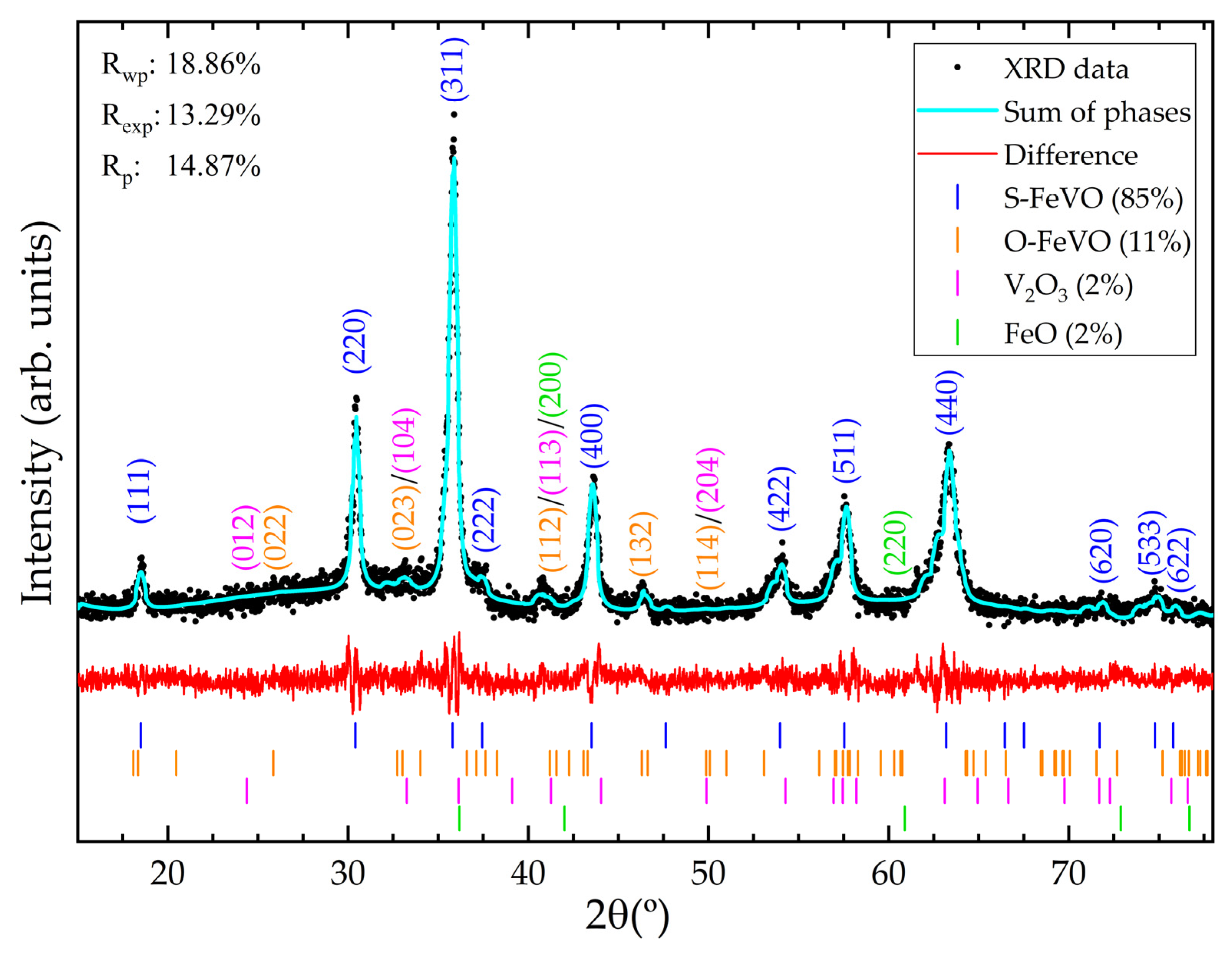
3.1. Ambient Conditions Sample Characterization of Starting Material
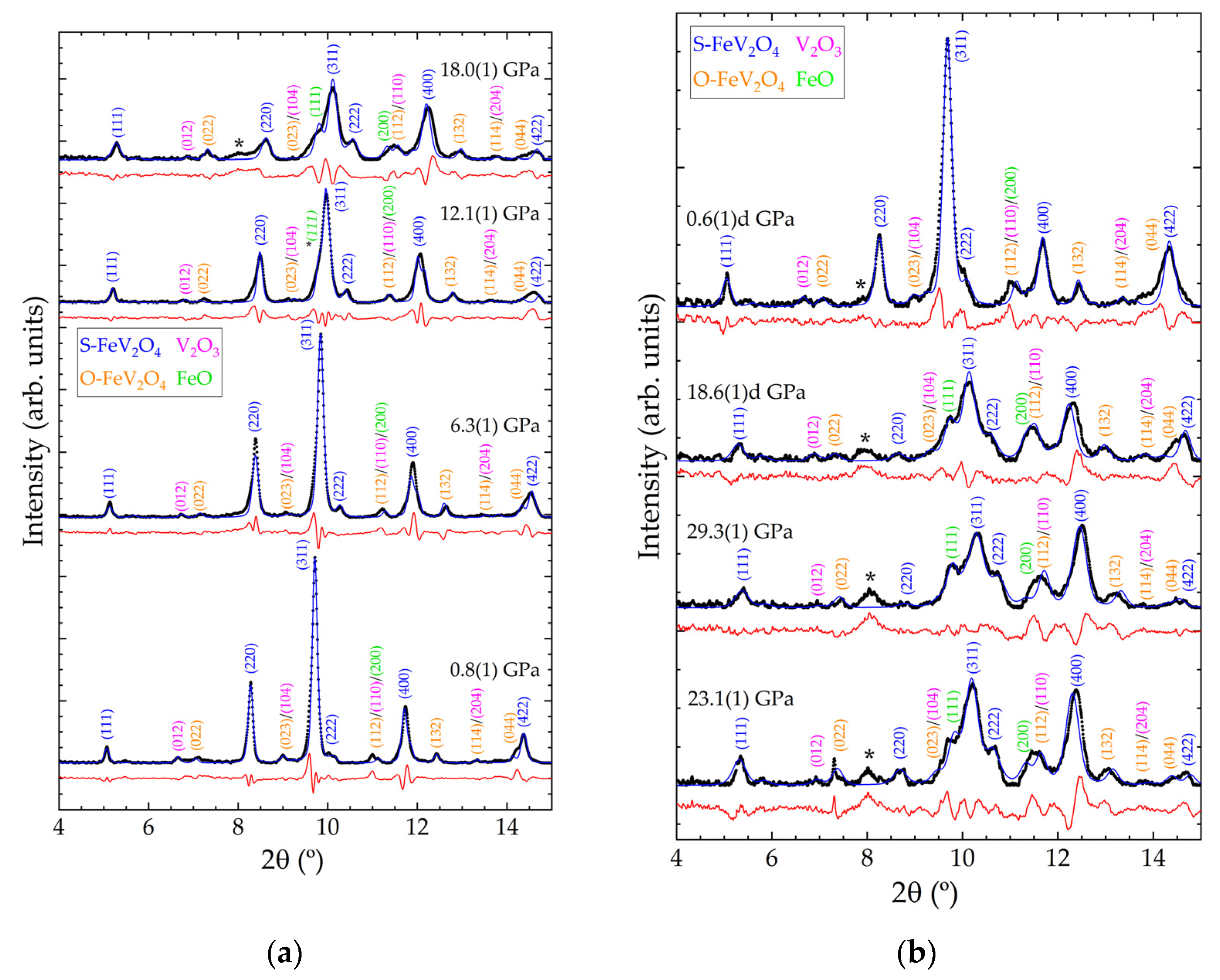
3.2. High-Pressure Powder X-ray Diffraction Analysis
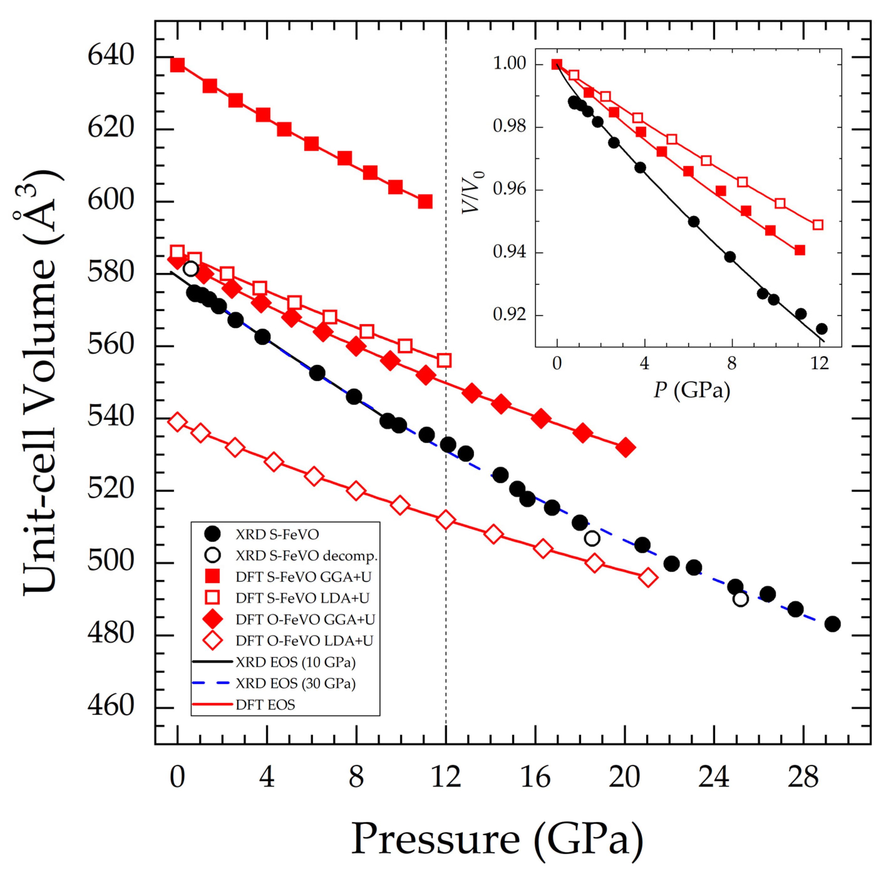
3.3. Unit-Cell Volume and Equation of State (EOS) Parameters
4. Conclusions
Author Contributions
Funding
Data Availability Statement
Acknowledgments
Conflicts of Interest
References
- Bragg, W.H. XXX. The structure of the spinel group of crystals. Philos. Mag. 1915, 6, 305–315. [Google Scholar] [CrossRef] [Green Version]
- Arita, R.; Held, K.; Lukoyanov, A.V.; Anisimov, V.I. Doped Mott Insulator as the Origin of Heavy-Fermion Behavior in LiV2O4. Phys. Rev. Lett. 2007, 98, 166402. [Google Scholar] [CrossRef] [PubMed] [Green Version]
- Biagioni, C.; Pasero, M. The systematics of the spinel-type minerals: An overview. Am. Miner. 2014, 99, 1254–1264. [Google Scholar] [CrossRef]
- Krishna, J.; Singh, N.; Shallcross, S.; Dewhurst, J.K.; Gross, E.K.U.; Maitra, T.; Sharma, S. Complete description of the magnetic ground state in spinel vanadates. Phys. Rev. B 2019, 100, 081102. [Google Scholar] [CrossRef] [Green Version]
- Tsurkan, V.; von Nidda, H.-A.K.; Deisenhofer, J.; Lunkenheimer, P.; Loidl, A. On the complexity of spinels: Magnetic, electronic, and polar ground states. Phys. Rep. 2021, 926, 1–86. [Google Scholar] [CrossRef]
- Dunn, J. A The mineral deposits of eastern Singhbhum and surrounding areas. Mem. Geol. Surv. India 1937, 69, 21. [Google Scholar]
- Radtke, A.S. Coulsonite, FeV2O4, a spinel-type mineral from Lovelock, Nevada. Am. Miner. 1962, 47, 1284–1291. [Google Scholar]
- Reuter, B.; Riedel, E.; Hug, P.; Arndt, D.; Geisler, U.; Behnke, J. Zur kristallchemie der vanadin(III)-spinelle. Z. Anorg. Allg. Chem. 1969, 369, 306–312. [Google Scholar] [CrossRef]
- Siratori, K. Effect of the Crystal Deformation on the Lattice Vibration of Oxide Spinels. JPSJ 1967, 23, 5. [Google Scholar] [CrossRef]
- Shahi, P.; Singh, R.; Kumar, S.; Dubey, D.K.; Singh, D.N.; Tiwari, A.; Tripathi, A.; Ghosh, A.K.; Chatterjee, S. Effect of Li doping on magnetic and transport properties of CoV2O4 and FeV2O4. arXiv 2015, arXiv:1411.2415. [Google Scholar]
- Van Vuuren, C.P.J.; Stander, P.P. The oxidation kinetics of FeV2O4 in the range 200–580 °C. Thermochim. Acta 1995, 254, 227–233. [Google Scholar] [CrossRef]
- Gupta, M.P.; Mathur, H.B. The cation distribution in the ferrite FeV2O4: Mossbauer and X-ray diffraction studies. J. Phys. C Solid State Phys. 1975, 8, 370. [Google Scholar] [CrossRef]
- Roulland, F.; Roseau, G.; Pena-Corredor, A.; Wendling, L.; Krieger, G.; Lefevre, C.; Trassin, M.; Pourroy, G.; Viart, N. Promoting the magnetic exchanges in PLD deposited strained films of FeV2O4 thin films. Mater. Chem. Phys. 2022, 276, 125360. [Google Scholar] [CrossRef]
- Maggay, I.V.B.; De Juan, L.M.Z.; Lu, J.S.; Nguyen, M.T.; Yonezawa, T.; Chan, T.S.; Liu, W.R. Electrochemical properties of novel FeV2O4 as an anode for Na-ion batteries. Sci. Rep. 2018, 8, 8839. [Google Scholar] [CrossRef]
- Janani, B.; Swetha, S.; Syed, A.; Elgorban, A.M.; Zaghloul, N.S.S.; Thomas, A.M.; Raju, L.L.; Khan, S.S. Spinel FeV2O4 coupling on nanocube-like Bi2O3 for high performance white light photocatalysis and antibacterial applications. J. Alloys Compd. 2021, 887, 161432. [Google Scholar] [CrossRef]
- Zhao, X.; Han, D.; Dai, M.; Fan, Y.; Wang, Z.; Han, D.; Niu, L. Direct Z-scheme FeV2O4/g-C3N4 binary catalyst for highly selective reduction of carbon dioxide. J. Chem. Eng. 2022, 436, 132051. [Google Scholar] [CrossRef]
- Chinnathambi, A. Synthesis and characterization of spinel FeV2O4 coupled ZnO nanoplates for boosted white light photocatalysis and antibacterial applications. J. Alloys Compd. 2022, 890, 161742. [Google Scholar] [CrossRef]
- Kismarahardja, A.W. Dielectric and Conducting Properties of the Spinel Structures FeV2O4, MnV2O4 and CoV2O4 in High Magnetic Field and under Very High Pressure; FSU: Tallahassee, FL, USA, 2010. [Google Scholar]
- Li, Z.Y.; Li, X.; Cheng, J.G.; Marshall, L.G.; Li, X.Y.A.; dos Santos, M.; Yang, W.G.; Wu, J.J.; Lin, J.F.; Henkelman, G.; et al. Anomalous bulk modulus in vanadate spinels. Phys. Rev. B 2016, 94, 165159. [Google Scholar] [CrossRef] [Green Version]
- Ishii, T.; Sakai, T.; Kojitani, H.; Mori, D.; Inaguma, Y.; Matsushita, Y.; Yamaura, K.; Akagoi, M. High-Pressure Phase Relations and Crystal Structures of Postspinel Phases in MgV2O4, FeV2O4, and MnCr2O4: Crystal Chemistry of AB2O4 Postspinel Compounds. Inorg. Chem. 2018, 57, 6648–6657. [Google Scholar] [CrossRef]
- Fauth, F.; Peral, I.; Popescu, C.; Knapp, M. The new Material Science Powder Diffraction beamline at Alba synchrotron. Powder Diffr. 2013, 28, S360–S370. [Google Scholar] [CrossRef]
- Dewaele, A.; Loubreyre, P.; Mezouar, M. Equations of state of six metals above 94 GPa. Phys. Rev. B 2004, 70, 094112. [Google Scholar] [CrossRef] [Green Version]
- Bortolotti, M.; Lutterotti, L.; Lonardelli, I. ReX: A computer program for structural analysis using powder diffraction data. J. Appl. Cryst. 2009, 42, 538–539. [Google Scholar] [CrossRef]
- Kraus, W.; Nolze, G. POWDER CELL—A program for the representation and manipulation of crystal structures and calculation of the resulting X-ray powder patterns. J. Appl. Cryst. 1996, 29, 301–303. [Google Scholar] [CrossRef]
- Kresse, G.; Hafner, J. Ab Initio Molecular Dynamics for Liquid Metals. Phys. Rev. B 1993, 47, 558. [Google Scholar] [CrossRef]
- Kresse, G.; Furthmüller, J. Efficiency of Ab-Initio Total Energy Calculations for Metals and Semiconductors Using a Plane-Wave Basis Set. Comput. Mater. Sci. 1996, 6, 15–50. [Google Scholar] [CrossRef]
- Kresse, G.; Furthmüller, J. Efficient Iterative Schemes for Ab Initio Total-Energy Calculations Using a Plane-Wave Basis Set. Phys. Rev. B 1996, 54, 11169. [Google Scholar] [CrossRef]
- Perdew, J.P.; Burke, K.; Ernzerhof, M. Generalized Gradient Approximation Made simple. Phys. Rev. Lett. 1997, 77, 3865. [Google Scholar] [CrossRef] [Green Version]
- Alder, D.M.; Alder, B.J. Ground state of the electron gas by a stochastic method. Phys. Rev. Lett. 1980, 45, 566. [Google Scholar] [CrossRef] [Green Version]
- Dudarev, S.L.; Botton, G.A.; Savrasov, S.Y.; Humphreys, C.J.; Sutton, A.P. Electron-energy-loss spectra and the structural stability of nickel oxide: An LSDA+U study. Phys. Rev. B 1998, 57, 1505. [Google Scholar] [CrossRef]
- Blöchl, P.E. Projector Augmented-Wave Method. Phys. Rev. B 1994, 50, 17953. [Google Scholar] [CrossRef] [Green Version]
- Kresse, I.G.; Joubert, D. From ultrasoft pseudopotentials to the projector augmented-wave method. Phys. Rev. B 1999, 59, 1758. [Google Scholar] [CrossRef]
- Monkhorst, H.J.; Pack, J.D. Special Points for Brillouin-Zone Integration. Phys. Rev. B 1976, 13, 5188. [Google Scholar] [CrossRef]
- Le Page, Y.; Saxe, P. Symmetry-General Least-Squares Extraction of Elastic Data for Strained Materials from Ab Initio Calculations of Stress. Phys. Rev. B 2002, 65, 104104. [Google Scholar] [CrossRef]
- Togo, A.; Tanaka, I. First principles phonon calculations in materials science. Scr. Mater. 2015, 108, 1–5. [Google Scholar] [CrossRef] [Green Version]
- Shvets, P.; Dikaya, O.; Maksimova, K.; Goikhman, A. A review of Raman spectroscopy of vanadium oxides. J. Raman Spectrosc. 2019, 50, 1226–1244. [Google Scholar] [CrossRef]
- De Faria, D.L.A.; Silva, S.V.; de Oliveira, M.T. Raman microspectroscopy of some iron oxides and oxyhydroxides. J. Raman Spectrosc. 1998, 28, 873–878. [Google Scholar] [CrossRef]
- Saccone, F.D.; Ferrari, S.; Errandonea, D.; Grinblat, F.; Bilovol, V.; Agouram, S. Cobalt ferrite nanoparticles under high pressure. J. Appl. Phys. 2015, 118, 075903. [Google Scholar] [CrossRef] [Green Version]
- D’Ippolito, V.; Andreozzi, G.B.; Bersani, D.; Lottici, P.P. Raman fingerprint of chromate, aluminate and ferrite spinels. J. Raman Spectrosc. 2015, 46, 1255–1264. [Google Scholar] [CrossRef]
- Byrum, T.M. Raman Scattering Studies of Spinels CoV2O4 and MnV2O4. Ph.D. Dissertation, University of Illinois, Urbana-Champaign, IL, USA, 2016. Available online: https://hdl.handle.net/2142/92693 (accessed on 10 November 2014).
- Singha, M.; Paul, B.; Gupta, R. Low temperature phonon studies and evidence of structure–spin correlations in MnV2O4. J. Appl. Phys. 2020, 127, 145901. [Google Scholar] [CrossRef]
- Errandonea, D.; Ferrer-Roca, C.; Martínez-García, D.; Segura, A.; Muñoz, A.; Rodríguez-Hernández, P.; López-Solano, J.; Alconchel, S.; Sapiña, F. High-pressure X-ray diffraction and ab initio study of Ni2Mo3N, Pd2Mo3N, Pt2Mo3N, Co3Mo3N, and Fe3Mo3N: Two families of ultra-incompressible bimetallic interstitial nitrides. Phys. Rev. B 2010, 82, 174105. [Google Scholar] [CrossRef] [Green Version]
- Errandonea, D.; Kumar, R.S.; Gomis, O.; Manjón, F.J.; Ursaki, V.V.; Tiginyanu, I.M. X-ray diffraction study on pressure-induced phase transformations and the equation of state of ZnGa2Te4. J. Appl. Phys. 2013, 114, 233507. [Google Scholar] [CrossRef]
- Wang, J.; Yip, S. Crystal instabilities at finite strain. Phys. Rev. Lett. 1993, 71, 4182. [Google Scholar] [CrossRef] [PubMed]
- Grimvall, G.; Magyari-Köpe, B.; Ozoliņš, V.; Persson, K.A. Lattice instabilities in metallic elements. Rev. Mod. Phys. 2012, 84, 945. [Google Scholar] [CrossRef]
- Singh, J.; Sahoo, S.S.; Venkatakrishnan, K.; Vaitheeswaran, G.; Errandonea, D. High-pressure study of the aurophilic topological Dirac material AuI. J. Alloys Compd. 2022, 928, 167178. [Google Scholar] [CrossRef]
- Yong, W.; Botis, S.; Shieh, S.R.; Shi, W.; Withers, A.C. Pressure-induced phase transition study of magnesiochromite (MgCr2O4) by Raman spectroscopy and X-ray diffraction. Phys. Earth Planet. Int. 2012, 196, 75–82. [Google Scholar] [CrossRef]
- Errandonea, D.; Kumar, R.S.; Manjón, F.J.; Ursaki, V.V.; Rusu, E.V. Post-spinel transformations and equation of state in ZnGa2O4: Determination at high pressure by in situ X-ray diffraction. Phys. Rev. B 2009, 79, 024103. [Google Scholar] [CrossRef] [Green Version]
- Hazen, R.M.; Yang, H. Effects of cation substitution and order-disorder on P-V-T equations of state of cubic spinels. Am. Miner. 2015, 84, 1956–1960. [Google Scholar] [CrossRef]
- Díaz-Anichtchenko, D.; Turnbull, R.; Bandiello, E.; Anzellini, S.; Achary, S.N.; Errandonea, D. Pressure-induced chemical decomposition of copper orthovanadate (α-Cu3V2O8). J. Mater. Chem. C 2021, 9, 13402–13409. [Google Scholar] [CrossRef]
- Sakuntala, T.; Arora, A.K.; Sivasubramanian, V.; Rao, R.; Kalavathi, S.; Deb, S.K. Pressure-induced amorphization and decomposition in ZrV2O7: A Raman spectroscopic study. Phys. Rev. B 2007, 75, 174119. [Google Scholar] [CrossRef]
- Vyazovkin, S. Kinetic effects of pressure on decomposition of solids. Int. Rev. Phys. Chem 2020, 39, 35–66. [Google Scholar] [CrossRef]
- Quian, Z.; Xiang, W.; Shan, Q. Pressure-Induced Phase Transition of V2O3. Chin. Phys. Lett. 2012, 29, 106101. [Google Scholar] [CrossRef]
- Birch, F. Finite Elastic Strain of Cubic Crystals. Phys. Rev. 1947, 71, 809–824. [Google Scholar] [CrossRef]
- Gonzalez-Platas, J.; Alvaro, M.; Nestola, F.; Angel, R. EosFit7-GUI: A New Graphical User Interface for Equation of StateCalculations, Analyses and Teaching. J. Appl. Cryst. 2016, 49, 1377–1382. [Google Scholar] [CrossRef]
- Klotz, S.; Chervin, J.-C.; Munsch, P.; Le Marchand, G. Hydrostatic Limits of 11 Pressure Transmitting Media. J. Phys. D Appl. Phys. 2009, 42, 075413. [Google Scholar] [CrossRef]
- Janthon, P.; Luo, S.A.; Kozlov, S.M.; Viñes, F.; Limtrakul, J.; Truhlar, D.G.; Illas, F. Bulk Properties of Transition Metals: A Challenge for the Design of Universal Density Functionals. J. Chem. Theory Comput. 2014, 10, 3832–3839. [Google Scholar] [CrossRef] [Green Version]
- Errandonea, D.; Meng, Y.; Somayazulu, M.; Häusermann, D. Pressure-Induced α→ω Transition in Titanium Metal: A Systematic Study of the Effects of Uniaxial Stress. Phys. B 2005, 355, 116–125. [Google Scholar] [CrossRef] [Green Version]
- Pereira, A.L.J.; Errandonea, D.; Beltrán, A.; Gracia, L.; Gomis, O.; Sans, J.A.; García-Domene, B.; Miquel-Veyrat, A.; Manjón, F.J.; Muñoz, A. Structural study of α-Bi2O3 under pressure. J. Phys. Condens. Matter 2013, 25, 475402. [Google Scholar] [CrossRef] [Green Version]
- Rajeswaran, B.; Khomskii, D.I.; Zvezdin, A.K.; Rao, C.N.R.; Sundaseran, A. Field-induced polar order at the Néel temperature of chromium in rare-earth orthochromites: Interplay of rare-earth and Cr magnetism. Phys. Rev. B 2012, 86, 214409. [Google Scholar] [CrossRef] [Green Version]
- Cliffe, M.J.; Goodwin, A.L. PASCal: A Principal Axis Strain Calculator for Thermal Expansion and Compressibility Determination. J. Appl. Cryst. 2012, 45, 1321–1329. [Google Scholar] [CrossRef] [Green Version]
- Ishii, T.; Kojitani, H.; Tsukamoto, S.; Fujino, K.; Mori, D.; Inaguma, Y.; Tsujino, N.; Yoshino, T.; Yamazaki, D.; Higo, Y.; et al. High-pressure phase transitions in FeCr2O4 and structure analysis of new post-spinel FeCr2O4 and Fe2Cr2O5 phases with meteoritical and petrological implications. Am. Miner. 2014, 99, 1788–1797. [Google Scholar] [CrossRef]


| Mode | FeV2O4 (This Work) | CoV2O4 [40] | MnV2O4 [40,41] | |
|---|---|---|---|---|
| Theory (DFT) | Experiment | Experiment | Experiment | |
| F2g | 190 | 140(2) | 192 | 178 |
| Eg | 295 | 215(2) | - | - |
| F2g | 404 | 364(2) | 356 | 348 |
| F2g | 435 | 450(2) | 480 | 476 |
| A1g | 569 | 720(2) | 676 | 656 |
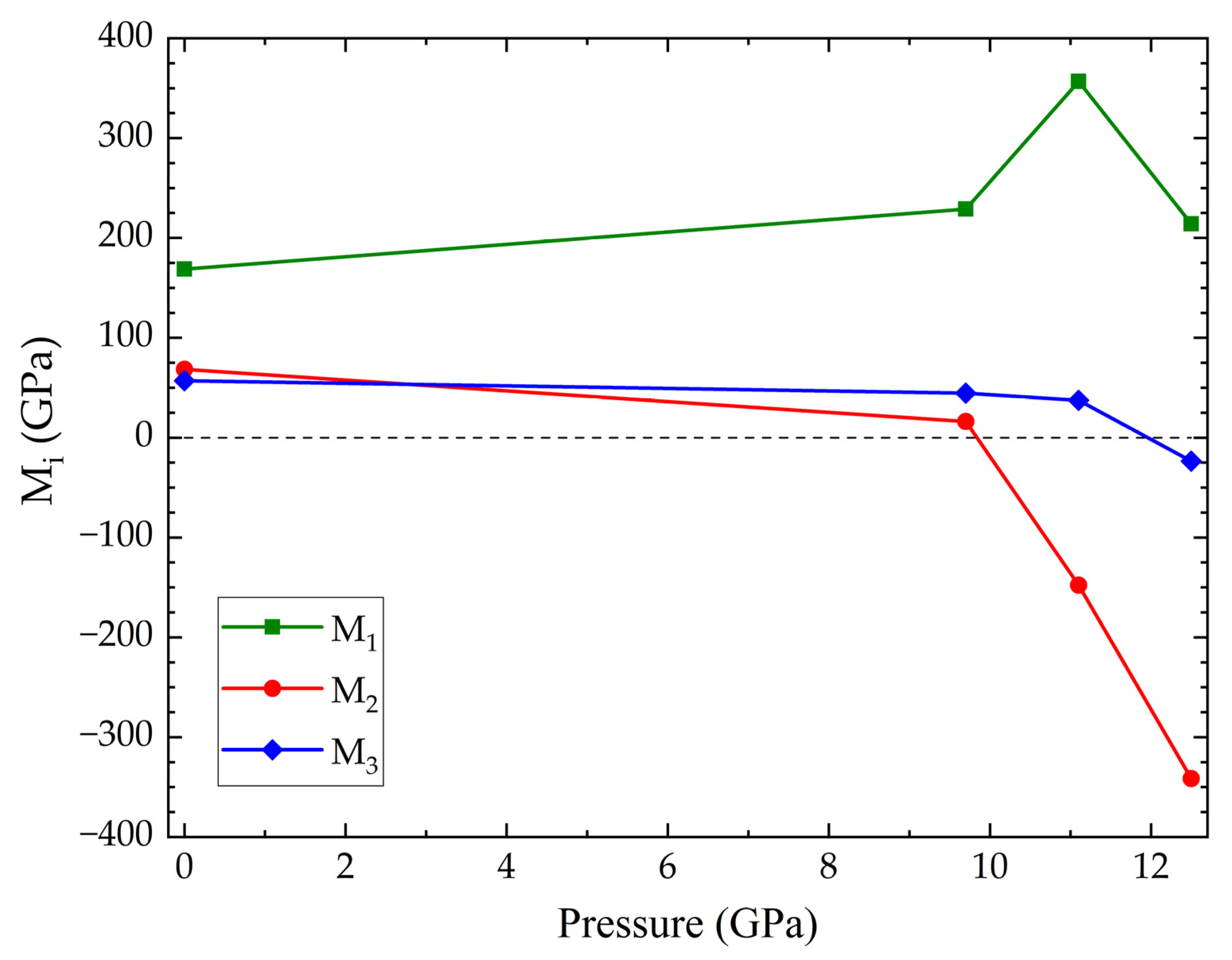
| Elastic Moduli (GPa) | Elastic Constants (GPa) | |||
|---|---|---|---|---|
| 0 GPa | 0 GPa | 9.7 GPa | 11.1 GPa | 12.5 GPa |
| Bulk modulus = 168.7 | C11 = 259.9 | C11 = 260 | C11 = 171 | C11 = −229 |
| Shear modulus = 61.3 | C12 = 123.2 | C12 = 54 | C12 = 444 | C12 = 429 |
| Young modulus = 164.1 | C44 = 57.0 | C44 = 49 | C44 = 49 | C44 = −11 |
| Source | V0(Å3) | B0(GPa) | B0′ |
|---|---|---|---|
| S-FeVO up to 9.9(1) GPa | 579.0(6) | 123(9) | 3.2(15) |
| 579.2(4) | 118.1(17) | 4(fixed) | |
| S-FeVO up to 29.3(1) GPa | 581.7(12) | 122(6) | 2.8(4) |
| 584.1(11) | 106(2) | 4(fixed) | |
| S-FeVO DFT calculations GGA+U | 638.3(3) | 154(2) | 4.99(18) |
| 637.2(3) | 166.5(10) | 4(fixed) | |
| S-FeVO DFT calculations LDA+U | 586.10(5) | 206.4(15) | 3.5(3) |
| 586.16(4) | 203.6(5) | 4(fixed) | |
| O-FeVO DFT calculations GGA+U | 291.95(14) | 173.3(4) | 4.66(5) |
| 291.82(4) | 179.1(5) | 4(fixed) | |
| O-FeVO DFT calculations LDA+U | 269.40(5) | 211(3) | 5.2(6) |
| 269.26(7) | 214.1(18) | 4(fixed) |
Disclaimer/Publisher’s Note: The statements, opinions and data contained in all publications are solely those of the individual author(s) and contributor(s) and not of MDPI and/or the editor(s). MDPI and/or the editor(s) disclaim responsibility for any injury to people or property resulting from any ideas, methods, instructions or products referred to in the content. |
© 2022 by the authors. Licensee MDPI, Basel, Switzerland. This article is an open access article distributed under the terms and conditions of the Creative Commons Attribution (CC BY) license (https://creativecommons.org/licenses/by/4.0/).
Share and Cite
Sánchez-Martín, J.; Turnbull, R.; Liang, A.; Díaz-Anichtchenko, D.; Rahman, S.; Saqib, H.; Ikram, M.; Popescu, C.; Rodríguez-Hernández, P.; Muñoz, A.; et al. High-Pressure X-ray Diffraction and DFT Studies on Spinel FeV2O4. Crystals 2023, 13, 53. https://doi.org/10.3390/cryst13010053
Sánchez-Martín J, Turnbull R, Liang A, Díaz-Anichtchenko D, Rahman S, Saqib H, Ikram M, Popescu C, Rodríguez-Hernández P, Muñoz A, et al. High-Pressure X-ray Diffraction and DFT Studies on Spinel FeV2O4. Crystals. 2023; 13(1):53. https://doi.org/10.3390/cryst13010053
Chicago/Turabian StyleSánchez-Martín, Josu, Robin Turnbull, Akun Liang, Daniel Díaz-Anichtchenko, Saqib Rahman, Hajra Saqib, Mujtaba Ikram, Catalin Popescu, Plácida Rodríguez-Hernández, Alfonso Muñoz, and et al. 2023. "High-Pressure X-ray Diffraction and DFT Studies on Spinel FeV2O4" Crystals 13, no. 1: 53. https://doi.org/10.3390/cryst13010053