

Formation of α-Hemihydrate Inside of a Gypsum Crystal during the Dehydration Process
by
 , , and
Christian Pritzel
1,2,*,
Mohammadamin Emami
1,3,
Sandra Afflerbach
4,
Manuela Killian
2 and
Reinhard Trettin
1 1
Institute of Building Material Chemistry, University of Siegen, Paul-Bonatz Str. 9-11, D-57076 Siegen, Germany
2
Institute for Chemistry and Structure of Novel Materials, University of Siegen, Paul-Bonatz Str. 9-11, D-57076 Siegen, Germany
3
Department of Conservation, Art University of Isfahan, Hakim Nezami St., Isfahan 8173877541, Iran
4
Department of Mechanical Engineering, University of Siegen, Paul-Bonatz Str. 9-11, D-57076 Siegen, Germany
*
Author to whom correspondence should be addressed.
Crystals 2022, 12(12), 1780; https://doi.org/10.3390/cryst12121780
Submission received: 31 October 2022
/
Revised: 27 November 2022
/
Accepted: 29 November 2022
/
Published: 8 December 2022
(This article belongs to the Special Issue Recent Developments of Inorganic Crystalline Materials)
Abstract
:Gypsum (calcium sulfate dihydrate) is one of the most used inorganic binding materials in the world. During calcination, calcium sulfate subhydrates are formed and, for technical reasons, are mixed with water to form dihydrate again. Therefore, the dehydration process of gypsum and the rehydration of hemihydrate were investigated. This dehydration process is technically performed in three different ways. Heating up, i.e., in a rotary kiln, leads to a preferred formation of β-hemihydrate, which crystallizes in comparatively small crystals. Similar results can be achieved by recrystallization from gypsum slurry around 100 °C in an autoclave or under a water steam atmosphere. However, in contrast, the recrystallization process here leads to the formation of a larger, needle-like morphology and sometimes branched α-hemihydrate crystals. The synthesis of β-hemihydrate was investigated in detail with a special thermal stage for optical microscopy on natural single gypsum crystals. It was observed that the crystal loses transparency because of the breaking surface of the crystals due to water evaporation. Furthermore, within a deeper layer of the crystal, new crystals become visible but disappear during dehydration of the upper layers. These are expected to be α-hemihydrate. This theory of the formation of α-hemihydrate inside of a gypsum crystal is experimentally proven in the present work. This work firstly shows that the observed crystallization inside of gypsum during dehydration is the formation of alpha-hemihydrate.
1. Introduction
Gypsum is one of the most used inorganic binding materials in the world. It is used for stucco, floor pavement, gypsum plaster board, gypsum fiber board or even as retarding agent for cement. The global crude gypsum (CaSO4 • 2H2O, calcium sulfate dihydrate: DH) supply was 261 Mt in 2016 according to the U.S. Geological Survey (USGS) [1], and in the EU, in 2020, 57 Mt of gypsum were used [2]. To use gypsum as binding material normally means to mine some raw gypsum from natural resources or to use by-product gypsum such as FGD-gypsum (flue gas desulfurization), followed by a calcination step. During calcination, calcium sulfate subhydrates (CaSO4 • ½ H2O or CaSO4 • ⅔ H2O) are formed. This subhydrate material is mixed with water at a construction site to form dihydrate (gypsum) again. Therefore, the dehydration process of gypsum and the rehydration of hemihydrate were investigated in several studies; the given references will give an overview of the research conducted up to now [3,4,5,6,7,8,9,10,11,12,13,14,15]. This dehydration process is technically performed in three different ways. One way is to heat up dihydrate in a rotary kiln or a vertical digester. The rotary kiln is a continuous process which is often used; the vertical digester is another common alternative batch process. The obtained product is β-hemihydrate (β-HH), which crystallizes in small crystals (Figure 1b). Another way is a recrystallization process at around 100 °C in an autoclave filled with gypsum slurry. In this process, larger hemihydrate crystals can grow with fewer surface defects. A comparable process was shown to be successful under a water steam atmosphere. For this process, mainly bricks of dihydrate powder were pressed and heated in a steam autoclave. The recrystallization process normally leads to larger hemihydrate crystals with a thin needle-like shape, and sometimes some branches were observed. Both syntheses lead to α-hemihydrate (α-HH) as a product. Here, the synthesis of β-HH was investigated in detail with a thermal stage for optical microscopy. For this experiment, a natural single gypsum crystal was used as the starting material. The temperature was increased with a slow heating rate in a thermal stage to investigate the dehydration process. It was obvious that the crystal becomes white and nontransparent because the surface of the crystals breaks due to the evaporating structure of water molecules. In addition to that, another mechanism is observable. In a deeper layer of the crystal, new crystals are visible, but during the dehydration at the upper layers, these crystallites are not detected at a later stage. To elucidate these buried crystals, the material was cleaved along the cleavage planes and investigated with scanning electron microscopy. Fowler et al. published in 1968 [13] that they found such crystallites and postulated that they are α-HH. That is the only paper that the authors found that explains the formation of crystals inside of a dihydrate crystal during dehydration, but it does not prove that it is a formation of alpha-hemihydrate. This theory has to be scrutinized, and the experimental proof has to be optimized. The main focus of the paper is on the formation of alpha-hemihydrate inside of a gypsum crystal during formation of beta-hemihydrate by heating up the gypsum crystal. Inside or on one gypsum crystal, both types of hemihydrates were formed. None of the experiments were performed in an autoclave; gypsum crystals were only heated in a furnace and the crystal seems to be acting as an autoclave. Through several experiments, it was clearly shown that the new-formed crystals inside of the gypsum crystal are alpha-hemihydrate and the experimental proof for Fowler et al. theory was provided. Some technical alpha- and beta-hemihydrates were used as reference. The paper does not deal with the formation of those alpha-hemihydrates in an autoclave, but some of the findings can be transferred to technical processes, especially to the formation of alpha-hemihydrate in steam autoclaves.
The reaction of calcium sulfate hemihydrate with water to form calcium sulfate dihydrate (Equation (1)) is used with inorganic binding materials for different technical uses, as mentioned in the beginning, or as gypsum-based casting material. The hemihydrate is obtained from gypsum in different ways (Equation (2)).
It is possible to heat up crude gypsum to produce β-HH. For this process, dihydrate is normally ground and heated up to ~140 °C to evaporate the crystalline water partly and to obtain β-HH (Figure 1b) in a dry process. The other way is the recrystallization process in water or under steam atmosphere. Below 100 °C, the solubility of dihydrate is lower than the solubility of hemihydrate, leading to the reaction from hemihydrate to dihydrate. The solubility of gypsum is nearly constant with regard to the temperature, but the solubility of hemihydrate decreases with increasing temperature. Above ~100 °C, hemihydrate becomes less soluble than dihydrate, and α-HH (Figure 1a) is formed by consuming dihydrate in a wet process.
Both reactions and the needed reactions’ temperatures can be influenced by several additives as well as applied pressure. The wet process can be performed in an autoclave with water or under a steam atmosphere. This recrystallization leads to larger crystals with a smaller surface and fewer defects compared to the dry process. This diverse crystal morphology might influence the hydration reaction since the dissolution rate of the two types of hemihydrates is different.
The dry process was investigated in more detail with optical microscopy to obtain a deeper understanding of the reaction mechanism. In addition, it was examined whether the dehydration, i.e., the evaporation of crystalline water from the gypsum crystal, proceeds by each layer or if it already starts in deeper layers inside of the gypsum crystal. It can be assumed that if the dehydration process also happens inside the deeper layers of the gypsum crystal, it should be possible to form conditions like in a steam autoclave process, and α-HH should be recrystallized. Alternatively, the dehydration could proceed from outside to inside. Because hemihydrate formation is endothermic, energy is consumed by dehydrating the outer layers of the gypsum crystal. In this case, the inner part of the crystal should not reach the temperature, which is necessary for the formation of α-HH. The formation of beta-hemihydrate is an endothermic reaction as well. During the phase decomposition of dihydrate to hemihydrate, the temperature inside of the crystal should not increase above the decomposition temperature in which dihydrate beta-hemihydrate can be formed.
2. Experimental Section
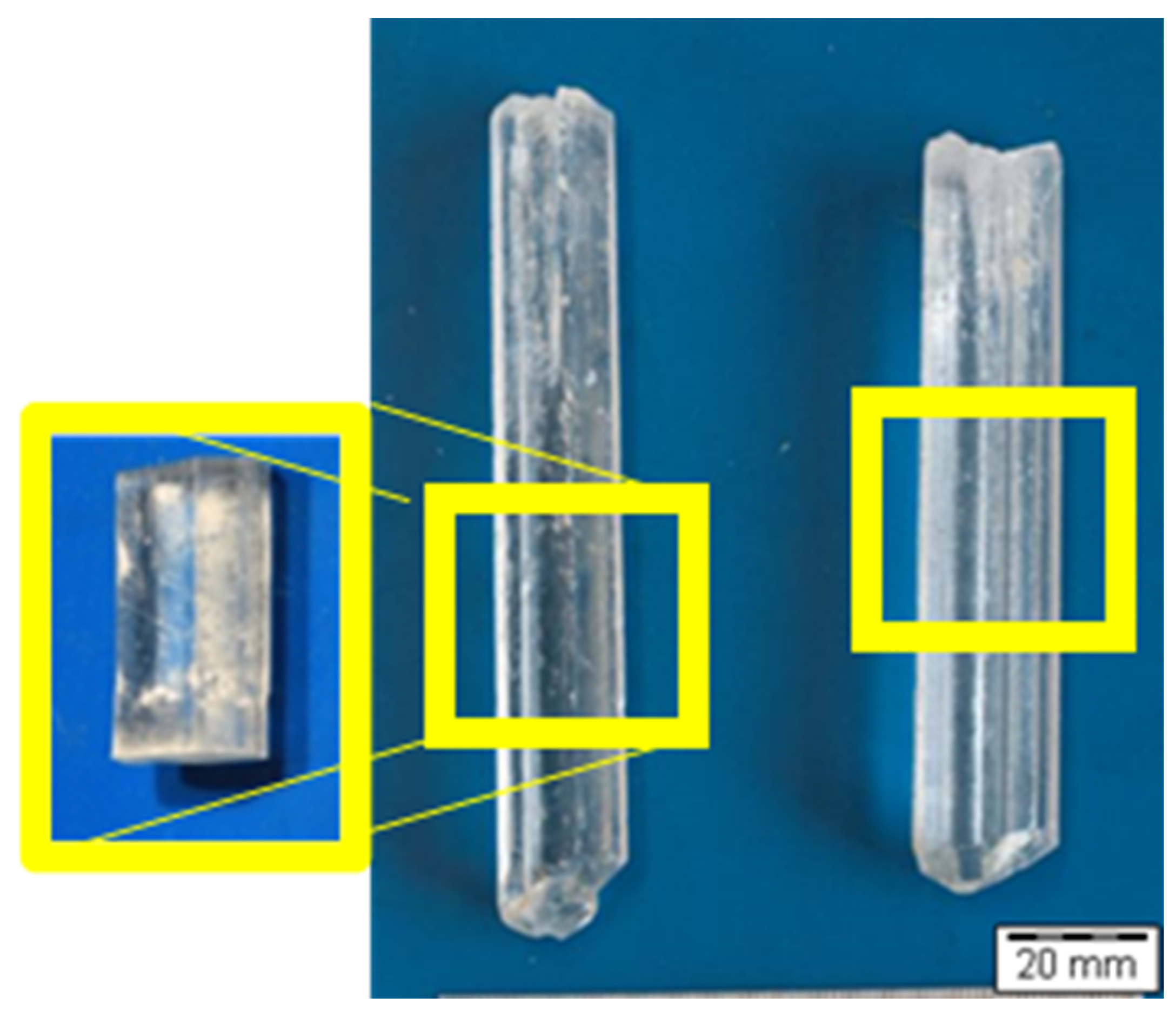
For all analyses, a commercially available natural gypsum crystal with a needle-like, dove-tailed twin shape was cut into pieces along the c-axis and prepared according to the analysis and method instruction. Pieces like the ones marked in yellow were cut for the measurements, and for optical microscopy, slices along the cleavage plane were used. Figure 2 shows the natural gypsum crystals which were used for the experiments. The formation of alpha-hemihydrate, observed in that paper, happened inside of these crystals during dehydration, and formation of beta-hemihydrate happened on the outside.
2.1. Optical Microscopy
For optical microscopy, a small chip was prepared by cutting a piece of the crystal along the cleavage plane. The crystal was placed in a Linkam thermal stage for optical microscopy. The sample was heated inside of the cell with 5K per minute starting from 20 °C, and the process was observed with an Olympus BX 61 optical microscope from Olympus Deutschland GmbH, Hamburg, Deutschland, in bright field and reflection light mode.
2.2. Scanning Electron Microscopy
A first step was a detailed phenomenological analysis of the two different structures. For a more detailed insight at a higher resolution, scanning electron microscopy (SEM) was applied. Therefore, the crystals from the optical microscopy analysis were adhered with silver glue to a sample holder and investigated with a FEI FEG Quanta 240 ESEM in low-vacuum mode at around 100 Pa and 30 keV. The advantages of the ESEM (Environmental Scanning Electron Microscopy) technique compared to common SEM are that sputtering with conductive material is nonessential and dehydration by low pressure is not to be expected, as it would happen in normal, high-vacuum SEM. With ESEM, the upper formed structures can be observed directly.
For investigation of deeper inner structures with SEM, another piece of the dehydrated gypsum crystal was stored for 6 h at 140 °C in advance of the cleavage along the cleavage planes. Afterwards, it was possible to observe deeper structures with SEM.
Additional EDX was carried out at a low voltage of 10 keV.
2.3. X-ray Powder Diffraction
The crystalline phase composition and decomposition which were formed inside of the gypsum crystal during dehydration was determined on manually collected crystals on a silicon zero-background holder using a Panalytical X’Pert Pro PW 3040/60 powder diffractometer from Malvern Panalytical Ltd., Malvern, UK.
3. Results and Discussion
3.1. Optical Microscopy
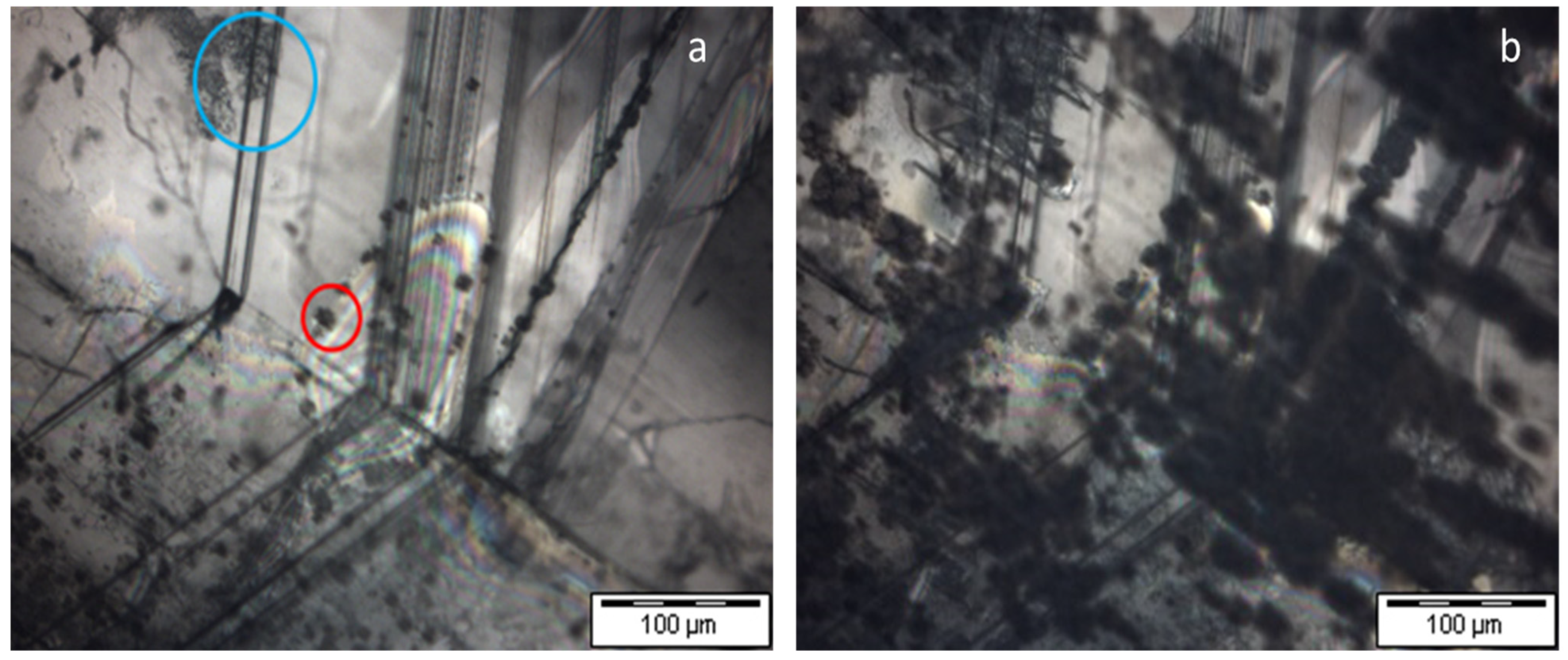
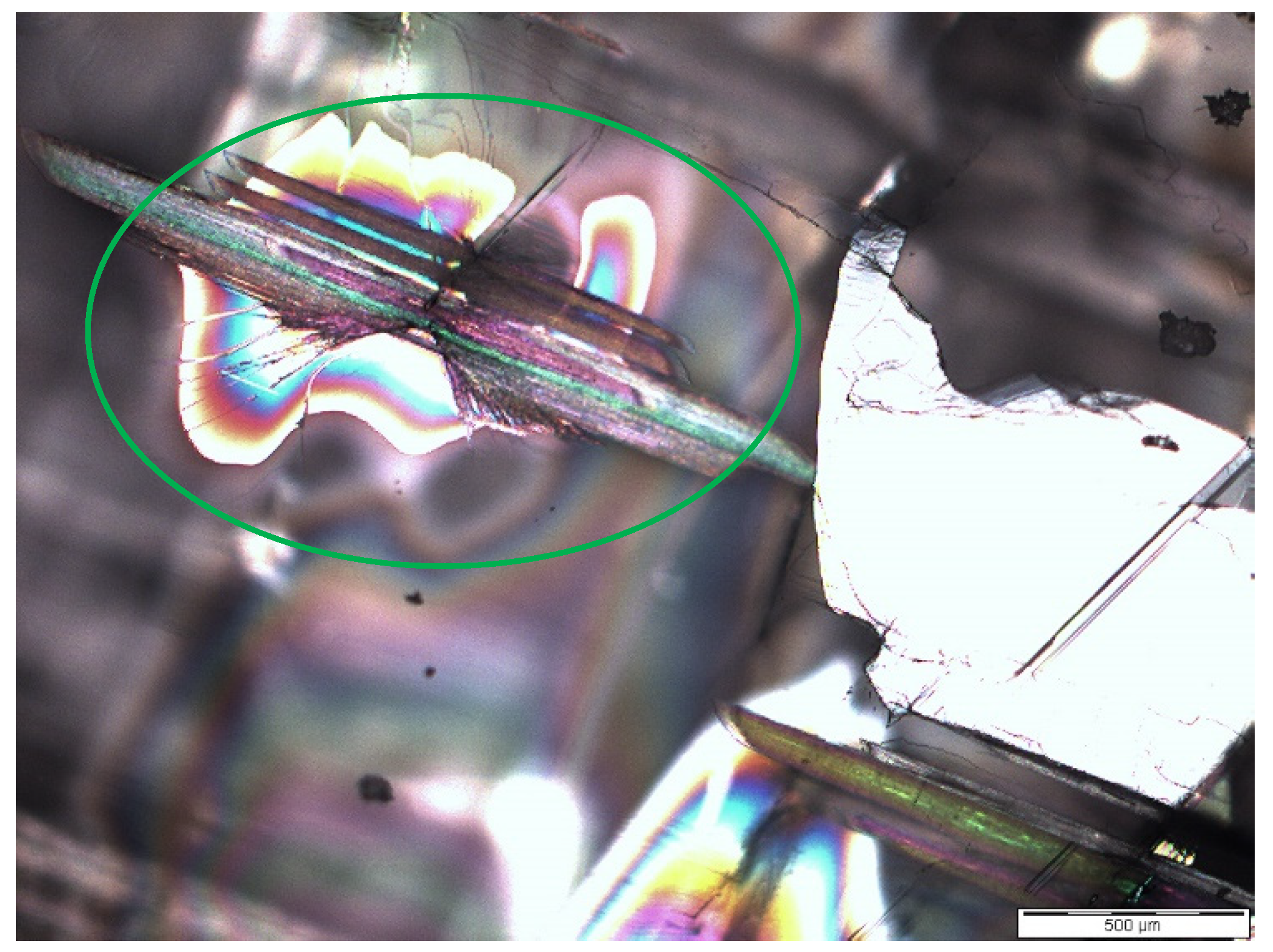
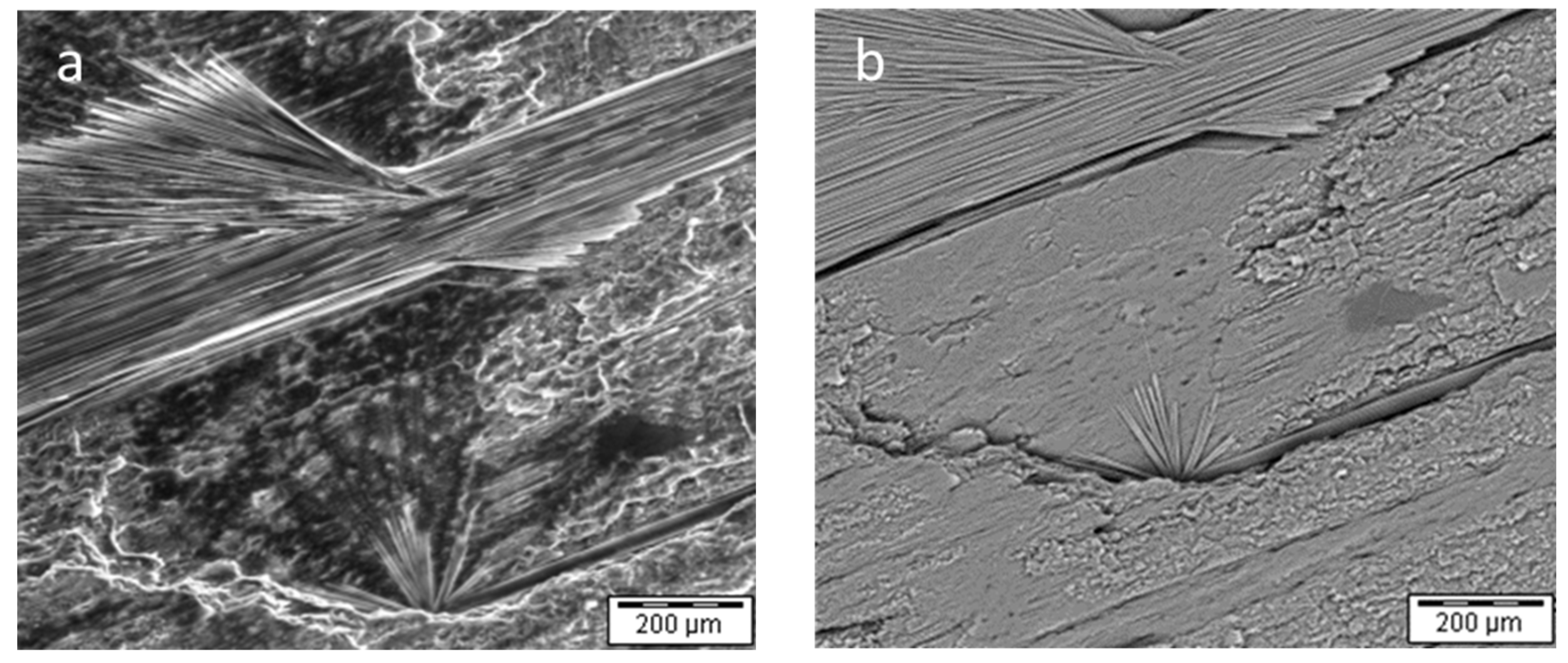
The decomposition process of dihydrate to beta-hemihydrate is investigated by optical microscopy. Small chips of gypsum crystals were subjected to heating on a heating stage from THMS 600-H form company Linkam (Linkam Scientific Instruments Ltd. Waterfield, Tadworth, Surrey, KT20 5LR, UK) and investigated with optical microscopy. After 23 min at ~135 °C, two different processes start. On the one hand, the refraction on the top of the crystal increases because of crack formation (e.g., red circle Figure 3a,b). That recrystallization takes place inside of a gypsum crystal. The crystal partly pushes up single layers along the cleavage planes and introduces new optical transitions from optical denser to less dense media and the other way around. Because of that mechanism, the newly formed crystals inside of the gypsum crystal cannot be observed perfectly. In addition, it is not possible to know in which layer of the gypsum crystal the new crystals are formed, but the formation of both alpha- and beta-hemihydrate is fast, which makes it even more complicated to obtain an as-good-as-possible picture. Consequently, some larger structures of crystals have been published previously by Fowler et al. Cracks (blue circle Figure 3a and green circle in Figure 4) are formed in a deeper layer of the gypsum crystals, which are only visible at the beginning of the reaction. These structures disappear when the first reaction mechanism leads to coverage of the whole surface of the gypsum crystal. If the reaction is stopped sufficiently early to still see these formed structures, they cannot be observed with SEM directly during its formation, which proves that they are formed in a deeper layer of the crystal. Because of the formation inside of a crystal with imperfections and stress during dehydration, it is tricky to obtain perfect pictures of the process even with a good microscope. It is impossible to tell where these crystals are formed, which makes it even more tricky to work with a high magnification, which will lead to less sharpness in the field. It is expected that the same structure may be observed at a fixed temperature after some minutes.
3.2. Scanning Electron Microscopy (SEM)
The surface character of the dehydrated gypsum was analyzed in SEM, additionally. It is obvious that the layer’s burst and broken parts and cracks are formed at the surface. The surface of the hemihydrate is much larger than that of the dihydrate crystal. Formed cracks are shown in Figure 5a,b. Larger, cross-like cracks can be observed beside smaller parallel line-like ones in direction of the c-axis.
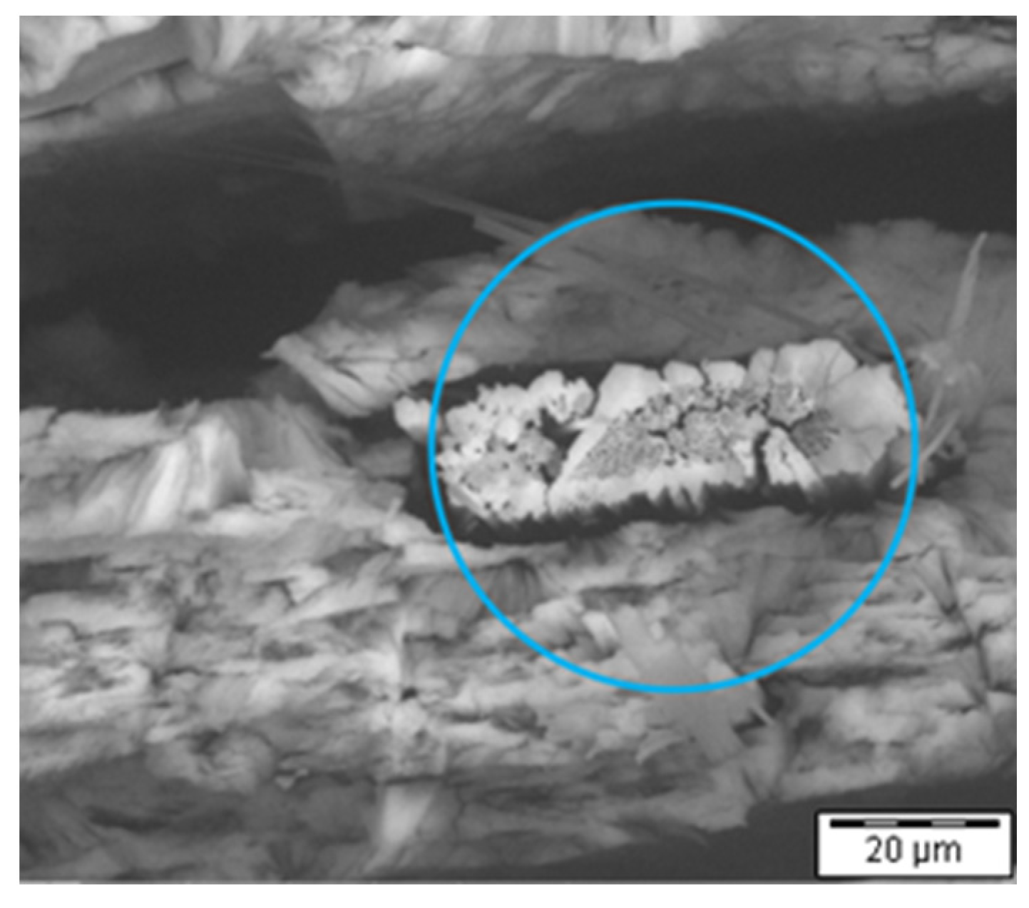
To elucidate the dehydration process in deeper layers of the material, a piece of gypsum cleaved after calcination was examined. Needle-like structures were observed within the crystal. These well-formed deeper structures (Figure 6a,b) could not be observed in the crystal before heating, as the crystal was completely transparent, and such crystallites should have increased the surface, and, therefore, white spots would have been visible.
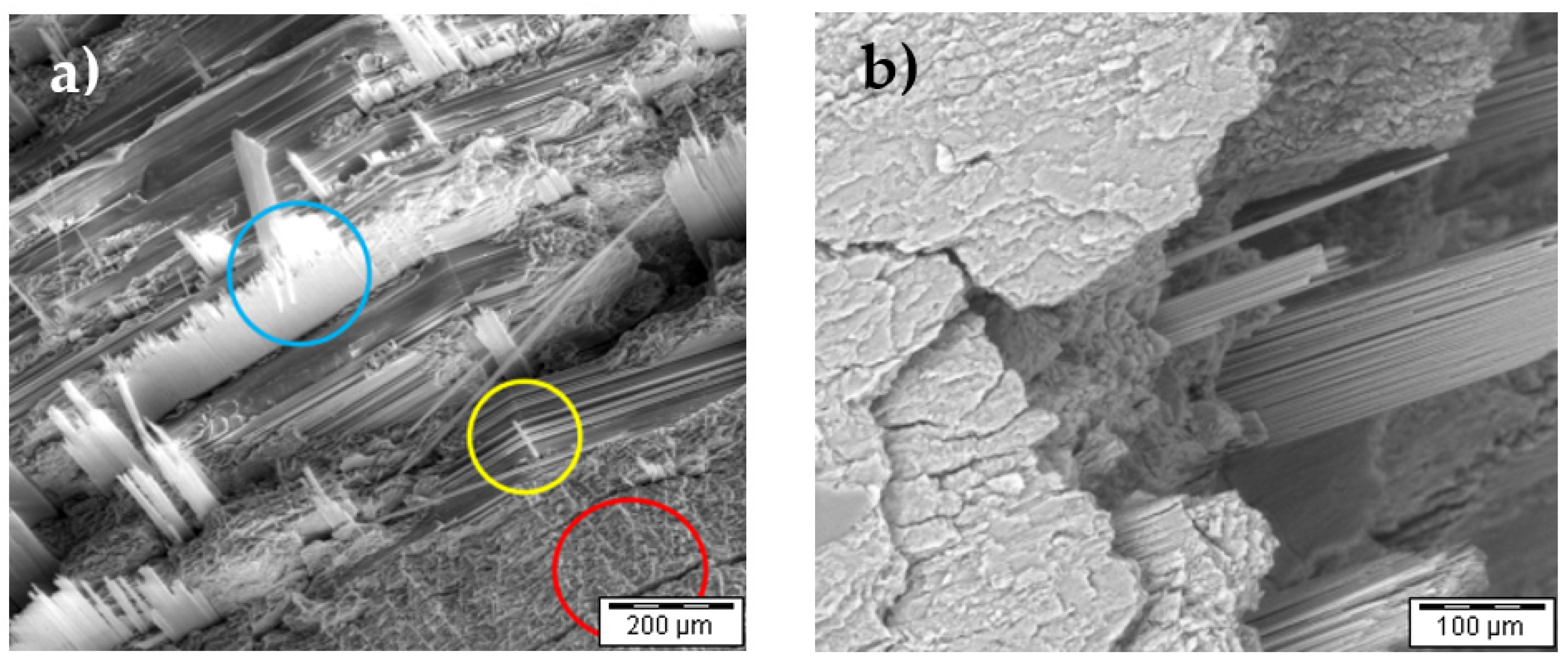
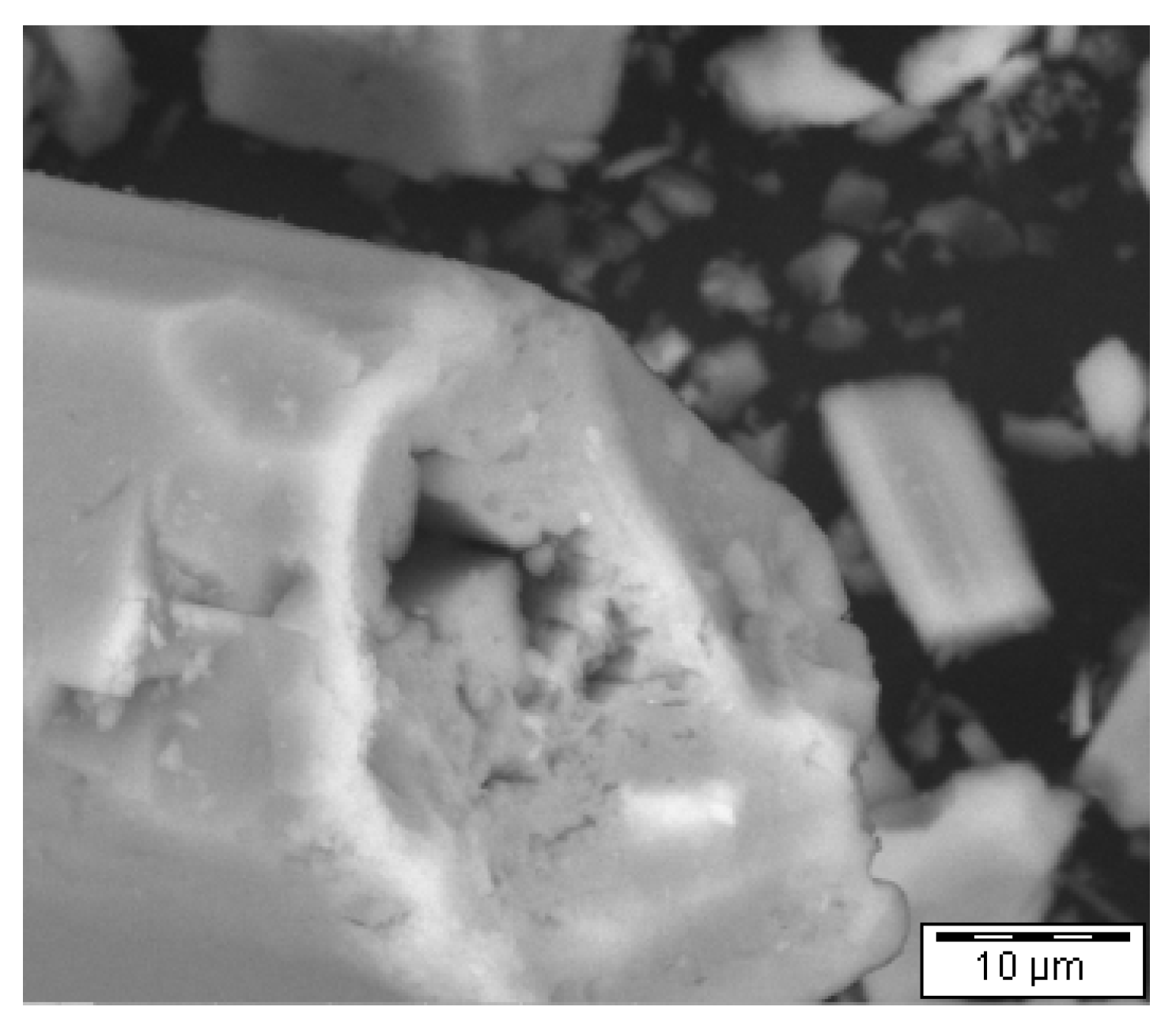
The structures were identified as crystals formed during the dehydration process of gypsum inside of a gypsum crystal. The hypothesis of Fowler et al. [13] remarked that α-HH is formed as water evaporates from the gypsum crystal but not directly by forming β-HH like on the top of the gypsum crystal. It is assumed that a steam atmosphere is formed inside the crystal, which would lead to conditions comparable to the inside of a steam autoclave during the technical formation of α-HH. If that hypothesis is correct, the calcium-to-oxygen ratio or the sulfur-to-oxygen ratio might be expected to be higher on the newly formed crystals compared to dihydrate, and are comparable to the structures of β- HH. Therefore, a semidehydrated gypsum crystal was analyzed with EDX and SEM. EDX is not capable of detecting hydrogen, but if water is evaporated or α-HH is formed, the water amount per formula unit inside of the crystals will be lowered, and, beside the hydrogen content, the oxygen content will decrease and can be quantified. Theoretically, hydrogen would be the better option, because it is only bounded in the water in gypsum and hemihydrate and oxygen is also bounded to sulfur in the sulfate. A new piece of the crystal was stored for a shorter time (around 30 min) at 140 °C and prepared in the same way as for the experiments shown in Figure 6a,b, leading to an incomplete conversion. Figure 7 shows starting material gypsum (yellow circle Figure 7), and the two products, β-HH (red circle Figure 7) and expected α-HH (blue circle Figure 7), on one picture.
Then, EDX was used to analyze the composition of the crystal at different sites (Figure 8). The quantitative EDX results are given in Table 1. It is obvious that in points 1 to 3 (points alpha-HH in crystal after furnace) and 6 (point beta-HH in crystal after furnace), the Ca/O and S/O ratio is comparable and lower than in points 4 and 5 (points DH in crystal after furnace). A piece of the crystal before oven curing and technical alpha- and beta-hemihydrate were measured as reference, and the results are also listed in Table 1. In addition, the theoretical values for hemihydrate and dihydrate were calculated (marked in yellow in Table 1).
Points 4 and 5 are comparable to the gypsum crystal without heat treatment. The measured values did not perfectly fit the calculated values because of the error of measurement of EDX.
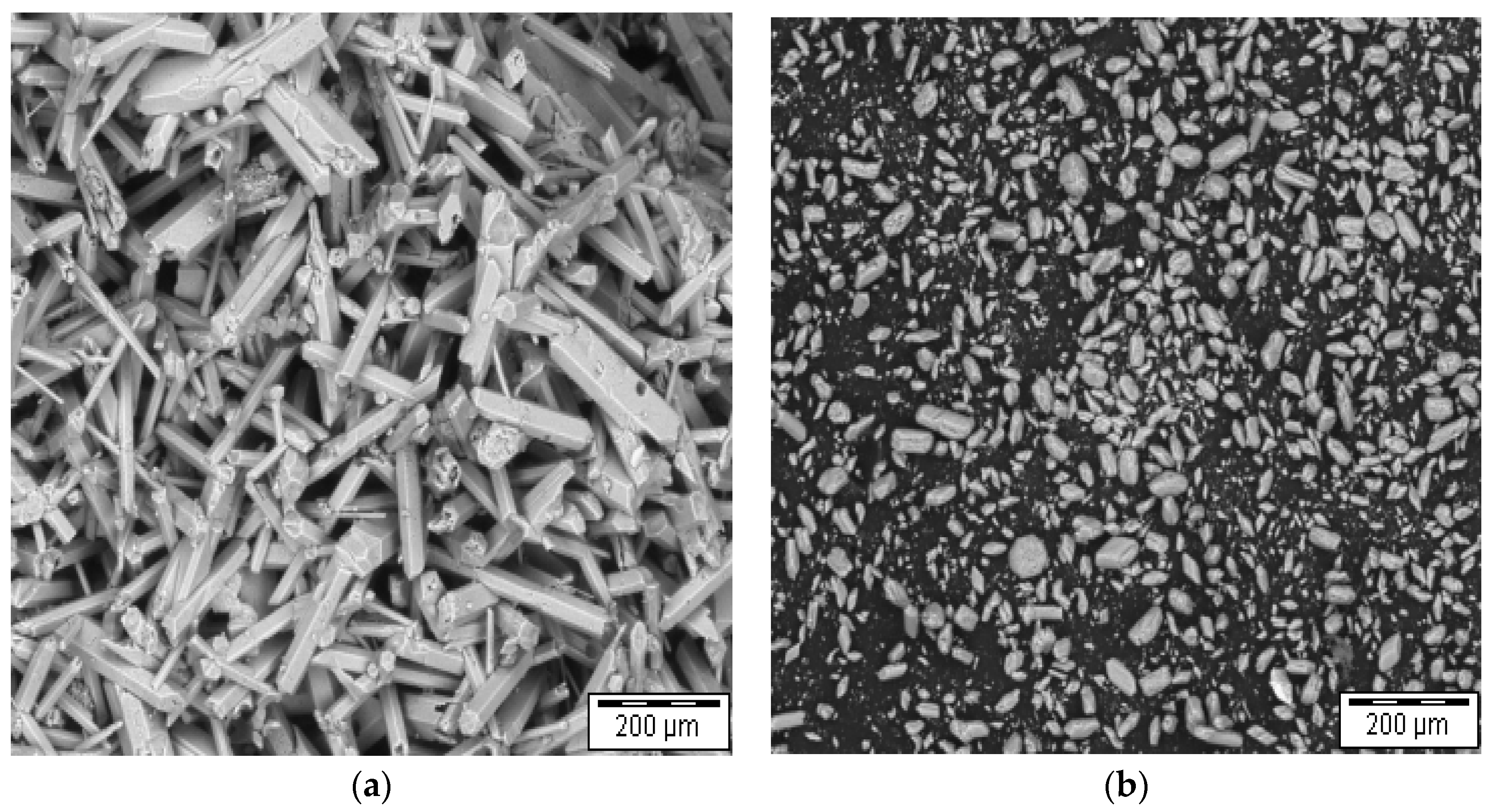
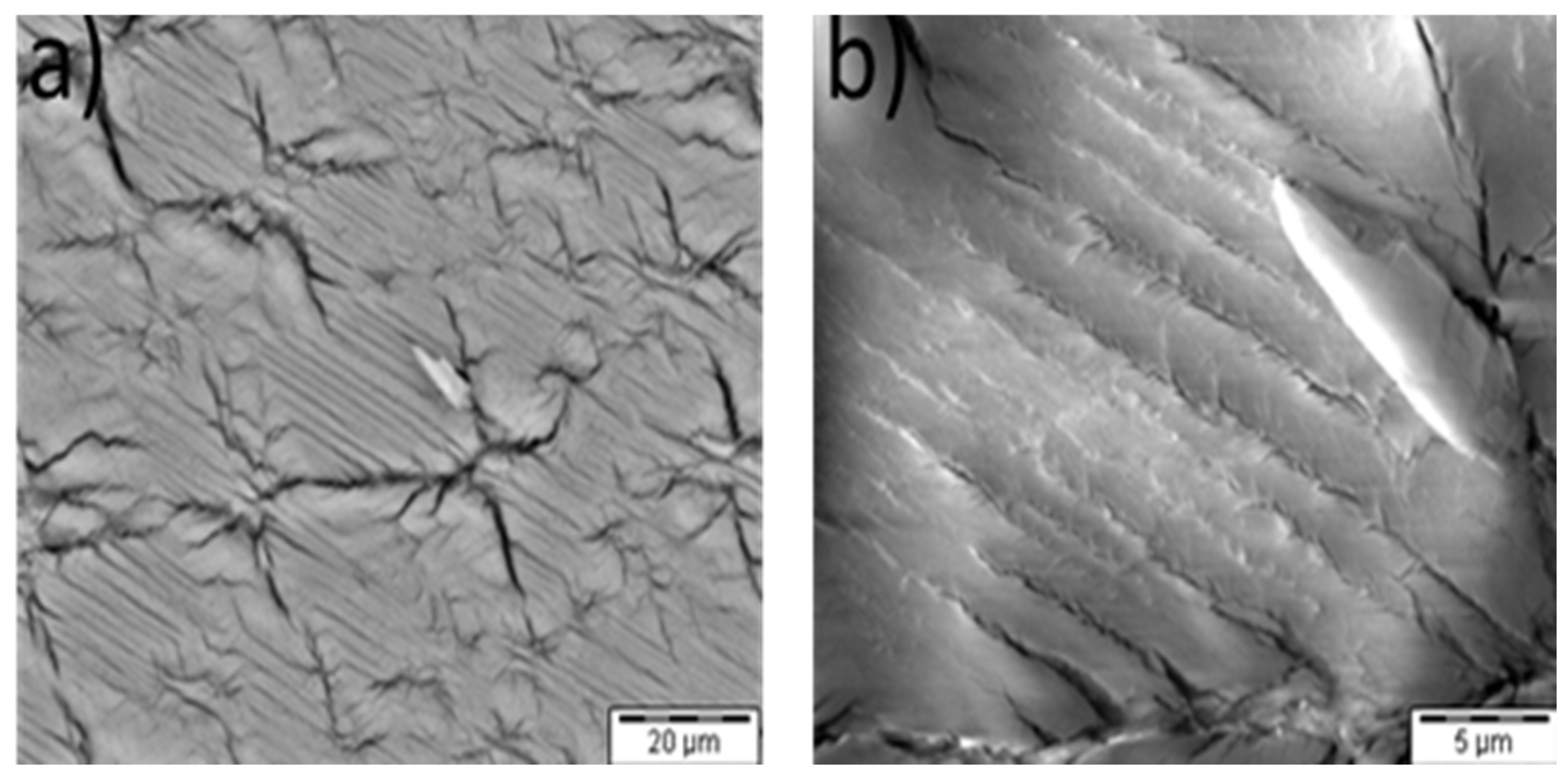
In order to qualify the formed structures with already known morphologies, typical technical steam autoclave α-HH (Figure 1a) and typical technical β-HH (Figure 1b) were investigated under SEM and compared to the observed structures. It is obvious that β-HH has a larger surface and smaller particle size compared to α-HH and a higher defect density. This leads to different hydration properties [16,17].
In Figure 7, the needle-like grown α-HH crystal is marked with a black arrow. It seems that the α-HH crystals are growing in the form of long needles. To prove this assumption, the surface of such crystals of dehydrated gypsum crystal broken along the c-axis were analyzed (Figure 9a,b). In Figure 9a α-HH (blue circle), β-HH (red circle) and dihydrate (yellow circle) are highlighted.
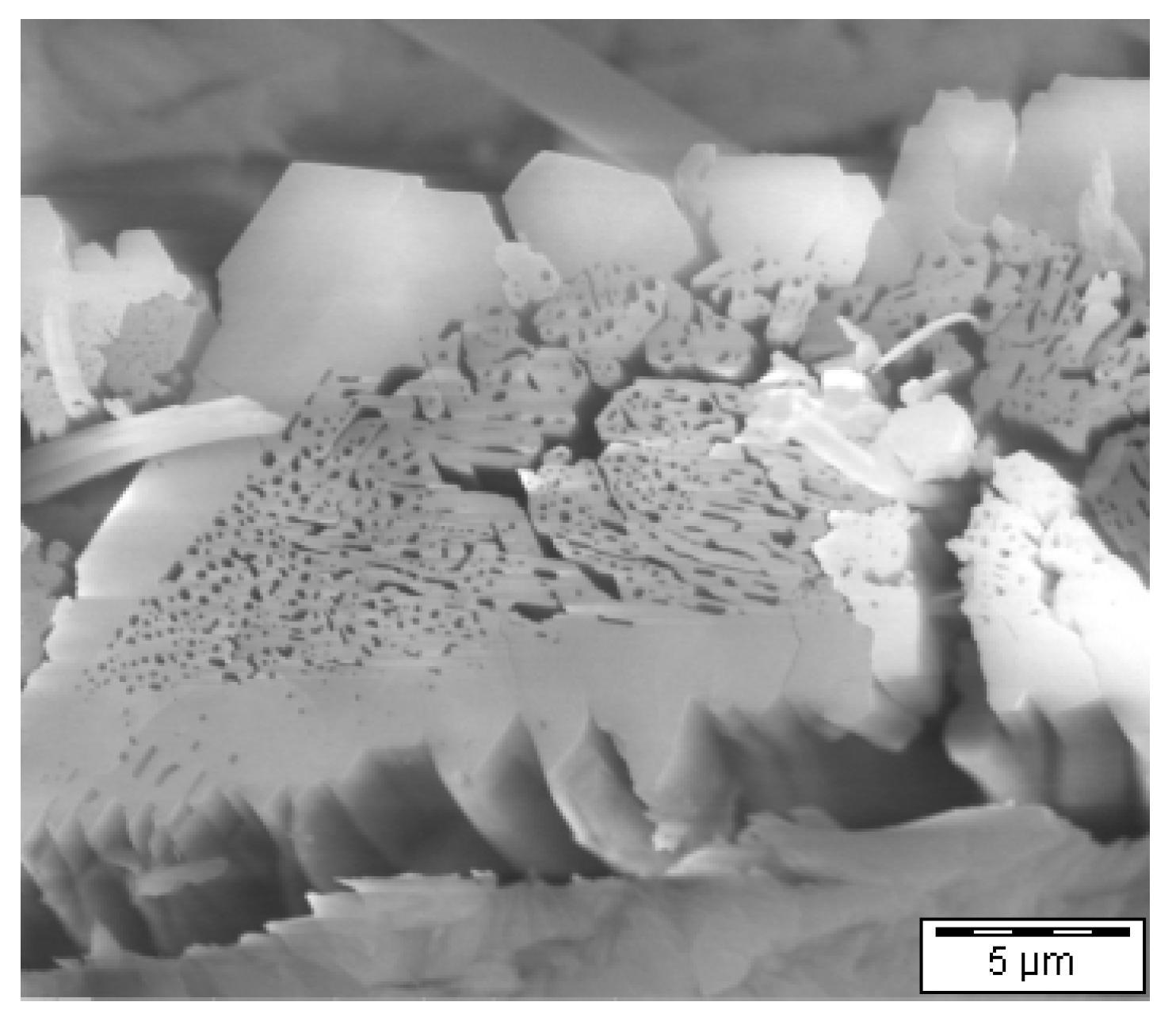
If the formed fiber-like crystals are DH, they are supposed to grow when the crystal in Figure 9a reacts with water, and they should be visible as a “second generation” of gypsum crystals beside smaller hydration products. Figure 9b shows such crystals in a side view. It becomes more obvious that the crystals are growing out of the surface. If it is hemihydrate, they should be consumed, and only one generation of newly formed gypsum crystals should be visible. In order to prove this postulation, the sample was covered with some water after the initial investigation for 4 h and the sample was examined again. In Figure 5 it can be seen that only one generation of dihydrate is visible, and the needle-like structures (blue circle Figure 10) are consumed, indicating that the needles indeed consist of α-HH. The voids in the applied technical α-HH were investigated in a more detailed fashion (Figure 11 and Figure 12) and compared with those of the α-HH formed inside of the gypsum crystal. Here, it is obvious that the morphology of the holes in the α-HH which is growing inside of the gypsum crystals is the same as the one of technical alpha-hemihydrate crystal (Figure 2 and Figure 3).
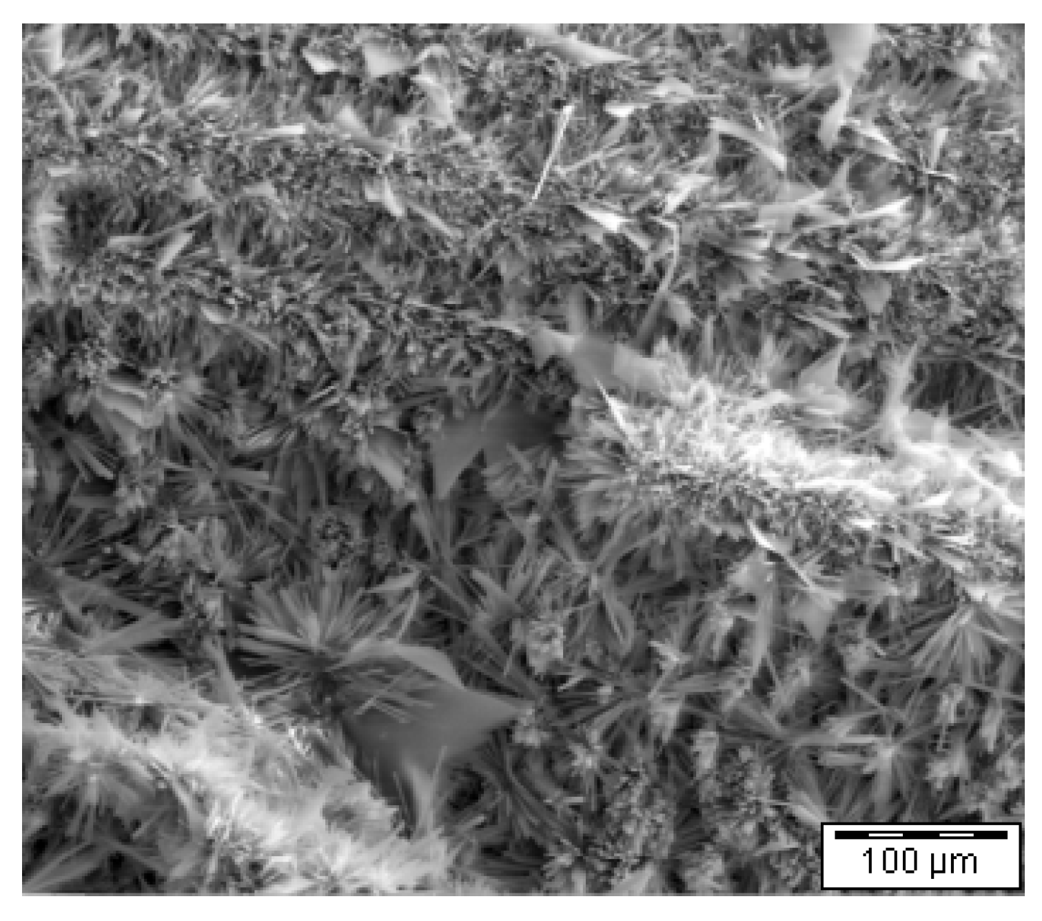
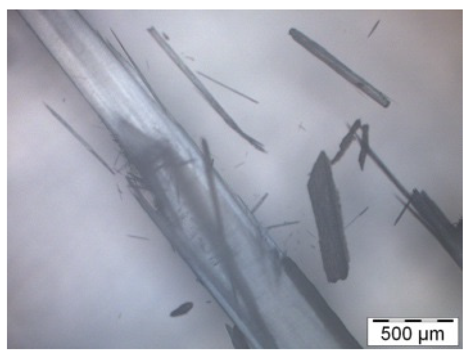
The newly formed α-HH crystals can be collected by tweezers; these crystals are not firmly attached to the gypsum or the β-HH. These α-HH crystals were placed in a special measuring cell for optical microscopy, as described by Pritzel et al. [18], and the rehydration process was followed. The results after 1 min and 240 min of hydration are given in Figure 13 (SEM) Figure 14 and Figure 15 (optical microscopy), respectively. Long whiskers of dihydrate are visible beside some smaller, star-like gypsum crystals with many branches. The created structures were also studied with ESEM after drying; most of the water was removed before drying to avoid artifacts. The observed structures are comparable in shape to fiber gypsum (Figure 16 and Figure 17). It is well-known that during the natural formation of fiber gypsum, the crystal is imbedded in environmental rock so that there is a force acting against the crystallization. In case of the optical microscopy experiment, the crystal can grow in any direction. This explains the small, highly branched gypsum crystals (Figure 15).
The crystals which are formed inside of the gypsum crystal during dehydration were collected and measured on a zero-background holder with X-ray powder diffraction. The only phase which can be found is calcium sulfate hemihydrate, so it is obviously proven that the formed crystals are α-HH (Figure 17).
4. Discussion of the Results
The formation of β-HH starts from small points at the top of gypsum crystals and these fields grow stepwise. The surface of the crystal bursts and the formed β-HH consists of small particles with many defects and a high surface. The deeper layers of the gypsum crystal react mainly in the same way.
In addition to that mechanism, it could be shown, for the first time, that the formation of α-HH inside of a dehydrating gypsum crystal is possible, as postulated by Fowler et al. With XRD, it has been proven that hemihydrate is formed during the dehydration process. Therefore, it is expected that something similar can happen in nature, too, but it is normally not visible because the formed crystals are covered by β-HH. It is hardly possible to find these α-HH crystals with any other technique than SEM on cleaved crystals and optical microscopy, as they are imbedded in a β-HH/DH mixture. Additionally, anhydrite and physically bound water in natural systems complicate the analysis. It opens a new point of view on the formation of fiber gypsum of whether such α-HH can be rehydrated under pressure to fibers of gypsum. The rehydration of the produced α-HH crystals was studied with optical microscopy, and, from the results of that study, such a mechanism can be derived.
It is concluded that the dehydration of gypsum occurs in several steps. The crystalline water which is in between the single layers of calcium sulfate gets evaporated and forms steam conditions inside of the crystal. Consequently, the pressure of the steam increases, and, after exceeding the strength of the calcium sulfate layers above the surface, pops up, and β-HH is formed. During this process, the surface of the formed hemihydrate increases compared to the dihydrate. If the strength is high enough, in case of many layers of calcium sulfate, i.e., the steam is trapped inside of the dehydrating crystal, it is possible that α-HH crystals are formed in deeper layers. The crystallization of α-HH is a solving and recrystallizing process. Because of the recrystallizing process, relatively large α-HH crystals can be formed compared to β-HH.
It is clearly shown that during the dehydration of dihydrate, two different types of hemihydrates are formed. On the one hand, there is beta-hemihydrate, which has a high surface on the outside of the crystal by breaking the surface of the gypsum crystal. On the other hand, an alpha-hemihydrate with larger crystals is formed inside of the gypsum crystal during a recrystallization, like is observed during the formation of alpha-hemihydrate in a steam autoclave. The inner recrystallization can only be overserved with optical microscopy before the beta-hemihydrate is formed on the outer layer of the crystal, because by breaking the surface during beta-hemihydrate formation, a very high surface is formed and the light scattering increases, so that the surface gets white and the reaction in the crystal is no more visible.
It is remarkable that inside of a dehydrating gypsum crystal, it is possible for “autoclave conditions” to form α-HH. The heat can be transported inside of the gypsum crystal and is not totally consumed by forming β-HH from the outside. The formed α-HH crystals are very loosely imbedded in the formed β-HH and can be collected easily with tweezers or a needle for further investigation.
5. Conclusions
The formation of calcium sulfate hemihydrate from calcium sulfate dihydrate is possible in two different ways. The first way is that water breaks the surface of a dihydrate crystal when the pressure inside of the crystal increases because of the higher mobility of the water atoms in the crystal structure. This process normally happens if gypsum is heated up in normal atmosphere. This process forms technical beta-hemihydrate. The second way is the recrystallization of hemihydrate after solving of dihydrate at higher temperatures in liquid water and a steam atmosphere. This recrystallization leads to longer hemihydrate crystals with fewer defects compared to the first one. The formed product is called alpha-hemihydrate. This alpha-hemihydrate process can also happen inside of a dehydrating gypsum crystal, like described by Fowler et al., which was proven by several methods.
Author Contributions
Conceptualization, C.P., R.T. and M.K.; methodology, C.P., S.A. and M.E.; validation, M.E. and S.A.; formal analysis, M.K.; investigation, C.P., S.A. and M.E.; resources, R.T. and M.K.; data curation, C.P.; writing—original draft preparation, C.P.; writing—review and editing, M.E., S.A., M.K. and R.T.; visualization, C.P., M.E. and S.A.; project administration, C.P.; All authors have read and agreed to the published version of the manuscript.
Funding
This research received no external funding.
Acknowledgments
The authors would like to thank company Linkam for lending the thermal stage for optical microscopy.
Conflicts of Interest
The authors declare no conflict of interest.
References
- Crangle, R.D., Jr. U.S. and Global Gypsum Supply Trends, Natural vs Synthetic vs. Recycled Gypsum. In Proceedings of the Global GypSupply Conference, Brussels, Belgium, 13–14 March 2018. [Google Scholar]
- Haneklaus, N.; Barbossa, S.; Basallote, M.D.; Bertau, M.; Bilal, E.; Chajduk, E.; Chernysh, Y.; Chuburi, V.; Cruz, J.; Dziarczykowski, K.; et al. Closing the upcoming EU gypsum gap with phosphogypsum. Resour. Conserv. Recycl. 2022, 182, 106328. [Google Scholar] [CrossRef]
- Eipeltauer, S. Stojydinovic. Aufbereitung und Verwertung von Gipsabfällen und Altgipsformen; Bericht Deutsche Keramische Gesellschaft 37 H. 9; Deutsche Keramische Gesellschaft: Köln, Germany, 1960; pp. 442–447. [Google Scholar]
- Autorenkollektiv. Der Baustoff Gips, 1st ed.; VEB Verlag Bauwesen: Berlin, Germany, 1977. [Google Scholar]
- Hall, C.; Cullen, D.C. Scanning Force Microscopy of Gypsum Dissolution and Crystal Growth. AIChE J. 1996, 42, 232–238. [Google Scholar] [CrossRef]
- Eipeltauer, E.; Moldan, K.; Podest, H. Quantitative Bestimmung von Calciumsulfat (gesamt) über Syngenit in Roh-und Brandgipsen. Zem. Kalk-Gips 1979, 4, 192–194. [Google Scholar]
- Le Chatelier, M.H. Crystalloids against colloids in the theorie of cements. Trans. Faraday Soc. 1919, 14, 8–11. [Google Scholar] [CrossRef]
- Cavazzi, A. Das gelatinöse Calciumsulfat und das Abbinden des Gipses. Kolloid-Zeitschr 1912, 11, 196–201. [Google Scholar]
- Perederij, I.A. Theorie der Bildung, Erhärtung und Festigkeit von normalem Gips und hochfestem Gips GP. Chem. Techn 1956, 8, 659–663. [Google Scholar]
- Eipeltauer, E. Erzeugung von kriechfesten Hartgipsen. Zem. Kalk-Gips 1960, 6, 259–264. [Google Scholar]
- Follner, S.; Wolter, A.; Preusser, A.; Indris, S.; Silber, C.; Follner, H. The Setting Behaviour of α- and β-CaSO4·½ H2O as a Function of Crystal Structure and Morphology. Cryst. Res. Technol. 2002, 37, 1075–1087. [Google Scholar] [CrossRef]
- Trettin, R. Untersuchungen zur Thermischen Zersetzung von CaSO4·2H2O; 1. Marburger Gipstagung (1996), Conference Book; Sondermann, U., Lehmann, K.M., Eds.; Institut für Mineralogie, Petrologie und Kristallographie: Marburg, Germany, 1996; Volume 4, p. 39. [Google Scholar]
- Fowler, A.; Howell, H.G.; Schiller, K.K. The Dihydrate-Hemihydrate Transformation in Gypsum. J. Appl. Chem. 1968, 18, 366–372. [Google Scholar] [CrossRef]
- Pritzel, C.; Trettin, R. Influencing the morphology of gypsum. In Proceedings of the 10th International Congress for Applied Mineralogy (ICAM); Broekmans, A.T.M., Ed.; Springer-Verlag: Berlin/Heidelberg, Germany, 2012; pp. 541–548. ISBN 978-3-642-27681-1. e-ISBN: 978-3-642-27682-8. [Google Scholar]
- Sakalli, Y.; Pritzel, C.; Trettin, R. Investigation of the hydration of calcium sulfate hemihydrate. ZKG Int. 2015, 68, 48–52. [Google Scholar]
- Pritzel, C.; Trettin, R. Investigation of the Hydration of Hemihydrate with Microscopic Methods. In Proceedings of the Thirty-Sixth International Conference on Cement Microscopy, Milan, Italy, 13–17 April 2014; pp. 325–341, ISBN 1-930787-09-X. [Google Scholar]
- Abu Zeitoun, E.; Pritzel, C.; Sakalli, Y.; Trettin, R. Investigation of Hydration of Various Mixtures of Alpha and Beta Hemihydrate; Tagungsbericht 20. In Proceedings of the Internationale Baustofftagung Ibausil Weimar 2018, Weimar, Germany, 12–14 September 2018; pp. 1-883–1-891, ISBN 978-3-00-059950-7. [Google Scholar]
- Pritzel, C.; Kowald, T.; Sakalli, Y.; Trettin, R. Binding materials based on calcium sulphates. In Cementitious Materials: Composition, Properties, Application; Pöllmann, H., Ed.; De Gruyter: Berlin, Germany, 2017; pp. 285–309. ISBN 978-3110473735. [Google Scholar]
Figure 1.
(a) α-HH, technical, steam autoclave; (b) β-HH, technical.

Figure 2.
Used natural gypsum crystals (photo).

Figure 3.
Gypsum crystal at (a) 135 °C, and (b) 142 °C.

Figure 4.
Gypsum crystal, reaction stopped after beginning of dehydration, OM.

Figure 5.
(a,b) Surface of the gypsum crystal after dehydration, SEM, low-vac mode.

Figure 6.
(a,b) Gypsum crystal cleaved along the cleavage plane after dehydration 4 h at 140 °C, SEM, low-vac mode.
Figure 6.
(a,b) Gypsum crystal cleaved along the cleavage plane after dehydration 4 h at 140 °C, SEM, low-vac mode.

Figure 7.
Gypsum crystal partly dehydrated, cleaved along cleavage plane, with observable dihydrate, α-hemihydrate and β-hemihydrate. Yellow circle = still dihydrate, blue circle = alpha hemihydrate, red circle = beta hemihydrate, black arrow = alpha hemihydrate growing direction of a needle like crystal.

Figure 8.
EDX measured at 6 points (highlighted points 1–6), 3× expected α-HH (1,2,3), 2× dihydrate (4,5) and 1× β-HH (6).
Figure 8.
EDX measured at 6 points (highlighted points 1–6), 3× expected α-HH (1,2,3), 2× dihydrate (4,5) and 1× β-HH (6).

Figure 9.
(a,b) α-HH on a broken gypsum crystal (in direction of c-axis of the original dihydrate crystal).
Figure 9.
(a,b) α-HH on a broken gypsum crystal (in direction of c-axis of the original dihydrate crystal).

Figure 10.
α-HH formed in gypsum crystal (blue circle) beside β-HH.

Figure 11.
Voids formed in technical-produced α-HH (steam autoclave).

Figure 12.
α-HH formed in gypsum along c-axis.

Figure 13.
Dehydrated crystal of Figure 9a, rehydration.
Figure 13.
Dehydrated crystal of Figure 9a, rehydration.

Figure 14.
α-HH out of DH after 1 min of hydration.

Figure 15.
α-HH out of DH after 240 min of hydration.

Figure 16.
α-HH out of dihydrate after 240 min of hydration.

Figure 17.
XRD measurement of the formed α-HH.


Table 1.
EDX results of crystal after 140 °C storage.
| Ca (Atomic %) | S (Atomic %) | O (Atomic %) | Ca + S + O (Atomic %) | Ca/S | Ca/O | S/O | |
|---|---|---|---|---|---|---|---|
| Calculated HH | 28 | 22 | 50 | 99 | 1,3 | 0,6 | 0,4 |
| Alpha HH in crystal after furnace point 1 | 30 | 22 | 48 | 100 | 1,3 | 0,6 | 0,5 |
| Alpha HH in crystal after furnace point 2 | 31 | 23 | 46 | 100 | 1,3 | 0,7 | 0,5 |
| Alpha HH in crystal after furnace point 3 | 30 | 22 | 48 | 100 | 1,4 | 0,6 | 0,5 |
| Beta HH in crystal after furnace | 30 | 23 | 47 | 100 | 1,3 | 0,6 | 0,5 |
| Alpha HH technical | 32 | 23 | 45 | 100 | 1,4 | 0,7 | 0,5 |
| Beta HH technical | 31 | 23 | 46 | 100 | 1,3 | 0,7 | 0,5 |
| Calculated DH | 23 | 19 | 56 | 98 | 1,3 | 0,4 | 0,3 |
| DH in crystal after furnace point 1 | 28 | 21 | 51 | 100 | 1,3 | 0,5 | 0,4 |
| DH in crystal after furnace point 2 | 28 | 21 | 52 | 100 | 1,3 | 0,5 | 0,4 |
| DH natural crystal before storage in furnace point 1 | 27 | 21 | 52 | 100 | 1,3 | 0,5 | 0,4 |
| DH natural crystal before storage in furnace point 2 | 28 | 21 | 51 | 100 | 1,3 | 0,6 | 0,4 |
| DH = dihydrate (gypsum); HH = Hemihydrate (bassanite) |
Publisher’s Note: MDPI stays neutral with regard to jurisdictional claims in published maps and institutional affiliations. |
© 2022 by the authors. Licensee MDPI, Basel, Switzerland. This article is an open access article distributed under the terms and conditions of the Creative Commons Attribution (CC BY) license (https://creativecommons.org/licenses/by/4.0/).
Share and Cite
MDPI and ACS Style
Pritzel, C.; Emami, M.; Afflerbach, S.; Killian, M.; Trettin, R. Formation of α-Hemihydrate Inside of a Gypsum Crystal during the Dehydration Process. Crystals 2022, 12, 1780. https://doi.org/10.3390/cryst12121780
AMA Style
Pritzel C, Emami M, Afflerbach S, Killian M, Trettin R. Formation of α-Hemihydrate Inside of a Gypsum Crystal during the Dehydration Process. Crystals. 2022; 12(12):1780. https://doi.org/10.3390/cryst12121780
Chicago/Turabian StylePritzel, Christian, Mohammadamin Emami, Sandra Afflerbach, Manuela Killian, and Reinhard Trettin. 2022. "Formation of α-Hemihydrate Inside of a Gypsum Crystal during the Dehydration Process" Crystals 12, no. 12: 1780. https://doi.org/10.3390/cryst12121780
Note that from the first issue of 2016, this journal uses article numbers instead of page numbers. See further details here.