

Nitrogen Structure Determination in Treated Fancy Diamonds via EPR Spectroscopy


1
Department of Chemical Sciences, Faculty of Natural Sciences, Ariel University, Ariel 4077625, Israel
2
Dianer Diamonds Company, Ltd., The Israeli Diamond Burse, Ramat-Gan 5252005, Israel
3
Department of Chemical Engineering and the Eastern R & D Center, Ariel University, Ariel 4077625, Israel
4
Department of Chemistry and Institute of Nanotechnology & Advanced Materials, Faculty of Exact Sciences, Bar Ilan University, Ramat Gan 5290002, Israel
5
Department of Chemistry, Faculty of Natural Sciences, Ben Gurion University of the Negev, Beer Sheva 8410501, Israel
*
Authors to whom correspondence should be addressed.
Crystals 2022, 12(12), 1775; https://doi.org/10.3390/cryst12121775
Submission received: 29 October 2022
/
Revised: 28 November 2022
/
Accepted: 5 December 2022
/
Published: 7 December 2022
(This article belongs to the Topic Advanced Structural Crystals)
Abstract
:Color induction in nitrogen-contaminated diamonds was carried out via various procedures that involve irradiation, thermal treatments (annealing), and more. These treatments affect vacancy defect production and atom orientation centers in the diamond lattice. Natural diamonds underwent color enhancement treatments in order to produce green, blue, and yellow fancy diamonds. The aim of this study was to follow the changes occurring during the treatment, mainly by EPR spectroscopy, which is the main source for the determination of the effect of paramagnetic centers (carbon-centered radicals) on the color centers produced via the treatments, but also via visual assessment, fluorescence, UV-vis, and FTIR spectroscopy. The results indicate that diamonds containing high levels of nitrogen contamination are associated with high carbon-centered radical concentrations. Four paramagnetic center structures (N1, N4, and P2/W21) were generated by the treatment. It is suggested that the N4 structure correlates with the formation of blue color centers, whereas yellow color centers are attributed to the presence of N1 species. While to produce blue and yellow colors, a thermal treatment is needed after irradiation, for treated green diamonds, no thermal treatment is needed (only irradiation).
1. Introduction
Fancy diamonds (colored diamonds) are rare in nature, as only 0.01% of diamonds that are mined are defined as fancy diamonds. In most cases, the color centers stem from nitrogen contamination in the crystal [1,2]. Diamonds can appear in every possible color: yellow, blue, red, green, purple, and even black [3,4,5]. Today, it is possible to produce desired colors that are found in natural, nontreated diamonds by conducting color enhancement treatments on natural diamonds [6] or by manufacturing synthetic-colored diamonds [7]. The difference in price between color-enhanced and nontreated, natural diamonds is very large. For example, a 1-carat fancy red natural diamond can cost ~USD 1,000,000 [8], whereas the color-enhanced equivalent will cost ~USD 10,000 [9], and the lab-grown version costs USD 2000 [10] (excluding the factors of shape and clarity).
Natural color-enhanced diamonds can be produced via different methods, usually a combination of several treatments. The most employed treatments are HPHT (High Pressure, High Temperature) [11], irradiation (usually in electron accelerators) [12], and thermal treatment (annealing) [13]. HPHT is also used as a method for the production of lab-grown diamonds. The HPHT process involves large presses to produce a ~5 GPa pressure at 1500 °C for several days. The second method is irradiation in a linear accelerator (β irradiation); the dose of irradiation depends on the type of diamond and the color to be induced. Lastly, thermal treatment or annealing is a controlled heating process that is often used after irradiation to bleach undesired colors and achieve an improved final color. For each color production, a different treatment protocol is utilized.
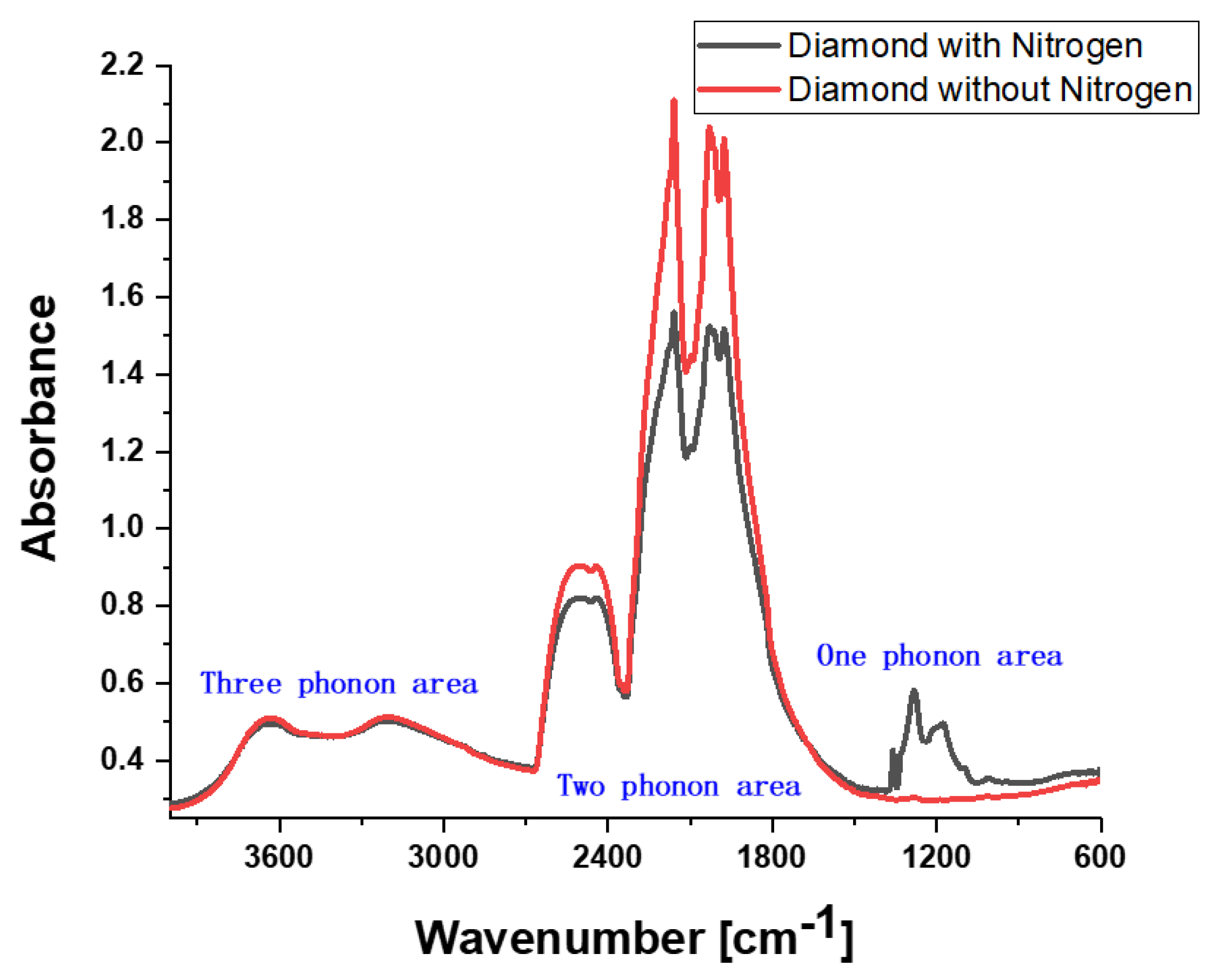
Diamonds are classified into two groups: type I with nitrogen atom/s contamination, which is the common type of diamond and accounts for >99% of natural diamonds, and type II with no nitrogen atom/s contamination. Type I is subdivided into two groups: aggregate nitrogen atoms (Ia type) and isolated nitrogen atoms (type Ib). Type II is also divided into two groups: no nitrogen contamination (IIa type) and no nitrogen but with boron atoms, which is very rare (type IIb). Usually, the type of diamond can be evaluated using Fourier Transform Infra-Red (FTIR) spectroscopy (Figure 1). The spectrum is divided into three areas: a one-phonon area related to nitrogen absorption, and two-phonon and three-phonon areas related to carbon–carbon bond absorption; boron absorption can also be detected in these areas. Hydrogen can be detected in the three-phonon area [14,15]. The nitrogen concentration in ppm can be calculated via spectra and can range from tens to thousands of parts per million [16].
The color of a diamond is a result of optical color centers that are present in the crystal lattice, for which some examples can be seen in Figure 2 [17,18]. There are different structural defects/optical centers in the diamond crystal, which are associated with different colors. A list of some of these defects is presented in Table 1. Some of the optical centers can be detected via different methods: e.g., the N3 center can be selected via EPR and UV.
In some diamonds, fluorescence is observed [19], which could be responsible for the special color centers. The fluorescence (detected by a handheld gemological UV lamp [manufactured by Systems Eickhorst, Hamburg, Germany]) can provide a clue to the diamond’s type. Many type Ia diamonds exhibit blue fluorescence due to nitrogen impurities. Type Ib diamonds are often inert or show weak orange fluorescence under both long- (365 nm) and short-wave (254 nm) UV radiation [14,20]. Synthetic type Ib diamonds commonly display uneven fluorescence patterns that can be used as an identification method for lab-grown diamonds [4]. Type IIb diamonds are often inert to long-wave UV and show weak blue fluorescence under short-wave UV, in addition to occasional blue or red phosphorescence under short-wave UV [21].
In the diamond crystal, there is a very low concentration (<10−5 M) of carbon-centered paramagnetic centers (stable carbon radicals). These paramagnetic centers can be evaluated using EPR (Electron Paramagnetic Resonance) spectroscopy [22,23,24,25]. In EPR, when a radical (defined as an atom with an unpaired electron) under a magnetic field is exposed to microwave radiation, it will subvert from its equilibrium state and will orient in a direction parallel or antiparallel to the direction of the magnetic field. There are two distinct energy levels for the unpaired electron, and measurements are taken as it is driven between the two levels. The two levels/states are denoted as α and β: α is for the high-energy state and β is for the low-energy state. This phenomenon is known as the Zeeman effect. This unpaired electron is characterized by the g-value, which is the fingerprint of each individual species. In addition, an electron-spin interaction with nearby nuclei spins can occur and is called the hyperfine interaction (A), which divides the energy levels and adds energy levels to the system. The difference between the levels is related to the distance of nuclei from the electron: far nuclei will cause a smaller split [26].
Stable carbon-centered paramagnetic centers are affected by the presence of defects (namely, different nitrogen atoms or vacancies) and will have different characteristics because of these defects. Thus, for example, the presence of a nitrogen atom, 14N, which holds a nuclear spin I = 1, will split the energy levels of carbon-centered radicals and will result in a different EPR spectrum. Table 2 presents different types of paramagnetic centers (defects) that have been reported and are correlated with type Ia diamonds and/or treated diamonds (irradiated or thermally treated) [27,28,29,30]. In some cases, a correlation between the EPR center and the optical center is observed, e.g., P2 and the optical center N3, which results in some diamonds having a yellow color [23,25,31].
The research hypothesis was that EPR spectroscopy can be used to determine the correlation between stable carbon-centered radicals in N-contaminated diamonds and the structure of the nitrogen atoms and their color for the following reasons:
- (1)
- It is possible to use EPR spectroscopy to determine the nitrogen atom structure in the diamond lattice surrounding the carbon-centered radicals.
- (2)
- The nitrogen distribution in the diamond crystal is homogeneous.
To test the research hypothesis, fancy color-enhanced diamonds underwent EPR spectroscopy to determine nitrogen contamination’s structural effects on the color achieved by the treatments. EPR spectroscopy provided information regarding the chemical properties of the carbon radical center and the nitrogen impurities surrounding it [32]. The data from EPR were analyzed, and an assignment protocol for the carbon-centered radicals was added to FTIR, fluorescence, and hyperspectral measurements in order to create the schematic mechanism of the change occurring during the treatments of different carbon-centered radicals.
The advantage of using EPR spectroscopy is that one can determine the structure of the nitrogen atoms that are adjacent to carbon-centered radicals, and the concentration of the radicals is much lower than the bulk concentration of nitrogen in the diamond (~1 × 10⁻⁵ M radicals compared to ~1 × 10⁻¹ M nitrogen).
In particular, if the structure of the nitrogen surrounding a carbon-centered paramagnetic center is established, this will provide important information on the structure of the whole crystal. EPR spectroscopy may characterize the structures of nitrogen atoms in Ia diamonds and their effects on the color of the fancy diamond. The data will be supported by FTIR measurements, fluorescence assessment, and hyperspectral measurements in the visible range of light.
2. Methods
The diamonds that were chosen for this study were purchased from a well-known diamond color enhancement company (Dianer Ltd., Ramat Gan Israel) operating in Israel for the last 30 years, which has been established as an excellent quality producer of color-enhanced treated fancy diamonds. Fifteen diamonds were used: five diamonds with similar properties for each color enhancement protocol. Diamonds that were intended for the green protocol weighed 0.1 ± 0.009 ct, blue weighed 0.1 ± 0.005 ct, and yellow weighed 0.1 ± 0.01 ct. All 15 diamonds were transparent (natural, nontreated) brilliantly shaped diamonds. The enhancement protocol was conducted to create green, blue, and yellow coloring. To produce green diamonds, only irradiation was applied in a LINAC (Linear Electron Accelerator, Kibutz Shaar Hagolan, Israel). For blue and yellow, irradiation was conducted after the thermal treatment. Each diamond color group was subjected to different conditions and exposure times in the treatment procedure.
Visual assessment photos of the diamonds were taken in daylight with a magnifying glass attached to the camera lens.
Fluorescence UV-visible spectra were measured pre- and post-treatment via a handheld gemological instrument of long-wave 365 nm radiation (Systems Eickhorst).
Hyperspectral measurements were performed in order to characterize the colors of the diamonds in the visible range; the spectra of the diamonds were measured to finally delineate the treated diamonds by an ASD Inc. Spectrometer with a spectral resolution of 1 mm. This was provided with its three detectors: 2151 channels in the areas of VNIR (0.35–1.0 microns), SWIR1 (1.0–1.79 microns), and SWIR2 (1.8–2.5 microns). After the measurements were completed, a correction was performed according to the SWIR-1 sensor (averaging the measurements according to the mid-range) for each measurement in MatLab software (Product version 8.5.0184244), and a spectral library was created in Excel with spectral data for wavelengths of 350–2500 microns.
FTIR and EPR measurements and data analysis (nitrogen concentration, spin concentration, g-value, hyperfine interaction, and species distribution percentage) were based on data evaluation from a previous paper published by Litvak et al. in 2022 [32].
3. Results and Discussion
3.1. Visual Assessment
All photos of the diamonds, pre- and post-treatment, are presented in Figure 3. The appearance of the pre-green diamonds was between that of a transparent crystal and a slightly yellow/brownish transparent crystal. Most of the pre-blue diamonds were transparent and only one had a slightly brownish tint. Three of the pre-yellow diamonds were clear crystals, one was transparent with a slight yellow tint, and one was transparent but had a slight opaque tint.
The irradiation process that was carried out for the diamonds is as follows.
Production of treated GREEN diamonds: The first stage was 1.5 h of irradiation in the LINAC with an electron energy range of 0.8–2 MeV. No further treatment was needed. The irradiated diamonds had varying green colors: two diamonds had a green-yellowish tint, two diamonds were deep green, and one was green-turquoise. There was no need for further thermal treatment to obtain a green color.
Production of treated BLUE diamonds: The first stage was 2 h of irradiation in the LINAC with an electron energy range of 0.8–2 MeV. In the second stage after the irradiation process, the pre-blue diamonds changed color; all of the diamonds turned green, green-turquoise, or azure, and the diamonds were exposed to thermal treatment at ~400–600 °C for 10–20 min. All of the diamonds acquired a clear blue color, except for one that was dark blue.
Production of treated YELLOW diamonds: The first stage was 2 h of irradiation in the LINAC with an electron energy range of 0.8–2 MeV. The irradiated diamonds turned turquoise to dark turquoise in color. In the second stage after irradiation, the diamonds were exposed to thermal treatment for 10–20 min at ~500–1000 °C, and the color changed to yellow. However, not all of the diamonds turned clear yellow: two turned green-yellow, and the other two turned to more yellow-brownish; only one turned vivid yellow.
The final diamond colors produced were green, blue, and yellow, diverging in their shade/tint/opaqueness; this is of great importance in the diamond trade market. Among the three colors, blue is the most popular, followed by yellow and finally green in the jewelry trade. In general, lighter and softer colors are more profitable to produce because they sell better; however, when a vivid color appears, it will also sell well. Therefore, the results of this study provide a comprehensive understanding of what occurs throughout various treatments and their significance in producing the final shade or color.
3.2. Fluorescence under 365 UV Light Excitation
The effect of exposure to ultraviolet (UV) light was investigated with a conventional long-wavelength-excitation (365 nm) lamp (Figure 4). Blue fluorescence was observed in five of the pretreated green diamonds, in three of five pretreated blue diamonds, and only in one pretreated yellow diamond. After irradiation, three of the green diamonds displayed blue fluorescence, and two displayed white fluorescence. Among the blue diamonds, blue fluorescence was displayed in two diamonds, and green to yellow was observed in one diamond. Only one yellow diamond displayed green-to-yellow fluorescence, and the rest displayed none. After the thermal treatment, blue fluorescence was observed in the same two blue diamonds, and all five yellow diamonds displayed strong yellow fluorescence. The appearance or disappearance of fluorescence in diamonds due to irradiation or thermal treatment results from either the formation of a fluorescence center due to the treatment or its disappearance, probably as a result of the formation of high-absorbing color centers formed by the treatment, which absorb fluorescence. Moreover, type Ia diamonds commonly display blue fluorescence with straight-to-wavy growth patterns, whereas type Ib diamonds often display orange fluorescence with green lines, which is caused by the H3 defect [16,20]; this corresponds to the pretreated type Ia diamond examined in this study.
3.3. UV-Visible Spectra of the Diamonds
The typical visible absorption spectra of the three final color-enhanced diamonds obtained are presented in Figure 5. The visible spectrum for yellow diamonds displays a rise in absorption from ~400 nm to ~470 nm, with a small N3 line (~415 nm) (N3 is also present at weak intensity in green diamonds) [6,33].
Blue and green diamonds display visible spectra with a large band between 400 and ~600 nm, which may be associated with the H3 defect (N-V-N defect) [2]. The GR1 center is attributed to the neutral isolated vacancy (V°) located at 741 and 744 nm, which is seen in both green and blue diamonds [34].
When the peak intensity of green diamonds decreased (absorbance) (~at 500 nm, Figure 5), an increase in the intensity of the green color was observed. The same trend was found with yellow diamonds (~400 nm). Since the yellow color center peaks have very low absorbance values, this trend could not be found.
The difference between pre-blue and pre-green diamonds lies in the N content, which is much higher in the pre-green diamonds (100 compared to 1000 ppm). However, blue diamonds need further thermal treatment in order to eliminate undesired color centers and to improve their blueish quality. By following the spectra of the treated stones presented in Figure 3, it is clear that the spectrum of the green diamond has a pattern close to that of the blue diamond. However, some absorption of the blue diamond is larger in the range of 420–510 nm. Indeed, the spectrum of the yellow diamond has almost no absorption in this range, which indicates that only yellow centers are present. After irradiation, the yellow diamond (Figure 3) exhibits a blue-greenish color, which is eliminated after the thermal treatment; this means that the blue centers are bleached by the treatment.
In yellow diamonds, the nitrogen concentration is significantly lower than in green and blue diamonds. Apparently, after the irradiation process, many blue color centers are formed, but also a few yellow color centers that result in a better blue color appearance (compared with the diamonds subjected to the blue protocol).
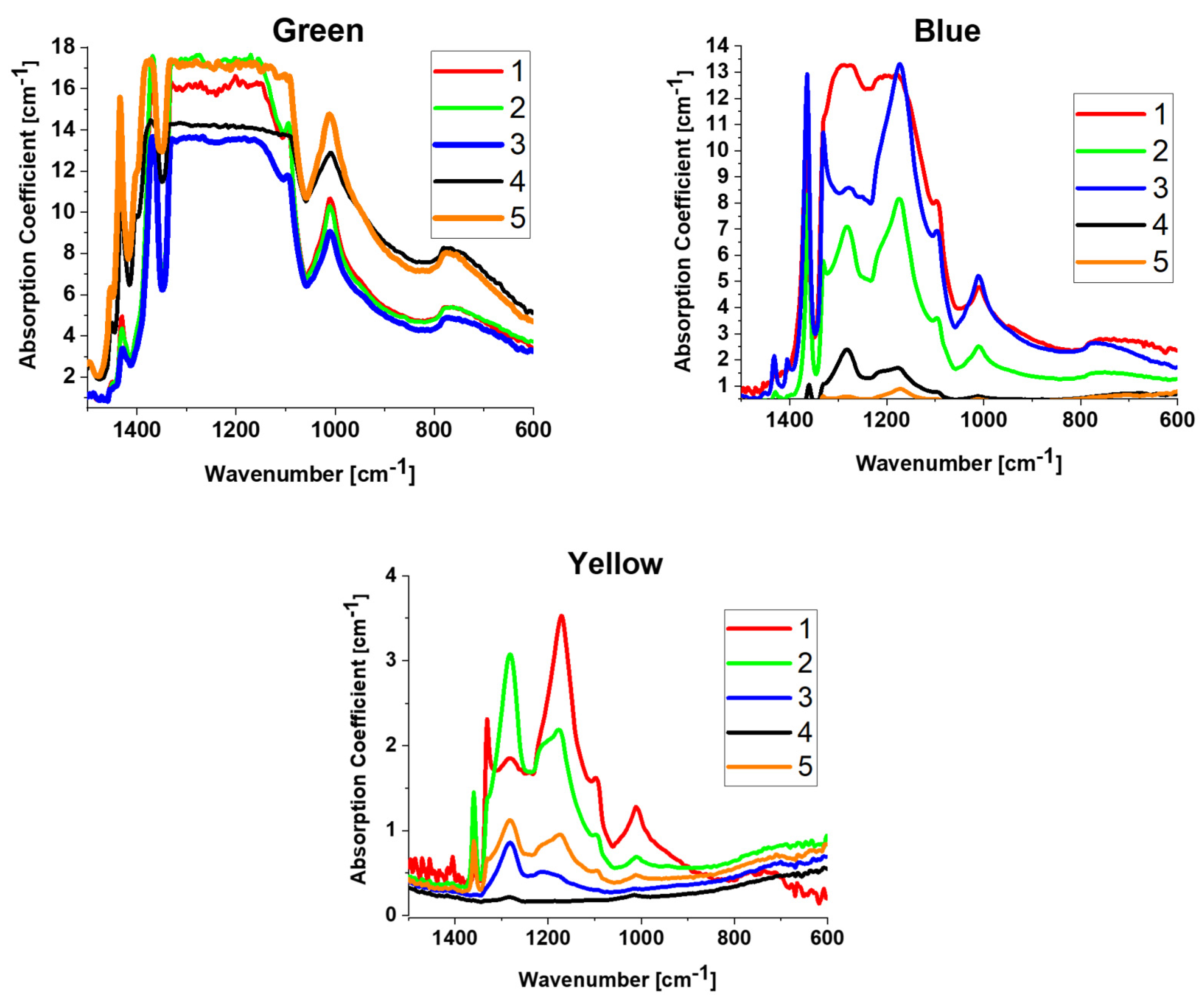
3.4. FTIR Spectra
There are no changes in the IR spectra of the nitrogen absorption peaks of the A, B, and C centers pre- and post-treatment (although the peak at 1450 cm−1 associated with interstitial nitrogen with no effect on color may change); hence, only the spectra of the final color-enhanced diamonds are given in Figure 6. The spectra presented were measured in the range of 600–1500 cm−1, the one-phonon region for nitrogen absorption. The diamonds are type Ia A/B diamonds (where A diamonds have a pair of nearest-neighbor nitrogen atoms, and B diamonds contain four N atoms surrounding a vacancy). The overall nitrogen concentration was calculated for each diamond, and the average values were >1500 ± 200 ppm for green (oversaturation for A and B centers; C center: 300 ± 100 ppm), ~671 ± 476 ppm (A center: 506 ± 358 ppm; B center: 105 ± 74 ppm; and C center: 60 ± 46 ppm) for blue, and 147 ± 97 ppm (A center: 113 ± 78 ppm; B center: 23 ± 16 ppm; and C center: 11 ± 4 ppm) for yellow diamonds.
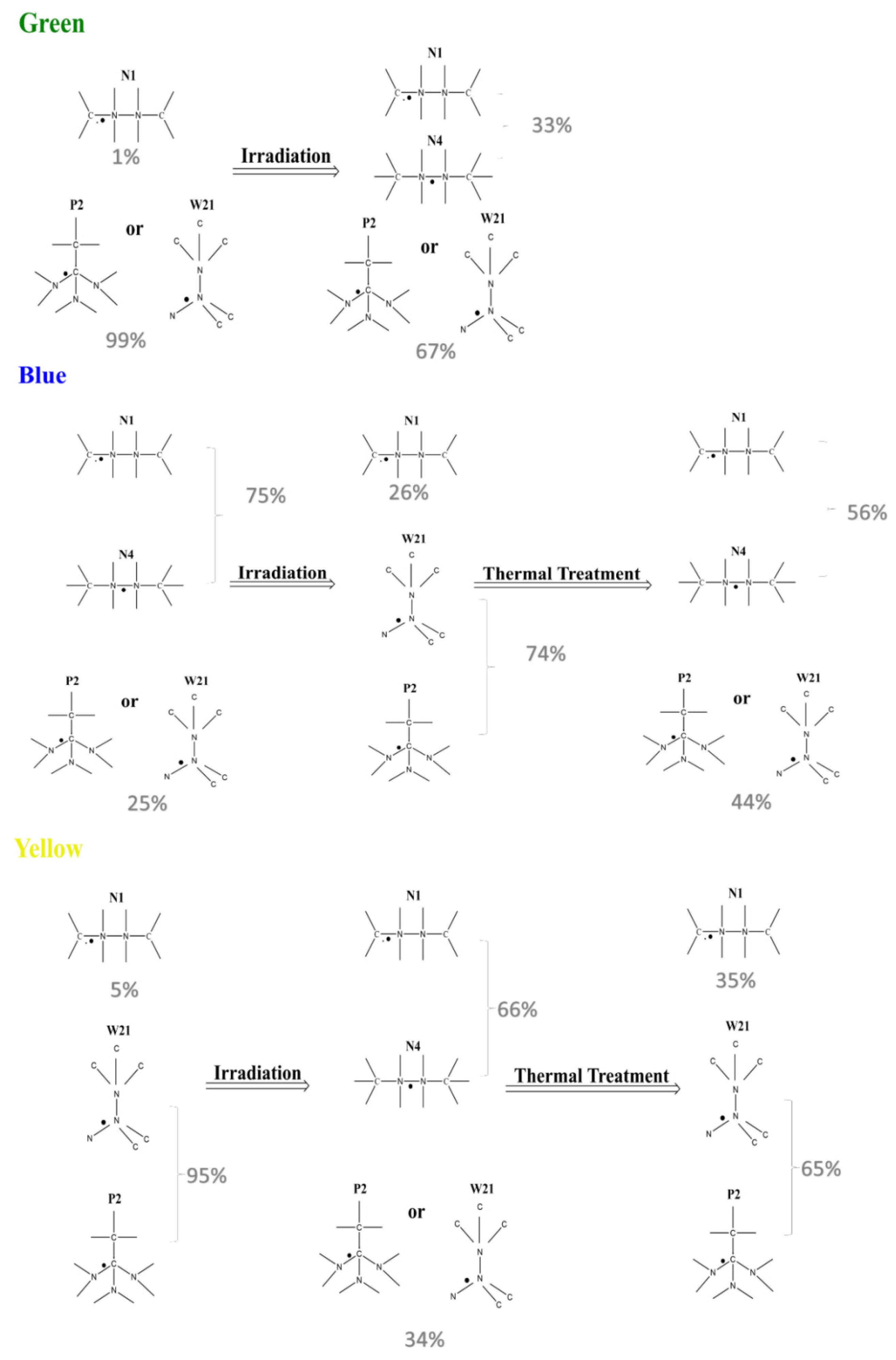
3.5. EPR and MATLAB EasySpin Simulations
This research focused on the relation between color centers that are produced as a result of color enhancement treatments and the changes in the radical configuration as a result of these treatments. For this purpose, an EPR spectra data analysis was conducted on the 15 diamonds, and only 1 orientation was chosen for the measurement (the crystallographic orientation in the magnetic field might affect the measurements) [32]. This study suggests a schematic mechanism (Figure 7), in which the change in the carbon radical center configuration before and after the treatments is presented. The stable carbon-centered radicals were assigned via the following g-values: 2.0024 for the N1 center, 2.0017–2.0022 for N4 centers, and 2.0027–2.0035 for P2/W21 (considering the hyperfine data obtained in a previous work [32], it was possible to assign the data of the simulation to defined carbon radical centers [24]).
3.6. Blue Evaluation of the Changes Observed (Figure 6)
N1 and N4 carbon-centered radicals are related to the green color. The green color is composed of a mixture of blue and yellow. If the yellow component is eliminated, the resulting color will be blue. Thus, the pre-blue diamonds, which turned green after the irradiation process, needed a thermal treatment to eliminate the yellow component in order to produce a blue color, which is characterized by N4 carbon radical species. However, when a yellow color is desired, the blue color component (in irradiated pre-yellow diamonds) needs to be eliminated (termination of N4 species), and this is achieved by exposing the diamonds to much higher temperatures in the thermal treatment protocol.
In order to explain our hypothesis that the structure of bulk nitrogen atoms in the diamond is identical to the nitrogen atoms adjacent to the carbon-centered radicals, it is important to compare the concentration of the nitrogen atoms adjacent to the carbon-centered radicals to the bulk nitrogen concentration in the diamond.
The calculations of the average nitrogen concentration and spin concentration of the five diamond samples were converted from ppm and #spins/mg units to Molar (Table 3). For example, converting ppm to M (Molar), 1000 ppm is 0.1%, meaning that if using the diamond density (3.53 gr/cm3), a volume of 1 Liter of diamond material weighs 3530 gr = 3.53 g/L. Next, dividing this value by the molar mass of nitrogen (14 g/mole) will give the result of 0.252 M. In order to convert #spins/mg to M, 4.5 × 10¹³ #spins/mg is multiplied by the diamond density (3.53 gr/cm3), and then to convert from spins to mol, it is necessary to divide by Avogadro’s number, which results in 4.5 × 10¹³
The average nitrogen and carbon-centered radical concentrations in the five samples of each color were converted from the standard ppm and #spins/mg units to the same concentration units in Molar (Table 3). Indeed, the bulk nitrogen concentration in the diamond is 3–4 orders of magnitude higher compared to the concentration of stable carbon radicals. This indicates that the absorption coefficient of the color centers is very high, meaning that they are allowed optical transitions.
As the structure of the nitrogen atoms adjacent to the carbon-centered radicals in the diamond can be determined by the EPR spectrum, and the nitrogen distribution in the diamond is homogeneous, the conclusion is that the bulk nitrogen structure in the diamond is the same.
In summary, using various spectroscopic techniques, one can correlate the color of the diamonds and relate them to several types of carbon-centered radicals. It was shown that each colored diamond has specific characteristics that are affected by irradiation and thermal treatments.
4. Conclusions
The aim of this study was to better understand the effect of color enhancement processes on treated diamonds and the source of the color centers produced. For this purpose, 15 natural diamonds, which underwent treatment protocols to enhance their color to green, blue, and yellow, were examined. A summary of the main conclusive results is presented in Table 4 based on a visual color assessment, fluorescence, FTIR, UV-vis, and EPR spectroscopy.
The results mainly indicate the following conclusions:
- (1)
- The optical color centers of green, blue, and yellow diamonds consist of blue optical centers (such as GR1) and yellow optical centers (such as N3 and H3), and the green color is a combination of blue and yellow color centers.
- (2)
- When high nitrogen concentrations are treated, a high spin concentration (stable carbon-centered radicals) is also established, as seen in our previous papers.
- (3)
- The paramagnetic centers that were found in this study were N1, N4, and P2/W21.
- (4)
- N4 is suggested to be correlated with a blue color, whereas a yellow color is attributed to the presence of N1 species.
Author Contributions
Conceptualization, I.L., H.C. and S.R.; methodology, I.L.; software, I.L.; validation, S.R., Y.A. and I.L.; formal analysis, I.L. and A.C.; investigation, I.L.; resources, A.C.; data curation, I.L.; writing—original draft preparation, I.L.; writing—review and editing, H.C.; visualization, I.L.; supervision, H.C.; project administration, H.C. All authors have read and agreed to the published version of the manuscript.
Funding
This research received no external funding.
Conflicts of Interest
The authors declare no conflict of interest.
References
- Shigley, J.E.; Breeding, C.M. Optical Defects in Diamond: A Quick Reference Chart. Gems Gemol. 2013, 49, 107–111. [Google Scholar] [CrossRef]
- Breeding, C.M.; Eaton-Magaña, S.; Shigley, J.E. Natural-Color Green Diamonds: A Beautiful Conundrum. Gems Gemol. 2018, 54, 2–27. [Google Scholar] [CrossRef]
- Wang, W.; Hall, M.; Breeding, C.M. Natural Type IA Diamond with Green-Yellow Color Due to Ni-Related Defects. Gems Gemol. 2007, 43, 240–243. [Google Scholar] [CrossRef] [Green Version]
- King, J.M.; Shigley, J.E.; Gelb, T.H.; Guhin, S.S.; Hall, M.; Wang, W. Characterization and Grading of Natural-Color Yellow Diamonds. Gems Gemol. 2005, 41, 88–115. [Google Scholar] [CrossRef] [Green Version]
- King, J.M.; Shigley, J.E.; Guhin, S.S.; Gelb, T.H.; Hall, M. Characterization and Grading of Natural-Color Pink Diamonds. Gems Gemol. 2002, 38, 128–147. [Google Scholar] [CrossRef] [Green Version]
- Collins, A.T. Investigating Artificially Coloured Diamonds. Nature 1978, 273, 654–655. [Google Scholar] [CrossRef]
- Borzdov, Y.; Pal’yanov, Y.; Kupriyanov, I.; Gusev, V.; Khokhryakov, A.; Sokol, A.; Efremov, A. HPHT Synthesis of Diamond with High Nitrogen Content from an Fe3N-C System. Diam. Relat. Mater. 2002, 11, 1863–1870. [Google Scholar] [CrossRef]
- Natural Fancy Red Diamond. Available online: https://www.leibish.com/red-diamonds/fancy-red-princess-40038#a_aid=yNaturalRedDiamond (accessed on 6 December 2022).
- Treated Fancy Vivid Pink Red. Available online: http://www.dianerdiamonds.com/view_diamonds/59.htmColorEnhancedRedDiamond (accessed on 6 December 2022).
- Synthetic Red Diamond. Available online: https://www.brilliantearth.com/lab-created-colored-diamonds/SyntheticRedDiamonds (accessed on 6 December 2022).
- Dobrinets, I.A.; Vins, V.G.; Zaitsev, A.M. Springer Series in Materials Science 181 HPHT-Treated Diamonds; Springer: Berlin/Heidelberg, Germany, 2013; ISBN 9783642374890. [Google Scholar]
- Hainschwang, T.; Respinger, A.; Notari, F.; Hartmann, H.J.; Günthard, C. A Comparison of Diamonds Irradiated by High Fluence Neutrons or Electrons, before and after Annealing. Diam. Relat. Mater. 2009, 18, 1223–1234. [Google Scholar] [CrossRef]
- Collins, A.T.; Kiflawi, I. The Annealing of Radiation Damage in Type Ia Diamond. J. Phys. Condens. Matter 2009, 21, 364209. [Google Scholar] [CrossRef]
- Hainschwang, T.; Notari, F.; Fritsch, E.; Massi, L. Natural, Untreated Diamonds Showing the A, B and C Infrared Absorptions (“ABC Diamonds”), and the H2 Absorption. Diam. Relat. Mater. 2006, 15, 1555–1564. [Google Scholar] [CrossRef]
- Goss, J.P.; Briddon, P.R.; Hill, V.; Jones, R.; Rayson, M.J. Identification of the Structure of the 3107 Cm-1 H-Related Defect in Diamond. J. Phys. Condens. Matter 2014, 26, 145801. [Google Scholar] [CrossRef] [PubMed]
- Breeding, C.M.; Shigley, J.E. The “Type” Classification System of Diamonds and Its Importance in Gemology. Gems Gemol. 2009, 45, 96–111. [Google Scholar] [CrossRef]
- Evans, T.; Phaal, C. Imperfections in Type I and Type II Diamonds. Proc. R. Soc. London Ser. A Math. Phys. Sci. 1962, 270, 538–552. [Google Scholar] [CrossRef]
- De Weerdt, F.; Van Royen, J. Defects in Coloured Natural Diamonds. Diam. Relat. Mater. 2001, 10, 474–479. [Google Scholar] [CrossRef]
- Dyer, H.B.; Matthews, I.G. The Fluorescence of Diamond. Proc. R. Soc. London Ser. A Math. Phys. Sci. 1958, 243, 320–335. [Google Scholar] [CrossRef]
- Luo, Y.; Breeding, C.M. Fluorescence Produced by Optical Defects in Diamond: Measurement, Characterization, and Challenges. Gems Gemol. 2013, 49, 82–97. [Google Scholar] [CrossRef]
- King, J.M.; Moses, T.M.; Shigley, J.E.; Welbourn, C.M.; Lawson, S.C.; Cooper, M. Characterizing Natural-Color Type IIb Blue Diamonds. Gems Gemol. 1998, 34, 246–268. [Google Scholar] [CrossRef] [Green Version]
- Baldwin, J.A., Jr. Electron Paramagnetic Resonance Investigation of the Vacancy in Diamond. Phys. Rev. Lett. 1963, 10, 220–222. [Google Scholar] [CrossRef]
- Loubser, J.H.N.; Van Wyk, J.A. Electron Spin Resonance in the Study of Diamond. Reports Prog. Phys. 1978, 41, 1201–1248. [Google Scholar] [CrossRef]
- Ammerlaan, C.A.J. Impurities and Defects in Group LVElements and Lll-VCompounds; Springer: Berlin/Heidelberg, Germany, 1998; Volume 22, ISBN 978-3-540-48331-1. [Google Scholar]
- Lee, C.W.Y.; Cheng, J.; Yiu, Y.C.; Chan, K.; Lau, D.; Tang, W.C.; Cheng, K.W.; Kong, T.; Hui, T.K.C.; Jelezko, F. Correlation between EPR Spectra and Coloration of Natural Diamonds. Diam. Relat. Mater. 2020, 103, 107728. [Google Scholar] [CrossRef]
- John, A.; Weil, J.R.B. Electron Paramagnetic Resonance Elemntry Theory and Practical Applications; John Wiley & Sons, Inc.: Hoboken, NJ, USA, 2007; pp. 1–118. [Google Scholar]
- Felton, S.; Cann, B.L.; Edmonds, A.M.; Liggins, S.; Cruddace, R.J.; Newton, M.E.; Fisher, D.; Baker, J.M. Electron Paramagnetic Resonance Studies of Nitrogen Interstitial Defects in Diamond. J. Phys. Condens. Matter 2009, 21, 364212. [Google Scholar] [CrossRef] [PubMed] [Green Version]
- Isoya, J.; Kanda, H.; Uchida, Y.; Lawson, S.C.; Yamasaki, S.; Itoh, H.; Morita, Y. EPR Identification of the Negatively Charged Vacancy in Diamond. Phys. Rev. B 1992, 45, 1436–1439. [Google Scholar] [CrossRef] [PubMed]
- Stepanov, V.; Takahashi, S. Determination of Nitrogen Spin Concentration in Diamond Using Double Electron-Electron Resonance. Phys. Rev. B 2016, 94, 024421. [Google Scholar] [CrossRef] [Green Version]
- Loubser, J.H.N.L.; Wrightj, A.C.J. A Singly Ionized N-c-n Centre in Diamond. J. Phys. D Appl. Phys. 1973, 6, 1129–1141. [Google Scholar] [CrossRef]
- Nadolinny, V.A.; Yuryeva, O.P.; Rakhmanova, M.I.; Shatsky, V.S.; Palyanov, Y.N.; Kupriyanov, I.N.; Zedgenizov, D.A.; Ragozin, A.L. Distribution of OK1, N3 and NU1 Defects in Diamond Crystals of Different Habits. Eur. J. Miner. 2012, 24, 645–650. [Google Scholar] [CrossRef]
- Litvak, I.; Cahana, A.; Anker, Y.; Ruthstein, S.; Cohen, H. The Effects of Thermal Treatment and Irradiation on the Chemical Properties of Natural Diamonds. Phys. Chem. Chem. Phys. 2022, 24, 11696–11703. [Google Scholar] [CrossRef] [PubMed]
- Tretiakova, L. Spectroscopic Methods for the Identification of Natural Yellow Gem-Quality Diamonds. Eur. J. Miner. 2009, 21, 43–50. [Google Scholar] [CrossRef]
- Wang, M.; Shi, G.; Yuan, J.C.C.; Han, W.; Bai, Q. Spectroscopic Characteristics of Treated-Color Natural Diamonds. J. Spectrosc. 2018, 2018, 8153941. [Google Scholar] [CrossRef]
Figure 1.
Example of IR spectra of diamonds with and without nitrogen contamination in the range of 600–4000 cm−1 (marked in the figure are the three phonon areas).
Figure 1.
Example of IR spectra of diamonds with and without nitrogen contamination in the range of 600–4000 cm−1 (marked in the figure are the three phonon areas).

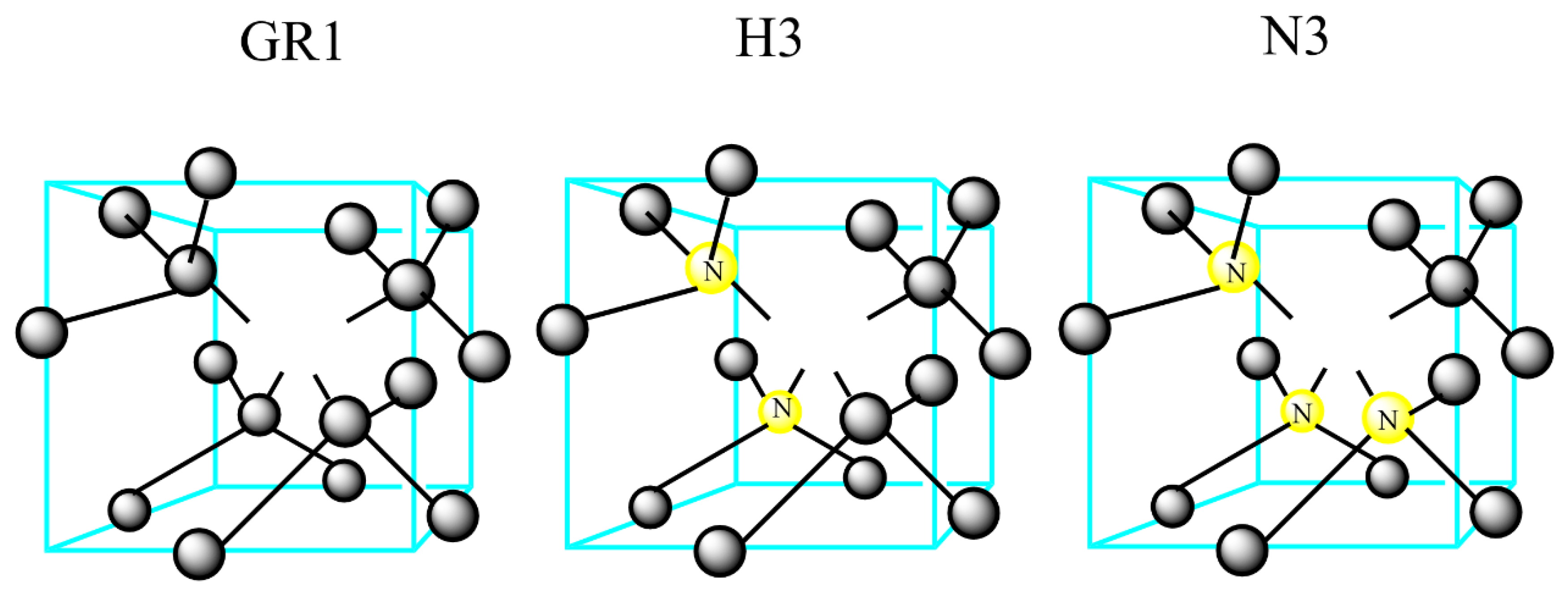
Figure 2.
Examples of optical defect structures in diamonds: GR1, H3, and N3. GR1: vacancy in a neutral charge state (V0); H3: four substitutional nitrogen atoms surrounding two vacancies (4N + 2V); and N3: three substitutional nitrogen atoms surrounding a vacancy (3N + V).
Figure 2.
Examples of optical defect structures in diamonds: GR1, H3, and N3. GR1: vacancy in a neutral charge state (V0); H3: four substitutional nitrogen atoms surrounding two vacancies (4N + 2V); and N3: three substitutional nitrogen atoms surrounding a vacancy (3N + V).

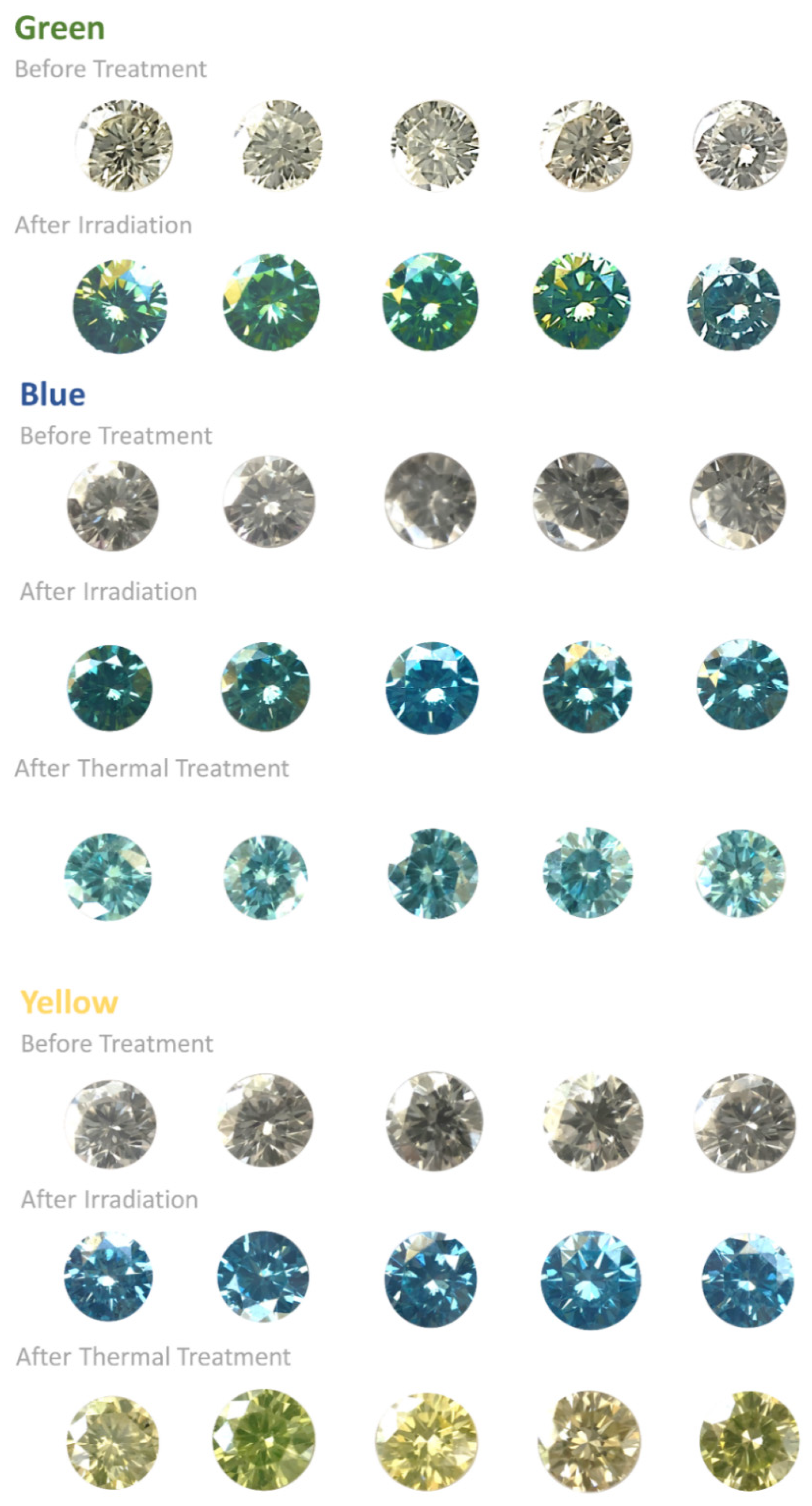
Figure 3.
Daylight photographs of the green, blue and yellow diamonds: pre- and post-irradiation and, for the blue and yellow diamonds, post-thermal treatment. Green: from clear crystal to green-yellowish/green-turquoise (irradiation); blue: from clear to dark-greenish (irradiation) to blue (annealing); yellow: from clear to blue (irradiation) to yellow (annealing).

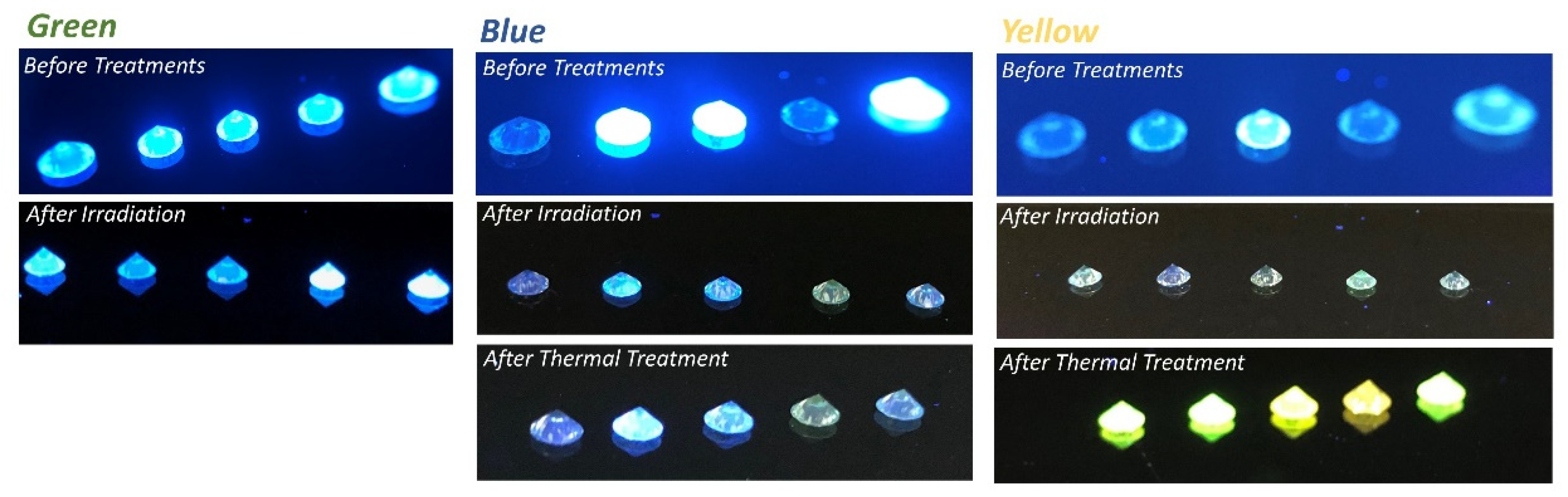
Figure 4.
Long-wave UV fluorescence photographs of the diamonds: pre- and post-irradiation and, for blue and yellow, post-thermal treatment. If green after the irradiation treatment, some diamonds’ fluorescence changed to white; if blue after the irradiation treatment, some diamonds’ fluorescence changed to green-yellow, and after annealing, there was no change in fluorescence; if yellow after the irradiation treatment, 4/5 diamonds showed no fluorescence, but, after thermal annealing, all diamonds showed strong yellow fluorescence.

Figure 5.
Visible absorption spectra after final treatments of the fancy diamonds: green (A), blue (B), and yellow (C). Green and blue display intense absorption bands between 400 and ~600 nm (H3) and a second absorption band located at 741 and 744 nm (GR1). Yellow displays an increase in absorption intensity from ~400 nm to ~470 nm, with a small N3 line (~415 nm). Numbers 1–5 denote the numbers of the sample diamonds (for yellow diamonds, sample 4 was omitted).

Figure 6.
FTIR spectra of the one-phonon area (nitrogen absorption: 600–1500 cm−1) of color-enhanced diamonds. No change in the concentrations of the A, B, and C centers of the nitrogen absorption peaks occurred during the treatments. Numbers 1–5 denote the numbers of the sample diamonds.
Figure 6.
FTIR spectra of the one-phonon area (nitrogen absorption: 600–1500 cm−1) of color-enhanced diamonds. No change in the concentrations of the A, B, and C centers of the nitrogen absorption peaks occurred during the treatments. Numbers 1–5 denote the numbers of the sample diamonds.

Figure 7.
Schematic paramagnetic centers before and after treatments for green, blue, and yellow diamonds. In green diamonds, there is an increase in the N1 species concentration and appearance of the N4 (formed by the irradiation treatment) species; in blue diamonds, there is an increase in the P2/W21 species concentration (formed by the irradiation treatment) and appearance of the N4 species concentration (formed by the annealing treatment); in yellow diamonds, there is an increase in the N1 species concentration and appearance of N4 (formed by the irradiation treatment) and also an increase in the concentration of the P2/W21 species (formed by the irradiation treatment) and disappearance of the N4 species (formed by the annealing treatment).


Table 1.
Examples of structural defects in the crystal, which are the sources of the green, blue, and yellow colors in the diamonds.
Table 1.
Examples of structural defects in the crystal, which are the sources of the green, blue, and yellow colors in the diamonds.
| Optical Center | Absorption Line [nm] | Defect Structure | Detection Method | Color | Occurs/Produced |
|---|---|---|---|---|---|
| GR1 | 740.9 and 744.4 | Vacancy in a neutral charge state (V°) | UV, PL | Green/blue | Natural or by irradiation |
| 595 band | 594.4 | Uncertain structure | UV | Contributes to other colors | By irradiation and thermal annealing |
| H3 | 503.2 | Two substitutional nitrogen atoms separated by a vacancy in a neutral charge state (N-V-N)° | UV, PL | Yellow | Natural or by irradiation and thermal annealing or HPHT |
| H4 | 496.2 | Four substitutional nitrogen atoms surrounding two vacancies (4N + 2V) | UV, PL | Yellow | Natural or by irradiation and thermal annealing |
| N3 | 415.2 | Three substitutional nitrogen atoms surround a vacancy (3N + V). | UV, PL, EPR | Yellow | Natural |
Table 2.
Examples of known carbon-centered paramagnetic species of different diamond types.
| Defect Type | Diamond Type | Structure | g-Value | Hyperfine Interaction (A) |
|---|---|---|---|---|
| N1 | Natural type Ia and Ib | Ionized nitrogen pair, negative (vacancy + nitrogen pair) complex, nonplanar N1CCN2 complex | 2.0024 | A A A |
| N4 | Natural, brown, with plastic deformation | Substitutional nitrogen pair near dislocation | 2.002 ± 0.001 | A A |
| OKI | Natural type Ib | (Nitrogen + vacancy) complex, (Nitrogen + vacancy + oxygen) complex | g1 = 2.0031 ± 0.0003 g2 = 2.0019 ± 0.0003 g3 = 2.0025 ± 0.0003 | A1=l5.48 MHz A2=21–66 MHz A3=15.19 MHz |
| S2 | Natural, after electron irradiation | Substitutional nitrogen + other defects | 2.0023 | A A |
| W30 | Natural type Ia, after irradiation and annealing at 450 °C | Center with four or more nitrogen atoms | 2.00 | A A |
Table 3.
Comparison between average nitrogen concentration and carbon-centered radical concentration in the diamond.
Table 3.
Comparison between average nitrogen concentration and carbon-centered radical concentration in the diamond.
| Color | Nitrogen Conc. [ppm] | Radical Conc. [#spins/mg] | Nitrogen Conc. [M] | Radical Conc. [M] | Diamond Density [g/cm3] |
|---|---|---|---|---|---|
| Green | 1000 ± 185 | 4.5 × 10¹³ ± 0.3 × 10¹³ | 2.52 × 10⁻¹ | 2.64 × 10⁻⁴ | 3.53 |
| Blue | 650 ± 350 | 6.5 × 10¹² ± 0.15 × 10¹³ | 1.64 × 10⁻¹ | 3.81 × 10⁻⁵ | |
| Yellow | 80 ± 50 | 5.2 × 10¹² ± 1 × 10¹³ | 2.02 × 10⁻² | 3.05 × 10⁻⁵ |
Table 4.
Summary of the main results: FTIR, UV-Vis, fluorescence, and EPR for the color enhancement process to produce green, blue and yellow diamonds.
Table 4.
Summary of the main results: FTIR, UV-Vis, fluorescence, and EPR for the color enhancement process to produce green, blue and yellow diamonds.
| Green | Blue | Yellow |
|---|---|---|
| High nitrogen concentration (>1000 ppm/mg) | Intermediate nitrogen concentration (~300–600 ppm) | Low nitrogen concentration (<200 ppm/mg) |
| GR1 and H3 optical defects after treatments | N3 and H3 optical defects after treatments | |
| Blue fluorescence in all five pretreated diamonds | Blue fluorescence in three pretreated diamonds | Blue fluorescence in one pretreated diamond |
| High spin concentration of >4.5 × 10¹⁵ spins/mg before treatment. After irradiation, there was a pronounced increase in the spin concentration. | Moderate changes in the spin concentration after treatment | Moderate changes in the spin concentration after treatment |
| Pretreated: dominant P2/W21 centers | Pretreated: dominant N1/N4 centers | Pretreated: dominant P2/W21 centers |
| Post-irradiation: the appearance of N4 centers | Post-irradiation: an increase in P2/W21 centers | Post-irradiation: large increase in the N1/N4 centers compared with P2/W21 |
| Post-thermal treatment: about half of the centers are P2/W21 and the other half are N1/N4 | Post-thermal treatment: only N1 and an increase in P2/W21 | |
Publisher’s Note: MDPI stays neutral with regard to jurisdictional claims in published maps and institutional affiliations. |
© 2022 by the authors. Licensee MDPI, Basel, Switzerland. This article is an open access article distributed under the terms and conditions of the Creative Commons Attribution (CC BY) license (https://creativecommons.org/licenses/by/4.0/).
Share and Cite
MDPI and ACS Style
Litvak, I.; Cahana, A.; Anker, Y.; Ruthstein, S.; Cohen, H. Nitrogen Structure Determination in Treated Fancy Diamonds via EPR Spectroscopy. Crystals 2022, 12, 1775. https://doi.org/10.3390/cryst12121775
AMA Style
Litvak I, Cahana A, Anker Y, Ruthstein S, Cohen H. Nitrogen Structure Determination in Treated Fancy Diamonds via EPR Spectroscopy. Crystals. 2022; 12(12):1775. https://doi.org/10.3390/cryst12121775
Chicago/Turabian StyleLitvak, Ira, Avner Cahana, Yaakov Anker, Sharon Ruthstein, and Haim Cohen. 2022. "Nitrogen Structure Determination in Treated Fancy Diamonds via EPR Spectroscopy" Crystals 12, no. 12: 1775. https://doi.org/10.3390/cryst12121775
Note that from the first issue of 2016, this journal uses article numbers instead of page numbers. See further details here.