Article

Effect of WO3 Nanoparticles on the Radiative Attenuation Properties of SrTiO3 Perovskite Ceramic

1
Department of Physics, Faculty of Science, Isra University, Amman 11622, Jordan
2
Physics Department, Faculty of Science, Universiti Teknologi Malaysia, Skudai 81310, Johor, Malaysia
3
Department of Nuclear Medicine Research, Institute for Research and Medical Consultations (IRMC), Imam Abdulrahman Bin Faisal University, P.O. Box 1982, Dammam 31441, Saudi Arabia
4
Department of Biophysics, Institute for Research and Medical Consultations (IRMC), Imam Abdulrahman Bin Faisal University, P.O. Box 1982, Dammam 31441, Saudi Arabia
5
Physics Department, Faculty of Science, Alexandria University, Alexandria 21511, Egypt
*
Author to whom correspondence should be addressed.
Crystals 2022, 12(11), 1602; https://doi.org/10.3390/cryst12111602
Submission received: 17 October 2022 / Revised: 7 November 2022 / Accepted: 8 November 2022 / Published: 10 November 2022

Abstract

:
In the present work, an experimental study is performed to study the radiation shielding characteristics of SrTiO3 (STO) perovskite ceramic added with different amounts (x = 0, 2, 5, and 10%) of tungsten trioxide nanoparticles (WO3 NPs). The four ceramic samples were prepared using the solid-state reaction method. The structural properties were examined using X-ray diffraction (XRD) and Fourier transform infrared spectroscopy (FTIR) techniques. The analysis showed the successful formation of WO3- doped STO samples. The crystallite size, estimated using the Scherrer equation, was found in the range of 50.86–41.17 nm. The effect of WO3 NPs on the radiation shielding performance of these ceramics was studied. Different parameters, such as linear attenuation coefficient (LAC) and other related factors, were experimentally determined. The linear attenuation coefficient results demonstrated that the additional amount of WO3 in the ceramics correlates with an improvement in their shielding abilities. The half-value layer (HVL) values for the ceramics with 2% WO3 nanoparticles are equal to 0.071, 1.760, 2.407, and 2.564 cm at 0.060, 0.662, 1.173, and 1.333 MeV, respectively. As the energy increases, more radiation can pass through the material; therefore, a larger thickness is required to absorb half of the total photons, leading to a greater HVL. The tenth value results reaffirmed that increasing the WO3 content in the STO ceramics improves their shielding efficiency. The radiation protection efficiency (RPE) of the four prepared STO ceramics was reported. From the RPE, we found that more photons can be attenuated at lower energies.

1. Introduction

The use of radiation has been growing during the past decades in various areas, including agricultural, industrial, and medical fields. Despite the enormous profits of radiation, it has the prospect of causing a significant risk hazard to human safety and the environment. Three basic concepts are followed for better radiation protection: minimizing the exposure time, increasing the distance that separates the entity of question and the source of radiation as much as possible, and using a protective material to isolate the entity of question from radiation. These three concepts are applied as far as practicable to reduce the entire absorbed dose due to radiation exposure [1].
The rendering of a material as a radiation shield is frequently evaluated by its ability to stop the penetration of incident radiation through various mechanisms of interaction. γ radiation, categorized as high penetrating potential, interacts with the material in three distinct processes: Compton scattering (CS), pair production (PP), and photoelectric absorption (PA) [2]. The probability of each interaction occurring is dependent on the incident γ radiation power and the composition of the protective material. PA is the main process for low energy γ radiation that interacts with materials with high atomic numbers. For high-energy γ radiation, PP became the dominant process. The γ interaction behavior with the shield is characterized by the linear attenuation coefficient (LAC), which depends on the properties of the material, the energy, and the incident radiation. Appropriate shielding material is able to attenuate γ radiation with minimum change to its thermal, mechanical, and physical properties as well as its chemical stability. In general, many factors must be considered when designing a radiation shield, including the kind and intensity of radiation, its energy level, cost, toxicity, and weight of material [3,4,5,6].
The frequently employed shielding materials are lead, concrete, and alloys. Studies searching for developed materials for radiation shielding applications have increased owing to their important role in progressed technologies that need ionizing radiations, such as nuclear medicine, radiology, and the characterization of materials. This necessitates advanced materials to be employed in the fabrication of protective structures to shelter humans and the environment from the hurtful impact of ionizing radiation. Aygün et al. studied heavy concretes holding chrome-ore for nuclear radiation shielding applications [7]. Their results showed better shielding properties for concretes with aggregates and additives compared to some heavy concretes and standard concrete [7]. M.I. Sayyed et al. prepared four alloys, including CrTe, CrTe0.95Sb0.05, CrTe0.90Sb0.10, and CrTe0.80Sb0.20, using a solid-state reaction. The results showed that replacing Te with Sb in the different alloys causes an improvement in their linear attenuation coefficient [8].
Glass-based materials are also common shielding materials and are possible alternatives to concretes for radiation-shielding purposes [9,10,11,12,13,14]. Glass materials display unique properties that make them useful for some industrial and technical applications. For example, T. H. Khazaalah et al. used soda–lime–silica (SLS) glass waste to fabricate free-Pb glass shielding [8]. γ-ray shielding properties of borate and phosphate glasses containing BaO, Bi2O3, and PbO in different ratios were previously studied. The results showed good attenuation properties of these glass systems [10,11]. In addition, the radiation shielding efficiency of borosilicate glass, which consists of silica and boron oxide, has been studied [12]. A recent report showed that borosilicate glass (30 mol%) mixed with 20 mol% of Bi2O3 and 50 mol% of BaO displays the highest density value and better radiation shielding performances [14].
Perovskite-based ceramic materials have attracted great interest due to their unique characteristics [15,16,17]. SrTiO3 (STO) is a model of the perovskite-like ceramic material with many important properties and possible uses in diverse applications such as optoelectronic [18], photovoltaic [19,20], photocatalytic [20,21], and energy storage applications [22]. STO displays a high dielectric constant, average permittivity, and low tangent loss [14]. STO has also proven its efficacity in the domain of radiation shielding [23,24,25]. Moreover, STO is a mixed conducting perovskite [23,24] which can be influenced by doping and/or addition, temperature, and oxygen partial pressure (pO2). For instance, E. Hannachi et al. studied the radiation-protecting properties of STO doped with tin oxide. The results showed that tin oxide addition enhanced the radiation shielding capacity of STO ceramic [23].
Metal oxide semiconductors play an interesting role in different areas of science, such as engineering, medicine, physics, chemistry, etc. Metal oxide semiconductors have gained much interest owing to their particular optical, magnetic, and electrical properties. Among the most useful metal oxide semiconductors, WO3 has been employed for different applications such as electrocatalyst [26], photocatalyst [27], electrochromic [28], anticancer [29], and gas sensing [30]. WO3 is an n-type semiconductor with a large band gap (around 2.7–3.5 eV) and is non-toxic. One of the most important characteristics of WO3 is its morphological assortment. It can be designed in different dimensional morphologies, such as nanoparticles [31], nanowires [32], nanofibers [33], nanorods [31], nanosheets [34], and nanoplates [35]. It is shown that some special morphologies of WO3 nanostructures are more appropriate for particular applications. With the emergence of nanotechnology as a progressive branch of science in recent years, the radiation protection system has also used various types of nanoparticles [36,37,38]. The fundamental supposition of the investigations was that the application of nanoparticles leads to a more even distribution within the matrix compared to particles with micrometric size, and, hence, a better attenuation will be attained. Dong Yu found that the γ attenuation of nano-sized WO3 was greater than the micro-sized particles [39]. Tekin et al. showed that the materials doped with nano-sized WO3 displayed better shielding characteristics than materials doped with micro-sized WO3 particles [37]. Kazemi et al. demonstrated, using the MCNPX model, that composites with WO3 nanoparticles had mass attenuation coefficients greater than those of composites with WO3 microparticles [40].
To the best of our knowledge, there is no study regarding the effect of WO3 nanoparticles on the radiation-shielding characteristics of ceramic materials. Thus, we aim in this work to study, for the first time, the radiation attenuation of STO ceramic mixed with WO3 nanoparticles. Different radiation shielding parameters were experimentally measured and discussed.

2. Experimental Procedure

2.1. Synthesis of Materials

All chemicals, strontium carbonate (SrCO3), titanium dioxide (TiO2), and tungsten trioxide nanoparticles (WO3 NPs), were reagent grade and used without further purification. First, stoichiometric strontium titanate SrTiO3 (STO for brevity) was synthesized using the solid-state reaction by mixing finely in ball milling media SrCO3 and TiO2 powders. The obtained mixture was sintered at 1300 °C for 5 h to form SrTiO3 ceramic (STO for brevity), as shown in Equation (1).
SrCO3 + TiO2 ⇒ SrTiO3 + CO2
In the second stage, different amounts of WO3 NPs (x = 0, 2, 5, and 10 wt.%) were added to the STO sample. The mixtures were then compacted into pellets using PVA, which plays the role of the binder. The resulting pellets were placed in crucible alumina, then sintered at 1500° to obtain compact and dense ceramics of pristine STO ceramic (x = 0 wt.%) and WO3-doped STO ceramics (x = 2, 5, and 10 wt.%). Then, the obtained ceramic samples were given for various characterizations.

2.2. Characterization of Ceramics

The crystallinity and phase analysis of the prepared ceramics were analyzed using a Rigaku Benchtop Miniflex powder XRD instrument with Cu Kα radiation. Using the Bruker alpha-II FT-IR spectrometer, the FT-IR spectra were recorded in the region of 400–4000 cm−1. The γ-ray shielding parameters of STO-WO3 NPs ceramics, such as linear attenuation coefficient (LAC), were experimentally determined using a high pure germanium (HPGe) detector and different point sources (Co-60, Cs-137, and Am-241). Readings resulting from the detector were processed using the Genie 2000 program. This program displays the intensity (count rate) for each photon that fell on the detector and was discovered during the measured time. From this point, the intensity of each specific energy was measured in the presence (I) and the absence (I0) of the absorption sample. Then, by knowing the thickness of the absorption sample (t), the following law is used to calculate the LAC, which represents the possibility of the photon interaction through a certain distance within the absorption sample [41].
I = I 0   e LAC   .   t   then , LAC = 1 t   ln I 0 I
The other attenuation factors-based LAC determination, such as half and tenth value layers (HVL, TVL) and radiation absorption factor (RAP). The definition and laws of these factors were reported in previously published papers [42,43].

3. Results and Discussion

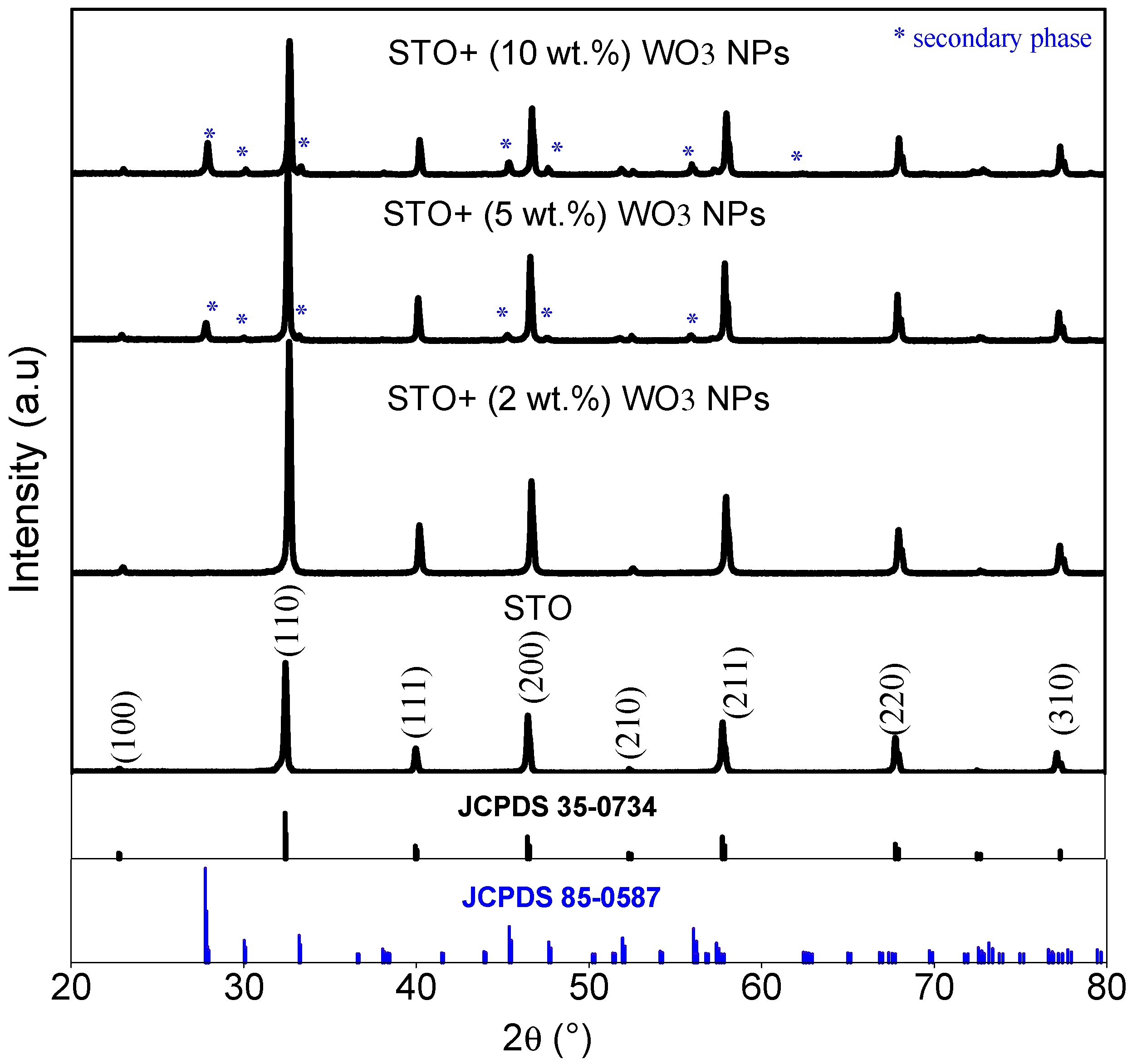
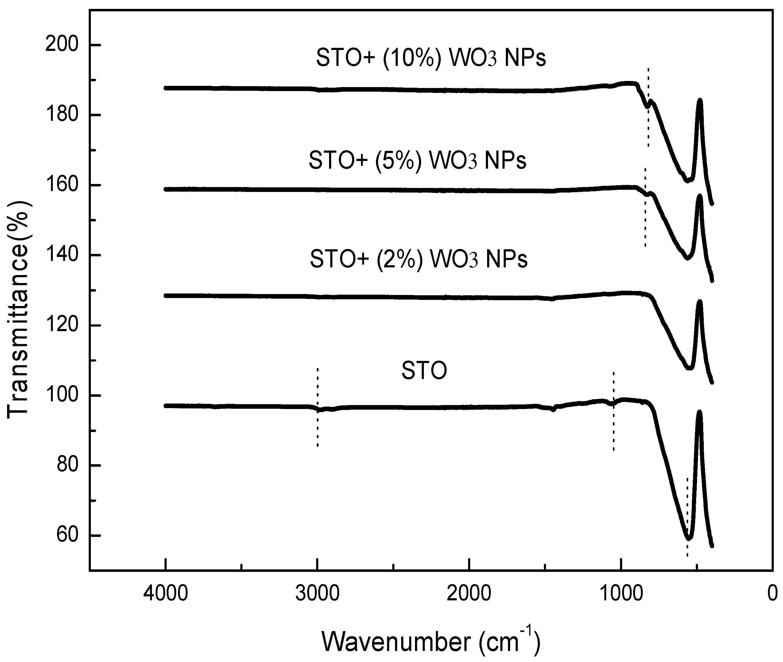
3.1. Structural and Functional Analysis

The synthesized ceramics were analyzed using XRD and FT-IR to investigate the structural and compositional properties. Figure 1 depicts the XRD patterns of SrTiO3 (STO) doped with different amounts of WO3 NPs. Pure STO exhibits (100), (110), (111), (200), (210), (211), (220), and (310) planes corresponding to cubic perovskite structure (space group: Pm-3 m) of SrTiO3 which are in agreement with JCPDS card no. 35–0734. The ceramic doped with x = 2 wt.% of WO3 NPs does not contain any impurity. However, the secondary phase of SrWO4 (marked as ‘*’ in the figure; card no. 85–0587) appeared for x = 5 and 10% of WO3 NPs, indicating that WO3 NPs may react with the host material (STO) during the sintering process. The peak intensity of the impurity phase increases remarkably as the content of WO3 NPs increases. The structural parameters, such as crystal cell parameter ‘a’ as well as the unit cell volume ‘V’ for all as-prepared ceramics calculated from the XRD data, are listed in Table 1. It is evident that the structural parameters ‘a’ and V’ are higher for doped samples compared to the STO pristine sample. This effect can be attributed to the difference in ionic radii between the W element (0.66 Å) derived from WO3 NPs dopant and Ti element (0.605 Å) derived from the host SrTiO3. The presence of the SrWO4 secondary phase can also be another reason for the lattice expansion in WO3- doped STO ceramics.
The crystallites size (dXRD) is estimated for all ceramics by using the Scherrer equation [44]:
d XRD = k   λ β   cos θ
where k is a constant close to unity (k = 0.9), β is full width at half maximum, and θ is Bragg’s angle. The calculated dXRD values for various ceramics are registered in Table 1 and are found to be in the range of 50.86–41.17 nm. Compared to the pristine sample, the crystallite size first reduces for the sample doped with a low amount of WO3, suggesting that WO3 NPs act as inhibitors for the grain growth of STO ceramic [45]. With further increasing the amount of WO3, the dXRD return to increase. This unexpected result may be due to the transformation of tungsten oxide into metal tungstate. Indeed, for high amounts of WO3, a new second phase of SrWO4 was detected, which is mainly attributed to the possible reaction between the host STO phase and WO3 NPs. It can be then assumed that the newly formed phase may result in initiating the growth process of aggregate particles by coalescence [46]. Further analysis is needed to validate this assumption, which will be the aim of our future work.
The functional groups of all ceramics were characterized using FTIR analysis, as depicted in Figure 2. In the measured range, several distinct absorption bands were detected for the pristine STO ceramic. The signal at about 560 cm−1 is related to the characteristic stretching vibration of the Sr-Ti-O bond [47], which confirms the successful formation of the SrTiO3 phase. This peak is maintained in the FTIR spectra of doped ceramics, confirming that the STO host perovskite structure is preserved. Another characteristic band with small intensity is observed around 1100 cm−1, which can be attributed to the C=O stretching modes [48]. A large band around 3000 cm−1 is also observed for STO ceramic, which may be assigned to the O–H stretching modes [49]. Additional bands around 825 cm−1 appeared for ceramics doped with x = 5 and 10% of WO3 NPs. This band is most probably a result of the signals of the W-O stretching vibration [50].

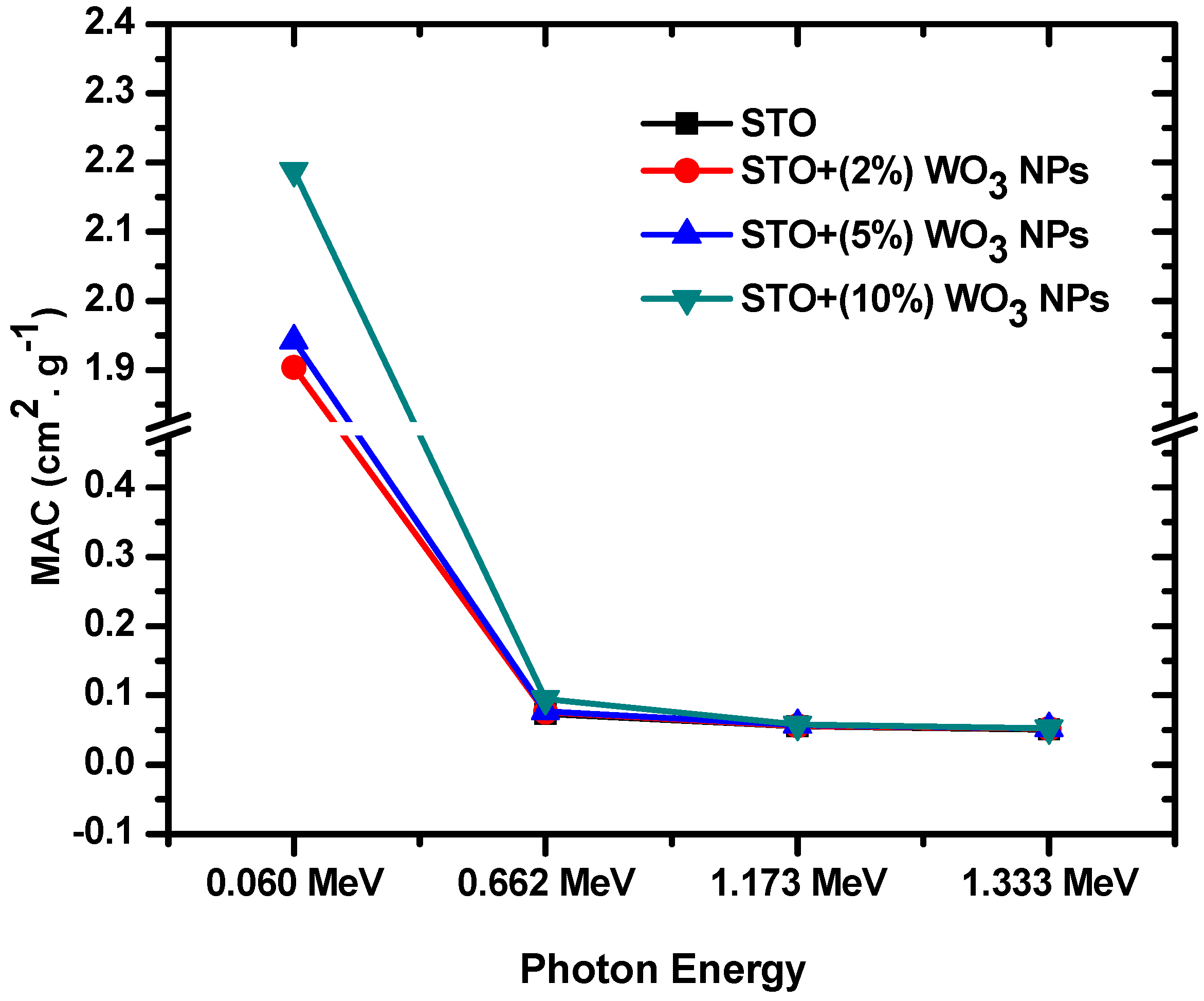
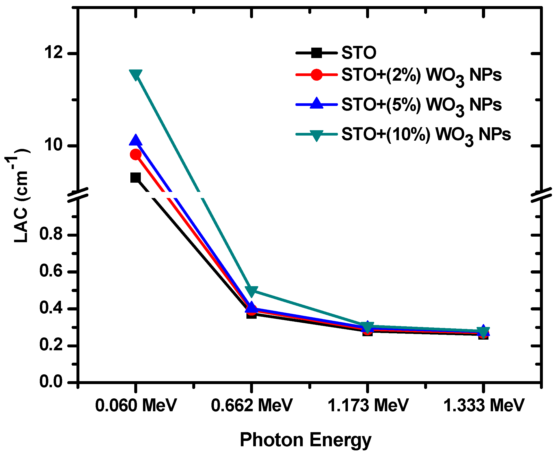
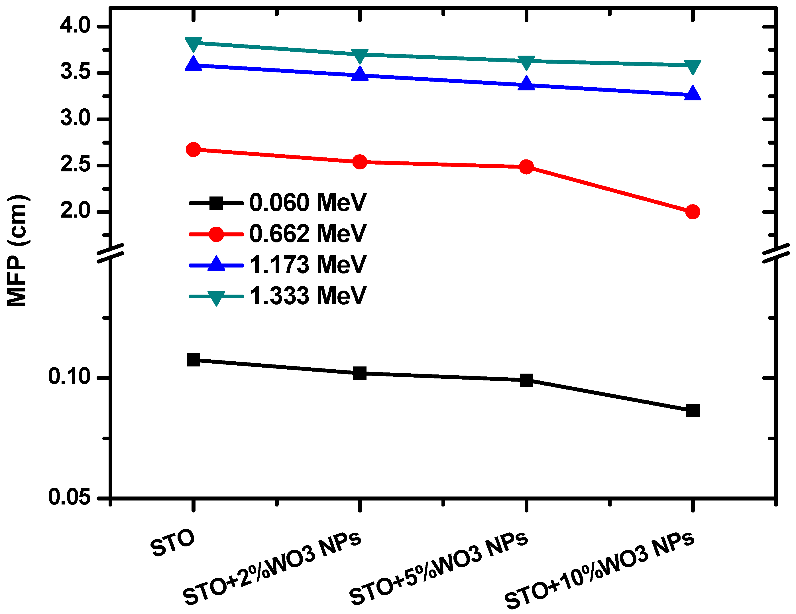
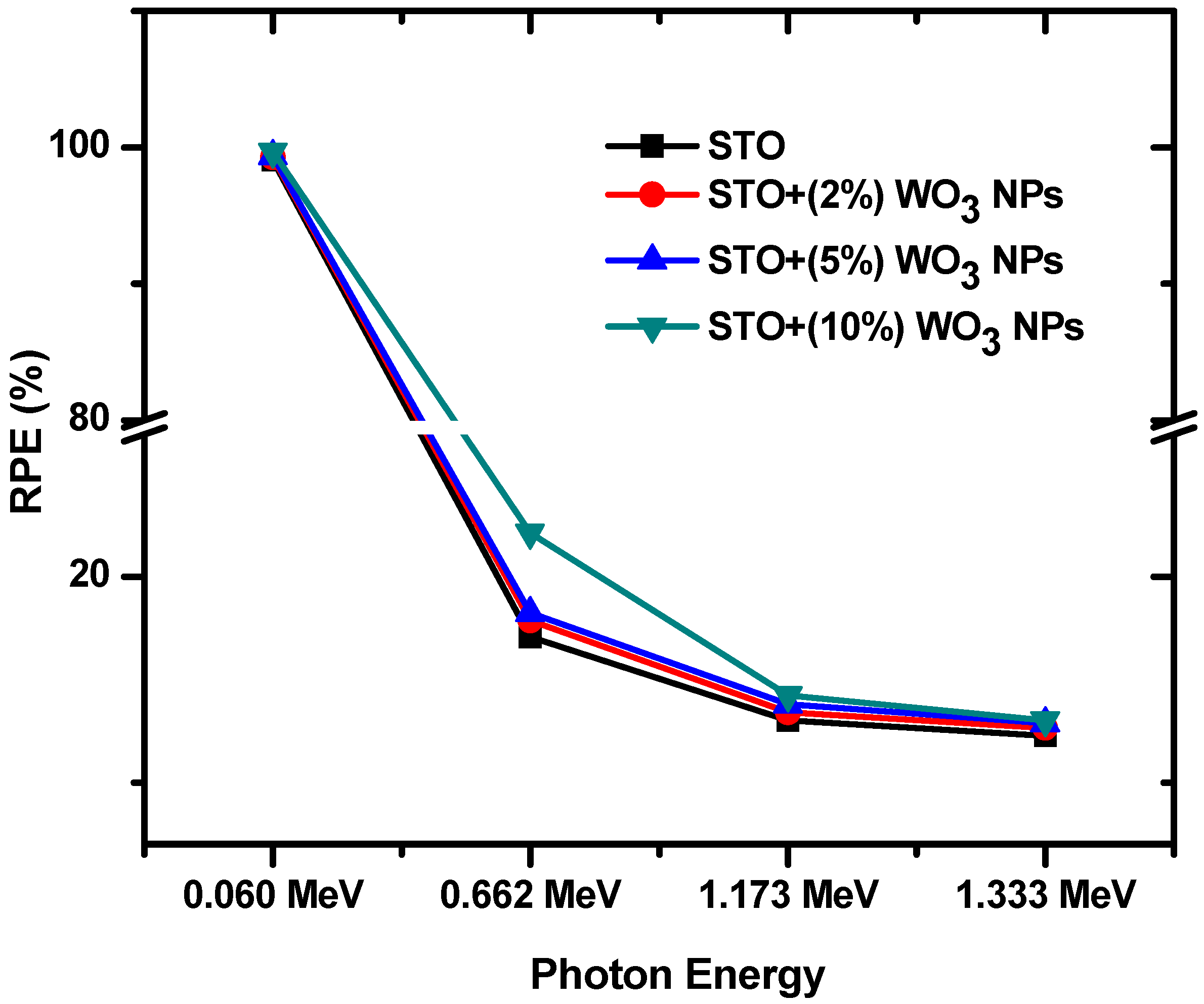
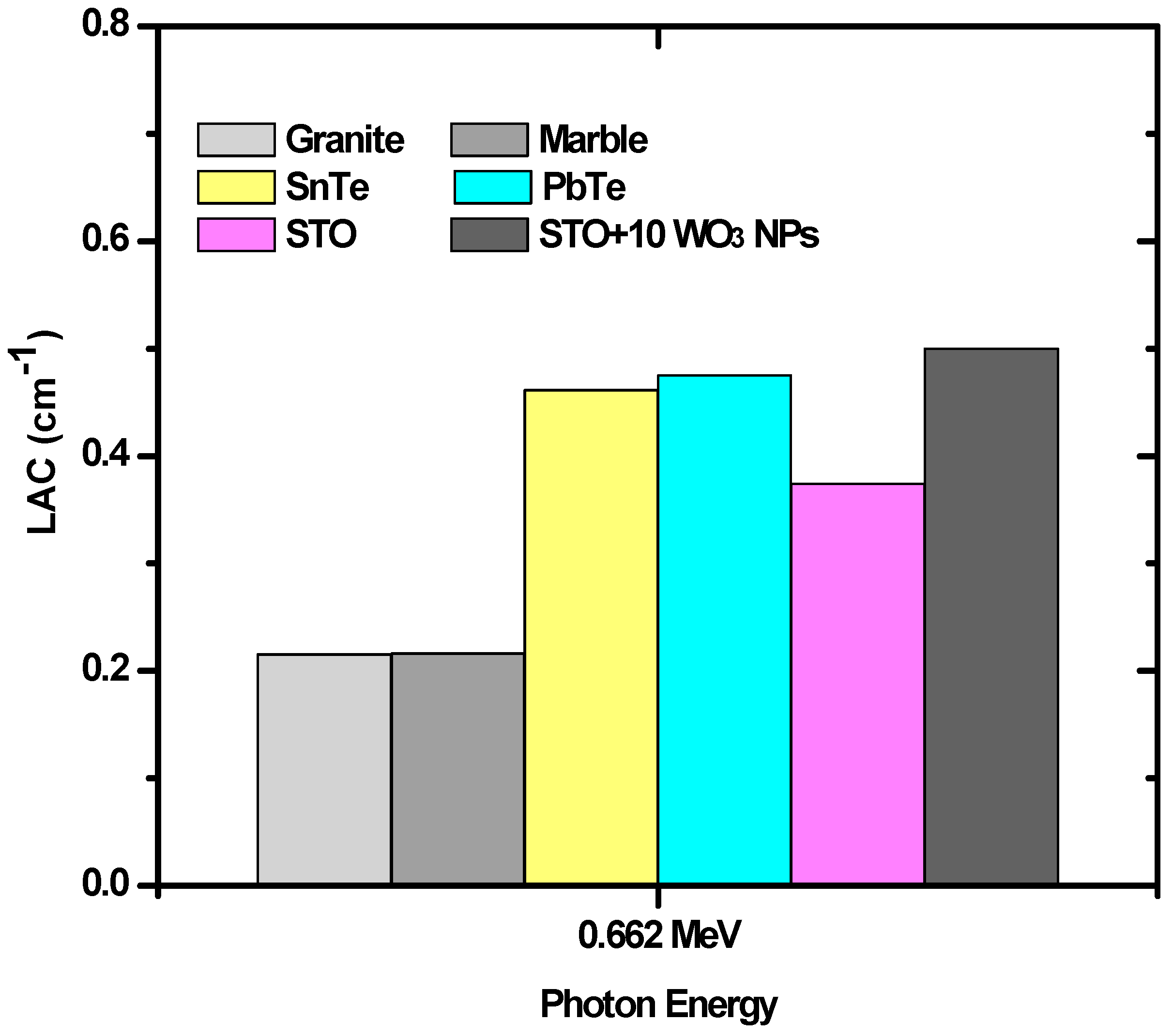
3.2. Radiation Attenuation Analysis

Figure 3 demonstrates the MAC (mass attenuation coefficient) of the prepared samples at four tested energies. At the first energy, the MAC values increase as more WO3 nanoparticles are added to the ceramic. For example, the MAC of the WO3-free ceramic is equal to 1.821 cm−1 at this energy, while the MAC of the ceramic with 10% WO3 nanoparticles is equal to 2.188 cm−1. From MAC determination and the density of ceramic samples, the LACs (linear attenuation coefficients) of the prepared ceramic samples can be calculated. Figure 4 demonstrates the LACs of the prepared ceramic samples at four tested energies. At the first energy, the LAC values increase as more WO3 nanoparticles are added to the ceramic. For example, the LAC of the WO3-free ceramic is equal to 9.308 cm−1 at this energy, while the LAC of the ceramic with 10% WO3 nanoparticles is equal to 11.563 cm−1. The same trend can be observed at higher energies, however, with a smaller difference. Nevertheless, these results show how the amount of WO3 in the ceramics correlates with an improvement in their shielding abilities. In addition, this figure also shows that LACs have an inverse relationship with energy. More specifically, the STO ceramic with 2% WO3 has an LAC equal to 9.810, 0.394, 0.288, and 0.270 cm−1 at 0.060, 0.662, 1.173, and 1.333 MeV, respectively. Thus, ceramics are most effective against low-energy photons.
The half-value layer (HVL) of the investigated samples is graphed in Figure 5 against energy. For all the tested ceramics, their HVL values correlate with energy. For example, for the ceramic with 2% WO3 nanoparticles, its HVL values are equal to 0.071 cm, 1.760 cm, 2.407 cm, and 2.564 cm at 0.060 MeV, 0.662 MeV, 1.173 MeV, and 1.333 MeV, respectively. As the energy increases, more radiation can pass through the material, which is why this relationship occurs. Therefore, a larger thickness is required to absorb half of the total photons, leading to a greater HVL. The figure also shows that HVL decreases with increasing WO3 content. This trend is most evident at higher energies, such as 1.173 MeV. Moreover, at 1.173 MeV, the WO3 values are equal to 2.483, 2.407, 2.334, and 2.259 cm for the samples with 0, 2, 5, and 10% WO3 nanoparticles. Thus, increasing the amount of WO3 nanoparticles in the ceramics improves their HVL values, lowering how thick a material needs to be to attenuate the same number of photons.
The tenth value layer (TVL) is like HVL, except it is defined as how thick a material needs to be to absorb nine-tenths of the incoming photons. Because of this, similar trends can be observed between the two figures; however, all the TVL values are higher than their HVL counterpart. The same two trends can be observed in Figure 6. Mainly, the TVL values increase with energy. For example, the TVL values for the STO with no WO3 nanoparticles are equal to 0.247, 6.158, 8.250, and 8.809 cm at 0.060, 0.662, 1.173, and 1.333 MeV, respectively. The second trend involves the relationship between TVL and photon energy. At 0.662 MeV, the STO ceramics have TVL values of 6.158, 5.848, 5.724, and 4.604 cm for the samples with 0, 2, 5, and 10% WO3 nanoparticles. Thus, increasing the WO3 content in the STO ceramics improves their shielding efficiency.
The mean free path (MFP) of the STO ceramics is shown in Figure 7 at the four tested energies. The maximum MFP values occur for the STO ceramic, while the ceramic with 10% WO3 nanoparticles has the minimum MFP values. For example, at 0.060 MeV, the STO ceramic has an MFP value of 0.107 cm, while the ceramic with 10% WO3 NP has an MFP value of 0.086 cm. Thus, the amount of WO3 in the ceramic mixture correlates with more collisions occurring within the sample, leading to greater attenuation. Furthermore, for any single ceramic, its MFP values increase with energy. For instance, STO-5WO3s is equal to 0.099 cm, 2.486 cm, 3.367 cm, and 3.628 cm at 0.060, 0.662, 1.173, and 1.333 MeV. Higher energy radiation tends to pass through the samples more easily, decreasing the number of collisions and increasing the distance between collisions. Thus, ceramics are able to attenuate lower energy photons than higher energy ones. The results of LAC, HVL, and MFP, as well as the mass attenuation coefficient (MAC), are reported in Table 2.
The radiation protection efficiency (RPE) of the four prepared STO ceramics was calculated and graphed in Figure 8 at a thickness of 0.5 cm. RPE increases with WO3 content at all tested energies. For instance, at 1.333 MeV, the RPE values are 12.25%, 12.64%, 12.87%, and 13.02% for the samples with 0, 2, 5, and 10% WO3 nanoparticles. A greater RPE indicates a more effective shielding capability, so raising the amount of WO3 in the ceramics leads to greater attenuation. Furthermore, RPE decreases as the energy of the incoming photons increases. The RPE of the sample with no WO3 is equal to 99.05 at 0.060 MeV and drops to 12.25% at 1.333 MeV. Meanwhile, the RPE of the sample with 10% of WO3 nanoparticles is equal to 99.69% and 13.02% at 0.060 MeV and 1.333 MeV, respectively. This conclusion reinforces some of the results from the previous figures that more photons can be attenuated at lower energies.
To compare the shielding abilities of the prepared ceramics with other radiation shields, Figure 9 graphs the LACs of the STO and STO + 10% of WO3 NPs ceramics alongside four other shielding materials at 0.662 MeV. The STO and STO + 10% of WO3 NPs ceramics had a greater LAC value than granite and marble [51,52]. SnTe and PbTe shields have a greater LAC than the STO ceramic but less than the STO + 10% of WO3 ceramic [53]. Thus, ceramics have an overall very desirable shielding ability compared to other commonly used materials for radiation shielding purposes.

4. Conclusions

In this work, we proposed a strategy for developing new ceramic materials consisting of STO doped with WO3 nanoparticles for the purpose of radiation protection. The different samples were prepared using the simple solid-state reaction method. The successful formation of the required compositions was confirmed by XRD and FTIR analyses. All as-prepared ceramics crystallized into the cubic structure. A new phase belonging to SrWO4 was detected for 5 and 10% doped ceramics suggesting an interaction between the host STO and the dopant WO3 NPs has occurred. The lattice constant ‘a’ and the unit cell volume ‘V’ were found to be higher for doped ceramic compared to the STO pristine ceramic. The crystallite size varied with the content of WO3 NPs. In the second part, we focused on the role of adding WO3 NPs in improving the radiation-protective characteristics of STO ceramics. The LAC was experimentally measured using a high pure germanium (HPGe) detector and three-point sources (60C, 241Am, and 137Cs). The LAC of the WO3-free ceramic at 0.06 MeV is equal to 9.308 cm−1, while it is 11.563 cm−1 for the ceramic with 10% WO3 nanoparticles. This shows that the LAC values increase as more WO3 nanoparticles are added to the ceramic. We investigated the relationship between the LAC and the energy, and we concluded that the ceramics are most effective against low-energy photons. In order to examine the thickness of the ceramics that required attenuating certain levels of the photons, we evaluated the HVL and TVL. From the findings of both factors, we found that a larger thickness is required to absorb the photons with higher energies. Moreover, we examined the impact of WO3 nanoparticles on these two factors. In brief, increasing the amount of WO3 nanoparticles in the ceramics improves their HVL and TVL values. In addition to these factors, the MFP results also proved that the ceramics are able to attenuate lower energy photons than higher energy ones. At 1.333 MeV, the RPE for the samples with 0, 2, 5, and 10% WO3 nanoparticles are 12.25%, 12.64%, 12.87%, and 13.02%, respectively. Hence, from the RPE data, we found that increasing the amount of WO3 in the ceramics leads to greater attenuation.

Author Contributions

Conceptualization, M.E.; Data curation, M.I.S..; Funding acquisition, M.I.S. and S.H..; Investigation, M.I.S.; Methodology, M.E.; Project administration, S.H.; Resources, Y.S. and E.H.; Software, M.I.S.; Supervision, S.H.; Validation, Y.S.; Writing—original draft, Y.S. and E.H.; Writing—review and editing, Y.S. and E.H. All authors have read and agreed to the published version of the manuscript.

Funding

M.I. Sayyed and S. Hashim gratefully acknowledge Universiti Teknologi Malaysia for supporting their Prominent Visiting Researcher Scheme (RJ3000.7113.3F000) through the Department of Deputy Vice-Chancellor (Research and Innovation).

Institutional Review Board Statement

Not applicable.

Informed Consent Statement

Not applicable.

Data Availability Statement

All relevant data are within this paper.

Acknowledgments

M.I. Sayyed and S. Hashim gratefully acknowledge Universiti Teknologi Malaysia for providing Prominent Visiting Researcher Scheme (RJ3000.7113.3F000) by the Department Deputy of Vice-Chancellor (Research and Innovation).

Conflicts of Interest

The authors declare no conflict of interest.

References

  1. Shultis, J.K.; Faw, R.E. Radiation shielding technology. Health Phys. 2005, 88, 297–322. [Google Scholar] [CrossRef] [PubMed] [Green Version]
  2. Amana, M.S.; Aldhuhaibat, M.J.R.; Salim, A.A. Evaluation of the absorption, scattering and overall probability of gamma rays in lead and concrete interactions. SCIOL Biomed. 2021, 4, 191–199. [Google Scholar]
  3. Hassan, H.E.; Badran, H.M.; Aydarous, A.; Sharshar, T. Studying the effect of nano lead compounds additives on the concrete shielding properties for γ-rays. Nucl. Instrum. Methods Phys. Res. Sect. B Beam Interact. Mater. At. 2015, 360, 81–89. [Google Scholar] [CrossRef]
  4. Hannachi, E.; Sayyed, M.I.; Slimani, Y.; Almessiere, M.A.; Baykal, A.; Elsafi, M. Synthesis, characterization, and performance assessment of new composite ceramics towards radiation shielding applications. J. Alloys Compd. 2022, 899, 163173. [Google Scholar] [CrossRef]
  5. Sayyed, M.I.; Hannachi, E.; Mahmoud, K.A.; Slimani, Y. Synthesis of different (RE) BaCuO ceramics, study their structural properties, and tracking their radiation protection efficiency using Monte Carlo simulation. Mater. Chem. Phys. 2022, 276, 125412. [Google Scholar] [CrossRef]
  6. Sayyed, M.I.; El-Mesady, I.A.; Abouhaswa, A.S.; Askin, A.; Rammah, Y.S. Comprehensive study on the structural, optical, physical and gamma photon shielding features of B2O3-Bi2O3-PbO-TiO2 glasses using WinXCOM and Geant4 code. J. Mol. Struct. 2019, 1197, 656–665. [Google Scholar] [CrossRef]
  7. Aygün, B.; Şakar, E.; Agar, O.; Sayyed, M.I.; Karabulut, A.; Singh, V.P. Development of new heavy concretes containing chrome-ore for nuclear radiation shielding applications. Prog. Nucl. Energy 2021, 133, 103645. [Google Scholar] [CrossRef]
  8. Khazaalah, T.H.; Shahrim Mustafa, I.; Al-Ghamdi, H.; Abdul Rahman, A.; Sayyed, M.I.; Almuqrin, A.H.; Mohd Zaid, M.H.; Hisam, R.; Abdul Malik, M.F.I.; Seth Ezra, N.; et al. The Effect of WO3-Doped Soda Lime Silica SLS Waste Glass to Develop Lead-Free Glass as a Shielding Material against Radiation. Sustainability 2022, 14, 2413. [Google Scholar] [CrossRef]
  9. Sayyed, M.I.; Hamad, M.K.; Mhareb, M.H.A.; Kurtulus, R.; Dwaikat, N.; Saleh, M.; Elsafi, M.; Taki, M.M.; Kavas, T.; Ziq, K.A.; et al. Assessment of radiation attenuation properties for novel alloys: An experimental approach. Radiat. Phys. Chem. 2022, 200, 110152. [Google Scholar] [CrossRef]
  10. Bagheri, R.; Shirmardi, S.P. Gamma-ray shielding studies on borate glasses containing BaO, Bi2O3, and PbO in different concentrations. Radiat. Phys. Chem. 2021, 184, 109434. [Google Scholar] [CrossRef]
  11. Bagheri, R.; Adeli, R. Gamma-ray shielding properties of phosphate glasses containing Bi2O3, PbO, and BaO in different rates. Radiat. Phys. Chem. 2020, 174, 108918. [Google Scholar] [CrossRef]
  12. Kavaz, E.; El_Agawany, F.I.; Tekin, H.O.; Perişanoğlu, U.; Rammah, Y.S. Nuclear radiation shielding using barium borosilicate glass ceramics. J. Phys. Chem. Solids 2020, 142, 109437. [Google Scholar] [CrossRef]
  13. Bagheri, R.; Moghaddam, A.K.; Shirmardi, S.P.; Azadbakht, B.; Salehi, M. Determination of gamma-ray shielding properties for silicate glasses containing Bi2O3, PbO, and BaO. J. Non-Cryst. Solids 2018, 479, 62–71. [Google Scholar] [CrossRef]
  14. Bagheri, R.; Moghaddam, A.K.; Yousefnia, H. Gamma ray shielding study of barium–bismuth–borosilicate glasses as transparent shielding materials using MCNP-4C code, XCOM program, and available experimental data. Nucl. Eng. Technol. 2017, 49, 216–223. [Google Scholar] [CrossRef]
  15. Slimani, Y.; Unal, B.; Almessiere, M.A.; Hannachi, E.; Yasin, G.; Baykal, A.; Ercan, I. Role of WO3 nanoparticles in electrical and dielectric properties of BaTiO3–SrTiO3 ceramics. J. Mater. Sci. Mater. Electron. 2020, 31, 7786–7797. [Google Scholar] [CrossRef]
  16. Shi, X.L.; Wu, H.; Liu, Q.; Zhou, W.; Lu, S.; Shao, Z.; Dargusch, M.; Chen, Z.G. SrTiO3-based thermoelectrics: Progress and challenges. Nano Energy 2020, 78, 105195. [Google Scholar] [CrossRef]
  17. Slimani, Y.; Selmi, A.; Hannachi, E.; Almessiere, M.A.; Baykal, A.; Ercan, I. Impact of ZnO addition on structural, morphological, optical, dielectric and electrical performances of BaTiO3 ceramics. J. Mater. Sci. Mater. Electron. 2019, 30, 9520–9530. [Google Scholar] [CrossRef]
  18. Abbas, M.I.; El-Khatib, A.M.; Dib, M.F.; Mustafa, H.E.; Sayyed, M.I.; Elsafi, M. The Influence of Bi2O3 Nanoparticle Content on the γ-ray Interaction Parameters of Silicon Rubber. Polymers 2022, 14, 1048. [Google Scholar] [CrossRef]
  19. Enzlberger, L. Photovoltaic Properties of SrTiO3-δ in Single Crystalline and Thin Film cells. Ph.D. Thesis, Technische Universität Wien, Vienna, Austria, 2022. [Google Scholar]
  20. Aravinthkumar, K.; Praveen, E.; Mary, A.J.R.; Mohan, C.R. Investigation on SrTiO3 nanoparticles as a photocatalyst for enhanced photocatalytic activity and photovoltaic applications. Inorg. Chem. Commun. 2022, 140, 109451. [Google Scholar] [CrossRef]
  21. Wang, J.; Wang, T.; Zhao, Z.; Wang, R.; Wang, C.; Zhou, F.; Li, S.; Zhao, L.; Feng, M. Regulation of oxygen vacancies in SrTiO3 perovskite for efficient photocatalytic nitrogen fixation. J. Alloys Compd. 2022, 902, 163865. [Google Scholar] [CrossRef]
  22. Liu, L.; Chu, B.; Li, P.; Fu, P.; Du, J.; Hao, J.; Li, W.; Zeng, H. Achieving high energy storage performance and ultrafast discharge speed in SrTiO3-based ceramics via a synergistic effect of chemical modification and defect chemistry. Chem. Eng. J. 2022, 429, 132548. [Google Scholar] [CrossRef]
  23. Hannachi, E.; Sayyed, M.I.; Albarzan, B.; Almuqrin, A.H.; Mahmoud, K.A. Synthesis and study of structural, optical and radiation-protective peculiarities of MTiO3 (M = Ba, Sr) metatitanate ceramics mixed with SnO2 oxide. Ceram. Int. 2021, 47, 28528–28535. [Google Scholar] [CrossRef]
  24. Slimani, Y.; Hamad, M.K.; Olarinoye, I.O.; Alajerami, Y.S.; Sayyed, M.I.; Almessiere, M.A.; Mhareb, M.H.A. Determination of structural features of different Perovskite ceramics and investigation of ionizing radiation shielding properties. J. Mater. Sci. Mater. Electron. 2021, 32, 20867–20881. [Google Scholar] [CrossRef]
  25. Hannachi, E.; Sayyed, M.I.; Mahmoud, K.A.; Slimani, Y.; Akhtar, S.; Albarzan, B.; Almuqrin, A.H. Impact of tin oxide on the structural features and radiation shielding response of some ABO3 perovskites ceramics (A = Ca, Sr, Ba; B = Ti). Appl. Phys. A 2021, 127, 1–12. [Google Scholar] [CrossRef]
  26. Pehlivan, I.B.; Atak, G.; Niklasson, G.A.; Stolt, L.; Edoff, M.; Edvinsson, T. Electrochromic solar water splitting using a cathodic WO3 electrocatalyst. Nano Energy 2021, 81, 105620. [Google Scholar] [CrossRef]
  27. Dutta, V.; Sharma, S.; Raizada, P.; Thakur, V.K.; Khan, A.A.P.; Saini, V.; Asiri, A.M.; Singh, P. An overview on WO3 based photocatalyst for environmental remediation. J. Environ. Chem. Eng. 2021, 9, 105018. [Google Scholar] [CrossRef]
  28. Chang, C.M.; Chiang, Y.C.; Cheng, M.H.; Lin, S.H.; Jian, W.B.; Chen, J.T.; Cheng, Y.J.; Ma, Y.R.; Tsukagoshi, K. Fabrication of WO3 electrochromic devices using electro-exploding wire techniques and spray coating. Sol. Energy Mater. Sol. Cells 2021, 223, 110960. [Google Scholar] [CrossRef]
  29. Qureshi, N.; Lee, S.; Chaudhari, R.; Mane, P.; Pawar, J.; Chaudhari, B.; Shinde, M.; Rane, S.; Kim, T.; Amalnerkar, D. Hydrothermal Generation of 3-Dimensional WO3 Nanocubes, Nanobars and Nanobricks, Their Antimicrobial and Anticancer Properties. J. Nanosci. Nanotechnol. 2021, 21, 5337–5343. [Google Scholar] [CrossRef]
  30. Du, Q.; Wang, L.; Yang, J.; Liu, J.; Yuan, Y.; Wang, M.; Liu, B.; Zhang, X.; Ren, Y.; Zhao, H.; et al. Enhancing gas sensing performances and sensing mechanism at atomic and molecule level of WO3 nanoparticles by hydrogenation. Sens. Actuators B Chem. 2018, 273, 1786–1793. [Google Scholar] [CrossRef]
  31. Hemily, H.M.; Saleh, I.H.; Ghataas, Z.F.; Abdel-Halim, A.A.; Hisam, R.; Shah, A.Z.; Sayyed, M.I.; Yasmin, S.; Elsafi, M. Radiation Shielding Enhancement of Polyester Adding Artificial Marble Materials and WO3 Nanoparticles. Sustainability 2022, 14, 13355. [Google Scholar] [CrossRef]
  32. Zeb, S.; Sun, G.; Nie, Y.; Cui, Y.; Jiang, X. Synthesis of highly oriented WO3 nanowire bundles decorated with Au for gas sensing application. Sens. Actuators B Chem. 2020, 321, 128439. [Google Scholar] [CrossRef]
  33. Xiao, Y.; He, Z.; Wang, R.; Tao, X.; Li, B. Synthesis of WO3 nanofibers decorated with BiOCl nanosheets for photocatalytic degradation of organic pollutants under visible light. Colloids Surf. A Physicochem. Eng. Asp. 2019, 580, 123752. [Google Scholar] [CrossRef]
  34. Lei, J.; Liu, H.; Yuan, C.; Chen, Q.; Liu, J.A.; Wen, F.; Jiang, X.; Deng, W.; Cui, X.; Duan, T.; et al. Enhanced photoreduction of U (VI) on WO3 nanosheets by oxygen defect engineering. Chem. Eng. J. 2021, 416, 129164. [Google Scholar] [CrossRef]
  35. Kong, L.; Guo, X.; Xu, J.; Mo, Z.; Li, L. Morphology control of WO3 nanoplate film on W foil by oxalic acid for photocatalytic gaseous acetaldehyde degradation. J. Photochem. Photobiol. A Chem. 2020, 401, 112760. [Google Scholar] [CrossRef]
  36. Ghavami, S.M.; Ghiasi, H.; Mesbahi, A. Monte Carlo modeling of the yttrium-90 nanospheres application in the liver radionuclide therapy and organs doses calculation. Nucl. Technol. Radiat. Prot. 2016, 31, 89–96. [Google Scholar] [CrossRef] [Green Version]
  37. Tekin, H.O.; Singh, V.P.; Manici, T. Effects of micro-sized and nano-sized WO3 on mass attenauation coefficients of concrete by using MCNPX code. Appl. Radiat. Isot. 2017, 121, 122–125. [Google Scholar] [CrossRef] [PubMed]
  38. Mesbahi, A.; Famouri, F.; Ahar, M.J.; Ghaffari, M.O.; Ghavami, S.M. A study on the imaging characteristics of Gold nanoparticles as a contrast agent in X-ray computed tomography. Pol. J. Med. Phys. Eng. 2017, 23, 9. [Google Scholar] [CrossRef] [Green Version]
  39. Al-Ghamdi, H.; Hemily, H.M.; Saleh, I.H.; Ghataas, Z.F.; Abdel-Halim, A.A.; Sayyed, M.I.; Yasmin, S.; Almuqrin, A.H.; Elsafi, M. Impact of WO3-Nanoparticles on Silicone Rubber for Radiation Protection Efficiency. Materials 2022, 15, 5706. [Google Scholar] [CrossRef]
  40. Elsafi, M.; Dib, M.F.; Mustafa, H.E.; Sayyed, M.I.; Khandaker, M.U.; Alsubaie, A.; Almalki, A.S.A.; Abbas, M.I.; El-Khatib, A.M. Enhancement of Ceramics Based Red-Clay by Bulk and Nano Metal Oxides for Photon Shielding Features. Materials 2021, 14, 7878. [Google Scholar] [CrossRef]
  41. El-Khatib, A.M.; Elsafi, M.; Almutiri, M.N.; Mahmoud, R.M.M.; Alzahrani, J.S.; Sayyed, M.I.; Abbas, M.I. Enhancement of Bentonite Materials with Cement for Gamma-Ray Shielding Capability. Materials 2021, 14, 4697. [Google Scholar] [CrossRef]
  42. Hannachi, E.; Sayyed, M.I.; Slimani, Y.; Elsafi, M. Experimental investigation on the physical properties and radiation shielding efficiency of YBa2Cu3Oy/M@ M3O4 (M = Co, Mn) ceramic composites. J. Alloys Compd. 2022, 904, 164056. [Google Scholar] [CrossRef]
  43. Al-Hadeethi, Y.; Sayyed, M.I.; Barasheed, A.Z.; Ahmed, M.; Elsafi, M. Fabrication of lead free borate glasses modified by bismuth oxide for gamma ray protection applications. Materials 2022, 15, 789. [Google Scholar] [CrossRef]
  44. Hannachi, E.; Almessiere, M.A.; Slimani, Y.; Baykal, A.; Azzouz, F.B. AC susceptibility investigation of YBCO superconductor added by carbon nanotubes. J. Alloys Compd. 2020, 812, 152150. [Google Scholar] [CrossRef]
  45. Hikichi, Y.; Niimi, T.; Sato, H. Preparation and Dielectric Properties of WO3-Doped Small-Grained BaTiO3 Ceramics. Jpn. J. Appl. Phys. 1985, 24, 1039. [Google Scholar] [CrossRef]
  46. Sczancoski, J.C.; Cavalcante, L.S.; Joya, M.R.; Espinosa, J.W.M.; Pizani, P.S.; Varela, J.A.; Longo, E. Synthesis, growth process and photoluminescence properties of SrWO4 powders. J. Colloid Interface Sci. 2009, 330, 227–236. [Google Scholar] [CrossRef] [PubMed]
  47. Hou, D.; Hu, X.; Ho, W.; Hu, P.; Huang, Y. Facile fabrication of porous Cr-doped SrTiO3 nanotubes by electrospinning and their enhanced visible-light-driven photocatalytic properties. J. Mater. Chem. A 2015, 3, 3935–3943. [Google Scholar] [CrossRef]
  48. Šetinc, T.; Spreitzer, M.; Vengust, D.; Jerman, I.; Suvorov, D. Inherent defects in sol-precipitation/hydrothermally derived SrTiO3 nanopowders. Ceram. Int. 2013, 39, 6727–6734. [Google Scholar] [CrossRef]
  49. Adireddy, S.; Lin, C.; Cao, B.; Zhou, W.; Caruntu, G. Solution-based growth of monodisperse cube-like BaTiO3 colloidal nanocrystals. Chemi. Mater. 2010, 22, 1946–1948. [Google Scholar] [CrossRef]
  50. Adhikari, S.; Sarkar, D.; Maiti, H.S. Synthesis and characterization of WO3 spherical nanoparticles and nanorods. Mater. Res. Bull. 2014, 49, 325–330. [Google Scholar] [CrossRef]
  51. Elsafi, M.; Alrashedi, M.F.; Sayyed, M.I.; Al-Hamarneh, I.F.; El-Nahal, M.A.; El-Khatib, M.; Khandaker, M.U.; Osman, H.; Askary, A.E. The Potentials of Egyptian and Indian Granites for Protection of Ionizing Radiation. Materials 2021, 14, 3928. [Google Scholar] [CrossRef]
  52. Elsafi, M.; El-Nahal, M.A.; Alrashedi, M.F.; Olarinoye, O.I.; Sayyed, M.I.; Khandaker, M.U.; Osman, H.; Alamri, S.; Abbas, M.I. Shielding properties of some marble types: A comprehensive study of experimental and XCOM results. Materials 2021, 14, 4194. [Google Scholar] [CrossRef] [PubMed]
  53. Mhareb, M.H.A.; Zeama, M.; Elsafi, M.; Alajerami, Y.S.; Sayyed, M.I.; Saleh, G.; Hamad, R.M.; Hamad, M.K. Radiation shielding features for various tellurium-based alloys: A comparative study. J. Mater. Sci. Mater. Electron. 2021, 32, 26798–26811. [Google Scholar] [CrossRef]
Figure 1. XRD patterns of STO ceramic and STO ceramics doped with x wt.% of WO3 NPs.
Crystals 12 01602 g001
Figure 2. FT-IR results of STO ceramic and STO ceramic doped with x% of WO3 NPs.
Crystals 12 01602 g002
Figure 3. Mass attenuation coefficients of the STO ceramic and STO ceramic doped with x% of WO3 NPs.
Crystals 12 01602 g003
Figure 4. Linear attenuation coefficients of the STO ceramic and STO ceramic doped with x% of WO3 NPs.
Crystals 12 01602 g004
Figure 5. Half value layer of STO ceramic and STO ceramic doped with x% of WO3 NPs.
Crystals 12 01602 g005
Figure 6. Tenth value layer of STO ceramic and STO ceramic doped with x% of WO3 NPs.
Crystals 12 01602 g006
Figure 7. Mean free path of STO ceramic and STO ceramic doped with x% of WO3 NPs.
Crystals 12 01602 g007
Figure 8. Radiation protection efficiency of STO ceramic and STO ceramic doped with x% of WO3 NPs.
Crystals 12 01602 g008
Figure 9. Linear attenuation coefficient for STO and STO + 10% WO3 NPs ceramics alongside four other shielding materials at 0.662 MeV.
Crystals 12 01602 g009
Table 1. Structural parameters deduced from XRD analysis.
CeramicsSTOSTO + (2%) WO3 NPsSTO + (5%) WO3 NPsSTO + (10%) WO3 NPs
a (Å)=3.90203.90473.90323.9042
V (Å3)=59.410359.533759.465159.5108
StructureCubicCubicCubicCubic
dXRD (nm)=45.4841.1745.4950.86
Table 2. The experimental values of the attenuator parameters for STO-ceramic samples.
CodeEnergy (MeV)STOSTO + 2% WO3 NPsSTO + 5% WO3 NPsSTO + 10% WO3 NPs
LAC, cm−10.0609.3089.81010.09711.563
0.6620.3740.3940.4020.500
1.1730.2790.2880.2970.307
1.3330.2610.2700.2760.279
HVL, cm0.0600.0740.0710.0690.060
0.6621.8541.7601.7231.386
1.1732.4832.4072.3342.259
1.3332.6522.5642.5152.484
TVL, cm0.0600.2470.2350.2280.199
0.6626.1585.8485.7244.604
1.1738.2507.9977.7537.505
1.3338.8098.5178.3538.253
MAC, cm2.g−10.0601.8211.9041.9432.188
0.6620.0730.0760.0770.095
1.1730.0550.0560.0570.058
1.3330.0510.0520.0530.053
Publisher’s Note: MDPI stays neutral with regard to jurisdictional claims in published maps and institutional affiliations.

