
Composition Engineering of (Lu,Gd,Tb)3(Al,Ga)5O12:Ce Film/Gd3(Al,Ga)5O12:Ce Substrate Scintillators
 , ,
, , 
Abstract
:1. Introduction
2. Materials and Methods
2.1. Fabrication of Substrates
2.2. Film Deposition via LPE Method
2.3. Determination of Optical and Scintillation Parameters
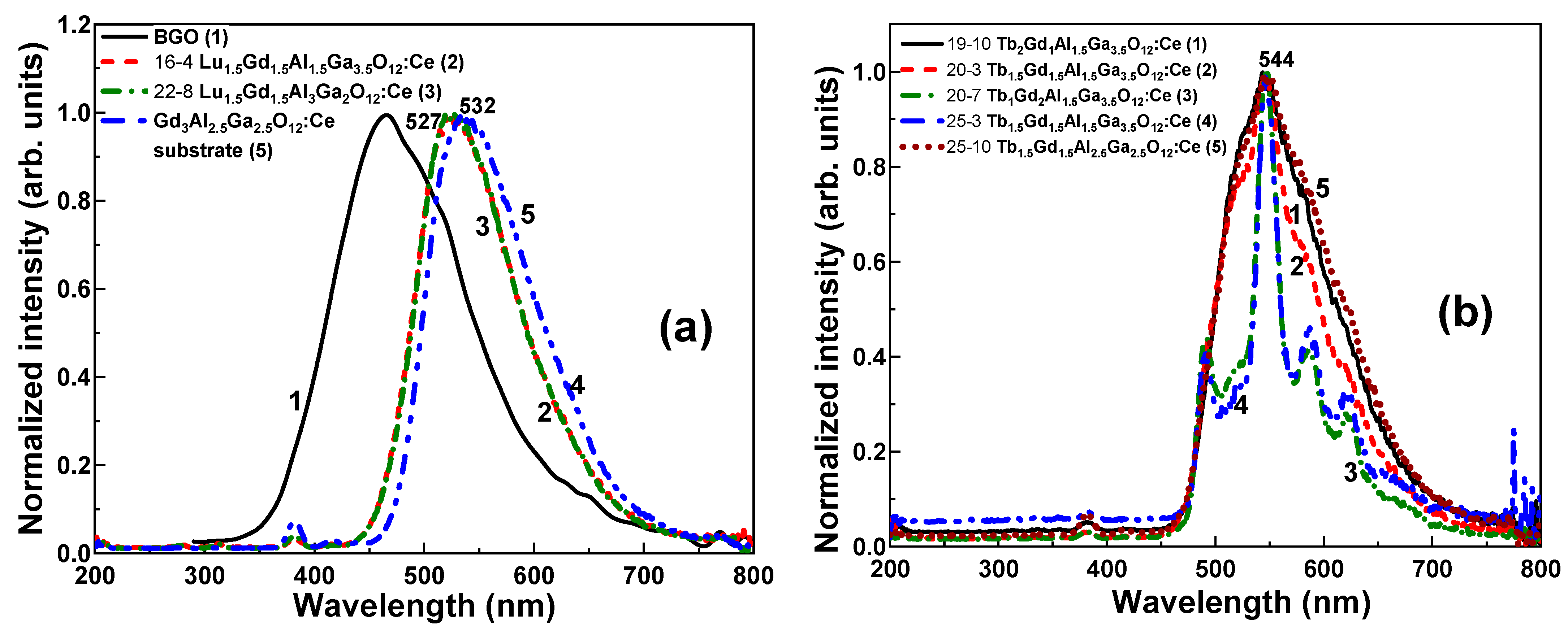
3. Results
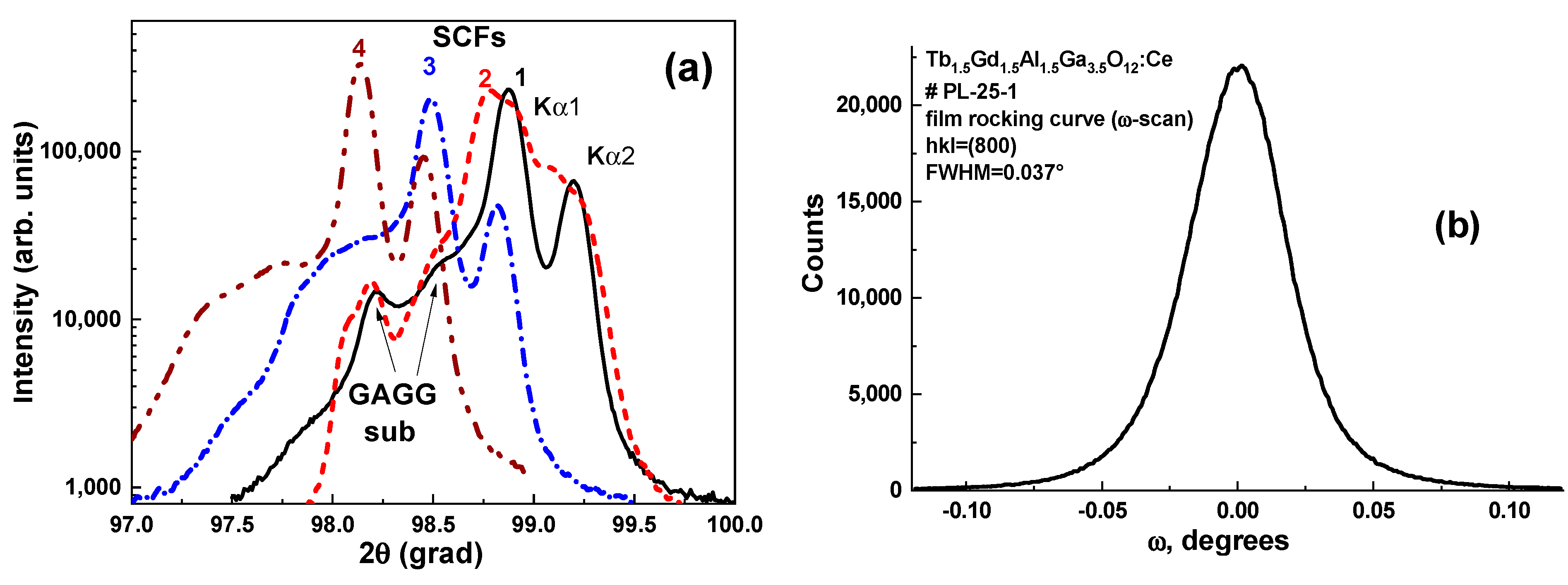
3.1. Determination of Film Structure and Composition
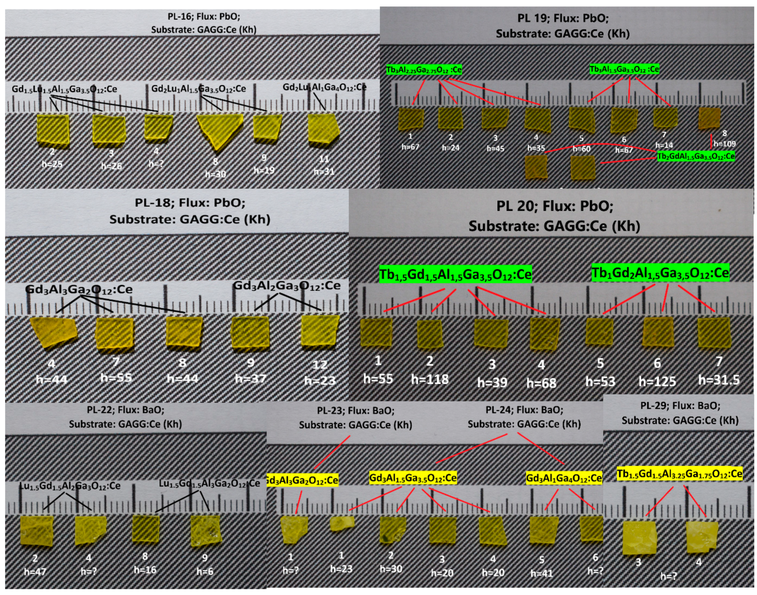
3.2. Primary Selection of Promising Compositions
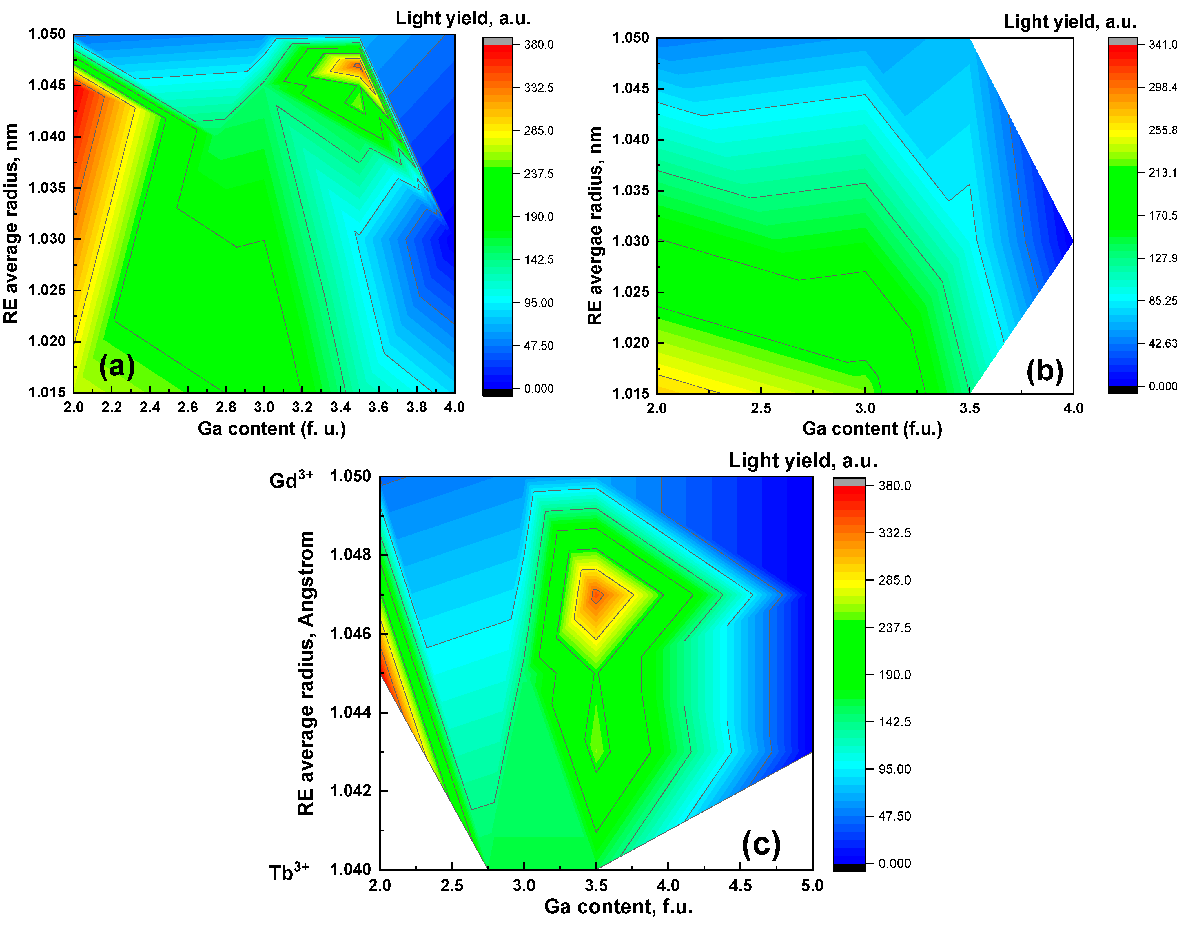
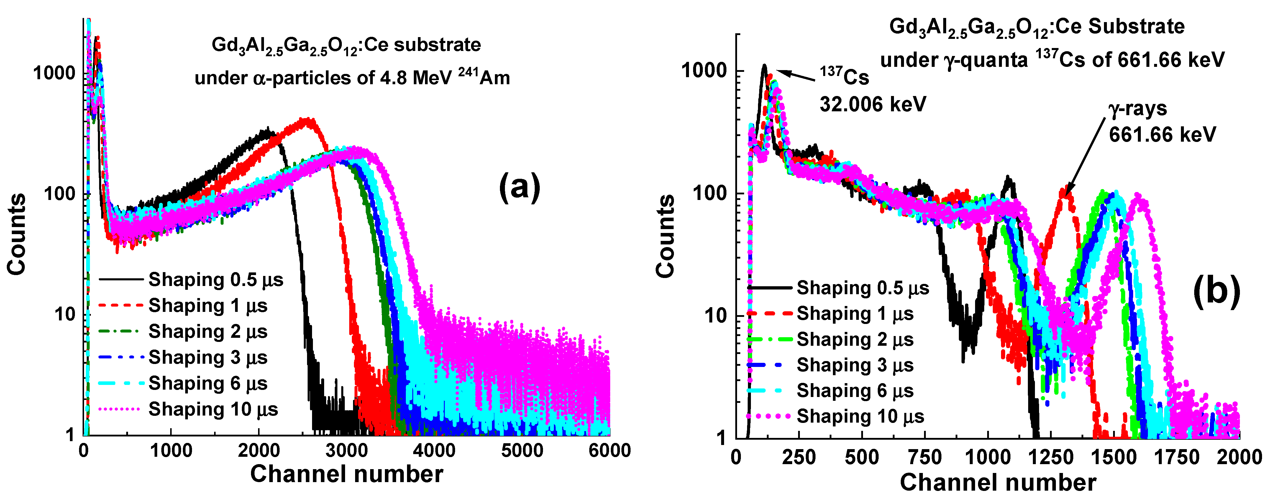
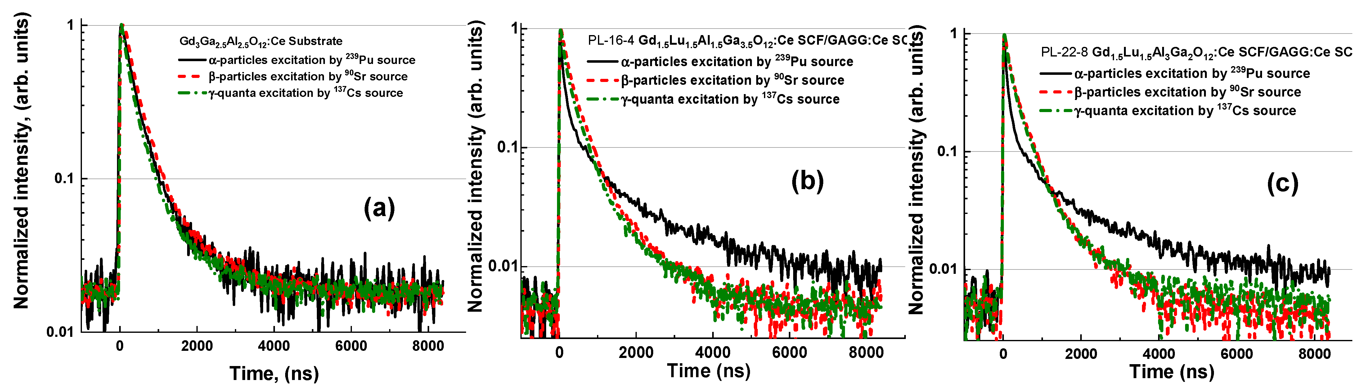
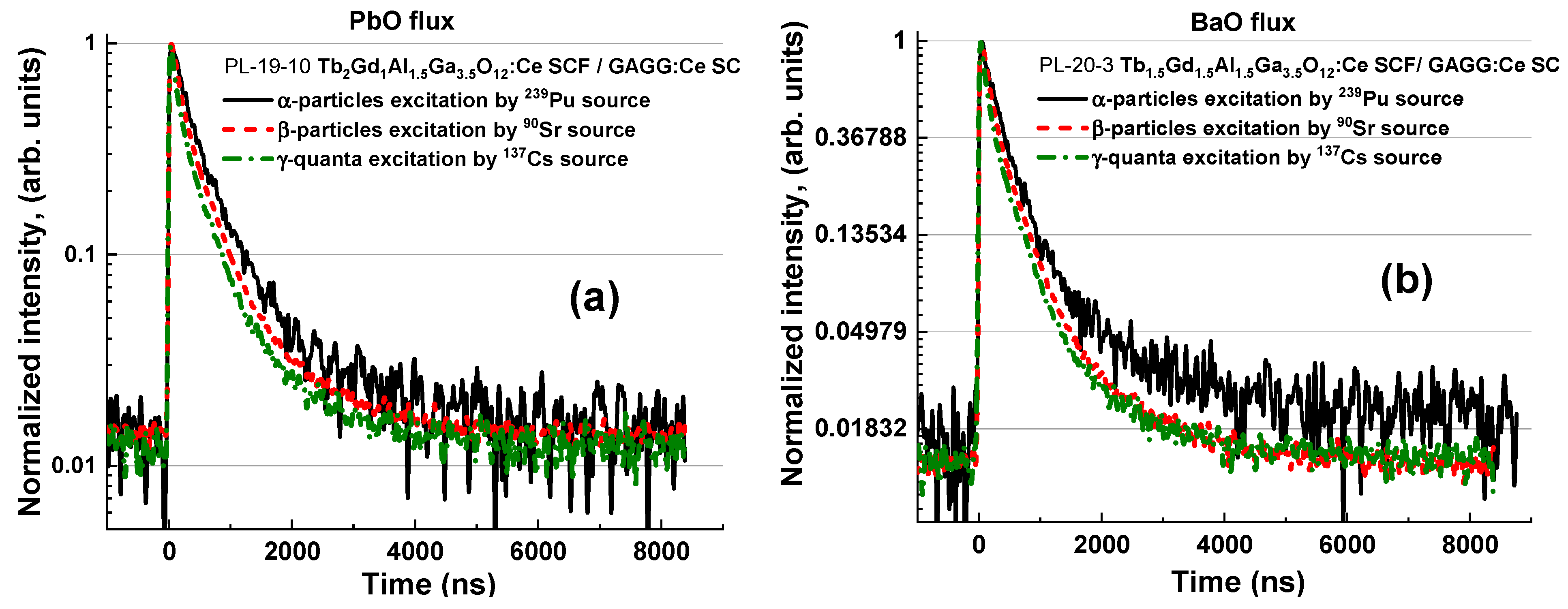
3.3. Characterization of Selected Composite Scintillators
Scintillation Parameters
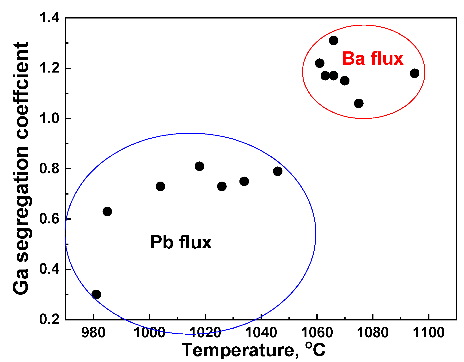
4. Discussion
5. Conclusions
Author Contributions
Funding
Data Availability Statement
Conflicts of Interest
References
- Zaitseva, N.; Rupert, B.L.; Pawelczak, I.; Glenn, A.; Martinez, P.H.; Carman, L.; Faust, M.; Cherepy, N.; Payne, S. Plastic scintillators with efficient neutron/gamma pulse shape discrimination. Nucl. Instrum. Methods Phys. Res. A 2012, 688, 88–93. [Google Scholar] [CrossRef]
- Sénoville, M.; Delaunay, F.; Pârlog, M.; Achouri, N.L.; Orr, N.A. Neutron-gamma discrimination with organic scintillators: Intrinsic pulse shape and light yield contributions. Nucl. Instrum. Meth. Phys. Res. A 2020, 971, 164080. [Google Scholar] [CrossRef]
- Zorenko, Y.; Novosad, S.S.; Pashkovskii, M.V.; Lyskovich, A.B.; Savitskii, V.G.; Batenchuk, M.M.; Malyutenkov, P.S.; Patsagan, N.I.; Nazar, I.V.; Gorbenko, V.I. Epitaxial structures of garnets as scintillation detectors of ionizing radiation. J. Appl. Spectrosc. 1990, 52, 645–649. [Google Scholar] [CrossRef]
- Zorenko, Y.; Batenchuk, M.; Gorbenko, V.; Pashkovsky, M. Single-crystalline oxide films of the Al2O3-Y2O3-R2O3 system as optical sensors of various types of ionizing radiation: Significant advantages over volume analogs. Proc. SPIE 1997, 2967, 101. [Google Scholar] [CrossRef]
- Globus, M.; Grinyov, B.; Ratner, M.; Tarasov, V.; Lyubinskiy, V.; Vydai, Y.; Ananenko, A.; Zorenko, Y.; Gorbenko, V.; Konstankevych, I. New type of scintillation detectors for biological, medical, and radiation monitoring applications. IEEE Trans. Nucl. Sci. 2004, 51, 1297–1303. [Google Scholar] [CrossRef]
- Zorenko, Y.; Gorbenko, V. Growth peculiarities of the R3Al5O12 (R = Lu, Yb, Tb, Eu–Y) single crystalline film phosphors by liquid phase epitaxy. Radiat. Meas. 2007, 42, 907–910. [Google Scholar] [CrossRef]
- Witkiewicz-Lukaszek, S.; Gorbenko, V.; Zorenko, T.; Syrotych, Y.; Mares, J.A.; Nikl, M.; Sidletskiy, O.; Bilski, P.; Yoshikawa, A.; Zorenko, Y. Composite Detectors Based on Single-Crystalline Films and Single Crystals of Garnet Compounds. Materials 2022, 15, 1249. [Google Scholar] [CrossRef]
- Witkiewicz-Lukaszek, S.; Gorbenko, V.; Zorenko, T.; Paprocki, K.; Sidletskiy, O.; Gerasymov, I.; Mares, J.A.; Kucerkova, R.; Nikl, M.; Zorenko, Y. Novel all-solid-state composite scintillators based on the epitaxial structures of LuAG garnet doped with Pr, Sc and Ce ions. IEEE Trans. Nucl. Sci. 2018, 65, 2114–2119. [Google Scholar] [CrossRef]
- Witkiewicz-Lukaszek, S.; Gorbenko, V.; Zorenko, T.; Paprocki, K.; Sidletskiy, O.; Gerasymov, I.; Mares, J.A.; Kucerkova, R.; Nikl, M.; Zorenko, Y. Composite scintillators based on the crystals and single crystalline films of LuAG garnet doped with Ce3+, Pr3+ and Sc3+ ions. Opt. Mater. 2018, 84, 593–599. [Google Scholar] [CrossRef]
- Witkiewicz-Lukaszek, S.; Gorbenko, V.; Zorenko, T.; Sidletskiy, O.; Arhipov, P.; Fedorov, A.; Mares, J.A.; Kucerkova, R.; Nikl, M.; Zorenko, Y. Liquid phase epitaxy growth of high-performance composite scintillators based on single crystalline films and crystals of LuAG. CrystEngComm. 2020, 22, 3713–3724. [Google Scholar] [CrossRef]
- Gorbenko, V.; Witkiewicz-Lukaszek, S.; Zorenko, T.; Syrotych, Y.; Mares, J.A.; Kucerkova, R.; Nikl, M.; Sidletskiy, O.; Fedorov, A.; Zorenko, Y. Development of composite scintillators based on the LuAG:Pr single crystalline films FILMS and LuAG:Sc single crystals. Crystals 2021, 11, 846. [Google Scholar] [CrossRef]
- Mares, J.A.; Witkiewicz-Lukaszek, S.; Gorbenko, V.; Zorenko, T.; Kucerkova, R.; Beitlerova, A.; D′Ambrosio, C.; Dlouhy, J.; Nikl, M.; Zorenko, Y. Alpha and gamma spectroscopy of Composite scintillators based on the LuAG:Pr crystals and single crystalline films of LuAG:Ce and (Lu,Gd,Tb)AG:Ce garnets. Opt. Mater. 2019, 96, 109268. [Google Scholar] [CrossRef]
- Witkiewicz-Lukaszek, S.; Gorbenko, V.; Zorenko, T.; Sidletskiy, O.; Gerasymov, I.; Fedorov, A.; Yoshikawa, A.; Mares, J.A.; Nikl, M.; Zorenko, Y. Development of composite scintillators Based on single crystalline films and Crystals of Ce3+-Doped (Lu,Gd)3(Al,Ga)5O12 Mixed Garnet Compounds. Cryst. Growth Des. 2018, 18, 1834–1842. [Google Scholar] [CrossRef]
- Witkiewicz-Lukaszek, S.; Gorbenko, V.; Zorenko, T.; Syrotych, Y.; Kucerkova, R.; Mares, J.A.; Nikl, M.; Sidletskiy, O.; Fedorov, A.; Kurosawa, S.; et al. New types of composite scintillators based on the single crystalline films and crystals of Gd3(Al,Ga)5O12:Ce mixed garnets. Mater. Sci. Eng. B 2021, 264, 114909. [Google Scholar] [CrossRef]
- Witkiewicz-Lukaszek, S.; Gorbenko, V.; Zorenko, T.; Paprocki, K.; Sidletskiy, O.; Fedorov, A.; Kucerkova, R.; Mares, J.A.; Nikl, M.; Zorenko, Y. Epitaxial growth of composite scintillators based on Tb3Al5O12:Ce single crystalline films and Gd3Al2.5Ga2.5O12:Ce crystal substrates. CrystEngComm 2018, 20, 3994–4002. [Google Scholar] [CrossRef]
- Kamada, K.; Yanagida, T.; Pejchal, J.; Nikl, M. Scintillator-oriented combinatorial search in Ce-doped (Y,Gd)3(Ga,Al)5O12 multicomponent garnet compounds. J. Phys. D Appl. Phys. 2011, 44, 505104. [Google Scholar] [CrossRef]
- Sidletskiy, O.; Kononets, V.; Lebbou, K.; Neicheva, S.; Voloshina, O.; Bondar, V.; Baumer, V.; Belikov, K.; Gektin, A.; Grinyov, B.; et al. Structure and scintillation yield of Ce-doped Al–Ga substituted yttrium garnet. Mater. Res. Bull. 2012, 47, 3249–3252. [Google Scholar] [CrossRef]
- Philip, O.; Gunow, G.; Shestakova, I.; Berheide, M.; Durner, E.; Stoller, C.; Cherepy, N. Scintillation properties of single-crystal and ceramic GGAG(Ce) and ceramic GYGAG(Ce) at temperatures up to 200 °C. In Proceedings of the 2015 IEEE Nuclear Science Symposium and Medical Imaging Conference (NSS/MIC), San Diego, CA, USA, 31 October–7 November 2015; pp. 1–7. [Google Scholar] [CrossRef]
- Korzhik, M.; Alenkov, V.; Buzanov, O.; Fedorov, A.; Dosovitskiy, G.; Grigorjeva, L.; Mechinsky, V.; Sokolov, P.; Tratsiak, Y.; Zolotarjovs, A.; et al. Nanoengineered Gd3Al2Ga3O12 scintillation materials with disordered garnet structure for novel detectors of ionizing radiation. Cryst. Res. Technol. 2019, 54, 1800172. [Google Scholar] [CrossRef]
- Zorenko, Y.; Gorbenko, V.; Zorenko, T.; Sidletskiy, O.; Fedorov, A.; Bilski, P.; Twardak, A. High-perfomance Ce-doped multicomponent garnet single crystalline film scintillators. Phys. Status Solidi RRL 2015, 9, 489–4931. [Google Scholar] [CrossRef]
- Zorenko, Y.; Nikl, M.; Gorbenko, V.; Savchyn, V.; Voznyak, T.; Kucerkova, R.; Sidletskiy, O.; Grynyov, B.; Fedorov, A. Growth and luminescent properties of Lu2SiO5 and Lu2SiO5:Ce single crystalline films. Opt. Mater. 2011, 33, 846–852. [Google Scholar] [CrossRef]
- Zorenko, Y.; Gorbenko, V.; Savchyn, V.; Voznyak, T.; Gorbenko, V.V.; Nikl, M.; Mares, J.A.; Sidletskiy, O.; Grynyov, B.; Fedorov, A.; et al. Scintillation and luminescent properties of undoped and Ce3+ doped Y2SiO5 and Lu2SiO5 single crystalline films grown by LPE method. Opt. Mater. 2012, 34, 1969–1974. [Google Scholar] [CrossRef]
- Zorenko, Y.; Gorbenko, V.; Zorenko, T.; Malinowski, P.; Jary, V.; Kucerkova, R.; Beitlerova, A.; Mares, J.A.; Nikl, M.; Fedorov, A. Luminescent and scintillation properties of Bi3+ doped Y2SiO5 and Lu2SiO5 single crystalline films. J. Lum. 2014, 154, 525–530. [Google Scholar] [CrossRef]
- Kilian, A.; Bilski, P.; Gorbenko, V.; Zorenko, T.; Witkiewicz, S.; Paprocki, K.; Zorenko, Y. Thermoluminescent properties of cerium doped Lu2SO5 and Y2SiO5 single crystalline films grown from PbO-B2O3 and Bi2O3 fluxes. Crystals 2018, 8, 120. [Google Scholar] [CrossRef]
- Zorenko, Y.; Gorbenko, V.; Konstankevych, I.; Voznjak, T.; Savchyn, V.; Nikl, M.; Mares, J.A.; Nejezchleb, K.; Mikhailin, V.; Kolobanov, V.; et al. Peculiarities of luminescence and scintillation properties of YAP:Ce and LuAP:Ce single crystals and single crystalline films. Radiat. Meas. 2007, 42, 528–532. [Google Scholar] [CrossRef]
- Zorenko, Y.; Gorbenko, V.; Zorenko, T.; Voznyak, T.; Riva, F.; Douissard, P.A.; Martin, T.; Fedorov, A.; Suchocki, A.; Zhydachevski, Y. Growth and luminescent properties of single crystalline films of Ce3+ doped Pr1-xLuxAlO3 and Gd1-xLuxAlO3 perovskites. J. Cryst. Growth 2017, 457, 220–226. [Google Scholar] [CrossRef]
- Riva, F.; Douissard, P.-A.; Martin, T.; Carla, F.; Zorenko, Y.V.; Dujardin, C. Epitaxial growth of gadolinium and lutetium-based aluminum perovskites thin film for X-rays micro-imaging applications. CrystEngComm 2016, 18, 608–615. [Google Scholar] [CrossRef]
- Gorbenko, V.; Zorenko, T.; Paprocki, K.; Riva, F.; Douissard, P.A.; Martin, T.; Zhydachevskii, Y.; Suchocki, A.; Fedorov, A.; Zorenko, Y. Epitaxial growth of the single crystalline films scintillating screens based on the Eu3+ doped RAlO3 (R = Y, Lu, Gd, Tb) perovskites. CrystEngComm 2018, 20, 937–945. [Google Scholar] [CrossRef]
- Robertson, J.M. Liquid phase epitaxy of garnets. J. Cryst. Growth 1978, 45, 233–242. [Google Scholar] [CrossRef]
- Van Erk, W.; Robertson, J.M. Segregation in liquid phase epitaxy of garnets. J. Cryst. Growth 1982, 59, 543–547. [Google Scholar] [CrossRef]
- Fasoli, M.; Vedda, A.; Nikl, M.; Jiang, C.; Uberuaga, B.P.; Andersson, D.A.; McClellan, K.J.; Stanek, C.R. Band-gap engineering for removing shallow traps in rare-earth Lu3Al5O12 garnet scintillators using Ga3+ doping. Phys. Rev. B 2011, 84, 081102. [Google Scholar] [CrossRef]
- Dorenbos, P. Electronic structure engineering of lanthanide activated materials. J. Mater. Chem. 2012, 22, 22344. [Google Scholar] [CrossRef]
- Dorenbos, P. Electronic structure and optical properties of the lanthanide activated RE3(Al1−xGax)5O12 (RE = Gd, Y, Lu) garnet compounds. J. Lum. 2013, 134, 310–318. [Google Scholar] [CrossRef]
- Song, Z.; Zhou, D.; Liu, Q. Tolerance factor and phase stability of the garnet structure. Acta Cryst. C 2019, 75, 1353. [Google Scholar] [CrossRef]
- Song, Z.; Xia, Z.; Liu, Q. Insight into the Relationship between Crystal Structure and Crystal-Field Splitting of Ce3+ Doped Garnet Compounds. J. Phys. Chem. C 2018, 122, 3567. [Google Scholar] [CrossRef]
- Gu, X.; He, Z.; Sun, X.Y.; Xu, L.-D.; Shi, M.-F. Preparation and luminescence properties of Tb3+-doped garnet Y2Mg2Al2Si2O12 luminescent materials. Luminescence 2021, 36, 834–838. [Google Scholar] [CrossRef] [PubMed]
- Gektin, A.V.; Vasil’ev, A.N. Fluctuations of ionizing particle track structure and energy resolution of scintillators. Funct. Mater. 2017, 24, 621–627. [Google Scholar] [CrossRef]
- Belsky, A.; Lebbou, K.; Kononets, V.; Sidletskiy, O.; Gektin, A.; Auffray, E.; Spassky, D.; Vasil’ev, A.N. Mechanisms of luminescence decay in YAG-Ce,Mg fibers excited by γ- and X-rays. Opt. Mater. 2019, 92, 341–346. [Google Scholar] [CrossRef]


| BaO Flux | PbO Flux |
|---|---|
| Gd3Al1.5Ga3.5O12 Gd3Al3Ga2O12 Gd3Al1Ga4O12 | Gd3Al3Ga2O12 Gd3Al2Ga3O12 |
| Tb1.5Gd1.5Al1.5Ga3.5O12 Tb1.5Gd1.5Al2Ga3O12 Tb1.5Gd1.5Al3.25Ga1.75O12 | Tb3Al5O12 Tb2GdAl1.5Ga3.5 O12 Tb3Al2.25Ga2.75O12 Tb3Al1.5Ga3.5O12 |
| Lu1.5Gd1.5Al2Ga3O12 Lu1.5Gd1.5Al3Ga2O12 | Lu1Gd2Al1Ga4O12 Lu1.5Gd1.5Al1.5Ga3.5O12 Lu1Gd2Al1.5Ga3.5O12 |
| Sample Number | Sample Composition | Used Flux | Sample Thickness, μm |
|---|---|---|---|
| Substrate | Gd3Al2.5Ga2.5O12:Ce (GAGG:Ce) | 1 mm | |
| PL16-4 | Gd1.5Lu1.5Al1.5Ga3.5O12:Ce/GAGG:Ce | PbO | 40 |
| PL19-10 | Tb2GdAl1.5Ga3.5O12:Ce/GAGG:Ce | PbO | 33 |
| PL20-3 | Tb1.5Gd1.5Al1.5Ga3.5O12:Ce/GAGG:Ce | PbO | 39 |
| PL20-7 | Tb1Gd2Ga3.5Al1.5O12:Ce/GAGG:Ce | PbO | 31.5 |
| PL22-8 | Lu1.5Gd1.5Al3Ga2O12:Ce/GAGG:Ce | BaO | 16 |
| PL25-3 | Tb1.5Gd1.5Al1.5Ga3.5O12:Ce/GAGG:Ce | BaO | 42 |
| PL25-10 | Tb1.5Gd1.5Al2Ga3O12:Ce/GAGG:Ce | BaO | 63 |
| Sample | LY 241Am at 1 µs and 10 µs (ph/MeV) | LY Difference between 0.5 and 10 µs of Shaping Time (%) | LY 137Cs at 1 µs and 10 µs (ph/MeV) | LY Difference between 0.5 and 10 μs of Shaping Time (%) | LY(γ-rays)/ LY(α-rays) at 1 µs |
|---|---|---|---|---|---|
| GAGG:Ce substrate | 5084–5314 | 23.0 | 38,500–41,352 | 20.0 | 7.6 |
| PL 16-4 Lu1.5Gd1.5Al1.5 Ga3.5O12:Ce/GAGG:Ce | 1224–1607 | 53.1 | 36,873–39,202 | 19.2 | 30 |
| PL 19-10 Tb2GdAl1.5 Ga3.5O12:Ce/GAGG:Ce | 110–281 | 284.9 | 32,269–34,728 | 21.2 | 293 |
| PL 20-3 Tb1.5Gd1.5Al1.5 Ga3.5O12:Ce/GAGG:Ce | 167–272 | 147.8 | 33,530–35,057 | 23.9 | 201 |
| PL 20-7 Tb1Gd2Al1.5 Ga3.5O12:Ce/GAGG:Ce | 80–182 | 169.3 | 31700–33,759 | 22.5 | 398 |
| PL 22-8 Lu1.5Gd1.5Al3 Ga2O12:Ce/GAGG:Ce | 2103–2720 | 53.1 | 32,946–35,619 | 20.4 | 15.65 |
| PL 25-3 Tb1.5Gd1.5Al1.5 Ga3.5O12:Ce/GAGG:Ce | 244–263 | 19.2 | 31,718–32,139 | 16.1 | 130 |
| PL 25-10 Tb1.5Gd1.5Al2 Ga3O12:Ce/GAGG:Ce | 245–254 | 17.8 | 31,334–31,758 | 15.3 | 128 |
| α-Particle Excitation by 239Pu Source | β-Particle Excitation by 90Sr Source | γ-Quantum Excitation by 137Cs Source | |||||||
|---|---|---|---|---|---|---|---|---|---|
| τ1/e | τ1/10 | τ1/100 | τ1/e | τ1/10 | τ1/100 | τ1/e | τ1/10 | τ1/100 | |
| GAGG:Ce Substrate | 373 | 992 | - | 409 | 1058 | - | 300 | 899 | - |
| PL16-4 Gd1.5Lu1.5Al1.5Ga3.5O12:Ce SCF | 124 | 630 | 6570 | 316 | 890 | 2930 | 245 | 785 | 2930 |
| PL 20-7 Tb1Gd2Ga3.5Al1.5O12:Ce SCF | 441 | 1054 | 4130 | 370 | 952 | 3880 | 300 | 847 | 3260 |
| PL 20-3 Lu1.5Gd1.5Al3Ga2O12:Ce SCF | 119 | 493 | 7090 | 290 | 854 | ~3100 | 263 | 799 | ~3100 |
| PL 20-7 Tb2GdAl1.5Ga3.5O12:Ce SCF | 434 | 1230 | - | 345 | 988 | - | 260 | 860 | - |
| PL 22-8 Tb1.5Gd1.5Al1.5Ga3.5O12:Ce SCF | 422 | 1207 | - | 341 | 1002 | - | 265 | 910 | - |
| PL 25-3 Tb1.5Gd1.5Al1.5Ga3.5O12:Ce SCF | 455 | ~2230 | - | 336 | ~2230 | - | 306 | ~2230 | - |
| PL 25-10 Tb1.5Gd1.5Al2Ga3O12:Ce SCF | 359 | 915 | - | 257 | 778 | - | 242 | 721 | - |
Publisher’s Note: MDPI stays neutral with regard to jurisdictional claims in published maps and institutional affiliations. |
© 2022 by the authors. Licensee MDPI, Basel, Switzerland. This article is an open access article distributed under the terms and conditions of the Creative Commons Attribution (CC BY) license (https://creativecommons.org/licenses/by/4.0/).
Share and Cite
Sidletskiy, O.; Gorbenko, V.; Zorenko, T.; Syrotych, Y.; Witkiwicz-Łukaszek, S.; Mares, J.A.; Kucerkova, R.; Nikl, M.; Gerasymov, I.; Kurtsev, D.; et al. Composition Engineering of (Lu,Gd,Tb)3(Al,Ga)5O12:Ce Film/Gd3(Al,Ga)5O12:Ce Substrate Scintillators. Crystals 2022, 12, 1366. https://doi.org/10.3390/cryst12101366
Sidletskiy O, Gorbenko V, Zorenko T, Syrotych Y, Witkiwicz-Łukaszek S, Mares JA, Kucerkova R, Nikl M, Gerasymov I, Kurtsev D, et al. Composition Engineering of (Lu,Gd,Tb)3(Al,Ga)5O12:Ce Film/Gd3(Al,Ga)5O12:Ce Substrate Scintillators. Crystals. 2022; 12(10):1366. https://doi.org/10.3390/cryst12101366
Chicago/Turabian StyleSidletskiy, Oleg, Vitalii Gorbenko, Tetiana Zorenko, Yurii Syrotych, Sandra Witkiwicz-Łukaszek, Jiri A. Mares, Romana Kucerkova, Martin Nikl, Iaroslav Gerasymov, Daniil Kurtsev, and et al. 2022. "Composition Engineering of (Lu,Gd,Tb)3(Al,Ga)5O12:Ce Film/Gd3(Al,Ga)5O12:Ce Substrate Scintillators" Crystals 12, no. 10: 1366. https://doi.org/10.3390/cryst12101366