Review

Recent Trends in Fascinating Applications of Nanotechnology in Allied Health Sciences

1
Department of Microbiology, Shri Sarvajanik Science College, Mehsana 384001, Gujarat, India
2
Research and Development Centre, YNC ENVIS PVT. LTD, New Delhi 110059, Delhi, India
3
Department of Chemistry, HVHP Institute of Post Graduate Studies and Research, Sarva Vishwavidyalaya, Kadi 382715, Gujarat, India
4
Mechanical Engineering Department, College of Engineering, King Khalid University, Abha 61411, Asir, Saudi Arabia
5
Research Center for Advanced Materials Science (RCAMS), King Khalid University Guraiger, P.O. Box No. 9004, Abha 61413, Asir, Saudi Arabia
6
Department of Microbiology, School of Sciences, P P Savani University, Surat 394125, Gujarat, India
7
Faculty of Science and Technology, Madhyanchal Professional University, Ratibad, Bhopal 462044, Madhya Pradesh, India
8
Civil Engineering Department, College of Engineering, King Khalid University, Abha 61411, Asir, Saudi Arabia
9
Global Institute of Technology & Management (GITM), Gurugram 122506, Haryana, India
10
Department of Earth Resources and Environmental Engineering, Hanyang University, Seoul 04763, Korea
*
Author to whom correspondence should be addressed.
Crystals 2022, 12(1), 39; https://doi.org/10.3390/cryst12010039
Submission received: 20 October 2021 / Revised: 18 December 2021 / Accepted: 21 December 2021 / Published: 27 December 2021

Abstract

:
The increased advancement in nanosciences in recent times has led to fascinating innovations. It has potential applications for altering the structural, surface, and physicochemical properties of nano-ranged metamaterials. The adaptable optical, structural, and surface characteristics of the nanoscopic regimes enhance the quality of integrated nanodevices and sensors. These are further used in optoelectronics, biomedicines, and catalysis. The use of nanomaterials for constructing nano-biosensors and various other organic and inorganic functional nanomaterials is quite promising. They have excellent electronic and surface-to-volume reactivity. Their various applications include metal and metal-oxides-based nanoparticles, clusters, wires, and 2D nanosheets as carbon nanotubes. More recently, hybrid nanomaterials are being developed to regulate sensing functionalities in the field of nanomedicine and the pharmaceutical industry. They are used as nano-markers, templates, and targeted agents. Moreover, the mechanical strength, chemical stability, durability, and flexibility of the hybrid nanomaterials make them appropriate for developing a healthy life for humans. This consists of a variety of applications, such as drug delivery, antimicrobial impacts, nutrition, orthopedics, dentistry, and fluorescence fabrics. This review article caters to the essential importance of nanoscience for biomedical applications and information for health science and research. The fundamental characteristics and functionalities of nanomaterials for particular biomedical uses are specifically addressed here.

1. Introduction

Researchers all over the world are working towards solutions for human welfare and health-related issues. The Allied Health Professionals (AHP) are developing a platform to sense and treat diseases by doing therapeutic and diagnostic research. Their field of study includes dental hygiene, sonographic diagnosis, diet, and medical technologies. The implementation of nanosciences in all these fields has improved the quality of all the services and human life in general. The fundamentals of nanosciences and their implementations in improving human health quality are summarized in Figure 1 [1].
Nanomaterials are synthesized in different shapes and sizes, these include 1D, 2D, and 3D structures (inorganic, organic, and dendrimers). The particles can be molded in the form of particles, sheets, rods, and wires based on their dimensionally confined electronic properties. The surface sites and band structures of such designed nanostructures have been prepared via different techniques and processed for multiple uses in various fields of medicine. Two-dimensional carbon materials, such as graphene, or CNTs, quantum dots (TiO2, ZnO, CuO, etc.), and semiconductors are also used to enhance the quality and safety of medicinal therapies. A schematic representation is presented in Figure 2 to highlight the different nanostructures for medicinal uses.
There are several technologies already available for improving paramedical or allied health services. This article particularly highlights the fundamental significance of nano-ranged substances for health and medicine-based challenges. This includes diagnosis, drug delivery, gene therapy, and nanomedicine. Nanomaterials have found their way in almost every area of science and daily life. Nanotechnology tends to take advantage of recent phenomena when the sizes of the materials are in the range of nanoscale (1–100 nm). To obtain different features from the bulk, electron fluctuations are controlled, or electronic characteristics are manipulated. The macro- to nano-sized particles have been explored in various physicochemical processes due to their surface and bandgap alterations. Some techniques, such as the luminescence optical emission of specific nanomaterials, are being studied using photoluminescence, which is commonly used in biomedical active nanocomposites. Although drug binding and releasing actions are studied by a UV–vis spectrophotometer for several nanomaterials, the inorganic material contained a metallic, and metal-oxide nanostructures absorb and emit a definite frequency of light. Thus, this spectral response helps us to study the biomedical sensing of many nano-based constituents [2,3].
The essential alteration of nano-based substances has opened a new window in the area of medical sciences. They are more cost-effective and give quality health and medical equipment, advanced facilities, and treatment schemes through continuous research and studies across the world. It helped create a healthy environment, including innovative thoughts and concepts to resolve health-based issues in the medical sector. This will be effective in studying drug resistance of pathogens for antibiotics, vaccine development, and cancer therapy [4,5]. Nano-theragnostic encompasses advanced tools that sense the symptoms of syndromes or diseases with higher precisions timely.
Nanosheets or thin films, such as graphene, MoS2, and carbon nitride, are found to have catalytic applications in antibacterial and antifungal activities. Such layered structures provide higher strength and a surface area to disperse the nano-regime under uniform manners. The 2D materials doped with noble metals (Ag, Cu, and Au NPs) are studied as efficient tools for medicinal application in nanoscience and technology. The recent advances in graphitic carbon nitrides and their composites have improved performances due to the sensitive optical, structural, and surface structures. This is a key factor that involves atomic layered structures for designing the nanodevices and sensors for biomedical applications. Many metal oxides, such as ZnO, CuO, MgO, TiO2, etc., along with the transitional (Cu, Zn, Ti, etc.), lanthanides (Sm, Gd, etc.), and other alkalis (Rb, Li, etc.) metals, have been investigated for manufacturing the nanocomposites materials with 2D structures [6,7]. The hybrid nanomaterials are also employed for wider uses, such as optoelectronics, biomedicine, and catalytic actions. The catalytic action of such meta-oxides is regulated by controlled electron-hole recombination, making the utilized 2D layer a phenomenon. The exciting free electrons are easily captured by the 2D materials, which can delay a recombination process. This results in the high performance of catalytic functions during antibacterial or antifungal activities. The role of nanotechnology in biomedical applications highlights the different nanomaterials and their characteristic features. A systematic report based on nano-concepts and their impacts on allied science has been constructed by discussing the various aspects of health questions and tasks. Applications of different nanomaterials in the field of medical sciences are shown in the given Table 1.

2. Nanomedicine

Nanomedicine is a special branch of nanosciences for identifying diseases by a specific diagnosis. The treatment is offered using nanomaterials as agents or biomarkers [8]. Figure 3 represents the various applications of nanotechnology in medical sciences [9]. Highly effective pharmaceutical carriers are essential for simplifying the various health factors and diseases with minimum toxicity to normal tissues. These kinds of necessities prompted extraordinary research in several nano-sized systems, such as liposome structures, for medicinal uses. Bangham explored the categories of several nanoparticles in anticancer applications [10]. The liposomal systems were carried out by many other scientists, and they collectively launched these applications in society for healthy human life. In the development of liposomal-based drugs, specific lipid units play an important role. It significantly increases the pharmacological impacts [11]. The semiconducting nanomaterials, such as ZnO, CuO, and TiO2, are mostly used in drug delivery due to their functionalized stabilities and actions. The functionalized metal-oxide nanoparticles are found more effective towards drug loading and delivery. They have surface modifications and quick actions in biological systems. The decorating surface of such nanoparticles is very studied and commonly employed for biomedical applications, including their confinement effects and surface-to-volume area properties. Apart from the liposomes, CNT, atonic layered structures of carbon (graphene), and its oxides have also been employed. However, silicon-based pure and doped nanomaterials have assembled molecular dendrimers [12]. They have also stabilized micelles systems, noble metal-based nanosystems, and other materials that can also be applied for the efficient delivery of drugs. This is shown in Figure 4. Metal-based nanostructures, including organic and inorganic nanomaterials, have potential in biomedical fields. The efficiency and accuracy of these nano-ranged materials have multiple merits to overcome some serious health issues by the implementation of nanocarriers, markers, and bioimaging. The optical sensitivities and spectral characteristics of such advanced nanomaterials make them reliable for multiple biomedical practices.
Nanoscale has countless applications in the field of medicines, drugs, and nanodevices. These are well commercialized and now easily available in the market [13]. They change the outer appearance using dimensional confinement in electron movements, and thus, it plays a key role in optimizing the properties of the nanomaterials. The optical, structural, and surface modifications are being explored to improve the quality of the nano-based materials. The metal and its derivatives are highly modified by using different physical and chemical routes. This is employed for controlled electronic and physicochemical activities of hybrid nanostructures. The details of various nanomaterials discovered in biosystems are listed in Table 2.
A few research projects have been granted by the DBT under various healthcare schemes. A combined effort is made by the industries and academicians to promote the research and development based on nano- and micro-level advanced materials. Some of the research projects are carried by the Biotechnology Industrial Research Assistance Council (BIRAC). This is mentioned in Table 3.

3. Nanotechnology in Nutrition

Nanotechnology has significantly contributed to the improvement of the food and fitness sectors, as well as the general well-being, of human life. They help in delivering nutrients by the range of food articles having unique chemical and physical activities combined with the regularity and quality [29]. It is applied to modify taste and color, investigate microorganisms found in food materials, and decompose the bacteria. The improved quality of nutritional substances and unusual carrier for nutrients transfer into the body parts in the form of vitamins are also employed by using nano-concepts. Nanotechnology serves as a significant tool to enable further explanation of nutrient metabolism and food physiology [30,31].

4. Nanotechnology in Sport Equipment

The global manufacturing of high-quality sports tools, materials, and kits is geared towards increasing the durability and functionality of sports equipment. Several industries and businesses are using nano-based building blocks to create high-strength apparatus that will revolutionize sports. To make sports equipment and clothing, nanomaterials, such as noble metal-based structures, metal oxides, carbon-based graphene, and their derivatives, are mixed into diverse starting materials [32]. Nanomaterials are lightweight but have stronger stability, resistance, and durability. Inwati, G.K. and co-authors have studied the antibacterial impact of hybrid nanomaterials and the fundamental mechanism of bacterial cell damage [2]. The metallic and metal-doped semiconducting inorganic nanoparticles are being used for biomedical applications by following their free radical oxygen species in the bacterial cytoplasm. The destruction of cell organelles and killing activities could be well understood by the schematic diagram shown in Figure 5.

5. Nanotechnology in Audiology

The development of hearing aid equipment is significantly improving. New technologies, such as feedback reduction circuitry trainable hearing, aids, nano-coatings to protect from moisture and corrosion, innovative noise reduction algorithms, and efficient power options, present strong merit towards the individual’s hearing disability. This is also a great help for those who are dependent on the hearing centers on the international platforms. Some semiconducting nanomaterials for medicinal impacts include antipathogenic and antibacterial actions [33]. In 2016, Hummel developed the nano-coating technique, which could stop moisture from entering the hearing aid shell. The study was conducted to check the effect of moisture on nano-coatings applied hearing aids. This enhanced the efficiency of the hearing aid against the moist environment by comparing the qualitative and quantitative outcomes. In this regard, the considered results involved different hearing aid creators. They were later compared with hearing aids exposed to chlorinated water and saltwater with three different times of exposure: 30, 180 and 480 min, respectively. Recently, Lin (2018) used hydrophobic nano-coatings to protect the hearing aid from foreign materials. However, the Hydra Shield2 nanotech was discovered to diminish the influx of water and oily broth constituents in hearing aids. This served as an immensely efficacious solution to moisture and earwax obstruction compared to other classical approaches [34].

5.1. Nanotechnology in Dentistry

Nano-dentistry is an emerging and flourishing science in the field of dental applications [35]. New approaches in the field of dentistry comprise native anesthesia, completely dealing with hypersensitivity, dentition denaturalization, and desired orthodontic readjustment in a single official meeting. Often, covalently attached diamondized enamel and regular oral health preservations with the use of mechanical dentist robots (nanorobotic dentifrice) is another potential application. This helps to deteriorate caries-forming microbes and fixes teeth decay blemishes [36,37].

5.2. Nanotechnology in Gene Therapy

Gene therapy refers to any treatment that involves the introduction of novel genes into cells, the repair or replacement of existing defective genes, or the regulation of gene expression [38,39,40]. Nanotechnology proved to be one of the most efficient methods for delivering bare therapeutic nucleic acids to target cells without the use of biological or synthetic carrier molecules. Dendrimers have proven to have potential applications in the crucial process of gene and drug deliveries. Gene delivery with the use of dendrimers was first time attempted by Dufes et al., 2005 [41]. Polyamidoamine (PAMAM) dendrimers with few branching points and an ammonium or ethylenediamine core molecule were successfully loaded with the gene. Gold nanoparticles are also being studied to offer improved drug transfer schemes in the treatment of gene healing. They have impressive features, such as chemical stabilities and simplicity while interacting with thio functional groups. Apart from this, fluctuating electrons on top of the conduction bands called plasmon resonance have given new directions for extensive research to explore their therapeutic potentials [42].

5.3. Nanotechnology in Diagnostic Techniques

Nanoparticles attached to biomolecules, such as proteins and a variety of other molecules, act as excellent disease markers. They are employed to be detected in laboratory samples at an early stage of infection or sickness. Implementation of nano-constituents in in vitro studies has expressed sensing functionalities. They have excellent accuracy and applicability. Various ranges of nanomaterials are reported as an attractive tool for the in vitro diagnostic tests, such as zero-dimensional (0D) metallic nanoparticles, magnetic nanoclusters, dispersed quantum dots (QDs), spherical silica nanostructures, CNTs (1D), silicon 2D structure, and nanopores. They are also used as atomic carbon, such as graphene, metal-based layered thin films that are categorized under two-dimensional (2D) systems (Table 4) [43].

5.4. Nanoparticles in MRI

Iron oxide nanoparticles are used to improve MRI imaging of cancer tumors. Iron oxide nanoparticles are functionalized with epithelial growth factor receptor antibodies, short peptides, such as Arginyl glycyl aspartic acid (RGD), or aptamers. They have been proposed for several cancer diagnoses, including kidney, stomach, liver, breast, colon, and brain cancer. Apart from that, synthesized iron oxide nanoparticles can be used for other purposes, such as early thrombosis detection and brain inflammation studies [69]. MRI imaging can also be conducted using nanoparticles made of manganese (Mn), gadolinium (Gd), and iron nanoparticles [70]. Due to the electronic and structural band gap positions, these nano-objects are commonly used in MRI studies in the field of biomedical applications. The d-d, d-f, and f-f intra-band spectral response of certain light energy allow them to be used as the desired alternative. The metal and metal oxides of such nanostructures are easy to employ in the medicinal branches. The elements and their compositions are targeted for the same. Another widely used and medically acceptable substance is superparamagnetic iron oxide nanoparticles (SPION). SPION increases imagining by shortening the T2 relaxation time of nearby water protons. This results in visible signal gaps on T2 weighted images that appear as dark spots. [71]. The photoactive spectral intensity in the form of absorption and emission is generally considered a vast factor for MRI and other biomedical applications. The electronic transitions under certain electromagnetic radiations are mentioned for the light active sensing, and thus, these metallic and metal-oxides are widely studied. The d-d and f-f transition are considered very narrow and clear for the studies based on absorption, luminescence, and electrochemical catalysis. Hence, these materials have been appreciated for their efficient durability [72,73].

5.5. Nanotechnology in Implants

The use of nanoparticles has a significant impact on processes, such as protein adsorption, blood clot formation, and cell behavior. This occurs during the placement of dental implants. Nanotechnology has the potential to improve spinal fusion efficiency while also minimizing the cost and danger of complications caused by bone morphogenetic protein (rhBMP). Advanced orthopedic implants frequently use nanomaterials. The use of complex high molecular structure polyethylene (UHMWPE) in arthroplasty areas has been limited due to concerns about probable breakage. However, due to its acceptable biocompatibility and wear-resistant features, nano-ranging techniques or tools have raised awareness in improving the mechanical asset of UHMWPE. CNT incorporation frequently results in unique nanocomposites, which represent a significant achievement and could be applied to the acetabular liner or tibial components in the future [74,75].

5.6. Nanotechnology in Vaccine Production

Nanoparticles offer numerous advantages over traditional vaccinations and adjuvants. Nanoparticles improve hydrophobic antigen solubility and reduce post-vaccine adverse effects. Uses of nanoparticles offer a controlled sustainable release of the antigens, with smaller volume and fewer doses [75]. Modifications of nanoparticles can result in their more immunogenic properties with adjuvant. They help to facilitate securely carrying antigens for different pathogens all at the same time. Researchers have also published their work on the development of an efficient spore-based vaccine that was proved to be effective against spores of Bacillus subtilis and anti-clostridium tetani [76,77]. Vaccines are the most effective way to prevent viral strains, such as SARS-CoV-2. Shin et al., 2020, reviewed contemporary approaches to advancing the COVID-19 vaccine, emphasizing the importance of nano-based techniques that enhanced the production approach for vaccines. Peptide-based vaccines are the most basic type of vaccination, and they may be easily created, validated, and prepared at a lower duration [78]. DNA vaccines are synthesized as an effective solution for diseases and are able to produce cellular immunity, including humoral; these are the safe vaccines so far. DNA vaccines, encapsulated with specific nanoparticles, stabilize DNA formulation and avoid its degradation [79]. Porous silicon micro-particle (PSM)-based therapeutic dendritic cell-vaccination (Nano-DC vaccine) serves as an antigen peptide carrier and an adjuvant both. There is a stronger association between the shape of PSM objects and their absorption owing to circulating dendritic cells. The intravenously approached vaccines highly gathered on the spleens and inguinal lymph nodes. Conversely, popliteal lymph nodes respond higher amounts by intradermal inoculated vaccines. Additionally, it is found that mice have large tumors received a high number of vaccines in lymph nodes compared to those with small or medium-sized tumors [80]. Thus, nanotechnology plays an efficient role in future therapeutic cancer vaccines.
Understanding the nanomedicine’s behavior in the human body requires controlled processing and a proper risk evaluation, which necessitates its characterization. The number of parameters needed for an accurate and full identification is not agreed upon in this categorization. Assessment of a nanocomposite should optimally take place at several phases of the lifecycle, from formulation to in vitro and in vivo evaluation processes. Interactions with living organisms, as well as specimen processing and extraction techniques, can alter specific attributes and cause interference with results. Furthermore, determining the biophysical characteristics of the materials in vivo and in vitro is critical for understanding their perceived hazard. For the analysis of these characteristics, a variety of methodologies are accessible, such as counting, separation, ensemble, integral approaches, etc. [81]. The particle tracking investigation using Transmission Electron Microscopy (TEM), HR-TEM, Scanning Electron Microscopy (SEM), cryo-SEM, and AFM are found disadvantageous as they require a high-vacuum operation. However, recently, cryo-SEM has been increasingly used, which prevents sample dehydration under high-vacuum circumstances. A further important aspect in the development of targeted drug delivery is biocompatibility. A biocompatible environment, in a nutshell, does not cause the organism to respond unfavorably. Biocompatibility can also be defined as “the capacity of a substance to function in a certain operation with a realistic solution.” The pharmaceutical companies, the government, and academia are all working to develop unique and suitable hazard analysis standards for nanomaterials. Nanotoxicology, which studies the impact of nanomaterials on biological systems, is being applied and explored. Additionally, the scale-up and reproducibility of nanomedicines will be a future issue in formulation development. A large proportion of nanomedicines fail to meet such criteria, and as a result, they are not available in the pharmaceutical sector. Conventional production procedures are incapable of producing nanoscopic 3D pharmaceuticals. As previously stated, nanomaterial fabrication methods involve top-down and bottom-down strategies, involving various phases, such as homogenization, ultrasound-assisted, crushing, gelatinization, and, in certain cases, the use of organic chemical reagents [82,83]. It is simple to monitor and accomplish a formulation for improvement on a limited scale. Furthermore, doing so on a wide level is difficult. The minor modifications in the production process can induce significant events in the physicochemical features, affecting the health and reliability of nanomedicines. The nano-range of constructed materials are now the strongest available particles to overcome several drawbacks, including efficiency, toxicity, and reproducibility, in the field of medical and health sciences.

5.7. Future Prospects

The development of nanotechnologies and their impacts in the biomedical field have been explored by implementing the distinct structures of nanomaterials or nanocomposites with their different morphologies and surfaces. The advanced hybrid metamaterials, including inorganic and organic substances, have significant importance in medical science and medicinal studies. Therefore, combining both nanomaterials and nanotechnology is covered with suitable surface-modified structures (spheres, wires, rods, sheets, etc.) for the rapid progress in human health and science. The semiconducting metal-oxides, metals, and organic constituents are well explained to study the biomedical sensors and their uses. The different aspects of medical issues, such as audiology, dentistry, nutrition, nanomedicines, diagnosis, and imaging, are explained in this review. The structural, optical, surface, and spectral properties of the nano-ranged materials are explained with authentic literature. Consequently, the obstacles of implementing nanotechnology, particularly in the pharmaceutical creation of novel drug products and resolving complicated health issues, are also outlined in this review. These are the features granted by the nanoscale that serve as the biggest challenges. Concerns about the implementation of nanostructures include their physical characteristics, which can lead to a change in pharmacokinetic, pharmacodynamic, and metabolic activity. Their ability to pass biological membranes, noxious assets, and persistence more easily in the environment and biology is an outstanding achievement. The importance of nanotechnology and nanosciences open a wider scope for further environmental, energy, and biomedical-based applications using nanoparticles and their derivatives.

Author Contributions

Conceptualization, G.K.I., V.K.Y., P.G. and D.-H.K.; data curation, N.D., G.K.I. and S.I., methodology, P.G., D.-H.K. and V.T.; validation, K.K.Y., S.I., P.G., B.-H.J. and R.P.; formal analysis, V.K.Y., G.K.I. and R.P.; resources, K.K.Y., B.-H.J. and N.D.; writing—original draft preparation, S.M., R.P., G.K.I. and D.-H.K.; writing—review and editing, V.K.Y., V.T., G.K.I., R.P. and S.M.; supervision, V.K.Y., S.I. and B.-H.J.; project administration V.K.Y., K.K.Y., D.-H.K. and N.D.; funding acquisition, B.-H.J., S.I., V.T. and S.M.; investigation, V.K.Y., B.-H.J. and V.T.; software, P.G., N.D., K.K.Y. and D.-H.K.; visualization, V.T., V.K.Y., S.M. and B.-H.J. All authors have read and agreed to the published version of the manuscript.

Funding

The authors gratefully acknowledge the Deanship of Scientific Research, King Khalid University (KKU), Abha-Asir, Kingdom of Saudi Arabia, for funding this research work under the grant number RGP.2/58/42. This study was also supported by the Korea Institute of Energy Technology Evaluation and Planning (KETEP) grants funded by the Ministry of Trade, Industry & Energy (MOTIE) of the South Korean government (No.20206410100040).

Acknowledgments

The authors gratefully acknowledge the Deanship of Scientific Research, King Khalid University (KKU), Abha-Asir, Kingdom of Saudi Arabia, for funding this research work under the grant number RGP.2/58/42. This study was also supported by the Korea Institute of Energy Technology Evaluation and Planning (KETEP) grants funded by the Ministry of Trade, Industry & Energy (MOTIE) of the South Korean government (No.20206410100040).

Conflicts of Interest

The authors declare that there is no conflict of interest.

References

  1. Monsky, W.L.; Vien, D.S.; Link, D.P. Nanotechnology Development and Utilization: A Primer for Diagnostic and Interventional Radiologists. Radiographics 2011, 31, 1449–1462. [Google Scholar] [CrossRef] [Green Version]
  2. Heinz, H.; Pramanik, C.; Heinz, O.; Ding, Y.; Mishra, R.; Marchon, D.; Flatt, R.; Estrela-Lopis, I.; Llop, J.; Moya, S.; et al. Nanoparticle decoration with surfactants: Molecular interactions, assembly, and applications. Surf. Sci. Rep. 2017, 72, 1–58. [Google Scholar] [CrossRef]
  3. Inwati, G.; Rao, Y.; Singh, M. Thermodynamically induced in Situ and Tunable Cu Plasmonic Behaviour. Sci. Rep. 2018, 8, 3006. [Google Scholar] [CrossRef] [PubMed]
  4. Muktar, Y.; Bikila, T.; Keffale, M. Application of nanotechnology for animal health and production improvement: A review. World Appl. Sci. J. 2015, 33, 1588–1596. [Google Scholar]
  5. Patra, J.K.; Das, G.; Fraceto, L.F.; Campos, E.V.R.; Rodriguez-Torres, M.D.P.; Acosta-Torres, L.S.; Diaz-Torres, L.A.; Grillo, R.; Swamy, M.K.; Sharma, S.; et al. Nano based drug delivery systems: Recent developments and future prospects. J. Nanobiotechnology 2018, 16, 71. [Google Scholar] [CrossRef] [PubMed] [Green Version]
  6. Zhao, J.; Huang, S.; Ravisankar, P.; Zhu, H. Two-Dimensional Nanomaterials for Photoinduced Antibacterial Applications. ACS Appl. Bio Mater. 2020, 3, 8188–8210. [Google Scholar] [CrossRef]
  7. Han, F.; Lv, S.; Li, Z.; Jin, L.; Fan, B.; Zhang, J.; Zhang, R.; Zhang, X.; Han, L.; Li, J. Triple-synergistic 2D material-based dual-delivery antibiotic platform. NPG Asia Mater. 2020, 12, 1–11. [Google Scholar] [CrossRef]
  8. Cheng, Y.; Yang, H.; Yang, Y.; Huang, J.; Wu, K.; Chen, Z.; Wang, X.; Lin, C.; Lai, Y. Progress in TiO2 nanotube coatings for biomedical applications: A review. J. Mater. Chem. B 2018, 6, 1862–1886. [Google Scholar] [CrossRef]
  9. El-Sayed, A.; Kamel, M. Advances in nanomedical applications: Diagnostic, therapeutic, immunization, and vaccine production. Environ. Sci. Pollut. Res. 2020, 27, 19200–19213. [Google Scholar] [CrossRef]
  10. Jha, R.K.; Jha, P.K.; Chaudhury, K.; Rana, S.V.; Guha, S.K. An emerging interface between life science and nanotechnology: Present status and prospects of reproductive healthcare aided by nano-biotechnology. Nano Rev. 2014, 5, 3. [Google Scholar] [CrossRef] [Green Version]
  11. Lai, Y.-K.; Wang, Q.; Huang, J.-Y.; Li, H.-Q.; Chen, Z.; Zhao, A.Z.-J.; Wang, Y.; Zhang, K.-Q.; Sun, H.-T.; Al-Deyab, S.S. TiO2 nanotube platforms for smart drug delivery: A review. Int. J. Nanomed. 2016, 11, 4819–4834. [Google Scholar] [CrossRef] [PubMed] [Green Version]
  12. Spivak, M.Y.; Bubnov, R.V.; Yemets, I.M.; Lazarenko, L.M.; Tymoshok, N.O.; Ulberg, Z.R. Gold nanoparticles-the theranostic challenge for PPPM: Nanocardiology application. EPMA J. 2013, 4, 18. [Google Scholar] [CrossRef] [Green Version]
  13. Zhu, T.; Mao, J.; Cheng, Y.; Liu, H.; Lv, L.; Ge, M.; Li, S.; Huang, J.; Chen, Z.; Li, H.; et al. Recent Progress of Polysaccharide-Based Hydrogel Interfaces for Wound Healing and Tissue Engineering. Adv. Mater. Interfaces 2019, 6, 1900761. [Google Scholar] [CrossRef] [Green Version]
  14. Qin, W.; Ding, D.; Liu, J.; Yuan, W.Z.; Hu, Y.; Liu, B.; Tang, B.Z. Biocompatible nanoparticles with aggregation-induced emission characteristics as far-red/near-infrared fluorescent bioprobes for in vitro and in vivo imaging applications. Adv. Funct. Mater. 2012, 22, 771–779. [Google Scholar] [CrossRef]
  15. Schwartz, J.A.; Shetty, A.M.; Price, R.E.; Stafford, R.J.; Wang, J.C.; Uthamanthil, R.K.; Pham, K.; McNichols, R.J.; Coleman, C.L.; Payne, J.D. Feasibility study of particle-assisted laser ablation of brain tumors in orthotopic canine model. Cancer Res. 2009, 69, 1659–1667. [Google Scholar] [CrossRef] [Green Version]
  16. Wang, Y.; Chen, L. Quantum dots, lighting up the research and development of nanomedicine. Nanomed. Nanotechnol. Biol. Med. 2011, 7, 385–402. [Google Scholar] [CrossRef]
  17. Wagh, A.; Qian, S.Y.; Law, B. Development of Biocompatible Polymeric Nanoparticles for in Vivo NIR and FRET Imaging. Bioconjugate Chem. 2012, 23, 981–992. [Google Scholar] [CrossRef]
  18. Zhu, T.; Cheng, Y.; Cao, C.; Mao, J.; Li, L.; Huang, J.; Gao, S.; Dong, X.; Chen, Z.; Lai, Y. A semi-interpenetrating network ionic hydrogel for strain sensing with high sensitivity, large strain range, and stable cycle performance. Chem. Eng. J. 2020, 385, 123912. [Google Scholar] [CrossRef]
  19. Sanna, V.; Pintus, G.; Roggio, A.M.; Punzoni, S.; Posadino, A.M.; Arca, A.; Marceddu, S.; Bandiera, P.; Uzzau, S.; Sechi, M. Targeted Biocompatible Nanoparticles for the Delivery of (−)-Epigallocatechin 3-Gallate to Prostate Cancer Cells. J. Med. Chem. 2011, 54, 1321–1332. [Google Scholar] [CrossRef]
  20. Wang, Q.; Huang, J.-Y.; Li, H.-Q.; Zhao, A.Z.-J.; Wang, Y.; Zhang, K.-Q.; Sun, H.-T.; Lai, Y.-K. Recent advances on smart TiO2 nanotube platforms for sustainable drug delivery applications. Int. J. Nanomed. 2016, 12, 151–165. [Google Scholar] [CrossRef] [Green Version]
  21. Aswathy, R.G.; Sivakumar, B.; Brahatheeswaran, D.; Fukuda, T.; Yoshida, Y.; Maekawa, T.; Kumar, D.S. Biocompatible fluorescent zein nanoparticles for simultaneous bioimaging and drug delivery application. Adv. Nat. Sci. Nanosci. Nanotechnol. 2012, 3, 025006. [Google Scholar] [CrossRef]
  22. Roullin, V.G.; Callewaert, M.; Molinari, M.; Delavoie, F.; Seconde, A.; Andry, M.C. Optimised NSAIDs-loaded biocompatible nanoparticles. Nano-Micro Lett. 2010, 2, 247–255. [Google Scholar] [CrossRef]
  23. Bhatia, P.; Vasaikar, S.; Wali, A. A landscape of nanomedicine innovations in India. Nanotechnol. Rev. 2018, 7, 131–148. [Google Scholar] [CrossRef]
  24. Kulkarni, A.S.; Ghugre, P.S.; Udipi, S.A. Applications of nanotechnology in nutrition: Potential and safety issues. Nov. Approaches Nanotechnol. Food 2016, 509–554. [Google Scholar] [CrossRef]
  25. Moraru, C.I.; Panchapakesan, C.P.; Huang, Q.; Takhistov, P.; Liu, S.; Kokini, J.L. Nanotechnology: A new frontier in food science understanding the special properties of materials of nanometer size will allow food scientists to design new, healthier, tastier, and safer foods. Nanotechnology 2003, 57, 24–29. [Google Scholar]
  26. Nickols-Richardson, S.M. Nanotechnology: Implications for food and nutrition professionals. J. Am. Diet. Assoc. 2007, 107, 1494–1497. [Google Scholar] [CrossRef]
  27. Omanović-Mikličanin, E. Application and Impact of Nanotechnology in Sport. In Proceedings of the 30th Scientific-Experts Conference of Agriculture and Food Industry, Sarajevo, Bosnia, Herzegovina, 26–27 September 2019; pp. 349–362. [Google Scholar]
  28. McPherson, B. Innovative technology in hearing instruments: Matching needs in the developing world. Trends Amplif. 2011, 15, 209–214. [Google Scholar] [CrossRef] [Green Version]
  29. Lin, W. Improve reliability of hearing instruments using nano technology. In Proceedings of the 2015 International Conference on Manipulation, Manufacturing and Measurement on the Nanoscale (3M-NANO), Changchun, China, 5–9 October 2015. [Google Scholar]
  30. Abiodun-Solanke, I.M.F.; Ajayi, D.M.; Arigbede, A.O. Nanotechnology and its application in dentistry. Ann. Med. Health Sci. Res. 2014, 4, 171–177. [Google Scholar] [CrossRef] [Green Version]
  31. Rybachuk, A.V.; Chekman, I.S.; Nebesna, T.Y. Nanotechnology and nanoparticles in dentistry. Pharm. Pharm 2009, 1, 18–21. [Google Scholar]
  32. Matar, R.; Soleimani, M.; Merheb, M. Human gene therapy—The future of health care. Hamdan Med. J. 2015, 8, 101–110. [Google Scholar] [CrossRef]
  33. Kumar, P.; Mathpal, M.C.; Inwati, G.; Ghosh, S.; Kumar, V.; Roos, W.; Swart, H. Optical and surface properties of Zn doped CdO nanorods and antimicrobial applications. Colloids Surf. A: Physicochem. Eng. Asp. 2020, 605, 125369. [Google Scholar] [CrossRef]
  34. Dufes, C.; Uchegbu, I.; Schatzlein, A. Dendrimers in gene delivery. Adv. Drug Deliv. Rev. 2005, 57, 2177–2202. [Google Scholar] [CrossRef] [Green Version]
  35. Ghosh, P.; Han, G.; De, M.; Kim, C.K.; Rotello, V.M. Gold nanoparticles in delivery applications. Adv. Drug Deliv. Rev. 2008, 60, 1307–1315. [Google Scholar] [CrossRef]
  36. Lyberopoulou, A.; Efstathopoulos, E.P.; Gazouli, M. Nanotechnology-Based Rapid Diagnostic Tests. Proof Concepts Rapid Diagn. Tests Technol. 2016, 7, 89–105. [Google Scholar]
  37. Crich, S.G.; Bussolati, B.; Tei, L.; Grange, C.; Esposito, G.; Lanzardo, S.; Camussi, G.; Aime, S. Magnetic Resonance Visualization of Tumor Angiogenesis by Targeting Neural Cell Adhesion Molecules with the Highly Sensitive Gadolinium-Loaded Apoferritin Probe. Cancer Res. 2006, 66, 9196–9201. [Google Scholar] [CrossRef] [PubMed] [Green Version]
  38. Preda, A.; van Vliet, M.; Krestin, G.P.; Brasch, R.C.; van Dijke, C.F. Magnetic resonance macromolecular agents for monitoring tumor microvessels and angiogenesis inhibition. Investig. Radiol. 2006, 41, 325–331. [Google Scholar] [CrossRef]
  39. Kiessling, F.; Morgenstern, B.; Zhang, C. Contrast agents and applications to assess tumor angiogenesis in vivo by magnetic resonance imaging. Curr. Med. Chem. 2007, 14, 77–91. [Google Scholar] [CrossRef]
  40. Winter, P.; Cai, K.; Chen, J.; Adair, C.R.; Kiefer, G.E.; Athey, P.S.; Gaffney, P.J.; Buff, C.E.; Robertson, J.; Caruthers, S.D.; et al. Targeted PARACEST nanoparticle contrast agent for the detection of fibrin. Magn. Reson. Med. 2006, 56, 1384–1388. [Google Scholar] [CrossRef] [PubMed] [Green Version]
  41. Phinikaridou, A.; Andia, M.E.; Saha, P.; Modarai, B.; Smith, A.; Botnar, R.M. In vivo magnetization transfer and diffusion-weighted magnetic resonance imaging detects thrombus composition in a mouse model of deep vein thrombosis. Circ. Cardiovasc. Imaging 2013, 6, 433–440. [Google Scholar] [CrossRef] [Green Version]
  42. Cassidy, M.C.; Chan, H.R.; Ross, B.D.; Bhattacharya, P.K.; Marcus, C.M. In vivo magnetic resonance imaging of hyperpolarized silicon particles. Nat. Nanotechnol. 2013, 8, 363–368. [Google Scholar] [CrossRef] [PubMed] [Green Version]
  43. Leike, J.; Sachse, A.; Ehritt, C. Biodistribution and Ct-Imaging Characteristics of Iopromide-Carrying Liposomes in Rats. J. Liposome Res. 1996, 6, 665–680. [Google Scholar] [CrossRef]
  44. Torchilin, V.P.; Frank-Kamenetsky, M.D.; Wolf, G.L. CT visualization of blood pool in rats by using long-circulating, iodine-containing micelles. Acad. Radiol. 1999, 6, 61–65. [Google Scholar] [CrossRef]
  45. Liu, Y.; Ai, K.; Liu, J.; Yuan, Q.; He, Y.; Lu, L. Hybrid BaYbF5 Nanoparticles: Novel Binary Contrast Agent for High-Resolution in Vivo X-ray Computed Tomography Angiography. Adv. Healthc. Mater. 2012, 1, 461–466. [Google Scholar] [CrossRef]
  46. Jakhmola, A.; Anton, N.; Vandamme, T.F. Inorganic Nanoparticles Based Contrast Agents for X-ray Computed Tomography. Adv. Heal. Mater. 2012, 1, 413–431. [Google Scholar] [CrossRef]
  47. Bzyl, J.; Lederle, W.; Rix, A.; Grouls, C.; Tardy, I.; Pochon, S.; Siepmann, M.; Penzkofer, T.; Schneider, M.; Kiessling, F.; et al. Molecular and functional ultrasound imaging in differently aggressive breast cancer xenografts using two novel ultrasound contrast agents (BR55 and BR38). Eur. Radiol. 2011, 21, 1988–1995. [Google Scholar] [CrossRef]
  48. Rabin, O.; Perez, J.M.; Grimm, J.; Wojtkiewicz, G.; Weissleder, R. An X-ray computed tomography imaging agent based on long-circulating bismuth sulphide nanoparticles. Nat. Mater. 2006, 5, 118–122. [Google Scholar] [CrossRef]
  49. Kiessling, F.; Bzyl, J.; Fokong, S.; Siepmann, M.; Schmitz, G.; Palmowski, M. Targeted Ultrasound Imaging of Cancer: An Emerging Technology on its Way to Clinics. Curr. Pharm. Des. 2012, 18, 2184–2199. [Google Scholar] [CrossRef]
  50. Pysz, M.A.; Foygel, K.; Rosenberg, J.; Gambhir, S.S.; Schneider, M.; Willmann, J.K. Antiangiogenic cancer therapy: Monitoring with molecular US and a clinically translatable contrast agent (BR55). Radiology 2010, 256, 519–527. [Google Scholar] [CrossRef] [Green Version]
  51. Gao, X.; Cui, Y.; Levenson, R.M.; Chung, L.W.K.; Nie, S. In vivo cancer targeting and imaging with semiconductor quantum dots. Nat. Biotechnol. 2004, 22, 969–976. [Google Scholar] [CrossRef] [PubMed]
  52. Kang, E.; Min, H.S.; Lee, J.; Han, M.H.; Ahn, H.J.; Yoon, I.C.; Choi, K.; Kim, K.; Park, K.; Kwon, I.C. Nanobubbles from gas-generating polymeric nanoparticles: Ultrasound imaging of living subjects. Angew. Chem. Int. Ed. 2010, 49, 524–528. [Google Scholar] [CrossRef] [PubMed]
  53. Kim, Y.H.; Jeon, J.; Hong, S.H.; Rhim, W.K.; Lee, Y.S.; Youn, H.; Chung, J.K.; Lee, M.C.; Lee, D.S.; Kang, K.W.; et al. Tumor targeting and imaging using cyclic RGD-PEGylated gold nanoparticle probes with directly conjugated iodine-125. Small 2011, 7, 2052–2060. [Google Scholar] [CrossRef] [PubMed]
  54. Santra, S.; Dutta, D.; Walter, G.A.; Moudgil, B.M. Fluorescent Nanoparticle Probes for Cancer Imaging. Technol. Cancer Res. Treat. 2005, 4, 593–602. [Google Scholar] [CrossRef] [Green Version]
  55. Cai, X.; Li, W.; Kim, C.H.; Yuan, Y.; Wang, L.V.; Xia, Y. In vivo quantitative evaluation of the transport kinetics of gold nanocages in a lymphatic system by noninvasive photoacoustic tomography. ACS Nano 2011, 5, 9658–9667. [Google Scholar] [CrossRef] [Green Version]
  56. De La Zerda, A.; Zavaleta, C.; Keren, S.; Vaithilingam, S.; Bodapati, S.; Liu, Z.; Levi, J.; Smith, B.; Ma, T.-J.; Oralkan, O.; et al. Carbon nanotubes as photoacoustic molecular imaging agents in living mice. Nat. Nanotechnol. 2008, 3, 557–562. [Google Scholar] [CrossRef]
  57. Xie, H.; Wang, Z.J.; Bao, A.; Goins, B.; Phillips, W.T. In vivo PET imaging and biodistribution of radiolabeled gold nanoshells in rats with tumor xenografts. Int. J. Pharm. 2010, 395, 324–330. [Google Scholar] [CrossRef]
  58. Majmudar, M.D.; Yoo, J.; Keliher, E.J.; Truelove, J.J.; Iwamoto, Y.; Sena, B.; Dutta, P.; Borodovsky, A.; Fitzgerald, K.; Di Carli, M.F.; et al. Polymeric nanoparticle PET/MR imaging allows macrophage detection in atherosclerotic plaques. Circ. Res. 2013, 112, 755–761. [Google Scholar] [CrossRef] [Green Version]
  59. Ocampo-García, B.E.; Ramírez, F.D.M.; Ferro-Flores, G.; De León-Rodríguez, L.M.; Santos-Cuevas, C.L.; Morales-Avila, E.; de Murphy, C.A.; Pedraza-López, M.; Medina, L.A.; Camacho-López, M.A. 99mTc-labelled gold nanoparticles capped with HYNIC-peptide/mannose for sentinel lymph node detection. Nuclear Med. Biol. 2011, 38, 1–11. [Google Scholar] [CrossRef]
  60. Koukouraki, K.S.; Giatromanolaki, A.; Kakolyris, S.; Georgoulias, V.; Velidaki, A.; Archimandritis, S.; Karkavitsas, N.N. High intratumoral accumulation of stealth liposomal doxorubicin in sarcomas: Rationale for combination with radiotherapy. Acta Oncol. 2000, 39, 207–211. [Google Scholar]
  61. Beltrán-Gracia, E.; López-Camacho, A.; Higuera-Ciapara, I.; Velázquez-Fernández, J.B.; Vallejo-Cardona, A.A. Nanomedicine review: Clinical developments in liposomal applications. Cancer Nanotechnol. 2019, 10, 11. [Google Scholar] [CrossRef]
  62. Avasthi, A.; Caro, C.; Pozo-Torres, E.; Leal, P.M.; García-Martí, M.L. Magnetic Nanoparticles as MRI Contrast Agents. Top. Curr. Chem. Vol. 2020, 40, 1–43. [Google Scholar] [CrossRef]
  63. Caspani, S.; Magalhães, R.; Araújo, J.P.; Sousa, C.T. Magnetic nanomaterials as contrast agents for MRI. Materials 2020, 13, 2586. [Google Scholar] [CrossRef] [PubMed]
  64. Shapiro, E.M.; Skrtic, S.; Sharer, K.; Hill, J.M.; Dunbar, C.; Koretsky, A. MRI detection of single particles for cellular imaging. Proc. Natl. Acad. Sci. USA 2004, 101, 10901–10906. [Google Scholar] [CrossRef] [Green Version]
  65. Lavenus, S.; Louarn, G.; Layrolle, P. Nanotechnology and Dental Implants. Int. J. Biomater. 2010, 2010, 915327. [Google Scholar] [CrossRef] [PubMed]
  66. Puértolas, J.; Kurtz, S. Evaluation of carbon nanotubes and graphene as reinforcements for UHMWPE-based composites in arthroplastic applications: A review. J. Mech. Behav. Biomed. Mater. 2014, 39, 129–145. [Google Scholar] [CrossRef]
  67. Dobrovolskaia, M.A.; Shurin, M.R.; Shvedova, A.A. Current understanding of interactions between nanoparticles and the immune system. Toxicol. Appl. Pharmacol. 2016, 299, 78–89. [Google Scholar] [CrossRef] [PubMed] [Green Version]
  68. Nordly, P.; Madsen, H.B.; Nielsen, H.M.; Foged, C. Status and future prospects of lipid-based particulate delivery systems as vaccine adjuvants and their combination with immunostimulators. Expert Opin. Drug Deliv. 2009, 6, 657–672. [Google Scholar] [CrossRef]
  69. Shin, M.D.; Shukla, S.; Chung, Y.H.; Beiss, V.; Chan, S.K.; Ortega-Rivera, O.A.; Wirth, D.M.; Chen, A.; Sack, M.; Pokorski, J.K.; et al. COVID-19 vaccine development and a potential nanomaterial path forward. Nat. Nanotechnol. 2020, 15, 646–655. [Google Scholar] [CrossRef]
  70. Li, W.; Joshi, M.D.; Singhania, S.; Ramsey, K.H.; Murthy, A.K. Peptide Vaccine: Progress and Challenges. Vaccines 2014, 2, 515–536. [Google Scholar] [CrossRef] [Green Version]
  71. Shah, M.A.A.; He, N.; Li, Z.; Ali, Z.; Zhang, L. Nanoparticles for DNA vaccine delivery. J. Biomed. Nanotechnol. 2014, 10, 2332–2349. [Google Scholar] [CrossRef]
  72. Inwati, G.; Rao, Y.; Singh, M. Single step aqueous synthesis of unsupported PtNi nanoalloys using flower extract as reducing agent and their compositional role to enhance electrocatalytic activity. AIP Conf. Proc. 2017, 1837, 040048. [Google Scholar]
  73. Kumar, P.; Inwati, G.K.; Mathpal, M.C.; Ghosh, S.; Roos, W.; Swart, H. Defects induced Enhancement of Antifungal activities of Zn doped CuO nanostructures. Appl. Surf. Sci. 2021, 560, 150026. [Google Scholar] [CrossRef]
  74. Inwati, G.; Rao, Y. Metal Oxide based Nanoparticles use for Pressure Sensor. Int. J. Curr. Eng. Technol. 2014, 4, 2347–5161. [Google Scholar]
  75. Malik, P.; Inwati, G.K.; Mukherjee, T.K.; Singh, S.; Singh, M. Green silver nanoparticle and Tween-20 modulated pro-oxidant to antioxidant curcumin transformation in aqueous CTAB stabilized peanut oil emulsions. J. Mol. Liq. 2019, 291, 111252. [Google Scholar] [CrossRef]
  76. Gnanamoorthy, G.; Ramar, K.; Padmanaban, A.; Yadav, V.K.; Babu, K.S.; Karthikeyan, V.; Narayanan, V. Implementation of ZnSnO3 nanosheets and their RE (Er, Eu, and Pr) materials: Enhanced photocatalytic activity. Adv. Powder Technol. 2020, 31, 1209–1219. [Google Scholar] [CrossRef]
  77. Yadav, V.K.; Suriyaprabha, R.; Khan, S.H.; Singh, B.; Gnanamoorthy, G.; Choudhary, N.; Yadav, A.K.; Kalasariya, H. A novel and efficient method for the synthesis of amorphous nanosilica from fly ash tiles. Mater. Today Proc. 2020, 26, 701–705. [Google Scholar] [CrossRef]
  78. Khan, M.; Khan, A.; Hasan, M.; Yadav, K.; Pinto, M.; Malik, N.; Yadav, V.; Khan, A.; Islam, S.; Sharma, G. Agro-Nanotechnology as an Emerging Field: A Novel Sustainable Approach for Improving Plant Growth by Reducing Biotic Stress. Appl. Sci. 2021, 11, 2282. [Google Scholar] [CrossRef]
  79. Gnanamoorthy, G.; Ali, D.; Yadav, V.K.; Dhinagaran, G.; Venkatachalam, K.; Narayanan, V. New construction of Fe3O4/rGO/ZnSnO3 nanocomposites enhanced photoelectro chemical properties. Opt. Mater. 2020, 109, 110353. [Google Scholar] [CrossRef]
  80. Gnanamoorthy, G.; Priya, P.; Ali, D.; Lakshmi, M.; Yadav, V.K.; Varghese, R. A new CuZr2S4/rGO and their reduced graphene oxide nanocomposities enhanced photocatalytic and antimicrobial activities. Chem. Phys. Lett. 2021, 781, 139011. [Google Scholar] [CrossRef]
  81. Soares, S.; Sousa, J.; Pais, A.; Vitorino, C. Nanomedicine: Principles, properties, and regulatory issues. Front. Chem. 2018, 6, 360. [Google Scholar] [CrossRef]
  82. Agrahari, V.; Hiremath, P. Challenges associated and approaches for successful translation of nanomedicines into commercial products. Nanomedicine 2017, 12, 819–823. [Google Scholar] [CrossRef] [Green Version]
  83. Desai, N. Challenges in Development of Nanoparticle-Based Therapeutics. AAPS J. 2012, 14, 282–295. [Google Scholar] [CrossRef] [PubMed] [Green Version]
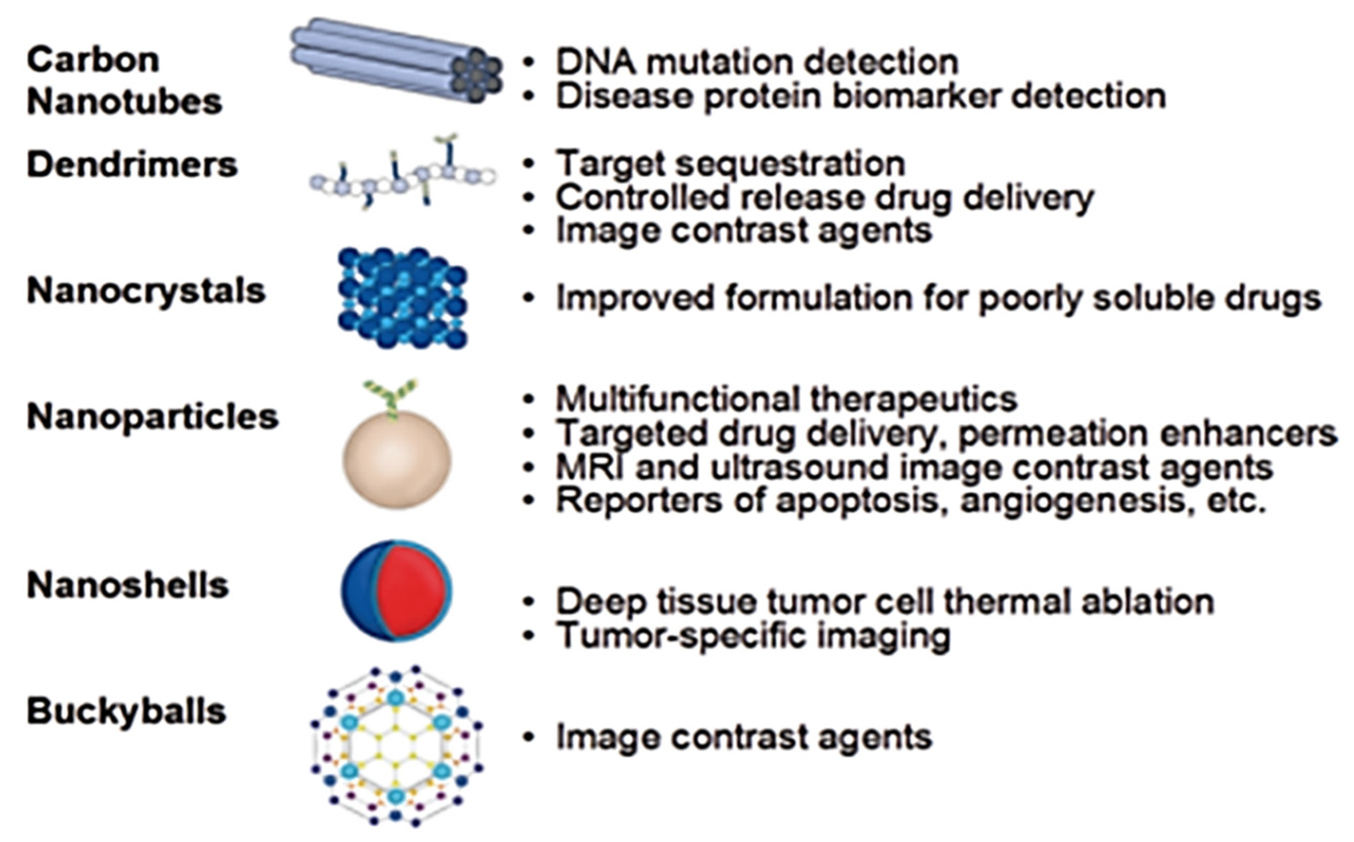
Figure 1. Various nanostructures and their possible biomedical uses [1].
Crystals 12 00039 g001
Figure 2. Various morphologies of nanoparticles and their applications in biomedical fields. Reprinted with permission from ref. [2]. Copyright 2014 Copyright John Wiley and Sons.
Crystals 12 00039 g002
Figure 3. Application of nanotechnology in medical science [9].
Crystals 12 00039 g003
Figure 4. Types of different nanocarriers.
Crystals 12 00039 g004
Figure 5. Schematic representation for antibacterial impacts of nanomaterials.
Crystals 12 00039 g005
Table 1. Application of different nanomaterials in the field of medical sciences.
Types of Nanomaterials Applications
Metal/metal-oxide nanoparticlesEnhanced drug loading and releasing action, permeation characters,
Carriers or agents for MRI and ultrasound image
Used in apoptosis and angiogenesis.
CNTDiagnosis in DNA transformation
biomarker for changes in protein structure
NanocrystalsEnhanced soluble drug formulation
Nanocore shellsContrast imaging for tumors
1D, 2D nanostructuresAccurate throughput scanning
Detector for protein diseases
Detection of DNA mutation
Diagnosis of gene mutation
0D (quantum dots)Diagnosis of gene and protein structures due to optical properties
Detection of tumor and lymph nodes
Table 2. Examples of biocompatible nanomaterials for healthcare applications.
NanomaterialRespective NanomedicineBiomedical ApplicationsPropertiesReferences
Gold NPsVerigeneIn vitro studies Genetic[14]
Nanogoldhn nm, mmv, or colloid Au NPsLoading and releasing agents for Drugs. Enhanced bioimagingOptoelectronic features due to Controlled Surface and band positions[15]
Aurimm uneAnticancerAnticancer impacts[16]
AIE-active fluorogen-loaded BSA NPsFluorogen, 2-(2,6,bis((E)-4-(phenyl(4-(1,2,2-triphenylvinyl)-[1,1 biphenyl]4-yl)amino)styryl)-4Hpyran-4-ylidene)malononitrite(TPE-TPA-DCM)advanced uptake tendency for cancer cells and in vitro and in vivo studiesImproved penetrability with good stability[17]
Nano-shellAuro-shellAeroshell SemiconductorNeck and head targets [18]
Quantum-dotsQdots, EviTagsIn vitro studiesTumor-based cell studies[19]
SemiconductorNanoco, CrystalPlex, cytodiagnosticsEnhanced Fluorescence studyMolecular sensing inside tissue cells [20,21]
Self-assembled chitosan (CHI) and modified lecithin (ML)Biosuitable and stable nanosystems Several applications, such as reversible hemostatic activities in wounds, nanocarriers for drugs, etc.Higher encapsulation performance with strong ionic nature, solubility, or lyophilized solid or rigorous colloidal system [22]
Targeted polymer NPs loaded with (-) epigallocatechin 3-gallate (EGCG)Chemotherapeutic markersStronger anticancer for prostate cancer (PCa)Marker for prostate-specific membrane antigen (PSMA)[23]
Organically modified silica nanoparticlesBiocompatible NPsIn vivo neuron targeting without harming the whole organism or causing neuronal deathActively useful for insertion into neuronal cell bodies, living brains, and suitable axonal projections [24]
Polydopamine fluorescent organic NPsBiocompatible NPsBioimaging of tissue, and cellsControlled photoluminescence response [18]
5-Fluorouracil (5-FU) loaded biocompatible fluorescence zein NPsSemisolids, solution-based, and solid nanosystems Drug delivery and imaging in biosystem Kinetic rate and controlled delivery of drug under biocompatible process [25]
Non-steroidal anti-inflammatory (NSAIDs)-loaded NPsA biocompatible formulation for a drug nanosystemSurface modifications in a prosthesis superficial alterationsControlled drug release[26]
Table 3. Research projects sponsored by BIRAC under industrial and academic collaboration [27,28].
CompanyProject Title
Lifecare Innovations Pvt Ltd.Production of poly(lactide-co-glycolide) nanoparticles (PLG-NP) and poly (lactide-co-glycollde)nanoparticles encapsulating antitubercular drugs (rifampicin, Isonlazld, and pyrazinamide-PLG-NP-ATDs)In GMP facilities
Rasayanl Biologics Pvt Ltd.Evaluation of platinum nanoparticles for the treatment of hormone-refractory prostate cancer
Imgenex India Pvt Ltd.Nanotechnological-based delivery of peptide inhibitors for the treatment of Osteoporosis
Jupiter Bioscience Ltd.Development, optimization, and characterization of ligand RGD peptide-targeted nano-constructs encapsulating anticancer chemotherapeutic agents for the effective treatment of lung cancer with Gemcitabine and stabilization of lyophilized or spray-dried formulation for direct local delivery or by injection via systemic circulation
Nanosniff Technologies Pvt Ltd.Development of a prototype instrument (sensor and detection electronics) to detect heart binding protein (hFAbP)
Onisome Healthcare Pvt Ltd.Bleomycin sulfate bearing nanostructured lipid particles for targeting brain cancer
Rellsys Medical Devices Ltd.Manufacture and clinical evaluation of non-polymeric nanocarbon porous matrix drug-eluting stent DES
V.B. Medicare Pvt Ltd.Development and characterization of lipid carrier-based nanogel formulation for 5-Fluorouracil
Table 4. Application of nanotechnology for different imaging approaches.
Imaging MethodAdvantagesDisadvantagesNanoparticles UsedReference
MRI
-
Increased spatial resolution (~10–500 µm)
-
Better contrast of soft tissues
-
Advanced and differential choices for metabolic structural, and functional analysis of tissues
-
Less sensitivity to contrast agents
-
Expensive
-
Time-consuming
-
Gadolinium containing probes
-
paramagnetic liposomes and polymers
-
ParaCEST agents
[44,45,46,47,48,49]
CT
-
Improved better resolution (~20–200 µm);
-
Infinite penetration depth;
-
Better contrast of soft tissues upon the introduction of contrast agents
-
Cost-effective
-
Faster
-
Inadequate contrast of soft tissues without injection of contrast agents
-
Radiation exposure
-
Less sensitivity to contrast causing agents
-
Micelles based on iodine and liposomes
-
Barium-based nanoparticles
-
Nanoparticles based on gold
-
Bismuth nanoparticles
[50,51,52,53]
Ultrasound
-
Better temporal and spatial resolutions (~50–100 µm)
-
Easily and swiftly operable
-
Real-time visualization;
-
Cost-cutting
-
Dependency on user
-
Inappropriate for full-body imaging
-
Targeted and non-targeted gas-filled microbubbles
-
Polymers releasing air
[54,55,56,57,58,59]
Optical Imaging
-
Best sensitivity for contrasting agents
-
Wide range of probes
-
Least expensive
-
Less penetration depth (<10 cm)
-
Increased background signal
-
Sensitivity to artifacts
-
Near-infrared fluorochrome-labeled nanoparticles
-
Quantum dots
-
Fluorescent nanoparticles probes
[58,60,61]
Photoacoustic imaging
-
Better sensitivity
-
Real-time visualization
-
Low cost
-
Finite penetration depth (up to ~5–6 cm)
-
Comparatively less specificity to contrast agents (signal from hemoglobin)
-
Gold nanoparticles, Gold nanorods
-
Carbon nanotubes
-
Fluorescent/dye-loaded NPs
[62,63] Akers WJ 2011
Positron emission tomography
-
Extremely high sensitivity
-
High penetration depth
-
Quantitative
-
Low spatial resolution (1–2 mm)
-
No anatomical information
-
Radiation exposure
-
High costs
-
Radioactive contrast agents (e.g., radiolabeled gold nanoshells)
-
Polymeric NPs
[64,65]
Single-photon emission CT
-
Very high sensitivity
-
Unlimited penetration depth
-
Long-circulating radionuclides
-
Low spatial resolution (1–2 mm)
-
No anatomical information
-
Radioactive probes
-
High cost
-
Technetium-labeled gold NPs
-
Indium-labeled liposomes
-
Nano- and micro-colloids
[66,67,68]
Publisher’s Note: MDPI stays neutral with regard to jurisdictional claims in published maps and institutional affiliations.

