Review

Synthetic Methods and Applications of Carbon Nanodots

1
Department of Chemistry, Banasthali Vidyapith, Banasthali 304022, India
2
Facultaed de Ciencias Quimicas, Departamento de Quimica Analitica e Inorganica, Universidad de Concepcion, Edmundo Larenas 129, Concepcion 4070371, Chile
3
School of Science, GSFC University, Vadodara 391750, India
4
Department of Chemistry, Central University of Allahabad, Allahabad 211002, India
*
Authors to whom correspondence should be addressed.
Catalysts 2023, 13(5), 858; https://doi.org/10.3390/catal13050858
Submission received: 30 December 2022 / Revised: 9 March 2023 / Accepted: 6 May 2023 / Published: 9 May 2023

Abstract

:
In the recent decade, carbon dots have drawn immense attention and prompted intense investigation. The latest form of nanocarbon, the carbon nanodot, is attracting intensive research efforts, similar to its earlier analogues, namely, fullerene, carbon nanotube, and graphene. One outstanding feature that distinguishes carbon nanodots from other known forms of carbon materials is its water solubility owing to extensive surface functionalization (the presence of polar surface functional groups). These carbonaceous quantum dots, or carbon nanodots, have several advantages over traditional semiconductor-based quantum dots. They possess outstanding photoluminescence, fluorescence, biocompatibility, biosensing and bioimaging, photostability, feedstock sustainability, extensive surface functionalization and bio-conjugation, excellent colloidal stability, eco-friendly synthesis (from organic matter such as glucose, coffee, tea, and grass to biomass waste-derived sources), low toxicity, and cost-effectiveness. Recent advances in the synthesis and characterization of carbon dots have been received and new insight is provided. Presently known applications of carbon dots in the fields of bioimaging, drug delivery, sensing, and diagnosis were highlighted and future applications of these astounding materials are speculated.

1. Introduction

Nanoparticles are microscopic particles with a size range of 1–100 nm. During the past decade, considerable research was conducted on the fabrication and application of nanoparticles in many fields. Based on their unique properties, nanoparticles have a substantial impact in various industries, including health, cosmetics, energy, pharmaceuticals, and food.
Enormous work was completed in recent years to design nanostructured materials with specific characteristics that will ultimately influence their function and application. In this era of carbon nanotechnology, special emphasis is laid on the organic functionality of nanomaterials or organic nanomaterials, including graphene, carbon nanotubes, and fullerenes. Because of their biocompatibility, ease of fabrication, and fascinating features, especially their water solubility fluorescence emission, carbon nanodots with a size in the range of 1–10 nm have taken the central stage of materials research. Carbon nanodots (CDs) are known to have zero dimension with almost spherical geometry. This material has become a rising star in the field of luminescent nanomaterials [1]. Due to their desirable qualities, such as hydrophilicity, ease of functionalization, outstanding biocompatibility, bright luminescence, good solubility, high chemical inertness, and low toxicity, they are potent candidates for various applications in solar cells, biosensors [2,3,4,5,6,7,8,9,10], bioimaging, and optoelectronic devices, etc. CNDs exhibit many remarkable properties including outstanding photoinduced electron transfer, stable chemical inertness, low cytotoxicity [11,12,13,14,15], good biocompatibility, and efficient light harvesting. Dots made of carbon, such as carbon nanodots (CDs) and graphene quantum dots (GQDs), are a brand-new carbonaceous nanomaterial with zero dimensions [16,17,18,19,20,21,22]. Until now, a lot of work has been carried out, and substantial advances have been made in the synthesis and uses of carbon-based dots [23,24,25,26,27].
This class of carbon-based nanomaterials was initially found by the top-down approach of minimizing huge carbon nanomaterials, and has recently advanced at a startling rate. Their purity standards, classification, and fluorescence mechanisms have made an impact on the research community, as evident from the rapid pace of research and publications in this area. The emphasis of the review is mainly on the synthesis and applications of carbon nanodots. A schematic of the methods of synthesis of CNDs and their structure was depicted in Figure 1 and Figure 2, respectively. They usually have the inner hybridization of sp2 and outer hybridization of sp3, and these hybridized structures tend to have functional groups containing oxygen atoms (such as -OH, -COOH, -CO, and many more).
Due to their excellent characteristics, CNDs are prospective replacement probes for bioimaging and bioassay [28,29,30,31,32,33]. So far, a number of methods have been investigated to synthesize CNDs, including thermal oxidation, chemical oxidation, and arc discharge, laser ablation of graphite, electrochemical synthesis, microwave synthesis, and ultrasonic methods [34,35,36,37,38,39,40]. The use of most of these techniques are, however, constrained since they sometimes require an expensive carbon source, intricate reactions, lengthy process time, and post-treatment steps. Therefore, there is a significant demand for synthetic methods that are easy to use, sustainable, and ecologically friendly for the mass production of high-quality CNDs [41].
Figure 2. Commonly available carbon nanodot structures: spherical particles, nanosheets of graphene, and amorphous structures. Reproduced with the permission from Ref. [41], 2019, American Chemical Society.
Catalysts 13 00858 g002
There are not many reviews focused explicitly on the synthetic pathways, characteristics, and uses of the CDs, despite the fact that they have all been thoroughly summarized elsewhere [42,43,44,45]. For improved clarity and understanding, a general evaluation of the most recent developments in the synthetic methods of CNDs is presented. Moreover, the advances in the understanding of the properties of carbon dots and the resulting applications were highlighted, and a new insight is provided by correlating the synthetic strategy, property, and application [46,47,48,49,50]. The organization of the review comprises a discussion of the approaches for the fabrication of carbon nanodots for various sustainable resources (including biomass), with an emphasis on cost-effectiveness and eco-friendliness. Afterward, the major applications of CNDs were highlighted, with an emphasis on bioimaging and photocatalysis.

2. Novel Methods for the Synthesis of Carbon Nanodots

During the last ten years, namely 2013–2023, many methods were developed to synthesize carbon nanodots with attractive features and applications in a specific field. These well-known CD synthesis techniques are typically categorized into “top-down” and “bottom-up” categories. The top-down techniques involve the exfoliation of nanomaterials chemically, laser ablation, electrical and chemical oxidation, arc discharge, and ultrasonic synthesis. Graphene quantum dots, or two-dimensional nanomaterials, are often produced via a “top-down” technique by exfoliating and cutting the macroscale framework of carbon species, such as carbon rods, tubes, graphite powder, activated carbon, carbon black, carbon soot, and carbon fibers possessing the graphene lattices. Top-down strategies typically demand a lengthy processing time, challenging reaction environments, and expensive materials and machinery [51,52,53], and these methods work well for the mass production of CNDs. On the contrary, the bottom-up method is used for producing carbonized polymer dots and carbon quantum dots (3D nanoparticles with spherical centers) by polymerizing molecular precursors, including glucose, sucrose, and citric acid, using processes such as chemical vapor deposition, plasma treatment, microwave pyrolysis, and solvothermal reactions, with a high degree of controllability. Of course, these methods are not flawless altogether [54].
Hawrylak et al. [55] synthesized carbon nanodots (CNDs) for the first time by surprise in 2004 in their effort to purify single-walled carbon nanotubes. Arc-discharge soot was used in the experiment as a source of carbon nanotubes. The components of the soot suspension were separated during the procedure using gel-electrophoresis, which exposed a brand-new band of fluorescent material. Currently, CNDs can be synthesized by chemical or physical processes. Thermal therapy, electrochemistry, acidic or hydrothermal oxidation, or ultrasonic treatment are examples of chemical processes. Arc discharge, plasma therapy, and laser ablation are examples of physical approaches [56]. The processes for synthesizing CNDs can be roughly categorized as top-down or bottom-up syntheses, as mentioned earlier [57,58,59,60,61,62,63,64,65,66]. Top-down techniques often include etching, intercalation, hydrothermal or solvothermal cutting, chemical oxidation, and laser ablation to break down a larger carbon structure into progressively smaller pieces. In bottom-up methods, CNDs are synthesized by carbonizing organic precursors. These include dehydration with sulfuric acid, microwave pyrolysis with solvent mediation, and refluxing pyrolysis. Using solvothermal or direct thermal breakdown, organic precursors such as allotropic carbon forms, natural gas, and carbohydrates are transformed into CNDs using direct thermal or solvothermal breakdown [67]. To achieve uniformity, the final product can be processed through electrophoresis, chromatography, centrifugation, dialysis, or through some other processes [68]. The carbon precursor, preparation technique, and experimental circumstances have a significant impact on the shape and structure of CNDs. For instance, depending on the parent material, CNDs made using top-down techniques have a different size and shape (coal, graphite powder, or graphene nanosheets). UV radiation or hydrothermal treatments can be used in a single preparation technique such as etching, and both would yield different outcomes. CNDs produced from the top-down approach would typically have dimensions below 10 nm, a spherical or sheet-like shape, and a size below 3 nm. Data indicate that bottom-up synthesized CNDs may be layered with a size of less than 10 nm.
Arc discharge and laser ablation are likely the most well-known top-down techniques for producing carbon-based nanomaterials. The term “arc discharge” refers to producing a current between two electrodes, often graphite rods, which causes them to vaporize. As a result, soot is formed, which may contain various nanoparticles of carbon. In contrast, the technique of laser ablation comprises applying a pulse of laser energy to a solid surface, resulting in carbon nanomaterials. Laser ablation in solution (LAS) has attracted interest as a top-down, single-step method for producing nanomaterials quickly and affordably, as shown in Figure 3 [69].
Amorphous (a) nanoparticles (a-CDs) are often produced at relatively low temperatures (<300 °C), whereas graphitized (g) structures are produced at higher temperatures (g-CDs). The surface functional groups of the resulting nanoparticles are significantly influenced by the precursors used. The most prevalent surface functional groups are amine and carboxylate, using precursors such as citric acid or polyamines. Such functionality can be generated via post-synthetic functionalization reactions. At the beginning of the research in this area, researchers found that proteins were the suitable precursors for the synthesis of carbon nanodots because they were readily available, affordable, and capable of undergoing dehydration and decarboxylation reactions to produce CNDs with heteroatom doping [70,71]. N-doping is accountable for better photoluminescence quantum yields, red-shifted absorption, and more favorable optoelectronic features. Citric acid in combination with amino acids (arginine, Arg) was also explored, leading to the formation of carbogenic nanoparticles’ molecular precursor, which benefits from its distinct reactive behavior and capacity to serve as a “passivating agent” or “capping agent” for the outer surface [72,73].
The hydrothermal approach, aided by microwave heating, is one among numerous potential synthesis processes. It has been widely employed for manufacturing a variety of carbon materials. Hydrothermal synthesis offers a minimal toxicological impact on materials and processes [74]. The use of hydrothermal conditions causes the reagent’s solubility to rise or change, enhances their chemical and physical interactions, and makes it easier for the carbonaceous structures to form. The production of nanomaterials with higher amounts of carbon, such as graphitic carbon compounds, and nanotubes at higher temperatures is a reliable process. Microwave-assisted methods have also grown in popularity as a method for synthesizing nanomaterials. Issues with the conventional heating process used for preparing nanomaterials, such as the tendency for insoluble compounds to cause heterogeneous heating, leading to an increase in the size of nanomaterials, is solved by microwave heating [75,76]. Due to its high energy consumption efficiency, MW irradiation offers a safe, inexpensive, and practical mode of heating, producing higher yields of the desired products [77]. As a result, the MW-assisted hydrothermal approach, which combines the benefits of both MW and hydrothermal processes, has become essential for the production of carbon dots.
The carbonization of small molecule precursors is achieved in the bottom-up fabrication of carbon dots. The most commonly available methods for the fabrication of carbon dots via the bottom-up method include a mixture of molecules having nitrogen atoms (such as urea) and citric acid [78,79,80,81,82,83,84]. The pyrolysis of these molecular precursors in an autoclave or microwave forms a black nanopowder of CDs. These CDs are easily dissolved in water and have exceptional fluorescent characteristics. These CDs are capable of emitting blue [85], green [86], and red emissions depending on their surface properties and circumstances (excitation source of radiation) [87,88], albeit a thorough purification is frequently required to separate the carbon dots [89]. When it comes to top-down methods, the starting materials include carbon structures such as amorphous activated carbon, carbon fibers, graphite, nanotubes, and fullerenes that are physically or chemically fragmented to produce very small carbon nanoparticles [90,91,92,93]. One such instance is graphitic oxidation in an extremely acidic environment [94,95], enabling the surface to be functionalized and the bulk-precursor to be broken up, resulting in the optical characteristics that are typical of CDs. Top-down synthetic methods frequently produce CDs with lower quantum yields of emission. These top-down synthetic methods of CDs are far more complex and time-consuming. However, they enable better structural control and end-product purity [96,97].

2.1. Sonochemical/Ultra-Sonic Fabrication of CDs

Sonochemistry is exploited for the synthesis of nanostructured materials. In this acoustic activation technique, severe physical and chemical conditions are generated as a result of the use of high-intensity ultrasound [98,99,100,101,102,103,104,105,106]. As the molecular dimensions are smaller than acoustic wavelengths, the chemical features of the resulting materials are not a result of the interaction between the ultrasonic and chemical species in the liquid state. Sonochemical fabrication is the result of the high compression heating of gas and vapor, resulting in incredibly high temperature and pressure conditions [107].
For the first time, Zhuo et al. [108] described the synthesis of graphene quantum dots using the ultra-sonic exfoliation of graphene. A simple sonochemical approach for the production of extremely photoluminescent CDs was devised by Wei et al. [109] in 2014. High-intensity ultrasound forms collapsing bubbles that serve as microreactors and offer intense, momentary conditions ideal for the pyrolysis of carbon precursors. Sono-chemically produced CDs in the presence of surface passivation agents have a high quantum yield and outstanding photostability.
The ultrasonic technique has merits of being inexpensive and easy to operate for the synthesis of carbon dots. The ultrasound method involves alternate high-pressure and low-pressure waves, which cause small bubbles in liquid to form and break. Thus, by means of powerful hydrodynamic shear forces resulting from the cavitation of tiny bubbles, macroscopic carbon materials were reduced to nanoscale CDs. Generally, the ultrasonic power, reaction time, and solvent and carbon source ratio were altered to produce CDs with various properties.
Huang et al. [110] used a direct ultrasonic exfoliation method to produce the chlorine-infused graphene quantum dots. Park et al. [111] conducted a typical experiment in which they first produced water-soluble CQDs from food waste. From ethanol and food waste mixture, approximately 120 g of carbon dots with an average diameter of 2–4 nm can be produced. The benefits of the as-prepared CDs for in vitro bioimaging include photostability, low cytotoxicity, and good PL characteristics.
Jiang et al. [112] reported a one-pot eco-friendly fabrication of silver nanoparticles supported on carbon from carbon and silver nitrate liquid solution without additional capping or reducing agents. Simply altering the molar ratio of carbon nanodots and AgNO3 would change the size of the silver nanoparticles supported on carbon. The simple method used to create amorphous carbon-supported Ag NPs was significant because it is synthesized in the absence of reducing or capping agents. Moreover, the stability and electrical and catalytic activities of the silver nanoparticles for the electrocatalytic reduction of H2O2 were also enhanced. The great sensitivity and low detection limit of the Ag/C nanocomposites make them excellent non-enzymatic H2O2 sensors. The Ag/C nanocomposites acted as a non-enzymatic H2O2 sensor due to their high sensitivity and selectivity, as shown in Scheme 1.
Manoharan et al. [113] used an easy and affordable technique to turn coconut water into vivid eco-friendly fluorescent carbon nanodots. Coconut water, as an environmentally friendly and less expensive carbon precursor, is used to fabricate finely dispersible carbon nanodots with both the amorphous and nano-crystalline carbon-phase. High-resolution transmission electron microscopy was used to demonstrate the monodispersed CNDs’ spherical shape, (4 ± 1 nm) nm particle size. FT-IR measurements revealed extensive surface functionalization. Using UV-visible absorption and photoluminescence spectroscopic techniques, the eco-friendly luminescent properties of the carbon nanodots were assessed. The fluorescence quantum yield of the carbon nanodots having a core of carbon with extensive surface functionalization was found to be 60.18%. These CNDs, fabricated from tender coconut water, are more favorable than those from other resources due to their stability, high quality, fast reaction rate, and fine dispersion.
Currently, microalgae productivity has been increasing worldwide. To take advantage of the situation, Choi et al. [114] set out to show that the aqueous-type biofriendly luminescent carbon nanodots (C-paints) could be successfully applied for enhancing the growth rate of microalgae, Haematococcus pluviali. A straightforward procedure of ultrasonic irradiation with the passivating agent, polyethylene glycol, was used to prepare C-paints. The end product, called a C-paint, has a carbonyl-rich surface, outstanding particle size homogeneity, high water-solubility, photo-stability, fluorescence efficacy, and biocompatibility.
Using polymer dots enclosed in NIR emissive hydrophobic carbon nanodots, Huang et al. [115] proposed the first endoplasmic reticulum (ER) focused nearinfrared (NIR) nanosensor for detecting Cu2+ in biosystems. With a detection limit of 13 nM, this nanosensor with stable fluorescence can be utilized to quantify Cu2+ in a linear range from 0.25 to 9.0 M. It responded quickly to Cu2+ (120 s). In addition, compared to other metal ions and amino acids, the nanosensor’s fluorescence fluctuations are extremely selective to Cu2+. Moreover, the developed nanosensor showed low cytotoxicity, superior biocompatibility, and ER targeting capability [116].

2.2. Hydrothermal Synthesis

The hydrothermal method for producing CDs is inexpensive and non-toxic. This is an easy method for creating carbon quantum dots compared to other synthetic approaches. Teflon lined stainless steel autoclaves are used as reaction vessels for the aqueous solution of the CD precursor and chemical agents. The autoclave is then placed in an air oven where the contents are hydrothermally reacted at a high pressure and high temperature to form the CDs [117,118,119,120,121,122,123]. Mehta et al. [124] developed a plant-based (sugarcane juice) source for producing luminescent carbon quantum dots, which are soluble in an aqueous medium with a size of less than 5 nm. These CQDs were used for the sensitive and specific detection of Cu2+. In their proposal, Lu et al. [125] suggested the production of carbon quantum dots via this method from pomelo peel having the size of less than 5 nm. The fabricated carbon quantum dots, which had remarkable yield, were used for the sensitive detection of Hg2+ at lower concentrations for the examination of water samples collected from the lake.
Li et al. [126] successfully manufactured CDs in 2018 using a one-step hydrothermal process that was ecologically friendly, easy, and affordable. Oxidation resistance, stability, excellent solubility, and high quantum yield (18.67%) were all features of the CDs. It was discovered that a charge transfer procedure could cause picric acid (PA) to quench the CDs’ early fluorescence. These CDs worked well as fluorescent probes to identify PA in our study. The technique was successfully used on actual and laboratory-derived tap water samples, and it was found to have beneficial properties, such as an outstanding selective nature, excellent sensitive nature, and lower detection limit of 10 nM.
Gao et al. [127] proposed a simple and inexpensive technique in which they showed the coupling of graphene quantum dots with carbon nitride (hexagonal-structure) via freeze-drying. The result showed enhanced photocatalytic activity, improved absorption in the visible region, and the effective separation of photon-generated electron-hole pairs. We have successfully completed the simple synthesis of B/N co-doped, fluorescent surface passivated carbon nanodots with a high quantum yield at a low cost, as reported by Jahan et al. [128]. Further employing these carbon dots results in the production of supramolecular moieties, which turns off fluorescence before being turned on by SR III.
For the first time, Soni et al. [129] have shown the precise source of light absorption and its emission of carbon nanodots. They demonstrated that molecular fluorophore, which is generally found in the fabrication mixture as a by-product, is the true source of the emission in red emissive carbon nanodots.
Using p-phenylenediamine and urea, Ding et al. [130] developed multiple multicolor-emitting carbon nanodots via this process. After being purified using column chromatography, the carbon nanodots were obtained without excitation, causing fluorophores to show different colors. Along with a progressive increase in the red-shifted fluorescence emission, the oxidation on the surface of the carbon nanodots also became enhanced. The band width narrows as the oxygen content of the carbon nanodots’ surface increases; as a result, the higher level of surface oxidation causes the red-shifted emission.
Bakier et al. [131] proposed a new turn-off fluorescent chemical sensor for the ultra-sensitive detection of aniline in the liquid phase, via the formation of colloidal carbon nanodots supported on nitrogen. They demonstrated a susceptible fluorescent aniline liquid sensor based on incredibly tiny carbon dots supported on nitrogen (Figure 4).
The carbon dots supported on nitrogen were produced using folic acid that had undergone ultrasonic processing at lower temperatures. They further discovered that the sensor’s operation followed the static N-CD fluorescence quenching caused by electrostatic contact with aniline. The detection limit for aniline via any method was 3.75 nM (0.332 ppb), which is the sensor’s detection limit. Furthermore, real sample analysis was investigated using the N-CDs’ nano-probe with real tap water, and excellent results were obtained with 99.7–101% recovery (Figure 5). Hence, this proposal could prove to be helpful in developing a simple and environmentally benign nano-sensing process with excellent sensitivity, outstanding selectivity, and good quantitative value to monitor harmful aniline against the degradation of the environment [131].

2.3. Carbonization/Pyrolysis

In recent years, pyrolysis has emerged as a potent technique for producing fluorescent CDs by using precursors that are microscopic carbon structures. Short reaction times, minimal costs, simple procedures, the absence of any solvent, and high quantum yields are all benefits of this technology. Under high temperatures, the following basic processes of heating, dehydrating, degrading, and carbonization are essential for converting the molecules with organic carbon into carbon quantum dots. During the pyrolysis process, strong concentrations of alkali perform the cleavage of carbon initiators into carbon nanoparticles.
Ma et al. [132] produced nitrogen-doped graphene quantum dots by directly carbonizing ethylene diamine tetra acetic acid at 260–280 °C, and this study also offered a growth mechanism for GQDs. It is important to note that ion doping has been found to produce a variety of CQD kinds. Li and colleagues created chlorine-doped graphene quantum dots by using HCl and fructose as precursors in a typical experiment. The average size of the quantum dots was found to be 5.4 nm. They altered the color of the emission by alternating the excitation wavelength from 300 to 600 nm, and the color changed from blue to red, respectively [133]. The fluorescent carbon quantum dots were also made by Praneerad et al. [134] by carbonizing the durian peel biomass waste. The produced CQDs were used to develop a composite-based electrode that displayed a significantly greater specific capacitance value as compared to the electrode made of pure carbon. According to 135.Zhang et al. [135], the quantum dots of carbon with increased sulphur and nitrogen contents were created by carbonizing hair fiber combined with H2SO4 through sonication.
Gunjal et al. [136] used a straightforward carbonization process to create waste tea residue carbon dots from surplus and inexpensive kitchen waste biomass, so that it is cheaper, greener, and more environmentally friendly than previous techniques. As soon as they are created, waste tea residue carbon dots exhibit excitation dependent emission and are very stable in ionic media. Furthermore, due to the oxidative nature of the ion, it has demonstrated excellent fluorescence quenching for ClO. The fabricated sensor has the advantage in that it is highly sensitive and selective in comparison to the other 21 common interfering ions which were tested against it. Its detection limit is comparatively lower than other biomass-made carbon dots. Its quick rate of reaction enables easy and feasible ClO detection in real samples with excellent precision and reliability. A simple method to covalently immobilize nanoscale carbon dots upon conducting carbon surface for sensing purposes is reported by Gutiérrez-Sánchez et al. [137]. The carbon nanodots (N-CD) containing amine functionalization on the surface can be electro-grafted upon the electrodes of carbon, where they are then readily covalently immobilized. They were made using a carbonization approach with microwave aid and cost-effective, biocompatible initiators, such as D-fructose as the primary carbon source and urea as the N-donor reagent, to produce peripheral enhanced nitrogen CD. It has been determined through various methods of analysis that the synthesized nanomaterial comprises regular-sized amorphous structures that glow blue when exposed to UV light. Through the relatively stable immobilization of nitrogen carbon dots onto the electrode surfaces through electrografting, hybrid electrodes with higher relative surface areas and enhanced electron transfer capacities are generated, holding great potential for electrochemical sensing. Because of their conductive nature, electrical properties, abundant edges sites, and high catalytic activity, N-CDs that are immobilized on carbon electrodes efficiently amplify the electro-chemiluminiscence (ECL) signal from the luminophore [Ru(bpy)3]2+ in a taurine sensor.

2.4. Electrochemical Synthesis

The one-pot electrochemical method is used to controllably synthesize fluorescent or luminescent carbon nanodots (C-dots) from small molecular alcohols as a single carbon source for the first time. By adjusting the applied potential, it is possible to control the size of the resulting C-dots, which can then be used to image cells using luminescence microscopy.
A titanium tube cathode and a pure graphite loop electrode were assembled in the center, according to Pender et al. [138]. Distilled water was used for synthesizing both the electrolyte and the cathode while the anode was isolated from them by an insulating O-ring. Luminescent blue-colored carbon dots were broadly employed in pure water thanks to the use of electronic voltage and ultrasonic control, which eliminated the need for laborious cleaning. The amount yield was 8.9%, while the size of the synthesized C-dots was 2–3 nm. The C-dots offered good fluorescent properties and thermodynamic constancy in the aqueous phase. Fluorescent CDs were made from ethanol by electrochemical carbonization, according to Miao et al. [139]. The synthesizing procedure is easy, economical, and environmentally benign. The synthesized carbon dots were amorphous, spherical, and easily dispensable in water, making them ideal for analytical uses. A strong fluorescence intensity with a QY of 10.04% was attained in the absence of a surface passivation reagent. By identifying Fe3+ induced fluorescence quenching, carbon nanodots were successfully used for the Fe3+ test.
Keerthana and Ashraf [140] highlighted the hydrothermal carbonization approach for synthesizing carbon dots from chitosan. Chitosan was totally transformed into carbon dots, according to an analysis using UV-Visible spectroscopy. With one step microwave synthesis, Arvapalli et al. [141] were able to synthesize carbon nanodots that had remarkable selectivity and sensitivity for the detection of Fe (III) ions. The synthesized carbon nanodots exhibit excellent stability, high photoluminescence, and strong water solubility. Bright blue fluorescence from carbon nanodots was successfully internalized inside endothelial cells, and when the cells were nurtured with iron, the fluorescence quenching phenomenon was seen, demonstrating the possibility of sensing iron in living cells. The transfer of charge specifically between the carbon nanodots and iron was responsible for the fluorescence quenching of the carbon nanodots, and cyclic voltammetry experiments have further confirmed this.
Tyrosinase was immobilized on carbon-based nanoparticles and cysteamine (electrically active layer) covering the gold electrode in the small gold-epinephrine biosensor reported by Baluta et al. [142]. This sensor system made use of the differential pulse and cyclic voltammetry voltammetric methods to monitor the oxidation of norepinephrinetonorepinephrine-quinone via catalysis.

2.5. Microwave-Assisted Synthesis

The electromagnetic wave known as the microwave has a vast wavelength range of 1 mm to 1 m and is frequently employed in daily life and scientific study. Microwaves can also deliver high energy to breakdown the chemical bonds in a substrate, just like lasers can. It is believed that using a microwave to create CDs is an energy-efficient method. Additionally, the reaction time may be significantly reduced. The substrate is typically pyrolyzed and the surface functionalized during microwave-aided synthesis [143,144].
The CDs are synthesized more quickly using a green, economical microwave-aided method. For the creation of CDs, microwave irradiation can deliver consistent heat. For the first time, Li et al. [145] produced green-colored luminescent graphene quantum dots via the cleavage of graphene oxide sheets chemically in the presence of acids under microwave conditions. They have an emission peak of 500 nm when excited at 260 nm and 340 nm. For the first time, electrochemiluminescence has been observed from the graphene quantum dots and is highly applicable in imaging and bio-sensing.
Liu et al. [146] produced carbon dots under microwave conditions. They used glutaraldehyde as a cross-linking agent for the fluorescent system. The luminescent emissions of the carbon dots come in a range on the basis of the amount of glutaraldehyde used. The as-prepared carbon dots showed remarkable luminescent characteristics and were less toxic, highly stable, and water soluble.
A simple microwave-assisted hydrothermal was used to produce CDs from Mangifera indica leaves [147]. The resulting carbon dots were employed as temperature sensors inside the cells, had good biocompatibility, and strong photostability. To create carbon dots from raw cashew gum, Pires et al. [148] devised a heating method that is microwave-assisted and has dual steps. The carbon quantum dots have an average size of nearly 9 nm. The synthesis involves two steps: the first step is the partial depolymerization, i.e., autohydrolysis of the gum and production of 5-hydroxymethyl furfural, while the second step involves poly-condensation for the production of the polyfuranic structure, accompanied by carbonization and nucleation. The generated carbon quantum dots have been used in the cell imaging of live cells because they exhibit good biocompatibility and low cytotoxicity.
Simsek et al. [149] showed that under different physical conditions, a quick and one-step green synthesis of carbon nanodots from Nerium oleander leaves may be achieved using a household oven and a microwave-assisted hydro-thermal synthesizer (Figure 6). The effects of the synthesizer system, the kind of extract based on the plant extraction of the plant solvents, and the synthetic conditions, including the time of reaction, temperature of reaction, surface-passivation reagent inclusion into the reaction medium, physical and chemical properties, and optical characteristics of carbon dots, were examined.
Ren et al. [150] reported the synthesis of 5.6 nm-diameter N-doped graphene quantum dots using microwave-assisted heat. The resulting N-doped graphene quantum dots were used in metal ion detection and exhibit strong and constant blue fluorescence emission with an 8% quantum yield.
Sendao et al. [151] suggested a microwave method to synthesize blue-emitting carbon quantum dots and looked into photoluminescent emission features. They discovered that the synthesis technique created green-emitting molecular fluorophore that can hide the photoluminescent emission of the carbon dots. It is important to note that in the same solution, these fluorophores and the carbon dots do not function as different species with independent emissions. Instead, their interaction results in a hybrid luminescence which is seen. This method demonstrates that the reactive nature and the characteristics in the excited-state are indistinct in comparison to their individual characteristics. The impurities of the fluorescence generated from its formation have formed a critical drawback in the investigation of the photoluminescent property of the carbon quantum dots (Table 1 and Table 2).

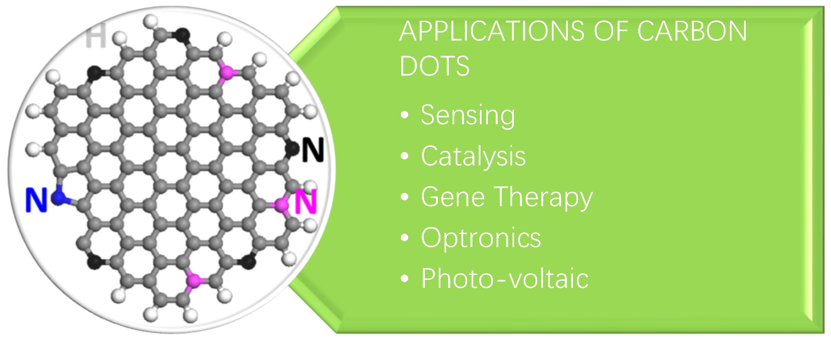
3. Applications of Carbon Dots (CDs)

There are various applications which are associated with carbon dots. CDs also show a number of biomedical applications. The application of CDs is shown in Figure 7.

3.1. Sensing

One of the most common and potentially significant uses of CDs is sensing [152,153,154]. Due to their superior optical qualities, high fluorescence sensitivity to the surrounding environment [155,156], and ability to function as effective electron donors [157,158,159], CDs are frequently suggested as detectors for a variety of harmful substances, including heavy metals such as mercury [160,161,162], copper, and iron [163,164,165,166]. To make CDs more sensitive to one or more of these analytes, persistent work is being conducted in this direction. Only a handful of studies, however, have attempted to examine the interactions of CDs with metal ions at a more fundamental level; for example, Goncalves and colleagues demonstrated that the fluorescence emissions of both CQD solution and CQDs immobilized in sol–gel are sensitive to the presence of Hg2+ [167]. In their study, laser-ablated and NH2-PEG200 and N-acetyl-L-cysteine-passivated CQDs were used as fluorescent probes. It was observed that the fluorescence intensity of the CQDs is efficiently quenched by micro molar amounts of Hg2+ with a Stern–Volmer constant of 1.3 × 10⁵ M−1. Therefore, judging from the relatively large magnitude of the Stern–Volmer constant [168], the quenching provoked by Hg2+ is probably due to the static quenching arising from the formation of a stable non-fluorescent complex between CQD and Hg2+. A substantial improvement in the sensitivity down to nanomolars was later realized by replacing the laser-ablated CQDs with N-CQDs. Again, static quenching is thought to be responsible for the quenching of fluorescence, but with a much larger Stern–Volmer constant of 1.4 × 10⁷ M−1, two orders of magnitude higher than that of the previous system [169]. It was suggested that the presence of the nitrogen element in the N-CQDs, most probably -CN groups on the N-CQD surface, is responsible for the much-improved performance of Hg2+ sensing.

3.2. Bio Imaging Probes

An intriguing application of C dots is their use as a potential agent for in vivo and in vitro bioimaging of cells and species due to their photoluminescence, which is an important property of C dots [170,171,172]. The bioimaging of cells and tissues is an important part of the diagnosis of many diseases, particularly cancer. Various fluorescent systems for diagnostic purposes have been reported, ranging from organic and inorganic dyes to the most recent nanoparticle-based systems.
To be considered suitable for use as an imaging probe, a bioimaging agent must have excellent biocompatibility, a tunable emission spectrum, and be free of cytotoxicity. Rapid progress in implementing a new class of nanoparticles has resulted in a material that meets these criteria and can be used for both diagnostic and therapeutic purposes. Chemical functionalization is used to successfully conjugate the required drug molecule to the fluorescent nanoprobes for these theranostic applications. Sahu et al. [173] reported the synthesis of C dots from orange juice hydrothermal treatment. This was one of the first examples of making fluorescent C dots from readily available natural resources [174,175]. The C dots were non-cytotoxic and efficiently taken up by MG-63 human osteosarcoma cells for cellular imaging.
The “central dogma” states that genetic information flows from DNA to RNA to proteins. Researchers investigated the physiological activity of RNA during cancer research by using RNA dynamics in cellular functions and the real-time monitoring of their temporo-spatial distribution. The experiments were carried out using fluorescent carbon dots created by the one-pot hydrothermal treatment of o-, m-, or p-phenylenediamines with triethylenetetramine by Chen et al. [176]. Because carbon has excellent biocompatibility and negligible cytotoxicity, there has been a lot of interest in using carbon nanodots as bioimaging probes instead of other types of nanoparticles. C dots are ideal candidates for theranostic applications due to their ease of synthesis, acceptable emission spectra, high photostability, and lack of cytotoxicity.
Tao et al. [177] used a mixed acid treatment to create C dots from carbon nanotubes (CNTs) and graphite. Under UV light, the C dots emit a strong yellow fluorescence with no cellular toxicity. They also demonstrated in vivo bioimaging in the near-infrared region using a rat model, and this experiment exemplified the possibilities for the development of fluorescent imaging probes in both the ultraviolet (UV) and infrared (IR) range spectra.

3.3. Photodynamic Therapy

Photodynamic therapy is a relatively new advancement in biomedical nanotechnology that uses energy transfer to destroy damaged cells and tissues. This method is useful in dealing with cancer cells because it effectively targets and destroys malignant tissue while leaving normal, healthy tissue alone. This targeted destruction in photodynamic therapy can be accomplished with fluorescent C dots that have adequate photostability [178].
Shi et al. [179] used the hydrothermal method to create N-doped C dots from rapeseed flowers and bee pollen. The authors demonstrated that C dots had no cytotoxic effect up to a limiting concentration of 0.5 mg/mL after this successful large-scale synthesis. Human colon carcinoma cells were imaged successfully in this study, and the C dots were found to have good photostability and biocompatibility.
Wang et al. [180] reported C dot synthesis from the condensation carbonization of linear polyethylenic amine (PEA) analogues and citric acid (CA) of different ratios. The authors successfully demonstrated that the extent of conjugated π-domains with CN in the carbon backbone was correlated with their photoluminescence quantum yield. The main conclusion from this study is that the emission arises not only from the sp2/sp3 carbon core and surface passivation of C nanodots, but also from the molecular fluorophores integrated into the C dot framework. This work provided an insight into the excellent biocompatibility, low cytotoxicity, and enhanced bioimaging properties of N-doped C dots, which opens the possibilities for new bioimaging applications.
Bankoti et al. [181] fabricated C dots from onion peel powder waste using the microwave method and studied cell imaging and wound healing aspects. The C dots exhibited stable fluorescence at an excitation wavelength of 450 nm and an emission wavelength of 520 nm at variable pH, along with the ability to scavenge free radicals, which can be further explored for antioxidant activity. The radical scavenging ability leads to an enhanced wound healing ability in a full-thickness wound in a rat model.

3.4. Photocatalysis

There has been significant research interest in photocatalysts over the past decade due to the scenario of environmental safety and sustainable energy. The applications of nanomaterials for the efficient fabrication of photocatalysts made the journey fast and effective.
Ming et al. [182] successfully developed C dots using a one-pot electrochemical method that only used water as the main reagent. This is an extremely promising synthetic methodology because it is a green protocol that is also cost-effective, with good photocatalytic activity of C dots for methyl orange degradation.
Song et al. [183] devised a two-step hydrothermal method for the creation of a C dot–WO2 photocatalyst. The authors used this system to photocatalytically degrade rhodamine B. It is worth noting that the reaction rate constant reported in this study is 0.01942 min−1, which is approximately 7.7 times higher than the catalytic rate using WO2 alone.
For photocatalytic hydrogen generation, a C-dot/g-C3N4 system was used. The authors created C dots from rapeseed flower pollen and hydrothermally incorporated them into g-C3N4. Under visible light irradiation, this system was able to photocatalytically generate hydrogen via sound with an output greater than that of bulk g-C3N4.

3.5. Biological Sensors and Chemical Sensors

There is great interest in using nanoparticles as biochemical sensors because C dots have been found to be useful in sensing chemical compounds or elements. Based on the properties of C dots, particularly their fluorescence properties and surface-functionalized chemical groups, various sensors for biological and chemical applications have been developed.
Qu et al. [184] developed ratiometric fluorescent nano-sensors using C dots in a single step of microwave-assisted synthesis. This research is significant in C-dot sensor research because the developed nanosensors are multi-sensory and can detect temperature, pH, and metal ions such as Fe (III). Because it can detect and estimate multiple metabolic parameters at the same time, this exciting feature is proving to be widely applicable in the biological environment. The sensory mechanism is non-cytotoxic and based on ratiometric fluorescence, which is a promising feature for future research.
Vedamalai et al. [185] developed C dots that are highly sensitive to copper (II) ions in cancer cells. They used a relatively simple hydrothermal synthesis method based on ortho-phenylenediamine (OPD). The orange color was caused by the formation of the Cu(OPD)2 complex on the surface of the C dots. Further investigation revealed that the C dots were highly water dispersible, photostable, chemically stable, and biocompatible.
Shi et al. [186] used C dots to detect Cu(II) ions in living cells as well. The hydrothermal pyrolysis of leeks resulted in blue and green fluorescent C dots. In a single step of hydrothermal carbonization, the C dots were modified with boronic acid using phenylboronic acid as the precursor. This C-dot-based sensor successfully detected blood sugar levels and demonstrated good selectivity with minimal chemical interference from other species [187].
Nie et al. [188] used a novel bottom-up method to develop a pH sensor out of C dots. This method yielded C dots with high crystallinity and stability. The procedure involved a one-pot synthesis with high reproducibility using chloroform and diethylamine. The authors were able to use the technique for cancer diagnosis after successfully implementing the pH detection of two C dots with different emission wavelengths.
Wang et al. [189] described an intriguing C-dot sensor for hemoglobin detection (Hb). The C dots were developed from glycine using an electrochemical method that included multiple steps, such as electro-oxidation, electro-polymerization, carbonization, and passivation. The authors successfully validated the sensitivity of Hb detection and discovered that the luminescence intensity varied inversely with Hb concentration in the 0.05–250 nM range.

3.6. Drug Delivery

Carbon dots’ excellent biocompatibility and clearance from the body meet the requirements for in vivo applications. Carbon dots with rich and tunable function groups, such as amino, carboxyl, or hydroxyl, can carry therapeutic agents, resulting in theranosticnanomedicines [190,191,192,193,194,195]. The bright emission of carbon dots allows for the dynamic and real-time monitoring of drug distribution and response. Zheng et al. [196] used carbon dots synthesized through the thermal pyrolysis of citric acid and polyene polyamine to transport oxaliplatin, a platinum-based drug, because platinum-based drugs are the most effective anticancer drugs and are used in more than 50% of clinical cancer patients’ chemotherapeutic treatments.

3.7. Micro-Fluidic Marker

The study of fluidic physics at the micro-scale is now best conducted using microfluidic systems. Because of their considerably higher surface-to-volume ratio, surface tension and viscosity dominate those of inertia, making the fluid easier to control. Static laminar flows and dynamic droplet formation are typical microfluidic situations. Both exhibit many advantages, including minimal reagent use, high sensitivity, and high output, which leads to a wide range of applications in bioassays, chemical reactions, drug delivery, etc. The majority of applications rely on the microfluidic circuit’s ability to visualize fluid flow. However, the biocompatibility and cheap cost of the fluorescent materials currently in use cannot be balanced, which is a critical issue for microfluidic applications, particularly for bio-applications. Sun’s colleagues used carbon dots, synthesized by heating glucose and urea in a microwave, to visualize microfluid flows for the first time to address this problem [197,198,199]. The scientists used carbon dots dissolved in the deionized water as a fluorescent marker to investigate the dynamics of the mixture of glycerol and deionized water. When the interface is ruptured by an electric field above a threshold, fast mixing occurs at the microscale. In addition to laminar flow, the authors also synthesized mono-dispersed droplets in a flow focusing system, where the continuous phase was mineral oil while the aqueous solution of carbon dots appeared as the dispersed phase. The diameter of the droplets will shrink because a higher capillary number results in a greater interfacial shear force. Additionally, the authors successfully demonstrated the multiple component droplet, merged droplet, and double emulsion, each of which has a distinct core-shell structure. To more accurately determine the speed of the flow field, luminescent seeding carbon dots were made via a mixture of carbon dots (liquid state) and polystyrene microparticles [200,201,202].

3.8. Bioimaging

Carbon dots have significant advantages over fluorescent organic dyes and genetically engineered fluorescent proteins, such as high PL quantum yield, photostability, and resistance to metabolic degradation, which endows them with enormous potential for use in bioapplications. While the toxicity testing of carbon dots is required before exploring their bioapplications, Yang et al. [203] used human breast cancer MCF-7 cells and human colorectal adenocarcinoma HT-29 cells (previously reported by other scientists, Yang modified and used it) to assess the in vitro toxicity of carbon dots synthesized by the laser ablation of graphite powder and cement with PEG1500N [204,205,206] as a surface passivation agent. All the observations of cell proliferation, mortality, and viability from both cell lines indicated that the carbon dots exhibited superior biocompatibility, even at concentrations as high as 50 mg/mL, which is much higher than the practical application demand, for example, in living cell imaging.

3.9. Carbon Dots Chiral Photonics

Chirality is essential in a number of practical application fields, such as chiral drug recognition, chiral molecular biology, and chiral chemistry [207,208,209]. As a result, as previously proposed by M. Va’zquez-Nakagawa et al. [210], chirality and carbon dots can be combined to form intriguing chiral optics based on carbon dots. The carbon dots used in their groundbreaking research were created by chemically exfoliating graphite with strong sulfuric and nitric acids. The carbon dots’ surface carboxylic acid groups were subsequently converted to acid chlorides using thionyl chloride. When the acid chlorides and the (R) or (S)-2-phenyl-1-propanol reacted simultaneously, enantiomerically pure esters and chiral carbon dots were created (chiral molecular). Enantiomerically esters and chiral carbon dots were formed, and their formation was verified using 13C-NMR and FTIR spectroscopy. The presence of phenyl substituents was suggested by the appearance of peaks in the 13C–NMR. The recent work in this field is the most notable development in the chiral regulation of bioreactions for chiral carbon dots. Xin et al. [211] described the destruction of the cell walls of gram-positive and gram-negative bacteria via carbon dots in the presence of D-glutamic acid, which resulted in the fatality of bacteria. In contrast, the carbon dots formed in the presence of L-glutamic acid demonstrated an insignificant effect on bacterial cells. This implied that antimicrobial nanoagents with chirality can be synthesized from carbon dots. The D-form and L-form of cysteine-based carbon dots were used to regulate the chirality of the enzyme. For instance, L-form cysteine carbon dots reduce the enzymatic activity while D-form cysteine carbon dots enhance the enzymatic activity of the enzyme. According to Li et al. [212], these cysteine-based carbon nanodots have the capacity to affect cellular energy metabolism. We anticipate that other chiral carbon dots-based applications will be investigated in the future [213], and that carbon dots with chirality will emerge as a novel but exciting topic because of their wide applications.

4. Conclusions

Carbon dots have drawn rigorous attention since they possess outstanding photoluminescence, fluorescence, biocompatibility, sensing and imaging, photostability, excellent colloidal stability, eco-friendly synthesis, low toxicity, and are cost-effective. In this review, widespread synthesis procedures have been discussed in detail, including bottom-up and top-down methods, along with biological and eco-friendly synthetic ways. This concludes numerous synthesizing routes that could be helpful to many scientific and research areas, since carbon nanodots can be easily synthesized for various applications. Earlier, the synthetic methods were limited because of unreliable quantum yields. However, in recent years, the synthesizing methods have seen a remarkable lift in yield, hence enhancing their use in different fields for varied applications. Despite many advancements in the field of carbon nanodots, there is still room for improvement in its synthetic methods. Several bio-related fields are left undiscovered and need special attention.

Author Contributions

Writing—original draft, A.B.; review and editing, S.G.; Funding acquisition, S.J.; investigation, formal analysis, data curation, N.K.J.: conceptualization, methodology, A.S.; Chemistry and English language editing, I.N.P.; methodology, data curation, J.D.; Supervision, M.S. All authors have read and agreed to the published version of the manuscript.

Funding

This work is financially supported by the Department of Science & Technology—Fund for Improvement of S&T Infrastructure in Universities and Higher Educational Institutions (DST—FIST), India (Order No. SR/FST/CS-II/2022/252).

Data Availability Statement

Data is available upon request.

Acknowledgments

The authors are thankful to the Department of Chemistry, Banasthali Vidyapith for providing the necessary infrastructure. We are thankful to publishing houses, namely ACS and Elsevier, Springer Nature for providing copyright permissions for the figures used in this review article.

Conflicts of Interest

This research received no external funding.

References

  1. Xu, X.Y.; Ray, R.; Gu, Y.L.; Ploehn, H.J.; Gearheart, L.; Raker, K.; Scrivens, W.A. Electrophoretic Analysis and Purification of Fluorescent Single-Walled Carbon Nanotube Fragments. J. Am. Chem. Soc. 2004, 126, 12736–12737. [Google Scholar] [CrossRef] [PubMed]
  2. Sun, Y.P.; Zhou, B.; Lin, Y.; Wang, W.; Fernando, K.A.S.; Pathak, P.; Meziani, M.J.; Harruff, B.A.; Wang, X.; Wang, H.; et al. QuantumSized Carbon Dots for Bright and Colorful Photoluminescence. J. Am. Chem. Soc. 2006, 128, 7756–7757. [Google Scholar] [CrossRef] [PubMed]
  3. Zheng, L.; Chi, Y.; Dong, Y.; Lin, J.; Wang, B. Electrochemiluminescence of Water-Soluble Carbon Nanocrystals Released Electrochemically from Graphite. J. Am. Chem. Soc. 2009, 131, 4564–4565. [Google Scholar] [CrossRef] [PubMed]
  4. Baker, S.N.; Baker, G.A. Luminescent Carbon Nanodots: Emergent Nanolights. Angew. Chem. Int. Ed. 2010, 49, 6726–6744. [Google Scholar] [CrossRef] [PubMed]
  5. Li, H.T.; He, X.D.; Kang, Z.H.; Huang, H.; Liu, Y.; Liu, J.L.; Lian, S.Y.; Tsang, C.C.A.; Yang, X.B.; Lee, S.T. Water-Soluble Fluorescent Carbon Quantum Dots and Catalyst Design. Angew. Chem. Int. Ed. 2010, 49, 4430–4434. [Google Scholar] [CrossRef] [PubMed]
  6. Ma, Z.; Zhang, Y.L.; Wang, L.; Ming, H.; Li, H.T.; Zhang, X.; Wang, F.; Liu, Y.; Kang, Z.H.; Lee, S.T. Bioinspired Photoelectric Conversion System Based on Carbon Quantum Dot Doped Dye—Semiconductor Complex. ACS Appl. Mater. Interfaces 2013, 5, 5080–5084. [Google Scholar] [CrossRef] [PubMed]
  7. Mu, X.W.; Wu, M.X.; Zhang, B.; Liu, X.; Xu, S.M.; Huang, Y.B.; Wang, X.H.; Sun, Q. A sensitive “off-on” carbon dots-Ag nanoparticles fluorescent probe for cysteamine detection via the inner filter effect. Talanta 2021, 221, 121463. [Google Scholar] [CrossRef]
  8. Huang, H.L.; Ge, H.; Ren, Z.P.; Huang, Z.J.; Xu, M.; Wang, X.H. Controllable Synthesis of Biocompatible Fluorescent Carbon Dots from Cellulose Hydrogel for the Specific Detection of Hg2+. Front. Bioeng. Biotechnol. 2021, 9, 617097. [Google Scholar] [CrossRef]
  9. Wang, Y.; Zhuang, Q.; Ni, Y. Facile Microwave Assisted Solid Phase Synthesis of Highly Fluorescent Nitrogen Sulfur Co-doped Carbon Quantum Dots for Cellular Imaging Applications. Chem. Eur. J. 2015, 21, 13004–13011. [Google Scholar] [CrossRef]
  10. Li, D.; Li, W.; Zhang, H.; Zhang, X.; Zhuang, J.; Liu, Y.; Hu, C.; Lei, B. Far-Red Carbon Dotsas Efficient Light-Harvesting Agents for Enhanced Photosynthesis. ACS Appl. Mater. Interfaces 2020, 12, 21009–21019. [Google Scholar] [CrossRef]
  11. Wang, J.; Li, R.S.; Zhang, H.Z.; Wang, N.; Zhang, Z.; Huang, C.Z. Highly fluorescent carbon dots as selective and visual probes for sensing copper ions in living cells via an electron transfer process. Biosens. Bioelectron. 2017, 97, 157–163. [Google Scholar] [CrossRef] [PubMed]
  12. Jana, J.; Lee, H.J.; Chung, J.S.; Kim, M.H.; Hur, S.H. Blue emitting nitrogen-doped carbon dots as a fluorescent probe for nitrite ion sensing and cell-imaging. Anal. Chim. Acta 2019, 1079, 212–219. [Google Scholar] [CrossRef]
  13. Hu, J.; Tang, F.; Jiang, Y.; Liu, C. Rapid screening and quantitative detection of Salmonella using a quantum dot nanobead-based biosensor. Analyst 2020, 145, 2184–2190. [Google Scholar] [CrossRef] [PubMed]
  14. Liu, M.L.; Chen, B.B.; Li, C.M.; Huang, C.Z. Carbon Dots: Synthesis, Formation Mechanism, Fluorescence Origin and Sensing Applications. Green Chem. 2019, 21, 449–471. [Google Scholar] [CrossRef]
  15. Qin, K.H.; Zhang, D.F.; Ding, Y.F.; Zheng, X.D.; Xiang, Y.Y.; Hua, J.H.; Zhang, Q.; Ji, X.L.; Li, B.; Wei, Y.L. Applications of hydrothermal synthesis of Escherichia coli derived carbon dots in in vitro and in vivo imaging and p-nitrophenol detection. Analyst 2020, 145, 177–183. [Google Scholar] [CrossRef]
  16. Cui, L.; Wang, J.; Sun, M.T. Graphene plasmon for optoelectronics. Rev. Phys. 2021, 6, 100054. [Google Scholar] [CrossRef]
  17. Yuan, F.; Wang, Y.K.; Sharma, G.; Dong, Y.; Zheng, X.; Li, P.; Johnston, A.; Bappi, G.; Fan, J.Z.; Kung, H. Bright High-Colour Purity Deep-Blue Carbon Dot Light-Emitting Diodes via Efficient Edge Amination. Nat. Photonics 2020, 14, 171–176. [Google Scholar] [CrossRef]
  18. Hu, C.; Li, M.Y.; Qiu, J.S.; Sun, Y.P. Design and fabrication of carbon dots for energy conversion and storage. Chem. Soc. Rev. 2019, 48, 2315–2337. [Google Scholar] [CrossRef]
  19. Cao, Y.; Cheng, Y.; Sun, M.T. Graphene-based SERS for sensor and catalysis. Appl. Spectrosco. Rev. 2021, 58, 1–38. [Google Scholar] [CrossRef]
  20. Hsu, P.C.; Shih, Z.Y.; Lee, C.H.; Chang, H.T. Synthesis and analytical applications of photoluminescent carbon nanodots. Green Chem. 2012, 14, 917–920. [Google Scholar] [CrossRef]
  21. Chandra, S.; Pathan, S.H.; Mitra, S.; Modha, B.H.; Goswami, A.; Pramanik, P. Tuning of photoluminescence on different surface functionalized carbon quantum dots. RSC Adv. 2012, 2, 3602–3606. [Google Scholar] [CrossRef]
  22. Liu, M.; Chen, W. Green synthesis of silver nanoclusters supported on carbon nanodots: Enhanced photoluminescence and high catalytic activity for oxygen reduction reaction. Nanoscale 2013, 5, 12558–12564. [Google Scholar] [CrossRef] [PubMed]
  23. Kottam, N.; Smrithi, S.P. Luminescent carbon nanodots: Current prospects on synthesis, properties and sensing applications. Methods Appl. Fluoresc. 2021, 9, 012001. [Google Scholar] [CrossRef] [PubMed]
  24. Xu, A.; Wang, G.; Li, Y.; Dong, H.; Yang, S.; He, P.; Ding, G. Carbon-based quantum dots with solid-state photoluminescent: Mechanism, implementation, and application. Small 2020, 16, 2004621. [Google Scholar] [CrossRef]
  25. Roy, P.; Chen, P.C.; Periasamy, A.P.; Chen, Y.N.; Chang, H.T. Photoluminescent carbon nanodots: Synthesis, physicochemical properties and analytical applications. Mater. Today 2015, 18, 447–458. [Google Scholar] [CrossRef]
  26. Li, H.; Kang, Z.; Liu, Y.; Lee, S.T. Carbon nanodots: Synthesis, properties and applications. J. Mat. Chem. 2012, 22, 24230–24253. [Google Scholar] [CrossRef]
  27. Philippidis, A.; Stefanakis, D.; Anglos, D.; Ghanotakis, D. Microwave heating of arginine yields highly fluorescent nanoparticles. J. Nanopart. Res. 2013, 15, 1–9. [Google Scholar] [CrossRef]
  28. Zhu, L.; Yin, Y.; Wang, C.F.; Chen, S. Plant leaf-derived fluorescent carbon dots for sensing, patterning and coding. J. Mater. Chem. C 2013, 1, 4925–4932. [Google Scholar] [CrossRef]
  29. Tang, J.; Zhang, Y.; Kong, B.; Wang, Y.; Da, P.; Li, J.; Elzatahry, A.A.; Zhao, D.; Gong, X.; Zheng, G. Solar-driven photoelectrochemical probing of nanodot/nanowire/cell interface. Nano Lett. 2014, 14, 2702–2708. [Google Scholar] [CrossRef]
  30. Li, Q.; Ohulchanskyy, T.Y.; Liu, R.; Koynov, K.; Wu, D.; Best, A.; Kumar, R.; Bonoiu, A.; Prasad, P.N. Photoluminescent carbon dots as biocompatible nanoprobes for targeting cancer cells in vitro. J. Phys. Chem. C. 2010, 114, 12062–12068. [Google Scholar] [CrossRef]
  31. Da Silva, J.C.E.; Gonçalves, H.M. Analytical and bioanalytical applications of carbon dots. TrAC Trends Anal. Chem. 2011, 30, 1327–1336. [Google Scholar] [CrossRef]
  32. Sanderson, K. Quantum dots go large: A small industry could be on the verge of a boom. Nature 2009, 459, 760–762. [Google Scholar] [CrossRef]
  33. Cao, L.; Wang, X.; Meziani, J.M.; Lu, F.; Wang, H.; Luo, P.G.; Lin, Y.; Harruff, B.A.; Veca, L.M.; Murray, D.; et al. Carbon Dots for Multiphoton Bioimaging. J. Am. Chem. Soc. 2007, 129, 11318–11319. [Google Scholar] [CrossRef] [PubMed]
  34. Yang, S.T.; Cao, L.; Luo, P.G.; Lu, F.S.; Wang, X.; Wang, H.F.; Meziani, M.J.; Liu, Y.F.; Qi, G.; Sun, Y.P. Carbon Dots for Optical Imaging in Vivo. J. Am. Chem. Soc. 2009, 131, 11308–11309. [Google Scholar] [CrossRef] [PubMed]
  35. Sun, D.; Ban, R.; Zhang, P.H.; Wu, G.H.; Zhang, J.R.; Zhu, J.J. Hair Fiber as a Precursor for Synthesizing of Sulfur- and Nitrogen-Codoped Carbon Dots with Tunable Luminescence Properties. Carbon 2013, 64, 424–434. [Google Scholar] [CrossRef]
  36. Miao, P.; Han, K.; Tang, Y.; Wang, B.; Lin, T.; Cheng, W. Recent advances in carbon nanodots: Synthesis, properties and biomedical applications. Nanoscale 2015, 7, 1586–1595. [Google Scholar] [CrossRef]
  37. Wang, H.; Boghossian, A.A. Covalent conjugation of proteins onto fluorescent single-walled carbon nanotubes for biological and medical applications. Mater. Adv. 2023, 4, 823–834. [Google Scholar] [CrossRef]
  38. Hu, S.L.; Niu, K.Y.; Sun, J.; Yang, J.; Zhao, N.Q.; Du, X.W. One-Step Synthesis of Fluorescent Carbon Nanoparticles by Laser Irradiation. J. Mater. Chem. 2009, 19, 484–488. [Google Scholar] [CrossRef]
  39. Ma, Z.; Ming, H.; Huang, H.; Liu, Y.; Kang, Z. One-step ultrasonic synthesis of fluorescent N-doped carbon dots from glucose and their visible-light sensitive photocatalytic ability. New J. Chem. 2012, 36, 861–864. [Google Scholar] [CrossRef]
  40. Dong, Y.; Pang, H.; Ren, S.; Chen, C.; Chi, Y.; Yu, T. Etching Single-Wall Carbon Nanotubes into Green and Yellow Single-Layer Graphene Quantum Dots. Carbon 2013, 64, 245–251. [Google Scholar] [CrossRef]
  41. Cadranel, A.; Margraf, J.T.; Strauss, V.; Clark, T.; Guldi, D.M. Carbon nanodots for charge-transfer processes. Acc. Chem. Res. 2019, 52, 955–963. [Google Scholar] [CrossRef] [PubMed]
  42. Li, H.; He, X.; Liu, Y.; Huang, H.; Lian, S.; Lee, S.T.; Kang, Z. One-Step Ultrasonic Synthesis of Water-Soluble Carbon Nanoparticles with Excellent Photoluminescent Properties. Carbon 2011, 49, 605–609. [Google Scholar] [CrossRef]
  43. Li, H.T.; He, X.D.; Liu, Y.; Yu, H.; Kang, Z.H.; Lee, S.T. Synthesis of Fluorescent Carbon Nanoparticles Directly from Active Carbon via a One-Step Ultrasonic Treatment. Mater. Res. Bull. 2011, 16, 147–151. [Google Scholar] [CrossRef]
  44. Dong, Y.; Pang, H.; Yang, H.B.; Guo, C.; Shao, J.; Chi, Y.; Li, C.M.; Yu, T. Carbon-based Dots Co-doped with Nitrogen and Sulfur for High Quantum Yield and Excitation-Independent Emission. Angew. Chem. Int. Ed. 2013, 52, 7800–7804. [Google Scholar] [CrossRef]
  45. Tabaraki, R.; Abdi, O. Microwave assisted synthesis of N-doped carbon dots: Aneasy, fast and cheap sensor for determination of aspartic acid in sport supplements. J. Fluoresc. 2019, 29, 751–756. [Google Scholar] [CrossRef] [PubMed]
  46. Tian, M.; Wang, Y.T.; Zhang, Y. Synthesis of fluorescent nitrogen-doped carbon quantum dots for selective detection of picric acid in water samples. J. Nanosci. Nanotechnol. 2018, 18, 8111–8117. [Google Scholar] [CrossRef]
  47. Luo, J.B.; Sun, Z.S.; Zhou, W.Y.; Mo, F.W.; Wu, Z.C.; Zhang, X.G. Hydrothermal synthesis of bright blue-emitting carbon dots for bioimaging and fluorescent determination of baicalein. Opt. Mater. 2021, 113, 110796. [Google Scholar] [CrossRef]
  48. Li, C.; Sun, X.Y.; Li, Y.; Liu, H.L.; Long, B.B.; Xie, D.; Chen, J.J.; Wang, K. Rapid and Green Fabrication of Carbon Dots for Cellular Imaging and Anti-Counterfeiting Applications. ACS Omega 2021, 6, 3232–3237. [Google Scholar] [CrossRef]
  49. Prasannan, A.; Imae, T. One-Pot Synthesis of Fluorescent Carbon Dots from Orange Waste. Ind. Eng. Chem. Res. 2013, 52, 15673–15678. [Google Scholar] [CrossRef]
  50. Aji, M.P.; Wiguna, P.A.; Susanto-Rosita, N.; Suciningtyas, S.A. Sulhadi Performance of Photocatalyst Based Carbon Nanodots from Waste Frying Oil in Water Purification. AIP Conf. Proc. 2016, 1725, 79–94. [Google Scholar]
  51. Achilleos, D.S.; Kasap, H.; Reisner, E. Photocatalytic hydrogen generation coupled to pollutant utilization using carbon dots produced from biomass. Green Chem. 2020, 22, 2831–2839. [Google Scholar] [CrossRef]
  52. Aggarwal, R.; Saini, D.; Singh, B.; Kaushik, J.; Garg, A.K.; Sonkar, S.K. Bitter apple peel derived photoactive carbon dots for the sunlight induced photocatalytic degradation of crystal violet dye. Sol. Energy 2020, 197, 326–331. [Google Scholar] [CrossRef]
  53. Tyagi, A.; Tripathi, K.M.; Singh, N.; Choudhary, S.; Gupta, R.K. Green synthesis of carbon quantum dots from lemon peel waste: Applications in sensing and photocatalysis. RSC Adv. 2016, 6, 72423–72432. [Google Scholar] [CrossRef]
  54. Guclu, A.D.; Potasz, P.; Korkusinski, M.; Hawrylak, P. Graphene Quantum Dots; Springer: Berlin/Heidelberg, Germany, 2014. [Google Scholar]
  55. Hawrylak, P.; Peeters, F.; Ensslin, K. Carbononics-integrating electronics, photonics and spintronics with graphene quantum dots. Phys. Status Solidi RRL 2016, 10, 11–12. [Google Scholar] [CrossRef]
  56. Hai, K.; Feng, J.; Chen, X.W.; Wang, J.H. Tuning the optical properties of graphene quantum dots for biosensing and bioimaging. J. Mater. Chem. B 2018, 6, 3219–3234. [Google Scholar] [CrossRef]
  57. Bisker, G.; Bakh, N.A.; Lee, M.A.; Ahn, J.; Park, M.; O’Connell, E.B.; Strano, M.S. Insulin detection using a corona phase molecular recognition site on single-walled carbon nanotubes. ACS Sens. 2018, 3, 367–377. [Google Scholar] [CrossRef]
  58. Xu, Z.Q.; Lan, J.Y.; Jin, J.C.; Dong, P.; Jiang, F.L.; Liu, Y. Highly Photoluminescent Nitrogen-Doped Carbon Nanodots and Their Protective Effects against Oxidative Stress on Cells. ACS Appl. Mater. Interfaces 2015, 7, 28346–28352. [Google Scholar] [CrossRef]
  59. Zhao, L.; Zhao, L.; Li, H.; Ma, J.; Bian, L.; Wang, X.; Pu, Q. Controlled synthesis of fluorescent carbon materials with the assistance of capillary electrophoresis. Talanta 2021, 228, 122224. [Google Scholar] [CrossRef]
  60. Yuvali, D.; Narin, I.; Soylak, M.; Yilmaz, E. Green synthesis of magnetic carbon nanodot/graphene oxide hybrid material (Fe3O4@C-nanodot@GO) for magnetic solid phase extraction of ibuprofen in human blood samples prior to HPLC-DAD determination. J. Pharm. Biomed. Anal. 2020, 179, 113001. [Google Scholar] [CrossRef]
  61. Yang, D.; Li, L.; Cao, L.; Chang, Z.; Mei, Q.; Yan, R.; Ge, M.; Jiang, C.; Dong, W.F. Green Synthesis of Lutein-Based Carbon Dots Applied for Free-Radical Scavenging within Cells. Materials 2020, 13, 4146. [Google Scholar] [CrossRef]
  62. Xu, Y.; Li, P.; Cheng, D.; Wu, C.; Lu, Q.; Yang, W.; Zhu, X.; Yin, P.; Liu, M.; Li, H. Group IV nanodots: Synthesis, surface engineering and application in bioimaging and biotherapy. J. Mater. Chem. B 2020, 8, 10290–10308. [Google Scholar] [CrossRef]
  63. Wang, Y.; Zhang, Y.; Yan, J.; Yu, J.; Ding, B. One-step synthesis of a macroporous Cu-g/C3N4 nanofiber electrocatalyst for efficient oxygen reduction reaction. Chem. Commun. 2020, 56, 14087–14090. [Google Scholar] [CrossRef]
  64. Qin, X.; Liu, J.; Zhang, Q.; Chen, W.; Zhong, X.; He, J. Synthesis of Yellow-Fluorescent Carbon Nano-dots by Microplasma for Imaging and Photocatalytic Inactivation of Cancer Cells. Nanoscale Res. Lett. 2021, 16, 14. [Google Scholar] [CrossRef]
  65. Ha, J.; Seo, Y.; Kim, Y.; Lee, J.; Lee, H.; Kim, S.; Choi, Y.; Oh, H.; Lee, Y.; Park, E. Synthesis of nitrogen-doped carbon nanodots to destroy bacteria competing with Campylobacter jejuni in enrichment medium, and development of a monoclonal antibody to detect C. jejuni after enrichment. Int. J. Food Microbiol. 2021, 339, 109014. [Google Scholar] [CrossRef]
  66. Guo, F.; Bao, L.; Wang, H.; Larson, S.L.; Ballard, J.H.; Knotek-Smith, H.M.; Zhang, Q.; Su, Y.; Wang, X.; Han, F. A simple method for the synthesis of biochar nanodots using hydrothermal reactor. MethodsX 2020, 7, 101022. [Google Scholar] [CrossRef] [PubMed]
  67. Sharma, V.; Tiwari, P.; Mobin, S.M. Sustainable carbon-dots: Recent advances in green carbon dots for sensing and bioimaging. J. Mater. Chem. B 2017, 5, 8904–8924. [Google Scholar] [CrossRef] [PubMed]
  68. Yang, N.; Jiang, X.; Pang, D.W. Carbon Nanoparticles and Nanostructures; Springer International Publishing: Cham, Switzerland, 2016; p. 360. [Google Scholar]
  69. Sharma, A.; Das, J. Small molecules derived carbon dots: Synthesis and applications in sensing, catalysis, imaging, and biomedicine. J. Nanobiotechnol. 2019, 17, 92. [Google Scholar] [CrossRef]
  70. Jiang, J.; He, Y.; Li, S.; Cui, H. Amino Acids as the Source for Producing Carbon Nanodots: Microwave Assisted One-Step Synthesis, Intrinsic Photoluminescence Property and Intense Chemiluminescence Enhancement. Chem. Commun. 2012, 48, 9634–9636. [Google Scholar] [CrossRef]
  71. Mazzier, D.; Favaro, M.; Agnoli, S.; Silvestrini, S.; Granozzi, G.; Maggini, M.; Moretto, A. Synthesis of Luminescent 3D Microstructures Formed by Carbon Quantum Dots and Their Self Assembly Properties. Chem. Commun. 2014, 50, 6592–6595. [Google Scholar] [CrossRef]
  72. Krysmann, M.J.; Kelarakis, A.; Dallas, P.; Giannelis, E.P. Formation Mechanism of Carbogenic Nanoparticles with Dual Photoluminescence Emission. J. Am. Chem. Soc. 2012, 134, 747–750. [Google Scholar] [CrossRef] [PubMed]
  73. Zhu, S.; Meng, Q.; Wang, L.; Zhang, J.; Song, Y.; Jin, H.; Zhang, K.; Sun, H.; Wang, H.; Yang, B. Highly Photoluminescent Carbon Dots for Multicolor Patterning, Sensors, and Bioimaging. Angew. Chem. Int. Ed. 2013, 52, 3953–3957. [Google Scholar] [CrossRef]
  74. Hu, B.; Wang, K.; Wu, L.; Yu, S.-H.; Antonietti, M.; Titirici, M.-M. Engineering Carbon Materials from the Hydrothermal Carbonization Process of Biomass. Adv. Mater. 2010, 22, 813–828. [Google Scholar] [CrossRef] [PubMed]
  75. Schwenke, A.M.; Hoeppener, S.; Schubert, U.S. Synthesis and Modification of Carbon Nanomaterials Utilizing Microwave Heating. Adv. Mater. 2015, 27, 4113–4141. [Google Scholar] [CrossRef]
  76. Vazquez, E.; Giacalone, F.; Prato, M. Non-Conventional Methods and Media for the Activation and Manipulation of Carbon Nanoforms. Chem. Soc. Rev. 2014, 43, 58–69. [Google Scholar] [CrossRef]
  77. Vazquez, E.; Prato, M. Carbon Nanotubes and Microwaves: Interactions, Responses, and Applications. ACS Nano 2009, 3, 3819–3824. [Google Scholar] [CrossRef]
  78. Liu, Q.; Zhang, N.; Shi, H.; Ji, W.; Guo, X.; Yuan, W.; Hu, Q. One-step microwave synthesis of carbon dots for highly sensitive and selective detection of copper ions in aqueous solution. New, J. Chem. 2018, 42, 3097. [Google Scholar] [CrossRef]
  79. Schneider, J.; Reckmeier, C.J.; Xiong, Y.; von Seckendorff, M.; Susha, A.S.; Kasák, P.; Rogach, A.L. Molecular fluorescence in citric acid-based carbon dots. J. Phys. Chem. C 2017, 121, 2014. [Google Scholar] [CrossRef]
  80. Cayuela, A.; Soriano, M.L.; Valcàrcel, M. Strong luminescence of carbon dots induced by acetone passivation: Efficient sensor for a rapid analysis of two different pollutants. Anal. Chim. Acta 2013, 804, 246. [Google Scholar] [CrossRef]
  81. Ðordevi’c, L.; Arcudi, F.; Prato, M. Preparation, functionalization and characterization of engineered carbon nanodots. Nat. Protoc. 2019, 14, 2931–2953. [Google Scholar] [CrossRef]
  82. Sciortino, A.; Mauro, N.; Buscarino, G.; Sciortino, L.; and Popescu, R.; Schneider, R.; Giammona, G.; Gerthsen, D.; Cannas, M.; Messina, F. β-C3N4 nanocrystals: Carbon dots with extraordinary morphological, structural, and optical homogeneity. Chem. Mater. 2018, 30, 1695. [Google Scholar] [CrossRef]
  83. Ji, Z.; Yin, Z.; Jia, Z.; Wei, J. Carbon nanodots derived from urea and citric acid in living cells: Cellular uptake and antioxidation effect. Langmuir 2020, 36, 8632. [Google Scholar] [CrossRef]
  84. Shan, X.; Chai, L.; Ma, J.; Qian, Z.; Chen, J.; Feng, H. B-doped carbon quantum dots as a sensitive fluorescence probe for hydrogen peroxide and glucose detection. Analyst 2014, 139, 2322. [Google Scholar] [CrossRef]
  85. Malfatti, L.; Innocenzi, P. Sol-gel chemistry for carbon dots. Chem. Rev. 2018, 18, 1192. [Google Scholar] [CrossRef]
  86. Ðordevi’c, L.; Arcudi, F.; Prato, M. Synthesis, separation, and characterization of small and highly fluorescent nitrogen-doped carbon nanodots. Angew. Chem. Int. Ed. 2016, 55, 2107. [Google Scholar] [CrossRef] [PubMed]
  87. Gan, Z.; Wu, X.; Hao, Y. The mechanism of blue photoluminescence from carbon nanodots. Cryst. Eng. Comm. 2014, 16, 4981. [Google Scholar] [CrossRef]
  88. Sai, L.; Yinzi, C.; Chun, L.; Jiali, F.; Weidong, X.; Xiaojuan, L. Carbon nanodots with intense emission from green to red and their multifunctional applications. J. Alloys Compd. 2018, 742, 212. [Google Scholar]
  89. Holá, K.; Sudolská, M.; Kalytchuk, S.; Nachtigallová, D.; Rogach, A.L.; Otyepka, M.; Zboril, R. Graphitic nitrogen triggers red fluorescence in carbon dots. ACS Nano 2017, 11, 12402. [Google Scholar] [CrossRef]
  90. Reckmeier, C.J.; Schneider, J.; Xiong, Y.; Häusler, J.; Kasák, P.; Schnick, W.; Rogach, A.L. Aggregated molecular fluorophores in the ammonothermal synthesis of carbon dots. Chem. Mater. 2017, 29, 10352. [Google Scholar] [CrossRef]
  91. Bao, L.; Liu, C.; Zhang, Z.L.; Pang, D.W. Photoluminescence-tunable carbon nanodots: Surface-state energy-gap tuning. Adv. Mater. 2015, 27, 1663. [Google Scholar] [CrossRef]
  92. Le Croy, G.E.; Sonkar, S.K.; Yang, F.; Monica Veca, L.; Wang, P.; Tackett II, K.N.; Yu, J.-J.; Vasile, E.; Qian, H.; Liu, Y. Towards structurally defined carbon dots as ultracompact fluorescent probes. ACS Nano 2014, 8, 4522–4529. [Google Scholar] [CrossRef] [PubMed]
  93. Campuzano, S.; Yáñez-Sedeño, P.; Pingarrón, J.M. Carbon dots and graphene quantum dots in electrochemical biosensing. Nanomaterials 2019, 9, 634. [Google Scholar] [CrossRef] [PubMed]
  94. Fresco-Cala, B.; Soriano, M.L.; Sciortino, A.; Cannas, M.; Messina, F.; Cardenas, S. One-pot synthesis of graphene quantum dots and simultaneous nanostructured self-assembly via a novel microwave-assisted method: Impact on triazine removal and efficiency monitoring. RSC Adv. 2018, 8, 29939. [Google Scholar] [CrossRef]
  95. Sun, Y.; Wang, S.; Li, C.; Luo, P.; Tao, L.; Wei, Y.; Shi, G. Large scale preparation of graphene quantum dots from graphite with tunable fluorescence properties. Phys. Chem. Chem. Phys. 2013, 15, 9907. [Google Scholar] [CrossRef] [PubMed]
  96. Wang, L.; Wang, Y.; Xu, T.; Liao, H.; Yao, C.; Liu, Y.; Li, Z.; Chen, Z.; Pan, D.; Sun, L. Gram-scale synthesis of single-crystalline graphene quantum dots with superior optical properties. Nat. Commun. 2014, 5, 5357. [Google Scholar] [CrossRef] [PubMed]
  97. Kwon, W.; Lee, G.; Do, S.; Joo, T.; Rhee, S.W. Size-controlled soft-template synthesis of carbon nanodots toward versatile photoactive materials. Small 2014, 10, 506. [Google Scholar] [CrossRef]
  98. Bang, J.H.; Suslick, K.S. Applications of ultrasound to the synthesis of nanostructured materials. Adv. Mater. 2010, 22, 1039–1059. [Google Scholar] [CrossRef]
  99. Shchukin, D.G.; Radziuk, D.; Möhwald, H. Ultrasonic fabrication of metallic nanomaterials and nanoalloys. Ann. Rev. Mater. Res. 2010, 40, 345–362. [Google Scholar] [CrossRef]
  100. Vinodgopal, K.; Neppolian, B.; Lightcap, I.V.; Grieser, F.; Ashokkumar, M.; Kamat, P.V. Sonolytic design of graphene−Au nanocomposites. Simultaneous and sequential reduction of graphene oxide and Au (III). Phys. Chem. Lett. 2010, 1, 1987. [Google Scholar] [CrossRef]
  101. Geng, J.; Jiang, L.; Zhu, J. Crystal formation and growth mechanism of inorganic nanomaterials in sonochemical syntheses. Sci. China Chem. 2012, 55, 2292–2310. [Google Scholar] [CrossRef]
  102. Skrabalak, S.E. Ultrasound-assisted synthesis of carbon materials. Phys. Chem. Chem. Phys. 2009, 11, 4930. [Google Scholar] [CrossRef]
  103. Gedanken, A. Using sonochemistry for the fabrication of nanomaterials. Ultrason. Sonochem. 2004, 11, 47–55. [Google Scholar] [CrossRef]
  104. Gao, T.; Wang, T. Sonochemical synthesis of SnO2nanobelt/CdS nanoparticle core/shell heterostructures. Chem. Commun. 2004, 22, 2558–2559. [Google Scholar] [CrossRef]
  105. Hasin, P.; Wu, Y. Sonochemical synthesis of copper hydride (CuH). Chem. Commun. 2012, 48, 1302–1304. [Google Scholar] [CrossRef] [PubMed]
  106. Xu, H.; Zeiger, B.W.; Suslick, K.S. Sonochemical synthesis of nanomaterials. Chem. Soc. Rev. 2013, 42, 2555–2567. [Google Scholar] [CrossRef] [PubMed]
  107. Suslick, K.S.; Flannigan, D.J. Inside a collapsing bubble: Sonoluminescence and the conditions during cavitation. Annu. Rev. Phys. Chem. 2008, 59, 659–683. [Google Scholar] [CrossRef]
  108. Zhuo, S.; Shao, M.; Lee, S.T. Upconversion and downconversion fluorescent graphene quantum dots: Ultrasonic preparation and photocatalysis. ACS Nano 2012, 6, 1059–1064. [Google Scholar] [CrossRef]
  109. Wei, K.; Li, J.; Ge, Z.; You, Y.; Xu, H. Sonochemical synthesis of highly photoluminescent carbon nanodots. RSC Adv. 2014, 4, 52230–52234. [Google Scholar] [CrossRef]
  110. Huang, H.Y.; Cui, Y.; Liu, M.Y.; Chen, J.Y.; Wan, Q.; Wen, Y.Q.; Wei, Y. A one-step ultrasonic irradiation assisted strategy for the preparation of polymer-functionalized carbon quantum dots and their biological imaging. J. Colloid Interface Sci. 2018, 532, 767–773. [Google Scholar] [CrossRef]
  111. Park, S.Y.; Lee, H.U.; Park, E.S.; Lee, S.C.; Lee, J.W.; Jeong, S.W.; Chi, H.K.; Lee, Y.C.; Yun, S.H.; Lee, J. Photoluminescent Green Carbon Nanodots from Food-Waste-Derived Sources: Large-Scale Synthesis, Properties, and Biomedical Applications. ACS Appl. Mater. Interfaces 2014, 6, 3365–3370. [Google Scholar] [CrossRef]
  112. Jiang, D.; Zhang, Y.; Huang, M.; Liu, J.; Wan, J.; Chu, H.; Chen, M. Carbon nanodots as reductant and stabilizer for one-pot sonochemical synthesis of amorphous carbon-supported silver nanoparticles for electrochemical nonenzymatic H2O2 sensing. J. Electroanal. Chem. 2014, 728, 26–33. [Google Scholar] [CrossRef]
  113. Manoharan, P.; Dhanabalan, S.C.; Alagan, M.; Muthuvijayan, S.; Ponraj, J.S.; Somasundaram, C.K. Facile synthesis and characterisation of green luminescent carbon nanodots prepared from tender coconut water using the acid-assisted ultrasonic route. Micro Nano Lett. 2020, 15, 920–924. [Google Scholar] [CrossRef]
  114. Choi, S.A.; Jeong, Y.; Lee, J.; Huh, Y.H.; Choi, S.H.; Kim, H.S.; Cho, D.H.; Lee, J.S.; Kim, H.; An, H.R.; et al. Biocompatible liquid-type carbon nanodots (C-paints) as light delivery materials for cell growth and astaxanthin induction of Haematococcus pluvialis. Mater. Sci. Eng. C 2020, 109, 110500. [Google Scholar] [CrossRef] [PubMed]
  115. Huang, H.; Li, S.; Chen, B.; Wang, Y.; Shen, Z.; Qiu, M.; Pan, H.; Wang, W.; Wang, Y.; Li, X. Endoplasmic reticulum-targeted polymer dots encapsulated with ultrasonic synthesized near-infrared carbon nanodots and their application for in vivo monitoring of Cu2+. J. Colloid Interface Sci. 2022, 627, 705–715. [Google Scholar] [CrossRef]
  116. Bandi, R.; Gangapuram, B.R.; Dadigala, R.; Eslavath, R.; Singh, S.S.; Guttena, V. Facile and green synthesis of fluorescent carbon dots from onion waste and their potential applications as sensor and multicolour imaging agents. RSC Adv. 2016, 6, 28633–28639. [Google Scholar] [CrossRef]
  117. Sachdev, A.; Gopinath, P. Green synthesis of multifunctional carbon dots from coriander leaves and their potential application as antioxidants, sensors and bioimaging agents. Analyst 2015, 140, 4260–4269. [Google Scholar] [CrossRef]
  118. Zhao, S.; Lan, M.; Zhu, X.; Xue, H.; Ng, T.W.; Meng, X.; Lee, C.S.; Wang, P.; Zhang, W. Green synthesis of bifunctional fluorescent carbon dots from garlic for cellular imaging and free radical scavenging. ACS Appl. Mater. Interfaces 2015, 7, 17054–17060. [Google Scholar] [CrossRef]
  119. Wang, N.; Wang, Y.; Guo, T.; Yang, T.; Chen, M.; Wang, J. Green preparation of carbon dots with papaya as carbon source for effective fluorescent sensing of Iron (III) and Escherichia coli. Biosens. Bioelectron. 2016, 85, 68–75. [Google Scholar] [CrossRef]
  120. Kasibabu, B.S.B.; D’Souza, S.L.; Jha, S.; Singhal, R.K.; Basu, H.; Kailasa, S.K. One-step synthesis of fluorescent carbon dots for imaging bacterial and fungal cells. Anal. Methods 2015, 7, 2373–2378. [Google Scholar] [CrossRef]
  121. Zhang, J.; Yuan, Y.; Liang, G.; Yu, S.H. Scale-up synthesis of fragrant nitrogen-doped carbon dots from bee pollens for bioimaging and catalysis. Adv. Sci. 2015, 2, 1500002. [Google Scholar] [CrossRef]
  122. Latief, U.; ul Islam, S.; Khan, Z.M.; Khan, M.S. A facile green synthesis of functionalized carbon quantum dots as fluorescent probes for a highly selective and sensitive detection of Fe3+ ions. Spectrochim. Acta Part A Mol. Biomol. Spectrosc. 2021, 262, 120132. [Google Scholar] [CrossRef] [PubMed]
  123. Xu, J.Y.; Zhou, Y.; Liu, S.X.; Dong, M.T.; Huang, C.B. Low-cost synthesis of carbon nanodots from natural products used as a fluorescent probe for the detection of ferrum(III) ions in lake water. Anal. Methods 2014, 6, 2086. [Google Scholar] [CrossRef]
  124. Mehta, V.N.; Jha, S.; Kailasa, S.K. One-pot green synthesis of carbon dots by using Saccharum officinarum juice for fluorescent imaging of bacteria (Escherichia coli) and yeast (Saccharomyces cerevisiae) cells. Mater. Sci. Eng. C 2014, 38, 20–27. [Google Scholar] [CrossRef] [PubMed]
  125. Lu, W.B.; Qin, X.Y.; Liu, S.; Chang, G.H.; Zhang, Y.W.; Luo, Y.L.; Asiri, A.M.; Al-Youbi, A.O.; Sun, X.P. Economical, Green Synthesis of Fluorescent Carbon Nanoparticles and Their Use as Probes for Sensitive and Selective Detection of Mercury(II) Ions. Anal. Chem. 2012, 84, 5351–5357. [Google Scholar] [CrossRef]
  126. Li, J.; Zhang, L.; Li, P.; Zhang, Y.; Dong, C. One step hydrothermal synthesis of carbon nanodots to realize the fluorescence detection of picric acid in real samples. Sens. Actuator B Chem. 2018, 258, 580–588. [Google Scholar] [CrossRef]
  127. Gao, Y.; Hou, F.; Hu, S.; Wu, B.G.; Wang, Y.; Zhang, H.Q.; Jiang, B.J.; Fu, H.G. Graphene Quantum-Dot-Modified Hexagonal Tubular Carbon Nitride for Visible-Light Photocatalytic Hydrogen Evolution. RSC Adv. 2018, 10, 1330–1335. [Google Scholar] [CrossRef]
  128. Jahan, S.; Mansoor, F.; Naz, S.; Lei, J.; Kanwal, S. Oxidative synthesis of highly fluorescent boron/nitrogen co-doped carbon nanodots enabling detection of photosensitizer and carcinogenic dye. Anal. Chem. 2013, 85, 10232–10239. [Google Scholar] [CrossRef]
  129. Soni, N.; Singh, S.; Sharma, S.; Batra, G.; Kaushik, K.; Rao, C.; Verma, N.C.; Mondal, B.; Yadav, A.; Nandi, C.K. Absorption and emission of light in red emissive carbon nanodots. Chem. Sci. 2021, 12, 3615–3626. [Google Scholar] [CrossRef]
  130. Ding, H.; Yu, S.B.; Wei, J.S.; Xiong, H.M. Full-color light-emitting carbon dots with a surface-state-controlled luminescence mechanism. ACS Nano 2016, 10, 484–491. [Google Scholar] [CrossRef]
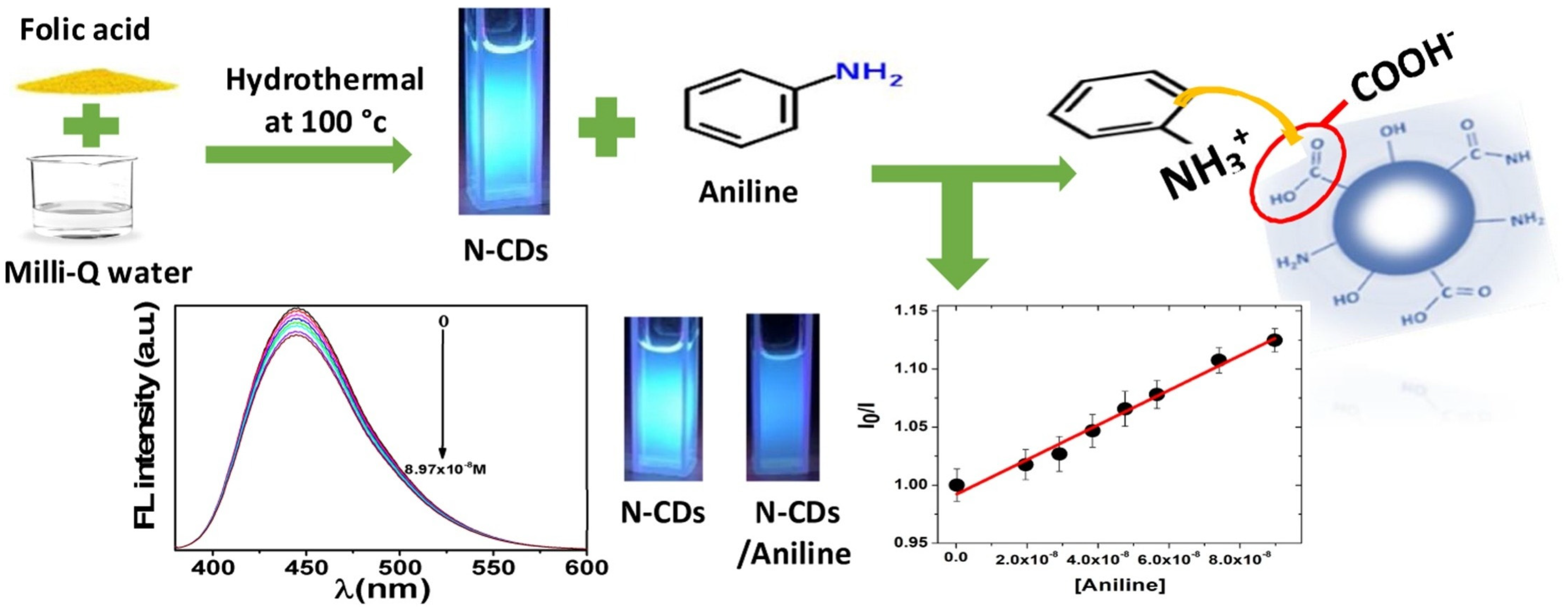
  131. Bakier, Y.M.; Ghali, M.; Elkun, A.; Beltagi, A.M.; Zahra, W.K. Static interaction between colloidal carbon nano-dots and aniline: A novel platform for ultrasensitive detection of aniline in aqueous media. Mater. Res. Bull. 2021, 134, 111119. [Google Scholar] [CrossRef]
  132. Ma, C.; Zhu, Z.; Wang, H.; Huang, X.; Zhang, X.; Qi, X.; Zhang, H.L.; Zhu, Y.; Deng, X.; Peng, Y. A General Solid-State Synthesis of Chemically-Doped Fluorescent Graphene Quantum Dots for Bioimaging and Optoelectronic Applications. Nanoscale 2015, 7, 10162–10169. [Google Scholar] [CrossRef]
  133. Li, X.; Lau, S.; Tang, L.; Ji, R.; Yang, P. Multicolour light emission from chlorine-doped graphene quantum dots. J. Mater. Chem. C 2013, 1, 7308–7313. [Google Scholar] [CrossRef]
  134. Praneerad, J.; Neungnoraj, K.; In, I.; Paoprasert, P. Environmentally friendly supercapacitor based on carbon dots from durian peel as an electrode. Key Eng. Mater. 2019, 803, 115–119. [Google Scholar] [CrossRef]
  135. Zhang, Y.Q.; Ma, D.K.; Zhuang, Y.; Zhang, X.; Chen, W.; Hong, L.L.; Huang, S.M. One-pot synthesis of N-doped carbon dots with tunable luminescence properties. J. Mater. Chem. 2012, 22, 16714–16718. [Google Scholar] [CrossRef]
  136. Gunjal, D.B.; Naik, V.M.; Waghmare, R.D.; Patil, C.S.; Shejwal, R.V.; Gore, A.H.; Kolekar, G.B. Sustainable carbon nanodots synthesised from kitchen derived waste tea residue for highly selective fluorimetric recognition of free chlorine in acidic water: A waste utilization approach. J. Taiwan Inst. Chem. Eng. 2019, 95, 147–154. [Google Scholar] [CrossRef]
  137. Gutiérrez-Sánchez, C.; Mediavilla, M.; Guerrero-Esteban, T.; Revenga-Parra, M.; Pariente, F.; Lorenzo, E. Direct covalent immobilization of new nitrogen-doped carbon nanodots by electrografting for sensing applications. Carbon 2020, 159, 303–310. [Google Scholar] [CrossRef]
  138. Pender, J.P.; Jha, G.; Youn, D.H.; Ziegler, J.M.; Andoni, I.; Choi, E.; Heller, A.; Dunn, B.S.; Weiss, P.S.; Penner, R.M. Electrode Degradation in Lithium-Ion Batteries. ACS Nano 2020, 14, 1243–1295. [Google Scholar] [CrossRef]
  139. Miao, P.; Tang, Y.; Han, K.; Wang, B. Facile synthesis of carbon nanodots from ethanol and their application in ferric (III) ion assay. J. Mater. Chem. A 2015, 3, 15068–15073. [Google Scholar] [CrossRef]
  140. Keerthana, A.K.; Ashraf, P.M. Carbon nanodots synthesized from chitosan and its application as a corrosion inhibitor in boat-building carbon steel BIS2062. Appl. Nano. 2020, 10, 1061–1071. [Google Scholar] [CrossRef]
  141. Arvapalli, D.M.; Sheardy, A.T.; Alapati, K.C.; Wei, J. High quantum yields fluorescent carbon nanodots for detection of Fe (III) Ions and electrochemical study of quenching mechanism. Talanta 2020, 209, 120538. [Google Scholar] [CrossRef]
  142. Baluta, S.; Lesiak, A.; Cabaj, J. Simple and cost-effective electrochemical method for norepinephrine determination based on carbon dots and tyrosinase. Sensors 2020, 20, 4567. [Google Scholar] [CrossRef]
  143. Zhai, X.; Zhang, P.; Liu, C.; Bai, T.; Li, W.; Dai, L.; Liu, W. Highly luminescent carbon nanodots by microwave-assisted pyrolysis. Chem. Commun. 2012, 48, 7955–7957. [Google Scholar] [CrossRef] [PubMed]
  144. Zhang, P.; Li, W.; Zhai, X.; Liu, C.; Dai, L.; Liu, W. A facile and versatile approach to biocompatible “fluorescent polymers” from polymerizable carbon nanodots. Chem. Commun. 2012, 48, 10431–10433. [Google Scholar] [CrossRef] [PubMed]
  145. Li, L.; Ji, J.; Fei, R.; Wang, C.; Lu, Q.; Zhang, J.; Jiang, L.P.; Zhu, J.J. A facile microwave avenue to electrochemiluminescenttwo-color graphene quantum dots. Adv. Funct. Mater. 2012, 22, 2971–2979. [Google Scholar] [CrossRef]
  146. Liu, H.; He, Z.; Jiang, L.P.; Zhu, J.J. Microwave-assisted synthesis of wavelength-tunablephotoluminescent carbon nanodots and their potential applications. ACS Appl. Mater. Interfaces 2015, 7, 4913–4920. [Google Scholar] [CrossRef]
  147. Kumawat, M.K.; Thakur, M.; Gurung, R.B.; Srivastava, R. Graphene quantum dots from mangiferaindica: Application in near-infrared bioimaging and intracellular nanothermometry. ACS Sustain. Chem. Eng. 2017, 5, 1382–1391. [Google Scholar] [CrossRef]
  148. Pires, N.R.; Santos, C.M.W.; Sousa, R.R.; De Paula, R.C.M.; Cunha, P.L.R.; Feitosa, J.P.A. Novel and Fast Microwave-Assisted Synthesis of Carbon Quantum Dots from Raw Cashew Gum. J. Brazil. Chem. Soc. 2015, 26, 1274–1282. [Google Scholar] [CrossRef]
  149. Simsek, S.; Alas, M.O.; Ozbek, B.; Genc, R. Evaluation of the physical properties of fluorescent carbon nanodots synthesized using Nerium oleander extracts by microwave-assisted synthesis methods. J. Mater. Res. Technol. 2019, 8, 2721–2731. [Google Scholar] [CrossRef]
  150. Ren, Q.; Ga, L.; Ai, J. Rapid synthesis of highly fluorescent nitrogendoped graphene quantum dots for effective detection of ferric ions and as fluorescent ink. ACS Omega 2019, 4, 15842–15848. [Google Scholar] [CrossRef]
  151. Sendao, R.M.S.; Crista, J.D.M.A.; Afonso, A.C.P.; Yuso, M.V.M.; Algarra, M.; Silva, J.C.E.; Silva, L.P. Insight into the hybrid luminescence showed by carbon dots and molecular fluorophores in solution. Phys. Chem. Chem. Phys. 2019, 21, 20919. [Google Scholar] [CrossRef]
  152. Luo, X.; Huang, G.; Bai, C.; Wang, C.; Yu, Y.; Tan, Y.; Tang, C.; Kong, J.; Huang, J.; Li, Z. A versatile platform for colorimetric, fluorescence and photothermal multi-mode glyphosate sensing by carbon dots anchoring ferrocene metal-organic framework nanosheet. J. Hazard. Mat. 2023, 443, 130277. [Google Scholar] [CrossRef]
  153. Wang, C.; Xu, Z.; Zhang, C. Polyethyleneimine-functionalized fluorescent carbon dots: Water stability, pH sensing, and cellular imaging. ChemNanoMat 2015, 1, 122–127. [Google Scholar] [CrossRef]
  154. Mandal, P.; Sahoo, D.; Sarkar, P.; Chakraborty, K.; Das, S. Fluorescence turn-on andturn-off sensing of pesticides by carbon dot-based sensor. New J. Chem. 2019, 43, 12137–12151. [Google Scholar] [CrossRef]
  155. Papaioannou, N.; Marinovic, A.; Yoshizawa, N.; Goode, A.E.; Fay, M.; Khlobystov, A.; Titirici, M.M.; Sapelkin, A. Structure and solvents effects on the optical properties of sugar-derivedcarbon nanodots. Sci. Rep. 2018, 8, 6559. [Google Scholar] [CrossRef]
  156. Wang, J.C.; Violette, K.; Ogunsolu, O.O.; Hanson, K. Metal ion mediated electrontransfer at dye–semiconductor interfaces. Phys. Chem. Chem. Phys. 2017, 19, 2679–2682. [Google Scholar] [CrossRef]
  157. Zheng, H.; Wang, Q.; Long, Y.; Zhang, H.; Huang, X.; Zhu, R. Enhancing theluminescence of carbon dots with a reduction pathway. Chem. Commun. 2011, 47, 10650–10652. [Google Scholar] [CrossRef]
  158. Deshmukh, S.; Deore, A.; Mondal, S. Ultrafast dynamics in carbon dots as photosensitizers: A review. ACS Appl. Nano Mater. 2021, 4, 7587–7606. [Google Scholar] [CrossRef]
  159. Sciortino, A.; Madonia, A.; Gazzetto, M.; Sciortino, L.; Rohwer, E.J.; Feurer, T.; Gelardi, F.M.; Cannas, M.; Cannizzo, A.; Messina, F. The interaction of photoexcited carbon nanodotswith metal ions disclosed down to the femtosecond scale. Nanoscale 2017, 9, 11902–11911. [Google Scholar] [CrossRef]
  160. Liu, R.; Li, H.; Kong, W.; Liu, J.; Liu, Y.; Tong, C.; Zhang, X.; Kang, Z. Ultra-sensitiveand selective Hg2+ detection based on fluorescent carbon dots. Mater. Res. Bull. 2013, 48, 2529–2534. [Google Scholar] [CrossRef]
  161. Huang, H.; Lv, J.J.; Zhou, D.L.; Bao, N.; Xu, Y.; Wang, A.J.; Feng, J.J. One-pot greensynthesis of nitrogen-doped carbon nanoparticles as fluorescent probes for mercury ions. RSC Adv. 2013, 3, 21691–21696. [Google Scholar] [CrossRef]
  162. Wang, X.; Zhang, J.; Zou, W.; Wang, R. Facile synthesis of polyaniline/carbon dotnanocomposites and their application as a fluorescent probe to detect mercury. RSC Adv. 2015, 5, 41914–41919. [Google Scholar] [CrossRef]
  163. Liu, C.; Tang, B.; Zhang, S.; Zhou, M.; Yang, M.; Liu, Y.; Zhang, Z.L.; Zhang, B.; Pang, D.W. Photoinduced electron transfer mediated by coordination between carboxyl on carbon nanodotsand Cu2+ quenching photoluminescence. J. Phy. Chem. C 2018, 122, 3662–3668. [Google Scholar] [CrossRef]
  164. Zhu, A.; Qu, Q.; Shao, X.; Kong, B.; Tian, Y. Carbon-dot-based dual-emissionnanohybrid produces a ratiometric fluorescent sensor for in vivo imaging of cellular copperions. Angew. Chem. Int. Ed. 2012, 51, 7185–7189. [Google Scholar] [CrossRef] [PubMed]
  165. Mohammed, L.J.; Omer, K.M. Dual functional highly luminescence B, N Co-dopedcarbon nanodots as nanothermometer and Fe3+/Fe2+ sensor. Sci. Rep. 2020, 10, 3028. [Google Scholar] [CrossRef] [PubMed]
  166. Sekar, A.; Yadav, R.; Basavaraj, N. Fluorescence quenching mechanism and the application of green carbon nanodots in the detection of heavy metal ions: A review. New J. Chem. 2021, 45, 2326–2360. [Google Scholar] [CrossRef]
  167. Goncalves, H.M.; Duarte, A.J.; da Silva, J.C.E. Optical fiber sensor for Hg (II) based on carbon dots. Biosen. Bioelect. 2010, 26, 1302–1306. [Google Scholar] [CrossRef] [PubMed]
  168. Gharat, P.M.; Pal, H.; Choudhury, S.D. Photophysics and luminescence quenching ofcarbon dots derived from lemon juice and glycerol. Spectrochim. Acta Part A Mol. Biomol. Spectro. 2019, 209, 14–21. [Google Scholar] [CrossRef]
  169. Karali, K.K.; Sygellou, L.; Stalikas, C.D. Highly fluorescent N-doped carbon nanodotsas an effective multi-probe quenching system for the determination of nitrite, nitrate and ferric ions infood matrices. Talanta 2018, 189, 480–488. [Google Scholar] [CrossRef] [PubMed]
  170. Guo, J.; Liu, D.; Filpponen, I.; Johansson, L.S.; Malho, J.M.; Quraishi, S.; Liebner, F.; Santos, H.A.; Rojas, O.J. Photoluminescent hybrids of cellulose nanocrystals and carbon quantum dots as cytocompatible probes for in vitro bioimaging. Biomacromolecules 2017, 18, 2045–2055. [Google Scholar] [CrossRef]
  171. Cronican, J.J.; Thompson, D.B.; Beier, K.T.; McNaughton, B.R.; Cepko, C.L.; Liu, D.R. Potent delivery of functional proteins into Mammalian cells in vitro and in vivo using a superchargedprotein. ACS Chem. Biol. 2010, 5, 747–752. [Google Scholar] [CrossRef]
  172. Pan, L.; Sun, S.; Zhang, L.; Jiang, K.; Lin, H. Near-infrared emissive carbon dots for two-photon fluorescence bioimaging. Nanoscale 2016, 8, 17350–17356. [Google Scholar] [CrossRef]
  173. Sahu, S.; Behera, B.; Maiti, T.K.; Mohapatra, S. Simple one-step synthesis of highly luminescent carbon dots from orange juice: Application as excellent bio-imaging agents. Chem. Commun. 2012, 48, 8835–8837. [Google Scholar] [CrossRef] [PubMed]
  174. Sivasankarapillai, V.S.; Jose, J.; Shanavas, M.S.; Marathakam, A.; Uddin, M.; Mathew, B. Silicon quantum dots: Promising theranostic probes for the future. Curr. Drug Targets 2019, 20, 1255–1263. [Google Scholar] [CrossRef] [PubMed]
  175. Liu, C.; Zhang, P.; Tian, F.; Li, W.; Li, F.; Liu, W. One-step synthesis of surfacepassivated carbon nanodots by microwave assisted pyrolysis for enhanced multicolour photoluminescence and bioimaging. J. Mater. Chem. 2011, 21, 13163–13167. [Google Scholar] [CrossRef]
  176. Chen, B.B.; Wang, X.Y.; Qian, R.C. Rolling “wool-balls”: Rapid live-cell mapping of membrane sialic acids via poly-p-benzoquinone/ethylenediamine nanoclusters. Chem. Commun. 2019, 55, 9681–9684. [Google Scholar] [CrossRef] [PubMed]
  177. Tao, H.; Yang, K.; Ma, Z.; Wan, J.; Zhang, Y.; Kang, Z.; Liu, Z. In vivo NIR fluorescenceimaging, biodistribution, and toxicology of photoluminescent carbon dots produced from carbonnanotubes and graphite. Small 2012, 8, 281–290. [Google Scholar] [CrossRef]
  178. Choi, Y.; Kim, S.; Choi, M.H.; Ryoo, S.R.; Park, J.; Min, D.H.; Kim, B.S. Highly biocompatible carbon nanodots for simultaneous bioimaging and targeted photodynamic therapy invitro and in vivo. Adv. Functional Mater. 2014, 24, 5781–5789. [Google Scholar] [CrossRef]
  179. Shi, Q.Q.; Li, Y.H.; Xu, Y.; Wang, Y.; Yin, X.B.; He, X.W.; Zhang, Y.K. High-yield and high-solubility nitrogen-doped carbon dots: Formation, fluorescence mechanism and imaging application. RSC Adv. 2014, 4, 1563–1566. [Google Scholar] [CrossRef]
  180. Wang, J.; Zhang, P.; Huang, C.; Liu, G.; Leung, K.C.F.; Wáng, Y.X.J. High performancephotoluminescent carbon dots for in vitro and in vivo bioimaging: Effect of nitrogen dopingratios. Langmuir 2015, 31, 8063–8073. [Google Scholar] [CrossRef]
  181. Bankoti, K.; Rameshbabu, A.P.; Datta, S.; Das, B.; Mitra, A.; Dhara, S. Onion derived carbon nanodots for live cell imaging and accelerated skin wound healing. J. Mater. Chem. B 2017, 5, 6579–6592. [Google Scholar] [CrossRef]
  182. Ming, H.; Ma, Z.; Liu, Y.; Pan, K.; Yu, H.; Wang, F.; Kang, Z. Large scaleelectrochemical synthesis of high-quality carbon nanodots and their photocatalytic property. Dalton Trans. 2012, 41, 9526–9531. [Google Scholar] [CrossRef]
  183. Song, B.; Wang, T.; Sun, H.; Shao, Q.; Zhao, J.; Song, K.; Hao, L.; Wang, L.; Guo, Z. Two-step hydrothermally synthesized carbon nanodots/WO3 photocatalysts with enhanced photocatalytic performance. Dalton Trans. 2017, 46, 15769–15777. [Google Scholar] [CrossRef] [PubMed]
  184. Qu, S.; Chen, H.; Zheng, X.; Cao, J.; Liu, X. Ratiometric fluorescent nanosensor based on water soluble carbon nanodots with multiple sensing capacities. Nanoscale 2013, 5, 5514–5518. [Google Scholar] [CrossRef] [PubMed]
  185. Vedamalai, M.; Periasamy, A.P.; Wang, C.W.; Tseng, Y.T.; Ho, L.C.; Shih, C.C.; Chang, H.T. Carbon nanodots prepared from o-phenylenediamine for sensing of Cu2+ ions in cells. Nanoscale 2014, 6, 13119–13125. [Google Scholar] [CrossRef] [PubMed]
  186. Shi, L.; Li, Y.; Li, X.; Zhao, B.; Wen, X.; Zhang, G.; Dong, C.; Shuang, S. Controllable synthesis of green and blue fluorescent carbon nanodots for pH and Cu2+ sensing in living cells. Biosen. Bioelect. 2016, 77, 598–602. [Google Scholar] [CrossRef]
  187. Xu, B.; Zhao, C.; Wei, W.; Ren, J.; Miyoshi, D.; Sugimoto, N.; Qu, X. Aptamer carbon-nanodot sandwich used for fluorescent detection of protein. Analyst 2012, 137, 5483–5486. [Google Scholar] [CrossRef]
  188. Nie, H.; Li, M.; Li, Q.; Liang, S.; Tan, Y.; Sheng, L.; Shi, W.; Zhang, S.X.A. Carbon dots with continuously tunable full-color emission and their application in ratiometric pH sensing. Chem. Mater. 2014, 26, 3104–3112. [Google Scholar] [CrossRef]
  189. Wang, C.I.; Wu, W.C.; Periasamy, A.P.; Chang, H.T. Electrochemical synthesis of photoluminescent carbon nanodots from glycine for highly sensitive detection of hemoglobin. Green Chem. 2014, 16, 2509–2514. [Google Scholar] [CrossRef]
  190. Gogoi, N.; Chowdhury, D. Novel carbon dot coated alginate beads with superior stability, swelling and pH responsive drug delivery. J. Mater. Chem. B 2014, 2, 4089–4099. [Google Scholar] [CrossRef]
  191. Karthik, S.; Saha, B.; Ghosh, S.K.; Singh, N.P. Photoresponsivequinoline tethered fluorescent carbon dots for regulated anticancer drug delivery. Chem. Commun. 2013, 49, 10471–10473. [Google Scholar] [CrossRef]
  192. Chen, B.; Yang, Z.; Zhu, Y.; Xia, Y. Zeolitic imidazolate framework materials: Recent progress in synthesis and applications. J. Mater. Chem. A 2014, 2, 16811–16831. [Google Scholar] [CrossRef]
  193. He, L.; Wang, T.; An, J.; Li, X.; Zhang, L.; Li, L.; Li, G.; Wu, X.; Su, Z.; Wang, C. Carbon nanodots@ zeolitic imidazolate framework-8 nanoparticles for simultaneous pH-responsive drug delivery and fluorescence imaging. Cryst. Eng. Commun. 2014, 16, 3259–3263. [Google Scholar] [CrossRef]
  194. Kim, Y.; Jang, G.; Lee, T.S. New fluorescent metal-ion detection using a paper-based sensor strip containing tethered rhodamine carbon nanodots. ACS Appl. Mater. Interfaces 2015, 7, 15649–15657. [Google Scholar] [CrossRef]
  195. Wang, J.; Zhang, Z.; Zha, S.; Zhu, Y.; Wu, P.; Ehrenberg, B.; Chen, J.Y. Carbon nanodots featuring efficient FRET for two-photon photodynamic cancer therapy with a low fs laser power density. Biomaterials 2014, 35, 9372–9381. [Google Scholar] [CrossRef] [PubMed]
  196. Zheng, M.; Liu, S.; Li, J.; Qu, D.; Zhao, H.; Guan, X.; Hu, X.; Xie, Z.; Jing, X.; Sun, Z. Integrating oxaliplatin with highly luminescent carbon dots: An unprecedented theranostic agent for personalized medicine. Adv. Mat. 2014, 26, 3554–3560. [Google Scholar] [CrossRef] [PubMed]
  197. Huang, Y.; Chen, J.; Wong, T.; Liow, J.L. Experimental and theoretical investigations of non-Newtonian electro-osmotic driven flow in rectangular microchannels. Soft Matter 2016, 12, 6206–6213. [Google Scholar] [CrossRef]
  198. Atencia, J.; Beebe, D.J. Controlled microfluidic interfaces. Nature 2005, 437, 648–655. [Google Scholar] [CrossRef]
  199. Stewart, M.P.; Sharei, A.; Ding, X.; Sahay, G.; Langer, R.; Jensen, K.F. In vitro and ex vivo strategies for intracellular delivery. Nature 2016, 538, 183–192. [Google Scholar] [CrossRef]
  200. Song, H.; Chen, D.L.; Ismagilov, R.F. Reactions in droplets in microfluidic channels. Angew. Chem. Int. Ed. 2006, 45, 7336–7356. [Google Scholar] [CrossRef]
  201. Huang, Y.; Xiao, L.; An, T.; Lim, W.; Wong, T.; Sun, H. Fast dynamic visualizations in microfluidics enabled by fluorescent carbon nanodots. Small 2017, 13, 1700869. [Google Scholar] [CrossRef]
  202. Lin, H.; Storey, B.D.; Oddy, M.H.; Chen, C.H.; Santiago, J.G. Instability of electrokinetic microchannel flows with conductivity gradients. Phys. Fluids 2004, 16, 1922–1935. [Google Scholar] [CrossRef]
  203. Yang, S.T.; Wang, X.; Wang, H.; Lu, F.; Luo, P.G.; Cao, L.; Meziani, M.J.; Liu, J.H.; Liu, Y.; Chen, M.; et al. Carbon Dots as Nontoxic and High-Performance Fluorescence Imaging Agents. J. Phys. Chem. C 2009, 113, 18110–18114. [Google Scholar] [CrossRef] [PubMed]
  204. Zhu, A.; Luo, Z.; Ding, C.; Li, B.; Zhou, S.; Wang, R.; Tian, Y. A two-photon “turn-on” fluorescent probe based on carbon nanodots for imaging and selective biosensing of hydrogen sulfide in live cells and tissues. Analyst 2014, 139, 1945–1952. [Google Scholar] [CrossRef] [PubMed]
  205. Chandra, A.; Deshpande, S.; Shinde, D.B.; Pillai, V.K.; Singh, N. Mitigating the cytotoxicity of graphene quantum dots and enhancing their applications in bioimaging and drug delivery. ACS Macro Lett. 2014, 3, 1064–1068. [Google Scholar] [CrossRef] [PubMed]
  206. Song, Y.; Shi, W.; Chen, W.; Li, X.; Ma, H. Fluorescent carbon nanodots conjugated with folic acid for distinguishing folate-receptor-positive cancer cells from normal cells. J. Mater. Chem. 2012, 22, 12568–12573. [Google Scholar] [CrossRef]
  207. Milton, F.P.; Govan, J.; Mukhina, M.V.; Gun’ko, Y.K. The chiral nano-world: Chiroptically active quantum nanostructures. Nano. Hori. 2016, 1, 14–26. [Google Scholar] [CrossRef]
  208. Liu, X.L.; Tsunega, S.; Jin, R.H. Self-directing chiral information in solid–solid transformation: Unusual chiral-transfer without racemization from amorphous silica to crystalline silicon. Nano. Hori. 2017, 2, 147–155. [Google Scholar] [CrossRef]
  209. Walker, R.; Pociecha, D.; Abberley, J.P.; Martinez-Felipe, A.; Paterson, D.A.; Forsyth, E.; Lawrence, G.B.; Henderson, P.A.; Storey, J.M.; Gorecka, E.; et al. Spontaneous chirality through mixing achiral components: A twist-bend nematic phase driven by hydrogen-bonding between unlike components. Chem. Commun. 2018, 54, 3383–3386. [Google Scholar] [CrossRef]
  210. Vázquez-Nakagawa, M.; Rodríguez-Pérez, L.; Herranz, M.A.; Martín, N. Chirality transfer from graphene quantum dots. Chem. Commun. 2016, 52, 665–668. [Google Scholar] [CrossRef]
  211. Xin, Q.; Liu, Q.; Geng, L.; Fang, Q.; Gong, J.R. Chiral nanoparticles as a new efficient antimicrobial nanoagent. Adv. Healthcare Mater. 2017, 6, 1601011. [Google Scholar] [CrossRef]
  212. Li, F.; Li, Y.; Yang, X.; Han, X.; Jiao, Y.; Wei, T.; Yang, D.; Xu, H.; Nie, G. Highly fluorescent chiral N-S-doped carbon dots from cysteine: Affecting cellular energy metabolism. Angew. Chem. 2018, 130, 2401–2406. [Google Scholar] [CrossRef]
  213. Malishev, R.; Arad, E.; Bhunia, S.K.; Shaham-Niv, S.; Kolusheva, S.; Gazit, E.; Jelinek, R. Chiral modulation of amyloid beta fibrillation and cytotoxicity by enantiomeric carbon dots. Chem. Commun. 2018, 54, 7762–7765. [Google Scholar] [CrossRef] [PubMed]
Figure 1. Methods used for the synthesis of Carbon nanodots (1–10 nm).
Catalysts 13 00858 g001
Figure 3. General methods of synthesis carbon nanodots (CNDs: Bottom-up approach: CNDs were synthesized from smaller carbon units (small organic molecules) by applying energy (electrochemical/chemical, thermal. laser, microwave, etc.). The source molecules will get ionized, dissociated, evaporated, or sublimated and then condensed to form CNDs; Top-down approach: CNDs are synthesized by transformation of larger carbon structures into ultra-small fragments by applying energy (thermal, mechanical, chemical, ultrasonic, etc.). Reproduced with the permission from Ref. [69], 2019, Springer Nature.
Catalysts 13 00858 g003
Scheme 1. Schematic view of the formation of carbon-supported silver nanoparticles. Reproduced with permission from Ref. [112]. 2014, Elsevier.
Catalysts 13 00858 sch001
Figure 4. Fluorescent chemical sensor for ultra-sensitive detection of aniline in the liquid phase, via formation of colloidal carbon nanodots supported on nitrogen. Reproduced with permission from Ref. [131]. 2021, Elsevier.
Catalysts 13 00858 g004
Figure 5. Schematic view of the experimental procedure. Reproduced with permission from Ref. [131]. 2021, Elsevier.
Catalysts 13 00858 g005
Figure 6. Schematic illustration of the fabrication process. Reproduced with permission from Ref. [149]. 2019, Elsevier.
Catalysts 13 00858 g006
Figure 7. Application of Carbon dots.
Catalysts 13 00858 g007
Table 1. Advantages and disadvantages of different synthetic methods of carbon nanodots.
Synthetic MethodAdvantagesDisadvantagesReferences
Sonochemical/Ultra-sonic fabricationEasy-operationExpensive cost of energy[108,109,110,111,112,113,114,115,116]
Hydrothermal synthesisCost-effective, environmentally benign, non-toxicNo uniformity in size [124,125,126,127,128,129,130,131]
Carbonization/PyrolysisNo solvent required, cost-effective, bulk-productionNo uniformity in distribution of size[132,133,134,135,136]
Electrochemical synthesisEasy, cost-effective, environmentally benignUniformity in size distribution[137,138,139,140,141,142]
Microwave-assisted synthesisFast, cost-effective, environmentally benignNo uniformity in distribution of size[143,144,145,146,147,148,149,150,151]
Table 2. Methods for the conversion of biobased and chemical feedstock into functionalized carbon nanodots.
SourcesSynthetic MethodsReferences
Carbon, Silver nitrate liquid solutionSonochemical synthesis[112]
Saccharum officinarum (Sugarcane)Hydrothermal synthesis[124]
Coconut waterUltrasonication synthesis[113]
o-phenylenediamineHydrothermal synthesis[129]
Waste biomassCarbonization synthesis[136]
EthanolElectrochemical carbonization synthesis[139]
Graphene Sonochemical synthesis[108]
p-phenylenediamine and ureaHydrothermal synthesis[130]
Mangifera indica (Mango)Microwave-assisted hydrothermal synthesis[147]
ChitosanHydrothermal carbonization synthesis[140]
Disclaimer/Publisher’s Note: The statements, opinions and data contained in all publications are solely those of the individual author(s) and contributor(s) and not of MDPI and/or the editor(s). MDPI and/or the editor(s) disclaim responsibility for any injury to people or property resulting from any ideas, methods, instructions or products referred to in the content.

