Article

Biochemical and Spectroscopic Characterization of a Recombinant Laccase from Thermoalkaliphilic Bacillus sp. FNT with Potential for Degradation of Polycyclic Aromatic Hydrocarbons (PAHs)

by
Constanza Bueno-Nieto
2,
Rodrigo Cortés-Antiquera
1,2,
Giannina Espina
2,*,
Joaquín Atalah
2,
Javiera Villanueva
1,2,
Carolina Aliaga
1,
Gustavo E. Zuñiga
1 and
Jenny M. Blamey
1,2,*
1
Facultad de Química y Biología, Universidad de Santiago de Chile, Alameda 3363, Santiago 9170022, Chile
2
Fundación Biociencia, José Domingo Cañas 2280, Santiago 7750132, Chile
*
Authors to whom correspondence should be addressed.
Catalysts 2023, 13(4), 763; https://doi.org/10.3390/catal13040763
Submission received: 30 January 2023 / Revised: 10 March 2023 / Accepted: 22 March 2023 / Published: 17 April 2023
(This article belongs to the Section Biocatalysis)

Abstract

:
Laccases are industrially relevant enzymes that are known for the wide variety of substrates they can use. In recent years, fungal laccases have been progressively replaced by bacterial laccases in applied contexts due to their capacity to work on harsh conditions including high temperatures, pHs, and chloride concentrations. The focus of researchers has turned specifically towards enzymes from extremophilic organisms because of their robustness and stability. The recombinant versions of enzymes from extremophiles have shown to overcome the problems associated with growing their native host organisms under laboratory conditions. In this work, we further characterize a recombinant spore-coat laccase from Bacillus sp. FNT, a thermoalkaliphilic bacterium isolated from a hot spring in a geothermal site. This recombinant laccase was previously shown to be very active and thermostable, working optimally at temperatures around 70–80 °C. Here, we showed that this enzyme is also resistant to common inhibitors, and we tested its ability to oxidize different polycyclic aromatic hydrocarbons, as these persistent organic pollutants accumulate in the environment, severely damaging ecosystems and human health. So far, the enzyme was found to efficiently oxidize anthracene, making it a compelling biotechnological tool for biocatalysis and a potential candidate for bioremediation of aromatic contaminants that are very recalcitrant to degradation.

1. Introduction

Laccases (benzenediol: oxygen-oxidoreductase; EC 1.10.3.2) are redox enzymes that belong to the enzyme family of multicopper oxidases (MCOs) and catalyze the oxidation of a large number of phenolic and non-phenolic molecules, using oxygen as an electron acceptor and generating water as a by-product [1,2]. Laccases have great biotechnological potential and are useful in several eco-friendly applications such as pulp and paper biobleaching [3], decolorization and degradation of textile dyes/effluent, bioremediation [4,5], biofuels [6] and many other uses [7,8].
Among their various applications, an interesting possibility enabled by their high versatility, ease of use, and broad substrate range is their utilization in the degradation of polycyclic aromatic hydrocarbons (PAHs), which are a large group of organic compounds composed of two or more fused aromatic rings. While some PAHs are naturally occurring, a significant number of them are generated through anthropogenic processes, including the incomplete combustion of organic compounds, industrial and agricultural activities, forest fires, and the use of fossil fuels [9]. Many of them have been shown to be carcinogenic and harmful to the environment, and due to their hydrophobic nature, they are easily adsorbed on soil [10], where they become very difficult to remove. To date, 16 PAHs appeared in the list of persistent organic contaminants created by the United States Environmental Protection Agency (EPA). So far, only a few laccases have shown to degrade some PAHs, suggesting that their application could serve as a potential tool for the elimination of these recalcitrant pollutants from the environment [11,12,13].
Laccases are widely distributed in nature, being produced by a broad array of organisms including plants [14], lichens [15], sponges [16], insects [17], wood-rotting fungi [6,8], and bacteria [7,18]. Within this group, fungal laccases are often highlighted, because they are the most studied and widely used in industrial processes. Nevertheless, their production is usually hindered due to long fermentation periods and low yields, and their applicability is limited by their low thermal stability and because their activity is generally restricted to mesophilic and acid conditions [19,20]. As the majority of the industrial applications are performed under extreme temperature, pH, and salt concentration conditions, fungal laccases generally fail to optimally work under these reaction settings, requiring larger quantities to compensate loss of activity [21].
Current research suggests that the advantages of using bacterial laccases in the industry cause them to be a very attractive alternative to fungal enzymes, as they are more stable at alkaline pHs and higher salt and metal concentrations [22]. It has also been demonstrated that they possess an enormous stability against various inhibitory agents [23]. The advantages of bacterial laccases could be explained by the presence of a coiled subdomain in their structure, which connects the first and second domains of these enzymes. It is hypothesized that the conformation of these domains contributes to the higher thermal stability and more compact structure of bacterial over fungal laccases [24]. Furthermore, additional advantages, such as cost-effective use in industrial applications, short enzyme-production times, and easiness to clone and express [25], support bacterial laccases having a remarkable biotechnological potential.
More recently, laccases from extremophilic bacteria have gained increased interest for real-life applications that require harsh reaction conditions [26]. So far, only a few thermophilic bacterial laccases have been purified and characterized from extremophiles, including the ones from Thermobaculum terrenum [27], Thermobifida fusca [28], Thermus thermophilus [29,30], and from Geobacillus genus [13,26,31,32]. These thermozymes, and the search for new ones, are of interest for researchers, because thermoactivity and thermal stability at high temperatures are very attractive features for their use in industrial processes [31,33].
One of the biggest challenges of working with native laccases from thermophiles is the difficulty in their isolation and culture, as well as its low cellular density in liquid medium under laboratory conditions. On the other hand, developing recombinant laccases for biotechnological applications can be very advantageous, because recombinant enzymes can be produced in higher concentrations with inexpensive growth media in a shorter period [26,34]. Obtaining a stable, active, versatile, and easy-to-produce laccase is still a relevant goal that can positively impact diverse industries and enable different applications, as discussed above.
In this study, we further characterized a novel recombinant spore-coat laccase from the thermoalkaliphilic Bacillus sp. FNT strain, which was isolated from a hot spring in a geothermal site by Fundación Biociencia [35]. This recombinant laccase shows high activity in a wide range of temperatures (20–90 °C) and thermal stability, which makes it very interesting for biotechnological applications. Here, we determined the kinetic parameters for syringaldazine, its model synthetic substrate, and assessed the effect of various common inhibitors in time at two different concentrations. Additionally, its EPR and UV-Vis spectra, which are characteristic features of multicopper oxidases, were also determined. Finally, given its potential as a biotechnologically relevant biocatalyst, we assessed the oxidation of anthracene (ANT), benzo[a]anthracene (BaA), and benzo[a]pyrene (BaP) as a model for PAH degradation using this new recombinant laccase.
The obtained results further highlight the impact that this interesting thermophilic enzyme could have in different applications, especially in bioremediation. The pursuit of bacterial laccases exhibiting high activity, yield, and robustness is a critical research area, aimed towards achieving the full potential of these enzymes as industrial biocatalysts and their real-world application, especially in environmentally friendly developments in a climate-change context. As such, this area of research is of utmost importance to both academia and industry. Therefore, the study and development of a new thermophilic bacterial laccase that exhibits improved performance and stability in comparison to well-characterized and currently commercialized fungal enzymes, holds the promise of enabling more efficient and sustainable industrial processes.

2. Results and Discussion

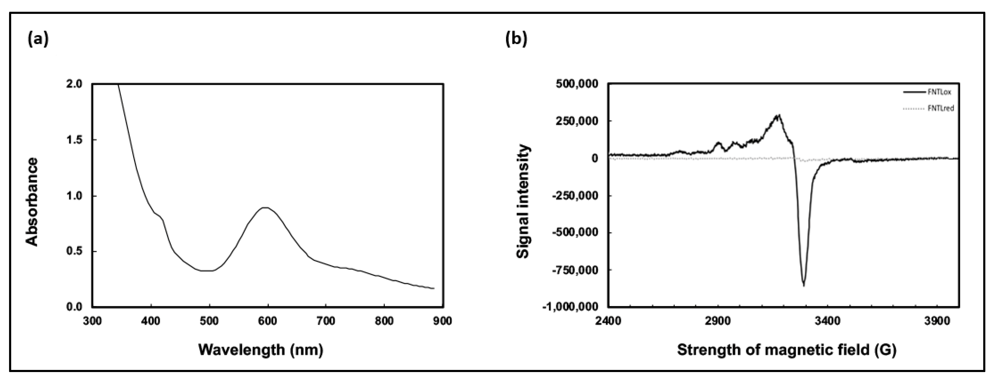
2.1. Further Characterization of Recombinant FNT Laccase (FNTL)

Laccases belong to the protein family of multicopper oxidases, and they are characterized by having four copper atoms in their catalytic center, divided into three types of structurally and functionally distinct copper sites, Type 1 (T1), Type 2 (T2), and binuclear Type 3 (T3), which can be distinguished by their unique spectroscopic features [36]. The three types of copper sites in the catalytic center of multicopper oxidases differ in their light absorbance: T1 copper gives a blue color to the protein from an absorbance at about 600 nm, T2 does not exhibit a strong feature in the visible absorption spectrum, and binuclear T3 provides a weak absorbance in the near UV [2,36].
The purified laccase in its oxidized state exhibited the typical blue-green color common to copper-containing enzymes, turning to a brown-yellow color in the absence of oxygen; this reduction is reversible and does not translate to a loss of activity. The T1 copper center was visible in the UV-visible absorption spectrum of the recombinant laccase showing a band at 580–620 nm with a peak at 600 nm, as expected (Figure 1a).
When the enzyme was studied using electron paramagnetic resonance spectroscopy (EPR) (Figure 1b), the spectrum obtained for the oxidized form of the enzyme was also consistent with the reported for other laccases [29,37], showing the presence of two copper ions with different coordination states, which could correspond to Type 1 and Type 2 copper atoms, given the regions of the spectrum in which the hyperfine signals appear. For the reduced sample of the enzyme after incubation with sodium dithionite, the EPR spectra was silent as expected.
In our previous work, we had determined that the activity of recombinant FNT laccase (FNTL) towards its model synthetic substrate, syringaldazine, was highest in the range of 70–80 °C and pH 6.0–7.0 [35]. To further characterize its catalytic properties, the kinetic parameters of the oxidation of syringaldazine by FNTL were determined at 70 °C and pH 6.0. The enzyme had a catalytic behavior consistent with the Michaelis–Menten equation (Figure S1), and exhibited a Km of 12.49 μM with a kcat = 33.77 s−1. The Km is slightly higher, whereas the kcat is lower than those reported for other laccases from the Bacillus genus, such as the recombinant spore-coat laccase of Bacillus licheniformis with Km = 4.2 μM, kcat = 100 s−1 [38], and the native spore-coat laccase of Bacillus sp. strain WT with Km = 3.7 μM, kcat = 51 s−1 [39]. As detailed in our previous publication, the FNTL enzyme has a very broad substrate range, being able to oxidize not only syringaldazine but also: ABTS; gallic acid; azo dyes: Congo red, methyl orange, and methyl red; triarylmethane dyes: Coomassie brilliant blue R250, bromophenol blue, malachite green, and crystal violet; and anthraquinonic dye: Remazol brilliant blue R. As described below, the FNTL enzyme is even able to utilize PAH as substrates. This wide substrate range could be related to the slightly lower affinity for syringaldazine reflected by the Km value. However, it is important to consider that comparing Km values between enzymes has some limitations due to the methodological differences among research groups, so it must be regarded as a general reference, with direct comparisons being preferable.

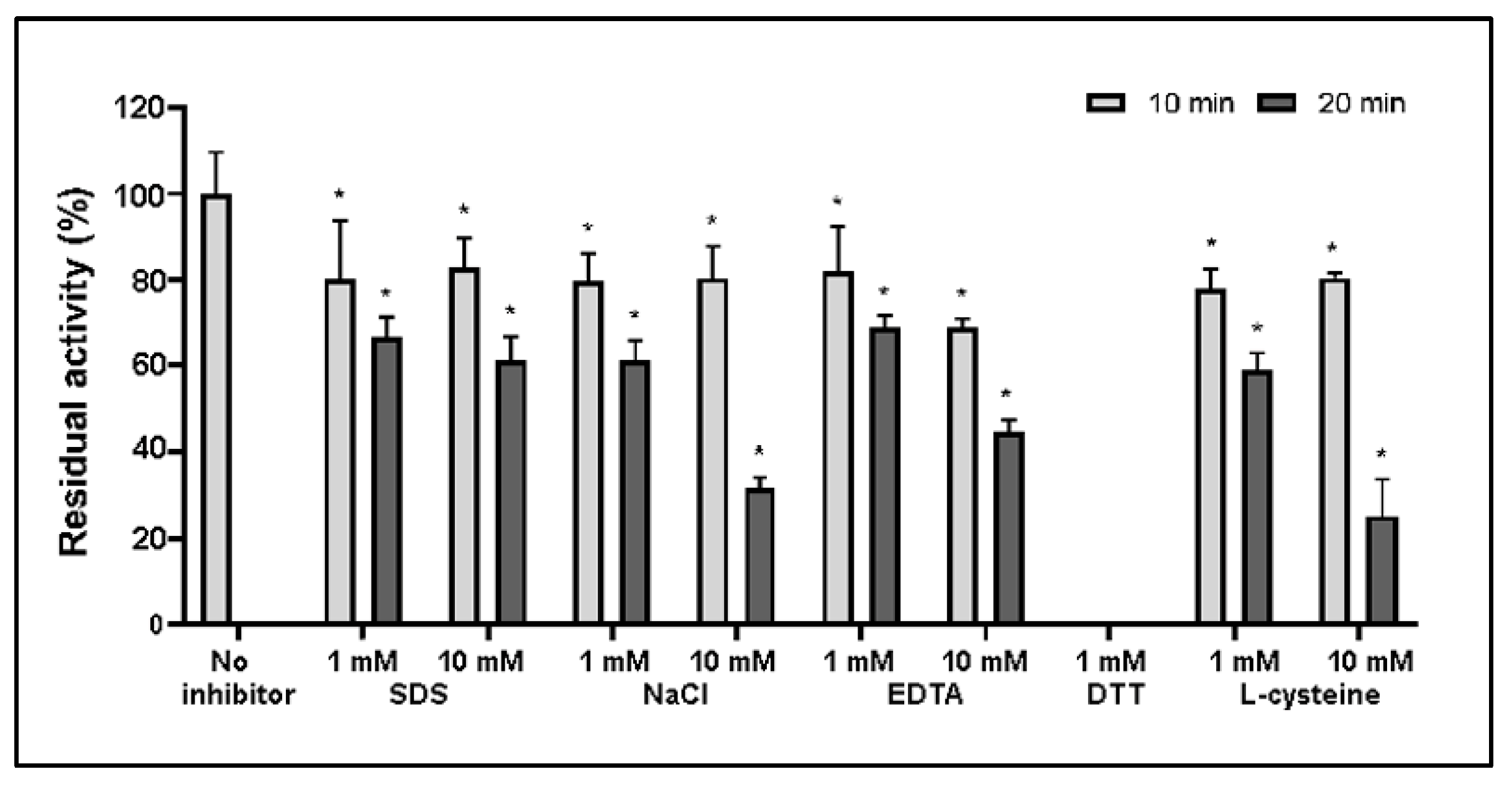
2.2. Effect of Inhibitors on FNTL

The influence of different chemical compounds on the catalytic activity of FNTL was further evaluated. In our previous work, the influence of 1 h exposure to 1 mM concentration of reducing reagents: dithiothreitol (DTT) and L-cysteine; salts: NaCl and NaN3; and sodium dodecyl sulfate (SDS) detergent was assessed [35]. This time, the effect of SDS, NaCl, DTT, L-cysteine, and ethylene diamine tetra-acid (EDTA) at 1 and 10 mM, was studied for 10 and 20 min to offer a more detailed description of FNTL inhibition in time (Figure 2).
The obtained results indicate that this enzyme maintained at least 70% of its activity after 10 min of incubation with all the different inhibitors tested at both concentrations, with the exception of DTT, which completely inhibited the enzyme. After 20 min of incubation, FNTL maintains more than 60% of its activity with 1 mM and 10 mM SDS, 1 mM NaCl. This is consistent with our previous results that indicate that, after 1 h incubation, the enzyme retained 67% and 50% of its activity with 1 mM of SDS and NaCl, respectively [35].
Chloride is a common laccase inhibitor [22,40]; consequently, chloride tolerance it is also a very valuable property for potential industrial and biotechnological applications [41]. The FNTL enzyme maintained around 80% of its activity after 10 min of incubation in the presence of 1 and 10 mM NaCl. However, after 20 min, the enzyme was able to retain around 60 and 30% of its activity in the presence of 1 and 10 mM NaCl, respectively. The chloride tolerance observed in FNTL is also a remarkable feature of this enzyme, although it is not among the most salt-tolerant bacterial laccases reported [39,42,43].
When incubated with 1 and 10 mM EDTA for 20 min, the enzyme maintains over 70% and 40% of its activity, respectively. The minor loss of activity displayed by FNTL in the presence of this chelating agent represents an advantage over other copper-ion dependent laccases described in the literature, where a direct correlation exists between the laccase activity and copper concentration present in the reaction mixture [44].
Both reducing agents were previously found to completely inhibit the FNTL activity after 1 h exposure at 1 mM concentration [35]. This time, DTT was found to completely inactivate the enzyme after only 10 min of incubation, while L-cysteine at 1 mM maintained near to 80% of its activity when incubating for 10 min and almost 60% after 20 min. However, when exposed at 10 mM of L-cysteine, the enzyme lost around 70% of its initial activity after 20 min.
Although laccases are typically sensitive to inhibitors due to the accessibility of their copper atoms [45], FNTL appears to exhibit a high tolerance toward chloride, EDTA, L-cysteine and SDS detergent. This observation supports the claim that FNTL is a very robust enzyme, needing over 20 min of incubation to significantly affect its activity with all inhibitors tested except for DTT, which had an immediate and drastic effect. This inhibition has also been reported for other laccases from Bacillus genus [43,46,47], highlighting the importance of the thiol group in the catalytic properties of the enzyme, as the T1 copper site is coordinated by two His and one Cys residues (arranged in a distorted trigonal geometry around the Cu ion), and a weaker fourth Met ligand that axially binds and completes a tetrahedral geometry [36,48].
Furthermore, the negative effect of some of the common inhibitors used is reported to be stronger for other bacterial laccases from the Bacillus genus, such as the native spore-coat laccase from Bacillus subtilis WD23 that maintained only 2.72% and 22.62% activity towards syringaldazine after just 3 min incubation with 1 mM EDTA and 1 mM L-cysteine, respectively [47], or the spore laccase from Bacillus vallismortis fmb-103, which using ABTS as substrate, maintained 13.2% of its activity after 10 min incubation with 5 mM EDTA and 58.3% with 1 mM SDS, while incubation with just 0.1 mM L-cysteine resulted in complete loss of activity [46].

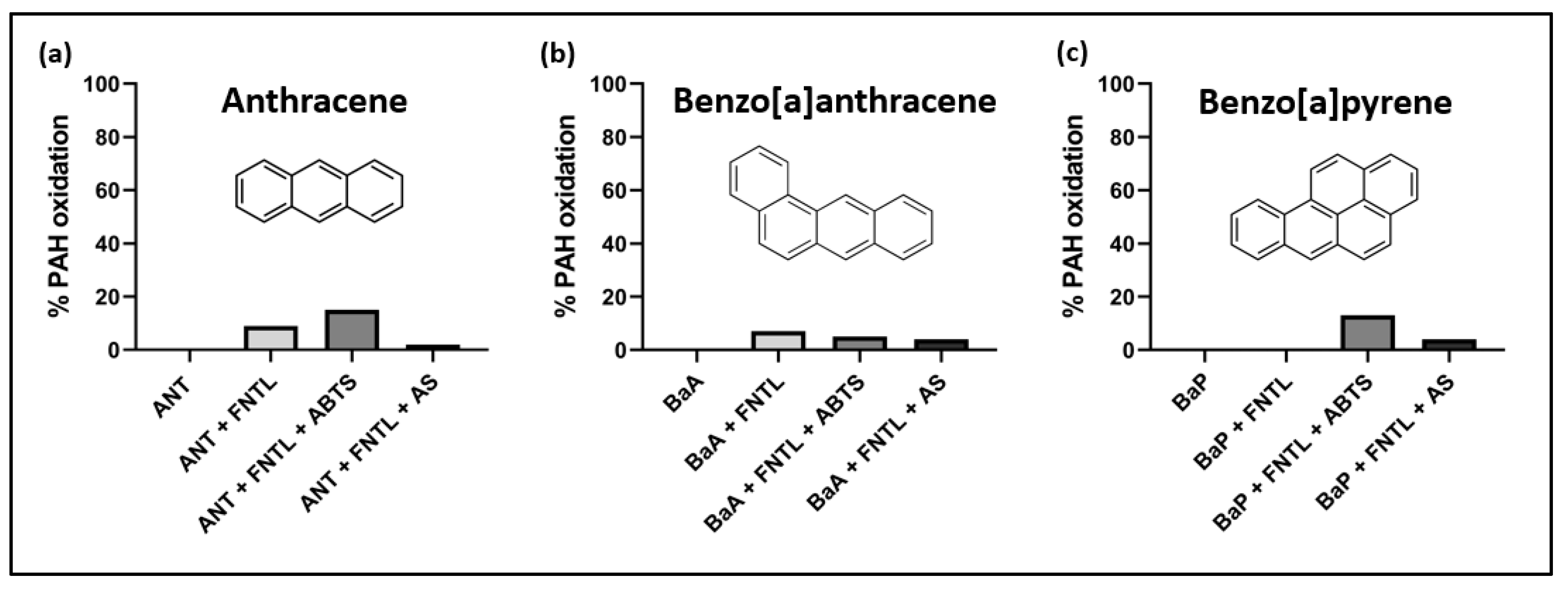
2.3. Evaluation of PAHs Oxidation

The catalytic properties of laccases allow their use in many different areas, including the very relevant ecological degradation of many toxic pollutants present in soil and water sources as a result of human activity, such as PAHs. For this reason, we tested the capacity of FNTL for oxidizing anthracene (ANT), benzo[a]nthracene (BaA), and benzo[a]pyrene (BaP). The three PAHs were exposed to the laccase in aqueous solution for 7 days at 30 °C, and in separate experiments, two different laccase redox mediator molecules, 2,2′-azino-bis(3-ethylbenzothiazoline-6-sulfonic acid (ABTS) and acetosyringone (AS), were added to the reaction (Figure 3). It is important to note that, as reported in our previous work, this thermophilic laccase is active in a wide range of temperatures (20–90 °C), and, at 30 °C, the enzyme exhibits only 20% of its optimum activity at 80 °C [35]. This was performed to evaluate the real biotechnological potential of this enzyme for bioremediation of PAH contaminants in soil and especially in water.
These preliminary results indicate that FNTL was able to directly oxidizing ANT and BaA, but not BaP. The mediated oxidation of BaP was observed (Figure 3c) when either AS or ABTS were added to the reaction mixture, with a greater effect produced by the latter. In the case of ANT (Figure 3a), the percentage of oxidation nearly doubled when ABTS was added as a mediator, but less oxidation was observed in the presence of AS. Notably, the oxidation of BaA (Figure 3b) shows no improvement in the presence of both redox mediators used, which could mean that direct oxidation of BaA happens faster than the oxidation of the mediators, although additional experiments would be necessary to confirm this. In the case of ANT and BaP, ABTS seemed to be the best redox mediator for the reaction in the conditions assayed.

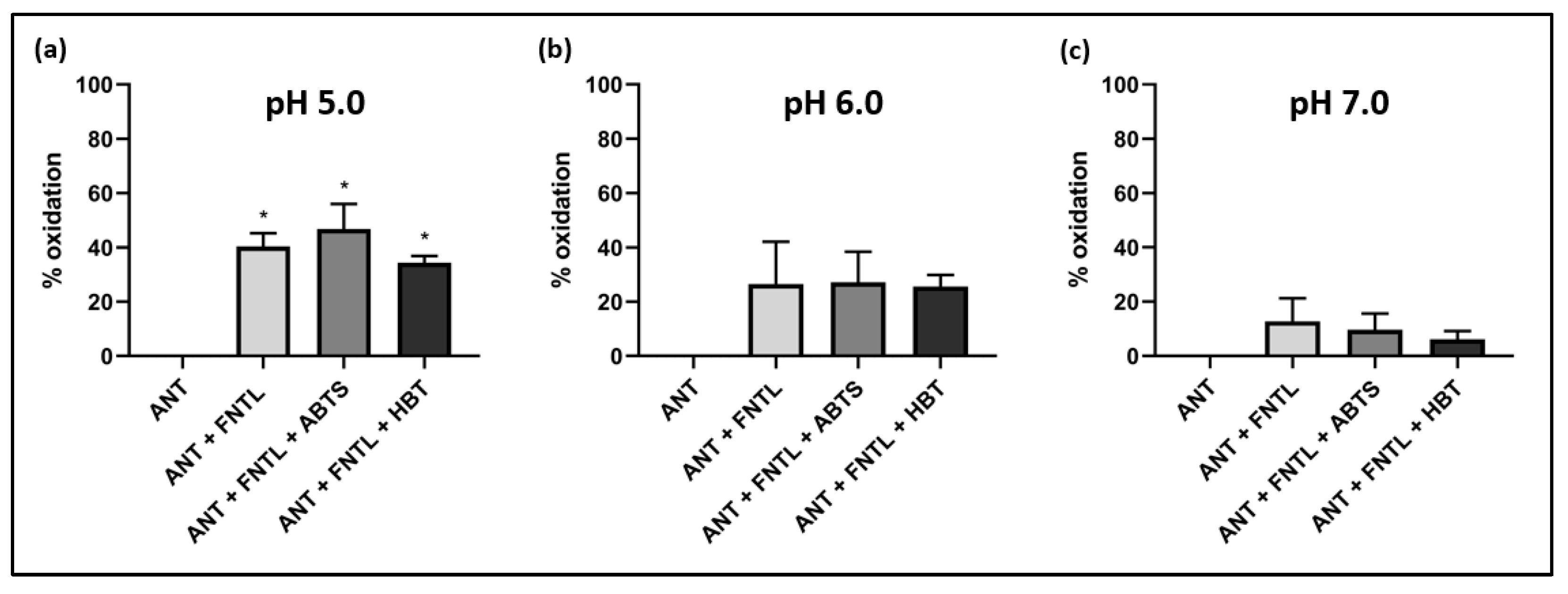
2.3.1. Evaluation of ANT Oxidation and Effect of Different pH

The oxidation of ANT was further studied, given that it showed the highest percentage of oxidation among the PAHs tested (Figure 4). When the effect of different pHs over the reaction was evaluated, the percentage of oxidation was highest at pH 5.0, reaching around 40% when ANT was exposed to FNTL alone, and near 50% in the presence of the mediator ABTS, while HBT seemed to slightly inhibit the reaction at this pH (Figure 4a). Interestingly, at pH 6.0, the reaction with the laccase alone reached around 25% oxidation, and none of the mediators used seemed to have any effect over the reaction at this pH (Figure 4b). In the case of pH 7.0, the oxidation reached its lowest percentages of oxidation (<20%), and no significant effect of the mediators was observed (Figure 4c).
The optimum pH condition for ANT degradation by FNTL is in agreement with previous studies, such as the ones with the fungal laccase from Trametes versicolor (pH 5.0) [49], or the bacterial laccases CueO from E. coli (pH 4.5) [12], CotA laccase from B. subtilis (pH 4.0) [50], and LacH5 from Bacillus atrophaeus (pH 5.0) [51].

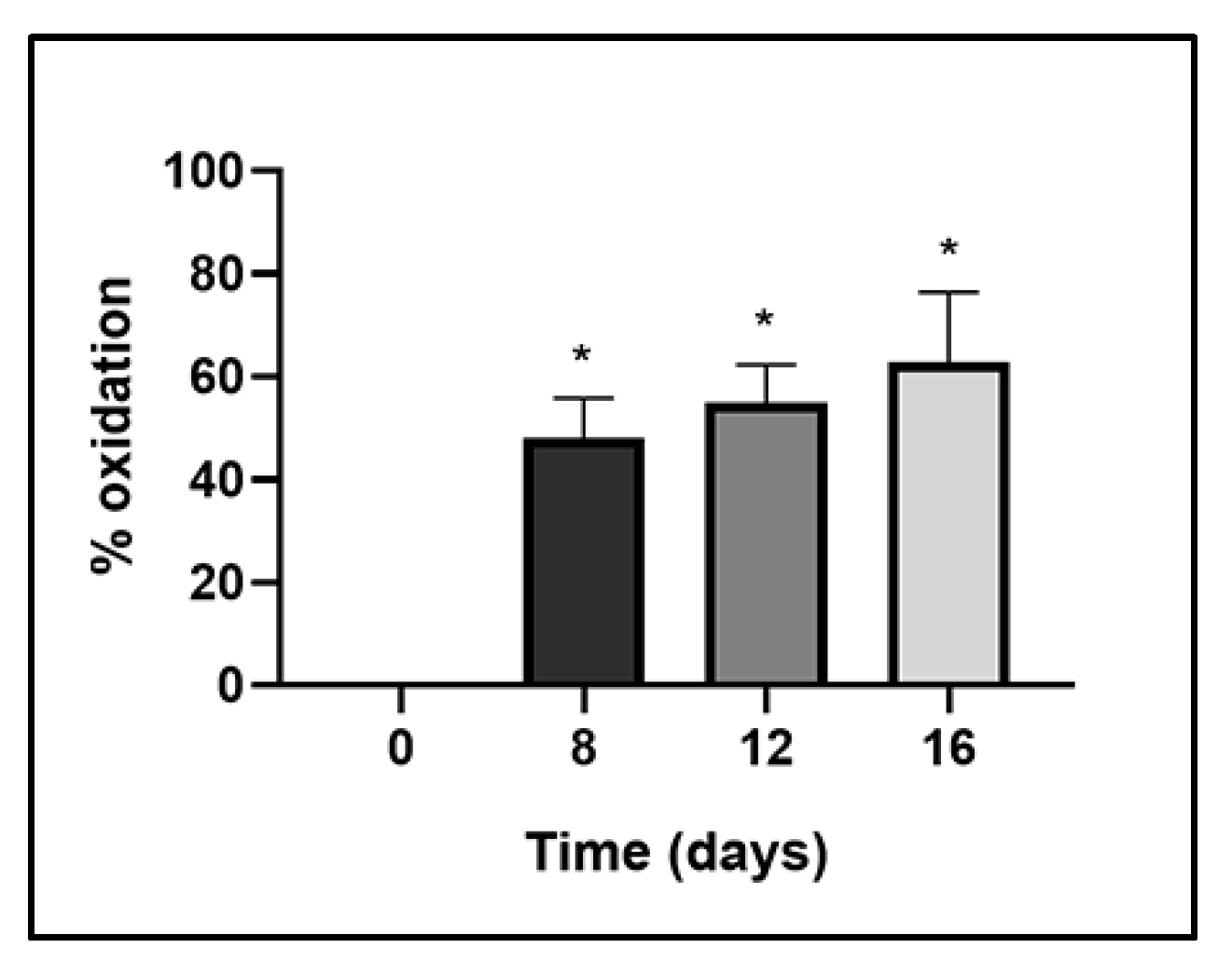
2.3.2. Evaluation of ANT Oxidation in Time

The effect of the incubation time over the oxidation of ANT by FNTL was also evaluated. When the reactions were incubated for 8, 12, and 16 days, there was a gradual increase in the oxidation, reaching over ~40%, ~50%, and ~60%, respectively (Figure 5). Suggesting this enzyme could be deemed suitable for the elimination of ANT from aqueous environments, although the nature of the obtained degradation products would have to be determined before further pursuing this enzyme as a bioremediation tool.
As previously mentioned, this thermophilic enzyme is active in a wide range of temperatures; however, at 40 °C, its activity is reported to be less than 40% of its optimum at 80 °C [35]. Therefore, ANT oxidation experiments performed at higher temperatures are expected to be more efficient, confirming the high biotechnological potential for bioremediation applications of FNTL.
It is well known that laccases have low specificity for PAHs as reducing substrates, which is in contrast to their strong preference for O2 as the oxidizing substrate [52]. Therefore, the degradation yield of PAHs could also be affected by many factors, such as the structure of the molecule, the redox potential of the laccase, the presence of detergents, or the enzyme concentration used [53]. In addition, different reaction settings could further improve PAHs degradation using this laccase. For example, reversed micellar systems have been proven to improve phenanthrene, anthracene, benzo(a)anthracene, benzo(b)fluoranthene [54], and pyrene degradation [55]. Meanwhile, laccases from T. versicolor immobilized on magnetic Fe3O4 particles coated with chitosan, demonstrated an efficient degradation of ANT and BaP in 48 h [56].
Finally, it has been shown that the products of ANT and BaP oxidation by CueO laccase from E. coli are the same that the ones obtained when using the fungal laccase from T. versicolor [12]. Quinones are the main products of PAHs oxidation, with anthraquinone been reported as the major end product of ANT oxidation [12,52,57]. This is a relatively inert compound that is not reduced by the usual reagents used for the reduction of quinones [52]. Indeed, 9,10-anthraquinone was the main product identified by HPLC separation when using CueO laccase from E. coli with 1 mM ABTS [12], and the same was found using the laccase from T. versicolor with 2 mM ABTS [49]. Furthermore, anthrone and anthraquinone were the products identified by GC-MS separation using Leucoagaricus gongylophorus laccase [58]. In the case of the BaP degradation pathway, when ABTS is present as redox mediator, laccase oxidized the substrate to benzo[a]pyrene-1,6-, 3,6-, and 6,12-quinones [12,59,60]. On the other hand, for BaA, it has been proposed that, first, the intermediate 9,10-dihydroxy benzo[a]anthracene is formed and, then, the oxidation and ring cleavage occur to form phenanthrene, being catalyzed in the same way to form diethyl phthalate. This diethyl phthalate is converted into 1,2-benzene dicarboxylic acid,1-ethylester by side-chain hydrolysis, concluding with the formation of butyric anhydride caused by ring rupture and dehydration [54]. Considering the similarity in biochemical characteristics between FNTL and other bacterial and fungal laccases previously described, it is possible to hypothesize a similar PAH degradation profile for the FNTL, although further experiments will be required to confirm its exact degradation mechanism.

3. Materials and Methods

3.1. Recombinant Expression

Escherichia coli BL21 cells (New England Biolabs, Ipswich, MA, USA) were chemically transformed using pJ444-fntlac plasmid [35]. Transformants were grown aerobically in 400 mL TBA culture medium supplemented with 2 mM CuSO4 and 30 μg mL−1 kanamycin, at 23 °C with shaking at 160 rev min−1 for 30 h [61]. The cells were harvested by centrifugation at 7690× g for 10 min at 4 °C, resuspended in a 5 mL aliquot of lysis buffer (50 mM Tris–HCl, pH 8.0) and incubated at 37 °C for 45 min with 1 mg mL−1 lysozyme. Cell disruption was carried out in ten 15 s bursts of sonication using a Branson 450 Digital Sonifier (Marshall Scientific, Hampton, NH, USA), and the cell lysate was centrifuged at 19,642× g for 30 min at 4 °C. The cell-free extract was then heat denatured at 85 °C for 5 min using a dry bath incubator (MS Major Science, Taoyuan, Taiwan). This treatment precipitate thermo sensitive native proteins from E. coli, allowing easy partial purification of the recombinant thermophilic FNTL. Finally, the soluble crude extract was obtained by centrifugation at 19,642× g for 30 min at 4 °C and and concentrated using an Amicon Ultra 15 (30 kDa MWCO) centrifugal filter device (Merck Millipore, Burlington, MA, USA), before utilization in PAH oxidation experiments.
For enzyme characterization, FNTL was further purified by anion exchange chromatography at room temperature on an ÄKTA Pure FPLC system (General Electric ™Healthcare, Boston, MA, USA) as described in our previous publication [35]. The soluble crude extract was diluted 10-fold in loading buffer (50 mM Tris-HCl, pH 8.0) and loaded onto a pre-equilibrated 25 ml Q-sepharose XK 16/20 column (General Electric ™Healthcare, Boston, MA, USA) at a flow rate of 1 mL min−1. The column was washed with 5 column volumes (CV) of the same buffer at 2 mL min−1, and the enzyme was eluted by a linear NaCl gradient (0–1 M) at a flow rate of 1 mL min−1. Fractions containing laccase activity were pooled and concentrated using an Amicon Ultra 15 (30 kDa MWCO) centrifugal filter device (Merck Millipore, Burlington, MA, USA).
Laccase overexpression was evaluated using 15% sodium dodecyl sulfate polyacrylamide gel electrophoresis (SDS-PAGE) and visualized using staining with Coomassie brilliant blue R-250 [62] (Figure S2). Protein concentration was determined using the method of Bradford [63] with the Bio-Rad protein reagent (Bio-Rad Laboratories, Irvine, CA, USA) and bovine serum albumin (BSA) as protein standard.

3.2. Enzyme Assay

Laccase activity was routinely spectrophotometrically assayed following the oxidation of syringaldazine (SGZ) substrate to tetramethoxy-azo-bis-methylene-quinone (II) at 530 nm. The assays were conducted at 70 °C in a volume of 3 mL containing 0.1 M potassium phosphate buffer pH 6.0 and 0.216 mM syringaldazine. The reaction was initiated by the addition of the substrate after preincubation of the reaction mixture at 70 °C for 2 min and all assays were conducted in triplicate.
The appearance of the product was monitored by measuring the change in absorbance at 530 nm over time using a UV/visible Spectrophotometer Shimadzu UV-1600 (Shimadzu Corporation, Kyoto, Japan). One unit (U) of laccase activity was defined as a change in absorbance at 530 nm of 0.001 per minute under the assay conditions [64].
The enzyme kinetics parameters were determined for the SGZ substrate (ε = 65,000 M−1cm−1) using a nonlinear regression, fitting the experimental data to the Michaelis–Menten equation using GraphPad Prism software (Version 8.0.2, GraphPad Software, Boston, MA, USA, 2019). The theoretical molecular mass of 59 kDa predicted with ExPASy ProtParam tool [65] from the translated fntl gene sequence was used to calculate the FNTL Turnover number (kcat).

3.3. Spectroscopic Analysis

The UV–visible absorption spectrum of pure FNTL was recorded in the range of 300–700 nm in a 50 mM potassium phosphate buffer, pH 6.0, using a UV/visible Spectrophotometer Shimadzu UV-1600 (Shimadzu Corporation, Kyoto, Japan). Preparation of the samples for the EPR analysis of the laccase was performed using purified protein with a concentration of 30 mg mL−1. For the electron paramagnetic resonance spectroscopy (EPR) spectroscopy, all manipulations of the laccase were performed under an atmosphere of N2. To prepare the EPR sample of the enzyme in a reduced form, the protein was reduced by adding sodium dithionite at a concentration 20 mM. Then, 300 µL of the reduced enzyme were injected into a quartz EPR tube and frozen under liquid N2. For the preparation of the oxidized form of laccase, the enzyme was flashed with oxygen until the protein turned totally blue. Then, 300 µL of the oxidized enzyme were injected into a quartz EPR tube and frozen under liquid N2. The EPR spectra were recorded at 8 K and a frequency of 9.45 GHz on a Brucker ER EMX 1572 spectrophotometer (Bruker, Billerica, MA, USA) equipped with a flow cryostat.

3.4. Effect of Inhibitors

The influence of different chemicals: sodium dodecyl sulfate (SDS), ethylene diamine tetra-acid (EDTA), NaCl, L-cysteine, and dithiothreitol (DTT) (all from Sigma-Aldrich, St. Louis, MO, USA) on the laccase activity was determined by incubating the enzyme in a 0.1 M potassium phosphate buffer, pH 6.0, with each potential inhibitor at a final concentration of 1 or 10 mM for 10 or 20 min at 25 °C. The assays were conducted in triplicate and controls were treated equally without inhibitors addition. Then, SGZ substrate was added to initiate the reaction and the laccase residual activity was measured under the standard conditions described in Section 3.2.

3.5. Oxidation of Polycyclic Aromatic Hydrocarbons by FNTL

The ability of FNTL enzyme to oxidize polycyclic aromatic hydrocarbons (PAHs) was evaluated using three different PAH: anthracene (ANT), benzo[a]anthracene (BaA), and benzo[a]pyrene (BaP); and three different redox mediators: 2,2′-Azino-bis (3-ethylbenzothiazoline-6-sulfonic acid) (ABTS), 1-hydroxybenzotriazole (HBT) or 3′,5′-Dimethoxy-4′-hydroxyacetophenone (AS) (all from Sigma-Aldrich, St. Louis, MO, USA).
PAH oxidation reactions were conducted in a volume of 1 mL in screw-cap glass tubes. ANT, BaA and BaP were dissolved in acetonitrile and added to a final concentration of 0.025 mg mL−1. Partially purified FNTL was added to each tube to a final protein concentration of 0.1 mg mL−1, plus 1 mM each redox mediator. The reaction tubes were tightly closed and incubated at 30 °C with shaking at 150 rev min−1 for 7 days, with enzyme refill at the 3rd day. The assays were conducted in triplicate and controls were treated equally without the addition of enzyme or mediator. After incubation, acetonitrile was added in a 3:1 ratio to stop the reactions. These were then centrifuged at 19,642× g for 20 min at 4 °C, and the supernatants were filtered through a 0.22 mm filter (Merck Millipore, Burlington, MA, USA) prior to analysis by high-efficiency liquid chromatography with a diode array detector (HPLC-DAD).
The oxidation of PAHs was determined using a Shimadzu LC-20AT HPLC-DAD (Shimadzu, Corporation, Kyoto, Japan). The separation of the analytes was carried out on a reverse phase NUCLEODUR 100-5 C18 ec column (Macherey-Nagel, Düren, Germany), with 50–100% gradient elution in 40 min, at a flow rate of 1 mL min−1, and detection at 254 nm. The PAH percentage of oxidation was calculated by comparison of peak areas of sample chromatograms with those of the controls containing only ANT, BaA or BaP, incubated under the same experimental conditions as the samples treated with the enzyme and mediators.

Effect of pH and Incubation Time on Anthracene Oxidation by FNTL

To determine the effect of pH on the oxidation of anthracene, different reactions were conducted in a volume of 1 mL containing 50 mM sodium citrate buffer for pH 5.0 and 6.0, or 50 mM of sodium phosphate buffer for pH 7.0, 0.05 mg mL−1 anthracene, 0.5 mg mL−1 partially purified FNTL and 2 mM mediator (ABTS or HBT). The reaction tubes were tightly closed and incubated at 40 °C with shaking at 150 rev min−1 for 8 days, with enzyme refill at the 3rd day. The assays were conducted in triplicate and controls were treated equally without the addition of enzyme or mediator. After the incubation, acetonitrile was added in a 3:1 ratio to stop the reactions. These were then centrifuged at 19,642× g for 20 min at 4 °C and the supernatants were filtered through a 0.22 mm filter prior to analysis by HPLC-DAD as described in Section 3.5.
The influence of the incubation time on anthracene oxidation was also evaluated. Reactions were performed in a volume of 1 mL containing 50 mM sodium citrate buffer pH 5.0, 0.05 mg mL−1 anthracene, 0.5 mg mL−1 partially purified FNTL and 2 mM ABTS. The reaction tubes were tightly closed and incubated at 40 °C with shaking at 150 rev min−1 for 8, 12 and 16 days, with enzyme refill every 4 days. The assays were conducted in triplicate and controls were treated equally without the addition of enzyme or mediator. After incubation, acetonitrile was added in a 3:1 ratio to stop the reactions. These were then centrifuged at 19,642× g for 20 min at 4 °C and the supernatants were filtered through a 0.22 mm filter prior to analysis by HPLC-DAD as described in Section 3.5.

3.6. Statistical Analysis

The statistical analysis was performed using GraphPad Prism software (Version 8.0.2, GraphPad Software, Boston, MA, USA, 2019). The results were expressed as the mean of triplicates ± standard deviation (SD). The statistical significance was determined by evaluating the p value with a 95% significance level obtained using a one-way analysis of variance (ANOVA), followed by multiple comparisons between the treatments and control group using Dunnett’s test.

4. Conclusions

The industrial implementation of bacterial laccases as biocatalysts still faces the substantial hurdle of the current scarcity of enzymes that exhibit the necessary high levels of activity, yield, and robustness. The practical utilization of laccases requires the development of highly efficient enzymes that can operate under harsh industrial conditions (e.g., elevated temperatures, pH levels, pressure, and presence of inhibitors) and can also be readily and easily produced. Owing to their intrinsic sensitivity to inhibitors, many laccases display a relatively low degree of tolerance toward various substances, which limits their industrial applicability. As a result, there is a pressing need to discover or engineer novel laccases with greater stability and enhanced performance. FNTL has demonstrated its ability to meet these expectations, while also exhibiting a high tolerance to typical laccase inhibitors and displaying high activity across a broad substrate range. These characteristics make this enzyme suitable for biotechnological applications and a good candidate for the removal of recalcitrant contaminants such as PAHs. The results obtained with FNTL show that the presence of ABTS improves the oxidation of ANT and the addition of a redox mediator is crucial for the oxidation of BaP. More interestingly, although this is a thermophilic laccase, it was still capable of catalyzing the oxidation of ANT at 30 and 40 °C (at only 20% and less than 40% of its optimum activity at 80 °C, respectively), which makes it suitable for the in situ application of contaminated waters; this is a possibility further underlined by its significant stability against denaturation and loss of activity. The latter is an absolute requisite for the practical implementation of enzymes and is met by FNTL, which is a significant finding for the future of biocatalysis. Furthermore, having a stable enzyme that is highly active toward structurally different substrates in a wide range of temperatures and conditions is a privileged starting point for protein engineering. This approach can be used to further improve its activity toward PAHs, as well as other persistent organic pollutants. Besides direct modification of the enzyme, different strategies such as reverse micellar systems could be used to enhance the PAH degradation by FNTL.

Supplementary Materials

The following supporting information can be downloaded at: https://www.mdpi.com/article/10.3390/catal13040763/s1, Figure S1: Michaelis–Menten plot of FNTL enzyme; Figure S2. Electrophoretic analysis of the heterologous overexpression and partial purification of FNTL.

Author Contributions

C.B.-N.: Conceptualization, Methodology, Investigation, Data Curation, Formal analysis and Visualization; R.C.-A.: Methodology, Investigation, Data Curation, and Formal analysis; G.E.: Conceptualization, Methodology, Formal analysis, Writing original draft, and Writing—Review and Editing; J.A.: Formal analysis, Writing original draft, and Writing—Review and Editing; J.V.: Investigation; C.A.: Investigation and Data curation; G.E.Z.: Resources and Funding acquisition; J.M.B.: Conceptualization, Resources, Supervision, Writing—Review and Editing, Funding acquisition, and Visualization. All authors have read and agreed to the published version of the manuscript.

Funding

This research has been supported by Universidad de Santiago de Chile, through funding of DICYT regular 022143BA project granted by Vicerrectoría de Investigación y Desarrollo, and also through funding by ANID CONICYT PCI NSFC190024 and Fundación Científica y Cultural Biociencia.

Data Availability Statement

Not applicable.

Acknowledgments

The authors gratefully acknowledge Professor Edwar Fuentes (University of Chile) for his expert advice, Sebastián Muñoz-Ibacache for his valuable assistance with the HPLC analysis, and Jorge Sánchez San Martín for his help with the enzyme assays.

Conflicts of Interest

The authors declare no conflict of interest. The funders had no role in the design of the study; in the collection, analyses, or interpretation of data; in the writing of the manuscript; or in the decision to publish the results.

References

  1. Arregui, L.; Ayala, M.; Gómez-Gil, X.; Gutiérrez-Soto, G.; Hernández-Luna, C.E.; Herrera De Los Santos, M.; Levin, L.; Rojo-Domínguez, A.; Romero-Martínez, D.; Saparrat, M.C.N.; et al. Laccases: Structure, Function, and Potential Application in Water Bioremediation. Microb. Cell Factories 2019, 18, 1–33. [Google Scholar] [CrossRef] [PubMed]
  2. Alcalde, M. Laccases: Biological Functions, Molecular Structure and Industrial Applications. In Industrial Enzymes Structure, Function and Applications; Polaina, J., MacCabe, A.P., Eds.; Springer: Dordrecht, The Netherlands, 2007; pp. 461–476. [Google Scholar] [CrossRef]
  3. Singh, G.; Arya, S.K. Utility of Laccase in Pulp and Paper Industry: A Progressive Step towards the Green Technology. Int. J. Biol. Macromol. 2019, 134, 1070–1084. [Google Scholar] [CrossRef] [PubMed]
  4. Unuofin, J.O.; Falade, A.O.; Aladekoyi, O.J. Applications of Microbial Laccases in Bioremediation of Environmental Pollutants: Potential Issues, Challenges, and Prospects. In Bioremediation for Environmental Sustainability; Elsevier: Amsterdam, The Netherlands, 2021; pp. 519–540. [Google Scholar] [CrossRef]
  5. Viswanath, B.; Rajesh, B.; Janardhan, A.; Kumar, A.P.; Narasimha, G. Fungal Laccases and Their Applications in Bioremediation. Enzyme Res. 2014, 2014, 163242. [Google Scholar] [CrossRef] [PubMed]
  6. Kudanga, T.; le Roes-Hill, M. Laccase Applications in Biofuels Production: Current Status and Future Prospects. Appl. Microbiol. Biotechnol. 2014, 98, 6525–6542. [Google Scholar] [CrossRef]
  7. Guan, Z.B.; Luo, Q.; Wang, H.R.; Chen, Y.; Liao, X.R. Bacterial Laccases: Promising Biological Green Tools for Industrial Applications. Cell. Mol. Life Sci. 2018, 75, 3569–3592. [Google Scholar] [CrossRef]
  8. Loi, M.; Glazunova, O.; Fedorova, T.; Logrieco, A.F.; Mulè, G. Fungal Laccases: The Forefront of Enzymes for Sustainability. J. Fungi 2021, 7, 1048. [Google Scholar] [CrossRef]
  9. Alegbeleye, O.O.; Opeolu, B.O.; Jackson, V.A. Polycyclic Aromatic Hydrocarbons: A Critical Review of Environmental Occurrence and Bioremediation. Environ. Manag. 2017, 60, 758–783. [Google Scholar] [CrossRef]
  10. Kuśmierz, M.; Oleszczuk, P.; Kraska, P.; Pałys, E.; Andruszczak, S. Persistence of Polycyclic Aromatic Hydrocarbons (PAHs) in Biochar-Amended Soil. Chemosphere 2016, 146, 272–279. [Google Scholar] [CrossRef]
  11. Punnapayak, H.; Prasongsuk, S.; Messner, K.; Danmek, K.; Lotrakul, P. Polycyclic Aromatic Hydrocarbons (PAHs) Degradation by Laccase from a Tropical White Rot Fungus Ganoderma Lucidum. Afr. J. Biotechnol. 2009, 8, 5897–5900. [Google Scholar] [CrossRef]
  12. Zeng, J.; Lin, X.; Zhang, J.; Li, X.; Wong, M.H. Oxidation of Polycyclic Aromatic Hydrocarbons by the Bacterial Laccase CueO from E. coli. Appl. Microbiol. Biotechnol. 2011, 89, 1841–1849. [Google Scholar] [CrossRef]
  13. Sharma, N.; Leung, I.K.H. Novel Thermophilic Bacterial Laccase for the Degradation of Aromatic Organic Pollutants. Front. Chem. 2021, 9, 711345. [Google Scholar] [CrossRef]
  14. Bai, Y.; Ali, S.; Liu, S.; Zhou, J.; Tang, Y. Characterization of Plant Laccase Genes and Their Functions. Gene 2023, 852, 147060. [Google Scholar] [CrossRef]
  15. Laufer, Z.; Beckett, R.P.; Minibayeva, F.V.; Luthje, S.; Bottger, M. Diversity of Laccases from Lichens in Suborder Peltigerineae. Bryologist 2009, 112, 418–426. [Google Scholar] [CrossRef]
  16. Li, Q.; Wang, X.; Korzhev, M.; Schröder, H.C.; Link, T.; Tahir, M.N.; Diehl-Seifert, B.; Müller, W.E.G. Potential Biological Role of Laccase from the Sponge Suberites Domuncula as an Antibacterial Defense Component. Biochim. Biophys. Acta 2015, 1850, 118–128. [Google Scholar] [CrossRef]
  17. Asano, T.; Seto, Y.; Hashimoto, K.; Kurushima, H. Mini-Review an Insect-Specific System for Terrestrialization: Laccase-Mediated Cuticle Formation. Insect Biochem. Mol. Biol. 2019, 108, 61–70. [Google Scholar] [CrossRef]
  18. Akram, F.; Ashraf, S.; Haq, I.U.; Shah, F.I.; Aqeel, A. Eminent Industrial and Biotechnological Applications of Laccases from Bacterial Source: A Current Overview. Appl. Biochem. Biotechnol. 2022, 194, 2336–2356. [Google Scholar] [CrossRef]
  19. Chauhan, P.S.; Goradia, B.; Saxena, A. Bacterial Laccase: Recent Update on Production, Properties and Industrial Applications. 3 Biotech 2017, 7, 323. [Google Scholar] [CrossRef]
  20. Liu, H.; Cheng, Y.; Du, B.; Tong, C.; Liang, S.; Han, S.; Zheng, S.; Lin, Y. Overexpression of a Novel Thermostable and Chloride-Tolerant Laccase from Thermus Thermophilus SG0.5JP17-16 in Pichia Pastoris and Its Application in Synthetic Dye Decolorization. PLoS ONE 2015, 10, e0119833. [Google Scholar] [CrossRef]
  21. Wang, T.N.; Zhao, M. A Simple Strategy for Extracellular Production of CotA Laccase in Escherichia coli and Decolorization of Simulated Textile Effluent by Recombinant Laccase. Appl. Microbiol. Biotechnol. 2017, 101, 685–696. [Google Scholar] [CrossRef]
  22. Fang, Z.; Li, T.; Wang, Q.; Zhang, X.; Peng, H.; Fang, W.; Hong, Y.; Ge, H.; Xiao, Y. A Bacterial Laccase from Marine Microbial Metagenome Exhibiting Chloride Tolerance and Dye Decolorization Ability. Appl. Microbiol. Biotechnol. 2011, 89, 1103–1110. [Google Scholar] [CrossRef]
  23. Guan, Z.B.; Shui, Y.; Song, C.M.; Zhang, N.; Cai, Y.J.; Liao, X.R. Efficient Secretory Production of CotA-Laccase and Its Application in the Decolorization and Detoxification of Industrial Textile Wastewater. Environ. Sci. Pollut. Res. Int. 2015, 22, 9515–9523. [Google Scholar] [CrossRef] [PubMed]
  24. Enguita, F.J.; Martins, L.O.; Henriques, A.O.; Carrondo, M.A. Crystal Structure of a Bacterial Endospore Coat Component: A Laccase with Enhanced Thermostability Properties. J. Biol. Chem. 2003, 278, 19416–19425. [Google Scholar] [CrossRef] [PubMed]
  25. Prins, A.; Kleinsmidt, L.; Khan, N.; Kirby, B.; Kudanga, T.; Vollmer, J.; Pleiss, J.; Burton, S.; le Roes-Hill, M. The Effect of Mutations near the T1 Copper Site on the Biochemical Characteristics of the Small Laccase from Streptomyces coelicolor A3(2). Enzyme Microb. Technol. 2015, 68, 23–32. [Google Scholar] [CrossRef] [PubMed]
  26. Basheer, S.; Rashid, N.; Ashraf, R.; Akram, M.S.; Siddiqui, M.A.; Imanaka, T.; Akhtar, M. Identification of a Novel Copper-Activated and Halide-Tolerant Laccase in Geobacillus thermopakistaniensis. Extremophiles 2017, 21, 563–571. [Google Scholar] [CrossRef] [PubMed]
  27. Brander, S.; Mikkelsen, J.D.; Kepp, K.P. TtMCO: A highly thermostable laccase-like multicopper oxidase from the thermophilic Thermobaculum terrenum. J. Mol. Catal. 2015, 112, 59–65. [Google Scholar] [CrossRef]
  28. Chen, C.Y.; Huang, Y.C.; Wei, C.M.; Meng, M.; Liu, W.H.; Yang, C.H. Properties of the newly isolated extracellular thermo-alkali-stable laccase from thermophilic actinomycetes, Thermobifida fusca and its application in dye intermediates oxidation. AMB Express 2013, 3, 49. [Google Scholar] [CrossRef]
  29. Miyazaki, K. A Hyperthermophilic Laccase from Thermus thermophilus HB27. Extremophiles 2005, 9, 415–425. [Google Scholar] [CrossRef]
  30. Navas, L.E.; Martínez, F.D.; Taverna, M.E.; Fetherolf, M.M.; Eltis, L.D.; Nicolau, V.; Estenoz, D.; Campos, E.; Benintende, G.B.; Berretta, M.F. A Thermostable Laccase from Thermus sp. 2.9 and Its Potential for Delignification of Eucalyptus Biomass. AMB Express 2019, 9, 24. [Google Scholar] [CrossRef]
  31. Verma, A.; Shirkot, P. Purification and Characterization of Thermostable Laccase from Thermophilic Geobacillus thermocatenulatus MS5 and Its Application in Removal of Textiles Dyes. Sch. Acad. J. Biosci. 2014, 2, 479–485. [Google Scholar]
  32. Rai, R.; Bibra, M.; Chadha, B.S.; Sani, R.K. Enhanced Hydrolysis of Lignocellulosic Biomass with Doping of a Highly Thermostable Recombinant Laccase. Int. J. Biol. Macromol. 2019, 137, 232–237. [Google Scholar] [CrossRef]
  33. Tripathy, S.; Maiti, N.K. Construction of Geobacillus thermoglucosidasius cDNA Library and Analysis of Genes Expressed in Response to Heat Stress. Mol. Biol. Rep. 2014, 41, 1639–1644. [Google Scholar] [CrossRef]
  34. Rosano, G.L.; Ceccarelli, E.A. Recombinant Protein Expression in Escherichia coli: Advances and Challenges. Front. Microbiol. 2014, 5, 172. [Google Scholar] [CrossRef]
  35. Espina, G.; Cáceres-Moreno, P.; Mejías-Navarrete, G.; Ji, M.; Sun, J.; Blamey, J.M. A Novel and Highly Active Recombinant Spore-Coat Bacterial Laccase, Able to Rapidly Biodecolorize Azo, Triarylmethane and Anthraquinonic Dyestuffs. Int. J. Biol. Macromol. 2021, 170, 298–306. [Google Scholar] [CrossRef]
  36. Jones, S.M.; Solomon, E.I. Electron Transfer and Reaction Mechanism of Laccases. Cell. Mol Life Sci. 2015, 72, 869–883. [Google Scholar] [CrossRef]
  37. Dedeyan, B.; Klonowska, A.; Tagger, S.; Tron, T.; Iacazio, G.; Gil, G.; Le Petit, J. Biochemical and Molecular Characterization of a Laccase from Marasmius quercophilus. Appl. Environ. Microbiol. 2000, 66, 925–929. [Google Scholar] [CrossRef]
  38. Koschorreck, K.; Richter, S.M.; Ene, A.B.; Roduner, E.; Schmid, R.D.; Urlacher, V.B. Cloning and Characterization of a New Laccase from Bacillus licheniformis Catalyzing Dimerization of Phenolic Acids. Appl. Microbiol. Biotechnol. 2008, 79, 217–224. [Google Scholar] [CrossRef]
  39. Siroosi, M.; Amoozegar, M.A.; Khajeh, K. Purification and Characterization of an Alkaline Chloride-Tolerant Laccase from a Halotolerant Bacterium, Bacillus sp. strain WT. J. Mol. Catal. 2016, 134, 89–97. [Google Scholar] [CrossRef]
  40. Couto, S.; Herrera, L. Inhibitors of Laccases: A Review. Curr. Enzym. Inhib. 2006, 2, 343–352. [Google Scholar] [CrossRef]
  41. Sharma, B.; Dangi, A.K.; Shukla, P. Contemporary Enzyme Based Technologies for Bioremediation: A Review. J. Environ. Manag. 2018, 210, 10–22. [Google Scholar] [CrossRef]
  42. Brander, S.; Mikkelsen, J.D.; Kepp, K.P. Characterization of an Alkali- and Halide-Resistant Laccase Expressed in E. coli: CotA from Bacillus clausii. PLoS ONE 2014, 9, e99402. [Google Scholar] [CrossRef]
  43. Lu, L.; Wang, T.N.; Xu, T.F.; Wang, J.Y.; Wang, C.L.; Zhao, M. Cloning and Expression of Thermo-Alkali-Stable Laccase of Bacillus licheniformis in Pichia pastoris and Its Characterization. Bioresour. Technol. 2013, 134, 81–86. [Google Scholar] [CrossRef] [PubMed]
  44. Li, X.; Wei, Z.; Zhang, M.; Peng, X.; Yu, G.; Teng, M.; Gong, W. Crystal Structures of E. coli Laccase CueO at Different Copper Concentrations. Biochem. Biophys. Res. Commun. 2007, 354, 21–26. [Google Scholar] [CrossRef] [PubMed]
  45. Johannes, C.; Majcherczyk, A. Laccase Activity Tests and Laccase Inhibitors. J. Biotechnol. 2000, 78, 193–199. [Google Scholar] [CrossRef] [PubMed]
  46. Zhang, C.; Zhang, S.; Diao, H.; Zhao, H.; Zhu, X.; Lu, F.; Lu, Z. Purification and characterization of a temperature-and pH-stable laccase from the spores of Bacillus vallismortis fmb-103 and its application in the degradation of malachite green. J. Agric. Food Chem. 2013, 61, 5468–5473. [Google Scholar] [CrossRef]
  47. Wang, C.; Zhao, M.; Lu, L.; Wei, X.; Li, T. Characterization of spore laccase from Bacillus subtilis WD23 and its use in dye decolorization. Afr. J. Biotechnol. 2011, 10, 2186–2192. [Google Scholar]
  48. Durão, P.; Chen, Z.; Fernandes, A.T.; Hildebrandt, P.; Murgida, D.H.; Todorovic, S.; Pereira, M.M.; Melo, E.P.; Martins, L.O. Copper Incorporation into Recombinant CotA Laccase from Bacillus subtilis: Characterization of Fully Copper Loaded Enzymes. J. Biol. Inorg. Chem. 2008, 13, 183–193. [Google Scholar] [CrossRef]
  49. Johannes, C.; Majcherczyk, A.; Hüttermann, A. Degradation of Anthracene by Laccase of Trametes versicolor in the Presence of Different Mediator Compounds. Appl. Microbiol. Biotechnol. 1996, 46, 313–317. [Google Scholar] [CrossRef]
  50. Zeng, J.; Zhu, Q.; Wu, Y.; Lin, X. Oxidation of Polycyclic Aromatic Hydrocarbons Using Bacillus subtilis CotA with High Laccase Activity and Copper Independence. Chemosphere 2016, 148, 1–7. [Google Scholar] [CrossRef]
  51. Wang, L.; Tan, Y.; Sun, S.; Zhou, L.; Wu, G.; Shao, Y.; Wang, M.; Xin, Z. Improving Degradation of Polycyclic Aromatic Hydrocarbons by Bacillus atrophaeus Laccase Fused with Vitreoscilla Hemoglobin and a Novel Strong Promoter Replacement. Biology 2022, 11, 1129. [Google Scholar] [CrossRef]
  52. Alcalde, M.; Bulter, T.; Arnold, F.H. Colorimetric Assays for Biodegradation of Polycyclic Aromatic Hydrocarbons by Fungal Laccases. J. Biomol. Screen. 2002, 7, 547–553. [Google Scholar] [CrossRef]
  53. Pozdnyakova, N.N.; Rodakiewicz-Nowak, J.; Turkovskaya, O.V.; Haber, J. Oxidative Degradation of Polyaromatic Hydrocarbons Catalyzed by Blue Laccase from Pleurotus ostreatus D1 in the Presence of Synthetic Mediators. Enzyme Microb. Technol. 2006, 39, 1242–1249. [Google Scholar] [CrossRef]
  54. Xu, P.; Du, H.; Peng, X.; Tang, Y.; Zhou, Y.; Chen, X.; Fei, J.; Meng, Y.; Yuan, L. Degradation of Several Polycyclic Aromatic Hydrocarbons by Laccase in Reverse Micelle System. Sci. Total Environ. 2020, 708, 134970. [Google Scholar] [CrossRef]
  55. Peng, X.; Yuan, X.Z.; Liu, H.; Zeng, G.M.; Chen, X.H. Degradation of Polycyclic Aromatic Hydrocarbons (PAHs) by Laccase in Rhamnolipid Reversed Micellar System. Appl. Biochem. Biotechnol. 2015, 176, 45–55. [Google Scholar] [CrossRef]
  56. Deng, J.; Wang, H.; Zhan, H.; Wu, C.; Huang, Y.; Yang, B.; Mosa, A.; Ling, W. Catalyzed Degradation of Polycyclic Aromatic Hydrocarbons by Recoverable Magnetic Chitosan Immobilized Laccase from Trametes versicolor. Chemosphere 2022, 301, 134753. [Google Scholar] [CrossRef]
  57. Cañas, A.I.; Alcalde, M.; Plou, F.; Martínez, M.J.; Martínez, Á.T.; Camarero, S. Transformation of Polycyclic Aromatic Hydrocarbons by Laccase Is Strongly Enhanced by Phenolic Compounds Present in Soil. Environ. Sci. Technol 2007, 41, 2964–2971. [Google Scholar] [CrossRef]
  58. Ike, P.T.L.; Birolli, W.G.; dos Santos, D.M.; Porto, A.L.M.; Souza, D.H.F. Biodegradation of Anthracene and Different PAHs by a Yellow Laccase from Leucoagaricus gongylophorus. Environ. Sci. Pollut. Res. Int. 2019, 26, 8675–8684. [Google Scholar] [CrossRef]
  59. Hu, X.; Zhang, Y.; Zhao, X.; Hwang, H.M. Biodegradation of Benzo[a]Pyrene with Immobilized Laccase: Genotoxicity of the Products in HaCaT and A3 Cells. Environ. Mol. Mutagen 2007, 48, 106–113. [Google Scholar] [CrossRef]
  60. Kadri, T.; Rouissi, T.; Kaur Brar, S.; Cledon, M.; Sarma, S.; Verma, M. Biodegradation of Polycyclic Aromatic Hydrocarbons (PAHs) by Fungal Enzymes: A Review. J. Environ. Sci. 2017, 51, 52–74. [Google Scholar] [CrossRef]
  61. Espina, G.; Muñoz-Ibacache, S.A.; Cáceres-Moreno, P.; Amenabar, M.J.; Blamey, J.M. From the Discovery of Extremozymes to an Enzymatic Product: Roadmap Based on Their Applications. Front. Bioeng. Biotechnol. 2022, 9, 1271. [Google Scholar] [CrossRef]
  62. Gallagher, S.; Sasse, J. Protein Analysis by SDS-PAGE and Detection by Coomassie Blue or Silver Staining. Curr. Protoc. Pharmacol. 2001, 2, A.3B.1–A.3B.10. [Google Scholar] [CrossRef]
  63. Bradford, M. A Rapid and Sensitive Method for the Quantitation of Microgram Quantities of Protein Utilizing the Principle of Protein-Dye Binding. Anal. Biochem. 1976, 72, 248–254. [Google Scholar] [CrossRef] [PubMed]
  64. Lante, A.; Crapisi, A.; Krastanov, A.; Spettoli, P. Biodegradation of Phenols by Laccase Immobilised in a Membrane Reactor. Process Biochem. 2000, 36, 51–58. [Google Scholar] [CrossRef]
  65. Gasteiger, E.; Hoogland, C.; Gattiker, A.; Duvaud, S.; Wilkins, M.R.; Appel, R.D.; Bairoch, A. Protein Identification and Analysis Tools on the ExPASy Server. In The Proteomics Protocols Handbook; Walker, J.M., Ed.; Humana Press: New York, NY, USA, 2005; pp. 571–607. [Google Scholar] [CrossRef]
Figure 1. (a) UV-visible absorption spectrum of purified FTNL. (b) EPR spectrum of the purified laccase.
Catalysts 13 00763 g001
Figure 2. Effect of different inhibitors on FNTL activity. SDS, NaCl, EDTA, DTT, and L-cysteine were tested at concentrations of 1 and 10 mM. Recombinant laccase activity was measured after 10 and 20 min of incubation with each inhibitor. Each bar represents the mean of three independent measurements. The significant differences were determined using one-way ANOVA, followed by multiple comparisons between the treatments and control using Dunnett’s test, indicated as * = p < 0.05.
Catalysts 13 00763 g002
Figure 3. Oxidation of different PAHs in the presence of FNTL. (a) ANT, (b) BaA, and (c) BaP, each at 0.025 mg mL−1, were exposed to partially purified FNTL (0.1 mg mL−1) in aqueous solution. The effect of 1 mM redox mediators ABTS and AS, was also evaluated. Reactions were incubated for 7 days at 30 °C with shaking at 150 rev min−1, with enzyme refill at the 3rd day.
Catalysts 13 00763 g003
Figure 4. Oxidation of anthracene in the presence of FNTL at three different pH. (a) pH 5.0, (b) pH 6.0, and (c) pH 7.0. Anthracene (0.05 mg mL−1) was exposed to partially purified FNTL (0.5 mg mL−1) and 2 mM redox mediator molecules ABTS and AS. Reactions were incubated for 8 days at 40 °C with shaking at 150 rev min−1, with enzyme refill at the 3rd day. The significant differences were determined using one-way ANOVA, followed by multiple comparisons between the treatments and control using Dunnett’s test, indicated as * = p < 0.05.
Catalysts 13 00763 g004
Figure 5. Influence of the incubation time on the oxidation of anthracene by FNTL. Anthracene (0.05 mg mL−1) was exposed to partially purified FNTL (0.5 mg mL−1) and 2 mM ABTS in 50 mM sodium citrate buffer pH 5.0. Reactions were incubated for 8, 12 and 16 days at 40 °C with shaking at 150 rev min−1, with enzyme refill every 4 days. The significant differences were determined using one-way ANOVA, followed by multiple comparisons between the treatments and control using Dunnett’s test, indicated as * = p < 0.05.
Catalysts 13 00763 g005
Disclaimer/Publisher’s Note: The statements, opinions and data contained in all publications are solely those of the individual author(s) and contributor(s) and not of MDPI and/or the editor(s). MDPI and/or the editor(s) disclaim responsibility for any injury to people or property resulting from any ideas, methods, instructions or products referred to in the content.

