Article

Coupling the Piezoelectric Effect and the Plasmonic Effect to Enhance the Photocatalytic Degradation of Ciprofloxacin in Au-Ferroelectric Bi4Ti3O12 Nanofibers

1
State Key Laboratory of NBC Protection for Civilian, Beijing 102205, China
2
School of Materials Design & Engineering, Beijing Institute of Fashion Technology, Beijing 100029, China
3
School of Chemistry and Chemical Engineering, Jiangxi University of Science and Technology, Ganzhou 341000, China
4
Institute of Photonics Technology, Jinan University, Guangzhou 511443, China
*
Authors to whom correspondence should be addressed.
Catalysts 2023, 13(3), 621; https://doi.org/10.3390/catal13030621
Submission received: 24 February 2023 / Revised: 13 March 2023 / Accepted: 15 March 2023 / Published: 20 March 2023

Abstract

:
In this study, ferroelectric Bi4Ti3O12 and Au-Bi4Ti3O12 nanofibers were synthesized by electrospinning and ion sputtering. The piezoelectric effect of Bi4Ti3O12 and the surface plasmon effect of Au were used to improve the photogenerated electron–hole separation and optical absorption. The results of the characterization showed successful preparation of the orthorhombic Bi4Ti3O12 nanofibers, in which the absorption band edge was 426 nm with a 2.91 eV band gap. The piezo-photocatalytic activity of the Bi4Ti3O12 was tested through the degradation of the antibiotic ciprofloxacin under three different experimental conditions: light, vibration, and light plus vibration. All of the ciprofloxacin was degraded after 80 min in piezo-photocatalytic conditions, with a piezo-photocatalytic degradation rate of 0.03141 min−1, which is 1.56 and 3.88 times, respectively, that of photocatalysis and piezo-catalysis. After loading Au on the Bi4Ti3O12, the degradation efficiency was improved under all three conditions, and the piezoelectric photocatalytic efficiency of Au-Bi4Ti3O12 for ciprofloxacin degradation was able to reach 100% in 60 min with a piezo-photocatalytic degradation rate of 0.06157 min−1. The results of the photocurrent and impedance tests indicated that the photocurrent density of Bi4Ti3O12 nanofibers loaded with Au is increased from 5.08 × 10⁻⁷ A/cm2 to 8.17 × 10⁻⁶ A/cm2, which is 16.08 times higher than without loading the Au. This work provides an effective way to improve the conversion efficiency of photocatalysis to degrade organic pollutants by combining the plasmon effect and the piezoelectric effect.

1. Introduction

According to research, China is the world’s largest producer and user of antibiotics. However, the abuse of antibiotics can make some pathogenic bacteria resistant, and their long-term survival in the environment would be a threat to human health and the security of the ecological environment. Avoiding the abuse of antibiotics and the degradation of antibiotics in rivers has become our research focus. Ciprofloxacin (CIP) is the third generation of synthetic quinolone antibacterial drugs, which have broad-spectrum antibacterial activity and good bactericidal effects [1,2]. It is widely used in animal husbandry to prevent and treat diseases. However, CIP is widely exposed to water ecosystems, and even at very low concentrations, it can cause a serious threat to human and ecological environments. Traditional physical separation technology, biological methods and other water treatment technologies cannot effectively remove these antibiotics [3,4]. Therefore, it is a challenge to develop effective and environment-friendly technologies for degrading antibiotics [5]. The emergence of photocatalytic technologies has brought hope to this urgent problem due to their environment-friendly nature and cost effectiveness [6,7]. Photocatalysis mainly involves the following processes: (1) a semiconductor is excited by photons with an energy greater than its band gap in order to generate electrons and holes [8,9,10]; (2) photogenerated electrons and holes are separated and migrated to the surface effectively [11,12,13,14]; (3) a surface redox reaction occurs to degrade the antibiotics [15,16,17]. However, for common semiconductor photocatalysts, the photogenerated electrons and holes are prone to recombination during the migration process and cannot generate highly reactive radicals. As a result, the catalytic activity and efficiency of the photocatalyst are greatly reduced.
Over the last few years, the piezoelectric polarization-based method known as piezo-catalysis has undergone rapid improvement in the domain of environmental mitigation and production of fuels [18,19,20]. On the application of mechanical force, the polarization of a piezoelectric semiconductor takes place, leading to the accumulation of oppositely charged localized polarizations on opposite surfaces. Consequently, the free charge carriers (electrons and holes) are separated after being driven through the built-in piezoelectric field, thereby allowing their participation in the surface redox reactions. Up to now, many piezoelectric materials have been demonstrated to be promising for application in the piezo-phototronic degradation of environmental pollutants [21,22,23]. However, a large proportion of piezoelectrical materials have poor electrical conductivity, which seriously restricts the transport of the charge carriers across the interface formed with other active materials.
Bi4Ti3O12 has a unique, layered structure with perovskite-like (Bi2Ti3O10)2− units alternating with (Bi2O2)2+ layers [24]. The (Bi2O2)2+ layers contain many oxygen vacancies, which can expand the wavelength range of light absorption. In addition, (Bi2O2)2+ and (Bi2Ti3O10)2− layers can form an inner electrical field [25], which could promote the separation of photo-generated electrons and holes. Its unique crystal and electronic structures endow Bi4Ti3O12 with excellent ferroelectricity and piezoelectricity, fatigue resistance and a high Curie temperature. Therefore, Bi4Ti3O12 can be used as an ideal material for piezo-phototronic degradation of organic pollutants.
It is generally accepted that the performance of different materials depends highly on their morphologies and microstructures [26,27]. Bi4Ti3O12 nanofibers have large specific surface areas, high porosity, small volumes, light weights, good flexibility and bending properties. The piezoelectric properties of continuous-filament nanofibers are better than those of granular and blocky ones. Moreover, the introduction of a noble metal, such as gold (Au), on the surface of Bi4Ti3O12 can not only help improve the absorbing efficiency of the light, but also enhance the separation efficiency of photogenerated electrons and holes due to the resonant oscillation of the surface plasmon effect [28,29].
In order to solve the problem of CIP release and to avoid rapid recombination between the electrons and holes of Bi4Ti3O12, we synthesized Bi4Ti3O12 nanofibers using the electrospinning method and then investigated the catalytic degradation of ciprofloxacin under different conditions to verify the photocatalytic properties of Bi4Ti3O12 nanofibers. To enhance the photocatalytic activity in the degradation of the substrate and explore the internal mechanism of photocatalysis, Au-Bi4Ti3O12 material was prepared. The results show that the catalytic effect and light absorption were all enhanced due to the local surface plasmon effect after loading Au on the Bi4Ti3O12, which is of great significance for improving the photocatalytic effects and studying the mechanism of the degradation. This work introduces the synergistic effects of SPR and piezotronic effects to promote the separation of electron–hole pairs and improve the piezo-photocatalytic degradation of ciprofloxacin using Au-Bi4Ti3O12 nanofiber heterostructures, which offer an efficient technology to design high-performance catalysts for environmental purification.

2. Results and Discussion

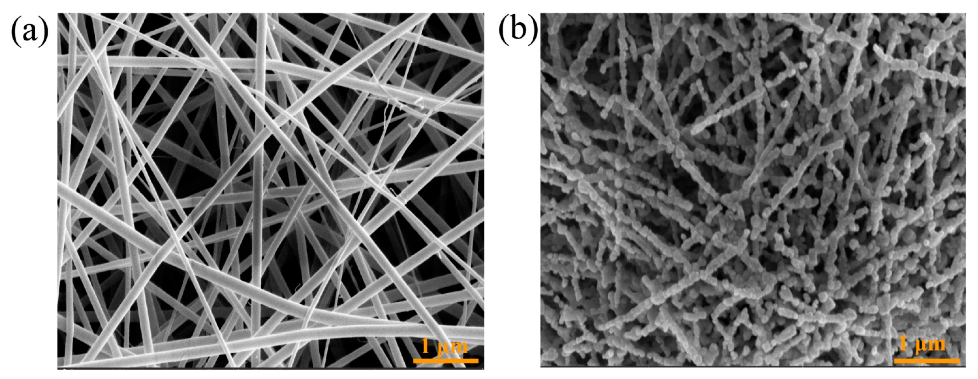
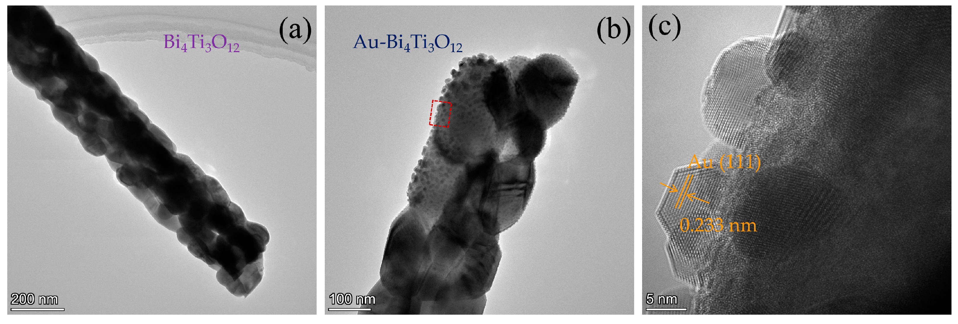
2.1. Characterization of the Morphology

The SEM images of Bi4Ti3O12 nanofibers before calcining and after calcining are shown in Figure 1. The SEM image of the dried Bi4Ti3O12 precursor without calcination after electrospinning shows that the nanofibers are interlaced (Figure 1a), which indicates that the electrospinning was in good condition. The SEM image presented in Figure 1b shows that the Bi4Ti3O12 still maintains a fibrous structure after calcining, which indicates that only the PVP was removed, and the structure of the Bi4Ti3O12 nanofibers was not affected by calcining. However, when the PVP was removed in the calcination process, the surface of the Bi4Ti3O12 was no longer smooth and became a porous nanofiber structure with a diameter of about 60–80 nm.
The TEM and HRTEM images were measured to further investigate the microstructure of the Bi4Ti3O12 nanofibers and the Au-Bi4Ti3O12 nanofibers. The low-magnification TEM image of the Bi4Ti3O12 shows nanofibers inlaid with particles (Figure 2a). Meanwhile, the TEM image of the Au-Bi4Ti3O12 (Figure 2b) shows that the nanofibers are made up of many Au particles, with sizes ranging from 5 to 15 nm. The HRTEM image (Figure 2c) shows a lattice spacing of 0.233 nm, corresponding to the (111) planes of Au and proving that the Au nanoparticles were successfully embedded in the Bi4Ti3O12 nanofiber [30].

2.2. Characterization of the Crystal Structure

The phase structures of the Bi4Ti3O12 nanofibers and the Au-Bi4Ti3O12 nanofibers were studied using X-ray diffraction. As shown in Figure 3a, all of the diffraction peaks of the Bi4Ti3O12 and Au-Bi4Ti3O12 nanofibers were in accord with the standards of the database card. The characteristic peaks located at 2θ equaled 23.0°, 29.9° and 32.7° and corresponded to crystal planes (111), (117) and (020), respectively. The peak shapes demonstrated that the prepared sample had good crystallinity. Other diffraction peaks were located at 21.35°, 38.1°, 39.7°, 47.0°, 51.35°, 53.13°, 56.8° and 62.2° corresponded to crystal facets (008), (0014), (208), (202), (262), (0214), (137) and (2214), respectively. These were all part of the orthorhombic crystalline phase of Bi4Ti3O12 (JCPDS 72-1019) [24]. The orthogonal crystal Bi4Ti3O12 was described by the general formula of (Bi2O2)2+(Bi2Ti3O10)2−, which is composed of three layers of TiO6 octahedron and (Bi2O2)2+ monolayers that are alternately stacked, so that its structure cannot be easily changed. The average crystallite size of Bi4Ti3O12 was calculated by using the modified Scherer method [31]:
In β = In ( k λ D SM cos θ )
where DSM is the average crystallite size, k is a constant (equal to 0.9), β is the line broadening in radians (FWHM), θ is the Bragg angle and λ is the X-ray wavelength (λ = 0.1546 nm). The calculated crystallite size value was 18.53 nm. No typical diffraction peaks of Au were detected in the Au-Bi4Ti3O12. This is because the amount of Au was too small (less than 5%) and could not be detected by the X-ray diffraction.
Raman spectra of the Bi4Ti3O12 and Au-Bi4Ti3O12 nanofibers are shown in Figure 3b, which illustrates that the Bi4Ti3O12 nanocrystals have strong phonon modes near the Raman frequency peaks of 58, 89,118, 267, 472, 537 and 846 cm−1. When we compared the distribution of the Bi4Ti3O12, the phonon modes corresponded to the peaks on the left or right of 200 cm−1, as the boundaries were different. Low-frequency modes of less than 200 cm−1 were related to the vibration of Bi3+ and (Bi2O2)2+ layers with large masses, and high-frequency modes larger than 200 cm−1 were related to internal vibrations or distortions of Ti4+ and TiO6 octahedra with relatively small masses. The Raman peaks at 267, 472, 537 and 846 cm−1 were attributed to the internal vibration modes of the TiO6 octahedron. The peak at 267 cm−1 was associated with the torsional vibration mode of the TiO6 octahedron. In accordance with the Raman rule, only the octahedral distortion was active, and the corresponding positions of peaks at 472 and 537 cm−1 depended on O-Ti-O bending and stretching vibrations, while the phonon mode at 846 cm−1 reflected the internal vibration of the octahedra and the stretching vibration of O-Ti-O. The strong and sharp mode at 55 cm−1 was a rigid layer mode, which originated from the layer motion, similar to the rigid-unit crystal in the layered structure [24]. The appearance of a TiO6 octahedron vibration mode and a rigid layer mode indicated that a layered perovskite structure had formed in the prepared Bi4Ti3O12 nanocrystals, which is consistent with the XRD characterization results. Au deposition on the Bi4Ti3O12 surface did not change the crystalline structures of the bare Bi4Ti3O12, since the spectra were similar to Bi4Ti3O12.

2.3. Characterization of the Optical Properties

In this study, UV-Vis absorbance spectra were measured to clarify the optical properties of the Bi4Ti3O12 nanofibers and the Au-Bi4Ti3O12 nanofibers. The UV-Vis diffuse reflection absorption spectrum showed an absorption edge of 426 nm of Bi4Ti3O12 (Figure 3c), which indicated that the Bi4Ti3O12 had a visible light absorption capacity. Based on the equation of Eg = 1240/λ, the band gap value were able to be obtained [32], and the band gaps of the samples were estimated to be 2.91 and 2.89 eV for the Bi4Ti3O12 and the Au-Bi4Ti3O12, respectively (Figure 3d). However, an absorption peak was observed at around 500–600 nm after loading Au nanoparticles onto the Bi4Ti3O12 nanofibers. This is attributed to the surface plasmon resonance (SPR) excitation of the Au, where the hot electrons on the surface of the Au oscillated together and resonated with incidental light, thus increasing the electromagnetic field at the vicinity of the Au surface [33].
The chemical states of the elements in Bi4Ti3O12 nanofibers were analyzed using X-ray photoelectron spectroscopy (XPS) survey scan spectra, as shown in Figure 3e. The results showed that the Bi4Ti3O12 nanofibers were mainly composed of Bi, Ti and O. Figure 3f shows the XPS spectrum of Bi 4f, which contains two isolated peaks at the binding energies of 158.88 eV and 164.18 eV, ascribing to Bi 4f7/2 and Bi 4f5/2, respectively. Thus, the element Bi can be ascribed to Bi3+ [34]. The peaks at 457.88 and 463.88 eV correspond to the binding energies of Ti 2p3/2 and Ti 2p1/2, respectively (Figure 3g), suggesting that Ti exists in the form of Ti4+ [24]. Figure 3h shows the high-resolution XPS spectrum of O 1s. The binding energies of O 1s at 529.48 and 530.98 eV correspond to the binding energy of the Bi-O bond and the surface hydroxyl oxygen, respectively [35,36]. The high-resolution XPS spectrum of the Au 4f in Au-Bi4Ti3O12 presents two peaks at 83.0 and 86.6 eV, which are assigned to metallic Au 4f7/2 and Au 4f5/2, respectively (Figure 3i) [37].

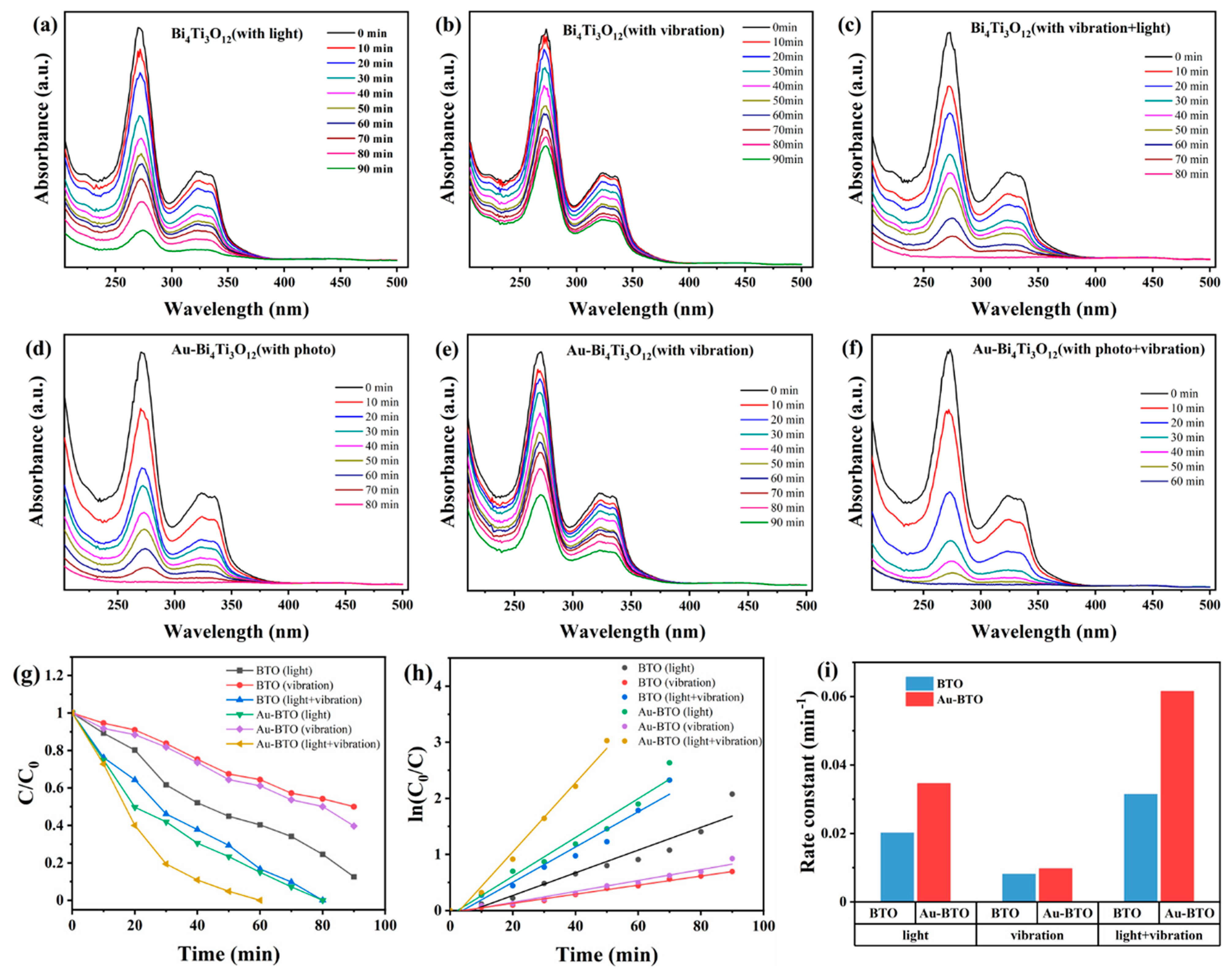
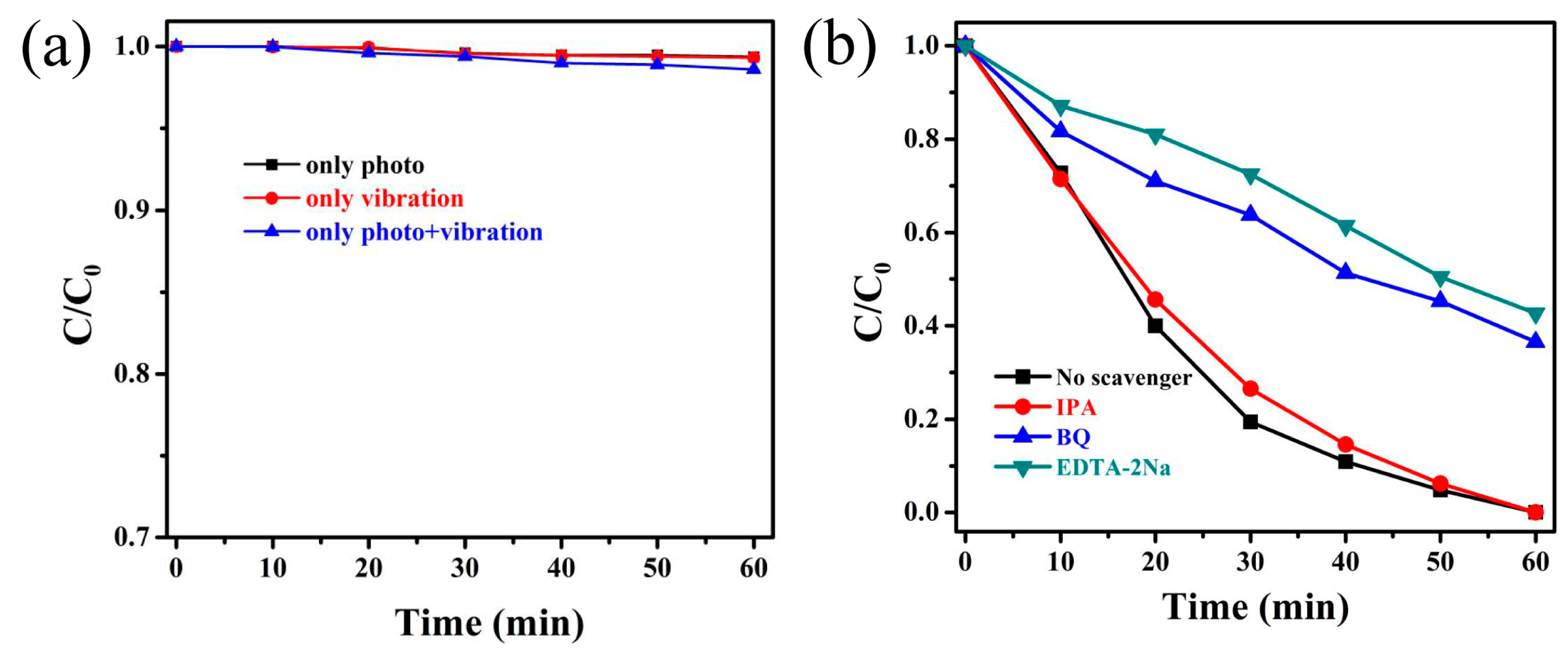
2.4. Degradation of Ciprofloxacin

Bi4Ti3O12 nanofibers and Au-Bi4Ti3O12 nanofibers were used to study the photocatalytic degradation of the ciprofloxacin antibiotic under three different experimental conditions: light, vibration, and light plus vibration. The photocatalytic performance of Bi4Ti3O12 nanofibers for the degradation of CIP under Xenon lamp illumination is presented in Figure 4a. According to the results of the ultraviolet visible spectrophotometer, as the photocatalytic reaction proceeded, the main extinction peak of CIP at 270 nm and the wider peak from 300–350 nm decreased with time, which means that the CIP was effectively degraded by the light. However, there was still some CIP that was not degraded 90 min into the reaction. When the Xenon lamp illumination was replaced with vibration, the main extinction peak of CIP and the wider peak were also decreased as the reaction proceeded, but the peak decreased more slowly than in the photocatalytic condition during the same 90-min time interval (Figure 4b). By using both Xenon lamp illumination and vibration, the CIP was almost completely degraded in 80 min (Figure 4c), which confirms our original hypothesis that piezo-photocatalytic degradation is better than either type alone.
The above results prove that the degradation performance of ferroelectric Bi4Ti3O12 is significantly enhanced under ultrasonic conditions, which may be ascribed to the piezoelectric effect that promotes the generation of free electrons on the surface of a piezo-catalyst. Bi4Ti3O12 has ferroelectric properties and can spontaneously polarize to generate an internal electric field. Vibration pressurization of Bi4Ti3O12 nanofibers can enhance the polarization electric field and light absorption. Under the enhanced polarization electric field, more photogenerated electrons and holes quickly move to both sides separately and combine with corresponding ions to the active sites of the surface. Both of these factors can enhance the catalytic effect.
When Au-Bi4Ti3O12 was substituted for Bi4Ti3O12, the efficiency of the degradation of CIP was increased under Xenon lamp illumination, with vibration, and with both Xenon lamp illumination and vibration (Figure 4d–f). The photocatalytic degradation trend of CIP by Au-Bi4Ti3O12 was similar to that of Bi4Ti3O12, but the degradation efficiency was able to reach 100% in 80 min. Moreover, by using both Xenon lamp illumination and vibration, the piezo-photocatalytic degradation efficiency of CIP by Au-Bi4Ti3O12 reached 100% just in 60 min.
For the purposes of comparison, chemical kinetics experiments were carried out for the degradation of CIP by Bi4Ti3O12 and Au-Bi4Ti3O12. Figure 4g displays a plot of C/C0 in the degradation of CIP vs. time using Bi4Ti3O12 and Au-Bi4Ti3O12 under different conditions. This graph demonstrates that the concentration ratio of the CIP decreased with respect to the time period. Based on the graph of C/C0 vs. time, a plot was drawn to demonstrate ln(C/C0) vs. time (Figure 4h). Using this plot, we calculated the reaction rate constants (k) for the degradation efficiencies of the CIP solution by Bi4Ti3O12 and Au-Bi4Ti3O12 nanofibers under Xenon lamp illumination, with vibration, and with both Xenon lamp illumination and vibration conditions, as shown in Figure 4i. Based on this figure, the degradation efficiencies of CIP by Au-Bi4Ti3O12 nanofibers are higher than those of the Bi4Ti3O12 nanofibers under Xenon lamp illumination, with vibration, and with both Xenon lamp illumination and vibration. The degradation efficiency of the CIP solution by Au-Bi4Ti3O12 nanofibers reached 100% within 60 min, with a peak reaction rate of 0.06187 min−1 under simultaneous light and ultrasonic vibration, which is twice as high as the peak reaction rate of the Bi4Ti3O12 nanofibers. This suggests that the introduction of Au on the surface of Bi4Ti3O12 can enhance the performance of Bi4Ti3O12 due to the piezo-plasmonic coupling effect.
Control experiments showed that no obvious self-decolorization was observed (Figure 5a) in the absence of catalyst under light irradiation with ultrasonic excitation. To understand the mechanism of the photocatalytic reaction process, a series of comparative tests were conducted by employing holes and radical trapping agents for the degradation of ciprofloxacin. The results are shown in Figure 5b. It is clear that the ciprofloxacin degradation was barely influenced with the addition of isopropanol (IPA) (as a quencher of •OH) [38]. However, after adding benzoquinone (BQ) (as a quencher of •O2) and ethylenediaminetetraacetic acid disodium (EDTA-2Na) (as a quencher of h+) into the reactions, the degradation rate of the ciprofloxacin was reduced significantly. The results indicate that the •O2 and the photogenerated holes were the major active species participating in the photocatalytic reaction process. After light irradiation, the electron and hole appeared and separated to the surface of the Au-Bi4Ti3O12 catalyst. Then the adsorbed oxygen combined with the electron and became the reactive species of superoxide radicals. With their strong oxidative ability, the •O2 and holes directly oxidated the pollutants.

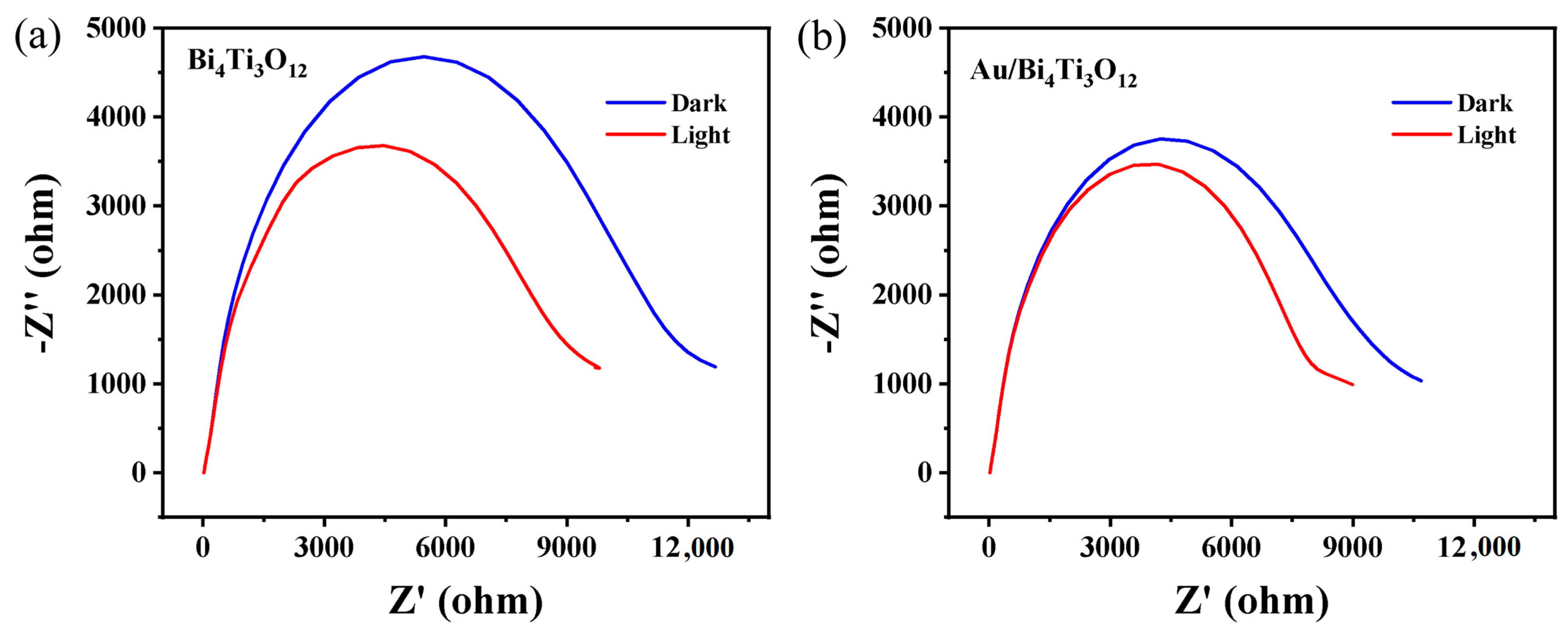
2.5. Photoelectrochemical Measurement

Electrochemical impedance spectroscopy (EIS) was used to understand the direction and resistance of the charge transfer [39]. The diameter of the semicircle in the figure represents the charge transfer resistance. The smaller the diameter of the semicircle is, the smaller the transfer resistance of the photo electrode is, and the higher the charge transfer efficiency is. As shown from left to right in Figure 6, the impedance diagrams were drawn of Bi4Ti3O12 nanofibers and Au-Bi4Ti3O12 nanofibers under light and dark. It was found that the impedance of Bi4Ti3O12 nanofibers without Au was large, and the change of additive optical impedance was very small, which indicates that the Bi4Ti3O12 nanofibers were insensitive to light absorption, whereas the impedance of Au-Bi4Ti3O12 nanofibers material became noticeably smaller under the light.
Subsequently, transient photocurrent response measurements were conducted to study the separation behaviors of the photoexcited carriers during the photoreactions. As shown in Figure 7, there was generation of photocurrents with good reproducibility for both the Bi4Ti3O12 and the Au-Bi4Ti3O12 nanofibers. The Bi4Ti3O12 nanofibers and the Au-Bi4Ti3O12 nanofibers were characterized at 0 V and 0.1 V, respectively. At the same time, the photocurrent density of Bi4Ti3O12 nanofibers at 0 V was 2.89 × 10⁻⁷ A/cm2. When the voltage was changed to 0.1 V, the photocurrent density increased to 5.04 × 10⁻⁷ A/cm2. The photocurrent density of Au-Bi4Ti3O12 nanofibers at 0 V was 2.74 × 10⁻⁶ A/cm2, and the photocurrent density at 0.1 V was 7.54 × 10⁻⁶ A/cm2.
The current-time curve is the observation of the change of the current with time under different voltage conditions. The fact that the I-T result was consistent under EIS indicates that loaded Au can enhance the photocatalytic performance of Bi4Ti3O12 nanofibers. After absorbing light, the Au nanoparticles are excited by the light to generate hot electrons. Because of the Fermi levels generated on the contact surfaces of the Au nanoparticles and the Bi4Ti3O12 nanofibers, the electrons in the Au nanoparticles move to the conduction band of the Bi4Ti3O12 nanofibers [40]. At the same time, the Bi4Ti3O12 nanofibers can absorb light to generate electron–hole pairs. Under the action of the polarization field, more electrons and holes are generated through transfer. On the other hand, after the local surface plasmon is excited, a large scattering cross section will be produced, and the generated hot electrons will then travel through the semiconductor and participate in the photocatalytic reaction process. Thus, loading Au can not only enhance the light absorption capacity, but also promote the separation of electrons and holes by participating in the reaction, which can explain why the introduction of Au enhances the photocatalytic performance.

3. Experimental Section

3.1. Synthesis of Bi4Ti3O12 Nanofibers

First, 0.75 g of Bi(NO3)·5H2O was poured into a bottle, and then 8 mL of N, N-dimethylformamide (DMF); 2 mL of glacial acetic acid; and 0.4 mL tetrabutyl titanate (TBT) were added into the bottle and continuously stirred for about 2 h. Subsequently, 1.25 g PVP was added into the premixed solution and stirred for about 8 h to form a precursor solution. Then the precursor solution was transferred into a 10 mL needle tube, followed by adjusting the high voltage to 18 V and the low voltage to −2 V and pushing the speed to 0.1 mm/min, with a rotation speed of 50 r/min. Then the solution was stripped and calcined in a tube furnace at 550 °C for 2 h (heating rate: 3 °C min−1). After naturally cooling to room temperature, the product was washed and dried to obtain the sample Bi4Ti3O12 nanofibers.

3.2. Synthesis of Au-Bi4Ti3O12 Nanofibers

First, the Au was plated with a small ion sputtering instrument to obtain Au-loaded Bi4Ti3O12 nanofibers. The relevant parameters were a current of 30 mA and an air pressure of 5 × 10⁻² mbar/Pa that was maintained in the cavity, along with a gold plating time of 20 s.

3.3. Material Characterization

The diffraction patterns of the samples and the pellets were collected with the Rigaku D/Max 3 C X-ray diffraction (XRD) system equipped with a Cu Kα tube (wavelength = 0.154 nm) and operated at 40 kV/40 mA. The scanner was set to scan from 10° to 80° with a scan rate of 1°/min. Raman spectra of the samples were measured using a Raman spectroscopy (Thermo scientific, with a laser wavelength of 785 nm) at a laser power of 0.2 mW and a magnification of 50×. The size and morphologies of the Bi4Ti3O12 nanofibers were characterized using a scanning electron microscope (SEM, Hitachi S-4800). The nanostructures of the Bi4Ti3O12 nanofibers and the Au-Bi4Ti3O12 nanofibers were observed using transmission electron microscopy (TEM, Hitachi H-8100IV). Prior to the SEM observation, the electrodes were covered with a Au coating to improve their conductivity. The absorption spectra of the samples were obtained using a UV-vis near-infrared spectrophotometer (UV-3600, Shimadzu). X-ray photoelectron spectroscopy (XPS, Thermo ESCALAB 250) was performed with an Al target (15 kV, 10 mA) to determine the surface chemical composition of the samples.

3.4. Degradation of Ciprofloxacin

The piezo-photocatalytic behaviors of the Bi4Ti3O12 nanofibers and the Au-Bi4Ti3O12 nanofibers were measured by the decomposition of ciprofloxacin. The change in concentration of the ciprofloxacin was measured using a UV-Vis spectrophotometer. Moreover, the degradation effect was reflected by the change of absorbance. In typical conditions, a concentration of 20mg/L of ciprofloxacin solution (100 mL) and 50 mg of catalyst were used during each experiment for every catalyst. Before testing the photocatalytic activity (under dark conditions), a reaction solution of ciprofloxacin and 50 mg catalyst was put into the beaker and stirred for 30 min using a magnetic stirrer to attain equilibrium (adsorption-desorption). The beaker was placed in a small ultrasonic machine (with ultrasonic power of 40 KHz) under a xenon lamp (300 W, light power 99.99 mW/cm2). The experiment was conducted according to the requirements of using just the lamp (photocatalytic reaction), just the ultrasonic machine (piezoelectric reaction), or the lamp and ultrasonic machine together (piezo-photocatalytic reaction) as the experimental instruments. During the photocatalytic reactions, 1 mL solution was taken out of the reaction mixture every 10 min. Then the reaction mixture was centrifuged to remove traces of the catalyst.

3.5. Photoelectrochemistry Measurements

The photocurrent densities and electrochemical impedance spectroscopy of the Bi4Ti3O12 nanofibers and the Au-Bi4Ti3O12 nanofibers were tested using an electrochemical workstation (CHI760E, China). This workstation was equipped with a standard three-electrode system with a platinum plate, a saturated Ag/AgCl electrode, and obtained samples as the counter, reference, and working electrodes, respectively. The prepared samples were directly spread onto an indium tin oxide glass electrode to prepare the working electrodes, and a Na2SO4 (0.1 M, pH 7.0) aqueous solution was used as the electrolyte for the photocurrent measurements. The studied area of the thin films was fixed to 1 cm2. The light source was supplied by a 300 W Xe arc lamp equipped with filter (AM 1.5 G) and with a power density of 100 mW cm−2.

4. Conclusions

In summary, in order to obtain high efficiency in the photocatalytic degradation of antibiotics, Au-loaded ferroelectric Bi4Ti3O12 nanofibers were obtained by electrospinning and ion sputtering. The characterization of the morphology, crystal structure and optical properties indicated that the Bi4Ti3O12 nanofibers with orthorhombic crystal phases were prepared successfully. Furthermore, the degradation efficiency of ciprofloxacin by the Bi4Ti3O12 nanofibers and the Au-Bi4Ti3O12 nanofibers was investigated under light, with ultrasonic vibration and with both light and ultrasonic vibration. The results are as follows: In a 90 min reaction, 50% of the CIP was degraded with vibration only, and the reaction rate constant was 0.00809 min−1. Under the condition of light only, 87.45% of the CIP was degraded, and the reaction rate constant was 0.02019 min−1. Under both light and vibration, 100% of the CIP was degraded, and the reaction rate constant was 0.03141 min−1, which is 0.01122 min−1 faster than that under light only. At the same time, after loading the Au, the catalytic performance was significantly enhanced under all three conditions. Under light and ultrasonic vibration, the CIP degradation efficiency of the Au-Bi4Ti3O12 nanofibers reached 100% within 60 min, and the reaction rate constant was 0.06187 min−1, which is twice as fast as the Bi4Ti3O12 nanofibers. It is further observed that the photocurrent density was increased from 5.08 × 10⁻⁷ A/cm2 to 8.17 × 10⁻⁶ A/cm2. This work provides a basis for more research on the piezoelectric coupling effect in photocatalytic degradation and promotes the practical application of piezoelectric catalysis technology in the environmental field.

Author Contributions

C.M. and J.P. contributed equally to this work. D.Y., C.M., H.H. and K.Y. conducted the experiments, data analysis, writing-original manuscript, writing-review & editing. J.P. conducted the experiments, data analysis. L.W. conducted the experiments. All authors have read and agreed to the published version of the manuscript.

Funding

This research is funded by the Open Project Program of the State Key Laboratory of NBC Protection for Civilian (SKLNBC2021-20); the Open Project Program of the State Key Laboratory of Photocatalysis on Energy and Environment (SKLPEE-KF202203), Fuzhou University; the special funding project for the instruction of high-level teachers of the Beijing Institute of Fashion Technology (BIFTXJ202221); and the National Natural Science Foundation of China (62204097).

Data Availability Statement

Not applicable.

Acknowledgments

In this section, we thank Peizhu Zheng and Wenhui Feng for their constructive discussions.

Conflicts of Interest

The authors declare no conflict of interest.

References

  1. Guo, F.; Zhang, H.; Li, H.; Shen, Z.R. Modulating the oxidative active species by regulating the valence of palladium cocatalyst in photocatalytic degradation of ciprofloxacin. Appl. Catal. B Environ. 2022, 306, 121092. [Google Scholar] [CrossRef]
  2. Hunge, Y.M.; Yadav, A.A.; Kang, S.W.; Lim, S.J.; Kim, H. Visible light activated MoS2/ZnO composites for photocatalytic degradation of ciprofloxacin antibiotic and hydrogen production. J. Photochem. Photobiol. A Chem. 2023, 434, 114250. [Google Scholar] [CrossRef]
  3. Lu, N.; Wang, P.; Su, Y.; Yu, H.T.; Liu, N.; Quan, X. Construction of Z-Scheme g-C3N4/RGO/WO3 with in situ photoreduced graphene oxide as electron mediator for efficient photocatalytic degradation of ciprofloxacin. Chemosphere 2019, 215, 444–453. [Google Scholar] [CrossRef] [PubMed]
  4. Xiong, Z.K.; Zhang, H.; Zhang, W.C.; Lai, B.; Yao, G. Removal of nitrophenols and their derivatives by chemical redox: A review. Chem. Eng. J. 2019, 359, 13–31. [Google Scholar] [CrossRef]
  5. Hu, K.; Li, R.Q.; Ye, C.L.; Wang, A.Q.; Wei, W.Q.; Hu, D.; Qiu, R.L.; Yan, K. Facile synthesis of Z-scheme composite of TiO2 nanorodig-C3N4 nanosheet efficient for photocatalytic degradation of ciprofloxacin. J. Clean. Prod. 2020, 253, 120055. [Google Scholar] [CrossRef]
  6. Hoffmann, M.R.; Martin, S.T.; Choi, W.Y.; Bahnemann, D.W. Environmental applications of semiconductor photocatalysis. Chem. Rev. 1995, 95, 69–96. [Google Scholar] [CrossRef]
  7. Meng, C.; Weng, B. Steric effects of a homogeneous CuCl2/solvent system for photocatalytic selective oxidation of benzyl alcohol. New J. Chem. 2022, 46, 13345–13351. [Google Scholar] [CrossRef]
  8. Tamaki, Y.; Furube, A.; Murai, M.; Hara, K.; Katoh, R.; Tachiya, M. Dynamics of efficient electron-hole separation in TiO2 nanoparticles revealed by femtosecond transient absorption spectroscopy under the weak-excitation condition. Phys. Chem. Chem. Phys. 2007, 9, 1453–1460. [Google Scholar] [CrossRef]
  9. Meng, C.; Yang, K.; Fu, X.Z.; Yuan, R.S. Photocatalytic Oxidation of Benzyl Alcohol by Homogeneous CuCl2/Solvent: A Model System to Explore the Role of Molecular Oxygen. ACS Catal. 2015, 5, 3760–3766. [Google Scholar] [CrossRef]
  10. Meng, C.; Wang, H.; Wu, Y.B.; Fu, X.Z.; Yuan, R.S. Study on Selective Photocatalytic Oxidation of Ethanol During TiO2 Promoted Water-Splitting Process. Acta Chim. Sin. 2017, 75, 508–513. [Google Scholar] [CrossRef] [Green Version]
  11. Huang, H.; Zhao, J.; Weng, B.; Lai, F.; Zhang, M.; Hofkens, J.; Roeffaers, M.B.J.; Steele, J.A.; Long, J. Site-Sensitive Selective CO(2) Photoreduction to CO over Gold Nanoparticles. Angew. Chem. Int. Ed. Engl. 2022, 61, e202204563. [Google Scholar] [PubMed]
  12. Huang, H.; Verhaeghe, D.; Weng, B.; Ghosh, B.; Zhang, H.; Hofkens, J.; Steele, J.A.; Roeffaers, M.B.J. Metal Halide Perovskite Based Heterojunction Photocatalysts. Angew. Chem. Int. Ed. Engl. 2022, 61, e202203261. [Google Scholar] [PubMed]
  13. Wang, H.; Hu, P.; Zhou, J.; Roeffaers, M.B.J.; Weng, B.; Wang, Y.; Ji, H. Ultrathin 2D/2D Ti3C2Tx/semiconductor dual-functional photocatalysts for simultaneous imine production and H2 evolution. J. Mater. Chem. A 2021, 9, 19984–19993. [Google Scholar] [CrossRef]
  14. Liu, S.; Qi, W.; Adimi, S.; Guo, H.; Weng, B.; Attfield, J.P.; Yang, M. Titanium Nitride-Supported Platinum with Metal-Support Interaction for Boosting Photocatalytic H(2) Evolution of Indium Sulfide. ACS Appl. Mater. Interfaces 2021, 13, 7238–7247. [Google Scholar] [CrossRef] [PubMed]
  15. Long, Z.Q.; Li, Q.G.; Wei, T.; Zhang, G.M.; Ren, Z.J. Historical development and prospects of photocatalysts for pollutant removal in water. J. Hazard. Mater. 2020, 395, 122599. [Google Scholar] [CrossRef]
  16. Luo, L.; Zhang, T.T.; Wang, M.; Yun, R.P.; Xiang, X. Recent Advances in Heterogeneous Photo-Driven Oxidation of Organic Molecules by Reactive Oxygen Species. Chemsuschem 2020, 13, 5173–5184. [Google Scholar] [CrossRef]
  17. Nosaka, Y.; Nosaka, A.Y. Generation and Detection of Reactive Oxygen Species in Photocatalysis. Chem. Rev. 2017, 117, 11302–11336. [Google Scholar] [CrossRef]
  18. Wang, Y.; Wen, X.R.; Jia, Y.M.; Huang, M.; Wang, F.F.; Zhang, X.H.; Bai, Y.Y.; Yuan, G.L.; Wang, Y.J. Piezo-catalysis for nondestructive tooth whitening. Nat. Commun. 2020, 11, 1328. [Google Scholar] [CrossRef] [Green Version]
  19. Wang, S.S.; Wu, Z.; Chen, J.; Ma, J.P.; Ying, J.S.; Cui, S.C.; Yu, S.G.; Hu, Y.M.; Zhao, J.H.; Jia, Y.M. Lead-free sodium niobate nanowires with strong piezo-catalysis for dye wastewater degradation. Ceram. Int. 2019, 45, 11703–11708. [Google Scholar] [CrossRef]
  20. Lan, S.Y.; Feng, J.X.; Xiong, Y.; Tian, S.H.; Liu, S.W.; Kong, L.J. Performance and Mechanism of Piezo-Catalytic Degradation of 4-Chlorophenol: Finding of Effective Piezo-Dechlorination. Environ. Sci. Technol. 2017, 51, 6560–6569. [Google Scholar] [CrossRef]
  21. Xu, S.W.; Qian, W.Q.; Zhang, D.; Zhao, X.; Zhang, X.M.; Li, C.B.; Bowen, C.R.; Yang, Y. A coupled photo-piezo-catalytic effect in a BST-PDMS porous foam for enhanced dye wastewater degradation. Nano Energy 2020, 77, 105305. [Google Scholar] [CrossRef]
  22. Yu, C.Y.; He, J.J.; Tan, M.X.; Hou, Y.X.; Zeng, H.; Liu, C.B.; Meng, H.M.; Su, Y.J.; Qiao, L.J.; Lookman, T.; et al. Selective Enhancement of Photo-Piezocatalytic Performance in BaTiO3 Via heterovalent Ion Doping. Adv. Funct. Mater. 2022, 32, 2209365. [Google Scholar] [CrossRef]
  23. Liu, Q.; Li, Z.Y.; Li, J.; Zhan, F.Q.; Zhai, D.; Sun, Q.W.; Xiao, Z.D.; Luo, H.; Zhang, D. Three dimensional BaTiO3 piezoelectric ceramics coated with TiO2 nanoarray for high performance of piezo-photoelectric catalysis. Nano Energy 2022, 98, 107267. [Google Scholar] [CrossRef]
  24. Hervoches, C.H.; Lightfoot, P. A variable-temperature powder neutron diffraction study of ferroelectric Bi4Ti3O12. Chem. Mater. 1999, 11, 3359–3364. [Google Scholar] [CrossRef]
  25. Chen, Z.W.; Jiang, H.; Jin, W.L.; Shi, C.K. Enhanced photocatalytic performance over Bi4Ti3O12 nanosheets with controllable size and exposed {001} facets for Rhodamine B degradation. Appl. Catal. B Environ. 2016, 180, 698–706. [Google Scholar] [CrossRef]
  26. Liu, G.; Yang, H.G.; Pan, J.; Yang, Y.Q.; Lu, G.Q.; Cheng, H.M. Titanium Dioxide Crystals with Tailored Facets. Chem. Rev. 2014, 114, 9559–9612. [Google Scholar] [CrossRef]
  27. Liu, X.T.; Shen, X.F.; Sa, B.S.; Zhang, Y.G.; Li, X.; Xue, H. Piezotronic-enhanced photocatalytic performance of heterostructured BaTiO3/SrTiO3 nanofibers. Nano Energy 2021, 89, 106391. [Google Scholar] [CrossRef]
  28. Hu, J.Y.; Chen, Y.X.; Zhou, Y.Y.; Zeng, L.X.; Huang, Y.C.; Lan, S.Y.; Zhu, M.S. Piezo-enhanced charge carrier separation over plasmonic Au-BiOBr for piezo-photocatalytic carbamazepine removal. Appl. Catal. B Environ. 2022, 311, 121369. [Google Scholar] [CrossRef]
  29. Zhang, Y.; Wang, S.; Zhao, Y.; Ding, Y.; Zhang, Z.; Jiang, T.; Wang, Z.L.; Li, L. Piezo-phototronic effect boosted catalysis in plasmonic bimetallic ZnO heterostructure with guided fermi level alignment. Mater. Today Nano 2022, 18, 100177. [Google Scholar] [CrossRef]
  30. Fan, Z.X.; Zhu, Y.H.; Huang, X.; Han, Y.; Wang, Q.X.; Liu, Q.; Huang, Y.; Gan, C.L.; Zhang, H. Synthesis of Ultrathin Face-Centered-Cubic Au@Pt and Au@Pd Core-Shell Nanoplates from Hexagonal-Close-Packed Au Square Sheets. Angew. Chem. Int. Ed. 2015, 54, 5672–5676. [Google Scholar] [CrossRef]
  31. Tatarchuk, T.; Myslin, M.; Mironyuk, I.; Bououdina, M.; Pedziwiatr, A.T.; Gargula, R.; Bogacz, B.F.; Kurzydlo, P. Synthesis, morphology, crystallite size and adsorption properties of nanostructured Mg-Zn ferrites with enhanced porous structure. J. Alloy. Compd. 2020, 819, 152945. [Google Scholar] [CrossRef]
  32. Xie, Y.; Zhou, Y.P.; Gao, C.M.; Liu, L.J.; Zhang, Y.F.; Chen, Y.; Shao, Y. Construction of AgBr/BiOBr S-scheme heterojunction using ion exchange strategy for high-efficiency reduction of CO2 to CO under visible light. Sep. Purif. Technol. 2022, 303, 122288. [Google Scholar] [CrossRef]
  33. Li, B.S.; Lai, C.; Zhang, M.M.; Liu, S.Y.; Yi, H.; Liu, X.G.; An, N.; Zhou, X.R.; Li, L.; Fu, Y.K.; et al. N, S-GQDs and Au nanoparticles co-modified ultrathin Bi2MoO6 nanosheet with enhanced charge transport dynamics for full-spectrum-light-driven molecular oxygen activation. Chem. Eng. J. 2021, 409, 128281. [Google Scholar] [CrossRef]
  34. You, D.T.; Wang, R.; Xie, J.W.; Liu, L.; Li, K.W.; Han, X.L.; Guo, T.; Xu, C.X. Synergistic SERS enhancement and in situ monitoring of photocatalytic reactions in a plasmonic metal/ferroelectric hybrid system by the light-induced pyroelectric effect. J. Mater. Chem. A 2022, 10, 14078–14089. [Google Scholar] [CrossRef]
  35. Chen, X.D.; Zhang, H.; Ci, C.G.; Sun, W.W.; Wang, Y. Few-Layered Boronic Ester Based Covalent Organic Frameworks/Carbon Nanotube Composites for High-Performance K-Organic Batteries. ACS Nano 2019, 13, 3600–3607. [Google Scholar] [CrossRef]
  36. Guo, W.X.; Sun, W.W.; Lv, L.P.; Kong, S.F.; Wang, Y. Microwave-Assisted Morphology Evolution of Fe-Based Metal-Organic Frameworks and Their Derived Fe2O3 Nanostructures for Li-Ion Storage. ACS Nano 2017, 11, 4198–4205. [Google Scholar] [CrossRef]
  37. Cuevas, A.J.S.; Cabrera, C.B.P.; Aguilar, C.A.H.; Padilla-Martinez, I.I.; Thangarasu, P.; Contreras, E.F.V.; Alonzo, F.R.; Narayanan, J. Effect of the structural integrity on the size and porosity of gold-implanted mixed-metal oxide nanocomposites: Their influence on the photocatalytic degradation of thioanisole. Dalton Trans. 2022, 51, 17671–17687. [Google Scholar] [CrossRef]
  38. You, D.T.; Liu, L.; Yang, Z.Y.; Xing, X.X.; Li, K.W.; Mai, W.J.; Guo, T.; Xiao, G.Z.; Xu, C.X. Polarization-induced internal electric field to manipulate piezo-photocatalytic and ferro-photoelectrochemical performance in bismuth ferrite nanofibers. Nano Energy 2022, 93, 106852. [Google Scholar] [CrossRef]
  39. Amulya, M.A.S.; Nagaswarupa, H.P.; Kumar, M.R.A.; Ravikumar, C.R.; Prashantha, S.C.; Kusuma, K.B. Sonochemical synthesis of NiFe2O4 nanoparticles: Characterization and their photocatalytic and electrochemical applications. Appl. Surf. Sci. Adv. 2020, 1, 100023. [Google Scholar] [CrossRef]
  40. Xu, S.Y.; Guo, L.M.; Sun, Q.J.; Wang, Z.L. Piezotronic Effect Enhanced Plasmonic Photocatalysis by AuNPs/BaTiO3 Heterostructures. Adv. Funct. Mater. 2019, 29, 1808737. [Google Scholar] [CrossRef]
Figure 1. Scanning electron microscopy (SEM) images of Bi4Ti3O12 nanofibers (a) before calcination and (b) after calcination.
Catalysts 13 00621 g001
Figure 2. Transmission electron microscopy (TEM) images of (a) Bi4Ti3O12 nanofibers and (b) Au-Bi4Ti3O12 nanofibers, and (c) high-resolution TEM images of Au-Bi4Ti3O12 nanofibers.
Catalysts 13 00621 g002
Figure 3. (a) X-Ray Diffraction, (b) Raman spectra, (c) UV-Vis diffuse reflectance spectra, and (d) A plot of the (αhν)2 versus the hν of Bi4Ti3O12 and Au–Bi4Ti3O12; (e) X-ray photoelectron spectroscopy and high-resolution XPS spectra of (f) Bi 4f, (g) Ti 2p and (h) O 1s core levels of Bi4Ti3O12; and (i) High-resolution XPS spectrum of Au 4f in Au–Bi4Ti3O12.
Catalysts 13 00621 g003
Figure 4. Absorbance spectra of the decomposition activity of ciprofloxacin with 10 min intervals under the following condition: Bi4Ti3O12 (a) under the light, (b) with vibration, and (c) with vibration and under the light; Au–Bi4Ti3O12 (d) under the light, (e) with vibration, and (f) with vibration and under the light; (g) comparison of the degradation proficiency in various test conditions; (h) sample kinetics fit to the information; and (i) comparison of the rate constant (k) values for the decomposition activity of ciprofloxacin under different conditions.
Catalysts 13 00621 g004
Figure 5. (a) Control experiments without catalyst under light irradiation with ultrasonic excitation. (b) Photodegradation curves of ciprofloxacin over Au-Bi4Ti3O12 with different scavengers.
Catalysts 13 00621 g005
Figure 6. Electrochemical impedance spectroscopy of (a) Bi4Ti3O12 and (b) Au-Bi4Ti3O12 under light and dark.
Catalysts 13 00621 g006
Figure 7. Photocurrent response of (a) Bi4Ti3O12 and (b) Au-Bi4Ti3O12 under lamp irradiation at 0 V and 0.1 V.
Catalysts 13 00621 g007
Disclaimer/Publisher’s Note: The statements, opinions and data contained in all publications are solely those of the individual author(s) and contributor(s) and not of MDPI and/or the editor(s). MDPI and/or the editor(s) disclaim responsibility for any injury to people or property resulting from any ideas, methods, instructions or products referred to in the content.

