

Green Carbon Dots: Applications in Development of Electrochemical Sensors, Assessment of Toxicity as Well as Anticancer Properties
 , , and
Abstract
:1. Introduction
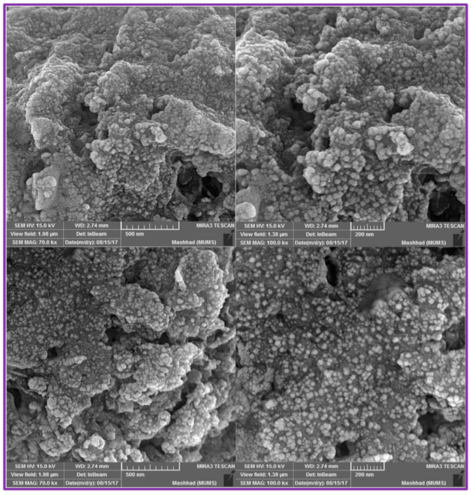
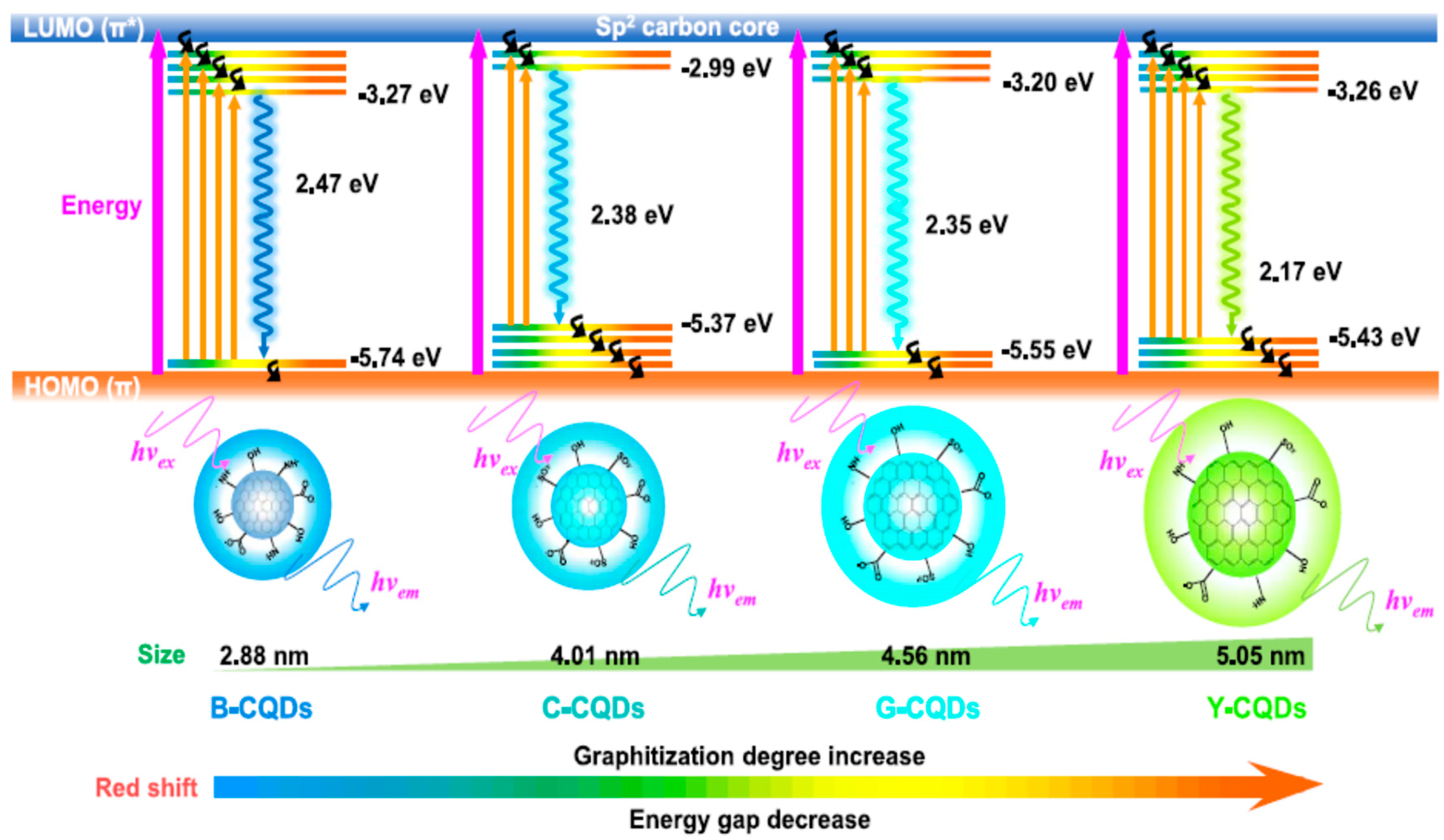
2. Green Synthesis Methods and Optical Properties of GCDs
3. Electrochemical Sensing Ability of GCDs
4. Toxicity Assessment and Anticancer Properties of Green Carbon Dots (GCDs)
5. Conclusions
- In order to bring GCDs into commercial applications, more study should be conducted on the search for high-quality natural precursors for their synthesis.
- In vivo toxicity studies are limited, hence, more in vivo studies involving GCDs should be considered for the implementation of GCDs in biological/clinical applications.
- In addition, toxicity of GCDs prepared from different natural sources under different synthesis conditions should be investigated to obtain comprehensive details on GCDs toxicity.
- Future studies should focus on evaluating the anticancer activity of GCDs using in vivo models.
- More research should be conducted to explore the possibility of using GCDs in photodynamic and photothermal therapy.
- GCDs-based sensors should also be used for the sensitive and selective detection of cancer biomarkers, such as miRNA and antigens, to explore the application of GCDs in clinical cancer diagnosis.
- It is also important to check the effect of GCDs’ size and surface modifications on anticancer as well as electrochemical sensing abilities.
Author Contributions
Funding
Data Availability Statement
Acknowledgments
Conflicts of Interest
References
- Nazri, N.A.A.; Azeman, N.H.; Luo, Y.; Bakar, A.A.A. Carbon quantum dots for optical sensor applications: A review. Opt. Laser Technol. 2021, 139, 106928. [Google Scholar] [CrossRef]
- Wang, B.; Wang, M.; Liu, F.; Zhang, Q.; Yao, S.; Liu, X.; Huang, F. Ti3C2: An Ideal Co-catalyst? Angew. Chem. Int. Ed. 2020, 59, 1914–1918. [Google Scholar] [CrossRef] [PubMed]
- Chen, Z.-L.; Wang, D.; Wang, X.-Y.; Yang, J.-H. Enhanced formaldehyde sensitivity of two-dimensional mesoporous SnO2 by nitrogen-doped graphene quantum dots. Rare Met. 2021, 40, 1561–1570. [Google Scholar] [CrossRef]
- Yan, J.; Ye, F.; Dai, Q.; Ma, X.; Fang, Z.; Dai, L.; Hu, C. Recent progress in carbon-based electrochemical catalysts: From structure design to potential applications. Nano Res. Energy 2023, 2, e9120047. [Google Scholar] [CrossRef]
- De Oliveira, B.P.; Da Silva Abreu, F.O.M. Carbon quantum dots synthesis from waste and by-products: Perspectives and challenges. Mater. Lett. 2021, 282, 128764. [Google Scholar] [CrossRef]
- Borna, S.; Sabzi, R.E.; Pirsa, S. Synthesis of carbon quantum dots from apple juice and graphite: Investigation of fluorescence and structural properties and use as an electrochemical sensor for measuring letrozole. J. Mater. Sci. Mater. Electron. 2021, 32, 10866–10879. [Google Scholar] [CrossRef]
- Zhu, L.; Shen, D.; Wang, Q.; Luo, K.H. Green synthesis of tunable fluorescent carbon quantum dots from lignin and their application in anti-counterfeit printing. ACS Appl. Mater. Interfaces 2021, 13, 56465–56475. [Google Scholar] [CrossRef] [PubMed]
- Desmond, L.J.; Phan, A.N.; Gentile, P. Critical overview on the green synthesis of carbon quantum dots and their application for cancer therapy. Environ. Sci. Nano. 2021, 8, 848. [Google Scholar] [CrossRef]
- Huo, X.; He, Y.; Ma, S.; Jia, Y.; Yu, J.; Li, Y.; Cheng, Q. Green synthesis of carbon dots from grapefruit and its fluorescence enhancement. J. Nanomater. 2020, 2020, 8601307. [Google Scholar] [CrossRef]
- Zhang, M.; Zhao, Y.; Cheng, J.; Liu, X.; Wang, Y.; Yan, X.; Zhang, Y.; Lu, F.; Wang, Q.; Qu, H.; et al. Novel carbon dots derived from schizonepetae herba carbonisata and investigation of their haemostatic efficacy. Artif. Cells Nanomed. Biotechnol. 2017, 46, 1562–1571. [Google Scholar] [CrossRef]
- Dias, C.; Vasimalai, N.; Sarria, M.P.; Pinheiro, I.; Vilas-Boas, V.; Peixoto, J.; Espina, B. Biocompatibility and bioimaging potential of fruit-based carbon dots. Nanomaterials 2019, 9, 199. [Google Scholar] [CrossRef] [PubMed] [Green Version]
- Malishev, R.; Arad, E.; Bhunia, S.K.; Shaham-Niv, S.; Kolusheva, S.; Gazit, E.; Jelinek, R. Chiral modulation of amyloid beta fibrillation and cytotoxicity by enantiomeric carbon dots. Chem. Commun. 2018, 54, 7762–7765. [Google Scholar] [CrossRef] [PubMed]
- Su, W.; Wu, H.; Xu, H.; Zhang, Y.; Li, Y.; Li, X.; Fan, L. Carbon dots: A booming material for biomedical applications. Mater. Chem. Front. 2020, 4, 821–836. [Google Scholar] [CrossRef]
- Chahal, S.; Macairan, J.-R.; Yousefi, N.; Tufenkji, N.; Naccache, R. Green synthesis of carbon dots and their applications. RSC Adv. 2021, 11, 25354–25363. [Google Scholar] [CrossRef]
- Lin, X.; Xiong, M.; Zhang, J.; He, C.; Ma, X.; Zhang, H.; Kuang, Y.; Yang, M.; Huang, Q. Carbon dots based on natural resources: Synthesis and applications in sensors. Microchem. J. 2021, 160, 105604. [Google Scholar] [CrossRef]
- Hassanvand, Z.; Jalali, F.; Nazari, M.; Parnianchi, F.; Santoro, C. Carbon nanodots in electrochemical sensors and biosensors: A review. ChemElectroChem 2021, 8, 15–35. [Google Scholar] [CrossRef]
- Liu, X.; Wang, Y.; Yan, X.; Zhang, M.; Zhang, Y.; Cheng, J.; Lu, F.; Qu, H.; Wang, Q.; Zhao, Y.; et al. Novel phellodendri cortex (Huang Bo)-derived carbon dots and their hemostatic effect. Nanomedicine 2018, 13, 391–405. [Google Scholar] [CrossRef]
- Moradi, S.; Sadrjavadi, K.; Farhadian, N.; Hosseinzadeh, L.; Shahlaei, M. Easy synthesis, characterization and cell cytotoxicity of green nano carbon dots using hydrothermal carbonization of gum tragacanth and chitosan bio-polymers for bioimaging. J. Mol. Liq. 2018, 259, 284–290. [Google Scholar] [CrossRef]
- Vasimalai, N.; Vilas-Boas, V.; Gallo, J.; de Fátima Cerqueira, M.; Menendez-Miranda, M.; Costa-Fernandez, J.M.; Dieguez, L.; Espina, B.; Fernandez-Arguelles, M.T. Green synthesis of fluorescent carbon dots from spices for in vitro imaging and tumour cell growth inhibition. Beilstein J. Nanotechnol. 2018, 9, 530–544. [Google Scholar] [CrossRef]
- Chan, M.-H.; Chen, B.-G.; Ngo, L.T.; Huang, W.-T.; Li, C.-H.; Liu, R.-S.; Hsiao, M. Natural carbon nanodots: Toxicity assessment and theranostic biological application. Pharmaceutics 2021, 13, 1874. [Google Scholar] [CrossRef]
- Wang, B.; Cai, H.; Waterhouse, G.I.N.; Qu, X.; Yang, B.; Lu, S. Carbon dots in bioimaging, biosensing and therapeutics: A comprehensive review. Small Sci. 2022, 2, 2200012. [Google Scholar] [CrossRef]
- Atchudan, R.; Edison, T.N.J.I.; Perumal, S.; Vinodh, R.; Lee, Y.R. Multicolor-emitting carbon dots from malus floribunda and their interaction with caenorhabditis elegans. Mater. Lett. 2020, 261, 127153. [Google Scholar] [CrossRef]
- Yao, H.; Li, J.; Song, Y.; Zhao, H.; Wei, Z.; Li, X.; Jin, Y.; Yang, B.; Jiang, J. Synthesis of ginsenoside Re-based carbon dots applied for bioimaging and effective inhibition of cancer cells. Int. J. Nanomed. 2018, 13, 6249–6264. [Google Scholar] [CrossRef] [Green Version]
- Tejwan, N.; Saha, S.K.; Das, J. Multifaceted applications of green carbon dots synthesized from renewable sources. Adv. Colloid Interface Sci. 2020, 275, 102046. [Google Scholar] [CrossRef] [PubMed]
- Tejwan, N.; Saini, A.K.; Sharma, A.; Singh, T.A.; Kumar, N.; Das, J. Metal-doped and hybrid carbon dots: A comprehensive review on their synthesis and biomedical applications. J. Control. Release 2021, 330, 132–150. [Google Scholar] [CrossRef]
- Karimian, N.; Fakhri, H.; Amidi, S.; Hajian, A.; Arduini, F.; Bagheri, H. A novel sensing layer based on metal–organic framework UiO-66 modified with TiO2-graphene oxide: Application to rapid, sensitive and simultaneous determination of paraoxon and chlorpyrifos. New J. Chem. 2019, 43, 2600–2609. [Google Scholar] [CrossRef]
- Hashemi, P.; Karimian, N.; Khoshsafar, H.; Arduini, F.; Mesri, M.; Afkhami, A.; Bagheri, H. Reduced graphene oxide decorated on Cu/CuO-Ag nanocomposite as a high-performance material for the construction of a non-enzymatic sensor: Application to the determination of carbaryl and fenamiphos pesticides. Mat. Sci. Eng. C 2019, 102, 764–772. [Google Scholar] [CrossRef] [PubMed]
- Wang, Q.; Pang, H.; Dong, Y.; Chi, Y.; Fu, F. Colorimetric determination of glutathione by using a nanohybrid composed of manganese dioxide and carbon dots. Microchim. Acta 2018, 185, 291. [Google Scholar] [CrossRef] [PubMed]
- Hashemi, P.; Bagheri, H.; Afkhami, A.; Amidi, S.; Madrakian, T. Graphene nanoribbon/FePt bimetallic nanoparticles/uric acid as a novel magnetic sensing layer of screen printed electrode for sensitive determination of ampyra. Talanta 2018, 176, 350–359. [Google Scholar] [CrossRef]
- Wang, X.; Feng, Y.; Dong, P.; Huang, J. A mini review on carbon quantum dots: Preparation, properties, and electrocatalytic application. Front. Chem. 2019, 7, 671. [Google Scholar] [CrossRef]
- Hoan, B.T.; Tam, P.D.; Pham, V.-H. Green synthesis of highly luminescent carbon quantum dots from lemon Juice. J. Nanotechnol. 2019, 2019, 2852816. [Google Scholar] [CrossRef] [Green Version]
- Anwar, S.; Ding, H.; Xu, M.; Hu, X.; Li, Z.; Wang, J.; Liu, L.; Jiang, L.; Wang, D.; Dong, C.; et al. Recent advances in synthesis, optical properties, and biomedical applications of carbon dots. ACS Appl. Bio. Mater. 2019, 2, 2317–2338. [Google Scholar] [CrossRef]
- Ahmadian-Fard-Fini, S.; Salavati-Niasari, M.; Ghanbari, D. Hydrothermal green synthesis of magnetic Fe3O4-carbon dots by lemon and grape fruit extracts and as a photoluminescence sensor for detecting of E. coli bacteria. Spectrochim. Acta-A Mol. Biomol. Spetrosc. 2018, 203, 481–493. [Google Scholar] [CrossRef] [PubMed]
- Zheng, J.; Xie, Y.; Wei, Y.; Yang, Y.; Liu, X.; Chen, Y.; Xu, B. An efficient synthesis and photoelectric properties of green carbon quantum dots with high fluorescent quantum yield. Nanomaterials 2020, 10, 82. [Google Scholar] [CrossRef] [PubMed] [Green Version]
- Mathew, S.; Thara, C.R.; John, N.; Mathew, B. Carbon dots from green sources as efficient sensor and as anticancer agent. J. Photochem. Photobiol. A Chem. 2023, 434, 114237. [Google Scholar] [CrossRef]
- Asghar, K.; Qasim, M.; Das, D. One-pot green synthesis of carbon quantum dot for biological application. In AIP Conference Proceedings; AIP Publishing LLC: Melville, NY, USA, 2017; Volume 1832, p. 050117. [Google Scholar]
- Zhao, P.; Zhang, Q.; Cao, J.; Qian, C.; Ye, J.; Xu, S.; Zhang, Y.; Li, Y. Facile and green synthesis of highly fluorescent carbon quantum dots from water hyacinth for the detection of ferric iron and cellular imaging. Nanomaterials 2022, 12, 1528. [Google Scholar] [CrossRef] [PubMed]
- Visheratina, A.; Hesami, L.; Wilson, A.K.; Baalbaki, N.; Noginova, N.; Noginov, M.A.; Kotov, N.A. Hydrothermal synthesis of chiral carbon dots. Chirality 2022, 34, 1503–1514. [Google Scholar] [CrossRef]
- Yen, Y.-C.; Lin, C.-C.; Chen, P.-Y.; Ko, W.-Y.; Tien, T.-R.; Lin, K.-J. Green synthesis of carbon quantum dots embedded onto titanium dioxide nanowires for enhancing photocurrent. R. Soc. Open Sci. 2017, 4, 161051. [Google Scholar] [CrossRef] [PubMed] [Green Version]
- Zhou, X.; Qu, Q.; Wang, L.; Li, L.; Li, S.; Xia, K. Nitrogen dozen carbon quantum dots as one dual function sensing platform for electrochemical and fluorescent detecting ascorbic acid. J. Nanopart. Res. 2020, 22, 20. [Google Scholar] [CrossRef]
- Ran, X.; Qu, Q.; Qian, X.; Xie, W.; Li, S.; Li, L.; Yang, L. Water-soluble pillar [6]arene functionalized nitrogen-doped carbon quantum dots with excellent supramolecular recognition capability and superior electrochemical sensing performance towards TNT. Sens. Actuators B Chem. 2018, 257, 362–371. [Google Scholar] [CrossRef]
- Ensafi, A.A.; Sefat, S.H.; Kazemifard, N.; Rezaei, B.; Moradi, F. A novel one-step and green synthesis of highly fluorescent carbon dots from saffron for cell imaging and sensing of prilocaine. Sens. Actuators B Chem. 2017, 253, 451–460. [Google Scholar] [CrossRef]
- Singh, A.K.; Singh, V.K.; Singh, M.; Singh, P.; Khadim, S.R.; Singh, U.; Koch, B.; Hasan, S.H.; Asthana, R.K. One pot hydrothermal synthesis of fluorescent NP-carbon dots derived from dunaliella salina biomass and its application in on-off sensing of Hg (II), Cr (VI) and live cell imaging. J. Photochem. Photobiol. A Chem. 2019, 376, 63–72. [Google Scholar] [CrossRef]
- Wang, X.; Yang, P.; Feng, Q.; Meng, T.; Wei, J.; Xu, C.; Han, J. Green preparation of fluorescent carbon quantum dots from cyanobacteria for biological imaging. Polymers 2019, 11, 616. [Google Scholar] [CrossRef] [PubMed] [Green Version]
- Atchudan, R.; Edison, T.N.J.I.; Shanmugam, M.; Perumal, S.; Somanathan, T.; Lee, Y.R. Sustainable synthesis of carbon quantum dots from banana peel waste using hydrothermal process for in vivo bioimaging. Phys. E Low-Dimens. Syst. Nanostruct. 2020, 126, 114417. [Google Scholar] [CrossRef]
- Liu, L.; Zhang, S.; Zheng, X.; Li, H.; Chen, Q.; Qin, K.; Ding, Y.; Wei, Y. Carbon dots derived from fusobacterium nucleatum for intracellular determination of Fe3+ and bioimaging both in vitro and in vivo. Anal. Methods 2021, 13, 1121–1131. [Google Scholar] [CrossRef] [PubMed]
- Raveendran, V.; Kizhakayil, R.N. Fluorescent carbon dots as biosensor, green reductant, and biomarker. ACS Omega 2021, 6, 23475–23484. [Google Scholar] [CrossRef] [PubMed]
- Arvapalli, D.M.; Sheardy, A.T.; Allado, K.; Chevva, H.; Yin, Z.; Wei, J. Design of curcumin loaded carbon nanodots delivery system: Enhanced bioavailability, release kinetics, and anticancer activity. ACS Appl. Bio Mater. 2020, 3, 8776–8785. [Google Scholar] [CrossRef] [PubMed]
- Chatzimitakos, T.; Kasouni, A.; Sygellou, L.; Avgeropoulos, A.; Troganis, A.; Stalikas, C. Two of a kind but different: Luminescent carbon quantum dots from citrus peels for iron and tartrazine sensing and cell imaging. Talanta 2017, 175, 305–312. [Google Scholar] [CrossRef]
- Hu, Y.; Zhang, L.; Li, X.; Liu, R.; Lin, L.; Zhao, S. Green preparation of S and N co-doped carbon dots from water chestnut and onion as well as their use as an off-on fluorescent probe for the quantification and imaging of coenzyme A. ACS Sustain. Chem. Eng. 2017, 5, 4992–5000. [Google Scholar] [CrossRef]
- Li, L.; Wang, X.; Fu, Z.; Cui, F. One-step hydrothermal synthesis of nitrogen- and sulfur-co-doped carbon dots from ginkgo leaves and application in biology. Mater. Lett. 2017, 196, 300–303. [Google Scholar] [CrossRef]
- Li, L.; Zhang, R.; Lu, C.; Sun, J.; Wang, L.; Qu, B.; Li, T.; Liu, Y.; Li, S. In situ synthesis of NIR-light emitting carbon dots derived from spinach for bio-imaging applications. J. Mater. Chem. B 2017, 5, 7328–7334. [Google Scholar] [CrossRef] [PubMed]
- Zhang, M.; Chi, C.; Yuan, P.; Su, Y.; Shao, M.; Zhou, N. A hydrothermal route to multicolor luminescent carbon dots from adenosine disodium triphosphate for bioimaging. Mater. Sci. Eng. C 2017, 76, 1146–1153. [Google Scholar] [CrossRef] [PubMed]
- Liu, X.; Liu, J.; Zheng, B.; Yan, L.; Dai, J.; Zhuang, Z.; Du, J.; Guo, Y.; Xiao, D. N-doped carbon dots: Green and efficient synthesis on a large-scale and their application in fluorescent pH sensing. New J. Chem. 2017, 41, 10607–10612. [Google Scholar] [CrossRef]
- Liu, X.; Yang, C.; Zheng, B.; Dai, J.; Yan, L.; Zhuang, Z.; Du, J.; Guo, Y.; Xiao, D. Green anhydrous synthesis of hydrophilic carbon dots on large-scale and their application for broad fluorescent pH sensing. Sens. Actuators B Chem. 2017, 255, 572–579. [Google Scholar] [CrossRef]
- Amin, N.; Afkhami, A.; Hosseinzadeh, L.; Madrakian, T. Green and cost-effective synthesis of carbon dots from date kernel and their application as a novel switchable fluorescence probe for sensitive assay of zoledronic acid drug in human serum and cellular imaging. Anal. Chim. Acta 2018, 1030, 183–193. [Google Scholar] [CrossRef] [PubMed]
- Ramezani, Z.; Qorbanpour, M.; Rabhar, N. Green synthesis of carbon quantum dots using quince fruit (Cydonia oblonga) powder as carbon precursor: Application in cell imaging and As3+ determination. Colloids Surf. A Physicochem. Eng. Asp. 2018, 549, 58–66. [Google Scholar] [CrossRef]
- Ma, H.; Sun, C.; Xue, G.; Wu, G.; Zhang, X.; Han, X.; Qi, X.; Lv, X.; Sun, H.; Zhang, J.; et al. Facile synthesis of fluorescent carbon dots from prunus cerasifera fruits for fluorescent ink, Fe3+ ion detection and cell imaging. Spectrochim. Acta-A Mol. Biomol. Spetrosc. 2019, 213, 281–287. [Google Scholar] [CrossRef]
- Qu, Y.; Yu, L.; Zhu, B.; Chai, F.; Su, Z. Green synthesis of carbon dots by celery leaves for use as fluorescent paper sensors for the detection of nitrophenols. New J. Chem. 2019, 44, 1500–1507. [Google Scholar] [CrossRef]
- Sahoo, N.K.; Jana, G.C.; Aktara, M.N.; Das, S.; Nayim, S.; Patra, A.; Bhattacharjee, P.; Bhadra, K.; Hossain, M. Carbon dots derived from lychee waste: Application for Fe3+ ions sensing in real water and multicolor cell imaging of skin melanoma cells. Mater. Sci. Eng. C 2019, 108, 110429. [Google Scholar] [CrossRef]
- Tadesse, A.; Hagos, M.; RamaDevi, D.; Basavaiah, K.; Belachew, N. Fluorescent-nitrogen-doped carbon quantum dots derived from citrus lemon juice: Green synthesis, mercury(II) ion sensing, and live cell imaging. ACS Omega 2020, 5, 3889–3898. [Google Scholar] [CrossRef] [Green Version]
- Wang, H.; Xie, Y.; Na, X.; Bi, J.; Liu, S.; Zhang, L.; Tan, M. Fluorescent carbon dots in baked lamb: Formation, cytotoxicity and scavenging capability to free radicals. Food Chem. 2019, 286, 405–412. [Google Scholar] [CrossRef]
- Wang, M.; Wan, Y.; Zhang, K.; Fu, Q.; Wang, L.; Zeng, J.; Xia, Z.; Gao, D. Green synthesis of carbon dots using the flowers of osmanthus fragrans (Thunb.) lour. as precursors: Application in Fe3+ and ascorbic acid determination and cell imaging. Anal. Bioanal. Chem. 2019, 411, 2715–2727. [Google Scholar] [CrossRef]
- Liu, S.; Liu, Z.; Li, Q.; Xia, H.; Yang, W.; Wang, R.; Li, Y.; Zhao, H.; Tian, B. Facile synthesis of carbon dots from wheat straw for colorimetric and fluorescent detection of fluoride and cellular imaging. Spectrochim. Acta-A Mol. Biomol. Spetrosc. 2020, 246, 118964. [Google Scholar] [CrossRef] [PubMed]
- Li, C.; Sun, X.; Li, Y.; Liu, H.; Long, B.; Xie, D.; Chen, J.; Wang, K. Rapid and green fabrication of carbon dots for cellular imaging and anti-counterfeiting applications. ACS Omega 2021, 6, 3232–3237. [Google Scholar] [CrossRef] [PubMed]
- Liu, Y.-Y.; Yu, N.-Y.; Fang, W.-D.; Tan, Q.-G.; Ji, R.; Yang, L.-Y.; Wei, S.; Zhang, X.-W.; Miao, A.-J. Photodegradation of carbon dots cause cytotoxicity. Nat. Commun. 2021, 12, 812. [Google Scholar] [CrossRef]
- Arul, V.; Chandrasekaran, P.; Sivaraman, G.; Sethuraman, M.G. Efficient green synthesis of N,B co-doped bright fluorescent carbon nanodots and their electrocatalytic and bio-imaging applications. Diam. Relat. Mater. 2021, 116, 108437. [Google Scholar] [CrossRef]
- Choppadandi, M.; Guduru, A.T.; Gondaliya, P.; Arya, N.; Kalia, K.; Kumar, H.; Kapusetti, G. Structural features regulated photoluminescence intensity and cell internalization of carbon and graphene quantum dots for bioimaging. Mater. Sci. Eng. C 2021, 129, 112366. [Google Scholar] [CrossRef]
- Emami, E.; Mousazadeh, M.H. pH-responsive zwitterionic carbon dots for detection of rituximab antibody. Luminescence 2021, 36, 1198–1208. [Google Scholar] [CrossRef]
- Li, Z.; Wang, Q.; Zhou, Z.; Zhao, S.; Zhong, S.; Xu, L.; Gao, Y.; Cui, X. Green synthesis of carbon quantum dots from corn stalk shell by hydrothermal approach in near-critical water and applications in detecting and bioimaging. Microchem. J. 2021, 166, 106250. [Google Scholar] [CrossRef]
- Paul, A.; Kurian, M. Facile synthesis of nitrogen doped carbon dots from waste biomass: Potential optical and biomedical applications. Clean. Eng. Technol. 2021, 3, 100103. [Google Scholar] [CrossRef]
- Rezaei, A.; Hashemi, E. A pseudohomogeneous nanocarrier based on carbon quantum dots decorated with arginine as an efficient gene delivery vehicle. Sci. Rep. 2021, 11, 13790. [Google Scholar] [CrossRef] [PubMed]
- Tejwan, N.; Sadhukhan, P.; Sharma, A.; Singh, T.A.; Hatimutia, M.; Pabbathi, A.; Das, J.; Sil, P.C. pH-responsive and targeted delivery of rutin for breast cancer therapy via folic acid-functionalized carbon dots. Diam. Relat. Mater. 2022, 129, 109346. [Google Scholar] [CrossRef]
- He, Z.; Cheng, J.; Yan, W.; Long, W.; Ouyang, H.; Hu, X.; Liu, M.; Zhou, N.; Zhang, X.; Wei, Y.; et al. One-step preparation of green tea ash derived and polymer functionalized carbon quantum dots via the thiol-ene click chemistry. Inorg. Chem. Commun. 2021, 130, 108743. [Google Scholar] [CrossRef]
- Arkan, E.; Barati, A.; Rahmanpanah, M.; Hosseinzadeh, L.; Moradi, S.; Hajialyani, M. Green synthesis of carbon dots derived from walnut oil and an investigation of their cytotoxic and apoptogenic activities toward cancer cell. Adv. Pharm. Bull. 2018, 8, 149–155. [Google Scholar] [CrossRef] [PubMed] [Green Version]


| Applications and Advantages | Green Carbon Dots (GCDs) | Carbon Quantum Dots (CQDs) |
|---|---|---|
| Precursors availability | High | Low |
| Preparation cost | Low | High |
| Aqueous solubility | Generally high | Generally low |
| Biocompatibility/therapeutic applications | High | Low |
| Requirement of additional surface passivation/Doping | Not/Less required | Highly required |
| Biodegradability | Generally high | Generally low |
| V(EDA) (mL) | QY% | GCDs Size | Reaction Duration (h) | Reaction Temperature (°C) |
|---|---|---|---|---|
| 0 | 5.29 | 3.31 nm | 12 | 180 |
| 2 | 41.07 | 3.31 nm | 12 | 180 |
| 4 | 62.98 | 3.31 nm | 12 | 180 |
| 8 | 36.22 | 3.31 nm | 12 | 180 |
| 4 | 15.07 | 3.31 nm | 12 | 160 |
| 4 | 62.98 | 3.31 nm | 12 | 180 |
| 4 | 43.05 | 3.31 nm | 12 | 200 |
| 4 | 25.42 | 3.31 nm | 10 | 180 |
| 4 | 62.98 | 3.31 nm | 12 | 180 |
| 4 | 24.02 | 3.31 nm | 14 | 180 |
| V(Lemon Juice) (mL) | Reaction Temperature (°C) | Reaction Duration (h) | QY% |
|---|---|---|---|
| 40 | 150 | 12 | 14.8 |
| 40 | 200 | 12 | 16.87 |
| 40 | 240 | 12 | 21.37 |
| 40 | 280 | 12 | 24.89 |
| Author | Treatment | GCDs Source | Size (nm) | Excitation Wavelength (nm) | Emission Wavelength (nm) | Ref. |
|---|---|---|---|---|---|---|
| Huo et al. | Hydrothermal | Natural grapefruit | 4.74–8.20 | 320–360 | 411–420 | [9] |
| Zhu et al. | Hydrolysis followed by hydrothermal | Alkali lignin | 2.88–5.05 | 450 | 520 | [7] |
| Zheng et al. | Solvothermal | 2,7-dihydroxynaphthalene | 3.31 | 460 | 513 | [34] |
| Hoan et al. | Hydrothermal | Lemon juice | 3–5 | 410–480 | 500–550 | [31] |
| Mathew et al. | Hydrothermal | Simarouba glauca leaves | 2.64 | 365 | 445 | [35] |
| Asghar et al. | Microwave | Honey | 2–7 | - | - | [36] |
| Visheratina et al. | Hydrothermal | L-cysteine D-cysteine | 4.4 and 5.3 4.4 and 5.3 | 350 | ~430 | [38] |
| Yen et al. | Electrochemical | Graphite-coated rod | 0.5–4 | 365 | 500 | [39] |
| Sensor | Material | Detection Limit (M) | Sensitivity (A/M) | GCDs Synthesized Method | Required Current for GCDs Synthesis | GCDs Size Range |
|---|---|---|---|---|---|---|
| GCE/GCDs | Letrozole | 1.85 × 10⁻⁵ | 0.111 | Electrochemical | 100 mA | 1–10 nm |
| GCE/GCDs | Clomifene | 70 × 10−5 | 0.041 | Electrochemical | 100 mA | 1–10 nm |
| GCE/GCDs | Letrozole | 4.23 × 10⁻⁵ | 0.076 | Electrochemical | 200 mA | 1–10 nm |
| GCE/GCDs | Clomifene | 85 × 10−5 | 0.033 | Electrochemical | 200 mA | 1–10 nm |
| GCE/GCDs | Letrozole | 5.15 × 10⁻⁵ | 0.067 | Electrochemical | 300 mA | 1–10 nm |
| GCE/GCDs | Clomifene | 90 × 10−5 | 0.028 | Electrochemical | 300 mA | 1–10 nm |
| GCE/GCDs | Letrozole | 4.27 × 10⁻⁵ | 0.069 | Hydrothermal | - | 1–10 nm |
| GCE/GCDs | Clomifene | 87 × 10−5 | 0.031 | Hydrothermal | - | 1–10 nm |
| Materials | Linear Range (µM) | LOD (nM) |
|---|---|---|
| Ionic liquid-graphene | 0.13–6.6 | 17.6 |
| Boron-doped diamond | 0.088–1.76 | 44 |
| Ordered mesoporous carbon | - | 0.88 |
| Nitrogen-doped graphene | 0.53–8.8 | 129.9 |
| Deposited graphene | 0.0044–0.88 | 0.88 |
| N-rich carbon nanodots | 5–30 | 1 |
| PtPd-rGONRs | 0.044–13.2 | 3.5 |
| Vanadium dioxide | 0.44–4.4 | 4.4 |
| N-doped graphene nanodots | 0.0044–1.76 | 0.88 |
| WP6-N-GCDs | 0.001–1; 1–20 | 0.95 |
| Modified Electrode | Target Compounds | Detection Limit | GCD Size | Electrochemical Method |
|---|---|---|---|---|
| GCDs/GCE | H2O2 | 3 × 10⁻⁹ M | - | Amperometry |
| GCDs/Cu2O/NF/GCE | H2O2 | 2.8 × 10⁻⁶ M | 10 nm | Amperometry |
| CuO/GCDs/CHNS/GCE | H2O2 | 2.4 × 10⁻⁹ M | ~4–6 nm | Amperometry |
| GCDs/Cu2O/GCE | Glucose | 6 × 10⁻⁶ M | - | Amperometry |
| GCDs/AuNPs-GOx/Au | Glucose | 17 × 10−6 M | - | Amperometry |
| GCDs/Au-NPs-Gox/GDAE | Glucose | 13.6 × 10⁻⁶ M | - | Amperometry |
| Modified Electrode | Method | Target Compound | GCDs Average Size | Detection Limit |
|---|---|---|---|---|
| GCDs/MoS2/Mo foil | CV | DA | - | 0.0090 µM |
| NF/NGCDs/GCE | DPV | DA | 7.4 nm | 1.0 nM |
| GCDs/GCE | LSV | DA | 3.3 nm | 2.7 µM |
| β-CD/GCDs/GCE | DPV | DA | 7.6 nm | 0.14 µM |
| Sl No. | Material | Source of Green Carbon Dot Synthesis | Average Size (nm) | Toxicity Assay in Cell Line | Concentration | Remark | Reference | |
|---|---|---|---|---|---|---|---|---|
| 1 | Fluorescent carbon dots | Saffron | 6.0 | Olfactory mucosa cells and bone marrow cells | 0.005–1.5 mg/mL | Low toxicity (more than 70% cell viability remains) | [42] | |
| 2 | Carbon dots | Schizonepetae herba carbonisata (SHC) | 0.8–4.0 | RAW 264.7 cells | 39.06–10,000 μg/mL | Negligible cytotoxicity up to 840 μg/mL concentration | [10] | |
| 3 | Carbon dots | Phellodendri cortex (PC) | 1.2–4.8 | RAW 264.7 cells | 0.01–10,000 μg/mL | Negligible cytotoxicity up to 1000 μg/mL concentration | [17] | |
| 4 | Enantiomeric carbon dots | L-lysine | 4.0 | SH-SY5Y cells | 0.2 and 0.4 mg/mL | L-lysine carbon dots showed negligible cytotoxicity | [12] | |
| D-lysine | ||||||||
| 5 | Carbon dots | Gum tragacanth (GT) | 70–90 | Human umbilical vein endothelial cells (HUVEC cell line) | 0–50 μg/mL | Low cytotoxicity (more than 80% cell viability remains) | [18] | |
| Gum tragacanth (GT) and chitosan | ||||||||
| 6 | Re-based carbon dots | Ginsenoside Re, citric acid and EDA | 4.6 | Human renal epithelial cells 293T, HL-7702 (L-02), MCF-10A and NSFbs | 0–1.0 mg/mL | Low toxicity (after 24 h of incubation, cell viability was more than 90%) | [23] | |
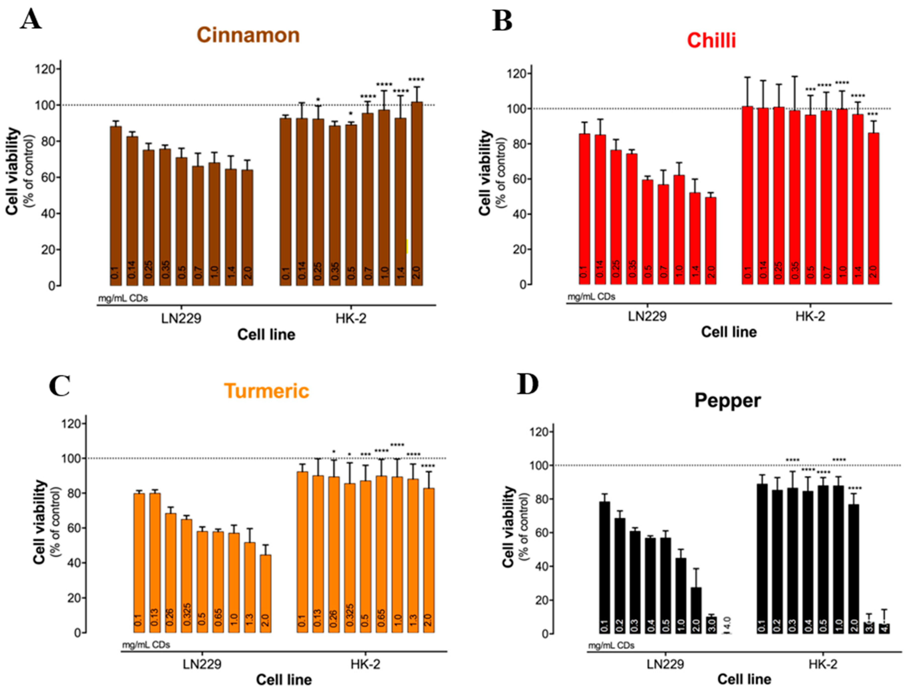
| 7 | Fluorescent carbon dots | Cinnamon | 3.4 | Human kidney cells (HK-2) | 0.1–2.0 mg/mL | Low toxicity (more than 80% cell viability remains) | [19] | |
| Red chili | 3.1 | 0.1–2.0 mg/mL | ||||||
| Turmeric | 4.3 | 0.1–2.0 mg/mL | ||||||
| Black pepper | 3.5 | 0.1–4.0 mg/mL | ||||||
| 8 | Carbon dots | Kiwi | 4.4 | Epithelial human kidney cells (HK-2) | 0.25–5.0 mg/mL | Low toxicity (up to 1 mg/mL concentration cell viability more than 60%) | [11] | |
| Avocado | 4.4 | |||||||
| Pear | 4.1 | |||||||
| 9 | Fluorescent NP-carbon dots | Wet algal biomass | 4.7 | HEK-293 (normal human embryonic kidney cell line) | 5–75 μg/mL | Negligible cytotoxicity | [43] | |
| 10 | Fluorescent carbon dots | Cyanobacteria powder | 2.5 | PC12 cells | 0–1000 µg/mL | Low cytotoxicity | [44] | |
| 11 | Carbon dots | Banana peel waste | 5 | Nematode | 0–200 µg/mL | Negligible cytotoxicity | [45] | |
| 12 | Carbon dots | Fusobacterium nucleatum cells | 4.1 | BEAS-2B (Lung normal epithelial cells) | 12.5–200 μg/mL | Low cytotoxicity (more than 80% cell viability remains) | [46] | |
| 13 | Fluorescent carbon dots | Fresh mint leaves | 6.5 | Primary H8 cells | 0–200 µg/mL | Negligible cytotoxicity | [47] | |
| 14 | Carbon dots (CDs) | E-CD | Citric acid and EDA | 10 | EA. hy926 cells | 0.1–3.2 mg/mL | Low cytotoxicity | [48] |
| U-CD | Urea and citric acid | 5 | ||||||
| Sl No. | Material | Source of Green Carbon Dot Synthesis | Average Size (nm) | Toxicity Assay in Cell Line | Concentration | Remark | Reference | |
|---|---|---|---|---|---|---|---|---|
| 1 | Luminescent carbon dots | Citrus sinensis | 6.5 | HeLa, A549, MDA-MB-231 and HEK-293 cells | 400 μg/mL | Extremely low cytotoxicity | [49] | |
| Citrus limon peels | 4.5 | |||||||
| 2 | S and N co-doped carbon dots | Water chestnut and onion | 3.5 | Human bladder cancer T24 cells | 0–300 μg/mL | Low cytotoxicity (after 24 h of incubation, cell viability remained at more than 80% for all the concentrations) | [50] | |
| 3 | Nitrogen- and sulfur-co-doped carbon dots | Ginkgo leaves juice | 2.2 | HeLa cells | N/A | Low cytotoxicity | [51] | |
| 4 | NIR-light emission carbon dots | Fresh spinach | 3–11 | A549 cells | 0–200 µg/mL | Negligible toxicity (after 24 h of co-incubation, cell viability showed above 94.2% at all the concentrations) | [52] | |
| 5 | Multicolor luminescent carbon dots | ATP | 3.8 | HeLa cells | 0–500 μg/mL | Negligible toxicity (very less change was observed between 24 h and 48 h incubation) | [53] | |
| 6 | N-doped carbon dots | Sucrose and urea | 1.6 | HeLa cells | 0–1.0 mg/mL | Negligible cytotoxicity (cell viability was more than 98.5% after 24 h of incubation, even at a high concentration, i.e., 1.0 mg/mL) | [54] | |
| 7 | Hydrophilic carbon dots | Glucose powder | 2.6 | HeLa cells | 0–1.0 mg/mL | Negligible cytotoxicity (cell viability was more than 98% after 24 h of incubation, even at a high concentration, i.e., 1.0 mg/mL) | [55] | |
| 8 | Carbon dots | Date kernels | 2.5 | Human MG-63 cells | 200.0 μg/mL | Low cytotoxicity (after 48 h of incubation, cell viability remains more than 85%) | [56] | |
| 9 | Carbon dots | Quince fruit (Cydonia oblonga) powder | 4.9 | HT-29 cells | 5–1000 μg/mL | Low toxicity | [57] | |
| 10 | Re-based carbon dots | Ginsenoside Re, citric acid and EDA | 4.6 | MCF-7, A375 HepG2 cells | 0–1.0 mg/mL | High cytotoxicity * While carbon dots were prepared separately from Ginsenoside; citric acid and EDA, cytotoxicity was relatively low. | [23] | |
| 11 | Fluorescent carbon dots | Cinnamon | 3.4 | Human glioblastoma cells (LN-229 cancer cell line) | 0.1–2.0 mg/mL | High toxicity | [19] | |
| Red chili | 3.1 | 0.1–2.0 mg/mL | ||||||
| Turmeric | 4.3 | 0.1–2.0 mg/mL | ||||||
| Black pepper | 3.5 | 0.1–4.0 mg/mL | ||||||
| 12 | Carbon dots | Kiwi | 4.4 | Epithelial human colorectal adenocarcinoma cells (Caco-2) | 0.25–5.0 mg/mL | Low toxicity (below 1.5 mg/mL concentration, cell viability was more than 80%, but cell death can induce in higher concentrations) | [11] | |
| Avocado | 4.4 | |||||||
| Pear | 4.1 | |||||||
| 13 | Fluorescent carbon dots | Prunus cerasifera fruits juice | 3–5 | HepG2 cells | 0–500 μg/mL | Low toxicity (after 24 h of incubation below 500 μg/mL concentration, cell viability was more than 90%) | [58] | |
| 14 | Carbon dots | Celery leaves | 2.1 | HepG2 cells | 0.01–0.022 g/mL | Low toxicity (cell viability was more than 85% for all the concentration after 24 h of incubation) | [59] | |
| 15 | Carbon dots | Lychee waste | 3.1 | A375 (Skin melanoma) cells | 0.0–1.2 mg/mL | Low cytotoxicity (after 48 h of incubation, cell viability was more than 89% for the highest concentration, i.e., 1.2 mg/mL) | [60] | |
| 16 | Fluorescent-N-doped carbon dots | Lemon juice and ethylenediamine | 3.0 | Human breast adenocarcinoma (MCF7) cells | 0.312–2.0 mg/mL | Low cytotoxicity (after 24 h of incubation cell viability for 2.0 mg/mL, the highest concentration was more than 88%) | [61] | |
| 17 | Fluorescent carbon dots | Fresh lamb | At 200 °C = 2.8 | HepG2 cells | 2.0 mg/mL | Low cytotoxicity (after 4 h of incubation cell, viability was more than 90% at this particular concentration) | [62] | |
| At 300 °C = 1.9 | ||||||||
| At 350 °C = 1.7 | ||||||||
| 18 | Carbon dots | Osmanthus fragrans flowers | 2.2 | A549 cells | 25–1000 μg/mL | Negligible toxicity (after 24 h of incubation, cell viability showed above 90% at all concentrations) | [63] | |
| 19 | Carbon dots | Dried wheat straw | 2.1 | HeLa cells | 0–0.8 mg/mL | Negligible cytotoxicity (cell viability remains more than 90% at all concentrations) | [64] | |
| 20 | Carbon dots | Gelatin and papain | 3.8 | A549 cells | 0–300 μg/mL | Negligible cytotoxicity (very less difference between 12 h and 24 h incubation for all concentrations, after 24 h incubation, cell viability remained above 91%) | [65] | |
| 21 | Carbon dots | Glucose | 3.0 | HeLa, HepG2 and HEK-293 cells | 0–300 mg/L | Negligible cytotoxicity (no change in cell viability for HeLa and HepG2 cells, but in the case of HEK-293 cell with increasing concentration, cell viability also increases) | [66] | |
| 22 | N, B co-doped bright fluorescent carbon dots | Solanum betaceum (S. betaceum) fruit extract | 5.0 | HeLa cells | 10–50 μg/mL | Low cytotoxicity (at minimum and maximum concentration, i.e., 10 μg/mL and 50 μg/mL, cell viability were 100% and 70%, respectively) | [67] | |
| 23 | Carbon dots | Gelatin | 5.0 | MCF-7 cell line | 20–120 μg/mL | Low cytotoxicity (cell viability was more than 80% even for the highest concentration after 24 h of incubation) | [68] | |
| 24 | Zwitterionic carbon dots | Citric acid and L-histidine | 8.5 | A549 cells | 0.01–1.5 mg/mL | Low cytotoxicity (after 24 h of incubation, cell viability was more than 90% even at a high concentration) | [69] | |
| 25 | Carbon dots | Corn stalk shell | 1.2–3.2 | A549 cells | 0–100 mg/L | Low cytotoxicity (after incubation for 24 h, cell viability remained more than 90% for all concentrations. Again, after 48 h of incubation, cell viability remained more than 75% for 100 mg/L concentration) | [70] | |
| 26 | Carbon dots | Fusobacterium nucleatum cells | 4.1 | HeLa cells | 12.5–200 μg/mL | Low cytotoxicity (more than 80% cell viability remains) | [46] | |
| 27 | Nitrogen-doped carbon dots | Jackfruit peel (JFP) | 6.4 | Dalton’s lymphoma ascites cells (DLA) | 50–200 μg/mL | Low cytotoxicity only in low concentrations, i.e., below 50 μg/mL (at 200 μg/mL concentration for JFP-carbon dots 100%, cell death was observed, but in the case of TP-carbon dots, only 60% cell death happened) | [71] | |
| Tamarind peel (TP) | 5.3 | |||||||
| 28 | Carbon dots | Arginine, chitosan, citric acid | 6–11 | AGS cells | 30:1–70:1 (carrier/DNA) | Negligible toxicity (at highest weight, cell viability decreased to 90%) | [72] | |
| 29 | Folic acid-functionalized carbon dots | Red Korean ginseng | 70 | MCF-7 cells | 10–50 μg/mL | High toxicity after 48 h of incubation | [73] | |
| 30 | Carbon dots | Tea leaves | 200 | HepG2 cells | 0–160 µg/mL | Low cytotoxicity (more 90% cell viability after 24 h of incubation at all concentrations) | [74] | |
| 31 | Carbon dots | Simarouba glauca leaf | 2.6 | Human breast cancer cell line (MCF-7) | 0–100 μg/mL | High toxicity with increasing concentration | [35] | |
| 32 | Carbon dots | Walnut oil | 12.3 | PC3, MCF-7, and HT-29 cells | 0–10 µg/mL | High cytotoxicity after 24 h of incubation | [75] | |
| 33 | Carbon dots (CDs) | E- CDs | Citric acid and EDA | 10.0 | HepG2 and A549 cells | 0.1–3.2 mg/mL | High cellular toxicity with increasing concentration | [48] |
| U-CDs | Urea and citric acid | 5.0 | ||||||
Disclaimer/Publisher’s Note: The statements, opinions and data contained in all publications are solely those of the individual author(s) and contributor(s) and not of MDPI and/or the editor(s). MDPI and/or the editor(s) disclaim responsibility for any injury to people or property resulting from any ideas, methods, instructions or products referred to in the content. |
© 2023 by the authors. Licensee MDPI, Basel, Switzerland. This article is an open access article distributed under the terms and conditions of the Creative Commons Attribution (CC BY) license (https://creativecommons.org/licenses/by/4.0/).
Share and Cite
Hatimuria, M.; Phukan, P.; Bag, S.; Ghosh, J.; Gavvala, K.; Pabbathi, A.; Das, J. Green Carbon Dots: Applications in Development of Electrochemical Sensors, Assessment of Toxicity as Well as Anticancer Properties. Catalysts 2023, 13, 537. https://doi.org/10.3390/catal13030537
Hatimuria M, Phukan P, Bag S, Ghosh J, Gavvala K, Pabbathi A, Das J. Green Carbon Dots: Applications in Development of Electrochemical Sensors, Assessment of Toxicity as Well as Anticancer Properties. Catalysts. 2023; 13(3):537. https://doi.org/10.3390/catal13030537
Chicago/Turabian StyleHatimuria, Madushmita, Plabana Phukan, Soumabha Bag, Jyotirmoy Ghosh, Krishna Gavvala, Ashok Pabbathi, and Joydeep Das. 2023. "Green Carbon Dots: Applications in Development of Electrochemical Sensors, Assessment of Toxicity as Well as Anticancer Properties" Catalysts 13, no. 3: 537. https://doi.org/10.3390/catal13030537