Next Issue
Volume 9, July
Previous Issue
Volume 9, May


Cancers, Volume 9, Issue 6 (June 2017) – 16 articles

Cover Story (view full-size image): 
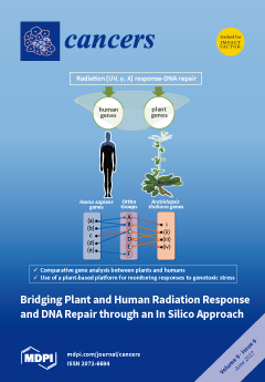
Bridging plant and animal stress responses. The conserved features of the DNA damage response and repair (DDR/R) in plants and animals might facilitate interdisciplinary studies that cross the traditional boundaries between animal and plant biology in order to discover new DNA damage biomarkers, particularly gene transcripts, for radiation exposure monitoring (REM) in environmental and biomedical settings. In this work, we have used a hybrid bioinformatics and experimental analysis, to reveal the common and divergent features of the most relevant DDR/R players in animals and plants focusing on ionizing radiation (IR) and UV-light. A plant-based platform for monitoring responses to genotoxic stresses is proposed. View this paper
  • Issues are regarded as officially published after their release is announced to the table of contents alert mailing list.
  • You may sign up for e-mail alerts to receive table of contents of newly released issues.
  • PDF is the official format for papers published in both, html and pdf forms. To view the papers in pdf format, click on the "PDF Full-text" link, and use the free Adobe Reader to open them.
Order results
Result details
Section
Select all
Export citation of selected articles as:
961 KiB  
Review
Selection of Nucleic Acid Aptamers Targeting Tumor Cell-Surface Protein Biomarkers
by Marie-Cécile Mercier, Monique Dontenwill and Laurence Choulier
Cancers 2017, 9(6), 69; https://doi.org/10.3390/cancers9060069 - 21 Jun 2017
Cited by 83 | Viewed by 11189
Abstract
Aptamers are nucleic acids referred to as chemical antibodies as they bind to their specific targets with high affinity and selectivity. They are selected via an iterative process known as ‘selective evolution of ligands by exponential enrichment’ (SELEX). Aptamers have been developed against [...] Read more.
Aptamers are nucleic acids referred to as chemical antibodies as they bind to their specific targets with high affinity and selectivity. They are selected via an iterative process known as ‘selective evolution of ligands by exponential enrichment’ (SELEX). Aptamers have been developed against numerous cancer targets and among them, many tumor cell-membrane protein biomarkers. The identification of aptamers targeting cell-surface proteins has mainly been performed by two different strategies: protein- and cell-based SELEX, when the targets used for selection were proteins and cells, respectively. This review aims to update the literature on aptamers targeting tumor cell surface protein biomarkers, highlighting potentials, pitfalls of protein- and cell-based selection processes and applications of such selected molecules. Aptamers as promising agents for diagnosis and therapeutic approaches in oncology are documented, as well as aptamers in clinical development. Full article
Show Figures

Figure 1

1376 KiB  
Review
Promotion of Tumor Invasion by Tumor-Associated Macrophages: The Role of CSF-1-Activated Phosphatidylinositol 3 Kinase and Src Family Kinase Motility Signaling
by Amy R. Dwyer, Eloise L. Greenland and Fiona J. Pixley
Cancers 2017, 9(6), 68; https://doi.org/10.3390/cancers9060068 - 18 Jun 2017
Cited by 53 | Viewed by 9580
Abstract
Macrophages interact with cells in every organ to facilitate tissue development, function and repair. However, the close interaction between macrophages and parenchymal cells can be subverted in disease, particularly cancer. Motility is an essential capacity for macrophages to be able to carry out [...] Read more.
Macrophages interact with cells in every organ to facilitate tissue development, function and repair. However, the close interaction between macrophages and parenchymal cells can be subverted in disease, particularly cancer. Motility is an essential capacity for macrophages to be able to carry out their various roles. In cancers, the macrophage’s interstitial migratory ability is frequently co-opted by tumor cells to enable escape from the primary tumor and metastatic spread. Macrophage accumulation within and movement through a tumor is often stimulated by tumor cell production of the mononuclear phagocytic growth factor, colony-stimulating factor-1 (CSF-1). CSF-1 also regulates macrophage survival, proliferation and differentiation, and its many effects are transduced by its receptor, the CSF-1R, via phosphotyrosine motif-activated signals. Mutational analysis of CSF-1R signaling indicates that the major mediators of CSF-1-induced motility are phosphatidyl-inositol-3 kinase (PI3K) and one or more Src family kinase (SFK), which activate signals to adhesion, actin polymerization, polarization and, ultimately, migration and invasion in macrophages. The macrophage transcriptome, including that of the motility machinery, is very complex and highly responsive to the environment, with selective expression of proteins and splice variants rarely found in other cell types. Thus, their unique motility machinery can be specifically targeted to block macrophage migration, and thereby, inhibit tumor invasion and metastasis. Full article
(This article belongs to the Special Issue PI3K/PDK1/Akt Pathways in Cancer)
Show Figures

Figure 1

1774 KiB  
Review
Androgen Receptor-Dependent and -Independent Mechanisms Involved in Prostate Cancer Therapy Resistance
by Daniel J. Crona and Young E. Whang
Cancers 2017, 9(6), 67; https://doi.org/10.3390/cancers9060067 - 12 Jun 2017
Cited by 89 | Viewed by 13477
Abstract
Despite the initial efficacy of androgen deprivation in prostate cancer, virtually all patients progress to castration-resistant prostate cancer (CRPC). Androgen receptor (AR) signaling is critically required for CRPC. A new generation of medications targeting AR, such as abiraterone and enzalutamide, has improved survival [...] Read more.
Despite the initial efficacy of androgen deprivation in prostate cancer, virtually all patients progress to castration-resistant prostate cancer (CRPC). Androgen receptor (AR) signaling is critically required for CRPC. A new generation of medications targeting AR, such as abiraterone and enzalutamide, has improved survival of metastatic CRPC (mCRPC) patients. However, a significant proportion of patients presents with primary resistance to these agents, and in the remainder, secondary resistance will invariably develop, which makes mCRPC the lethal form of the disease. Mechanisms underlying progression to mCRPC and treatment resistance are extremely complex. AR-dependent resistance mechanisms include AR amplification, AR point mutations, expression of constitutively active AR splice variants, and altered intratumoral androgen biosynthesis. AR-independent resistance mechanisms include glucocorticoid receptor activation, immune-mediated resistance, and neuroendocrine differentiation. The development of novel agents, such as seviteronel, apalutamide, and EPI-001/EPI-506, as well as the identification and validation of novel predictive biomarkers of resistance, may lead to improved therapeutics for mCRPC patients. Full article
Show Figures

Figure 1

2112 KiB  
Review
Carbon Ion Radiotherapy: A Review of Clinical Experiences and Preclinical Research, with an Emphasis on DNA Damage/Repair
by Osama Mohamad, Brock J. Sishc, Janapriya Saha, Arnold Pompos, Asal Rahimi, Michael D. Story, Anthony J. Davis and D.W. Nathan Kim
Cancers 2017, 9(6), 66; https://doi.org/10.3390/cancers9060066 - 09 Jun 2017
Cited by 121 | Viewed by 13673
Abstract
Compared to conventional photon-based external beam radiation (PhXRT), carbon ion radiotherapy (CIRT) has superior dose distribution, higher linear energy transfer (LET), and a higher relative biological effectiveness (RBE). This enhanced RBE is driven by a unique DNA damage signature characterized by clustered lesions [...] Read more.
Compared to conventional photon-based external beam radiation (PhXRT), carbon ion radiotherapy (CIRT) has superior dose distribution, higher linear energy transfer (LET), and a higher relative biological effectiveness (RBE). This enhanced RBE is driven by a unique DNA damage signature characterized by clustered lesions that overwhelm the DNA repair capacity of malignant cells. These physical and radiobiological characteristics imbue heavy ions with potent tumoricidal capacity, while having the potential for simultaneously maximally sparing normal tissues. Thus, CIRT could potentially be used to treat some of the most difficult to treat tumors, including those that are hypoxic, radio-resistant, or deep-seated. Clinical data, mostly from Japan and Germany, are promising, with favorable oncologic outcomes and acceptable toxicity. In this manuscript, we review the physical and biological rationales for CIRT, with an emphasis on DNA damage and repair, as well as providing a comprehensive overview of the translational and clinical data using CIRT. Full article
(This article belongs to the Special Issue DNA Repair Pathways in Cancer)
Show Figures

Figure 1

5324 KiB  
Article
Bridging Plant and Human Radiation Response and DNA Repair through an In Silico Approach
by Zacharenia Nikitaki, Athanasia Pavlopoulou, Marcela Holá, Mattia Donà, Ioannis Michalopoulos, Alma Balestrazzi, Karel J. Angelis and Alexandros G. Georgakilas
Cancers 2017, 9(6), 65; https://doi.org/10.3390/cancers9060065 - 06 Jun 2017
Cited by 11 | Viewed by 6285
Abstract
The mechanisms of response to radiation exposure are conserved in plants and animals. The DNA damage response (DDR) pathways are the predominant molecular pathways activated upon exposure to radiation, both in plants and animals. The conserved features of DDR in plants and animals [...] Read more.
The mechanisms of response to radiation exposure are conserved in plants and animals. The DNA damage response (DDR) pathways are the predominant molecular pathways activated upon exposure to radiation, both in plants and animals. The conserved features of DDR in plants and animals might facilitate interdisciplinary studies that cross traditional boundaries between animal and plant biology in order to expand the collection of biomarkers currently used for radiation exposure monitoring (REM) in environmental and biomedical settings. Genes implicated in trans-kingdom conserved DDR networks often triggered by ionizing radiation (IR) and UV light are deposited into biological databases. In this study, we have applied an innovative approach utilizing data pertinent to plant and human genes from publicly available databases towards the design of a ‘plant radiation biodosimeter’, that is, a plant and DDR gene-based platform that could serve as a REM reliable biomarker for assessing environmental radiation exposure and associated risk. From our analysis, in addition to REM biomarkers, a significant number of genes, both in human and Arabidopsis thaliana, not yet characterized as DDR, are suggested as possible DNA repair players. Last but not least, we provide an example on the applicability of an Arabidopsis thaliana—based plant system monitoring the role of cancer-related DNA repair genes BRCA1, BARD1 and PARP1 in processing DNA lesions. Full article
(This article belongs to the Special Issue DNA Repair Pathways in Cancer)
Show Figures

Graphical abstract

1862 KiB  
Article
Being-in-the-Chemotherapy-Suite versus Being-in-the-Oncology-Ward: An Analytical View of Two Hospital Sites Occupied by People Experiencing Cancer
by Catherine Hughes, Kate Van Heugten, Sally Keeling and Francisc Szekely
Cancers 2017, 9(6), 64; https://doi.org/10.3390/cancers9060064 - 05 Jun 2017
Cited by 3 | Viewed by 5776
Abstract
How do people with cancer occupy places within the health system during their journey through palliative care? The answer to this question was explored by the authors as part of a wider ethnographic study of eight people’s journeys from referral to palliative care [...] Read more.
How do people with cancer occupy places within the health system during their journey through palliative care? The answer to this question was explored by the authors as part of a wider ethnographic study of eight people’s journeys from referral to palliative care services to the end of life. This article reports on findings that have emerged from ongoing analysis that has been completed in the years proceeding data collection. An ethnographic research design was used to collect data about the participants and their family members over a three-year period. Data was collected using participant observation and semi-structured interviews. Over 380 transcripts based on field note entries and taped interviews were produced during the 1121 h of contact with participants and family members that made up the research period. Analysis of these texts identified two focal sites within Christchurch Hospital that were occupied by the participants. These were the Chemotherapy Suite and the Oncology Ward. Drawing on literature concerning previous anthropological analysis, research was conducted to understand how places affect people and how people affect places. The researchers have used a model outlined by the American ethnographer Miles Richardson to analyse two distinct sites within one hospital. As explained in Richardson’s article, whose title is used to model the title of this article, a sense of place becomes apparent when comparing and contrasting two sites within the same location. Richardson’s article is highly interpretative and relies not only on pre-existing theoretical frameworks but also on personal interpretation. The same approach has been used in the current article. Here, ethnographic methods require the researcher’s interpretation of how participants occupied these sites. Following this approach, the Chemotherapy Suite is presented as a place where medicine dominates illness, and appears as distinct from the Oncology Ward, where disease predominates and death is secreted away. Full article
(This article belongs to the Special Issue End-of-Life Cancer Care)
Show Figures

Figure 1

2070 KiB  
Article
Combination of Near Infrared Light-Activated Photodynamic Therapy Mediated by Indocyanine Green with Etoposide to Treat Non-Small-Cell Lung Cancer
by Ting Luo, Qinrong Zhang and Qing-Bin Lu
Cancers 2017, 9(6), 63; https://doi.org/10.3390/cancers9060063 - 05 Jun 2017
Cited by 15 | Viewed by 6254
Abstract
Indocyanine green (ICG) has been reported as a potential near-infrared (NIR) photosensitizer for photodynamic therapy (PDT) of cancer. However the application of ICG-mediated PDT is both intrinsically and physiologically limited. Here we report a combination of ICG-PDT with a chemotherapy drug etoposide (VP-16), [...] Read more.
Indocyanine green (ICG) has been reported as a potential near-infrared (NIR) photosensitizer for photodynamic therapy (PDT) of cancer. However the application of ICG-mediated PDT is both intrinsically and physiologically limited. Here we report a combination of ICG-PDT with a chemotherapy drug etoposide (VP-16), aiming to enhance the anticancer efficacy, to circumvent limitations of PDT using ICG, and to reduce side effects of VP-16. We found in controlled in vitro cell-based assays that this combination is effective in killing non-small-cell lung cancer cells (NSCLC, A549 cell line). We also found that the combination of ICG-PDT and VP-16 exhibits strong synergy in killing non-small-cell lung cancer cells partially through inducing more DNA double-strand breaks (DSBs), while it has a much weaker synergy in killing human normal cells (GM05757). Furthermore, by studying the treatment sequence dependence and the cytotoxicity of laser-irradiated mixtures of ICG and VP-16, we found that the observed synergy involves direct/indirect reactions between ICG and VP-16. We further propose that there exists an electron transfer reaction between ICG and VP-16 under irradiation. This study therefore shows the anticancer efficacy of ICG-PDT combined with VP-16. These findings suggest that ICG-mediated PDT may be applied in combination with the chemotherapy drug VP-16 to treat some cancers, especially the non-small-cell lung cancer. Full article
(This article belongs to the Special Issue Photodynamic Cancer Therapy)
Show Figures

Figure 1

3450 KiB  
Article
Local Immune Responsiveness of Mice Bearing Premalignant Oral Lesions to PD-1 Antibody Treatment
by Corinne A. Levingston and M. Rita I. Young
Cancers 2017, 9(6), 62; https://doi.org/10.3390/cancers9060062 - 02 Jun 2017
Cited by 8 | Viewed by 4122
Abstract
A carcinogen-induced premalignant oral lesion model that progresses to oral cancer was used to examine the immunological impact of a 5-week treatment regimen to block programmed cell death protein 1 (PD-1). PD-1 antibody treatment resulted in concurrent, but transient, increases in interleukin (IL)-2, [...] Read more.
A carcinogen-induced premalignant oral lesion model that progresses to oral cancer was used to examine the immunological impact of a 5-week treatment regimen to block programmed cell death protein 1 (PD-1). PD-1 antibody treatment resulted in concurrent, but transient, increases in interleukin (IL)-2, IFN-γ and IL-17, and delayed increases in IL-6 and IL-10 within the lesion-bearing tongue epithelium. In contrast, cytokine secretion by lymph node cells of PD-1 antibody-treated mice was lower than for mice treated with control antibodies, with the exception of interferon (IFN)-γ, whose secretion increased late in the treatment period. This delayed secretion of IFN-γ coincided with an increase in CD4+ lymph node cells expressing IFN-γ. Lymph node cells of PD-1 antibody-treated mice reacted to a challenge with lysates of lesions or cancer by early production of IFN-γ, but this rapidly subsided. There also was increased production IL-17 and tumor necrosis factor (TNF)-α in response to the challenge, but the response was greatest by cells of control lesion-bearing mice. Clinical assessment showed an early but transient, stabilization of disease in mice treated with PD-1 antibody. These results show an early beneficial, but time-limited, response to PD-1 antibody treatment, which then fails with continued lesion progression. Full article
Show Figures

Figure 1

3703 KiB  
Article
A Novel Micro Cold Atmospheric Plasma Device for Glioblastoma Both In Vitro and In Vivo
by Zhitong Chen, Hayk Simonyan, Xiaoqian Cheng, Eda Gjika, Li Lin, Jerome Canady, Jonathan H. Sherman, Colin Young and Michael Keidar
Cancers 2017, 9(6), 61; https://doi.org/10.3390/cancers9060061 - 30 May 2017
Cited by 92 | Viewed by 10301
Abstract
Cold atmospheric plasma (CAP) treatment is a rapidly expanding and emerging technology for cancer treatment. Direct CAP jet irradiation is limited to the skin and it can also be invoked as a supplement therapy during surgery as it only causes cell death in [...] Read more.
Cold atmospheric plasma (CAP) treatment is a rapidly expanding and emerging technology for cancer treatment. Direct CAP jet irradiation is limited to the skin and it can also be invoked as a supplement therapy during surgery as it only causes cell death in the upper three to five cell layers. However, the current cannulas from which the plasma emanates are too large for intracranial applications. To enhance efficiency and expand the applicability of the CAP method for brain tumors and reduce the gas flow rate and size of the plasma jet, a novel micro-sized CAP device (µCAP) was developed and employed to target glioblastoma tumors in the murine brain. Various plasma diagnostic techniques were applied to evaluate the physics of helium µCAP such as electron density, discharge voltage, and optical emission spectroscopy (OES). The direct and indirect effects of µCAP on glioblastoma (U87MG-RedFluc) cancer cells were investigated in vitro. The results indicate that µCAP generates short- and long-lived species and radicals (i.e., hydroxyl radical (OH), hydrogen peroxide (H2O2), and nitrite (NO2), etc.) with increasing tumor cell death in a dose-dependent manner. Translation of these findings to an in vivo setting demonstrates that intracranial µCAP is effective at preventing glioblastoma tumor growth in the mouse brain. The µCAP device can be safely used in mice, resulting in suppression of tumor growth. These initial observations establish the µCAP device as a potentially useful ablative therapy tool in the treatment of glioblastoma. Full article
Show Figures

Figure 1

565 KiB  
Review
Towards Targeting PI3K-Dependent Regulation of Gene Expression in Brain Cancer
by Theo Mantamadiotis
Cancers 2017, 9(6), 60; https://doi.org/10.3390/cancers9060060 - 30 May 2017
Cited by 25 | Viewed by 5133
Abstract
The PI3K pathway is one of the most highly perturbed cell signaling pathways in human cancer, including the most common malignant brain tumors, gliomas, where either activating mutations of positive pathway effectors or loss/inactivation of pathway inhibitors occurs. Knowledge of the precise transcription [...] Read more.
The PI3K pathway is one of the most highly perturbed cell signaling pathways in human cancer, including the most common malignant brain tumors, gliomas, where either activating mutations of positive pathway effectors or loss/inactivation of pathway inhibitors occurs. Knowledge of the precise transcription factors modulated by PI3K in tumor cells remains elusive but there are numerous PI3K-responsive signaling factors, including kinases, which can activate many transcription factors. In the context of cancer, these transcription factors participate in the regulation of target genes expression networks to support cancer cell characteristics such as survival, proliferation, migration and differentiation. This review focuses on the role of PI3K signaling-regulated transcription in brain cancer cells from a series of recent investigations. A deeper understanding of this regulation is beginning to provide the hope of developing more sophisticated anti-cancer targeting approaches, where both upstream and downstream components of the PI3K pathway may be targeted by existing and novel drugs. Full article
(This article belongs to the Special Issue PI3K/PDK1/Akt Pathways in Cancer)
Show Figures

Figure 1

206 KiB  
Review
Improving Outcomes in Patients with CRC: The Role of Patient Reported Outcomes—An ESDO Report
by Eric Van Cutsem, Aimery De Gramont, Geoffrey Henning, Philippe Rougier, Franck Bonnetain and Thomas Seufferlein
Cancers 2017, 9(6), 59; https://doi.org/10.3390/cancers9060059 - 26 May 2017
Cited by 6 | Viewed by 4098
Abstract
Colorectal cancer is one of the most commonly diagnosed cancers worldwide and nearly half of patients will develop metastatic disease at some point during the course of their disease. The goal of anticancer therapy in this context is to extend survival, while trying [...] Read more.
Colorectal cancer is one of the most commonly diagnosed cancers worldwide and nearly half of patients will develop metastatic disease at some point during the course of their disease. The goal of anticancer therapy in this context is to extend survival, while trying to maximise the patient’s health-related quality of life. To this end, we need to understand how to incorporate patient-reported outcomes into clinical trials and routine practice to accurately assess if treatment strategies are providing clinical benefit for the patient. This review reflects the proceedings of a 2016 European Society of Digestive Oncology workshop, where the authors discussed the use of patient-reported outcomes to measure health-related quality of life when evaluating treatment during the management of colorectal cancer. A summary of the challenges associated with implementing patient-reported outcomes in clinical trials is provided, as well as a review of the current clinical evidence surrounding patient-reported outcomes in metastatic colorectal cancer. Full article
558 KiB  
Review
The Therapeutic Targeting of HGF/c-Met Signaling in Hepatocellular Carcinoma: Alternative Approaches
by Chi-Tan Hu, Jia-Ru Wu, Chuan-Chu Cheng and Wen-Sheng Wu
Cancers 2017, 9(6), 58; https://doi.org/10.3390/cancers9060058 - 26 May 2017
Cited by 40 | Viewed by 9696
Abstract
The poor prognosis of hepatocellular carcinoma (HCC), one of the most devastating cancers worldwide, is due to frequent recurrence and metastasis. Among the metastatic factors in the tumor microenvironment, hepatocyte growth factor (HGF) has been well known to play critical roles in tumor [...] Read more.
The poor prognosis of hepatocellular carcinoma (HCC), one of the most devastating cancers worldwide, is due to frequent recurrence and metastasis. Among the metastatic factors in the tumor microenvironment, hepatocyte growth factor (HGF) has been well known to play critical roles in tumor progression, including HCC. Therefore, c-Met is now regarded as the most promising therapeutic target for the treatment of HCC. However, there are still concerns about resistance and the side effects of using conventional inhibitors of c-Met, such as tyrosine kinase inhibitors. Recently, many alternative strategies of c-Met targeting have been emerging. These include targeting the downstream effectors of c-Met, such as hydrogen peroxide-inducible clone 5 (Hic-5), to block the reactive oxygen species (ROS)-mediated signaling for HCC progression. Also, inhibition of endosomal regulators, such as PKCε and GGA3, may perturb the c-Met endosomal signaling for HCC cell migration. On the other hand, many herbal antagonists of c-Met-dependent signaling, such as saponin, resveratrol, and LZ-8, were identified. Taken together, it can be anticipated that more effective and safer c-Met targeting strategies for preventing HCC progression can be established in the future. Full article
(This article belongs to the Special Issue Hepatocyte Growth Factor Pathway in Cancer)
Show Figures

Figure 1

2344 KiB  
Review
Chemotherapeutic Drugs: DNA Damage and Repair in Glioblastoma
by Laura Annovazzi, Marta Mellai and Davide Schiffer
Cancers 2017, 9(6), 57; https://doi.org/10.3390/cancers9060057 - 26 May 2017
Cited by 59 | Viewed by 11285
Abstract
Despite improvements in therapeutic strategies, glioblastoma (GB) remains one of the most lethal cancers. The presence of the blood–brain barrier, the infiltrative nature of the tumor and several resistance mechanisms account for the failure of current treatments. Distinct DNA repair pathways can neutralize [...] Read more.
Despite improvements in therapeutic strategies, glioblastoma (GB) remains one of the most lethal cancers. The presence of the blood–brain barrier, the infiltrative nature of the tumor and several resistance mechanisms account for the failure of current treatments. Distinct DNA repair pathways can neutralize the cytotoxicity of chemo- and radio-therapeutic agents, driving resistance and tumor relapse. It seems that a subpopulation of stem-like cells, indicated as glioma stem cells (GSCs), is responsible for tumor initiation, maintenance and recurrence and they appear to be more resistant owing to their enhanced DNA repair capacity. Recently, attention has been focused on the pivotal role of the DNA damage response (DDR) in tumorigenesis and in the modulation of therapeutic treatment effects. In this review, we try to summarize the knowledge concerning the main molecular mechanisms involved in the removal of genotoxic lesions caused by alkylating agents, emphasizing the role of GSCs. Beside their increased DNA repair capacity in comparison with non-stem tumor cells, GSCs show a constitutive checkpoint expression that enables them to survive to treatments in a quiescent, non-proliferative state. The targeted inhibition of checkpoint/repair factors of DDR can contribute to eradicate the GSC population and can have a great potential therapeutic impact aiming at sensitizing malignant gliomas to treatments, improving the overall survival of patients. Full article
(This article belongs to the Special Issue DNA Repair Pathways in Cancer)
Show Figures

Figure 1

257 KiB  
Review
RGD-Binding Integrins in Head and Neck Cancers
by Hanadi Talal Ahmedah, Laurence H. Patterson, Steven D. Shnyder and Helen M. Sheldrake
Cancers 2017, 9(6), 56; https://doi.org/10.3390/cancers9060056 - 26 May 2017
Cited by 27 | Viewed by 6244
Abstract
Alterations in integrin expression and function promote tumour growth, invasion, metastasis and neoangiogenesis. Head and neck cancers are highly vascular tumours with a tendency to metastasise. They express a wide range of integrin receptors. Expression of the αv and β1 subunits has been [...] Read more.
Alterations in integrin expression and function promote tumour growth, invasion, metastasis and neoangiogenesis. Head and neck cancers are highly vascular tumours with a tendency to metastasise. They express a wide range of integrin receptors. Expression of the αv and β1 subunits has been explored relatively extensively and linked to tumour progression and metastasis. Individual receptors αvβ3 and αvβ5 have proved popular targets for diagnostic and therapeutic agents but lesser studied receptors, such as αvβ6, αvβ8, and β1 subfamily members, also show promise. This review presents the current knowledge of integrin expression and function in squamous cell carcinoma of the head and neck (HNSCC), with a particular focus on the arginine-glycine-aspartate (RGD)-binding integrins, in order to highlight the potential of integrins as targets for personalised tumour-specific identification and therapy. Full article
(This article belongs to the Special Issue Integrins in Cancer)
1247 KiB  
Article
Self-Esteem and Academic Difficulties in Preadolescents and Adolescents Healed from Paediatric Leukaemia
by Marta Tremolada, Livia Taverna, Sabrina Bonichini, Giuseppe Basso and Marta Pillon
Cancers 2017, 9(6), 55; https://doi.org/10.3390/cancers9060055 - 24 May 2017
Cited by 29 | Viewed by 5792
Abstract
Adolescents with cancer may demonstrate problems in their self-esteem and schooling. This study aims to screen the preadolescents and adolescents more at risk in their self-esteem perception and schooling difficulties post-five years from the end of therapy. Twenty-five paediatric ex-patients healed from leukaemia [...] Read more.
Adolescents with cancer may demonstrate problems in their self-esteem and schooling. This study aims to screen the preadolescents and adolescents more at risk in their self-esteem perception and schooling difficulties post-five years from the end of therapy. Twenty-five paediatric ex-patients healed from leukaemia were recruited at the Haematology-Oncologic Clinic (University of Padua). The mean age of the children was 13.64 years (Standard Deviation (SD)) = 3.08, range = 10–19 years), most were treated for acute lymphoblastic leukaemia (ALL) (84%) and relatively equally distributed by gender. They filled in the Multidimensional Self-Esteem Test, while parents completed a questionnaire on their child’s schooling. Global self-esteem was mostly below the 50 percentile (58.5%), especially regarding interpersonal relationships (75%). An independent sample t-test showed significant mean differences on the emotionality scale (t = 2.23; degree of freedom (df) = 24; p = 0.03) and in the bodily experience scale (t = 3.02; df = 24; p = 0.006) with survivors of Acute Myeloid Leukaemia (AML) having lower scores. An Analysis of Variance (ANOVA) showed significant mean differences in the bodily experience scale (F = 12.31; df = 2, p = 0.0001) depending on the survivors’ assigned risk band. The parent reports showed that 43.5% of children had difficulties at school. Childhood AML survivors with a high-risk treatment were more at risk in their self-esteem perceptions. Preventive interventions focusing on self-esteem and scholastic wellbeing are suggested in order to help their return to their normal schedules. Full article
(This article belongs to the Special Issue Quality of Life for Cancer Patients)
Show Figures

Figure 1

1344 KiB  
Review
Dysregulation of miRNA Expression in Cancer Associated Fibroblasts (CAFs) and Its Consequences on the Tumor Microenvironment
by Maren Schoepp, Anda Jana Ströse and Jörg Haier
Cancers 2017, 9(6), 54; https://doi.org/10.3390/cancers9060054 - 24 May 2017
Cited by 65 | Viewed by 7145
Abstract
The tumor microenvironment, including cancer-associated fibroblasts (CAF), has developed as an important target for understanding tumor progression, clinical prognosis and treatment responses of cancer. Cancer cells appear to transform normal fibroblasts (NF) into CAFs involving direct cell-cell communication and epigenetic regulations. This review [...] Read more.
The tumor microenvironment, including cancer-associated fibroblasts (CAF), has developed as an important target for understanding tumor progression, clinical prognosis and treatment responses of cancer. Cancer cells appear to transform normal fibroblasts (NF) into CAFs involving direct cell-cell communication and epigenetic regulations. This review summarizes the current understanding on miR involvement in cancer cell—tumor environment/stroma communication, transformation of NFs into CAFs, their involved targets and signaling pathways in these interactions; and clinical relevance of CAF-related miR expression profiles. There is evidence that miRs have very similar roles in activating hepatic (HSC) and pancreatic stellate cells (PSC) as part of precancerous fibrotic diseases. In summary, deregulated miRs affect various intracellular functional complexes, such as transcriptional factors, extracellular matrix, cytoskeleton, EMT/MET regulation, soluble factors, tyrosine kinase and G-protein signaling, apoptosis and cell cycle & differentiation, but also formation and composition of the extracellular microenvironment. These processes result in the clinical appearance of desmoplasia involving CAFs and fibrosis characterized by deregulated stellate cells. In addition, modulated release of soluble factors can act as (auto)activating feedback loop for transition of NFs into their pathological counterparts. Furthermore, epigenetic communication between CAFs and cancer cells may confer to cancer specific functional readouts and transition of NF. MiR related epigenetic regulation with many similarities should be considered as key factor in development of cancer and fibrosis specific environment. Full article
Show Figures

Figure 1

Previous Issue
Next Issue
Back to TopTop