Next Issue
Volume 9, June
Previous Issue
Volume 9, April


Cancers, Volume 9, Issue 5 (May 2017) – 14 articles

Cover Story (view full-size image): 
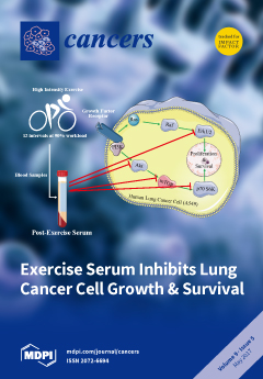
Lung cancer accounts for the most cancer-related deaths globally, indicating an urgent need to find strategies for its prevention and treatment. The Akt, mammalian target of rapamycin (mTOR) and p70 S6K, and the Ras-extracellular signal-regulated kinase (Erk1/2) pathways are activated in cancer leading to cell survival and growth. Exercise is associated with health benefits and in the present study we found that exposure of lung cancer cells to post exercise serum resulted in inhibition of cell proliferation and survival, as well as a significant reduction in activated Akt, mTOR, p70 S6K and Erk1/2. Our data suggest that post exercise serum has anti-cancer properties in lung cancer and deserves further systematic investigation. View this paper
  • Issues are regarded as officially published after their release is announced to the table of contents alert mailing list.
  • You may sign up for e-mail alerts to receive table of contents of newly released issues.
  • PDF is the official format for papers published in both, html and pdf forms. To view the papers in pdf format, click on the "PDF Full-text" link, and use the free Adobe Reader to open them.
Order results
Result details
Section
Select all
Export citation of selected articles as:
526 KiB  
Review
Physical Activity and Quality of Life in Cancer Survivors: A Meta-Synthesis of Qualitative Research
by Shaunna Burke, Amanda Wurz, Andrew Bradshaw, Stephanie Saunders, Malcolm A. West and Jennifer Brunet
Cancers 2017, 9(5), 53; https://doi.org/10.3390/cancers9050053 - 20 May 2017
Cited by 66 | Viewed by 10294
Abstract
Qualitative research on the impact of physical activity on quality of life (QoL) in adults diagnosed with cancer is accumulating. However, the field of physical activity and cancer survivorship lack a synthesis of this research to reliably understand the implications for future research [...] Read more.
Qualitative research on the impact of physical activity on quality of life (QoL) in adults diagnosed with cancer is accumulating. However, the field of physical activity and cancer survivorship lack a synthesis of this research to reliably understand the implications for future research and practice. The aim of this meta-synthesis was to identify, appraise, and synthesize qualitative research on cancer survivors’ perspectives of the impact of physical activity on their QoL. Seven electronic databases were searched for original studies published in English, and reference lists of relevant studies were hand-searched to identify additional studies. Forty studies met eligibility criteria and were included in this meta-synthesis. Study characteristics and major findings were extracted, and findings were summarized, compared, and synthesized. Themes identified in this review revealed that physical activity positively impacted four dimensions of cancer survivors’ QoL: physical (e.g., managing the physical consequences of cancer and its treatment), psychological (e.g., evoking positive self-perceptions), social (e.g., feeling understood by others), and spiritual (e.g., redefining life purpose). This meta-synthesis corroborates conclusions from reviews of quantitative research and illustrates that physical activity can be used to improve QoL in adult cancer survivors, regardless of diagnosis (i.e., stage, cancer type) and treatment status. It also provides detailed insight into specific aspects within each dimension of QoL impacted by physical activity from cancer survivors’ perspectives, which is important for understanding the meaning and utility of physical activity for them. However, more research is needed to further develop the qualitative evidence base in order to better understand how physical activity impacts on QoL experiences in men, young adults, and adults diagnosed with less common types of cancer at different points along cancer trajectory (i.e., diagnosis, treatment, post-treatment, palliation). Full article
(This article belongs to the Special Issue Quality of Life for Cancer Patients)
Show Figures

Figure 1

2311 KiB  
Review
Epidermal Growth Factor Receptor Cell Proliferation Signaling Pathways
by Ping Wee and Zhixiang Wang
Cancers 2017, 9(5), 52; https://doi.org/10.3390/cancers9050052 - 17 May 2017
Cited by 1099 | Viewed by 46514
Abstract
The epidermal growth factor receptor (EGFR) is a receptor tyrosine kinase that is commonly upregulated in cancers such as in non-small-cell lung cancer, metastatic colorectal cancer, glioblastoma, head and neck cancer, pancreatic cancer, and breast cancer. Various mechanisms mediate the upregulation of EGFR [...] Read more.
The epidermal growth factor receptor (EGFR) is a receptor tyrosine kinase that is commonly upregulated in cancers such as in non-small-cell lung cancer, metastatic colorectal cancer, glioblastoma, head and neck cancer, pancreatic cancer, and breast cancer. Various mechanisms mediate the upregulation of EGFR activity, including common mutations and truncations to its extracellular domain, such as in the EGFRvIII truncations, as well as to its kinase domain, such as the L858R and T790M mutations, or the exon 19 truncation. These EGFR aberrations over-activate downstream pro-oncogenic signaling pathways, including the RAS-RAF-MEK-ERK MAPK and AKT-PI3K-mTOR pathways. These pathways then activate many biological outputs that are beneficial to cancer cell proliferation, including their chronic initiation and progression through the cell cycle. Here, we review the molecular mechanisms that regulate EGFR signal transduction, including the EGFR structure and its mutations, ligand binding and EGFR dimerization, as well as the signaling pathways that lead to G1 cell cycle progression. We focus on the induction of CYCLIN D expression, CDK4/6 activation, and the repression of cyclin-dependent kinase inhibitor proteins (CDKi) by EGFR signaling pathways. We also discuss the successes and challenges of EGFR-targeted therapies, and the potential for their use in combination with CDK4/6 inhibitors. Full article
(This article belongs to the Special Issue EGFR Family Signaling in Cancer)
Show Figures

Figure 1

1155 KiB  
Article
A Feasibility Study of Personalized Prescription Schemes for Glioblastoma Patients Using a Proliferation and Invasion Glioma Model
by Minsun Kim, Jakob Kotas, Jason Rockhill and Mark Phillips
Cancers 2017, 9(5), 51; https://doi.org/10.3390/cancers9050051 - 13 May 2017
Cited by 8 | Viewed by 4472
Abstract
Purpose: This study investigates the feasibility of personalizing radiotherapy prescription schemes (treatment margins and fractional doses) for glioblastoma (GBM) patients and their potential benefits using a proliferation and invasion (PI) glioma model on phantoms. Methods and Materials: We propose a strategy to personalize [...] Read more.
Purpose: This study investigates the feasibility of personalizing radiotherapy prescription schemes (treatment margins and fractional doses) for glioblastoma (GBM) patients and their potential benefits using a proliferation and invasion (PI) glioma model on phantoms. Methods and Materials: We propose a strategy to personalize radiotherapy prescription schemes by simulating the proliferation and invasion of the tumor in 2D according to the PI glioma model. We demonstrate the strategy and its potential benefits by presenting virtual cases, where the standard and personalized prescriptions were applied to the tumor. Standard prescription was assumed to deliver 46 Gy in 23 fractions to the initial, gross tumor volume (GTV1) plus a 2 cm margin and an additional 14 Gy in 7 fractions to the boost GTV2 plus a 2 cm margin. The virtual cases include the tumors with a moving velocity of 0.029 (slow-move), 0.079 (average-move), and 0.13 (fast-move) mm/day for the gross tumor volume (GTV) with a radius of 1 (small) and 2 (large) cm. For each tumor size and velocity, the margin around GTV1 and GTV2 was varied between 0–6 cm and 1–3 cm, respectively. Equivalent uniform dose (EUD) to normal brain was constrained to the EUD value obtained by using the standard prescription. Various linear dose policies, where the fractional dose is linearly decreasing, constant, or increasing, were investigated to estimate the temporal effect of the radiation dose on tumor cell-kills. The goal was to find the combination of margins for GTV1 and GTV2 and a linear dose policy, which minimize the tumor cell-surviving fraction (SF) under a normal tissue constraint. The efficacy of a personalized prescription was evaluated by tumor EUD and the estimated survival time. Results: The personalized prescription for the slow-move tumors was to use 3.0–3.5 cm margins for GTV1, and a 1.5 cm margin for GTV2. For the average- and fast-move tumors, it was optimal to use a 6.0 cm margin for GTV1 and then 1.5–3.0 cm margins for GTV2, suggesting a course of whole brain therapy followed by a boost to a smaller volume. It was more effective to deliver the boost sequentially using a linearly decreasing fractional dose for all tumors. Personalized prescriptions led to surviving fractions of 0.001–0.465% compared to the standard prescription, and increased the tumor EUDs by 25.3–49.3% and estimated survival times by 7.6–22.2 months. Conclusions: Personalizing treatment margins based on the measured proliferative capacity of GBM tumor cells can potentially lead to significant improvements in tumor cell kill and related clinical outcomes. Full article
Show Figures

Figure 1

208 KiB  
Review
Immunotherapy for Colorectal Cancer
by Patrick M. Boland and Wen Wee Ma
Cancers 2017, 9(5), 50; https://doi.org/10.3390/cancers9050050 - 11 May 2017
Cited by 122 | Viewed by 13506
Abstract
The recent success of anti-PD1 drugs in metastatic colorectal cancer patients with mismatch repair deficiency generated overwhelming enthusiasm for immunotherapy in the disease. However, patients with mismatch repair deficient colorectal cancer represent only a small subset of the metastatic population. Current research focuses [...] Read more.
The recent success of anti-PD1 drugs in metastatic colorectal cancer patients with mismatch repair deficiency generated overwhelming enthusiasm for immunotherapy in the disease. However, patients with mismatch repair deficient colorectal cancer represent only a small subset of the metastatic population. Current research focuses on advancing immunotherapy to earlier stages of the disease including adjuvant and first-line metastatic settings, and on inducing sensitivity to immune checkpoint inhibitor therapy through a combinatorial approach. Here, we review the contemporary understanding of the immune and molecular landscape in colorectal cancer and discuss ongoing clinical trials evaluating novel combination regimens based on immune checkpoint inhibitors. Full article
(This article belongs to the Special Issue Cancer Immunotherapies)
237 KiB  
Review
Diagnostic and Therapeutic Potential of MicroRNAs in Lung Cancer
by Kentaro Inamura
Cancers 2017, 9(5), 49; https://doi.org/10.3390/cancers9050049 - 09 May 2017
Cited by 57 | Viewed by 6447
Abstract
Lung cancer is the leading cause of deaths resulting from cancer owing to late diagnosis and limited treatment intervention. MicroRNAs are short, non-coding RNA molecules that regulate gene expression post-transcriptionally by translational repression or target messenger RNA degradation. Accumulating evidence suggests various roles [...] Read more.
Lung cancer is the leading cause of deaths resulting from cancer owing to late diagnosis and limited treatment intervention. MicroRNAs are short, non-coding RNA molecules that regulate gene expression post-transcriptionally by translational repression or target messenger RNA degradation. Accumulating evidence suggests various roles for microRNAs, including development and progression of lung cancers. Because microRNAs are degraded to a much lesser extent in formalin-fixed paraffin-embedded specimens and are present not only in tumor tissues but also in body fluids, there is an increased potential in microRNA analyses for cancer research. In this review, recent studies of microRNA are introduced and briefly summarized, with a focus on the association of microRNAs with histological subtypes, genetic driver alterations, therapeutically-targeted molecules, and carcinogens. The reported circulating microRNA signature for the early detection of lung cancer and the implications of microRNAs as the modulators of tumor immune response are also introduced. Full article
1844 KiB  
Review
Targeting Intracellular Calcium Signaling ([Ca2+]i) to Overcome Acquired Multidrug Resistance of Cancer Cells: A Mini-Overview
by Dietrich Büsselberg and Ana-Maria Florea
Cancers 2017, 9(5), 48; https://doi.org/10.3390/cancers9050048 - 09 May 2017
Cited by 46 | Viewed by 6503
Abstract
Cancer is a main public health problem all over the world. It affects millions of humans no matter their age, gender, education, or social status. Although chemotherapy is the main strategy for the treatment of cancer, a major problem limiting its success is [...] Read more.
Cancer is a main public health problem all over the world. It affects millions of humans no matter their age, gender, education, or social status. Although chemotherapy is the main strategy for the treatment of cancer, a major problem limiting its success is the intrinsic or acquired drug resistance. Therefore, cancer drug resistance is a major impediment in medical oncology resulting in a failure of a successful cancer treatment. This mini-overview focuses on the interdependent relationship between intracellular calcium ([Ca2+]i) signaling and multidrug resistance of cancer cells, acquired upon treatment of tumors with anticancer drugs. We propose that [Ca2+]i signaling modulates gene expression of multidrug resistant (MDR) genes which in turn can be modulated by epigenetic factors which in turn leads to modified protein expression in drug resistant tumor cells. A precise knowledge of these mechanisms will help to develop new therapeutic strategies for drug resistant tumors and will improve current chemotherapy. Full article
Show Figures

Figure 1

1030 KiB  
Review
MTH1 as a Chemotherapeutic Target: The Elephant in the Room
by Govindi J. Samaranayake, Mai Huynh and Priyamvada Rai
Cancers 2017, 9(5), 47; https://doi.org/10.3390/cancers9050047 - 08 May 2017
Cited by 48 | Viewed by 7288
Abstract
Many tumors sustain elevated levels of reactive oxygen species (ROS), which drive oncogenic signaling. However, ROS can also trigger anti-tumor responses, such as cell death or senescence, through induction of oxidative stress and concomitant DNA damage. To circumvent the adverse consequences of elevated [...] Read more.
Many tumors sustain elevated levels of reactive oxygen species (ROS), which drive oncogenic signaling. However, ROS can also trigger anti-tumor responses, such as cell death or senescence, through induction of oxidative stress and concomitant DNA damage. To circumvent the adverse consequences of elevated ROS levels, many tumors develop adaptive responses, such as enhanced redox-protective or oxidatively-generated damage repair pathways. Targeting these enhanced oxidative stress-protective mechanisms is likely to be both therapeutically effective and highly specific to cancer, as normal cells are less reliant on such mechanisms. In this review, we discuss one such stress-protective protein human MutT Homolog1 (MTH1), an enzyme that eliminates 8-oxo-7,8-dihydro-2’-deoxyguanosine triphosphate (8-oxodGTP) through its pyrophosphatase activity, and is found to be elevated in many cancers. Our studies, and subsequently those of others, identified MTH1 inhibition as an effective tumor-suppressive strategy. However, recent studies with the first wave of MTH1 inhibitors have produced conflicting results regarding their cytotoxicity in cancer cells and have led to questions regarding the validity of MTH1 as a chemotherapeutic target. To address the proverbial "elephant in the room" as to whether MTH1 is a bona fide chemotherapeutic target, we provide an overview of MTH1 function in the context of tumor biology, summarize the current literature on MTH1 inhibitors, and discuss the molecular contexts likely required for its efficacy as a therapeutic target. Full article
(This article belongs to the Special Issue DNA Repair Pathways in Cancer)
Show Figures

Figure 1

2485 KiB  
Article
Inhibition of Human Lung Cancer Cell Proliferation and Survival by Post-Exercise Serum Is Associated with the Inhibition of Akt, mTOR, p70 S6K, and Erk1/2
by Nigel Kurgan, Evelyn Tsakiridis, Rozalia Kouvelioti, Jessy Moore, Panagiota Klentrou and Evangelia Tsiani
Cancers 2017, 9(5), 46; https://doi.org/10.3390/cancers9050046 - 08 May 2017
Cited by 35 | Viewed by 8617
Abstract
Non-small cell lung cancer (NSCLC) accounts for 85% of all lung cancer cases, and for the most cancer-related deaths. The survival pathway of Akt, its downstream effectors, the mammalian target of rapamycin (mTOR) and ribosomal protein S6 kinase (p70 S6K), and the Ras-extracellular [...] Read more.
Non-small cell lung cancer (NSCLC) accounts for 85% of all lung cancer cases, and for the most cancer-related deaths. The survival pathway of Akt, its downstream effectors, the mammalian target of rapamycin (mTOR) and ribosomal protein S6 kinase (p70 S6K), and the Ras-extracellular signal-regulated kinase (Erk1/2) pathways are activated in cancer leading to cell survival and growth. Thus, approaches that inhibit these signaling molecules may prove useful in the fight against lung cancer. Exercise is associated with health benefits and a limited number of studies indicate that serum from physically active individuals inhibit mammary and prostate cancer cell growth. In this study, we examined the effects of post exercise serum on proliferation, survival, and signaling cascades of human NSCLC cells. Blood was collected from male subjects prior to, 5 min, 1 h, and 24 h after a single bout of high intensity interval exercise on a cycle ergometer. Exposure of NSCLC cells to post exercise serum resulted in the inhibition of cell proliferation and survival, as well as significant reduction of phosphorylated/activated Akt, mTOR, p70 S6K, and Erk1/2 levels compared to cells treated with serum taken pre-exercise. Our data suggest that post exercise serum has anti-cancer properties in lung cancer and deserves further systematic investigation in animal models. Full article
Show Figures

Figure 1

254 KiB  
Review
Metformin in Lung Cancer: Review of in Vitro and in Vivo Animal Studies
by Michael Yousef and Evangelia Tsiani
Cancers 2017, 9(5), 45; https://doi.org/10.3390/cancers9050045 - 06 May 2017
Cited by 52 | Viewed by 6994
Abstract
Cancer cells display enhanced growth rates and a resistance to apoptosis. The ability of cancer cells to evade homeostasis and proliferate uncontrollably while avoiding programmed cell death/apoptosis is acquired through mutations to key signaling molecules, which regulate pathways involved in cell proliferation and [...] Read more.
Cancer cells display enhanced growth rates and a resistance to apoptosis. The ability of cancer cells to evade homeostasis and proliferate uncontrollably while avoiding programmed cell death/apoptosis is acquired through mutations to key signaling molecules, which regulate pathways involved in cell proliferation and survival and these mutations allow them to develop resistance to many chemotherapeutic agents, highlighting the need for development of new potent anti-cancer agents. Metformin has long been used as a treatment for type 2 diabetes and has recently attracted attention as a potential agent to be used in the treatment of cancer. The present review summarizes the existing in vitro and in vivo animal studies focusing on the anti-lung cancer effects of metformin and its effects on key proliferative and anti-apoptotic signaling pathways. Full article
1265 KiB  
Review
HGF/Met Signaling in Cancer Invasion: The Impact on Cytoskeleton Remodeling
by Chuan Xiang, Junxia Chen and Panfeng Fu
Cancers 2017, 9(5), 44; https://doi.org/10.3390/cancers9050044 - 05 May 2017
Cited by 41 | Viewed by 9437
Abstract
The invasion of cancer cells into surrounding tissue and the vasculature is essential for tumor metastasis. Increasing evidence indicates that hepatocyte growth factor (HGF) induces cancer cell migration and invasion. A broad spectrum of mechanisms underlies cancer cell migration and invasion. Cytoskeletal reorganization [...] Read more.
The invasion of cancer cells into surrounding tissue and the vasculature is essential for tumor metastasis. Increasing evidence indicates that hepatocyte growth factor (HGF) induces cancer cell migration and invasion. A broad spectrum of mechanisms underlies cancer cell migration and invasion. Cytoskeletal reorganization is of central importance in the development of the phenotype of cancer cells with invasive behavior. Through their roles in cell mechanics, intracellular trafficking, and signaling, cytoskeleton proteins participate in all essential events leading to cell migration. HGF has been involved in cytoskeleton assembly and reorganization, and its role in regulating cytoskeleton dynamics is still expanding. This review summarizes our current understanding of the role of HGF in regulating cytoskeleton remodeling, distribution, and interactions. Full article
(This article belongs to the Special Issue Hepatocyte Growth Factor Pathway in Cancer)
Show Figures

Figure 1

380 KiB  
Brief Report
Androgen Receptor Could Be a Potential Therapeutic Target in Patients with Advanced Hepatocellular Carcinoma
by Tatsuo Kanda, Koji Takahashi, Masato Nakamura, Shingo Nakamoto, Shuang Wu, Yuki Haga, Reina Sasaki, Xia Jiang and Osamu Yokosuka
Cancers 2017, 9(5), 43; https://doi.org/10.3390/cancers9050043 - 05 May 2017
Cited by 18 | Viewed by 5416
Abstract
Hepatocellular carcinoma (HCC) is a male-dominant disease with poor prognosis. Sorafenib is the only approved systemic chemotherapeutic drug for patients with advanced HCC. Previous studies have shown that androgen and androgen receptor (AR) are involved in human hepatocarcinogenesis and the development of HCC. [...] Read more.
Hepatocellular carcinoma (HCC) is a male-dominant disease with poor prognosis. Sorafenib is the only approved systemic chemotherapeutic drug for patients with advanced HCC. Previous studies have shown that androgen and androgen receptor (AR) are involved in human hepatocarcinogenesis and the development of HCC. Here, we discuss the recent data on AR and HCC, and the combination of sorafenib and inhibitors of AR for advanced-HCC patients. Androgen-dependent and androgen-independent AR activation exist in human hepatocarcinogenesis. AR could directly control hepatocarcinogenesis and regulate the innate immune system to influence HCC progression. Combination of sorafenib with AR inhibitors might represent a potential treatment for patients with advanced HCC. Full article
(This article belongs to the Special Issue AR Signaling in Human Malignancies: Prostate Cancer and Beyond)
Show Figures

Figure 1

452 KiB  
Review
KRAS, TP53, CDKN2A, SMAD4, BRCA1, and BRCA2 Mutations in Pancreatic Cancer
by Jonas Cicenas, Kotryna Kvederaviciute, Ingrida Meskinyte, Edita Meskinyte-Kausiliene, Aiste Skeberdyte and Jonas Cicenas
Cancers 2017, 9(5), 42; https://doi.org/10.3390/cancers9050042 - 28 Apr 2017
Cited by 180 | Viewed by 13423
Abstract
Pancreatic cancer is a disease that has a very high fatality rate and one of the highest mortality ratios among all major cancers, remaining the fourth leading cause of cancer-related deaths in developed countries. The major treatment of pancreatic cancer is surgery; however, [...] Read more.
Pancreatic cancer is a disease that has a very high fatality rate and one of the highest mortality ratios among all major cancers, remaining the fourth leading cause of cancer-related deaths in developed countries. The major treatment of pancreatic cancer is surgery; however, only 15–20% of patients are candidates for it at the diagnosis of disease. On the other hand, survival in patients, who undergo surgery, is less than 30%. In most cancers, genome stability is disturbed and pancreatic cancer is not the exception. Approximately 97% of pancreatic cancers have gene derangements, defined by point mutations, amplifications, deletions, translocations, and inversions. This review describes the most frequent genetic alterations found in pancreatic cancer. Full article
(This article belongs to the Special Issue Small GTPases in Cancer)
Show Figures

Figure 1

3036 KiB  
Review
Targeting the ATR-CHK1 Axis in Cancer Therapy
by Stuart Rundle, Alice Bradbury, Yvette Drew and Nicola J. Curtin
Cancers 2017, 9(5), 41; https://doi.org/10.3390/cancers9050041 - 27 Apr 2017
Cited by 160 | Viewed by 16857
Abstract
Targeting the DNA damage response (DDR) is a new therapeutic approach in cancer that shows great promise for tumour selectivity. Key components of the DDR are the ataxia telangiectasia mutated and Rad3 related (ATR) and checkpoint kinase 1 (CHK1) kinases. This review article [...] Read more.
Targeting the DNA damage response (DDR) is a new therapeutic approach in cancer that shows great promise for tumour selectivity. Key components of the DDR are the ataxia telangiectasia mutated and Rad3 related (ATR) and checkpoint kinase 1 (CHK1) kinases. This review article describes the role of ATR and its major downstream target, CHK1, in the DDR and why cancer cells are particularly reliant on the ATR-CHK1 pathway, providing the rationale for targeting these kinases, and validation of this hypothesis by genetic manipulation. The recent development of specific inhibitors and preclinical data using these inhibitors not only as chemosensitisers and radiosensitisers but also as single agents to exploit specific pathologies of tumour cells is described. These potent and specific inhibitors have now entered clinical trial and early results are presented. Full article
(This article belongs to the Special Issue DNA Repair Pathways in Cancer)
Show Figures

Figure 1

1497 KiB  
Review
HER2 in Breast Cancer Stemness: A Negative Feedback Loop towards Trastuzumab Resistance
by Babak Nami and Zhixiang Wang
Cancers 2017, 9(5), 40; https://doi.org/10.3390/cancers9050040 - 26 Apr 2017
Cited by 61 | Viewed by 14418
Abstract
HER2 receptor tyrosine kinase that is overexpressed in approximately 20% of all breast cancers (BCs) is a poor prognosis factor and a precious target for BC therapy. Trastuzumab is approved by FDA to specifically target HER2 for treating HER2+ BC. However, about 60% [...] Read more.
HER2 receptor tyrosine kinase that is overexpressed in approximately 20% of all breast cancers (BCs) is a poor prognosis factor and a precious target for BC therapy. Trastuzumab is approved by FDA to specifically target HER2 for treating HER2+ BC. However, about 60% of patients with HER2+ breast tumor develop de novo resistance to trastuzumab, partially due to the loss of expression of HER2 extracellular domain on their tumor cells. This is due to shedding/cleavage of HER2 by metalloproteinases (ADAMs and MMPs). HER2 shedding results in the accumulation of intracellular carboxyl-terminal HER2 (p95HER2), which is a common phenomenon in trastuzumab-resistant tumors and is suggested as a predictive marker for trastuzumab resistance. Up-regulation of the metalloproteinases is a poor prognosis factor and is commonly seen in mesenchymal-like cancer stem cells that are risen during epithelial to mesenchymal transition (EMT) of tumor cells. HER2 cleavage during EMT can explain why secondary metastatic tumors with high percentage of mesenchymal-like cancer stem cells are mostly resistant to trastuzumab but still sensitive to lapatinib. Importantly, many studies report HER2 interaction with oncogenic/stemness signaling pathways including TGF-β/Smad, Wnt/β-catenin, Notch, JAK/STAT and Hedgehog. HER2 overexpression promotes EMT and the emergence of cancer stem cell properties in BC. Increased expression and activation of metalloproteinases during EMT leads to proteolytic cleavage and shedding of HER2 receptor, which downregulates HER2 extracellular domain and eventually increases trastuzumab resistance. Here, we review the hypothesis that a negative feedback loop between HER2 and stemness signaling drives resistance of BC to trastuzumab. Full article
(This article belongs to the Special Issue EGFR Family Signaling in Cancer)
Show Figures

Figure 1

Previous Issue
Next Issue
Back to TopTop