Article

Cell-Free RNA from Plasma in Patients with Neuroblastoma: Exploring the Technical and Clinical Potential

by
Nathalie S. M. Lak
1,2,*,
Anne Seijger
3,
Lieke M. J. van Zogchel
1,2,
Nina U. Gelineau
1,2,
Ahmad Javadi
2,
Lily Zappeij-Kannegieter
2,
Laura Bongiovanni
3,4,
Anneloes Andriessen
3,
Janine Stutterheim
1,
C. Ellen van der Schoot
2,
Alain de Bruin
3 and
Godelieve A. M. Tytgat
1,2
1
Princess Máxima Center for Pediatric Oncology, 3584 CS Utrecht, The Netherlands
2
Department of Experimental Immunohematology, Sanquin Research, 1066 CX Amsterdam, The Netherlands
3
Department of Biomolecular Health Sciences, Faculty of Veterinary Medicine, Utrecht University, 3584 CL Utrecht, The Netherlands
4
Department of Veterinary Medicine, University of Teramo, 64100 Teramo, Italy
*
Author to whom correspondence should be addressed.
Cancers 2023, 15(7), 2108; https://doi.org/10.3390/cancers15072108
Submission received: 14 February 2023 / Revised: 17 March 2023 / Accepted: 28 March 2023 / Published: 31 March 2023
(This article belongs to the Collection Cancer Biomarkers in Body Fluids)

Abstract

:

Simple Summary

Neuroblastoma mostly affects young children and despite intensive treatment, many children die of progressive disease. It remains challenging to identify those patients at risk. Analyzing blood, as liquid biopsies, is not invasive and can help to identify these patients. We studied whether RNA molecules can be detected in these liquid biopsies. In blood plasma, RNA can be free-floating or packed in small particles, ‘extracellular vesicles’. We present a workflow to analyze this cell-free RNA from small volumes of blood plasma of children with neuroblastoma. We have used neuroblastoma-specific markers and markers involved in cell proliferation. These latter genes can be upregulated in many different tumor types. We demonstrate that both types of markers have a higher expression in patients with metastatic disease, compared to healthy controls and patients with localized disease. These findings are essential for future studies on cell-free RNA, hopefully leading to improved survival for these patients.

Abstract

Neuroblastoma affects mostly young children, bearing a high morbidity and mortality. Liquid biopsies, e.g., molecular analysis of circulating tumor-derived nucleic acids in blood, offer a minimally invasive diagnostic modality. Cell-free RNA (cfRNA) is released by all cells, especially cancer. It circulates in blood packed in extracellular vesicles (EV) or attached to proteins. We studied the feasibility of analyzing cfRNA and EV, isolated by size exclusion chromatography (SEC), from platelet-poor plasma from healthy controls (n = 40) and neuroblastoma patients with localized (n = 10) and metastatic disease (n = 30). The mRNA content was determined using several multiplex droplet digital PCR (ddPCR) assays for a neuroblastoma-specific gene panel (PHOX2B, TH, CHRNA3) and a cell cycle regulation panel (E2F1, CDC6, ATAD2, H2AFZ, MCM2, DHFR). We applied corrections for the presence of platelets. We demonstrated that neuroblastoma-specific markers were present in plasma from 14/30 patients with metastatic disease and not in healthy controls and patients with localized disease. Most cell cycle markers had a higher expression in patients. The mRNA markers were mostly present in the EV-enriched SEC fractions. In conclusion, cfRNA can be isolated from plasma and EV and analyzed using multiplex ddPCR. cfRNA is an interesting novel liquid biopsy-based target to explore further.

1. Introduction

Neuroblastoma is the most common extracranial solid tumor in children [1]. Most patients present with disseminated disease which requires intensive treatment, consisting of chemotherapy, surgery and immunotherapy [1]. Still, more than half of patients suffer from refractory disease or relapse, which is associated with low survival [2,3]. At initial diagnosis and during the first courses of chemotherapy, it is hard to identify patients with treatment-resistant disease or at risk for relapse. Currently, response evaluation depends on imaging which often demands general anesthesia in these young patients. Liquid biopsy-based monitoring might decrease the number of diagnostic procedures and potentially even improve sensitivity of response monitoring [4,5].
The presence of neuroblastoma-specific mRNA in the cellular compartment of blood and bone marrow, such as PHOX2B, TH and CHRNA3, has been shown to correlate with outcome, enabling response monitoring in patients with high-risk disease [6,7,8,9]. Additionally, several targets in cell-free DNA (cfDNA) from plasma have been described to track therapy response, disappearing as tumor burden decreases and re-appearing as the disease relapses [10,11,12,13]. However, the presence of tumor-specific mRNA is often attributed to circulating tumor cells, which are not always present in every stage of the disease. cfDNA targets such as mutations in the ALK gene, amplification of MYCN or hypermethylation of the tumor suppressor gene RASSF1A (RASSF1A-M) are only applicable in patients with high-risk disease [10,12]. Therefore, apart from cfDNA, other liquid biopsy-based biomarkers in the plasma compartment deserve to be investigated. cfDNA is often shed through apoptosis or necrosis [14], whereas RNA is also actively secreted by living cells [15], presumably presenting a more comprehensive perspective on the ongoing disease [16]. Due to the presence of RNases in plasma [17], RNA has historically been considered unstable in plasma and therefore cfRNA not suitable for biomarker studies. However, in recent years, it has been discovered that plasma contains several types of RNA, which are mostly protected from degradation through their association with extracellular vesicles (EVs) or protein aggregates [16,18,19,20,21]. Furthermore, platelets contain RNA which also bears biomarker potential [22,23,24].
In the field of neuroblastoma, Morini et al. identified a panel of miRNA and showed that upregulation of these miRNA in plasma after induction therapy was associated with better chemotherapy response [25]. Ma et al. identified a single miRNA (miR199a-3p) which was upregulated in plasma from patients with neuroblastoma in all risk groups [26]. Recently, Matthew et al. have performed an impressive sequencing effort and characterized cell-free mRNA from plasma from both healthy controls and adults with lung and breast cancer [27]. They demonstrated that cfRNA expression profiles in patients differed from healthy controls, and they were able to identify tumor tissue-specific signatures. So far, similar sequencing studies in neuroblastoma have not been performed.
Another example of the possibilities of cell-free mRNA from plasma in cancer comes from studies in canines. Duplication of genomic DNA and distribution amongst the new daughter cells is a normal process in healthy cells. This process, named ‘cell cycle’, consists of well-defined phases, all guarded by checkpoints and their respective regulatory genes [28]. Tumor cells are highly proliferative due to dysregulation of the cell cycle [29]. A pivotal gene for the progression of the G2 phase to the S phase is E2F1 [29,30]. In canines, Bongiovanni et al. identified several genes within the E2F1 pathway to be overexpressed in tissue from canine melanomas, amongst them E2F1, DHFR, CDC6, ATAD2, MCM2 and H2AFZ [31]. Subsequently, Andriessen et al. reported that CDC6, DHFR, H2AFZ and ATAD2 transcripts were present in plasma of canines with malignancies and that these genes were mainly associated with EV [32]. Cell cycle dysregulation is an important feature of the pathogenesis of neuroblastoma [1,33], and we therefore postulated that transcripts of cell cycle proteins might potentially serve as novel biomarkers for this disease.
In this study, we explore the feasibility of detecting and studying cfRNA in plasma from patients with neuroblastoma by studying both a neuroblastoma-specific and a cell cycle panel for use on cell-free mRNA from plasma in patients with neuroblastoma. We report on the development of several multiplex panels for droplet digital PCR (ddPCR) and investigate whether these mRNA targets from plasma are associated with EVs. Finally, we describe technical challenges arising from the study of cfRNA from plasma.

2. Methods

2.1. Patients and Samples

Peripheral blood samples from neuroblastoma patients were collected within the Minimal Residual Disease study of the DCOG high-risk protocol, approved by the ethical committee of the Academic Medical Center, Amsterdam, The Netherlands (MEC07/219#08.17.0836). Samples from patients with International Neuroblastoma Staging System (INSS) stage 1 (localized disease that can be fully resected) and INSS stage 4 (metastatic disease) were included. Peripheral blood was collected in EDTA tubes (Becton-Dickinson, Franklin Lakes, NJ, USA) and processed within 24 h. Plasma was obtained by centrifugating blood samples at 1375× g for 10 min and stored at −20 °C until further processing. For controls, blood was collected from healthy adult volunteers and prepared similar to patients’ samples, including storage at −20 °C.

2.2. Preparation of Platelets from Peripheral Blood

Peripheral blood was collected in EDTA tubes (Becton-Dickinson) and processed within 2 h. First, platelet-rich plasma was obtained by centrifugation at 235× g for 15 min. The supernatant was collected and 10% anticoagulant citrate dextrose, solution A (ACD-A, Terumo, Japan)) was added and centrifuged at 16,873× g for 4 min to pellet the platelets. Leukocyte and platelet counts were measured with the Sysmex XN1000 Hematology analyzer (Sysmex, Kobe, Japan) according to manufacturer’s protocol.

2.3. Isolation of Cell-Free RNA and cDNA Synthesis

RNA was isolated from 200 µL of plasma, unless otherwise specified, with the miRNeasy micro serum/plasma kit (Qiagen, Germantown, TN, USA) following manufacturer’s protocol. RNA was eluted in 12 µL of H2O and subsequently used for cDNA synthesis with the High Capacity RNA-to-cDNA kit (Thermo Fisher, Waltham, MA, USA).

2.4. Design and Optimization of the Multiplex ddPCR Assays

For the detection of PHOX2B, TH and CHRNA3, the same primers and probes as previously described for RT-qPCR were used [6,9,34]. As potential cfRNA reference genes, GUSB and B2M were included, as previously described for RT-qPCR [35]. Genes involved in the E2F1 pathway were CDC6, ATAD2, DHFR, H2AFZ and MCM2. To quantify the presence of platelets in the plasma, we applied an assay for platelet-specific ITG3B, designed to amplify and detect both polymorphic alleles (HPA-1A and HPA-1B) of this gene [29]. ddPCR assays were designed using Primer3Plus (www.primer3plus.com (accessed on 1 February 2021)). All sequences are shown in Supplemental Table S1. The QX200™ Droplet Generator (Bio Rad, Hercules, CA, USA) or QX200™ Automated Droplet Generator (Bio Rad) were used for droplet generation. Thermal cycling was performed using the C1000 Touch Thermal Cycler (Bio Rad) with the following program: 95 °C for 10 min; 40 cycles of 94 °C for 30 s, annealing temperature variable per assay for 1 min; 98 °C for 10 min; 4 °C hold. Following PCR, droplets were read and quantified using the QX200 Droplet reader (Bio Rad). Assays were optimized using RNA isolated from the neuroblastoma cell line IMR32 or RNA isolated from healthy platelets. All patient samples were tested in duplicate and ‘no template controls’ were included with every assay. ddPCR assay analyses were done in QX Manager 1.2 Standard Edition software (Bio Rad), except if indicated, then analyzed in Quantasoft 1.7.4 software (Bio Rad). Results are represented in copies/mL plasma, unless otherwise specified.

2.5. Isolation of Cell-Free DNA and ddPCR Assays

cfDNA was isolated using the Quick cfDNA Serum & Plasma kit (Zymo Research, Irvine, CA, USA). The methylation-sensitive restriction enzyme-based ddPCR for methylated tumor suppressor gene RASSF1A (RASSF1A-M) and ACTB was performed as described previously [10].

2.6. Isolation of EVs from Plasma and Electron Microscopy on EVs

EVs from plasma were isolated from 500 µL plasma by size exclusion chromatography (SEC) columns (qEV Original 70 nm from Izon Science, Christchurch, New Zealand) according to manufacturer’s protocol. SEC fractions 6 to 20 were collected. Electron microscopy was performed as reported previously [30].

2.7. Western Blot

Protein content of each SEC fraction was measured by micro BCA protein assay (Thermo Fisher Scientific), and input from the separate SEC fractions was adjusted accordingly to obtain equal loading of every SEC fraction onto the 4–12% SDS PAGE gel (Bio Rad). Protein concentration was eventually determined by a precipitation assay with trichloroacetic acid (Sigma, Kanagawa, Japan). After transfer to a nitrocellulose membrane (Bio Rad), the membrane was cut into two parts to allow for staining for different targets simultaneously. The membrane was blocked with PBS containing 5% (w/v) bovine serum albumin and then incubated with CD9 (Santa Cruz Biotechnology, Dallas, TX, USA, sc52519, 1:1000) and CD63 (BD Biosciences, San Jose, CA, USA, 556019, 1:1000). Antibody binding was visualized with anti-mouse IgG coupled to horse radish peroxidase at a 1:5000 dilution. Subsequently, the membranes were stripped by incubating with 1% NaN3 for an hour, and after blocking, incubated with CD81 (Santa Cruz Biotechnology, Santa Cruz, Dallas, TX, USA, SC9158, 1:1000) and TSG101 (Sigma, St. Louis, MI, USA, T5701, 1:1000).

2.8. Statistical Analysis

Statistical analyses were performed using SPSS version 23. Venn diagrams were generated using Lucid chart (www.lucidchart.com (accessed on 18 February 2022)). All other figures were generated using GraphPad Prism version 8. Continuous variables were analyzed using the non-parametric Mann–Whitney U test; differences were considered significant at p < 0.05.

3. Results

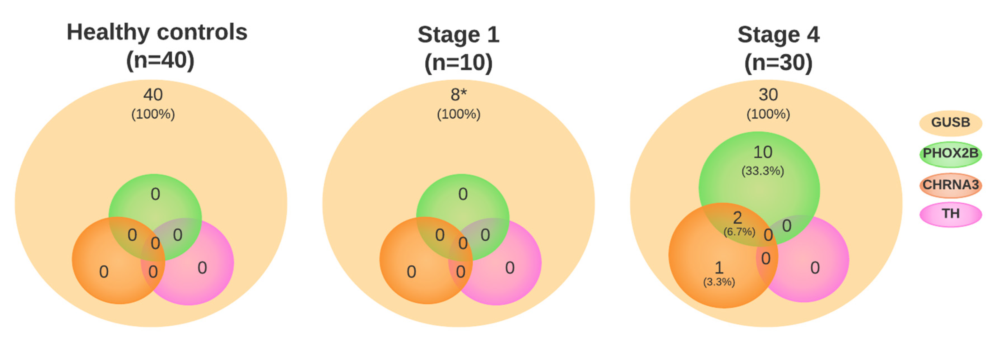
3.1. Neuroblastoma-Specific mRNA Is Present in Plasma

To study cfRNA in limited volume samples of pediatric patients with neuroblastoma, we first designed and optimized a multiplex ddPCR which included the neuroblastoma-specific targets PHOX2B, CHRNA3 and TH, and GUSB as a reference gene (Supplemental Figure S1). In 40 healthy controls, there were no transcripts of PHOX2B, TH or CHRNA3 detected, whereas in all donors, GUSB transcripts could be demonstrated (mean 96 copies/mL plasma, range 36–238 copies/mL) (Supplemental Table S2).
We tested the neuroblastoma-specific multiplex ddPCR panel in a first cohort, consisting of 38 samples from 22 patients with neuroblastoma, which were collected at different timepoints during treatment (patient characteristics and outcome in Supplemental Table S3). In these 38 samples, only 24 samples were positive for GUSB and at lower concentrations (mean of positive samples 14.9 copies/mL plasma (range 2.0–127 copies/mL)). In the 24 samples positive for GUSB, 2 samples were positive for PHOX2B and GUSB (1 at initial diagnosis (2.1 and 2.1 copies/mL, respectively) and 1 at relapse (11 and 127 copies/mL), Supplemental Table S4). No samples were positive for TH or CHRNA3. As it is known that freeze–thaw cycles can affect cfRNA quality [27], we hypothesized that the cfRNA in the samples from these archived samples might be degenerated. Unfortunately, no RNA or plasma was left for analysis of RNA quality through another modality, e.g., Bioanalyzer.
To overcome this problem, we subsequently used only pre-treatment plasma samples that had not been thawed before, 10 samples from patients with INSS stage 1 (localized disease) and 30 INSS stage 4 neuroblastoma patients (metastatic disease), to form a second cohort. Patient characteristics and outcomes are shown in Supplemental Table S3. Results for the neuroblastoma-specific markers are shown in Figure 1 and Supplemental Table S5. In all 40 neuroblastoma samples, GUSB was detectable; for patients with localized disease, the mean was 38.2 copies/mL plasma (range 2.3–95 copies/mL plasma) and metastatic disease, 53 copies/mL plasma (range 10–220 copies/mL plasma). In none of the samples of patients with localized disease were PHOX2B, TH and CHRNA3 detected. In contrast, in 14/30 samples of patients with metastatic disease, PHOX2B (n = 13, 9.2 copies/mL, range 0.4–47 copies/mL) and/or CHRNA3 (n = 4, mean 5.4 copies/mL, range 2.1–11 copies/mL) was detected. No samples were positive for TH. In the samples with at least one marker positive, 10/14 (71%) suffered from an event vs. 11/16 (69%) in the negative samples.

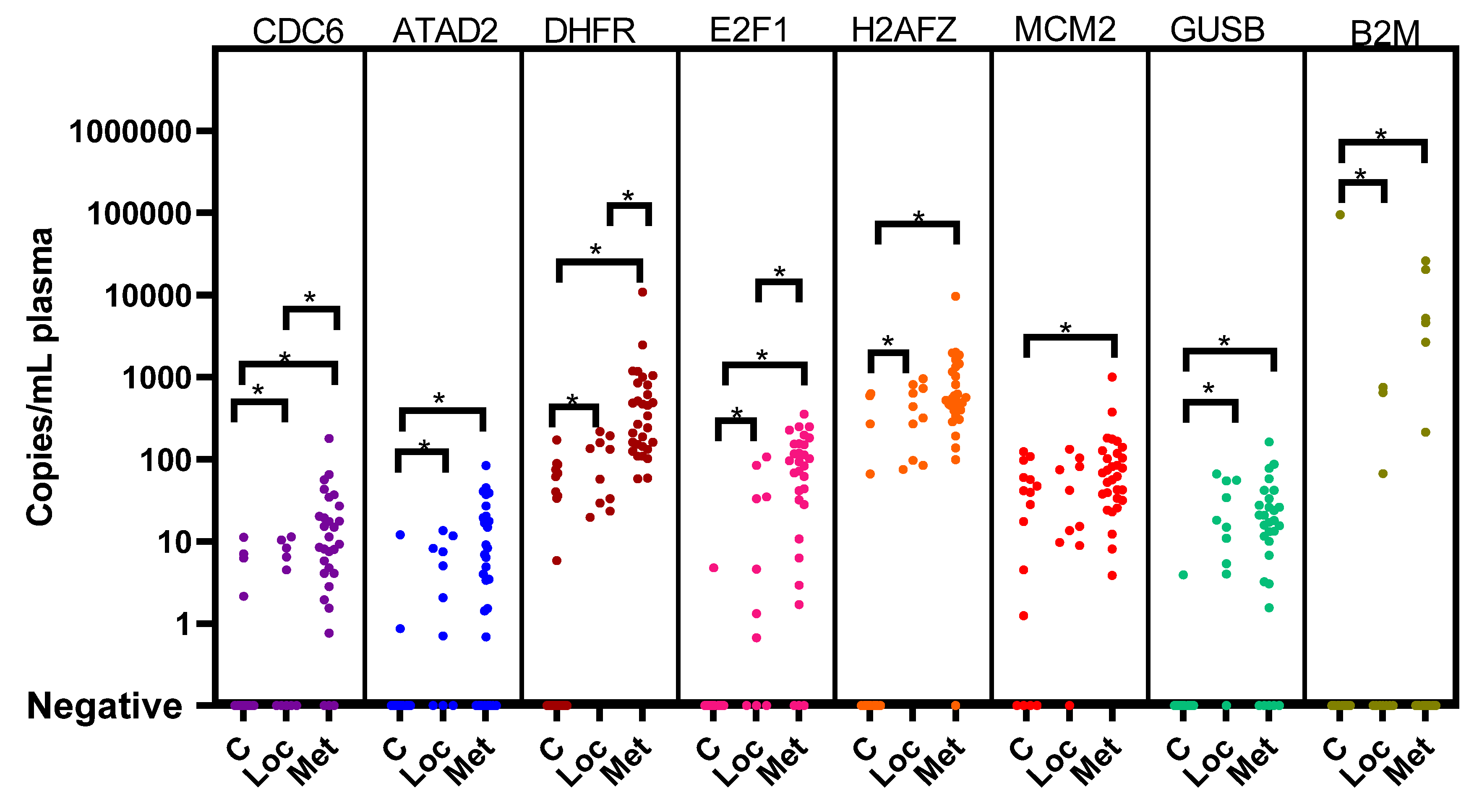
3.2. Cell Cycle Genes in Plasma and Correction for the Presence of Platelets

Next, we investigated the presence of transcripts of cell cycle genes in cfRNA (Supplemental Figure S2 for the cell cycle panel ddPCR assays). We found that platelets also contain these transcripts (Supplemental Figure S3 for expression in platelets). Since plasma was isolated by centrifugation at 1375× g for 10 min, contaminating platelets might have been present in the plasma thereby affecting the analysis. Indeed, in EDTA blood from four healthy controls that were treated similarly as our plasma samples, using the Sysmex system, we measured that after the centrifugation step, 25–50% of the platelets were still present in plasma (Supplemental Table S6). As platelet counts can vary between patients and healthy controls, the cfRNA was corrected for the presence of platelet-specific RNA using the platelet-specific ITGB3 ddPCR. The ratio between ITGB3 expression and the different cell cycle gene transcripts was stable between donors, which enabled a correction co-efficient for each marker, as indicated in Supplemental Table S7.

3.3. Cell Cycle Genes in Plasma from Patients at Diagnosis

We measured the expression of the six cell cycle genes (CDC6, ATAD2, E2F1, H2AFZ, MCM2 and DHFR), the two potential references genes (GUSB and B2M) and the platelet-specific marker ITGB3 in 200 µL of plasma from 20 healthy controls (Supplemental Table S8). We then proceeded to measure these genes in our cohort of 40 patients. After correcting for platelets, CDC6, ATAD2, DHFR, E2F1, H2AFZ, GUSB and B2M were significantly higher in patients with localized disease than in healthy controls. CDC6, DHFR, E2F1, H2AFZ, MCM2, GUSB and B2M were significantly higher in patients with metastatic disease than in healthy controls, and CDC6, DHFR and E2F1 were significantly higher in metastatic patients than in localized patients (Figure 2 and Supplemental Table S9).
We hypothesized that these cell cycle panels could assist in differentiating patients from healthy controls and could possibly differentiate between low- and high-risk disease. For this purpose, we determined the background expression of the cell cycle markers in 20 healthy plasma samples and set a threshold for positivity (Supplemental Table S8). When applying the thresholds for positivity per marker (after correcting for platelets), none of the 10 patients with localized disease were positive, whereas 14 out of 30 patients with metastatic disease had markers that were above the threshold (Supplemental Table S9). All of these 14 patients were positive for DHFR, and only 3 patients were also positive for MCM2 in combination with CDC6 (n = 2) and H2AFZ (n = 1). All three patients suffered from relapse. Eleven other patients were only positive for DHFR. A total of 7/11 suffered from relapse or refractory disease, and 4 eventually died from the disease. In this small cohort, we observed that, when correcting for platelets and background expression, DHFR was elevated in 14/30 patients with metastatic disease at diagnosis. When compared with the neuroblastoma genes, 7/14 DHFR-positive patients were also positive for PHOX2B and/or CHRNA3.

3.4. cfRNA during Treatment

To explore the potential of cfRNA measurements to monitor residual disease during treatment, we measured 66 samples drawn during treatment from 11 patients with metastatic disease (Supplemental Table S10). Patients were chosen according to their clinical outcome and availability of follow-up samples. All neuroblastoma-specific markers were negative in all follow-up samples, except for one sample at the first course of first-line chemotherapy in patient NBL 2187. This sample was positive for CHRNA3 (2.0 copies/mL plasma). We also measured the cell cycle markers in the sequential samples. Many neuroblastoma patients suffer from bone marrow depression due to toxicity of chemotherapy during treatment, which results in low platelet counts. Since we do not know how this affects the RNA content of platelets and if the cell cycle/ITGB3 ratios are affected, we decided not to use the ITGB3-corrected ratios for these samples but to only use the absolute number of copies present in the samples. Supplemental Figure S4 displays the course of the markers for all 11 patients, sorted per clinical outcome. From seven patients, at least three samples during the first line of therapy were available, and from three patients, two samples during the first line of therapy. In all patients, B2M always had the highest expression throughout the entire treatment. The other transcripts varied greatly per patient. No marker showed an evident increase or decrease in expression in patients with good vs. poor clinical outcome. Therefore, it is impossible to draw a conclusion on the level of specific markers in relation to clinical outcome in this small cohort, considering the variation in sampled time points, the unknown platelet counts and variation in expression levels between the different patients.

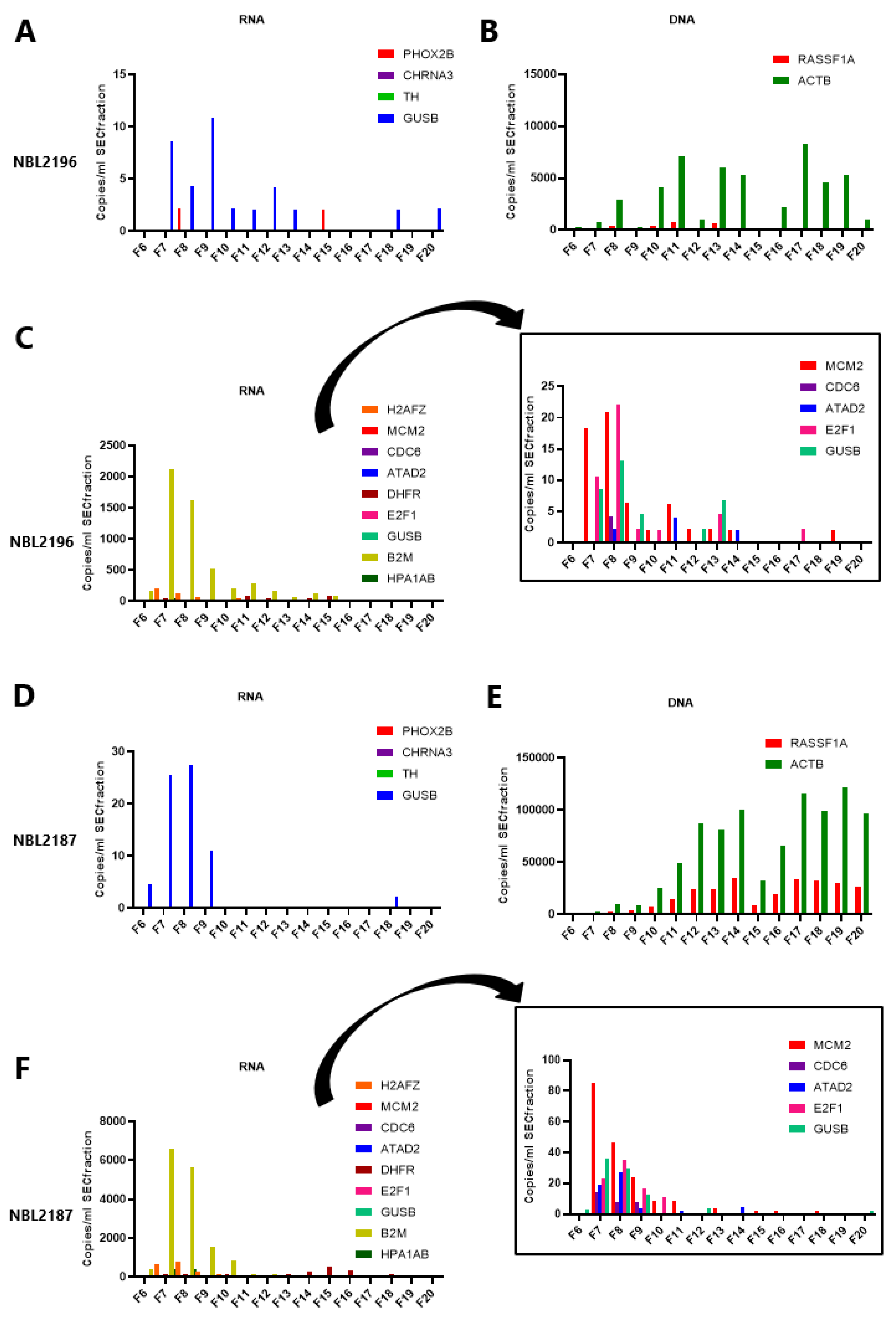
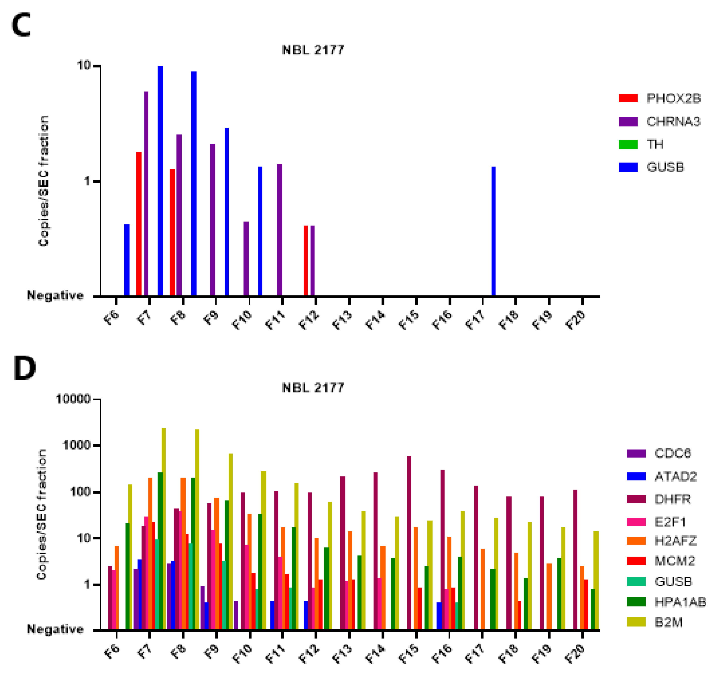
3.5. The mRNA in Plasma Is Concentrated in EV-Enriched SEC Fractions

Subsequently, we investigated in which compartment the neuroblastoma-derived transcripts were present in a patient with metastatic disease by size exclusion chromatography (SEC) on 500 µL of plasma, yielding SEC fractions of 500 µL each. The mRNA markers were tested in parallel to cfDNA using the reference gene ACTB and tumor-specific RASSF1A-M. The presence of EV was confirmed on western blot by the presence of EV-enriched proteins CD9, CD63, CD81 and TSG101 in SEC fractions 7 to 10 isolated from a healthy control and a patient with metastatic disease (Supplemental Figures S5–S7). Electron microscopy on SEC fractions from the same patient also confirmed the presence of EV in fractions 7 to 10 (Supplemental Figure S8), whereas the higher fractions contained aggregated proteins. ddPCR of the RNA markers (both neuroblastoma-specific and cell cycle) and DNA markers from 200 µL of each SEC fraction from two other patients with metastatic disease (one (NBL2196) being PHOX2B-positive in unfractionated plasma and one PHOX2B-negative (NBL2187)) showed that the mRNA markers were mostly present in the EV-enriched fractions, whereas the DNA targets were mostly present in the higher, protein-enriched fractions (Figure 3A,B,D,E, Supplemental Table S11).
Please note that due to a high concentration of B2M, H2AFZ and HPA1A/B, the insert in Figure 3C,F displays an adjusted y-axis without these markers to show the concentration of the other markers.
The presence of mRNA (neuroblastoma-specific and cell cycle markers) in the EV fractions was confirmed in a subsequent experiment with another patient (NBL2177) and a healthy control in which the input in the cDNA reaction was increased 2.5-fold by using the complete 500 µL of SEC fractions. The results are shown in Figure 4 and Supplemental Table S12. Overall, the sum of the positive droplets from all SEC fractions corresponds well to what is found in 500 µL of whole plasma. Unexpectedly, DHFR is increased in the higher, protein-enriched fraction in the patient sample and is even higher than B2M.

4. Discussion

This study addressed the potential for cfRNA analysis from small volumes of plasma using multiplex ddPCR assays and EV enrichment. Within this first exploratory study, we did not aim to draw conclusions on added value to current clinical practice. However, we demonstrated that in patients with neuroblastoma, neuroblastoma-specific cfRNA is only present in patients with metastatic disease and that this RNA is associated with EV. Even with low volumes of plasma, neuroblastoma-specific and quantifiable signals can be obtained when using multiplex ddPCR assays. In this small patient cohort, no correlation with outcome of the disease was observed.
However, we did identify several challenges that are essential to further studies on cfRNA in neuroblastoma. Firstly, pre-analytical variables concerning the preparation and storage of the plasma are critical [20]. The plasma samples we used were prepared within 24 h after collection and then stored at −20 °C. In our first cohort, we used plasma samples that had gone through several cycles of thawing and freezing, and from many of these plasmas, no intact mRNA could be isolated, in contrast to plasma from healthy controls that was only thawed once for the cfRNA isolation. This observation that one freeze–thaw cycle does not affect RNA content is also described by Matthew et al. [27].
The plasma preparation protocol is an equally important consideration if one aims to study cfRNA and the transcripts of interest are also expressed in platelets. In this cohort, a one-step centrifugation protocol was applied to obtain platelet-poor plasma (as we confirmed). Since cell cycle genes are expressed in healthy platelets, the quantitative data had to be corrected for the presence of the variable number of platelets. As we showed that the ratio between our transcripts of interest and the platelet-specific transcript ITGB3 was similar between platelets of different healthy donors, it was possible to correct our data. For future studies, the use of platelet-free plasma might be preferable. Platelet-free plasma was not collected for our cohort but is worth considering in future prospective studies on cfRNA.
Considering the lack of literature on reference genes for cfRNA, we pragmatically included GUSB, which is regularly used as a reference gene for the cellular compartment of peripheral blood for patients with neuroblastoma [9]. In addition, we included B2M as a reference gene [35] as it has been described that B2M is one of the genes highly expressed in plasma, although this finding might partly be caused by its high expression in platelets, as is shown in our data and known from the literature [23,27]. B2M is also an interesting gene specifically for neuroblastoma. It is part of the major histocompatibility complex (MHC), and neuroblastoma cells downregulate MHC proteins, probably in order to evade the immune system [36,37]. Our data suggest that this downregulation is not mediated by specific expulsion of B2M mRNA from the neuroblastoma cells.
The literature on cfRNA analysis in neuroblastoma patients is scarce. Only a single report by Corrias et al. reports on the analysis of cfRNA by RT-qPCR in neuroblastoma patients, stages 1 to 4, using TH as the only neuroblastoma-specific marker and several reference genes, including B2M [38]. This study aimed to investigate whether the analysis of cfRNA was useful in monitoring disease status, as compared to analysis with the same markers in whole blood. In this study, 6 out of 47 samples were positive for TH (1/4 patients with stage 3 disease at diagnosis, 0/15 patients with stage 4 at diagnosis, 1/13 patients with stage 4 during treatment, and 4/15 patients at relapse). In our study, we increased the number of neuroblastoma-specific genes, and by using the ddPCR instead of RT-qPCR, we could increase sensitivity and were able to precisely quantitate the number of transcripts. Interestingly, in our study, no samples were positive for TH, but almost half of the stage 4 samples were positive for PHOX2B and/or CHRNA3 at diagnosis. In contrast, none of the 10 diagnostic samples from patients with localized disease tested positive, strongly suggesting that the presence of cfRNA is related to the stage of the disease. Corrias et al. conclude that the analysis of cfRNA with TH was not superior for monitoring of treatment response compared to RNA analysis from blood cells. Although our discovery study was not aimed or powered to study this question, our study also found that in only one of the 66 samples obtained during treatment could tumor-specific cfRNA be demonstrated. In addition, we did not find a prognostic difference for patients testing positive for the neuroblastoma-specific cfRNA at diagnosis compared to the negative patients (71% vs. 69%, respectively).
It is known that cell cycle genes belonging to the E2F1 pathway can be highly expressed in malignancies. For neuroblastoma, it is known that MYCN amplification is a feature of aggressive disease [1] and that this in turn can upregulate E2F1 and MCM2 [39,40,41]. CDC6, one of the genes of the E2F1 pathway, is also described as an important player in cell proliferation and cell death in neuroblastoma cells, as illustrated by knockdown experiments by Feng et al. [42]. Recently, Andriessen et al. demonstrated that CDC6 was significantly elevated in plasma from canine patients with malignancies compared to healthy controls [32]. This prompted us to study cell cycle genes in our cohort as well, aiming to overcome the low expression of the neuroblastoma-specific markers in cfRNA. Although CDC6 as well as E2F1 and DHFR were, after correcting for platelet presence, significantly higher in patients with metastatic disease compared to patients with localized disease and healthy controls, these markers cannot be easily used to discriminate between healthy individuals and neuroblastoma patients at the individual level. Only two patients had increased levels of CDC6 and MCM2 after correcting for background expression; one patient had increased levels of MCM2 and other genes were not increased, except for DHFR. This latter gene was elevated in 14/30 patients with metastatic disease. DHFR plays a role in the cell cycle as an enzyme in the folate biosynthesis pathway and is thereby essential for cell proliferation. Inhibition of DHFR has been used historically in antimicrobial agents (e.g., trimethoprim), rheumatoid disease and cancer (e.g., methotrexate) [43,44]. Our findings on elevated DHFR at diagnosis in neuroblastoma patients with metastatic disease support its crucial role, corresponding to a high proliferation rate in metastatic disease. However, if the level of DHFR would have a linear association with tumor burden, we would expect this to be reflected in the longitudinal samples. But this was not observed in our cohort.
Indeed, in the longitudinal samples, all cell cycle markers had a high variation in expression between patients and within individual patients, whereas B2M was consistently high. Sample collection was not consistent in our cohort beyond the induction treatment phase, which further complicated speculations on their potential as markers for early treatment failure or relapse detection. This underlines that further studies with standardized sampling are essential for future cfRNA research. In addition, studies broadening the perspective on mRNA markers specifically for cfRNA research are necessary since current RNA markers are mostly based on the cellular compartment of blood and might not be suited for cfRNA. Specifically for patients with neuroblastoma, a study including RNA sequencing of the transcriptome of the cell-free compartment at different timepoints could improve understanding of this field immensely and help identify cfRNA markers with diagnostic and prognostic value.
We confirmed that most of the mRNA markers are concentrated in the EV-enriched fractions [18,21,32]. However, unexpectedly, DHFR was found to be mostly present in the higher SEC fractions. This could be due to elution of smaller EV in later fractions or also to packaging of mRNA into protein aggregates; both hypotheses are supported by the literature [21,45,46,47,48] and the presence of B2M and GUSB in these SEC fractions. Further studies using RNase and proteinase on the different SEC fractions could elucidate further if mRNA is truly packaged within EV or only associated with EV. The same approach is possible for cfDNA using DNase, since we mostly demonstrate the presence of cfDNA in the protein-enriched fractions. The literature is still conflicting on this subject [49,50], and it is not inconceivable that EV cargo could even differ per disease. Furthermore, the method chosen for EV enrichment heavily affects the result of the downstream analysis, and after SEC, the presence of similar-sized lipoprotein particles in the EV-enriched fractions might also result in less pure EV preparations [51,52].
Considering a possible implementation in clinical practice, our study does not immediately show a benefit for EV enrichment prior to cfRNA isolation, especially in respect to time and cost effectiveness. In children, the amount of plasma is the major limiting step, and it seems simpler to just isolate RNA from full plasma than first performing density gradient centrifugation for EV isolation. However, that the concentration of EV can result in enrichment of tumor RNA was indeed recently shown by Stegmaier et al. [53]. They show that their target of interest, the transcripts of the PAX-FOXO or SYT-SSX fusion genes from alveolar rhabdomyosarcoma and synovial sarcoma, respectively, had a higher concentration in EV-derived cfRNA from patient plasma than cfRNA directly from plasma [53]. In future studies, it is important to determine which question needs to be answered. The purity of EV through elaborate isolation procedures can be essential to increase the knowledge of EV cargo and function, whereas a translational goal to improve diagnostic procedures might benefit more from a quick EV enrichment procedure through commercially available precipitating agents which increase the target concentration and thereby the sensitivity of the test.

5. Conclusions

In this study, we explore the possibilities of different cfRNA markers from plasma as novel biomarkers in patients with neuroblastoma. We discuss the possible variables affecting the detection of cfRNA-based markers and present approaches for correcting for the presence of platelets and background marker expression in plasma. Considering the neuroblastoma-specific markers, we conclude that these are only present in patients with metastatic disease. For the cell cycle markers, we find that many markers are higher in patients than in healthy controls, but only elevated above background expression levels in some metastatic patients. Our experiments on EV using SEC isolation illustrate that the mRNA markers are mostly expressed in EV-enriched SEC fractions, whereas cfDNA is mostly present in EV-poor SEC fractions. This study can form a starting point for further research into the potential of cfRNA-based analysis of liquid biopsies since this can be an additional approach to the more common analysis of cfDNA for liquid biopsies.

Supplementary Materials

The following supporting information can be downloaded at: https://www.mdpi.com/article/10.3390/cancers15072108/s1. Supplemental Figure S1. 2D plots from the ddPCR assays illustrating gating strategies for the neuroblastoma-specific genes. Supplemental Figure S2. 2D plots from the ddPCR assays illustrating gating strategies for the cell cycle genes. Supplemental Figure S3. Expression of cell cycle genes, reference genes and platelet-specific genes in platelets from 4 healthy donors as measured by ddPCR. Supplemental Figure S4. Level of the cell cycle markers in sequential plasma samples from 11 patients with metastatic neuroblastoma. Supplemental Figure S5. Western blot images from the size exclusion chromatography (SEC) fractions isolated from 500 µL of plasma from 1 healthy control. Supplemental Figure S6. Western blot images from the size exclusion chromatography (SEC) fractions isolated from 500 ul of plasma from 1 patient with metastatic neuroblastoma. Supplemental Figure S7. Western blot images from the size exclusion chromatography (SEC) fractions isolated from 500 µL of plasma from a healthy control. Supplemental Figure S8. Electron microscopy images of fractions (6 to 15) purified by size exclusion chromatography from 500 µL of plasma from a patient with neuroblastoma. Supplemental Table S1. Sequences of ddPCR assays. Supplemental Table S2. Results of the neuroblastoma-specific panel and GUSB in plasma of 40 healthy controls. Supplemental Table S3. Patient characteristics of the first and second cohort. Supplemental Table S4. Results of ddPCR of the genes in the first cohort. Supplemental Table S5. Results of the ddPCR for DNA-based targets and mRNA-based targets, neuroblastoma-specific panel and cell cycle markers in the second cohort. Supplemental Table S6. Overview of number of leukocytes and platelets recovered at the different centrifugation steps during the preparation of platelet-poor plasma and their percentages to whole blood. Supplemental Table S7. Expression of cell cycle genes in 4 samples of platelets isolated from healthy controls and the corresponding correction co-efficients, as calculated by the following formula. Supplemental Table S8. Expression of cell cycle genes in plasma from 20 healthy controls with the levels after correction for platelets using the correction co-efficient per marker and the calculated thresholds for positivity. Supplemental Table S9. Overview of the cell cycle marker results and the levels corrected for presence of platelets using the correction co-efficients. Supplemental Table S10. Levels of the neuroblastoma-specific markers and cell cycle genes in sequential plasma samples from 11 patients with metastatic neuroblastoma. Supplemental Table S11. Results of the RNA-based ddPCR assays (both neuroblastoma-specific and cell cycle panels) and DNA-based ddPCR on 200 µL of SEC fractions isolated from 500 µL of plasma. Supplemental Table S12. Number of positive droplets per neuroblastoma-specific and cell cycle marker gene per 500 µL SEC fraction.

Author Contributions

Conceptualization, N.S.M.L., A.S., L.B., A.A., C.E.v.d.S., A.d.B. and G.A.M.T.; Methodology, N.S.M.L., A.S., L.M.J.v.Z., N.U.G., L.B., A.A., C.E.v.d.S., A.d.B. and G.A.M.T.; Validation, N.S.M.L., A.J. and L.Z.-K.; Formal Analysis, N.S.M.L., L.Z.-K., A.J. and G.A.M.T.; Investigation, N.S.M.L., A.S., A.J., L.Z.-K., C.E.v.d.S., A.d.B. and G.A.M.T.; Resources, C.E.v.d.S., J.S. and G.A.M.T.; Data Curation, N.S.M.L., L.Z.-K., A.J., J.S., C.E.v.d.S. and G.A.M.T.; Writing—Original Draft Preparation, N.S.M.L. and G.A.M.T.; Writing—Review & Editing, N.S.M.L., A.S., L.M.J.v.Z., N.U.G., A.J., L.Z.-K., L.B., A.A., J.S., C.E.v.d.S., A.d.B. and G.A.M.T.; Visualization, N.S.M.L.; Supervision, J.S., C.E.v.d.S., A.d.B. and G.A.M.T.; Project Administration, N.S.M.L., L.Z.-K. and A.J.; Funding Acquisition, J.S., C.E.v.d.S. and G.A.M.T. All authors have read and agreed to the published version of the manuscript.

Funding

N.S.M.L.: L.Z.-K. and J.S. were supported by grant 312, from Children Cancer-Free (KiKa).

Institutional Review Board Statement

The study was conducted in accordance with the Declaration of Helsinki and approved by the medical ethics committee of the Academic Medical Center, Amsterdam, the Netherlands; MEC07/219#08.17.0836.

Informed Consent Statement

Informed consent was obtained from all subjects involved in the study.

Data Availability Statement

The data presented in this study are available in this article (and Supplementary Materials). Further data can be shared up on request.

Acknowledgments

We are grateful to Jalenka van Wijk for performing the pilot experiments for this study. We thank Floris van Alphen for his help with the TCA assays and Herbert Koster for his help with the experiments on the platelets. We are grateful to Nicole van der Wel and Anita Grootemaat for their help with the electron microscopy on the SEC fractions.

Conflicts of Interest

The authors declare no conflict of interest.

References

  1. Matthay, K.K.; Maris, J.M.; Schleiermacher, G.; Nakagawara, A.; Mackall, C.L.; Diller, L.; Weiss, W.A. Neuroblastoma. Nat. Rev. Dis. Prim. 2016, 2, 16078. [Google Scholar] [CrossRef] [PubMed]
  2. Pearson, A.D.; Pinkerton, C.R.; Lewis, I.J.; Imeson, J.; Ellershaw, C.; Machin, D. High-dose rapid and standard induction chemotherapy for patients aged over 1 year with stage 4 neuroblastoma: A randomised trial. Lancet Oncol. 2008, 9, 247–256. [Google Scholar] [CrossRef] [PubMed]
  3. Kreissman, S.G.; Seeger, R.C.; Matthay, K.K.; London, W.B.; Sposto, R.; A Grupp, S.; A Haas-Kogan, D.; LaQuaglia, M.P.; Yu, A.L.; Diller, L.; et al. Purged versus non-purged peripheral blood stem-cell transplantation for high-risk neuroblastoma (COG A3973): A randomised phase 3 trial. Lancet Oncol. 2013, 14, 999–1008. [Google Scholar] [CrossRef] [Green Version]
  4. Van Paemel, R.; Vlug, R.; De Preter, K.; Van Roy, N.; Speleman, F.; Willems, L.; Lammens, T.; Laureys, G.; Schleiermacher, G.; Tytgat, G.A.M.; et al. The pitfalls and promise of liquid biopsies for diagnosing and treating solid tumors in children: A review. Eur. J. Pediatr. 2020, 179, 191–202. [Google Scholar] [CrossRef] [Green Version]
  5. Alix-Panabières, C.; Pantel, K. Clinical Applications of Circulating Tumor Cells and Circulating Tumor DNA as Liquid Biopsy. Cancer Discov. 2016, 6, 479–491. [Google Scholar] [CrossRef] [PubMed] [Green Version]
  6. Stutterheim, J.; Gerritsen, A.; Zappeij-Kannegieter, L.; Kleijn, I.; Dee, R.; Hooft, L.; van Noesel, M.M.; Bierings, M.; Berthold, F.; Versteeg, R.; et al. PHOX2B Is a Novel and Specific Marker for Minimal Residual Disease Testing in Neuroblastoma. J. Clin. Oncol. 2008, 26, 5443–5449. [Google Scholar] [CrossRef]
  7. Yáñez, Y.; Hervás, D.; Grau, E.; Oltra, S.; Pérez, G.; Palanca, S.; Bermúdez, M.; Márquez, C.; Cañete, A.; Castel, V. TH and DCX mRNAs in peripheral blood and bone marrow predict outcome in metastatic neuroblastoma patients. J. Cancer Res. Clin. Oncol. 2016, 142, 573–580. [Google Scholar] [CrossRef] [PubMed]
  8. Van Wezel, E.M.; van Zogchel, L.M.J.; van Wijk, J.; Timmerman, I.; Vo, N.K.; Zappeij-Kannegieter, L.; deCarolis, B.; Simon, T.; van Noesel, M.M.; Molenaar, J.J.; et al. Mesenchymal Neuroblastoma Cells Are Undetected by Current mRNA Marker Panels: The Development of a Specific Neuroblastoma Mesenchymal Minimal Residual Disease Panel. JCO Precis. Oncol. 2019, 3, 1–11. [Google Scholar] [CrossRef]
  9. Stutterheim, J.; Gerritsen, A.; Zappeij-Kannegieter, L.; Yalcin, B.; Dee, R.; van Noesel, M.M.; Berthold, F.; Versteeg, R.; Caron, H.N.; van der Schoot, C.E.; et al. Detecting Minimal Residual Disease in Neuroblastoma: The Superiority of a Panel of Real-Time Quantitative PCR Markers. Clin. Chem. 2009, 55, 1316–1326. [Google Scholar] [CrossRef] [Green Version]
  10. Van Zogchel, L.M.J.; Lak, N.S.M.; Verhagen, O.J.H.M.; Tissoudali, A.; Nuru, M.G.; Gelineau, N.U.; Zappeij-Kannengieter, L.; Javadi, A.; Zijtregtop, E.A.M.; Merks, J.H.M.; et al. Novel Circulating Hypermethylated RASSF1A ddPCR for Liquid Biopsies in Patients With Pediatric Solid Tumors. JCO Precis. Oncol. 2021, 5, 1738–1748. [Google Scholar] [CrossRef]
  11. Lodrini, M.; Sprüssel, A.; Astrahantseff, K.; Tiburtius, D.; Konschak, R.; Lode, H.N.; Fischer, M.; Keilholz, U.; Eggert, A.; Deubzer, H.E. Using droplet digital PCR to analyze MYCN and ALK copy number in plasma from patients with neuroblastoma. Oncotarget 2017, 8, 85234–85251. [Google Scholar] [CrossRef] [PubMed] [Green Version]
  12. Lodrini, M.; Graef, J.; Thole-Kliesch, T.M.; Astrahantseff, K.; Sprüssel, A.; Grimaldi, M.; Peitz, C.; Linke, R.B.; Hollander, J.F.; Lankes, E.; et al. Targeted Analysis of Cell-free Circulating Tumor DNA is Suitable for Early Relapse and Actionable Target Detection in Patients with Neuroblastoma. Clin. Cancer Res. 2022, 28, 1809–1820. [Google Scholar] [CrossRef] [PubMed]
  13. Van Zogchel, L.M.J.; van Wezel, E.M.; van Wijk, J.; Stutterheim, J.; Bruins, W.S.C.; Zappeij-Kannegieter, L.; Tirza, J.E.; Iedan, R.N.; Verly, C.; Godelieve, A.M.; et al. Hypermethylated RASSF1A as Circulating Tumor DNA Marker for Disease Monitoring in Neuroblastoma. JCO Precis. Oncol. 2020, 4, 291–306. [Google Scholar] [CrossRef]
  14. Wan, J.C.M.; Massie, C.; Garcia-Corbacho, J.; Mouliere, F.; Brenton, J.D.; Caldas, C.; Pacey, S.; Baird, R.; Rosenfeld, N. Liquid biopsies come of age: Towards implementation of circulating tumour DNA. Nat. Rev. Cancer 2017, 17, 223–238. [Google Scholar] [CrossRef]
  15. Skog, J.; Wurdinger, T.; van Rijn, S.; Meijer, D.H.; Gainche, L.; Curry, W.T., Jr.; Sena-Esteves, M.; Carter, B.S.; Krichevsky, A.M.; Breakefield, X.O. Glioblastoma microvesicles transport RNA and proteins that promote tumour growth and provide diagnostic biomarkers. Nat. Cell Biol. 2008, 10, 1470–1476. [Google Scholar] [CrossRef] [PubMed]
  16. Hulstaert, E.; Morlion, A.; Avila Cobos, F.; Verniers, K.; Nuytens, J.; Vanden Eynde, E.; Yigit, N.; Anckaert, J.; Geerts, A.; Hindryckx, P.; et al. Charting Extracellular Transcriptomes in The Human Biofluid RNA Atlas. Cell Rep. 2020, 33, 108552. [Google Scholar] [CrossRef] [PubMed]
  17. Herriott, R.M.; Connolly, J.H.; Gupta, S. Blood Nucleases and Infectious Viral Nucleic Acids. Nature 1961, 189, 817–820. [Google Scholar] [CrossRef]
  18. Bebelman, M.P.; Smit, M.J.; Pegtel, D.M.; Baglio, S.R. Biogenesis and function of extracellular vesicles in cancer. Pharmacol. Ther. 2018, 188, 1–11. [Google Scholar] [CrossRef]
  19. Xavier, C.P.R.; Caires, H.R.; Barbosa, M.A.G.; Bergantim, R.; Guimarães, J.E.; Vasconcelos, M.H. The Role of Extracellular Vesicles in the Hallmarks of Cancer and Drug Resistance. Cells 2020, 9, 1141. [Google Scholar] [CrossRef]
  20. Grölz, D.; Hauch, S.; Schlumpberger, M.; Guenther, K.; Voss, T.; Sprenger-Haussels, M.; Oelmüller, U. Liquid Biopsy Preservation Solutions for Standardized Pre-Analytical Workflows—Venous Whole Blood and Plasma. Curr. Pathobiol. Rep. 2018, 6, 275–286. [Google Scholar] [CrossRef] [Green Version]
  21. Arroyo, J.D.; Chevillet, J.R.; Kroh, E.M.; Ruf, I.K.; Pritchard, C.C.; Gibson, D.F.; Mitchell, P.S.; Bennett, C.F.; Pogosova-Agadjanyan, E.L.; Stirewalt, D.L.; et al. Argonaute2 complexes carry a population of circulating microRNAs independent of vesicles in human plasma. Proc. Natl. Acad. Sci. USA 2011, 108, 5003–5008. [Google Scholar] [CrossRef] [PubMed] [Green Version]
  22. Wurdinger, T.; In ‘t Veld, S.G.J.G.; Best, M.G. Platelet RNA as Pan-Tumor Biomarker for Cancer Detection. Cancer Res. 2020, 80, 1371–1373. [Google Scholar] [CrossRef] [Green Version]
  23. Rowley, J.W.; Oler, A.J.; Tolley, N.D.; Hunter, B.N.; Low, E.N.; Nix, D.A.; Yost, C.C.; Zimmerman, G.A.; Weyrich, A. Genome-wide RNA-seq analysis of human and mouse platelet transcriptomes. Blood 2011, 118, e101–e111. [Google Scholar] [CrossRef] [PubMed] [Green Version]
  24. Rowley, J.W.; Schwertz, H.; Weyrich, A.S. Platelet mRNA. Curr. Opin. Hematol. 2012, 19, 385–391. [Google Scholar] [CrossRef] [PubMed]
  25. Morini, M.; Cangelosi, D.; Segalerba, D.; Marimpietri, D.; Raggi, F.; Castellano, A.; Fruci, D.; de Mora, J.F.; Cañete, A.; Yáñez, Y.; et al. Exosomal microRNAs from Longitudinal Liquid Biopsies for the Prediction of Response to Induction Chemotherapy in High-Risk Neuroblastoma Patients: A Proof of Concept SIOPEN Study II. Cancers 2019, 11, 1476. [Google Scholar] [CrossRef] [Green Version]
  26. Ma, J.; Xu, M.; Yin, M.; Hong, J.; Chen, H.; Gao, Y.; Xie, C.; Shen, N.; Gu, S.; Mo, X. Exosomal hsa-miR199a-3p Promotes Proliferation and Migration in Neuroblastoma. Front. Oncol. 2019, 9, 459. [Google Scholar] [CrossRef]
  27. Larson, M.H.; Pan, W.; Kim, H.J.; Mauntz, R.E.; Stuart, S.M.; Pimentel, M.; Zhou, Y.; Knudsgaard, P.; Demas, V.; Aravanis, A.M.; et al. A comprehensive characterization of the cell-free transcriptome reveals tissue- and subtype-specific biomarkers for cancer detection. Nat. Commun. 2021, 12, 2357. [Google Scholar] [CrossRef]
  28. Harashima, H.; Dissmeyer, N.; Schnittger, A. Cell cycle control across the eukaryotic kingdom. Trends Cell Biol. 2013, 23, 345–356. [Google Scholar] [CrossRef]
  29. Matthews, H.K.; Bertoli, C.; de Bruin, R.A.M. Cell cycle control in cancer. Nat. Rev. Mol. Cell Biol. 2021, 23, 74–88. [Google Scholar] [CrossRef]
  30. Thurlings, I.; de Bruin, A. E2F Transcription Factors Control the Roller Coaster Ride of Cell Cycle Gene Expression. Methods Mol. Biol. 2016, 1342, 71–88. [Google Scholar]
  31. Bongiovanni, L.; Andriessen, A.; Silvestri, S.; Porcellato, I.; Brachelente, C.; de Bruin, A. H2AFZ: A Novel Prognostic Marker in Canine Melanoma and a Predictive Marker for Resistance to CDK4/6 Inhibitor Treatment. Front. Vet. Sci. 2021, 8, 705359. [Google Scholar] [CrossRef]
  32. Andriessen, A.; Bongiovanni, L.; Driedonks, T.A.P.; van Liere, E.; Seijger, A.; Hegeman, C.V.; van Nimwegen, S.A.; Galac, S.; Westendorp, B.; Hoen, E.N.M.N.; et al. CDC6: A novel canine tumour biomarker detected in circulating extracellular vesicles. Veter Comp. Oncol. 2021, 20, 381–392. [Google Scholar] [CrossRef] [PubMed]
  33. Ando, K.; Nakagawara, A. Acceleration or Brakes: Which Is Rational for Cell Cycle-Targeting Neuroblastoma Therapy? Biomolecules 2021, 11, 750. [Google Scholar] [CrossRef] [PubMed]
  34. Van Zogchel, L.; de Carolis, B.; van Wezel, E.; Stutterheim, J.; Zappeij-Kannegieter, L.; van Doornum, M.; Schumacher-Kuckelkorn, R.; Gecht, J.; Simon, T.; Caron, H.; et al. Detection of Minimal Residual Disease (MRD) in High Risk Neuroblastoma Correlates with Outcome: Final Results of International GPOH-DCOG Prospective Validation Study. In Pediatric Blood & Cancer; Wiley: Hoboken, NJ, USA, 2018; p. S38. [Google Scholar]
  35. Beillard, E.; Pallisgaard, N.; van der Velden, V.H.J.; Bi, W.; Dee, R.; van der Schoot, E.; Delabesse, E.; Macintyre, E.; Gottardi, E.; Saglio, G.; et al. Evaluation of candidate control genes for diagnosis and residual disease detection in leukemic patients using ‘real-time’ quantitative reverse-transcriptase polymerase chain reaction (RQ-PCR)—A Europe against cancer program. Leukemia 2003, 17, 2474–2486. [Google Scholar] [CrossRef] [Green Version]
  36. Corrias, M.V.; Occhino, M.; Croce, M.; De Ambrosis, A.; Pistillo, M.P.; Bocca, P.; Pistoia, V.; Ferrini, S. Lack of HLA-class I antigens in human neuroblastoma cells: Analysis of its relationship to TAP and tapasin expression. Tissue Antigens 2001, 57, 110–117. [Google Scholar] [CrossRef]
  37. Spel, L.; Schiepers, A.; Boes, M. NFκB and MHC-1 Interplay in Neuroblastoma and Immunotherapy. Trends Cancer 2018, 4, 715–717. [Google Scholar] [CrossRef]
  38. Corrias, M.V.; Pistorio, A.; Cangemi, G.; Tripodi, G.; Bs, B.C.; Scaruffi, P.; Fardin, P.; Garaventa, A.; Pistoia, V.; Haupt, R. Detection of cell-free RNA in children with neuroblastoma and comparison with that of whole blood cell RNA. Pediatr. Blood Cancer 2010, 54, 897–903. [Google Scholar] [CrossRef]
  39. Koppen, A.; Ait-Aissa, R.; Koster, J.; Van Sluis, P.G.; Øra, I.; Caron, H.N.; Volckmann, R.; Versteeg, R.; Valentijn, L.J. Direct regulation of the minichromosome maintenance complex by MYCN in neuroblastoma. Eur. J. Cancer 2007, 43, 2413–2422. [Google Scholar] [CrossRef]
  40. Liu, X.; Cai, Y.; Cheng, C.; Gu, Y.; Hu, X.; Chen, K.; Wu, Y.; Wu, Z. PCLAF promotes neuroblastoma G1/S cell cycle progression via the E2F1/PTTG1 axis. Cell Death Dis. 2022, 13, 178. [Google Scholar] [CrossRef] [PubMed]
  41. Wang, H.; Wang, X.; Xu, L.; Zhang, J.; Cao, H. Prognostic significance of MYCN related genes in pediatric neuroblastoma: A study based on TARGET and GEO datasets. BMC Pediatr. 2020, 20, 314. [Google Scholar] [CrossRef]
  42. Feng, L.; Barnhart, J.R.; Seeger, R.C.; Wu, L.; Keshelava, N.; Huang, S.-H.; Jong, A. Cdc6 knockdown inhibits human neuroblastoma cell proliferation. Mol. Cell. Biochem. 2008, 311, 189–197. [Google Scholar] [CrossRef] [PubMed]
  43. Bhagat, K.; Kumar, N.; Gulati, H.K.; Sharma, A.; Kaur, A.; Singh, J.V.; Singh, H.; Bedi, P.M.S. Dihydrofolate reductase inhibitors: Patent landscape and phases of clinical development (2001–2021). Expert Opin. Ther. Patents 2022, 32, 1079–1095. [Google Scholar] [CrossRef] [PubMed]
  44. Raimondi, M.V.; Randazzo, O.; La Franca, M.; Barone, G.; Vignoni, E.; Rossi, D.; Collina, S. DHFR Inhibitors: Reading the Past for Discovering Novel Anticancer Agents. Molecules 2019, 24, 1140. [Google Scholar] [CrossRef] [PubMed] [Green Version]
  45. Böing, A.N.; van der Pol, E.; Grootemaat, A.E.; Coumans, F.A.W.; Sturk, A.; Nieuwland, R. Single-step isolation of extracellular vesicles by size-exclusion chromatography. J. Extracell. Vesicles 2014, 3, 23430. [Google Scholar] [CrossRef] [PubMed]
  46. Clayton, A.; Boilard, E.; Buzas, E.I.; Cheng, L.; Falcón-Perez, J.M.; Gardiner, C.; Gustafson, D.; Gualerzi, A.; Hendrix, A.; Hoffman, A.; et al. Considerations towards a roadmap for collection, handling and storage of blood extracellular vesicles. J. Extracell. Vesicles 2019, 8, 1647027. [Google Scholar] [CrossRef] [Green Version]
  47. Théry, C.; Witwer, K.W.; Aikawa, E.; Alcaraz, M.J.; Anderson, J.D.; Andriantsitohaina, R.; Antoniou, A.; Arab, T.; Archer, F.; Atkin-Smith, G.K.; et al. Minimal information for studies of extracellular vesicles 2018 (MISEV2018): A position statement of the International Society for Extracellular Vesicles and update of the MISEV2014 guidelines. J. Extracell. Vesicles 2018, 7, 1535750. [Google Scholar] [CrossRef] [Green Version]
  48. Coumans, F.A.W.; Brisson, A.R.; Buzas, E.I.; Dignat-George, F.; Drees, E.E.E.; El-Andaloussi, S.; Emanueli, C.; Gasecka, A.; Hendrix, A.; Hill, A.F.; et al. Methodological Guidelines to Study Extracellular Vesicles. Circ. Res. 2017, 120, 1632–1648. [Google Scholar] [CrossRef]
  49. Liu, H.; Tian, Y.; Xue, C.; Niu, Q.; Chen, C.; Yan, X. Analysis of extracellular vesicle DNA at the single-vesicle level by nano-flow cytometry. J. Extracell. Vesicles 2022, 11, 12206. [Google Scholar] [CrossRef]
  50. Thakur, B.K.; Zhang, H.; Becker, A.; Matei, I.; Huang, Y.; Costa-Silva, B.; Zheng, Y.; Hoshino, A.; Brazier, H.; Xiang, J.; et al. Double-stranded DNA in exosomes: A novel biomarker in cancer detection. Cell Res. 2014, 24, 766–769. [Google Scholar] [CrossRef] [Green Version]
  51. Van Deun, J.; Mestdagh, P.; Sormunen, R.; Cocquyt, V.; Vermaelen, K.; Vandesompele, J.; Bracke, M.; De Wever, O.; Hendrix, A. The impact of disparate isolation methods for extracellular vesicles on downstream RNA profiling. J. Extracell. Vesicles 2014, 3, 24858. [Google Scholar] [CrossRef] [Green Version]
  52. Vergauwen, G.; Tulkens, J.; Pinheiro, C.; Cobos, F.A.; Dedeyne, S.; De Scheerder, M.; Vandekerckhove, L.; Impens, F.; Miinalainen, I.; Braems, G.; et al. Robust sequential biophysical fractionation of blood plasma to study variations in the biomolecular landscape of systemically circulating extracellular vesicles across clinical conditions. J. Extracell. Vesicles 2021, 10, e12122. [Google Scholar] [CrossRef] [PubMed]
  53. Stegmaier, S.; Sparber-Sauer, M.; Aakcha-Rudel, E.; Münch, P.; Reeh, T.; Feuchtgruber, S.; Hallmen, E.; Blattmann, C.; Bielack, S.; Klingebiel, T.; et al. Fusion transcripts as liquid biopsy markers in alveolar rhabdomyosarcoma and synovial sarcoma: A report of the Cooperative Weichteilsarkom Studiengruppe (CWS). Pediatr. Blood Cancer 2022, 69, 29652. [Google Scholar] [CrossRef] [PubMed]
Figure 1. Expression of neuroblastoma-specific genes in cell-free RNA from healthy controls (n = 40), and diagnostic plasmas from patients with neuroblastoma with localized (n = 10) and metastatic (n = 30) disease. * Not enough material was left for 2 patients to perform the ddPCR for these neuroblastoma-specific markers.
Cancers 15 02108 g001
Figure 2. Expression of cell cycle genes (CDC6, ATAD2, DHFR, E2F1, H2AFZ and MCM2) and reference genes (GUSB and B2M) in cell-free RNA from healthy controls (n = 40) and diagnostic plasmas from patients with neuroblastoma with localized (n = 10) and metastatic (n = 30) disease, as measured by ddPCR from 200 µL plasma and corrected for platelet contamination. C; healthy controls. Loc; patients with localized disease. Met; patients with metastatic disease. * Significance at p < 0.05.
Cancers 15 02108 g002
Figure 3. Expression of the mRNA markers and cell-free DNA markers in fractions isolated by size exclusion chromatography (SEC) and analyzed by ddPCR. Fractions F7 to F10 are considered as enriched in extracellular vesicles (EV). For patient NBL2196, (A) shows the expression of the neuroblastoma-specific mRNA markers, PHOX2B, CHRNA3 and TH, and reference gene GUSB in cell-free RNA from 200 µL per SEC fraction: only GUSB and PHOX2B are expressed. (B) shows the cfDNA tumor-specific target methylated RASSF1A (RASSF1A-M) and reference gene ACTB in cfDNA from 200 µL per SEC fraction. (C) illustrates the expression of the cell cycle markers (H2AFZ, MCM2, CDC6, ATAD2, DHFR, E2F1, GUSB, B2M and HPA1A/B) in 200 µL per SEC fraction from the same patient. For patient NBL2187, (D) shows the expression of the neuroblastoma-specific mRNA markers, PHOX2B, CHRNA3 and TH, and reference gene GUSB in cell-free RNA from 200 µL per SEC fraction: only GUSB is expressed. (E) shows the cfDNA tumor-specific target methylated RASSF1A (RASSF1A-M) and reference gene ACTB in cfDNA from 200 µL per SEC fraction. (F) illustrates the expression of the cell cycle markers (H2AFZ, MCM2, CDC6, ATAD2, DHFR, E2F1, GUSB, B2M and HPA1A/B) in 200 µL per SEC fraction from the same patient.
Cancers 15 02108 g003
Figure 4. Expression of neuroblastoma-specific and cell cycle genes in 500 µL of SEC (size exclusion chromatography) fractions, as isolated from 500 µL plasma, from one healthy control and one patient with metastatic neuroblastoma. Fractions F7 to F10 are considered as enriched in extracellular vesicles (EV). In the healthy control, (A) shows the expression of the neuroblastoma-specific genes (PHOX2B, CHRNA3 and TH with reference gene GUSB) and (B) shows the expression of the cell cycle markers (CDC6, ATAD2, DHFR, E2F1, H2AFZ and MCM2 with GUSB, HPA1A/B and B2M). In the patient NBL 2177, (C) shows the expression of the neuroblastoma-specific genes (PHOX2B, CHRNA3 and TH with reference gene GUSB) and (D) shows the expression of the cell cycle markers (CDC6, ATAD2, DHFR, E2F1, H2AFZ and MCM2 with GUSB, HPA1A/B and B2M). Please note that the y-axis is represented on a log scale.
Cancers 15 02108 g004aCancers 15 02108 g004b
Disclaimer/Publisher’s Note: The statements, opinions and data contained in all publications are solely those of the individual author(s) and contributor(s) and not of MDPI and/or the editor(s). MDPI and/or the editor(s) disclaim responsibility for any injury to people or property resulting from any ideas, methods, instructions or products referred to in the content.

