Review

Prospects of POLD1 in Human Cancers: A Review

by
Michał Gola
1,*,
Przemysław Stefaniak
2,
Janusz Godlewski
1,2,
Barbara Alicja Jereczek-Fossa
3,4 and
Anna Starzyńska
5
1
Department of Human Histology and Embryology, Collegium Medicum, School of Medicine, University of Warmia and Mazury, 10-082 Olsztyn, Poland
2
Department of Surgical Oncology, Hospital Ministry of Internal Affairs with Warmia and Mazury Oncology Centre, 10-228 Olsztyn, Poland
3
Division of Radiation Oncology, European Institute of Oncology (IEO), Istituto di Ricovero e Cura a Carattere Scientifico (IRCCS), 20141 Milan, Italy
4
Department of Oncology and Hemato-Oncology, University of Milan, 20122 Milan, Italy
5
Department of Oral Surgery, Medical University of Gdańsk, 7 Dębinki Street, 80-211 Gdańsk, Poland
*
Author to whom correspondence should be addressed.
Cancers 2023, 15(6), 1905; https://doi.org/10.3390/cancers15061905
Submission received: 7 February 2023 / Revised: 7 March 2023 / Accepted: 20 March 2023 / Published: 22 March 2023
(This article belongs to the Section Cancer Informatics and Big Data)

Abstract

:

Simple Summary

Polymerase delta 1 (POLD1), a catalytic and proofreading subunit of the human DNA polymerase delta (Polδ), plays a vital role in maintaining the stability of the genome. In recent years, POLD1 germline and somatic mutations, as well as gene-expression patterns, have been extensively studied in cancers. Nevertheless, the complex regulatory mechanisms of POLD1 expression and their clinical relevance are yet to be elucidated. This review aims to summarize the current research status regarding the role of POLD1 in neoplastic processes.

Abstract

Cancer is the second leading cause of death globally, exceeded only by cardiovascular disease. Despite the introduction of several survival-prolonging treatment modalities, including targeted therapy and immunotherapy, the overall prognosis for the metastatic disease remains challenging. Therefore, the identification of new molecular biomarkers and therapeutic targets related to cancer diagnosis and prognosis is of paramount importance. DNA polymerase delta 1 (POLD1), a catalytic and proofreading subunit of the DNA polymerase δ complex, performs a crucial role in DNA replication and repair processes. Recently, germline and somatic mutations of the POLD1 gene have been acknowledged in several malignancies. Moreover, diversified POLD1 expression profiles have been reported in association with clinicopathological features in a variety of tumor types. With this review, we aim to summarize the current knowledge on the role of POLD1 in cancers. In addition, we discuss the future prospects and clinical applications of the assessment of POLD1 mutation and expression patterns in tumors.

1. Introduction

Within the next twenty years the number of new cancer cases is expected to nearly double globally [1]. In addition, the observed increase in incidence will presumably be paralleled by a rise in cancer-related deaths [1]. The projected increase is expected to be driven by both aging and the growth of the human population worldwide [1]. Despite the rapid progress in cancer diagnosis and treatment, when it is diagnosed at a metastatic stage the prognosis remains poor [2]. Therefore, the identification of novel molecular diagnostic, prognostic, and predictive biomarkers may be highly warranted.
The polymerase delta 1 (POLD1) gene encodes the major catalytic and proofreading subunit of the DNA polymerase δ (Polδ) holoenzyme [3]. It is crucial that POLD1 expression is precisely regulated during the cell cycle [4]. With regard to the Polδ complex, it plays an essential role in DNA replication as it synthesizes the DNA at the lagging strand. Furthermore, the important error-correcting ability provided by the exonuclease activity of this enzyme contributes significantly to replication fidelity [5]. In addition, Polδ is involved in DNA repair upon exposure to mutagens [6].
Both germline and somatic alterations in the proofreading domain of POLD1 have been implicated in various diseases, including cancers [7]. Moreover, POLD1 mutation is thought to be strongly associated with genomic instability, mutator phenotype, and tumorigenesis [7,8,9].
Although POLD1 germline mutation was primarily linked to colorectal and endometrial cancers, more recent data have also found a connection between this mutation and an increased risk of many other tumor types [7,10]. Notably, two clinical conditions regarding the cancer-predisposing nature of the POLD1 mutation have been mentioned in the literature: the first, defined as polymerase proofreading-associated polyposis [11] and the second, the recently described POLE/POLD1-associated tumor syndrome, which have expanded the spectrum of possible tumors affecting people with inherited DNA polymerase-related mutations [12]. Nonetheless, somatic mutations of the POLD1 gene are inadequately studied in cancer [7]. Thus, they warrant further investigation to be appropriately addressed clinically and practically.
As for immunohistochemical POLD1 protein expression levels in malignancies, their correlation with clinicopathological characteristics has lately been confirmed [13,14,15,16,17,18,19,20,21,22,23,24,25]. Notably, the downregulation of POLD1 expression also occurs under physiological conditions, such as replicative senescence to mediate aging [26]. Advancing our understanding of the POLD1 expression patterns in various cancer types in relation to meaningful clinical and pathological parameters may facilitate the translation of research knowledge into clinical practice.
Past articles have thoroughly reviewed the role of POLD1 in tumors in 2016 [7,9]. Therefore, our aim is to summarize and update the current understanding of the POLD1 mutations and expression changes in human cancers. Moreover, we address possible therapeutic implications and future challenges in POLD1-mutated cancers.

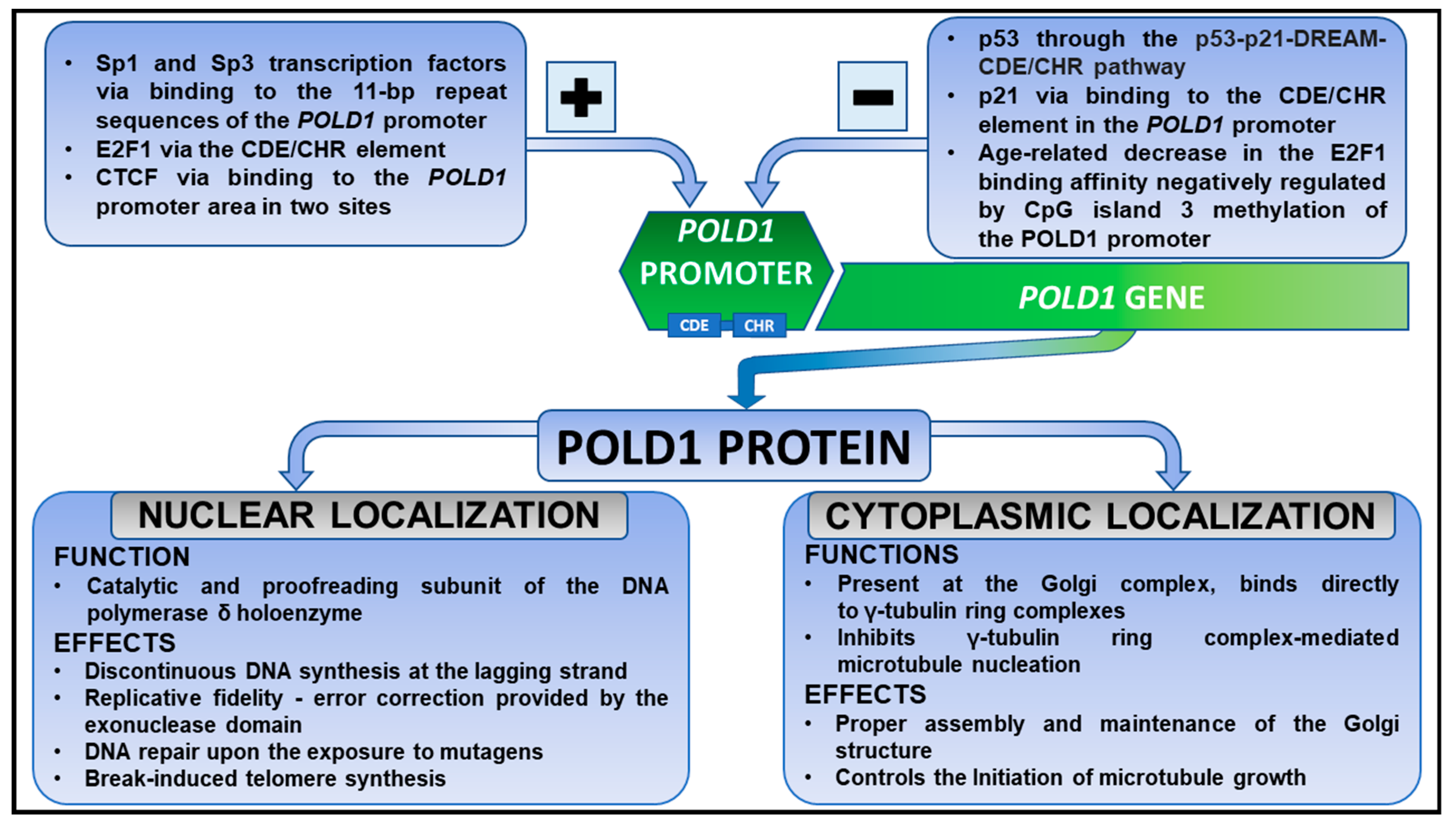
2. POLD1 Gene and a Polymerase Delta Holoenzyme in Health

The DNA polymerase delta 1 catalytic subunit (POLD1) gene is located on chromosome 19 at q13.3-q13.4 [27,28]. The human POLD1 gene is composed of 27 exons and 26 introns. It encodes a large 125-kDa protein known as the POLD1/p125 subunit, a catalytic and proofreading subunit of the human DNA polymerase delta (Polδ). The fluctuations of the POLD1 promoter activity imply that the transcriptional mRNA and protein levels are regulated during the cell cycle [4]. The POLD1 gene promoter is activated by Sp1, Sp3 transcription factors, cell cycle-related E2F1, and CCCTC-binding factor (CTCF) [29,30]. On the other hand, the p53 protein, the “genome guardian”, and cyclin-dependent kinase inhibitor p21 are able to inhibit the Sp1-stimulated POLD1 promoter activity, thus repressing POLD1 gene expression [31,32].
Song et al. discovered that a cell cycle-dependent element (CDE)/cell cycle gene homology region (CHR) element—is an active functional element in the POLD1 promoter, crucial for the cell cycle regulation of the POLD1 gene [32]. They also showed that E2F1 and p21 regulate the POLD1 promoter via binding to the CDE/CHR element [32]. Interestingly, p53-mediated inhibition of POLD1 transcription seems to be indirect [33]. p53 does not directly bind to the CDE–CHR motifs. Instead, p53 triggers indirect p21-dependent POLD1 downregulation via the p53–p21–DREAM–CDE/CHR pathway [33]. In addition, CTCF mainly binds to the POLD1 promoter in site 3 and 4 regions [30]. It has been shown that these two regions play crucial roles in CTCF-mediated POLD1 regulation [30]. Additionally, microRNA-155-mediated suppression of the forkhead transcription factor FOXO3a subsequently contributes to the decreased expression of POLD1 at the mRNA and protein levels [34].
Moreover, POLD1 expression has been causally linked to age-related cellular senescence and two POLD1-related mechanisms of aging have been proposed [26,30]. Gao et al. showed that the attenuation of the binding affinity of E2F1 for the POLD1 promoter, via age-related decline in E2F1 expression and increased CpG island 3 methylation in the POLD1 promoter region, causes a reduction in POLD1 expression in replicative senescence [26]. Hou et al. proposed, however, that an age-related decline in CTCF levels affects the binding affinity of CTFC to the POLD1 promoter, thus driving down POLD1 expression [30]. It is, therefore, well established that down-regulation of POLD1 is a characteristic age-related change occurring in humans [26,30,35].
Human Polδ holoenzyme has been believed to be a heterotetramer in unstressed, non-dividing cells composed of a catalytic subunit (POLD1) and three accessory subunits, POLD2 (p50), POLD3 (p66/p68), and POLD4 (p12) [36]. However, it has recently been suggested that the POLD4 subunit may occur as a dimer in the human Polδ complex [37]. Therefore, human Polδ potentially exists under a pentameric structure [37]. Through the catalytic POLD1 subunit, in association with both replication factor C (RFC) and proliferating cell nuclear antigen (PCNA), Polδ plays a pivotal role in discontinuous DNA synthesis at the lagging strand during genome replication. Moreover, its 3′–5′ exonuclease domain provides an error-correcting activity during DNA synthesis [38]. It has also been shown that Polδ is required for break-induced telomere synthesis [39].
A pivotal role for POLD1 in maintaining the enzymatic function of the Polδ complex suggests that adequate cellular POLD1 expression levels should be sustained for the smooth functioning of genomic DNA replication [40]. Novel data suggest that POLD1 is protected from degradation while being interconnected with the three different subunits in the Polδ complex, whereas excessive non-associated POLD1 is degraded to intercept the formation of incomplete and inactive Polδ units [40]. Moreover, results obtained in an experimental study using a murine model revealed the overall low levels of POLD1-3 subunits in POLD3-deficient cells [41].
Surprisingly, it has recently been shown that POLD1 plays a unique role in both the nucleus and the cytoplasm [42]. Subcellular fractionation has revealed that more than 60% of the POLD1 content occurs in the cytoplasm [42]. Regarding its cytoplasmic function, the POLD1 protein localized at the Golgi complex controls microtubule growth [42]. Shen et al. demonstrated that POLD1 undergoes bidirectional nucleocytoplasmic transport [43]. In addition, they also substantially unraveled the mechanisms underlying both the nuclear import and export of the POLD1 protein [43]. Figure 1 summarizes the mechanisms of POLD1 gene regulation. Moreover, it outlines the physiological role of the POLD1 protein within the cytoplasm and nucleus.
Interestingly, during normal cell cycle progression, conversion of heterotetrameric Polδ (Polδ4) to the heterotrimer (Polδ3) takes place via POLD4/p12 subunit degradation [40,41]. It has been shown that POLD4 undergoes proteasomal degradation initiated by its polyubiquitination, in synchrony with the S phase [44,45]. Contrarily, no alteration in POLD1, POLD2, and POLD3 levels with the progression of the cell cycle was observed in Zhang et al.’s studies [45]. Although both Polδ4 and Polδ3 are active polymerases, Polδ3 seems to be the predominating form of human Polδ during the S phase of the cell cycle [6,45]. Moreover, a direct comparison of Polδ3 with Polδ4 has shown that the heterotrimeric complex displays a higher ratio of exonuclease to polymerase activity in vitro than four-subunit Polδ4 [6]. In addition, POLD4 degradation in response to DNA damage after the application of ultraviolet, methyl methanesulfonate, hydroxyurea, and aphidicolin, causes the concomitant conversion of Polδ4 to a heterotrimeric form [46]. Therefore, exposure to the agents causing DNA damage following Polδ4 to Polδ3 conversion suppresses DNA synthesis, while at the same time favoring proofreading activity under conditions of DNA replication stress.

3. POLD1 Gene and a Polymerase Delta Holoenzyme in Non-Oncogenic Processes

Numerous studies have documented the physiological role of both Polδ holoenzyme and POLD1 protein. As they are involved in multiple biological processes, including DNA replication and DNA repair, a growing body of studies in recent years has highlighted the role of germline and somatic mutations in POLD1POLD4 genes in the genesis of human pathologies. Mandibular hypoplasia, deafness, progeroid features, and progressive lipodystrophy (MDPL) syndrome is a rare autosomal dominant disorder caused by heterozygous de novo mutations in the POLD1 gene [47,48]. Notably, exome sequencing revealed that mutations in different POLD1 domains may lead to a major phenotypic variability in MDPL syndrome [48]. Recent data have also revealed that POLD1 mutations causing reduced Polδ polymerase activity with a maintained exonuclease activity can lead to a non-syndromic sensorineural hearing loss in an autosomal recessive manner [49]. In addition, several cases of autosomal recessive POLD1-linked combined immunodeficiency have been reported [50,51,52]. Additionally, Nichols-Vinueza et al. described a potential role for POLD1 in B-cell maturation [52].

4. POLD1 in Cancers

Knowing that Polδ is crucial in genome maintenance and is involved in the DNA proofreading processes, it is not surprising that mutations in the POLD1 gene have been linked to genomic instability, mutator phenotype, and carcinogenesis [7,8,9]. In this paragraph, we aim to discuss the influence of both germline and somatic POLD1 mutations on different malignancies. Moreover, the changes in the POLD1 expression level in relation to clinicopathological parameters will be addressed.

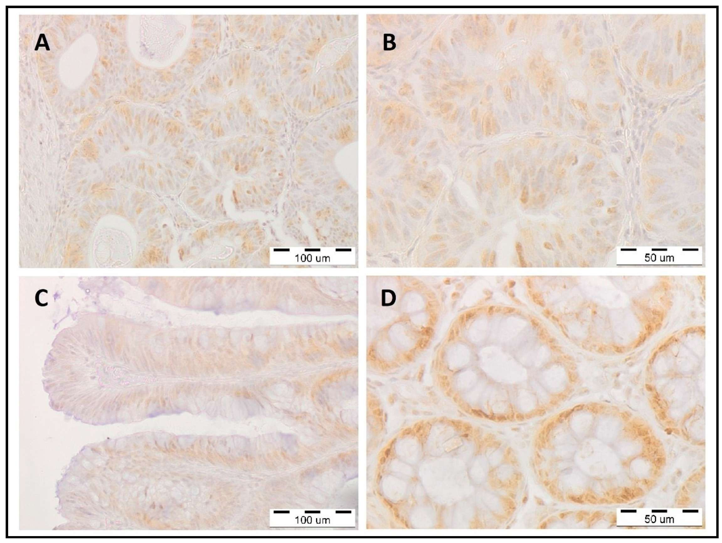
4.1. Colorectal Cancer

Colorectal cancer (CRC) is the third most frequently diagnosed cancer worldwide but is second in terms of mortality [1]. Almost a third of all CRC cases have a familial component, however, only about 5% of these develop as a consequence of well-defined inherited syndromes, whereas the majority of them do not possess a known genetic background [53,54]. To date, colorectal cancer (CRC) seems to be the most extensively studied malignancy concerning POLD1 mutations.
Among patients with a family history of CRC and multiple or large colorectal polyps, a minor fraction of carriers of POLD1 heterozygous germline mutations can be found [55,56]. Regarding somatic pathogenic variants affecting POLD1 in CRC, they are thought to be extremely rare findings [57]. Unfortunately, the overall incidence of POLD1 mutation in CRC patients remains unknown. Since the first description in 2013 of a correlation between the POLD1 mutation and dominantly inherited intestinal adenomas and carcinomas [55], two terms regarding this phenomenon have been coined. Originally, this syndrome was described as “polymerase proofreading-associated polyposis” [11], however, the term “POLE/POLD1-associated tumor syndrome” was recently proposed instead [12], as among some patients with a germline mutation in DNA polymerase subunits POLE and/or POLD1, expanded extracolonic tumor spectrum and absence of colonic polyposis have been noted. Interestingly, tumors with POLE/POLD1 mutations were found to be hypermutated, chromosomally unstable, and, surprisingly, microsatellite stable [55]. Moreover, recent data suggested the occurrence of an abundance of tumor-infiltrating lymphocytes (TILs) in POLE/POLD1-mutant CRCs [58]. In general, lymphocytes located in the tumor tissue region have emerged as the activators of a patient’s pre-existing intratumoral immunity after exposure to ICI treatment [58].
Unfortunately, data regarding POLE/POLD1 status to predict the efficacy of immunotherapy in CRC treatment are scarce, thus conducting research in this field would be reasonable.
Recently, Siraj et al. investigated the correlation of immunohistochemical POLD1 protein expression with clinicopathological features in Middle Eastern CRCs [13]. They observed that low POLD1 expression, found in more than half of CRC cases, was correlated with larger tumor size, adenocarcinoma histology, and stage III tumors [13]. Moreover, in our study focused on patients with CRC using IHC analysis, lower POLD1 nuclear expression in CRC cells compared with normal epithelial colon cells was observed. POLD1 expression levels in the tumor cells did not correlate with clinicopathological factors and the prognosis of CRC patients (data not published yet).
In Figure 2, we present representative immunohistochemical staining patterns of POLD1 within healthy, benign polypoid, and malignant tissues.

4.2. Endometrial Cancer

With its incidence continuing to rise, endometrial cancer (EC) is the most common gynecological malignancy in developed countries [1,59]. Although the POLD1 mutations in general are very scarce in EC [14,60], it is now well established that POLD1 germline mutations predispose people to EC [11,14]. Presumably, the frequency of POLD1 mutations in EC may be population dependent [60,61]. Among 47 South East Asian women with grade 3 (according to FIGO grades) endometrioid ECs, a presence of two pathogenic germline POLD1 mutations was detected [61]. Whereas Church et al.’s data revealed only one germline POLD1-mutated endometrioid EC among 154 patients with different FIGO grades enrolled in a British study [60].
Moreover, EC was found to be the most common malignancy among female POLD1 germline mutation carriers [62]. Palles et al.’s studies revealed that EC is the most frequent extraintestinal cancer in polymerase proofreading-associated polyposis syndrome [62]. Surprisingly, one recent study on Chinese genomic cancer population data suggests that EC may be the most common malignancy with POLD1 mutations among all cancers [63].
Even though POLD1-mutated cancers are believed to be primarily microsatellite stable [55], some ECs and CRCs possess microsatellite instability (MSI) [63,64,65,66,67]. Notably, Haraldsdottir et al. have suggested that the deficient DNA mismatch repair system in endometrial and colorectal cancers resulting in MSI phenotype, may occur mainly in tumors with somatic rather than germline POLD1/POLE mutations [64]. In addition, one study that assessed an endometrial dedifferentiated carcinoma molecular profile suggested that POLD1 and MLH1 (a gene involved in DNA mismatch repair), as well as high microsatellite instability (MSI-H) and increased tumor mutation burden (TMB), may be peculiar to this rare distinct subtype of EC [68].
As it is widely known that patients with MSI-H tumors can benefit from immunotherapy [69], routine clinical genetic testing for both POLD1/POLE and MSI-related mutations may help identify patients with endometrial and colorectal cancers who could respond well to immunotherapy [70]. To date, only one study evaluated POLD1 protein immunohistochemical expression in EC and its association with the clinicopathological profile [14]. Low expression of POLD1 was noted in almost 60% of Saudi EC cases and significantly correlated with grade 1 tumors [14].
Nevertheless, conducting research to establish both prognostic and predictive implications of POLD1 mutation and/or POLD1 protein expression for EC patients is justified.

4.3. Renal Cancer

Clear cell renal cell carcinoma (ccRCC) is the most common histopathological subtype of renal cancer, accounting for up to 85% of all kidney cancers [71]. Current studies have highlighted the role of DNA damage repair pathways in ccRCC. Two research groups have recently analyzed DNA polymerase epsilon and delta and their possible prognostic significance for patients with ccRCC [15,72]. Our team was the first to report that, unexpectedly, POLD1 nuclear protein expression levels correlated with better prognosis in patients with ccRCC [15]. Moreover, we have also demonstrated that the level of POLD1 immunoexpression in tumor cells does not correlate with the demographic and clinicopathological characteristics of ccRCC patients [15]. This unanticipated data demonstrating the positive correlation of POLD1 nuclear immunoreactivity with longer overall survival among ccRCC patients, in contrast to several reports regarding other malignancies, may suggest that the potential role of POLD1 can be considered as cancer specific [14,17].
Eventually, subsequent molecular studies will be crucial to clarify the role of POLD1 in ccRCC tumorigenesis.

4.4. Liver Cancer

Hepatocellular carcinoma (HCC), accounting for 75–85% of primary liver cancers [1], is the second most lethal malignancy after pancreatic cancer, with a 5-year relative sur-vival of 18% [59]. The role of POLD1 in HCC was first presented in 2010 [16]. Sanefuji et al. demonstrated that POLD1 overexpression induced by tp53 mutation correlates with a high degree of vascular invasion, poor cellular differentiation, and a poorer prognosis in HCC [16]. They have also suggested that POLD1 may contribute to cancer cell dedifferen-tiation in human HCC [16]. Furthermore, apoptosis of HCC cells was shown to be related to the inhibition of POLD1 gene expression [73]. Similarly, apoptosis of triple-negative breast cancer (BC) cells was driven by the reduction in POLD1 expression [74].
Recently, Tang et al. conducted a thorough analysis of the predictive and prognostic value of POLD1 in HCC [17]. It was shown that POLD1 is significantly upregulated in HCC in comparison with adjacent healthy liver tissue [17]. Additionally, POLD1 overexpression correlated with increased levels of alpha-fetoprotein, high TNM stage (III and IV), and predicted worse prognosis in HCC [17]. In addition, aberrantly high expression of POLD1 in HCC tissue was demonstrated to be caused by DNA copy number gain, low POLD1 methylation, and downregulation of small noncoding miR-139-3p [17]. Lastly, POLD1 expression positively correlated with the immune infiltration levels of B cells, macrophages, dendritic cells, and CD4+ T cells in the HCC tumor microenvironment [17]. Moreover, an in vivo orthotopic liver injection model revealed that POLD1 knockdown in HCC cells diminished tumor incidence, size, and lung metastases in mice [75].
Taken together, an in-depth exploration of HCC tumorigenesis can aid in the development of novel clinical treatment modalities.

4.5. Breast Cancer

With an estimated 2.3 million new cases in 2020, breast cancer is the current leading cause of global cancer incidence, constituting 11.7% of all cancers diagnosed worldwide [1]. Patients harboring germline POLD1 mutations are at higher risk of developing BC compared with the general population [76,77]. Somatic mutations, although insufficiently documented, have also been reported in BC [78,79]. Furthermore, even though BC cases are infrequently associated with MSI [80], this particular group of tumors may be POLD1-mutated as well [81].
Research conducted by Zhang et al. highlighted the mechanisms underlying changes in POLD1 expression in BC [18]. They have shown that POLD1 expression and the methylation level of the POLD1 promoter are both increased in BC tissues and cell lines [18]. Moreover, POLD1 expression and the methylation of the POLD1 gene promoter were found to be inhibited by the p53 protein through the suppression of DNA methyltransferase 1 and Sp1 activities in BC [18]. In the following studies, they observed that sirtuin 1, a member of the sirtuin family, enhances proliferative, migratory, and invasive properties of BC cells in vitro by inhibiting the p53 protein expression and, therefore, promoting the POLD1 upregulation [82].
To date, data regarding the expression patterns and clinical value of POLD1 among patients with BC are scarce. However, novel studies conducted by Qin et al. revealed that increased POLD1 expression is linked to poor prognosis in BC [19]. Elevated POLD1 levels were related to lymph node metastasis, histological grade, p53 status, and the ki-67 index [19]. Moreover, survival analysis showed that BC patients with high POLD1 levels had poorer disease-free survival in comparison with patients with low POLD1 expression [19]. In addition, POLD1 status likely affects the treatment response, especially in triple-negative breast cancer [83].
Thus, these data suggest that the POLD1 protein can be considered as a potential prognostic biomarker in BC [19].

4.6. Other Cancers

Although POLD1 mutations in cancers seem to be understudied, they may serve as promising prognostic/predictive biomarkers in the future and, importantly, future targets for novel POLD1-oriented therapies. Nevertheless, apart from malignancies mentioned in the previous paragraphs, several different cancers have been reported to harbor POLD1 mutation as well. Wang et al.’s studies that aimed to assess the clinical benefits of immune-checkpoint inhibitor (ICI) treatment have estimated that across almost 50,000 patients with different cancer types, POLD1 mutation occurred in 1.37% of cases [10].
For instance, the overall frequency of POLD1 mutations was estimated to be 2.5% among Chinese patients with lung cancer [84]. Furthermore, a recent analysis revealed that POLD1 is associated with the development of non-small cell lung cancer (NSCLC) from chronic obstructive pulmonary disease [20]. Interestingly, high expression levels of POLD1 indicated poor prognosis in lung adenocarcinoma but not in squamous cell carcinoma [20]. Additionally, in both mesothelioma, a rare and deadly cancer, and esophageal squamous cell carcinoma, POLD1 has been found to play a significant role in resistance to platinum-based chemotherapy [85,86].
In papillary thyroid cancer, low POLD1 expression implies poor clinicopathological characteristics [21]. Recently, POLD1 protein level was shown to be negatively correlated with the expression of cadherin-16 in this cancer [87].
In gastric adenocarcinoma, POLD1 mutation occurred in 2.77% of 613 patients [88]. Zhu et al. have also shown that using immunotherapy in POLE/POLD1-mutated tumors may have potentially positive implications [88].
Comprehensive molecular profiling has identified the presence of POLD1 mutations in brain tumors [62,89,90,91]. Additionally, POLD1 expression in this heterogeneous group of neoplasms was only described in 1p19q co-deleted lower-grade gliomas [24]. Yet, the clinical significance of POLD1 mutations and changes in the POLD1 protein level in central nervous system tumors is not fully understood.
Studies concerning uveal melanoma, the most common primary intraocular malignancy in adults, have discussed the changes in POLD1 expression patterns within patients with this cancer [22,23].
Much research has been conducted to investigate the possible clinical significance of POLD1 in gynecologic malignancies. Besides endometrial carcinoma, cervical and ovarian cancers were also assessed for the importance of POLD1 mutations and expression changes [92,93]. Interestingly, the POLD1 gene was more frequently altered among Chinese women with locally advanced cervical cancer than in Caucasian patients [92]. Furthermore, using cisplatin-resistant and cisplatin-sensitive ovarian cancer cell lines, Xing et al. demonstrated that POLD1 was highly expressed in the platinum-sensitive group, whereas chemoresistant cell lines possessed a reduced expression of POLD1 [93].
Intriguingly, in acute lymphoblastic leukemia (ALL), POLD1 upregulation may promote the relapse of this disease [25]. Thus, its expression could be proposed as the potential diagnostic marker and therapeutic target for the treatment of relapsed ALL [25].
A summary of studies discussing the relationship of POLD1 expression level with clinicopathological characteristics was provided in Table 1.

5. Conclusions and Future Directions

With cancer incidence rates continuing to rise globally, there is a strong need for the development of new biomarkers and treatment strategies [1]. The genetic mutations and expression profiles in POLD1-mutated cancers have recently gained remarkable attention. Nevertheless, the complex regulatory mechanisms of POLD1 expression in cancers remain to be fully characterized.
Both POLD1 germline and somatic mutations within the proofreading domain are considered rare in a variety of malignancies [9,10,63]. Additionally, the presence of pathogenic POLD1 germline alterations has been linked with some hereditary tumors, such as colorectal and endometrial cancers [55].
Based on the primary role of Polδ in genome maintenance, mutations in the catalytic POLD1 subunit lead to genomic instability, mutator phenotype, and malignant transformation [7,9]. Missense mutations are believed to be the most common mutations in POLD1-associated tumors, and no mutation hotspots have been identified so far [70,84].
It should be noted that POLD1 has been considered haploinsufficient in the literature [9,47,55]. Contrarily, a POLD1 haplosufficiency has been recently revealed in the yeast model [94] and in germline monoallelic POLD1 alteration carriers [95,96]. It has been speculated that a solitary heterozygous POLD1 mutation in the exonuclease domain without concurrent mutations in other repair systems causes only a modest increase in the mutation rate [95]. In addition, Schamschula et al. suggested that POLD1-mutated cancers acquire the ultra-high TMB only with concurrent mismatch repair deficiency [66]. It is hypothesized that there are no cancers with an exclusive heterozygous POLD1 mutation and only a complete loss of the two POLD1 alleles can lead to cancer [95]. Therefore, both germline and somatic POLD1 mutations triggering tumor formation may turn out to be biallelic [95]. Nevertheless, an attempt to organize the current knowledge and deepen understanding of POLD1 biology in carcinogenesis would be of paramount importance and could pave the way for novel anti-cancer drugs.
Whereas an overwhelming number of studies in the last decades have focused on POLD1 pathogenic variants in a wide range of neoplasms, only a few studies have validated the prognostic and predictive utility of POLD1 mRNA and protein expression in human cancers to date. So far, POLD1 expression and its clinical significance have been investigated in colorectal, endometrial, renal, liver, breast, lung, and thyroid cancers [13,14,15,17,19,20,21]. Nevertheless, these data are mainly limited to single-institution experiences.
In recent years, various immunotherapy-based cancer treatments have been approved for clinical use and many additional drugs are still being developed, thus opening a new era in cancer treatment [97,98]. Moreover, ICI treatment is considered a new standard of care across many cancer indications [97]. There are several biomarkers for predicting the outcome of ICI treatment, among which expression of programmed death-ligand 1 (PD-L1), MSI status, and TMB are the most commonly used [63,97]. Moreover, high levels of TILs (especially the CD8+ T cells subpopulation [99]), common in MSI cancers, may predict the response to ICI treatment in various malignancies [100]. As some ICI recipients fail to respond to the treatment, it is therefore crucial to identify accessory molecular biomarkers for predicting the ICI response to prevent immunotherapy overtreatment [98,99,100,101,102].
Some reports have suggested that POLE/POLD1 mutations and/or POLE/POLD1 expression patterns may be useful to predict immunotherapy effectiveness [10,70]. Previous studies investigating the association of POLE/POLD1 mutation with MSI status in various cancers have shown ambiguous results. Although cancers harboring POLD1 mutation are believed to be primarily microsatellite stable, some of them may display an MSI-H phenotype [55,63,64,65,66,67,68]. Wang et al.’s study on a cohort of almost 50,000 patients with different solid cancer types assessed the impact of POLE/POLD1 mutations on immunotherapy outcomes [10]. Their analysis demonstrated that among ICI-treated cancer individuals, patients with either POLE or POLD1 mutations have significantly longer overall survival in comparison with those without [10]. Notably, the ICI treatment outcomes were similar, regardless of whether these mutations were located within the exonuclease domain or outside of it [10]. Regrettably, POLE/POLD1 alterations cannot be independent factors for predicting the benefit from immunotherapy [101]. In addition, a study conducted by He et al. suggested that POLE/POLD1 mutations in the proofreading domain may result in DNA repair defects and an exceptionally high TMB, thereby generating high neoantigen load levels that, in turn, positively correlate with the sensitivity to ICI treatment [63]. Moreover, tumors expressing higher numbers of neoantigens are associated with a better response to immunotherapy in patients with CRC, melanoma, and NSCLC [63]. However, little is known about the influence of POLD1 expression level on the efficacy of ICI in cancer treatment.
In the last decade, whole-genome sequencing identified specific mutational signatures that describe the patterns of mutations that arise during tumorigenesis [103,104]. Several single-base substitution (SBS) signatures have been described concerning POLD1-mutated tumors; SBS10c and SBS10d are linked to defective POLD1 proofreading [105], whereas SBS20 is associated with concurrent POLD1 mutations and mismatch repair deficiency [65]. Groundbreaking studies have shown that the presence of specific SBS signatures may have prognostic and therapeutic implications [106]. Notably, Ma et al. reported that POLD1 functional mutations generating SBS signatures associated with POLD1 activity lead to augmented immunogenicity [107]. Moreover, patients with cancers generating POLD1-mutation-related SBS signatures demonstrated an enhanced response to ICI treatment [107].
Given the variety of immuno-oncology treatments currently in use for therapies in patients with different cancer types, including melanoma, NSCLC, CRC, and HCC [97,108], novel research examining the relationship between the expression of the POLD1 protein and the clinical outcomes in patients receiving immunotherapy would be of great importance. Several clinical trials are ongoing to evaluate the impact of POLD1 mutation on treatment outcomes (NCT05103969, NCT03810339, NCT03428802, NCT03491345, NCT04969029).
With the advancement of knowledge regarding cancer biology, substantial progress in the development of new diagnostic and prognostic molecular biomarkers as well as targeted therapeutic strategies against oncological diseases can be expected within the next few years.

Author Contributions

Conceptualization, M.G., P.S. and J.G.; writing—original draft preparation, M.G. and P.S.; writing—review and editing, M.G. and P.S.; figure preparation, M.G. and J.G.; review, J.G., A.S. and B.A.J.-F.; supervision, J.G. and A.S. All authors have read and agreed to the published version of the manuscript.

Funding

This research received no external funding.

Acknowledgments

Thanks to Marcela Burdzy-Pogonowska for proofreading the article.

Conflicts of Interest

The authors declare no conflict of interest.

References

  1. Sung, H.; Ferlay, J.; Siegel, R.L.; Laversanne, M.; Soerjomataram, I.; Jemal, A.; Bray, F. Global Cancer Statistics 2020: GLOBOCAN Estimates of Incidence and Mortality Worldwide for 36 Cancers in 185 Countries. CA Cancer J. Clin. 2021, 71, 209–249. [Google Scholar] [CrossRef]
  2. Ganesh, K.; Massagué, J. Targeting metastatic cancer. Nat. Med. 2021, 27, 34–44. [Google Scholar] [CrossRef] [PubMed]
  3. Fuchs, J.; Cheblal, A.; Gasser, S.M. Underappreciated Roles of DNA Polymerase δ in Replication Stress Survival. Trends Genet. 2021, 37, 476–487. [Google Scholar] [CrossRef] [PubMed]
  4. Zeng, X.R.; Hao, H.; Jiang, Y.; Lee, M.Y. Regulation of human DNA polymerase delta during the cell cycle. J. Biol. Chem. 1994, 269, 24027–24033. [Google Scholar] [CrossRef] [PubMed]
  5. Jain, R.; Rice, W.J.; Malik, R.; Johnson, R.E.; Prakash, L.; Prakash, S.; Ubarretxena-Belandia, I.; Aggarwal, A.K. Cryo-EM structure and dynamics of eukaryotic DNA polymerase δ holoenzyme. Nat. Struct. Mol. Biol. 2019, 26, 955–962. [Google Scholar] [CrossRef]
  6. Meng, X.; Zhou, Y.; Zhang, S.; Lee, E.Y.; Frick, D.N.; Lee, M.Y. DNA damage alters DNA polymerase delta to a form that exhibits increased discrimination against modified template bases and mismatched primers. Nucleic Acids Res. 2009, 37, 647–657. [Google Scholar] [CrossRef] [Green Version]
  7. Nicolas, E.; Golemis, E.A.; Arora, S. POLD1: Central mediator of DNA replication and repair, and implication in cancer and other pathologies. Gene 2016, 590, 128–141. [Google Scholar] [CrossRef]
  8. Lemoine, F.J.; Degtyareva, N.P.; Kokoska, R.J.; Petes, T.D. Reduced levels of DNA polymerase delta induce chromosome fragile site instability in yeast. Mol. Cell. Biol. 2008, 28, 5359–5368. [Google Scholar] [CrossRef] [Green Version]
  9. Rayner, E.; van Gool, I.C.; Palles, C.; Kearsey, S.E.; Bosse, T.; Tomlinson, I.; Church, D.N. A panoply of errors: Polymerase proofreading domain mutations in cancer. Nat. Rev. Cancer. 2016, 16, 71–81. [Google Scholar] [CrossRef]
  10. Wang, F.; Zhao, Q.; Wang, Y.N.; Jin, Y.; He, M.M.; Liu, Z.X.; Xu, R.H. Evaluation of POLE and POLD1 Mutations as Biomarkers for Immunotherapy Outcomes Across Multiple Cancer Types. JAMA Oncol. 2019, 5, 1504–1506. [Google Scholar] [CrossRef] [Green Version]
  11. Briggs, S.; Tomlinson, I. Germline and somatic polymerase ε and δ mutations define a new class of hypermutated colorectal and endometrial cancers. J. Pathol. 2013, 230, 148–153. [Google Scholar] [CrossRef] [PubMed] [Green Version]
  12. Schubert, S.A.; Morreau, H.; de Miranda, N.F.C.C.; van Wezel, T. The missing heritability of familial colorectal cancer. Mutagenesis 2020, 35, 221–231. [Google Scholar] [CrossRef] [PubMed] [Green Version]
  13. Siraj, A.K.; Bu, R.; Iqbal, K.; Parvathareddy, S.K.; Masoodi, T.; Siraj, N.; Al-Rasheed, M.; Kong, Y.; Ahmed, S.O.; Al-Obaisi, K.A.S.; et al. POLE and POLD1 germline exonuclease domain pathogenic variants, a rare event in colorectal cancer from the Middle East. Mol. Genet. Genom. Med. 2020, 8, e1368. [Google Scholar] [CrossRef]
  14. Siraj, A.K.; Parvathareddy, S.K.; Bu, R.; Iqbal, K.; Siraj, S.; Masoodi, T.; Concepcion, R.M.; Ghazwani, L.O.; AlBadawi, I.; Al-Dayel, F.; et al. Germline POLE and POLD1 proofreading domain mutations in endometrial carcinoma from Middle Eastern region. Cancer Cell Int. 2019, 19, 334. [Google Scholar] [CrossRef] [PubMed]
  15. Godlewski, J.; Stefaniak, P.; Kiezun, J.; Krazinski, B.E. DNA Polymerase Delta 1 Catalytic Subunit (POLD1) as a Prognostic Factor in Clear Cell Renal Cell Carcinoma Patients. In Vivo 2022, 36, 1188–1194. [Google Scholar] [CrossRef]
  16. Sanefuji, K.; Taketomi, A.; Iguchi, T.; Sugimachi, K.; Ikegami, T.; Yamashita, Y.; Gion, T.; Soejima, Y.; Shirabe, K.; Maehara, Y. Significance of DNA polymerase delta catalytic subunit p125 induced by mutant p53 in the invasive potential of human hepatocellular carcinoma. Oncology 2010, 79, 229–237. [Google Scholar] [CrossRef] [PubMed]
  17. Tang, H.; You, T.; Sun, Z.; Bai, C. A Comprehensive Prognostic Analysis of POLD1 in Hepatocellular Carcinoma. BMC Cancer 2022, 22, 197. [Google Scholar] [CrossRef]
  18. Zhang, L.; Yang, W.; Zhu, X.; Wei, C. p53 inhibits the expression of p125 and the methylation of POLD1 gene promoter by downregulating the Sp1-induced DNMT1 activities in breast cancer. Onco Targets Ther. 2016, 9, 1351–1360. [Google Scholar] [CrossRef] [Green Version]
  19. Qin, Q.; Tan, Q.; Li, J.; Yang, W.; Lian, B.; Mo, Q.; Wei, C. Elevated expression of POLD1 is associated with poor prognosis in breast cancer. Oncol. Lett. 2018, 16, 5591–5598. [Google Scholar] [CrossRef] [Green Version]
  20. Zhang, L.; Chen, J.; Yang, H.; Pan, C.; Li, H.; Luo, Y.; Cheng, T. Multiple microarray analyses identify key genes associated with the development of Non-Small Cell Lung Cancer from Chronic Obstructive Pulmonary Disease. J. Cancer 2021, 12, 996–1010. [Google Scholar] [CrossRef]
  21. Siraj, A.K.; Bu, R.; Arshad, M.; Iqbal, K.; Parvathareddy, S.K.; Masoodi, T.; Ghazwani, L.O.; Al-Sobhi, S.S.; Al-Dayel, F.; Al-Kuraya, K.S. POLE and POLD1 pathogenic variants in the proofreading domain in papillary thyroid cancer. Endocr. Connect. 2020, 9, 923–932. [Google Scholar] [CrossRef] [PubMed]
  22. Kucherlapati, M. Examining transcriptional changes to DNA replication and repair factors over uveal melanoma subtypes. BMC Cancer 2018, 18, 818. [Google Scholar] [CrossRef] [PubMed] [Green Version]
  23. Wang, T.; Liu, D.; Wang, L.; Liu, M.; Duan, W.; Yi, J.; Yi, Y. DNA Repair Genes Are Associated with Subtype Classification, Prognosis, and Immune Infiltration in Uveal Melanoma. J. Oncol. 2022, 2022, 1965451. [Google Scholar] [CrossRef] [PubMed]
  24. Tang, L.; Deng, L.; Bai, H.X.; Sun, J.; Neale, N.; Wu, J.; Wang, Y.; Chang, K.; Huang, R.Y.; Zhang, P.J.; et al. Reduced expression of DNA repair genes and chemosensitivity in 1p19q codeleted lower-grade gliomas. J. Neurooncol. 2018, 139, 563–571. [Google Scholar] [CrossRef] [PubMed]
  25. Li, S.; Wang, C.; Wang, W.; Liu, W.; Zhang, G. Abnormally high expression of POLD1, MCM2, and PLK4 promotes relapse of acute lymphoblastic leukemia. Medicine 2018, 97, e10734. [Google Scholar] [CrossRef]
  26. Gao, S.; Song, Q.; Liu, J.; Zhang, X.; Ji, X.; Wang, P. E2F1 mediates the downregulation of POLD1 in replicative senescence. Cell. Mol. Life Sci. 2019, 76, 2833–2850. [Google Scholar] [CrossRef] [Green Version]
  27. Chung, D.W.; Zhang, J.A.; Tan, C.K.; Davie, E.W.; So, A.G.; Downey, K.M. Primary structure of the catalytic subunit of human DNA polymerase delta and chromosomal location of the gene. Proc. Natl. Acad. Sci. USA 1991, 88, 11197–11201. [Google Scholar] [CrossRef] [Green Version]
  28. Kemper, R.R.; Ahn, E.R.; Zhang, P.; Lee, M.Y.; Rabin, M. Human DNA polymerase delta gene maps to region 19q13.3-q13.4 by in situ hybridization. Genomics 1992, 14, 205–206. [Google Scholar] [CrossRef]
  29. Zhao, L.; Chang, L.S. The human POLD1 gene. Identification of an upstream activator sequence, activation by Sp1 and Sp3, and cell cycle regulation. J. Biol. Chem. 1997, 272, 4869–4882. [Google Scholar] [CrossRef] [PubMed]
  30. Hou, Y.; Song, Q.; Gao, S.; Zhang, X.; Wang, Y.; Liu, J.; Fu, J.; Cao, M.; Wang, P. CTCF Mediates Replicative Senescence Through POLD1. Front. Cell Dev. Biol. 2021, 9, 618586. [Google Scholar] [CrossRef]
  31. Li, B.; Lee, M.Y. Transcriptional regulation of the human DNA polymerase delta catalytic subunit gene POLD1 by p53 tumor suppressor and Sp1. J. Biol. Chem. 2001, 276, 29729–29739. [Google Scholar] [CrossRef] [PubMed] [Green Version]
  32. Song, N.; Zhu, X.; Shi, L.; An, J.; Wu, Y.; Sang, J. Identification and functional analysis of a CDE/CHR element in the POLD1 promoter. Sci. China C Life Sci. 2009, 52, 551–559. [Google Scholar] [CrossRef] [PubMed]
  33. Fischer, M.; Quaas, M.; Steiner, L.; Engeland, K. The p53-p21-DREAM-CDE/CHR pathway regulates G2/M cell cycle genes. Nucleic Acids Res. 2016, 44, 164–174. [Google Scholar] [CrossRef] [PubMed] [Green Version]
  34. Czochor, J.R.; Sulkowski, P.; Glazer, P.M. miR-155 Overexpression Promotes Genomic Instability by Reducing High-fidelity Polymerase Delta Expression and Activating Error-Prone DSB Repair. Mol. Cancer Res. 2016, 14, 363–373. [Google Scholar] [CrossRef] [PubMed] [Green Version]
  35. Wang, J.L.; Guo, H.L.; Wang, P.C.; Liu, C.G. Age-dependent down-regulation of DNA polymerase δ1 in human lymphocytes. Mol. Cell. Biochem. 2012, 371, 157–163. [Google Scholar] [CrossRef]
  36. Lee, M.Y.W.T.; Wang, X.; Zhang, S.; Zhang, Z.; Lee, E.Y.C. Regulation and Modulation of Human DNA Polymerase δ Activity and Function. Genes 2017, 8, 190. [Google Scholar] [CrossRef] [Green Version]
  37. Khandagale, P.; Peroumal, D.; Manohar, K.; Acharya, N. Human DNA polymerase delta is a pentameric holoenzyme with a dimeric p12 subunit. Life Sci. Alliance 2019, 2, e201900323. [Google Scholar] [CrossRef] [Green Version]
  38. Morrison, A.; Bell, J.B.; Kunkel, T.A.; Sugino, A. Eukaryotic DNA polymerase amino acid sequence required for 3′----5′ exonuclease activity. Proc. Natl. Acad. Sci. USA 1991, 88, 9473–9477. [Google Scholar] [CrossRef] [Green Version]
  39. Dilley, R.L.; Verma, P.; Cho, N.W.; Winters, H.D.; Wondisford, A.R.; Greenberg, R.A. Break-induced telomere synthesis underlies alternative telomere maintenance. Nature 2016, 539, 54–58. [Google Scholar] [CrossRef] [Green Version]
  40. Drosopoulos, W.C.; Vierra, D.A.; Kenworthy, C.A.; Coleman, R.A.; Schildkraut, C.L. Dynamic Assembly and Disassembly of the Human DNA Polymerase δ Holoenzyme on the Genome In Vivo. Cell Rep. 2020, 30, 1329–1341.e5. [Google Scholar] [CrossRef]
  41. Murga, M.; Lecona, E.; Kamileri, I.; Díaz, M.; Lugli, N.; Sotiriou, S.K.; Anton, M.E.; Méndez, J.; Halazonetis, T.D.; Fernandez-Capetillo, O. POLD3 Is Haploinsufficient for DNA Replication in Mice. Mol. Cell. 2016, 63, 877–883. [Google Scholar] [CrossRef] [Green Version]
  42. Shen, Y.; Liu, P.; Jiang, T.; Hu, Y.; Au, F.K.C.; Qi, R.Z. The catalytic subunit of DNA polymerase δ inhibits γTuRC activity and regulates Golgi-derived microtubules. Nat. Commun. 2017, 8, 554. [Google Scholar] [CrossRef] [PubMed] [Green Version]
  43. Shen, Y.; Wang, K.; Qi, R.Z. The catalytic subunit of DNA polymerase δ is a nucleocytoplasmic shuttling protein. Exp. Cell Res. 2019, 375, 36–40. [Google Scholar] [CrossRef] [PubMed]
  44. Lee, M.Y.; Zhang, S.; Lin, S.H.; Wang, X.; Darzynkiewicz, Z.; Zhang, Z.; Lee, E.Y. The tail that wags the dog: p12, the smallest subunit of DNA polymerase δ, is degraded by ubiquitin ligases in response to DNA damage and during cell cycle progression. Cell Cycle 2014, 13, 23–31. [Google Scholar] [CrossRef] [PubMed] [Green Version]
  45. Zhang, S.; Zhao, H.; Darzynkiewicz, Z.; Zhou, P.; Zhang, Z.; Lee, E.Y.; Lee, M.Y. A novel function of CRL4(Cdt2): Regulation of the subunit structure of DNA polymerase δ in response to DNA damage and during the S phase. J. Biol. Chem. 2013, 288, 29550–29561. [Google Scholar] [CrossRef] [Green Version]
  46. Zhang, S.; Zhou, Y.; Trusa, S.; Meng, X.; Lee, E.Y.; Lee, M.Y. A novel DNA damage response: Rapid degradation of the p12 subunit of dna polymerase delta. J. Biol. Chem. 2007, 282, 15330–15340. [Google Scholar] [CrossRef] [Green Version]
  47. Weedon, M.N.; Ellard, S.; Prindle, M.J.; Caswell, R.; Lango Allen, H.; Oram, R.; Godbole, K.; Yajnik, C.S.; Sbraccia, P.; Novelli, G.; et al. An in-frame deletion at the polymerase active site of POLD1 causes a multisystem disorder with lipodystrophy. Nat. Genet. 2013, 45, 947–950. [Google Scholar] [CrossRef] [Green Version]
  48. Elouej, S.; Beleza-Meireles, A.; Caswell, R.; Colclough, K.; Ellard, S.; Desvignes, J.P.; Béroud, C.; Lévy, N.; Mohammed, S.; De Sandre-Giovannoli, A. Exome sequencing reveals a de novo POLD1 mutation causing phenotypic variability in mandibular hypoplasia, deafness, progeroid features, and lipodystrophy syndrome (MDPL). Metabolism 2017, 71, 213–225. [Google Scholar] [CrossRef]
  49. Oh, D.Y.; Matsumoto, Y.; Kitajiri, S.I.; Kim, N.K.D.; Kim, M.Y.; Kim, A.R.; Lee, M.; Lee, C.; Tomkinson, A.E.; Katsuno, T.; et al. POLD1 variants leading to reduced polymerase activity can cause hearing loss without syndromic features. Hum. Mutat. 2020, 41, 913–920. [Google Scholar] [CrossRef]
  50. Conde, C.D.; Petronczki, Ö.Y.; Baris, S.; Willmann, K.L.; Girardi, E.; Salzer, E.; Weitzer, S.; Ardy, R.C.; Krolo, A.; Ijspeert, H.; et al. Polymerase δ deficiency causes syndromic immunodeficiency with replicative stress. J. Clin. Investig. 2019, 129, 4194–4206. [Google Scholar] [CrossRef]
  51. Cui, Y.; Keles, S.; Charbonnier, L.M.; Julé, A.M.; Henderson, L.; Celik, S.C.; Reisli, I.; Shen, C.; Xie, W.J.; Schmitz-Abe, K.; et al. Combined immunodeficiency caused by a loss-of-function mutation in DNA polymerase delta 1. J. Allergy Clin. Immunol. 2020, 145, 391–401.e8. [Google Scholar] [CrossRef] [PubMed] [Green Version]
  52. Nichols-Vinueza, D.X.; Delmonte, O.M.; Bundy, V.; Bosticardo, M.; Zimmermann, M.T.; Dsouza, N.R.; Pala, F.; Dobbs, K.; Stoddard, J.; Niemela, J.E.; et al. POLD1 Deficiency Reveals a Role for POLD1 in DNA Repair and T and B Cell Development. J. Clin. Immunol. 2021, 41, 270–273. [Google Scholar] [CrossRef] [PubMed]
  53. Mao, R.; Krautscheid, P.; Graham, R.P.; Ganguly, A.; Shankar, S.; Ferber, M.; Hegde, M.; ACMG Laboratory Quality Assurance Committee. Genetic testing for inherited colorectal cancer and polyposis, 2021 revision: A technical standard of the American College of Medical Genetics and Genomics (ACMG). Genet. Med. 2021, 23, 1807–1817. [Google Scholar] [CrossRef]
  54. Jasperson, K.W.; Tuohy, T.M.; Neklason, D.W.; Burt, R.W. Hereditary and familial colon cancer. Gastroenterology 2010, 138, 2044–2058. [Google Scholar] [CrossRef] [PubMed] [Green Version]
  55. Palles, C.; Cazier, J.B.; Howarth, K.M.; Domingo, E.; Jones, A.M.; Broderick, P.; Kemp, Z.; Spain, S.L.; Guarino, E.; Salguero, I.; et al. Germline mutations affecting the proofreading domains of POLE and POLD1 predispose to colorectal adenomas and carcinomas. Nat. Genet. 2013, 45, 136–144. [Google Scholar] [CrossRef] [PubMed] [Green Version]
  56. Elsayed, F.A.; Tops, C.M.J.; Nielsen, M.; Ruano, D.; Vasen, H.F.A.; Morreau, H.J.; Hes, F.; van Wezel, T. Low frequency of POLD1 and POLE exonuclease domain variants in patients with multiple colorectal polyps. Mol. Genet. Genom. Med. 2019, 7, e00603. [Google Scholar] [CrossRef] [Green Version]
  57. Mur, P.; García-Mulero, S.; Del Valle, J.; Magraner-Pardo, L.; Vidal, A.; Pineda, M.; Cinnirella, G.; Martín-Ramos, E.; Pons, T.; López-Doriga, A.; et al. Role of POLE and POLD1 in familial cancer. Genet. Med. 2020, 22, 2089–2100. [Google Scholar] [CrossRef]
  58. Keshinro, A.; Vanderbilt, C.; Kim, J.K.; First, C.; Chen, C.T.; Yaeger, R.; Ganesh, K.; Segal, N.H.; Gonen, M.; Shia, J.; et al. Tumor-Infiltrating Lymphocytes, Tumor Mutational Burden, and Genetic Alterations in Microsatellite Unstable, Microsatellite Stable, or Mutant POLE/POLD1 Colon Cancer. JCO Precis. Oncol. 2021, 5, PO.20.00456. [Google Scholar] [CrossRef] [PubMed]
  59. Jemal, A.; Ward, E.M.; Johnson, C.J.; Cronin, K.A.; Ma, J.; Ryerson, B.; Mariotto, A.; Lake, A.J.; Wilson, R.; Sherman, R.L.; et al. Annual Report to the Nation on the Status of Cancer, 1975-2014, Featuring Survival. J. Natl. Cancer Inst. 2017, 109, djx030. [Google Scholar] [CrossRef] [Green Version]
  60. Church, D.N.; Briggs, S.E.; Palles, C.; Domingo, E.; Kearsey, S.J.; Grimes, J.M.; Gorman, M.; Martin, L.; Howarth, K.M.; Hodgson, S.V.; et al. DNA polymerase ε and δ exonuclease domain mutations in endometrial cancer. Hum. Mol. Genet. 2013, 22, 2820–2828. [Google Scholar] [CrossRef] [Green Version]
  61. Wong, A.; Kuick, C.H.; Wong, W.L.; Tham, J.M.; Mansor, S.; Loh, E.; Jain, S.; Vikas, N.N.; Tan, S.H.; Chan, S.H.; et al. Mutation spectrum of POLE and POLD1 mutations in South East Asian women presenting with grade 3 endometrioid endometrial carcinomas. Gynecol. Oncol. 2016, 141, 113–120. [Google Scholar] [CrossRef] [PubMed]
  62. Palles, C.; Martin, L.; Domingo, E.; Chegwidden, L.; McGuire, J.; Cuthill, V.; Heitzer, E.; CORGI Consortium; Kerr, R.; Kerr, D.; et al. The clinical features of polymerase proof-reading associated polyposis (PPAP) and recommendations for patient management. Fam. Cancer 2022, 21, 197–209. [Google Scholar] [CrossRef] [PubMed]
  63. He, J.; Ouyang, W.; Zhao, W.; Shao, L.; Li, B.; Liu, B.; Wang, D.; Han-Zhang, H.; Zhang, Z.; Shao, L.; et al. Distinctive genomic characteristics in POLE/POLD1-mutant cancers can potentially predict beneficial clinical outcomes in patients who receive immune checkpoint inhibitor. Ann. Transl. Med. 2021, 9, 129. [Google Scholar] [CrossRef] [PubMed]
  64. Haraldsdottir, S.; Hampel, H.; Tomsic, J.; Frankel, W.L.; Pearlman, R.; de la Chapelle, A.; Pritchard, C.C. Colon and endometrial cancers with mismatch repair deficiency can arise from somatic, rather than germline, mutations. Gastroenterology 2014, 147, 1308–1316.e1. [Google Scholar] [CrossRef] [Green Version]
  65. Haradhvala, N.J.; Kim, J.; Maruvka, Y.E.; Polak, P.; Rosebrock, D.; Livitz, D.; Hess, J.M.; Leshchiner, I.; Kamburov, A.; Mouw, K.W.; et al. Distinct mutational signatures characterize concurrent loss of polymerase proofreading and mismatch repair. Nat. Commun. 2018, 9, 1746. [Google Scholar] [CrossRef] [Green Version]
  66. Schamschula, E.; Kinzel, M.; Wernstedt, A.; Oberhuber, K.; Gottschling, H.; Schnaiter, S.; Friedrichs, N.; Merkelbach-Bruse, S.; Zschocke, J.; Gallon, R.; et al. Teenage-Onset Colorectal Cancers in a Digenic Cancer Predisposition Syndrome Provide Clues for the Interaction between Mismatch Repair and Polymerase δ Proofreading Deficiency in Tumorigenesis. Biomolecules 2022, 12, 1350. [Google Scholar] [CrossRef]
  67. Jansen, A.M.; van Wezel, T.; van den Akker, B.E.; Ventayol Garcia, M.; Ruano, D.; Tops, C.M.; Wagner, A.; Letteboer, T.G.; Gómez-García, E.B.; Devilee, P.; et al. Combined mismatch repair and POLE/POLD1 defects explain unresolved suspected Lynch syndrome cancers. Eur. J. Hum. Genet. 2016, 24, 1089–1092. [Google Scholar] [CrossRef]
  68. Kobayashi, Y.; Kitazono, I.; Akahane, T.; Yanazume, S.; Kamio, M.; Togami, S.; Nohara, S.; Sakamoto, I.; Yokoyama, S.; Tabata, K.; et al. Molecular Evaluation of Endometrial Dedifferentiated Carcinoma, Endometrioid Carcinoma, Carcinosarcoma, and Serous Carcinoma Using a Custom-Made Small Cancer Panel. Pathol. Oncol. Res. 2021, 27, 1610013. [Google Scholar] [CrossRef]
  69. Li, K.; Luo, H.; Huang, L.; Luo, H.; Zhu, X. Microsatellite instability: A review of what the oncologist should know. Cancer Cell Int. 2020, 20, 16. [Google Scholar] [CrossRef] [Green Version]
  70. Ma, X.; Dong, L.; Liu, X.; Ou, K.; Yang, L. POLE/POLD1 mutation and tumor immunotherapy. J. Exp. Clin. Cancer Res. 2022, 41, 216. [Google Scholar] [CrossRef]
  71. Cohen, H.T.; McGovern, F.J. Renal-cell carcinoma. N. Engl. J. Med. 2005, 353, 2477–2490. [Google Scholar] [CrossRef] [PubMed] [Green Version]
  72. Wu, X.; Tang, H.; Xu, W.H.; Tang, H.; Wei, S.; Anwaier, A.; Huang, H.; Qu, Y.Y.; Zhang, H.; Zhao, S.; et al. Protumorigenic Role of Elevated Levels of DNA Polymerase Epsilon Predicts an Immune-Suppressive Microenvironment in Clear Cell Renal Cell Carcinoma. Front. Genet. 2021, 12, 751977. [Google Scholar] [CrossRef] [PubMed]
  73. Cao, B.; Zhang, Z.; Zhang, Y.; Li, J.; Liang, G.; Ling, J. Effect of Smilax china L.-containing serum on the expression of POLD1 mRNA in human hepatocarcinoma SMMC-7721 cells. Exp. Ther. Med. 2013, 6, 1070–1076. [Google Scholar] [CrossRef] [PubMed]
  74. Liang, Z.J.; Wan, Y.; Zhu, D.D.; Wang, M.X.; Jiang, H.M.; Huang, D.L.; Luo, L.F.; Chen, M.J.; Yang, W.P.; Li, H.M.; et al. Resveratrol Mediates the Apoptosis of Triple Negative Breast Cancer Cells by Reducing POLD1 Expression. Front. Oncol. 2021, 11, 569295. [Google Scholar] [CrossRef]
  75. Ho, D.W.; Lam, W.M.; Chan, L.K.; Ng, I.O. Investigation of Functional Synergism of CENPF and FOXM1 Identifies POLD1 as Downstream Target in Hepatocellular Carcinoma. Front. Med. 2022, 9, 860395. [Google Scholar] [CrossRef] [PubMed]
  76. Sigurdson, A.J.; Hauptmann, M.; Chatterjee, N.; Alexander, B.H.; Doody, M.M.; Rutter, J.L.; Struewing, J.P. Kin-cohort estimates for familial breast cancer risk in relation to variants in DNA base excision repair, BRCA1 interacting and growth factor genes. BMC Cancer 2004, 4, 9. [Google Scholar] [CrossRef] [Green Version]
  77. Bellido, F.; Pineda, M.; Aiza, G.; Valdés-Mas, R.; Navarro, M.; Puente, D.A.; Pons, T.; González, S.; Iglesias, S.; Darder, E.; et al. POLE and POLD1 mutations in 529 kindred with familial colorectal cancer and/or polyposis: Review of reported cases and recommendations for genetic testing and surveillance. Genet. Med. 2016, 18, 325–332. [Google Scholar] [CrossRef] [Green Version]
  78. Nik-Zainal, S.; Davies, H.; Staaf, J.; Ramakrishna, M.; Glodzik, D.; Zou, X.; Martincorena, I.; Alexandrov, L.B.; Martin, S.; Wedge, D.C.; et al. Landscape of somatic mutations in 560 breast cancer whole-genome sequences. Nature 2016, 534, 47–54, Erratum in Nature 2019, 566, E1. [Google Scholar] [CrossRef] [PubMed] [Green Version]
  79. Encinas, G.; Sabelnykova, V.Y.; de Lyra, E.C.; Hirata Katayama, M.L.; Maistro, S.; de Vasconcellos Valle, P.W.M.; de Lima Pereira, G.F.; Rodrigues, L.M.; de Menezes Pacheco Serio, P.A.; de Gouvêa, A.C.R.C.; et al. Somatic mutations in early onset luminal breast cancer. Oncotarget 2018, 9, 22460–22479. [Google Scholar] [CrossRef] [Green Version]
  80. Anbazhagan, R.; Fujii, H.; Gabrielson, E. Microsatellite instability is uncommon in breast cancer. Clin. Cancer Res. 1999, 5, 839–844. [Google Scholar] [PubMed]
  81. da Silva, C.M.; Shimba, D.; Oishi, D.; Cagnacci, A.; de Gouvea, A.C.R.C.; Ades, F. VUS-type alteration in POLD1 and microsatellite instability in a metastatic luminal B breast cancer patient. Ecancermedicalscience 2020, 14, 1002. [Google Scholar] [CrossRef] [PubMed]
  82. Xu, Y.; Qin, Q.; Chen, R.; Wei, C.; Mo, Q. SIRT1 promotes proliferation, migration, and invasion of breast cancer cell line MCF-7 by upregulating DNA polymerase delta1 (POLD1). Biochem. Biophys. Res. Commun. 2018, 502, 351–357. [Google Scholar] [CrossRef] [PubMed]
  83. Anurag, M.; Jaehnig, E.J.; Krug, K.; Lei, J.T.; Bergstrom, E.J.; Kim, B.J.; Vashist, T.D.; Huynh, A.M.T.; Dou, Y.; Gou, X.; et al. Proteogenomic Markers of Chemotherapy Resistance and Response in Triple-Negative Breast Cancer. Cancer Discov. 2022, 12, 2586–2605. [Google Scholar] [CrossRef] [PubMed]
  84. Yao, J.; Gong, Y.; Zhao, W.; Han, Z.; Guo, S.; Liu, H.; Peng, X.; Xiao, W.; Li, Y.; Dang, S.; et al. Comprehensive analysis of POLE and POLD1 Gene Variations identifies cancer patients potentially benefit from immunotherapy in Chinese population. Sci. Rep. 2019, 9, 15767. [Google Scholar] [CrossRef] [PubMed] [Green Version]
  85. Røe, O.D.; Szulkin, A.; Anderssen, E.; Flatberg, A.; Sandeck, H.; Amundsen, T.; Erlandsen, S.E.; Dobra, K.; Sundstrøm, S.H. Molecular resistance fingerprint of pemetrexed and platinum in a long-term survivor of mesothelioma. PLoS ONE 2012, 7, e40521. [Google Scholar] [CrossRef] [Green Version]
  86. Sun, Y.; Zhai, L.; Ma, S.; Zhang, C.; Zhao, L.; Li, N.; Xu, Y.; Zhang, T.; Guo, Z.; Zhang, H.; et al. Down-regulation of RIP3 potentiates cisplatin chemoresistance by triggering HSP90-ERK pathway mediated DNA repair in esophageal squamous cell carcinoma. Cancer Lett. 2018, 418, 97–108. [Google Scholar] [CrossRef]
  87. Yang, X.; Li, Y.; Liu, G.; Zha, W.; Liu, Y. Cadherin-16 inhibits thyroid carcinoma cell proliferation and invasion. Oncol Lett. 2022, 23, 145. [Google Scholar] [CrossRef]
  88. Zhu, M.; Cui, H.; Zhang, L.; Zhao, K.; Jia, X.; Jin, H. Assessment of POLE and POLD1 mutations as prognosis and immunotherapy biomarkers for stomach adenocarcinoma. Transl. Cancer Res. 2022, 11, 193–205. [Google Scholar] [CrossRef]
  89. Johnson, A.; Severson, E.; Gay, L.; Vergilio, J.A.; Elvin, J.; Suh, J.; Daniel, S.; Covert, M.; Frampton, G.M.; Hsu, S.; et al. Comprehensive Genomic Profiling of 282 Pediatric Low- and High-Grade Gliomas Reveals Genomic Drivers, Tumor Mutational Burden, and Hypermutation Signatures. Oncologist 2017, 22, 1478–1490. [Google Scholar] [CrossRef] [Green Version]
  90. Mackay, A.; Burford, A.; Molinari, V.; Jones, D.T.W.; Izquierdo, E.; Brouwer-Visser, J.; Giangaspero, F.; Haberler, C.; Pietsch, T.; Jacques, T.S.; et al. Molecular, Pathological, Radiological, and Immune Profiling of Non-brainstem Pediatric High-Grade Glioma from the HERBY Phase II Randomized Trial. Cancer Cell 2018, 33, 829–842.e5. [Google Scholar] [CrossRef] [Green Version]
  91. Cho, Y.A.; Kim, D.; Lee, B.; Shim, J.H.; Suh, Y.L. Incidence, clinicopathologic, and genetic characteristics of mismatch repair gene-mutated glioblastomas. J. Neurooncol. 2021, 153, 43–53. [Google Scholar] [CrossRef] [PubMed]
  92. Wei, Y.; Wei, C.; Chen, L.; Liu, N.; Ou, Q.; Yin, J.C.; Pang, J.; Fang, Z.; Wu, X.; Wang, X.; et al. Genomic Correlates of Unfavorable Outcome in Locally Advanced Cervical Cancer Treated with Neoadjuvant Chemoradiation. Cancer Res. Treat. 2022, 54, 1209–1218. [Google Scholar] [CrossRef]
  93. Xing, L.; Mi, W.; Zhang, Y.; Tian, S.; Zhang, Y.; Qi, R.; Lou, G.; Zhang, C. The identification of six risk genes for ovarian cancer platinum response based on global network algorithm and verification analysis. J. Cell. Mol. Med. 2020, 24, 9839–9852. [Google Scholar] [CrossRef] [PubMed]
  94. Zhou, Z.X.; Lujan, S.A.; Burkholder, A.B.; St Charles, J.; Dahl, J.; Farrell, C.E.; Williams, J.S.; Kunkel, T.A. How asymmetric DNA replication achieves symmetrical fidelity. Nat. Struct. Mol. Biol. 2021, 28, 1020–1028. [Google Scholar] [CrossRef] [PubMed]
  95. Andrianova, M.A.; Seplyarskiy, V.B.; Terradas, M.; Sánchez-Heras, A.B.; Mur, P.; Soto, J.L.; Aiza, G.; Kondrashov, F.A.; Kondrashov, A.S.; Bazykin, G.A.; et al. Extended family with germline pathogenic variant in polymerase delta provides strong evidence for recessive effect of proofreading inactivation. bioRxiv 2022, 20, 500591. [Google Scholar] [CrossRef]
  96. Bonjoch, L.; Soares de Lima, Y.; Díaz-Gay, M.; Dotti, I.; Muñoz, J.; Moreira, L.; Carballal, S.; Ocaña, T.; Cuatrecasas, M.; Ortiz, O.; et al. Unraveling the impact of a germline heterozygous POLD1 frameshift variant in serrated polyposis syndrome. Front. Mol. Biosci. 2023, 10, 1119900. [Google Scholar] [CrossRef]
  97. Twomey, J.D.; Zhang, B. Cancer Immunotherapy Update: FDA-Approved Checkpoint Inhibitors and Companion Diagnostics. AAPS J. 2021, 23, 39. [Google Scholar] [CrossRef]
  98. Esfahani, K.; Roudaia, L.; Buhlaiga, N.; Del Rincon, S.V.; Papneja, N.; Miller, W.H., Jr. A review of cancer immunotherapy: From the past, to the present, to the future. Curr. Oncol. 2020, 27, S87–S97. [Google Scholar] [CrossRef]
  99. Li, F.; Li, C.; Cai, X.; Xie, Z.; Zhou, L.; Cheng, B.; Zhong, R.; Xiong, S.; Li, J.; Chen, Z.; et al. The association between CD8+ tumor-infiltrating lymphocytes and the clinical outcome of cancer immunotherapy: A systematic review and meta-analysis. EClinicalMedicine 2021, 41, 101134. [Google Scholar] [CrossRef]
  100. Brummel, K.; Eerkens, A.L.; de Bruyn, M.; Nijman, H.W. Tumour-infiltrating lymphocytes: From prognosis to treatment selection. Br. J. Cancer. 2023, 128, 451–458. [Google Scholar] [CrossRef]
  101. Wang, F.; Zhao, Q.; Xu, R.H. Evaluation of POLE/POLD1 Variants as Potential Biomarkers for Immune Checkpoint Inhibitor Treatment Outcomes-Reply. JAMA Oncol. 2020, 6, 590. [Google Scholar] [CrossRef] [PubMed]
  102. Marron, T.U.; Ryan, A.E.; Reddy, S.M.; Kaczanowska, S.; Younis, R.H.; Thakkar, D.; Zhang, J.; Bartkowiak, T.; Howard, R.; Anderson, K.G.; et al. Considerations for treatment duration in responders to immune checkpoint inhibitors. J. Immunother. Cancer 2021, 9, e001901. [Google Scholar] [CrossRef] [PubMed]
  103. Alexandrov, L.B.; Nik-Zainal, S.; Wedge, D.C.; Aparicio, S.A.; Behjati, S.; Biankin, A.V.; Bignell, G.R.; Bolli, N.; Borg, A.; Børresen-Dale, A.L.; et al. Signatures of mutational processes in human cancer. Nature 2013, 500, 415–421. [Google Scholar] [CrossRef] [Green Version]
  104. Alexandrov, L.B.; Kim, J.; Haradhvala, N.J.; Huang, M.N.; Tian Ng, A.W.; Wu, Y.; Boot, A.; Covington, K.R.; Gordenin, D.A.; Bergstrom, E.N.; et al. The repertoire of mutational signatures in human cancer. Nature 2020, 578, 94–101, Erratum in Nature 2023, 614, E412023. [Google Scholar] [CrossRef] [PubMed] [Green Version]
  105. Robinson, P.S.; Coorens, T.H.H.; Palles, C.; Mitchell, E.; Abascal, F.; Olafsson, S.; Lee, B.C.H.; Lawson, A.R.J.; Lee-Six, H.; Moore, L.; et al. Increased somatic mutation burdens in normal human cells due to defective DNA polymerases. Nat. Genet. 2021, 53, 1434–1442. [Google Scholar] [CrossRef] [PubMed]
  106. Brady, S.W.; Gout, A.M.; Zhang, J. Therapeutic and prognostic insights from the analysis of cancer mutational signatures. Trends Genet. 2022, 38, 194–208. [Google Scholar] [CrossRef]
  107. Ma, X.; Riaz, N.; Samstein, R.M.; Lee, M.; Makarov, V.; Valero, C.; Chowell, D.; Kuo, F.; Hoen, D.; Fitzgerald, C.W.R.; et al. Functional landscapes of POLE and POLD1 mutations in checkpoint blockade-dependent antitumor immunity. Nat. Genet. 2022, 54, 996–1012. [Google Scholar] [CrossRef]
  108. Liu, C.; Yang, M.; Zhang, D.; Chen, M.; Zhu, D. Clinical cancer immunotherapy: Current progress and prospects. Front. Immunol. 2022, 13, 961805. [Google Scholar] [CrossRef]
Figure 1. Regulation of POLD1 gene expression within the cell and physiological roles of POLD1 depending on its cellular localization. The figure highlights positive and negative regulators of the POLD1 promoter. Apart from well-known functions of POLD1 exhibited in the nucleus, recent studies have also noticed its role within the cytoplasm. bp, base pair; CDE/CHR, cycle-dependent element/cell cycle gene homology region; CTCF, CCCTC-binding factor; DREAM, dimerization partner, RB-like, E2F, and MuvB core complex.
Cancers 15 01905 g001
Figure 2. Immunohistochemical staining against POLD1 within tissues of the human large intestine. (A,B) Represent POLD1 expression in the colon cancer tissue; (C) POLD1 expression in villous adenomas; (D) POLD1 expression in epithelial cells of normal mucosa of the large intestine, view of the transverse section of intestinal crypts. All sections were stained using immunohistochemical methods: primary antibody—rabbit anti-human antibody against POLD1 (HPA046524; Sigma-Aldrich, St. Louis, MO, USA), next—secondary antibody HRP-conjugated (ready-to-use dilution; ImmPRESS Universal reagent Anti-Mouse/Rabbit Ig, Vector Laboratories). Finally, the sections were stained in diaminobenzidine (DAB; Dako, Glostrup, Denmark) and counterstained with hematoxylin (Sigma-Aldrich, St. Louis, MO, USA). The magnification was (A,C) 200× and (B,D) 400×; particular bars are labeled in each photo.
Cancers 15 01905 g002
Table 1. Changes in POLD1 expression levels and their implications in different types of cancer.
Type of CancerNumber
of Patients
POLD1 StatusStage/
Grade
Detection Method Clinicopathological Relevance Ref.
Colorectal
cancer
1069LowI–IVIHC
  • Low protein expression had a significant association with adenocarcinoma histology, larger tumor size, and stage III tumors
[13]
Endometrial cancer 419 Low I–IV; mainly type I EC (88.2%) IHC
  • Low protein expression had a significant association with grade 1 tumors and a trend toward type I EC
[14]
Clear cell renal cell carcinoma 56 High I–III;
mainly lower grades (G1–G2 in 73.2%)
IHC
  • POLD1 protein expression levels in the tumor cells did not correlate with clinicopathological data
  • Strong POLD1 protein nuclear immunoexpression in cancer cells correlated with better prognosis
[15]
Hepatocellular carcinoma 339 High I–IV;
mainly stage I and II tumors (50.1% and 24.8%, respectively)
mRNA
data retrieved from the GEPIA2 database
IHC
  • High POLD1 expression was significantly correlated with increased levels of alpha-fetoprotein and advanced TNM stage
  • POLD1 upregulation was an independent indicator of poor OS
  • DNA copy gain, low POLD1 methylation, and downregulation of miR-139-3p may cause POLD1 high expression
  • POLD1 expression correlated with the immune infiltration levels of B cells, macrophages, dendritic cells, and CD4+T cells
[17]
Breast
cancer
84 High G1–G3;
invasive breast cancer treated with radical mastectomy
RT-qPCR
Western blot
  • Increased POLD1 gene expression was significantly associated with lymph node metastasis, histological grade, p53 status, and ki-67 index
  • High POLD1 gene expression was associated with poor DFS
[19]
Lung
cancer
High I–IV RT-qPCR
  • Gradually higher POLD1 gene expression was associated with the gradually worse prognosis of lung adenocarcinoma
[20]
Papillary thyroid cancer 286
(females in 77.7%)
Low I–IV;
mainly stage I tumors (68.3%)
IHC
  • Decreased POLD1 protein expression was significantly associated with the follicular variant of PTC, distant metastasis, and stage IV tumors
[21]
Lower-grade gliomas with 1p19q codeletion 280 Low WHO grade II–III RT-qPCR
Western blot
  • Lower expression of the POLD1 gene was associated with longer PFS and OS in patients who received chemotherapy
[24]
Childhood Acute lymphoblastic leukemia 90 High
  • In 90 matched diagnosis and relapse pairs of ALL bone marrow samples, POLD1 expression was significantly upregulated in relapsed ALL in comparison to the newly diagnosed ALL
  • POLD1 upregulation was miR-520H mediated
[25]
Disclaimer/Publisher’s Note: The statements, opinions and data contained in all publications are solely those of the individual author(s) and contributor(s) and not of MDPI and/or the editor(s). MDPI and/or the editor(s) disclaim responsibility for any injury to people or property resulting from any ideas, methods, instructions or products referred to in the content.

