Article

Melanoma Brain Metastases: A Retrospective Analysis of Prognostic Factors and Efficacy of Multimodal Therapies

1
Department of Interdisciplinary Medicine, University of Bari Aldo Moro, 70121 Bari, Italy
2
Medical Oncology Unit, Azienda Ospedaliero-Universitaria, Policlinico di Bari, 70124 Bari, Italy
3
Medical Statistic and Biometry Unit, Department of Biomedical Sciences and Human Oncology, University of Bari Aldo Moro, 70121 Bari, Italy
4
IRCCS, Istituto Tumori Giovanni Paolo II, 70124 Bari, Italy
5
Medical Statistic and Biometry Unit, Department of Interdisciplinary Medicine, University of Bari Aldo Moro, 70124 Bari, Italy
*
Author to whom correspondence should be addressed.
These authors contributed equally to this work.
Cancers 2023, 15(5), 1542; https://doi.org/10.3390/cancers15051542
Submission received: 19 December 2022 / Revised: 20 February 2023 / Accepted: 27 February 2023 / Published: 28 February 2023
(This article belongs to the Special Issue Advanced Research in Metastatic Brain Tumors)

Abstract

:

Simple Summary

The treatment strategies of patients with melanoma brain metastases are continually evolving, although this remains a poor prognostic subset. We report a real-life retrospective analysis of 105 patients with melanoma brain metastases aiming to analyze the impact of clinical–pathological features and multimodal therapies, such as neurological symptom occurrence, on overall survival in the pre-combined immunotherapy era. We observed a significant improvement in the survival of patients treated with encephalic radiotherapy (eRT) despite the type of systemic treatment performed. The only subset of patients that did not experience survival improvement from eRT was identified by LDH levels higher than two times the upper limit normal. In our opinion, our results, if confirmed by prospective analysis, may help to identify the correct therapeutic strategy for the worst prognostic subgroup of patients with melanoma brain metastases.

Abstract

Brain metastasis in cutaneous melanoma (CM) has historically been considered to be a dismal prognostic feature, although recent evidence has highlighted the intracranial activity of combined immunotherapy (IT). Herein, we completed a retrospective study to investigate the impact of clinical–pathological features and multimodal therapies on the overall survival (OS) of CM patients with brain metastases. A total of 105 patients were evaluated. Nearly half of the patients developed neurological symptoms leading to a negative prognosis (p = 0.0374). Both symptomatic and asymptomatic patients benefited from encephalic radiotherapy (eRT) (p = 0.0234 and p = 0.011). Lactate dehydrogenase (LDH) levels two times higher than the upper limit normal (ULN) at the time of brain metastasis onset was associated with poor prognosis (p = 0.0452) and identified those patients who did not benefit from eRT. Additionally, the poor prognostic role of LDH levels was confirmed in patients treated with targeted therapy (TT) (p = 0.0015) concerning those who received immunotherapy (IT) (p = 0.16). Based on these results, LDH levels higher than two times the ULN at the time of the encephalic progression identify those patients with a poor prognosis who did not benefit from eRT. The negative prognostic role of LDH levels on eRT observed in our study will require prospective evaluations.

1. Introduction

Brain metastases (BMs) occur in almost 50% of patients with cutaneous melanoma (CM) and are the third most common metastatic site [1,2,3,4]. These patients commonly experience a dismal prognosis with a 4 month median overall survival (mOS) [5,6]. Immune checkpoint inhibitors (ICIs) and targeted therapies (TTs) have improved the outcome of unresectable and advanced CM but, in the recent past, patients with active BMs were routinely excluded from clinical trials. Therefore, the evaluation of the intracranial efficacy of systemic therapies was mostly based on retrospective analyses [7,8,9,10]. More recently, however, prospective studies have been designed to include patients with BMs [8,9,11,12,13]. In particular, the COMBI-MB Phase 2 study explored the impact of Dabrafenib plus Trametinib in BRAF v600-mutant melanoma brain metastases and revealed an intracranial response rate of 58% in the entire cohort. However, the duration of response was at least 6.5 months, and was significantly lower with respect to patients without BMs resulting in at least 12 months [14,15,16]. Both anti-PD1 and anti-CTLA-4 monoclonal antibodies showed poor efficacy for the treatment of BMs when administered as single agents, thus showing a 25% intracranial response rate [17,18,19], whereas the best response was obtained by their combination. The Australian anti-PD-1 brain collaboration (ABC) study showed a rate of response of about 50% with a median duration of response not reached after 34 months [20]. In addition, the CheckMate-204 trial demonstrated an OS rate of 72% after a 3 year follow-up in untreated and asymptomatic patients while a similar benefit was not reached in those who were symptomatic and showed an intracranial response of 22% and a survival rate of 33% at 36 months of follow-up [21]. Nowadays, many critical issues emerge in the choice of the best treatment for patients with BM, and the best strategy is still debated for those bearing the BRAF mutation. In this regard, preliminary data from the Phase 2 TRICOTEL study underlined the intracranial efficacy of combining TT with ICIs, with particular activity in patients receiving corticosteroids and/or in symptomatic ones [22]. Moreover, recent evidence has proved an early acquired resistance to the combination of ICIs for patients pre-treated with TT [23]. Furthermore, growing interest has emerged in the combination of ICIs with encephalic radiotherapy (eRT) to maximize the antitumor response [24,25,26,27,28,29]. Thus, trials evaluating the putative additive effect of eRT have been designed, but results are not still available (NCT03340129, NCT03430947 and NCT02097732). Apart from neurological symptoms, other factors are questioned to have a prognostic role in brain metastatic melanoma, such as the presence of more than three BMs, a poor performance status and the concomitant presence of extracranial metastases in specific sites, as well as elevated lactate dehydrogenase (LDH) levels at the time of encephalic progression. However, the role of these parameters needs to be further confirmed [30,31,32,33,34]. Herein, we performed a retrospective, real-life, multicentric analysis to explore the prognostic role of clinical, demographic and pathological features and the efficacy of multimodal therapeutic strategies in our cohort of CM patients with BMs.

2. Materials and Methods

2.1. Study Population

This is a retrospective observational study that enrolled 105 patients with histological CM and radiological diagnosis of BMs treated in two oncological centers of Bari (Medical Oncology Unit, Policlinico Hospital, and Rare Tumors and Melanoma Unit, IRCCS Istituto Tumori Giovanni Paolo II). This study was designed and performed by medical oncologists. Patients received standard treatments according to good clinical practice. Demographic data included histopathological parameters according to AJCC Eighth Edition (e.g., histotype, Breslow depth, ulceration, number of mitoses and lymphocyte infiltration), clinical features (such as neurological symptoms), access and type of systemic or local treatments (single-agent IT, TT with anti-BRAF plus anti-MEK drugs, chemotherapy and eRT performed as stereotactic radiosurgery or whole brain radiotherapy) and time of intra- and extra-cranial metastatic diagnosis and death. Other parameters were age, sex, melanoma primary site, nodal involvement, mutational status of both BRAF and NRAS analyzed on both primary and metastatic specimens, LDH levels, sites of extracranial metastases and leptomeningeal neoplastic infiltration. Moreover, detailed information regarding systemic treatments and sequences, as well as the time of extra-cranial and intra-cranial progression, were assessed. Written informed consent for clinical data collection was collected from all patients.

2.2. Statistical Analysis

A comparison of cumulative survival was performed using Kaplan–Meier curves for each variable (symptoms, radiotherapy, type of systemic therapy and LDH levels). Median overall survival (mOS) and its interquartile range (IQR) were determined in each subgroup, aided by the time of BM diagnosis. Median progression-free survival (mPFS) and its IQR were determined in each subgroup, aided by the date of BM diagnosis until the first intracranial progression according to response assessment in neuro-oncology (RANO) criteria. However, due to the retrospective nature of our study and the lack of a centralized radiological evaluation, we decided to consider only mOS for the final evaluation of treatment efficacy. The difference in survival was evaluated via the log-rank test run for each variable and in the other two analyses: for eRT adjusted accounting for symptoms, and for eRT adjusted accounting for systemic therapy and for LDH levels adjusted accounting for eRT. Furthermore, the Cox regression model was applied to evaluate the effect of each variable on the risk of death. All risk factors were evaluated to assess the assumption of a proportional hazard using a multivariable Cox model with the dependent OS. The model-independent variables were as follows: treatment (TT, chemotherapy and IT), eRT (yes/no), site of the primary melanoma (head and neck, trunk, limbs and other sites), neurological symptoms (presence/absence), Breslow (≤1 mm, 1–2 mm, 2.1–4 mm and >4 mm), age classes (the classes were lower than 56 years, 56–65 years, 66–75 years and more than 75) and sex (male = 1, female = 0). All of the analyses were performed using SAS 9.4 for a personal computer via PROC LIFE TEST and PROC PHREG. Statistical significance was set at p < 0.05.

2.3. Ethics Approval and Consent to Participate

Ethical review and approval were not required for this study due to its retrospective and observational nature and due to being conducted in accordance with national regulations that strictly impose ethical review and approval only for those observational studies designed as prospective pharmacological observational ones (Official Gazette of the Italian Republic n. 42, 19 February 2022, decree 30 November 2021, art. 6 subparagraph 2). The clinical data collection on human participants is in strict compliance with the ethical standards of the Declaration of Helsinki. All patients provided their written informed consent to participate in the study and to the publication of their data in an anonymous form.

3. Results

3.1. Baseline Demographic Features

Cutaneous melanoma patients with BM were enrolled from 2017 to 2021. The clinical and pathological features are described in Table 1.
About 90% of patients (95/105) also had extracranial metastases and 39% of them were diagnosed with more than two extracranial metastatic sites. The onset of BMs was mostly metachronous in 97% of patients after the melanoma diagnosis and 61% after an extracranial progression. Therefore, the spread of melanoma toward the brain mostly occurred in a delayed phase of the disease. The brain metastatic location was supratentorial in 51% of cases and supra- and infra-tentorial in 24% of patients, while infra-tentorial occurred in 6% of patients. About 59% (62/105) of patients showed less than four MRI-confirmed BMs. At the onset of BMs, 18% of patients had LDH levels higher than 2-fold the upper level normal (ULN), whereas 45% suffered from neurological symptoms (47/105) and all of them received steroids. Neurosurgical exeresis was performed on 13 patients (12%) due to emergency reasons or therapeutic strategy. Encephalic RT was completed in 71% of patients, stereotactic radiosurgery (SRS) was performed in 65% of patients and whole brain radiotherapy (WBRT) was performed for 35% of them. With regard to first-line systemic treatments after BM diagnosis, 15 patients did not receive any therapy due to their poor performance status. Among the others (n = 90), 48% received TT and 48% received IT, whereas only 4% underwent chemotherapy. These data are detailed in Table 2.

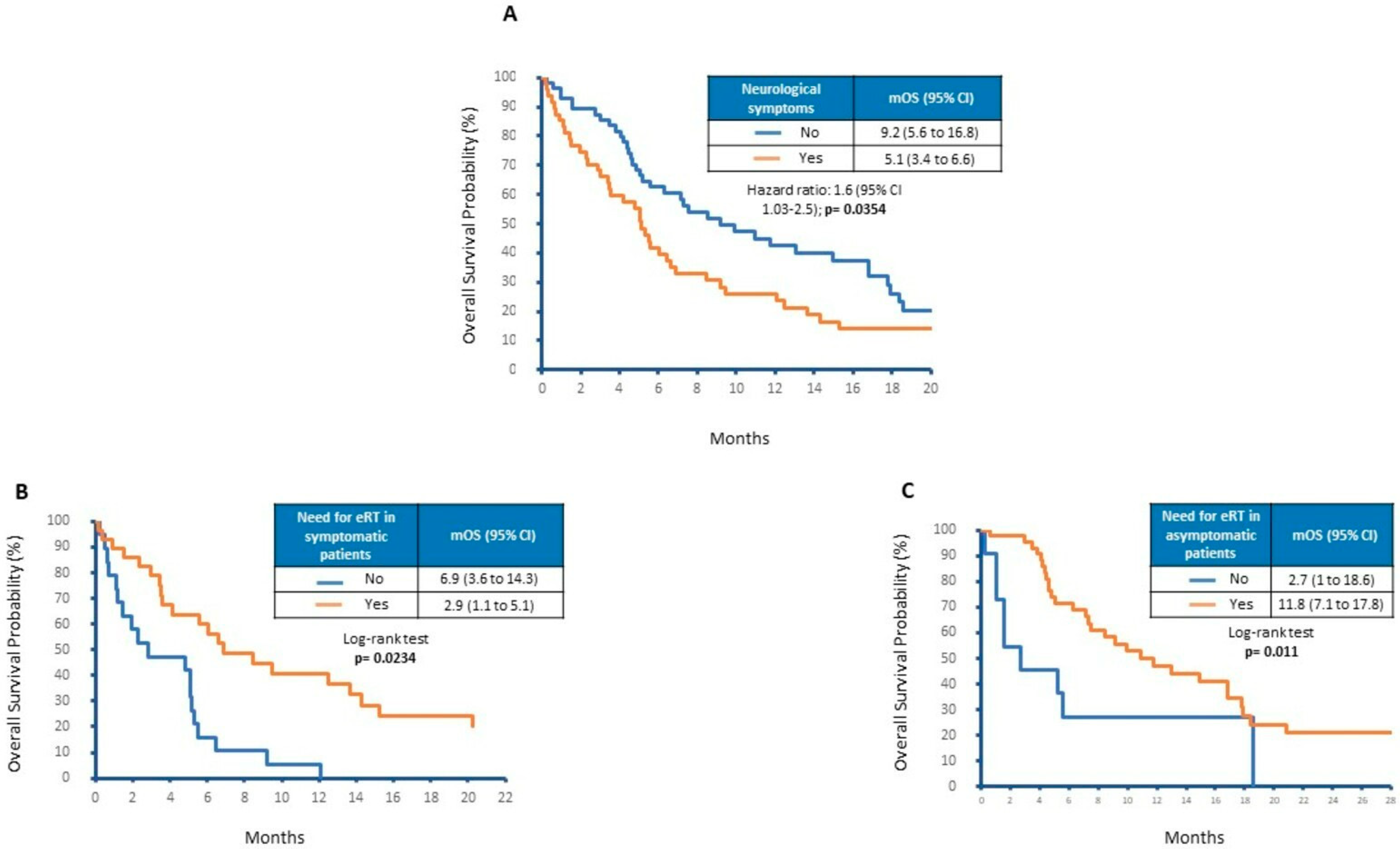
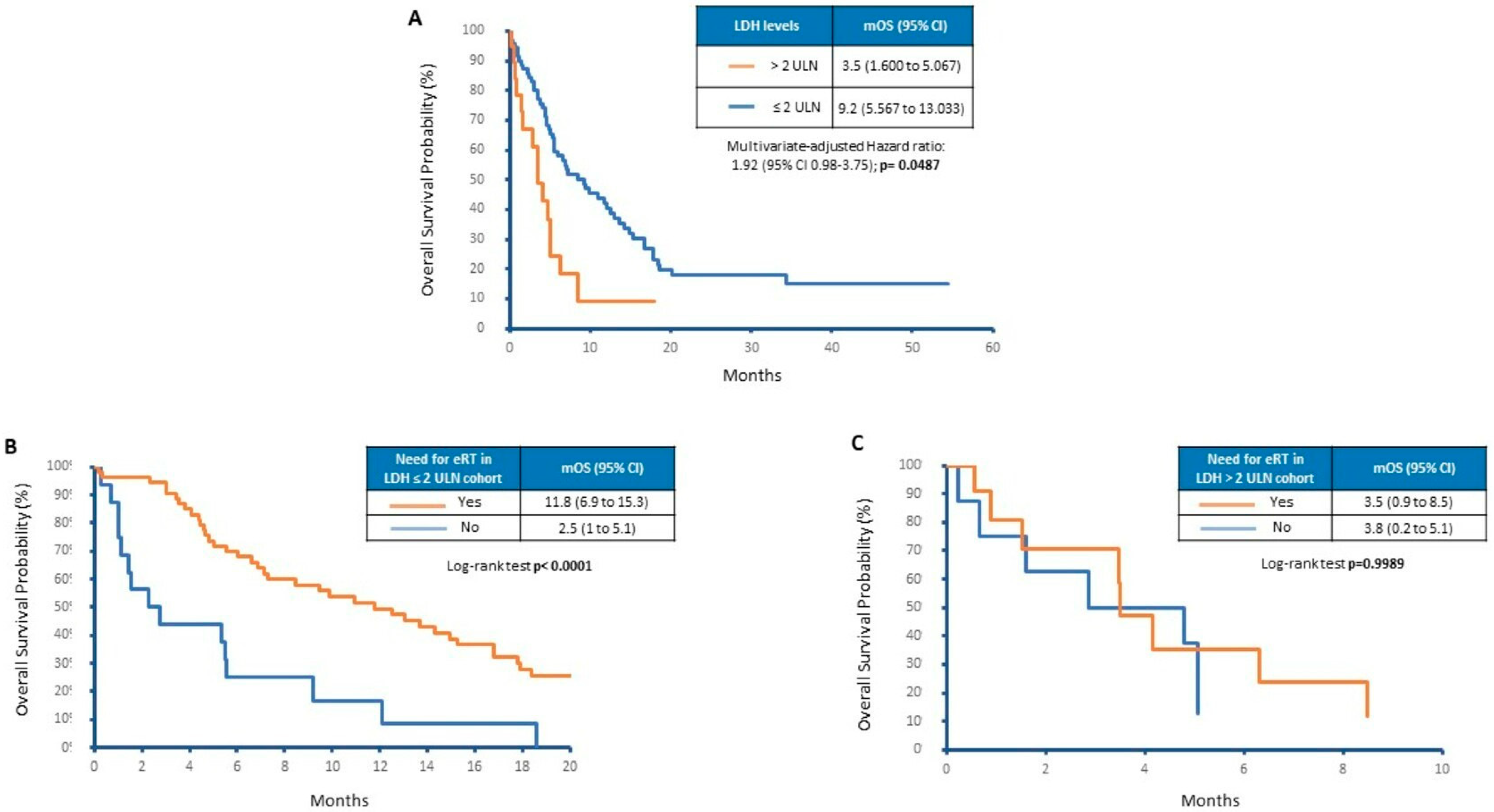
3.2. Neurological Symptoms and Factors Associated with Overall Survival

At the time of analysis, death occurred in 79 patients and 26 were still alive. The mOS from the BM diagnosis was 6.6 months (IQR: 5.1–9.2). The next step of the study explored the features of CM patients with symptomatic BMs (Table 3).
In our cohort, 45% of patients developed neurological symptoms. Their median age was 62 years and 57% were male. Many symptomatic patients showed supratentorial BMs (57%) as well as LDH levels ≤ 2 times the ULN (67%) at the time of their onset. Notably, only 47% of patients showed more than three BMs. The presence of neurological symptoms was a negative prognostic factor with a hazard ratio (HR) of 1.6 (95% CI 1.03–2.5). As shown in Figure 1A, mOS was 5.1 months for symptomatic patients versus 9.2 months for asymptomatic ones (p = 0.0354). Therefore, we questioned whether the eRT could have a role in the control of neurological symptoms, delaying clinical–neurological deterioration and then improving prognosis in symptomatic patients. In our analysis, patients with neurological symptoms showed a significant survival benefit from radiation therapy, achieving an mOS from BM diagnosis of 6.9 months with 2.9 months for those that did not receive eRT (p = 0.0234; Figure 1B). However, eRT also improved outcomes in asymptomatic patients (mOS: 11.8 vs. 2.7 months, p = 0.011; Figure 1C).
Other analyses demonstrated the poor prognostic role of LDH levels > 2 times the ULN at the time of encephalic progression. As shown in Figure 2A, by grouping patients by LDH levels we noticed a significant difference in terms of mOS (3.5 versus 9.2 months, p = 0.0014). Moreover, limited to the low number of patients, doubled LDH levels seem to select patients who do not benefit from eRT. Only patients with LDH levels ≤ 2 times the ULN levels showed an improvement in OS due to eRT. As shown in Figure 2B, OS was 2.5 months in the eRT untreated group with respect to 11.8 months observed in the group that received eRT (p < 0.0001). On the contrary, no survival difference was evidenced in patients showing LDH levels > 2 times the ULN after grouping them according to eRT (3.8 versus 3.5 months, p = 0.998; Figure 2C).
The Cox model applied to evaluate factors involved in survival showed that worse outcome was associated with the presence of more than three BMs (in the multivariable model: HR 1.78, 95% CI 1.04–3.06) and with LDH levels higher than 2-fold the ULN (HR 1.85, 95% CI 0.96–3.56). In addition, Table 4 shows that eRT has a protective effect (in the multivariable model: HR 0.37, 95% CI 0.21–0.67).

3.3. Impact of Treatments on Overall Survival

The next set of exploratory analyses investigated the additive effect of systemic therapies with eRT. As shown in Figure 3A,B, patients treated with TT or IT benefit from the concomitant use of eRT (mOS 2.2 vs. 9.5 months, p = 0.0062 and 2.7 vs. 9.9 months, p = 0.001, respectively). Moreover, the poor prognostic role of LDH levels was confirmed in patients treated with TT who experienced a worse outcome when LDH was >2 times the ULN (mOS 3.5 vs. 12.5 months, p = 0.0015; Figure 3C). On the contrary, patients who underwent IT did not show a significant difference in mOS when stratified according to LDH levels (mOS 4.17 vs. 8.43 months, p = 0.16; Figure 3D).

4. Discussion

The present study retrospectively explored the features of CM patients bearing BMs in a real-life cohort and examined the most used and effective multimodal therapeutic strategies in the pre-combined IT era or when clinical contraindications limit its practice.
In our population, BMs from CM occurred most frequently in males with a prevalent involvement of the supratentorial region. Most patients (90%) also evidenced extracranial metastases that in 63% of cases were diagnosed at least three months before the central nervous system (CNS) metastatic involvement. Thus, the intracranial progression apparently represents, at least in the vast majority of our patients, a delayed event in the natural history of CM. The aforementioned phenomenon has already been previously described, although a recent retrospective real-world study has assessed how BMs and extracranial metastases occur synchronously in nearly 70% of cases. This may be a potential consequence of intensive brain surveillance [34,35]. The incidence of BRAF mutations (55%) was similar to that reported in the general CM population. Nearly half of the patients (45%) suffered from neurological symptoms and, among them, only 46% showed more than three BMs, and only 24% were characterized by LDH levels > 2-fold the ULN. Therefore, neurological symptoms were not related to a multifocal metastatic brain disease or elevated LDH values.
The median survival after BM onset was 6.6 months, lower than the OS revealed by the recent Phase 3 randomized clinical trials using either TT or IT. However, our study reflects real-life data of patients usually excluded from clinical trials, such as those with poor performance status, previously treated with systemic therapies before the onset of BMs or those treated in the pre-combined IT era. In this setting, the prognosis is guided by factors mainly evidenced in retrospective cohorts of patients, including elevated LDH levels, neurological symptoms, three or more BMs and three or more extracranial metastatic sites [36]. In our population, the multivariate model was in line with these results.
Of note, neurological symptoms, occurring in 45% of our population, were the most common complication of BMs that negatively influenced both quality of life and survival. The percentage of symptomatic patients detected in our study differs from that evidenced in recent and quoted trials that highlight the high frequency of asymptomatic brain lesions (nearly 80%) [20,21]. In this regard, we have to underline that our study enrolled “real-life” patients who underwent radiological evaluation according to clinical practice with CT scans that have low specificity for BMs. Conversely, the gold standard for BM diagnosis is represented by the encephalic MRI, usually performed only after the evidence of neurological symptoms. Indeed, a retrospective study performed at the Memorial Sloan Kettering Cancer Center (MSKCC) extracted data from 355 real-life CM patients bearing BMs and found that 67% of them had neurological symptoms at BM onset [37]. Therefore, a universally accepted screening program, eventually involving encephalic MRI for high-risk patients, should be designed and diffused in clinical practice in order to detect BMs when the patients are still asymptomatic and improve the percentage of asymptomatic patients, as already happens in RCT.
Furthermore, we evaluated the eRT efficacy in improving outcomes across different subgroups of patients: symptomatic versus asymptomatic, IT-treated versus TT-treated and LDH levels upper versus lower than/equal to two times the ULN levels. The number of patients did not allow us to perform a subgroup analysis based on the different types of eRT. Anyway, this evaluation would not have significantly influenced any results considering that our aim was exploring the eRT additive effect with systemic treatments, its role in controlling neurological symptoms, improving quality of life and allowing access to further lines of systemic treatments, thus improving mOS. Our data showed that eRT improved outcomes both in the symptomatic and asymptomatic group as well as in patients treated with either TT or IT with a single agent. Thus, eRT might play a key role in prolonging OS by stabilizing BM growth and delaying clinical deterioration. Based on these results, while waiting for prospective data concerning the efficacy of eRT with combined IT, eRT should play a part in the therapeutic strategy at least for symptomatic brain metastatic CM patients or those who are asymptomatic and excluded from combined IT due to clinical contraindications.
The last step of our work focused on the negative prognostic role of LDH levels higher than 2-fold the ULN at the time of CNS metastatic spread and data parallel to those of previously published papers [38,39]. However, relevant data from our study concern the inefficacy of eRT in improving survival in patients with LDH > 2 times the ULN at BM onset. Thus, LDH values more than or equal to double the ULN could represent the sign of an aggressive and active brain metastatic disease that does not benefit from eRT despite the systemic treatments. On the other hand, the negative prognostic role of elevated LDH levels is confirmed only in patients treated with TT. Conversely, patients receiving IT with a single agent did not experience a significant difference in terms of OS when stratified according to LDH levels. The putative explanation of this finding could rely on a greater efficacy of IT in CM patients with BMs with LDH levels > 2-fold the ULN, confirming the recently published data regarding CM patients developing extracranial metastases [40]. The biological explanation of these findings relies on the role of LDH in downregulating the immune system due to the production of elevated levels of its oncometabolite, the lactate that was found to be associated with an increased number of metastatic sites and lower survival. Elevated levels of lactate induce an immune-suppressed microenvironment that sustains CM growth by promoting the expression of programmed cell death of protein-1 (PD-1) and the ligand (PD-L1) on tumor cells [41,42]. Once these data are confirmed in larger and prospective cohorts of CM patients bearing BMs, our results could underly that elevated LDH levels identify an aggressive subset of brain metastatic CM that should be oriented to IT-based therapy regardless of BRAF mutational status.
In conclusion, exploring the activity of the combined use of anti-CTLA4 and anti-PD1 agents in brain metastatic CM patients with LDH levels > 2-fold the ULN could represent an interesting strategy for the control of a severe complication that restrains survival in the majority of patients.

5. Conclusions

The present study demonstrated that neurological symptoms and high LDH values are negative prognostic factors in patients with CM developing BMs. In addition, preliminary results underlined the survival benefit due to the eRT in all subgroups, although patients with LDH levels ≥ 2-fold the ULN did not benefit from IT. These preliminary observations, therefore, suggest that IT may also have an active role in BMs showing elevated LDH levels. This may be at least explained by the immune suppressive microenvironment sustained by lactate, the LDH oncometabolite. However, further prospective studies are needed to understand the effective role of eRT and IT in melanoma patients characterized by BMs and elevated LDH.

Author Contributions

V.I., M.C.S. and M.T. conceived and designed the analysis and wrote the manuscript. M.C.S., S.S., S.C. and M.G. contributed to the data collection. M.E.M. and P.T. performed the statistics. M.T. and C.P. revised the text. All authors have read and agreed to the published version of the manuscript.

Funding

This research was granted funds from the University of Bari (research fellowship number 06.227 in neuro-oncology).

Institutional Review Board Statement

The study was conducted in accordance with the Declaration of Helsinki. Ethical review and approval were waived for this study due to its retrospective and observational nature and in accordance with national regulations that strictly impose ethical review and approval only for those observational studies designed as prospective pharmacological observational ones (Official Gazette of the Italian Republic n. 42, 19 February 2022, decree 30 November 2021, art. 6 subparagraph 2).

Informed Consent Statement

Informed consent was obtained from all subjects involved in the study.

Data Availability Statement

The datasets used and analyzed during the current study are available from the corresponding author upon reasonable request.

Acknowledgments

The authors are grateful to Pandora for Research, ONLUS Association for Oncology.

Conflicts of Interest

The authors declare no conflict of interest.

References

  1. Lamba, N.; Wen, P.Y.; Aizer, A.A. Epidemiology of brain metastases and leptomeningeal disease. Neuro Oncol. 2021, 23, 1447–1456. [Google Scholar] [CrossRef] [PubMed]
  2. Tadros, S.; Ray-Chaudhury, A. Pathological Features of Brain Metastases. Neurosurg. Clin. N. Am. 2020, 31, 549–564. [Google Scholar] [CrossRef] [PubMed]
  3. Sandhu, M.R.S.; Chiang, V.L.; Tran, T.; Yu, J.B.; Weiss, S.A.; Goldberg, S.B.; Aboian, M.S.; Kluger, H.M.; Mahajan, A. Incidence and characteristics of metastatic intracranial lesions in stage III and IV melanoma: A single institute retrospective analysis. J. Neuro-Oncol. 2021, 154, 197–203. [Google Scholar] [CrossRef] [PubMed]
  4. Bander, E.D.; Yuan, M.; Carnevale, J.A.; Reiner, A.S.; Panageas, K.S.; Postow, M.A.; Tabar, V.; Moss, N.S. Melanoma brain metastasis presentation, treatment, and outcomes in the age of targeted and immunotherapies. Cancer 2021, 127, 2062–2073. [Google Scholar] [CrossRef]
  5. Sperduto, P.W.; Mesko, S.; Li, J.; Cagney, D.; Aizer, A.; Lin, N.U.; Nesbit, E.; Kruser, T.J.; Chan, J.; Braunstein, S.; et al. Survival in Patients With Brain Metastases: Summary Report on the Updated Diagnosis-Specific Graded Prognostic Assessment and Definition of the Eligibility Quotient. J. Clin. Oncol. 2020, 38, 3773–3784. [Google Scholar] [CrossRef]
  6. Davies, M.A.; Liu, P.; McIntyre, S.; Kim, K.B.; Papadopoulos, N.; Hwu, W.J.; Hwu, P.; Bedikian, A. Prognostic factors for survival in melanoma patients with brain metastases. Cancer 2011, 117, 1687–1696. [Google Scholar] [CrossRef]
  7. Sacks, P.; Rahman, M. Epidemiology of Brain Metastases. Neurosurg. Clin. N. Am. 2020, 31, 481–488. [Google Scholar] [CrossRef]
  8. Chaung, K.V.; Sloan, A.E.; Choi, S. Limited brain metastases: A narrative review. Ann. Pall. Med. 2021, 10, 6016–6027. [Google Scholar] [CrossRef]
  9. Phillips, W.J.; Baghai, T.; Ong, M.; Lo, B.; Ibrahim, A.M.; Smith, T.K.T.; Song, X. A Contemporary Report of Clinical Outcomes in Patients with Melanoma Brain Metastases. Curr. Oncol. 2021, 28, 428–439. [Google Scholar] [CrossRef]
  10. Keung, E.Z.; Gershenwald, J.E. The eighth edition American Joint Committee on Cancer (AJCC) melanoma staging system: Implications for melanoma treatment and care. Exp. Rev. Anticanc. 2018, 18, 775–784. [Google Scholar] [CrossRef]
  11. Tawbi, H.; To, T.M.; Bartley, K.; Sadetsky, N.; Burton, E.; Haydu, L.; McKenna, E. Treatment patterns and clinical outcomes for patients with melanoma and central nervous system metastases: A real-world study. Cancer Med. 2022, 11, 139–150. [Google Scholar] [CrossRef] [PubMed]
  12. Gutzmer, R.; Vordermark, D.; Hassel, J.C.; Krex, D.; Wendl, C.; Schadendorf, D.; Sickmann, T.; Rieken, S.; Pukrop, T.; Höller, C.; et al. Melanoma brain metastases-Interdisciplinary management recommendations 2020. Cancer Treat Rev. 2020, 89, 102083. [Google Scholar] [CrossRef] [PubMed]
  13. Becco, P.; Gallo, S.; Poletto, S.; Frascione, M.P.M.; Crotto, L.; Zaccagna, A.; Paruzzo, L.; Caravelli, D.; Carnevale-Schianca, F.; Aglietta, M. Melanoma Brain Metastases in the Era of Target Therapies: An Overview. Cancers 2020, 12, 1640. [Google Scholar] [CrossRef] [PubMed]
  14. Davies, M.A.; Saiag, P.; Robert, C.; Grob, J.J.; Flaherty, K.T.; Arance, A.; Chiarion-Sileni, V.; Thomas, L.; Lesimple, T.; Mortier, L.; et al. Dabrafenib plus trametinib in patients with BRAFV600-mutant melanoma brain metastases (COMBI-MB): A multicentre, multicohort, open-label, phase 2 trial. Lancet Oncol. 2017, 18, 863–873. [Google Scholar] [CrossRef] [PubMed]
  15. Robert, C.; Grob, J.J.; Stroyakovskiy, D.; Karaszewska, B.; Hauschild, A.; Levchenko, E.; Chiarion Sileni, V.; Schachter, J.; Garbe, C.; Bondarenko, I.; et al. Five-Year Outcomes with Dabrafenib plus Trametinib in Metastatic Melanoma. N Eng. J. Med. 2019, 381, 626–636. [Google Scholar] [CrossRef]
  16. Grob, J.J.; Amonkar, M.M.; Karaszewska, B.; Schachter, J.; Dummer, R.; Mackiewicz, A.; Stroyakovskiy, D.; Drucis, K.; Grange, F.; Chiarion-Sileni, V.; et al. Comparison of dabrafenib and trametinib combination therapy with vemurafenib monotherapy on health-related quality of life in patients with unresectable or metastatic cutaneous BRAF Val600-mutation-positive melanoma (COMBI-v): Results of a phase 3, open-label, randomised trial. Lancet Oncol. 2015, 16, 1389–1398. [Google Scholar]
  17. Goldberg, S.B.; Gettinger, S.N.; Mahajan, A.; Chiang, A.C.; Herbst, R.S.; Sznol, M.; Tsiouris, A.J.; Cohen, J.; Vortmeyer, A.; Jilaveanu, L.; et al. Pembrolizumab for patients with melanoma or non-small-cell lung cancer and untreated brain metastases: Early analysis of a non-randomised, open-label, phase 2 trial. Lancet. Oncol. 2016, 17, 976–983. [Google Scholar] [CrossRef] [Green Version]
  18. Larkin, J.; Minor, D.; D’Angelo, S.; Neyns, B.; Smylie, M.; Miller, W.H., Jr.; Gutzmer, R.; Linette, G.; Chmielowski, B.; Lao, C.D.; et al. Overall Survival in Patients With Advanced Melanoma Who Received Nivolumab Versus Investigator’s Choice Chemotherapy in CheckMate 037: A Randomized, Controlled, Open-Label Phase III Trial. J. Clin. Oncol. 2018, 36, 383–390. [Google Scholar] [CrossRef]
  19. Patel, K.R.; Lawson, D.H.; Kudchadkar, R.R.; Carthon, B.C.; Oliver, D.E.; Okwan-Duodu, D.; Ahmed, R.; Khan, M.K. Two heads better than one? Ipilimumab immunotherapy and radiation therapy for melanoma brain metastases. Neuro-Oncol. 2015, 17, 1312–1321. [Google Scholar] [CrossRef] [Green Version]
  20. Long, G.V.; Atkinson, V.; Lo, S.; Sandhu, S.; Guminski, A.D.; Brown, M.P.; Wilmott, J.S.; Edwards, J.; Gonzalez, M.; Scolyer, R.A.; et al. Combination nivolumab and ipilimumab or nivolumab alone in melanoma brain metastases: A multicentre randomised phase 2 study. Lancet Oncol. 2018, 19, 672–681. [Google Scholar] [CrossRef]
  21. Tawbi, H.A.; Forsyth, P.A.; Hodi, F.S.; Lao, C.D.; Moschos, S.J.; Hamid, O.; Atkins, M.B.; Lewis, K.; Thomas, R.P.; Glaspy, J.A.; et al. Safety and efficacy of the combination of nivolumab plus ipilimumab in patients with melanoma and asymptomatic or symptomatic brain metastases (CheckMate 204). Neuro-Oncol. 2021, 23, 1961–1973. [Google Scholar] [CrossRef] [PubMed]
  22. Dummer, R.; Queirolo, P.; Abajo Guijarro, A.M.; Hu, Y.; Wang, D.; Azevedo, S.J.; Robert, C.; Ascierto, P.A.; Chiarion -Sileni, V.; Pronzato, P.; et al. Atezolizumab (A), cobimetinib (C), and vemurafenib (V) in patients (pts) with BRAFV600 mutation–positive melanoma with central nervous system (CNS) metastases (mets): Primary results from phase 2 Tricotel study. J. Clin. Oncol. 2022, 40 (Suppl. 16), 9515. [Google Scholar] [CrossRef]
  23. Lau, P.K.H.; Feran, B.; Smith, L.; Lasocki, A.; Molania, R.; Smith, K.; Weppler, A.; Angel, C.; Kee, D.; Bhave, P.; et al. Melanoma brain metastases that progress on BRAF-MEK inhibitors demonstrate resistance to ipilimumab-nivolumab that is associated with the Innate PD-1 Resistance Signature (IPRES). J. Immunother. Cancer 2021, 9, e002995. [Google Scholar] [CrossRef] [PubMed]
  24. Wegner, R.E.; Abel, S.; D’Amico, R.S.; Mehta, G.U.; Sheehan, J. Time from stereotactic radiosurgery to immunotherapy in patients with melanoma brain metastases and impact on outcome. J. Neuro-Oncol. 2021, 152, 79–87. [Google Scholar] [CrossRef] [PubMed]
  25. Kim, P.H.; Suh, C.H.; Kim, H.S.; Kim, K.W.; Kim, D.Y.; Lee, E.Q.; Aizer, A.A.; Guenette, J.P.; Huang, R.Y. Immune Checkpoint Inhibitor with or without Radiotherapy in Melanoma Patients with Brain Metastases: A Systematic Review and Meta-Analysis. K. J. Radiol. 2021, 22, 584–595. [Google Scholar] [CrossRef]
  26. Moyers, J.T.; Chong, E.G.; Peng, J.; Tsai, H.H.C.; Sufficool, D.; Shavlik, D.; Nagaraj, G. Real world outcomes of combination and timing of immunotherapy with radiotherapy for melanoma with brain metastases. Cancer Med. 2021, 10, 1201–1211. [Google Scholar] [CrossRef]
  27. Lamba, N.; Aizer, A.A. The evolving role of systemic therapy and local, brain-directed treatment in patients with melanoma and brain metastases. Neuro-Oncol. 2021, 23, 1816–1817. [Google Scholar] [CrossRef]
  28. White, R.J.; Abel, S.; Horne, Z.D.; Lee, J.; Edington, H.; Greenberg, L.; Younes, H.; Hilton, C.; Wegner, R.E. Melanoma brain metastases: Is it time to eliminate radiotherapy? J. Neuro-Oncol. 2020, 149, 27–33. [Google Scholar] [CrossRef]
  29. Cortellini, A.; Tucci, M.; Adamo, V.; Stucci, L.S.; Russo, A.; Tanda, E.T.; Spagnolo, F.; Rastelli, F.; Bisonni, R.; Santini, D.; et al. Integrated analysis of concomitant medications and oncological outcomes from PD-1/PD-L1 checkpoint inhibitors in clinical practice. J. Immunother. Cancer 2020, 8, e001361. [Google Scholar] [CrossRef]
  30. Frinton, E.; Tong, D.; Tan, J.; Read, G.; Kumar, V.; Kennedy, S.; Lim, C.; Board, R.E. Metastatic melanoma: Prognostic factors and survival in patients with brain metastases. J. Neuro-Oncol. 2017, 135, 507–512. [Google Scholar] [CrossRef] [Green Version]
  31. Weide, B.; Elsässer, M.; Büttner, P.; Pflugfelder, A.; Leiter, U.; Eigentler, T.K.; Bauer, J.; Witte, M.; Meier, F.; Garbe, C. Serum markers lactate dehydrogenase and S100B predict independently disease outcome in melanoma patients with distant metastasis. Br. J. Cancer 2012, 107, 422–428. [Google Scholar] [CrossRef] [PubMed]
  32. Marcus, D.M.; Lowe, M.; Khan, M.K.; Lawson, D.H.; Crocker, I.R.; Shelton, J.W.; Melton, A.; Maynard, N.; Delman, K.A.; Carlson, G.W.; et al. Prognostic factors for overall survival after radiosurgery for brain metastases from melanoma. Am. J. Clin. Oncol. 2014, 37, 580–584. [Google Scholar] [CrossRef] [PubMed]
  33. Neal, M.T.; Chan, M.D.; Lucas, J.T., Jr.; Loganathan, A.; Dillingham, C.; Pan, E.; Stewart, J.H., 4th; Bourland, J.D.; Shaw, E.G.; Tatter, S.B.; et al. Predictors of survival, neurologic death, local failure, and distant failure after gamma knife radiosurgery for melanoma brain metastases. World Neurosurg. 2014, 82, 1250–1255. [Google Scholar] [CrossRef] [PubMed]
  34. Rishi, A.; Yu, H.M. Current Treatment of Melanoma Brain Metastasis. Curr. Treat. Opt. Oncol. 2020, 21, 45. [Google Scholar] [CrossRef]
  35. Shibahara, I.; Kanamori, M.; Watanabe, T.; Utsunomiya, A.; Suzuki, H.; Saito, R.; Sonoda, Y.; Jokura, H.; Uenohara, H.; Tominaga, T. Clinical Features of Precocious, Synchronous, and Metachronous Brain Metastases and the Role of Tumor Resection. World Neurosurg. 2018, 113, e1–e9. [Google Scholar] [CrossRef]
  36. Anvari, A.; Sasanpour, P.; Kheradmardi, M.R. Radiotherapy and Immunotherapy in Melanoma Brain Metastases. Hematol Oncol Stem Cell Ther. 2023, 16, 1–20. [Google Scholar] [CrossRef]
  37. Raizer, J.J.; Hwu, W.J.; Panageas, K.S.; Wilton, A.; Baldwin, D.E.; Bailey, E.; von Althann, C.; Lamb, L.A.; Alvarado, G.; Bilsky, M.H.; et al. Brain and leptomeningeal metastases from cutaneous melanoma: Survival outcomes based on clinical features. Neur.-Oncol. 2008, 10, 199–207. [Google Scholar] [CrossRef] [Green Version]
  38. Wagner, N.B.; Forschner, A.; Leiter, U.; Garbe, C.; Eigentler, T.K. S100B and LDH as early prognostic markers for response and overall survival in melanoma patients treated with anti-PD-1 or combined anti-PD-1 plus anti-CTLA-4 antibodies. Br. J. Cancer 2018, 119, 339–346. [Google Scholar] [CrossRef] [Green Version]
  39. Placzke, J.; Teterycz, P.; Quaglino, P.; Cybulska-Stopa, B.; Tucci, M.; Rubatto, M.; Skora, T.; Interno, V.; Rosinska, M.; Borkowska, A.; et al. The Analysis of Trends in Survival for Patients with Melanoma Brain Metastases with Introduction of Novel Therapeutic Options before the Era of Combined Immunotherapy-Multicenter Italian-Polish Report. Cancers 2022, 14, 5763. [Google Scholar] [CrossRef]
  40. Knispel, S.; Gassenmaier, M.; Menzies, A.M.; Loquai, C.; Johnson, D.B.; Franklin, C.; Gutzmer, R.; Hassel, J.C.; Weishaupt, C.; Eigentler, T.; et al. Outcome of melanoma patients with elevated LDH treated with first-line targeted therapy or PD-1-based immune checkpoint inhibition. Eur. J. Cancer 2021, 148, 61–75. [Google Scholar] [CrossRef]
  41. Scott, K.E.; Cleveland, J.L. Lactate Wreaks Havoc on Tumor-Infiltrating T and NK Cells. Cell Metab. 2016, 24, 649–650. [Google Scholar] [CrossRef] [PubMed] [Green Version]
  42. Daneshmandi, S.; Wegiel, B.; Seth, P. Blockade of Lactate Dehydrogenase-A (LDH-A) Improves Efficacy of Anti-Programmed Cell Death-1 (PD-1) Therapy in Melanoma. Cancers 2019, 11, 450. [Google Scholar] [CrossRef] [PubMed]
Figure 1. Overall survival according to (A) neurological symptoms, (B) eRT in symptomatic patients and (C) eRT in asymptomatic patients.
Cancers 15 01542 g001
Figure 2. Overall survival of patients depending on (A) LDH levels, (B) eRT in LDH ≤ 2 ULN and (C) eRT in LDH > 2 ULN.
Cancers 15 01542 g002
Figure 3. Overall survival by (A) eRT in TT-treated patients, (B) eRT in IT-treated patients, (C) LDH levels in TT-treated patients and (D) LDH levels in IT-treated patients.
Cancers 15 01542 g003
Table 1. Patients’ demographics.
Number%
SexMale6158
Female4442
Primary siteLimbs3634
Head and neck99
Trunk5350
Other sites77
HistologySSM3938
Nodular4947
Animal type67
Unknown118
Breslow depth≤1 mm1010
>1–2 mm1111
>2–4 mm3533
>4 mm3432
Unknown1514
UlcerationPresent4643
Absent5048
Unknown99
Mitosis≤13836
>16764
BRAF statusMutated5855
Wild-type4745
NRAS statusMutated712
Wild-type5388
LDH at melanoma diagnosis≤2 × ULN6764
>2 × ULN55
Unknown3331
SSM, superficial spreading melanoma; LDH, lactate dehydrogenase.
Table 2. Metastatic disease features.
Number%
Extracranial metastasesYes9590
No1010
Number of extracranial metastatic sites≤25250
>23735
Unknown1615
Timing of BMs from melanoma diagnosisSynchronous33
Metachronous10297
Timing of BMs from extracranial progressionSynchronous3836
Subsequent6461
Unknown33
Site of BMsSupra-tentorial5451
Infra-tentorial66
Supra- and infra-tentorial2524
Unknown2019
Number of BMs≤36259
>33937
Unknown2019
LDH levels at BM diagnosis≤2 × ULN7168
>2 × ULN1918
Unknown1514
Symptomatic BMsYes4745
No5855
NeurosurgeryYes1312
No9288
Encephalic radiotherapyYes7571
No3029
Type of encephalic radiotherapyStereotactic radiotherapy4965
Whole brain radiotherapy2635
First-line systemic treatmentTargeted therapy with anti-BRAF + anti-MEK4341
Immunotherapy4442
Chemotherapy33
None1514
BM, brain metastasis; LDH, lactate dehydrogenase; ULN, upper limit normal.
Table 3. Features of symptomatic cohort.
Number%
SexMale2757
Female2043
BRAF statusMutated2655
Wild-type2145
LDH at BM diagnosis≤2 × ULN3167
>2 × ULN1021
Unknown612
Site of BMsSupra-tentorial2757
Infra-tentorial12
Supra- and infra-tentorial817
Unknown1124
Number of BMs≤32553
>32247
Steroid therapyYes47100
No00
BM, brain metastasis; LDH, lactate dehydrogenase; ULN, upper limit normal.
Table 4. Results of Cox regression for overall survival.
FactorsEffect TestedUnivariate AnalysisMultivariate Analysis
HR95% CIp-ValueHR95% CIp-Value
SexMale vs. female0.790.51–1.250.31610.920.54–0.570.7624
Age (years)56–65 vs. ≤552.031.11–3.710.02151.680.88–3.220.1174
66–75 vs. ≤551.590.86–2.920.13731.550.78–3.060.2070
>75 vs. ≤552.281.1–4.70.02641.970.86–4.490.1080
RadiotherapyYes vs. no0.30.18–0.49<0.00010.370.21–0.670.0009
Neurological symptomsYes vs. no1.61.03–2.490.03741.200.71–2.010.5020
Number of BM≥3 vs. <31.450.93–2.280.5711.781.04–3.060.0363
Liver metastasisYes vs. no1.440.89–2.340.13751.050.59–1.850.8763
LDH levels > 2 times the ULNYes vs. no2.581.41–4.740.00211.850.96–3.560.0452
BM, brain metastasis; LDH, lactate dehydrogenase; ULN, upper limit normal.
Disclaimer/Publisher’s Note: The statements, opinions and data contained in all publications are solely those of the individual author(s) and contributor(s) and not of MDPI and/or the editor(s). MDPI and/or the editor(s) disclaim responsibility for any injury to people or property resulting from any ideas, methods, instructions or products referred to in the content.

