Next Issue
Volume 15, February-1
Previous Issue
Volume 15, January-1


Cancers, Volume 15, Issue 2 (January-2 2023) – 233 articles

Cover Story (view full-size image): 
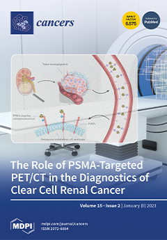
Carboxypeptidase type II, also known as prostate-specific membrane antigen (PSMA), is expressed on the surface of neovascular endothelial cells in various solid tumors, including clear cell renal cancer (ccRCC). Based on its genetic characteristics, it is considered a highly vascularized tumor, which might be a rationale for using PET/CT imaging with PSMA-targeting rather than [18F]F-FDG, which is not routinely recommended due to its excretion pathway and its low accuracy. In the studies included in this systematic review, PSMA-targeted PET/CT showed promising results in the diagnostics of ccRCC, being able to detect more lesions than conventional imaging examinations. It can identify ccRCC lesions with a more aggressive phenotype, predict treatment outcomes and change patient management in a significant percentage of patients. View this paper
  • Issues are regarded as officially published after their release is announced to the table of contents alert mailing list.
  • You may sign up for e-mail alerts to receive table of contents of newly released issues.
  • PDF is the official format for papers published in both, html and pdf forms. To view the papers in pdf format, click on the "PDF Full-text" link, and use the free Adobe Reader to open them.
Order results
Result details
Section
Select all
Export citation of selected articles as:
0 pages, 6103 KiB  
Article
TGFβ1-Induced EMT in the MCF10A Mammary Epithelial Cell Line Model Is Executed Independently of SNAIL1 and ZEB1 but Relies on JUNB-Coordinated Transcriptional Regulation
by Pablo Antón-García, Elham Bavafaye Haghighi, Katja Rose, Georg Vladimirov, Melanie Boerries and Andreas Hecht
Cancers 2023, 15(2), 558; https://doi.org/10.3390/cancers15020558 - 16 Jan 2023
Cited by 4 | Viewed by 3139 | Correction
Abstract
Epithelial-mesenchymal transition (EMT) fosters cancer cell invasion and metastasis, the main cause of cancer-related mortality. Growing evidence that SNAIL and ZEB transcription factors, typically portrayed as master regulators of EMT, may be dispensable for this process, led us to re-investigate its mechanistic underpinnings. [...] Read more.
Epithelial-mesenchymal transition (EMT) fosters cancer cell invasion and metastasis, the main cause of cancer-related mortality. Growing evidence that SNAIL and ZEB transcription factors, typically portrayed as master regulators of EMT, may be dispensable for this process, led us to re-investigate its mechanistic underpinnings. For this, we used an unbiased computational approach that integrated time-resolved analyses of chromatin structure and differential gene expression, to predict transcriptional regulators of TGFβ1-inducible EMT in the MCF10A mammary epithelial cell line model. Bioinformatic analyses indicated comparatively minor contributions of SNAIL proteins and ZEB1 to TGFβ1-induced EMT, whereas the AP-1 subunit JUNB was anticipated to have a much larger impact. CRISPR/Cas9-mediated loss-of-function studies confirmed that TGFβ1-induced EMT proceeded independently of SNAIL proteins and ZEB1. In contrast, JUNB was necessary and sufficient for EMT in MCF10A cells, but not in A549 lung cancer cells, indicating cell-type-specificity of JUNB EMT-regulatory capacity. Nonetheless, the JUNB-dependence of EMT-associated transcriptional reprogramming in MCF10A cells allowed to define a gene expression signature which was regulated by TGFβ1 in diverse cellular backgrounds, showed positively correlated expression with TGFβ signaling in multiple cancer transcriptomes, and was predictive of patient survival in several cancer types. Altogether, our findings provide novel mechanistic insights into the context-dependent control of TGFβ1-driven EMT and thereby may lead to improved diagnostic and therapeutic options. Full article
(This article belongs to the Section Molecular Cancer Biology)
Show Figures

Figure 1

17 pages, 3112 KiB  
Article
Naturally Isolated Sesquiterpene Lactone and Hydroxyanthraquinone Induce Apoptosis in Oral Squamous Cell Carcinoma Cell Line
by Afshan Shams, Ayaz Ahmed, Ajmal Khan, Shariqa Khawaja, Najeeb Ur Rehman, Asma Saleem Qazi, Adnan Khan, Sami Bawazeer, Syed Abid Ali and Ahmed Al-Harrasi
Cancers 2023, 15(2), 557; https://doi.org/10.3390/cancers15020557 - 16 Jan 2023
Cited by 4 | Viewed by 2388
Abstract
Oral squamous cell carcinoma (OSCC) is one of the most prevalent cancers worldwide, especially in Asian countries. The emergence of its drug resistance and its side effects demands alternatives, to improve prognosis. Since the majority of cancer drugs are derived from natural sources, [...] Read more.
Oral squamous cell carcinoma (OSCC) is one of the most prevalent cancers worldwide, especially in Asian countries. The emergence of its drug resistance and its side effects demands alternatives, to improve prognosis. Since the majority of cancer drugs are derived from natural sources, it provides a window to look for more biocompatible alternatives. In this study, two natural compounds, costunolide (CE) and aloe emodin (AE), were isolated from the stem of Lycium shawii. The compounds were examined for their anticancer and apoptotic potentials against OSCC (CAL 27) cells, using an in vitro analysis, such as a MTT assay, scratch assay, gene, and protein expressions. Both compounds, CE and AE, were found to be cytotoxic against the cancer cells with an IC50 value of 32 and 38 µM, respectively. Moreover, the compounds were found to be non-toxic against normal NIH-3T3 cells and comparable with the standard drug i.e., 5-fluorouracil (IC50 = 97.76 µM). These compounds were active against normal cells at higher concentrations. Nuclear staining displayed the presence of apoptosis-associated morphological changes, i.e., karyopyknosis and karyorrhexis in the treated cancer cells. Flow cytometry results further confirmed that these compounds induce apoptosis rather than necrosis, as the majority of the cells were found in the late apoptotic phase. Gene and protein expression analyses showed an increased expression of apoptotic genes, i.e., BAK, caspase 3, 6, and 9. Moreover, the compounds significantly downregulated the expression of the anti-apoptotic (BCL-2 L1), metastatic (MMP-2), and pro-inflammatory (COX-2) genes. Both compounds have shown promising anticancer, apoptotic, and anti-migratory activities against the OSCC cell line (i.e., CAL-27). However, further in vivo studies are required to explore these compounds as anticancer agents. Full article
(This article belongs to the Special Issue Advances in Anticancer Drugs and Pharmacotherapy of Cancer)
Show Figures

Figure 1

16 pages, 1419 KiB  
Review
Graph Theory Measures and Their Application to Neurosurgical Eloquence
by Onur Tanglay, Nicholas B. Dadario, Elizabeth H. N. Chong, Si Jie Tang, Isabella M. Young and Michael E. Sughrue
Cancers 2023, 15(2), 556; https://doi.org/10.3390/cancers15020556 - 16 Jan 2023
Cited by 3 | Viewed by 2749
Abstract
Improving patient safety and preserving eloquent brain are crucial in neurosurgery. Since there is significant clinical variability in post-operative lesions suffered by patients who undergo surgery in the same areas deemed compensable, there is an unknown degree of inter-individual variability in brain ‘eloquence’. [...] Read more.
Improving patient safety and preserving eloquent brain are crucial in neurosurgery. Since there is significant clinical variability in post-operative lesions suffered by patients who undergo surgery in the same areas deemed compensable, there is an unknown degree of inter-individual variability in brain ‘eloquence’. Advances in connectomic mapping efforts through diffusion tractography allow for utilization of non-invasive imaging and statistical modeling to graphically represent the brain. Extending the definition of brain eloquence to graph theory measures of hubness and centrality may help to improve our understanding of individual variability in brain eloquence and lesion responses. While functional deficits cannot be immediately determined intra-operatively, there has been potential shown by emerging technologies in mapping of hub nodes as an add-on to existing surgical navigation modalities to improve individual surgical outcomes. This review aims to outline and review current research surrounding novel graph theoretical concepts of hubness, centrality, and eloquence and specifically its relevance to brain mapping for pre-operative planning and intra-operative navigation in neurosurgery. Full article
(This article belongs to the Special Issue Advances of Brain Mapping in Cancer Research)
Show Figures

Figure 1

19 pages, 1305 KiB  
Review
Mass Spectrometry-Based Proteomics Workflows in Cancer Research: The Relevance of Choosing the Right Steps
by Paula Carrillo-Rodriguez, Frode Selheim and Maria Hernandez-Valladares
Cancers 2023, 15(2), 555; https://doi.org/10.3390/cancers15020555 - 16 Jan 2023
Cited by 7 | Viewed by 5658
Abstract
The qualitative and quantitative evaluation of proteome changes that condition cancer development can be achieved with liquid chromatography–mass spectrometry (LC-MS). LC-MS-based proteomics strategies are carried out according to predesigned workflows that comprise several steps such as sample selection, sample processing including labeling, MS [...] Read more.
The qualitative and quantitative evaluation of proteome changes that condition cancer development can be achieved with liquid chromatography–mass spectrometry (LC-MS). LC-MS-based proteomics strategies are carried out according to predesigned workflows that comprise several steps such as sample selection, sample processing including labeling, MS acquisition methods, statistical treatment, and bioinformatics to understand the biological meaning of the findings and set predictive classifiers. As the choice of best options might not be straightforward, we herein review and assess past and current proteomics approaches for the discovery of new cancer biomarkers. Moreover, we review major bioinformatics tools for interpreting and visualizing proteomics results and suggest the most popular machine learning techniques for the selection of predictive biomarkers. Finally, we consider the approximation of proteomics strategies for clinical diagnosis and prognosis by discussing current barriers and proposals to circumvent them. Full article
(This article belongs to the Special Issue Application of Proteomics in Cancers)
Show Figures

Graphical abstract

15 pages, 2734 KiB  
Article
Distinct Clinicopathological Features and Prognostic Values of High-, Low-, or Non-Expressing HER2 Status in Colorectal Cancer
by Zehua Wu, Yi Cheng, Huaiming Wang, Dian Liu, Xiaoxing Qi, Chao Wang, Yuanzhe Zhang, Yuting Zhang, Runkai Cai, Hong Huo, Jianwei Zhang, Yue Cai, Weiwei Li, Huabin Hu and Yanhong Deng
Cancers 2023, 15(2), 554; https://doi.org/10.3390/cancers15020554 - 16 Jan 2023
Cited by 1 | Viewed by 1730
Abstract
The encouraging effects of HER2-ADC in patients with HER2-low expression cancers indicated the classical classifications based on positive and negative HER2 might no longer be suitable. However, the biology and prognosis of colorectal cancer patients with different HER2 expression status were still not [...] Read more.
The encouraging effects of HER2-ADC in patients with HER2-low expression cancers indicated the classical classifications based on positive and negative HER2 might no longer be suitable. However, the biology and prognosis of colorectal cancer patients with different HER2 expression status were still not clear. This is a multi-center retrospective study that included patients with histologically confirmed colorectal cancer and determined HER2 status who received radical surgical resection. HER2 immunohistochemistry (IHC) 1+ and IHC 2+ groups were combined and defined as a HER2-low group because of the concordance of clinicopathological characteristics. As compared with the HER2-high group, both the HER2-zero and the HER2-low group had less tumor with perineural invasion (14.3%, 13.1% vs. 31.6%, p = 0.001 and p < 0.001), less stage III disease (41.8%, 39.9% vs. 56.1%, p = 0.044 and p = 0.022), more RAS/BRAF mutation (52.1%, 49.9% vs. 19.5%, p < 0.001 and p < 0.001) and better disease-free survival (DFS) (3y-DFS rate of 78.7%, 82.4% vs. 59.3%, p < 0.001 and p < 0.001). Multivariate analysis and propensity score matching also revealed that HER2-high expression was an independent prognostic factor of DFS. In conclusion, our study revealed that HER2-low colorectal cancer tumors are close to HER2-zero tumors, but different from HER2-high tumors. The routine examination of HER2 IHC is needed in early-stage colorectal cancer. Full article
(This article belongs to the Topic Advances in Anti-Cancer Drugs)
Show Figures

Figure 1

23 pages, 6771 KiB  
Article
Clinical and Biological Significances of FBLN5 in Gastric Cancer
by Xiulan Bian, Shengjie Yin, Xin Yin, Tianyi Fang, Yufei Wang, Shuo Yang, Xinju Jiang, Yingwei Xue, Fei Ye and Lei Zhang
Cancers 2023, 15(2), 553; https://doi.org/10.3390/cancers15020553 - 16 Jan 2023
Cited by 1 | Viewed by 2260
Abstract
Abnormal FBLN5 expression levels are related to various cancer types. This study is the first to explore its clinical and biological significances in gastric cancer (GC). We used The Cancer Genome Atlas-GC (TCGA-GC) and Gene Expression Omnibus (GEO) databases to identify the differential [...] Read more.
Abnormal FBLN5 expression levels are related to various cancer types. This study is the first to explore its clinical and biological significances in gastric cancer (GC). We used The Cancer Genome Atlas-GC (TCGA-GC) and Gene Expression Omnibus (GEO) databases to identify the differential expression of FBLN5, and its association with clinical pathological characteristics was analyzed. A Kaplan–Meier plotter was used to calculate the impact of FBLN5 on GC patient prognosis, and the biological functions of FBLN5 were analyzed. In addition, we constructed a GC tissue microarray, and performed an immunohistochemical staining of FBLN5 to verify our findings. Western blotting was conducted simultaneously to confirm that FBLN5 was overexpressed in GC. We found that the high level of FBLN5 mRNA in GC was associated with a poor prognosis. High FBLN5 expression levels were significantly correlated with INFc and N3 lymph node metastasis. Univariate and multivariate analyses showed that FBLN5 expression levels and lymph node metastasis rate were independent risk factors related to GC patient prognosis, which can be combined to construct a nomogram to serve patients. Therefore, we believe that FBLN5 is significantly related to the poor prognosis of GC patients. FBLN5 is a valuable prognostic indicator to evaluate the prognosis of GC. Full article
Show Figures

Figure 1

14 pages, 1282 KiB  
Review
Oncotherapeutic Strategies in Early Onset Colorectal Cancer
by Mary O’Reilly, Anna Linehan, Aleksandar Krstic, Walter Kolch, Kieran Sheahan, Des C. Winter and Ray Mc Dermott
Cancers 2023, 15(2), 552; https://doi.org/10.3390/cancers15020552 - 16 Jan 2023
Cited by 4 | Viewed by 2747
Abstract
Early onset colorectal cancer (EOCRC), defined as colorectal cancers in patients aged less than 50 years, is becoming an increasingly common issue, globally. Since 1994, the incidence of this condition has been rising by 2% annually. Approximately one in five patients under 50 [...] Read more.
Early onset colorectal cancer (EOCRC), defined as colorectal cancers in patients aged less than 50 years, is becoming an increasingly common issue, globally. Since 1994, the incidence of this condition has been rising by 2% annually. Approximately one in five patients under 50 years of age diagnosed with colorectal cancer have an underlying genetic predisposition syndrome. The detection of cancer among the other 80% of patients poses a considerable task, as there is no family history to advocate for commencing early screening in this group. Patients with EOCRC have distinct social, spiritual, fertility, and financial needs from their older counterparts that need to be addressed. This review discusses the risk factors associated with the development of EOCRC and current best practice for the management of this disease. Full article
(This article belongs to the Special Issue Early Age Onset Cancers)
Show Figures

Figure 1

17 pages, 2049 KiB  
Review
P-21 Activated Kinases in Liver Disorders
by Xun Qiu, Hanzhi Xu, Kai Wang, Fengqiang Gao, Xiao Xu and Hong He
Cancers 2023, 15(2), 551; https://doi.org/10.3390/cancers15020551 - 16 Jan 2023
Cited by 2 | Viewed by 1702
Abstract
The p21 Activated Kinases (PAKs) are serine threonine kinases and play important roles in many biological processes, including cell growth, survival, cytoskeletal organization, migration, and morphology. Recently, PAKs have emerged in the process of liver disorders, including liver cancer, hepatic ischemia-reperfusion injury, hepatitis, [...] Read more.
The p21 Activated Kinases (PAKs) are serine threonine kinases and play important roles in many biological processes, including cell growth, survival, cytoskeletal organization, migration, and morphology. Recently, PAKs have emerged in the process of liver disorders, including liver cancer, hepatic ischemia-reperfusion injury, hepatitis, and liver fibrosis, owing to their effects in multiple signaling pathways in various cell types. Activation of PAKs promotes liver cancer growth and metastasis and contributes to the resistance of liver cancer to radiotherapy and chemotherapy, leading to poor survival of patients. PAKs also play important roles in the development and progression of hepatitis and other pathological processes of the liver such as fibrosis and ischemia-reperfusion injury. In this review, we have summarized the currently available studies about the role of PAKs in liver disorders and the mechanisms involved, and further explored the potential therapeutic application of PAK inhibitors in liver disorders, with the aim to provide a comprehensive overview on current progress and perspectives of PAKs in liver disorders. Full article
Show Figures

Figure 1

18 pages, 3963 KiB  
Article
Recurrence Risk Evaluation in Patients with Papillary Thyroid Carcinoma: Multicenter Machine Learning Evaluation of Lymph Node Variables
by Sung-Woo Jang, Jae-Hyun Park, Hae-Rim Kim, Hyeong-Ju Kwon, Yu-Mi Lee, Suck-Joon Hong and Jong-Ho Yoon
Cancers 2023, 15(2), 550; https://doi.org/10.3390/cancers15020550 - 16 Jan 2023
Cited by 2 | Viewed by 1428
Abstract
Background: Lymph node (LN)-related risk factors have been updated to predict long-term outcomes in patients with papillary thyroid carcinoma (PTC). However, those factors’ analytic appropriateness and general applicability must be validated. This study aimed to assess LN-related risk factors, and suggest new LN-related [...] Read more.
Background: Lymph node (LN)-related risk factors have been updated to predict long-term outcomes in patients with papillary thyroid carcinoma (PTC). However, those factors’ analytic appropriateness and general applicability must be validated. This study aimed to assess LN-related risk factors, and suggest new LN-related risk categories. Methods: This multicenter observational cohort study included 1232 patients with PTC with N1 disease treated with a total thyroidectomy and neck dissection followed by radioactive iodine remnant ablation. Results: The median follow-up duration was 117 months. In the follow-up period, structural recurrence occurred in 225 patients (18.3%). Among LN-related variables, the presence of extranodal extension (p < 0.001), the maximal diameter of metastatic LN foci (p = 0.029), the number of retrieved LNs (p = 0.003), the number of metastatic LNs (p = 0.003), and the metastatic LN ratio (p < 0.001) were independent risk factors for structural recurrence. Since these factors showed a nonlinear association with the hazard ratio of recurrence-free survival (RFS) rates, we calculated their optimal cutoff values using the K-means clustering algorithm, selecting 0.2 cm and 1.1 cm for the maximal diameter of metastatic LN foci, 4 and 13 for the number of metastatic LN, and 0.28 and 0.58 for the metastatic LN ratio. The RFS curves of each subgroup classified by these newly determined cutoff values showed significant differences (p < 0.001). Each LN risk group also showed significantly different RFS rates from the others (p < 0.001). Conclusions: In PTC patients with an N1 classification, our novel LN-related risk estimates may help predict long-term outcomes and design postoperative management and follow-up strategies. After further validation studies based on independent datasets, these risk categories might be considered when redefining risk stratification or staging systems. Full article
(This article belongs to the Special Issue Advances in Understanding the Immune Network of Thyroid Cancers)
Show Figures

Figure 1

31 pages, 3116 KiB  
Systematic Review
DNA and RNA Alterations Associated with Colorectal Peritoneal Metastases: A Systematic Review
by Danique J. I. Heuvelings, Anne G. W. E. Wintjens, Julien Luyten, Guus E. W. A. Wilmink, Laura Moonen, Ernst-Jan M. Speel, Ignace H. J. T. de Hingh, Nicole D. Bouvy and Andrea Peeters
Cancers 2023, 15(2), 549; https://doi.org/10.3390/cancers15020549 - 16 Jan 2023
Cited by 3 | Viewed by 2187
Abstract
Background: As colorectal cancer (CRC) patients with peritoneal metastases (PM) have a poor prognosis, new treatment options are currently being investigated for CRC patients. Specific biomarkers in the primary tumor could serve as a prediction tool to estimate the risk of distant metastatic [...] Read more.
Background: As colorectal cancer (CRC) patients with peritoneal metastases (PM) have a poor prognosis, new treatment options are currently being investigated for CRC patients. Specific biomarkers in the primary tumor could serve as a prediction tool to estimate the risk of distant metastatic spread. This would help identify patients eligible for early treatment. Aim: To give an overview of previously studied DNA and RNA alterations in the primary tumor correlated to colorectal PM and investigate which gene mutations should be further studied. Methods: A systematic review of all published studies reporting genomic analyses on the primary tissue of CRC tumors in relation to PM was undertaken according to PRISMA guidelines. Results: Overall, 32 studies with 18,906 patients were included. BRAF mutations were analyzed in 17 articles, of which 10 found a significant association with PM. For all other reported genes, no association with PM was found. Two analyses with broader cancer panels did not reveal any new biomarkers. Conclusion: An association of specific biomarkers in the primary tumors of CRC patients with metastatic spread into peritoneum could not be proven. The role of BRAF mutations should be further investigated. In addition, studies searching for potential novel biomarkers are still required. Full article
(This article belongs to the Collection Hyperthermia in Cancer Therapy)
Show Figures

Figure 1

24 pages, 4897 KiB  
Article
Synergistic Combination of Luteolin and Asiatic Acid on Cervical Cancer In Vitro and In Vivo
by Ya-Hui Chen, Jyun-Xue Wu, Shun-Fa Yang and Yi-Hsuan Hsiao
Cancers 2023, 15(2), 548; https://doi.org/10.3390/cancers15020548 - 16 Jan 2023
Cited by 7 | Viewed by 2410
Abstract
Cervical cancer is an important issue globally because it is the second most common gynecological malignant tumor and conventional treatment effects have been shown to be limited. Lut and AsA are plant-derived natural flavonoid and triterpenoid products that have exhibited anticancer activities and [...] Read more.
Cervical cancer is an important issue globally because it is the second most common gynecological malignant tumor and conventional treatment effects have been shown to be limited. Lut and AsA are plant-derived natural flavonoid and triterpenoid products that have exhibited anticancer activities and can modulate various signaling pathways. Thus, the aim of the present study was to evaluate whether Lut combined with AsA could enhance the anticancer effect to inhibit cervical cancer cell proliferation and examine the underlying molecular mechanisms in vitro and in vivo. The results of a CCK-8 assay showed that Lut combined with AsA more effectively inhibited the proliferation of CaSki and HeLa cells than Lut or AsA treatment alone. Lut combined with AsA caused apoptosis induction and sub-G1-phase arrest in CaSki and HeLa cells, as confirmed by flow cytometry, mitoROS analysis, antioxidant activity measurement and western blot assay. In addition, Lut combined with AsA significantly inhibited the cell migration ability of CaSki and HeLa cells in a wound-healing assay. Furthermore, Lut combined with AsA induced apoptosis and inhibited migration through downregulated PI3K/AKT (PI3K, AKT and p70S6K), JNK/p38 MAPK and FAK (integrin β1, paxillin and FAK) signaling and upregulated ERK signaling. In an in vivo study, Lut combined with AsA markedly inhibited cervical cancer cell-derived xenograft tumor growth. Collectively, the present study showed that Lut combined with AsA may be used as an anticancer agent to improve the prognosis of cervical cancer. Indeed, with additional research to develop standardized dosages, Lut and AsA combination therapy could also be applied in clinical medicine. Full article
(This article belongs to the Special Issue Molecular Pathogenesis of Cervical Cancer)
Show Figures

Figure 1

11 pages, 2583 KiB  
Article
The Antineoplastic Effect of Dimethyl Fumarate on Virus-Negative Merkel Cell Carcinoma Cell Lines: Preliminary Results
by Thilo Gambichler, Lyn G. Brüggestrat, Marina Skrygan, Christina H. Scheel, Laura Susok and Jürgen C. Becker
Cancers 2023, 15(2), 547; https://doi.org/10.3390/cancers15020547 - 16 Jan 2023
Cited by 1 | Viewed by 1427
Abstract
Merkel cell carcinoma (MCC) is a rare, difficult-to-treat skin cancer once immunotherapy has failed. MCC is associated either with the clonal integration of the Merkel cell polyomavirus (MCPyV) or mutagenic UV-radiation. Fumaric acid esters, including dimethyl fumarate (DMF), have been shown to inhibit [...] Read more.
Merkel cell carcinoma (MCC) is a rare, difficult-to-treat skin cancer once immunotherapy has failed. MCC is associated either with the clonal integration of the Merkel cell polyomavirus (MCPyV) or mutagenic UV-radiation. Fumaric acid esters, including dimethyl fumarate (DMF), have been shown to inhibit cell growth in cutaneous melanoma and lymphoma. We aimed to explore the effects of DMF on MCPyV-negative MCC cell lines. Three MCC cell lines (MCC13, MCC14.2, and MCC26) were treated with different doses of DMF. The cytotoxic effects and cell proliferation were assessed by the MTT cytotoxicity assay and BrdU proliferation assay at different time points. A significant reduction in cell viability and proliferation were demonstrated for all the cell lines used, with DMF proving to be effective. Full article
(This article belongs to the Special Issue Merkel Cell Carcinoma: An Update and Review)
Show Figures

Figure 1

16 pages, 3575 KiB  
Article
DHODH Inhibition Exerts Synergistic Therapeutic Effect with Cisplatin to Induce Ferroptosis in Cervical Cancer through Regulating mTOR Pathway
by Mengying Jiang, Yizuo Song, Hejing Liu, Yanshan Jin, Ruyi Li and Xueqiong Zhu
Cancers 2023, 15(2), 546; https://doi.org/10.3390/cancers15020546 - 16 Jan 2023
Cited by 9 | Viewed by 2999
Abstract
Ferroptosis exhibits a potent antitumor effect and dihydroorotate dehydrogenase (DHODH) has recently been identified as a novel ferroptosis defender. However, the role of DHODH inhibition in cervical cancer cells is unclear, particularly in synergy with cisplatin via ferroptosis. Herein, shRNA and brequinar were [...] Read more.
Ferroptosis exhibits a potent antitumor effect and dihydroorotate dehydrogenase (DHODH) has recently been identified as a novel ferroptosis defender. However, the role of DHODH inhibition in cervical cancer cells is unclear, particularly in synergy with cisplatin via ferroptosis. Herein, shRNA and brequinar were used to knock down DHODH and directly inhibit DHODH, respectively. Immunohistochemistry and Western blotting assays were performed to measure the expression of proteins. CCK-8 and colony formation assays were employed to assess the cell viability and proliferation. Ferroptosis was monitored through flow cytometry, the malondialdehyde assay kit and JC-1 staining analyses. The nude mouse xenograft model was generated to examine the effect of combination of DHODH inhibition and cisplatin on tumor growth in vivo. The expression of DHODH was increased in cervical cancer tissues. DHODH inhibition inhibited the proliferation and promoted the ferroptosis in cervical cancer cells. A combination of DHODH inhibition and cisplatin synergistically induced both in vitro and in vivo ferroptosis and downregulated the ferroptosis defender mTOR pathway. Therefore, the combination of DHODH inhibition and cisplatin exhibits synergistic effects on ferroptosis induction via inhibiting the mTOR pathway could provide a promising way for cervical cancer therapy. Full article
Show Figures

Figure 1

13 pages, 380 KiB  
Systematic Review
Multimodal Deep Learning-Based Prognostication in Glioma Patients: A Systematic Review
by Kaitlyn Alleman, Erik Knecht, Jonathan Huang, Lu Zhang, Sandi Lam and Michael DeCuypere
Cancers 2023, 15(2), 545; https://doi.org/10.3390/cancers15020545 - 16 Jan 2023
Cited by 5 | Viewed by 3134
Abstract
Malignant brain tumors pose a substantial burden on morbidity and mortality. As clinical data collection improves, along with the capacity to analyze it, novel predictive clinical tools may improve prognosis prediction. Deep learning (DL) holds promise for integrating clinical data of various modalities. [...] Read more.
Malignant brain tumors pose a substantial burden on morbidity and mortality. As clinical data collection improves, along with the capacity to analyze it, novel predictive clinical tools may improve prognosis prediction. Deep learning (DL) holds promise for integrating clinical data of various modalities. A systematic review of the DL-based prognostication of gliomas was performed using the Embase (Elsevier), PubMed MEDLINE (National library of Medicine), and Scopus (Elsevier) databases, in accordance with PRISMA guidelines. All included studies focused on the prognostication of gliomas, and predicted overall survival (13 studies, 81%), overall survival as well as genotype (2 studies, 12.5%), and response to immunotherapy (1 study, 6.2%). Multimodal analyses were varied, with 6 studies (37.5%) combining MRI with clinical data; 6 studies (37.5%) integrating MRI with histologic, clinical, and biomarker data; 3 studies (18.8%) combining MRI with genomic data; and 1 study (6.2%) combining histologic imaging with clinical data. Studies that compared multimodal models to unimodal-only models demonstrated improved predictive performance. The risk of bias was mixed, most commonly due to inconsistent methodological reporting. Overall, the use of multimodal data in DL assessments of gliomas leads to a more accurate overall survival prediction. However, due to data limitations and a lack of transparency in model and code reporting, the full extent of multimodal DL as a resource for brain tumor patients has not yet been realized. Full article
Show Figures

Figure 1

25 pages, 8262 KiB  
Article
Revealing Prognostic and Immunotherapy-Sensitive Characteristics of a Novel Cuproptosis-Related LncRNA Model in Hepatocellular Carcinoma Patients by Genomic Analysis
by Zhenzhen Mao, Ye Nie, Weili Jia, Yanfang Wang, Jianhui Li, Tianchen Zhang, Xinjun Lei, Wen Shi, Wenjie Song and Xiao Zhang
Cancers 2023, 15(2), 544; https://doi.org/10.3390/cancers15020544 - 16 Jan 2023
Viewed by 1976
Abstract
Immunotherapy has shown strong anti-tumor activity in a subset of patients. However, many patients do not benefit from the treatment, and there is no effective method to identify sensitive immunotherapy patients. Cuproptosis as a non-apoptotic programmed cell death caused by excess copper, whether [...] Read more.
Immunotherapy has shown strong anti-tumor activity in a subset of patients. However, many patients do not benefit from the treatment, and there is no effective method to identify sensitive immunotherapy patients. Cuproptosis as a non-apoptotic programmed cell death caused by excess copper, whether it is related to tumor immunity has attracted our attention. In the study, we constructed the prognostic model of 9 cuproptosis-related LncRNAs (crLncRNAs) and assessed its predictive capability, preliminarily explored the potential mechanism causing treatment sensitivity difference between the high-/low-risk group. Our results revealed that the risk score was more effective than traditional clinical features in predicting the survival of HCC patients (AUC = 0.828). The low-risk group had more infiltration of immune cells (B cells, CD8+ T cells, CD4+ T cells), mainly with anti-tumor immune function (p < 0.05). It showed higher sensitivity to immune checkpoint inhibitors (ICIs) treatment (p < 0.001) which may exert the effect through the AL365361.1/hsa-miR-17-5p/NLRP3 axis. In addition, NLRP3 mutation-sensitive drugs (VNLG/124, sunitinib, linifanib) may have better clinical benefits in the high-risk group. All in all, the crLncRNAs model has excellent specificity and sensitivity, which can be used for classifying the therapy-sensitive population and predicting the prognosis of HCC patients. Full article
(This article belongs to the Collection Application of Bioinformatics in Cancers)
Show Figures

Figure 1

23 pages, 1863 KiB  
Review
Configuring Therapeutic Aspects of Immune Checkpoints in Lung Cancer
by Avinash Khadela, Vivek P. Chavda, Humzah Postwala, Ramya Ephraim, Vasso Apostolopoulos and Yesha Shah
Cancers 2023, 15(2), 543; https://doi.org/10.3390/cancers15020543 - 16 Jan 2023
Cited by 8 | Viewed by 2203
Abstract
Immune checkpoints are unique components of the body’s defense mechanism that safeguard the body from immune responses that are potent enough to harm healthy body cells. When proteins present on the surface of T cells recognize and bind to the proteins present on [...] Read more.
Immune checkpoints are unique components of the body’s defense mechanism that safeguard the body from immune responses that are potent enough to harm healthy body cells. When proteins present on the surface of T cells recognize and bind to the proteins present on other tumor cells, immune checkpoints are triggered. These proteins are called immunological checkpoints. The T cells receive an on/off signal when the checkpoints interact with companion proteins. This might avert the host’s immune system from eliminating cancer cells. The standard care plan for the treatment of non-small cell lung cancer (NSCLC) has been revolutionized with the use of drugs targeting immune checkpoints, in particular programmed cell death protein 1. These drugs are now extended for their potential to manage SCLC. However, it is acknowledged that these drugs have specific immune related adverse effects. Herein, we discuss the use of immune checkpoint inhibitors in patients with NSCLC and SCLC, their outcomes, and future perspectives. Full article
(This article belongs to the Special Issue Checkpoint Markers and Cancer Microenvironment: What Do We Know?)
Show Figures

Figure 1

17 pages, 1078 KiB  
Review
Therapeutic Management of Adults with Inflammatory Bowel Disease and Malignancies: A Clinical Challenge
by Francesca Ferretti, Rosanna Cannatelli, Giovanni Maconi and Sandro Ardizzone
Cancers 2023, 15(2), 542; https://doi.org/10.3390/cancers15020542 - 16 Jan 2023
Viewed by 2044
Abstract
Patients with chronic inflammatory bowel diseases (IBD) have increased risk of developing intestinal and extraintestinal cancers. However, once a diagnosis of malignancy is made, the therapeutic management of Crohn’s disease (CD) and ulcerative colitis (UC) can be challenging as major guidelines suggest discontinuing [...] Read more.
Patients with chronic inflammatory bowel diseases (IBD) have increased risk of developing intestinal and extraintestinal cancers. However, once a diagnosis of malignancy is made, the therapeutic management of Crohn’s disease (CD) and ulcerative colitis (UC) can be challenging as major guidelines suggest discontinuing the ongoing immunosuppressant and biological therapies for at least 2–5 years after the end of cancer treatment. Recently, new molecules such as vedolizumab and ustekinumab have been approved for IBD and limited data exist on the real risk of new or recurrent cancer in IBD patients with prior cancer, exposed to immunosuppressants and biologic agents. Thus, a multidisciplinary approach and case-by-case management is the preferred choice. The primary aim of our review was to summarize the current evidence about the safety of reintroducing an immunosuppressant or biologic agent in patients with a history of malignancy and to compare the different available therapies, including gut-selective agents. The secondary aim was to evaluate the clinical course of the IBD patients under cancer treatment who do not receive any specific immunosuppressant treatment after the diagnosis of cancer. Full article
Show Figures

Figure 1

9 pages, 660 KiB  
Article
Efficacy of Liver Chemoembolization after Prior Cetuximab Monotherapy in Patients with Metastatic Colorectal Cancer
by Marcin Szemitko, Elzbieta Golubinska-Szemitko, Jerzy Sienko, Aleksander Falkowski and Ireneusz Wiernicki
Cancers 2023, 15(2), 541; https://doi.org/10.3390/cancers15020541 - 16 Jan 2023
Cited by 2 | Viewed by 1342
Abstract
Purpose: Chemoembolization of liver lesions, metastatic from colorectal cancer (CRC), with irinotecan-loaded microspheres shows less efficacy if applied after previous systemic chemotherapy. This is because cancer cells acquire resistance to previously used chemotherapeutic agents, e.g., irinotecan or perhaps via, e.g., modulations of [...] Read more.
Purpose: Chemoembolization of liver lesions, metastatic from colorectal cancer (CRC), with irinotecan-loaded microspheres shows less efficacy if applied after previous systemic chemotherapy. This is because cancer cells acquire resistance to previously used chemotherapeutic agents, e.g., irinotecan or perhaps via, e.g., modulations of EGFR receptors after use of anti-EGFR antibodies. Objective: To evaluate the effects of prior treatment with anti-EGFR (cetuximab) antibodies on the efficacy of chemoembolization, with irinotecan-loaded microspheres, of liver lesions metastatic from CRC. Patients and methods: The study included 50 patients (27 female, 23 male) with inoperable liver metastases in the course of CRC who underwent a total of 192 chemoembolization procedures with microspheres loaded with 100 mg of irinotecan. Chemoembolization of the right or left liver lobes was performed alternately at three-week intervals. Patients were divided into two groups: group A (n = 26): patients who had previously received anti-EGFR (cetuximab) antibodies; and group B (n = 24): patients who had never received anti-EGFR antibodies. Response to treatment was assessed according to mRECIST criteria. Overall survival time (OS) was calculated using the Kaplan–Meier method. Evaluation of adverse effects was performed according to the Cancer Therapy Evaluation Program Common Terminology Criteria for Adverse Events (Version 5.0). Results: Analysis did not show a statistically significant difference in radiological response between the two groups: partial response: 36.2% in group A and 32.9% in group B (p = 0.139); and stable disease: 19.2% in group A and 21.7% in group B (p = 0.224). Post-treatment progression was comparable at 46.2% in group A and 41.6% in group B (p = 0.343). There was a significant difference in OS (p = 0.043 log-rank test), however, prior treatment with cetuximab showed no significant effect on OS in a Cox proportional hazards regression model HR 1.906 (0.977–3.716), p = 0.058. Mean OS was 15.2 months (95% confidence interval (Cl): 6 to 23 months) in group A and 13.1 months (95% Cl: 7 to 22 months) in group B. In both groups, there was a negative correlation between carcinoembryonic antigen (CEA) levels below 10 mg/mL before surgery and OS (hazard ratio (HR) 0.83 (0.47–8.43), p = 0.005 in group A and HR 1.02 (0.56–7.39), p = 0.003 in group B). There was no significant difference in the number of prominent complications between group A (7 complications) and group B (6 complications), p = 0.663. Conclusions: Previous therapy with anti-EGFR antibodies before treatment with irinotecan chemoembolization of liver metastatic lesions did not have a significant effect on radiological response to treatment or post-treatment progression. However, higher baseline levels of CEA (>10 ng/mL) were correlated with worse OS (p = 0.039). Full article
Show Figures

Figure 1

10 pages, 834 KiB  
Perspective
The Prime and Integral Cause of Cancer in the Post-Warburg Era
by Salvador Harguindey, Stephan J. Reshkin and Khalid O. Alfarouk
Cancers 2023, 15(2), 540; https://doi.org/10.3390/cancers15020540 - 16 Jan 2023
Viewed by 2596
Abstract
Back to beginnings. A century ago, Otto Warburg published that aerobic glycolysis and the respiratory impairment of cells were the prime cause of cancer, a phenomenon that since then has been known as “the Warburg effect”. In his early studies, Warburg looked at [...] Read more.
Back to beginnings. A century ago, Otto Warburg published that aerobic glycolysis and the respiratory impairment of cells were the prime cause of cancer, a phenomenon that since then has been known as “the Warburg effect”. In his early studies, Warburg looked at the effects of hydrogen ions (H+), on glycolysis in anaerobic conditions, as well as of bicarbonate and glucose. He found that gassing with CO2 led to the acidification of the solutions, resulting in decreased rates of glycolysis. It appears that Warburg first interpreted the role of pH on glycolysis as a secondary phenomenon, a side effect that was there just to compensate for the effect of bicarbonate. However, later on, while talking about glycolysis in a seminar at the Rockefeller Foundation, he said: “Special attention should be drawn to the remarkable influence of the bicarbonate…”. Departing from the very beginnings of this metabolic cancer research in the 1920s, our perspective advances an analytic as well as the synthetic approach to the new “pH-related paradigm of cancer”, while at the same time addressing the most fundamental and recent changing concepts in cancer metabolic etiology and its potential therapeutic implications. Full article
Show Figures

Figure 1

13 pages, 1748 KiB  
Article
E3 Ubiquitin Ligase NEDD4 Affects Estrogen Receptor α Expression and the Prognosis of Patients with Hormone Receptor-Positive Breast Cancer
by Yutaka Natori, Junko Suga, Emi Tokuda, Kazunoshin Tachibana, Jun-ichi Imai, Reiko Honma, Yusuke Azami, Masaru Noda, Eisaku Sasaki, Shinya Watanabe, Tohru Ohtake and Shigehira Saji
Cancers 2023, 15(2), 539; https://doi.org/10.3390/cancers15020539 - 16 Jan 2023
Cited by 1 | Viewed by 1776
Abstract
Neural precursor cell-expressed developmentally downregulated 4–1 (NEDD4) is an E3 ligase that leads to the degradation of proteins, including estrogen receptor α. We evaluated whether the expression level of NEDD4 affected the outcome of breast cancer patients. We performed a retrospective cohort study [...] Read more.
Neural precursor cell-expressed developmentally downregulated 4–1 (NEDD4) is an E3 ligase that leads to the degradation of proteins, including estrogen receptor α. We evaluated whether the expression level of NEDD4 affected the outcome of breast cancer patients. We performed a retrospective cohort study enrolling 143 patients with hormone receptor-positive, human epidermal growth factor receptor 2-negative early breast cancer. Of the 66 patients with high NEDD4 mRNA levels (high NEDD4 group) and 77 patients with low NEDD4 mRNA levels (low NEDD4 group), 98.4% and 96.1%, respectively, of the patients had received neoadjuvant/adjuvant hormone therapy. Disease-free survival and overall survival were significantly longer in the low NEDD4 group than in the high NEDD4 group (p = 0.048 and p = 0.022, respectively). Western blotting revealed a high expression of estrogen receptor α in the NEDD4-knockdown culture cells. The proliferation of NEDD4-knockdown cells treated with tamoxifen or estradiol deprivation was suppressed, compared with that of NEDD4-expressing cells. Knockdown of NEDD4 in breast cancer cells induced the accumulation of estrogen receptor α and increased sensitivity to hormone therapy. In summary, this mechanism may lead to a better prognosis in hormone receptor-positive breast cancer patients with a low expression of NEDD4. Full article
Show Figures

Figure 1

14 pages, 2735 KiB  
Article
Dual Function of CCAT2 in Regulating Luminal Subtype of Breast Cancer Depending on the Subcellular Distribution
by Heying Xie, Yuefan Guo, Zhen Xu, Qiong Wang, Tao Wang, Yi Gu, Danni Li, Yu Liu, Wenjing Ma, Pengfei Liu, Qian Zhao, Jinhui Lü, Junjun Liu and Zuoren Yu
Cancers 2023, 15(2), 538; https://doi.org/10.3390/cancers15020538 - 16 Jan 2023
Cited by 2 | Viewed by 1726
Abstract
Breast cancer is the most common cancer in women around the world. Emerging evidence has indicated the important roles that non-coding RNAs play in regulating tumor development and progression in breast cancer. Herein, we found a dual function of long non-coding RNA (LncRNA) [...] Read more.
Breast cancer is the most common cancer in women around the world. Emerging evidence has indicated the important roles that non-coding RNAs play in regulating tumor development and progression in breast cancer. Herein, we found a dual function of long non-coding RNA (LncRNA) CCAT2 in the luminal subtype of breast cancer, depending on its subcellular distribution. CCAT2 showed an overall downregulation in the tumor tissues from luminal breast cancer patients. Transient overexpression of CCAT2 in the luminal subtype of breast cancer cell MCF-7 or T47D significantly suppressed cell proliferation in vitro and inhibited tumor growth in vivo. Gene expression analysis of cancer stem cell markers including OCT4, NANOG, h-TERT, SOX2 and KLF4; flow cytometry analysis of breast cancer stem cell population, and mammosphere formation assay demonstrated inhibition of cancer cell stemness with transient transfection of CCAT2 in which exogenous CCAT2 mainly distributed in the cytoplasm and regulated miR-221-p27 signaling via RNA sequence interaction. However, overexpression of CCAT2 in MCF-7 cells through pMX retroviral nuclear expression vector accumulated CCAT2 in the nucleus, leading to upregulation of OCT4-PG1, a pseudogene of stem gene OCT4, thereby promoting the cancer cell stemness. In conclusion, the current study, for the first time, revealed a dual function of lncRNA CCAT2 as a tumor suppressor or oncogene depending upon its subcellular distribution. It also demonstrated the regulatory mechanism of cytoplasmic CCAT2 in suppressing tumorigenesis in the luminal subtype of breast cancer. Full article
Show Figures

Figure 1

3 pages, 195 KiB  
Editorial
Therapies in Cervical Cancer—Editorial
by Raj Naik, Nick Wood, Antonios Anagnostopoulos and Dennis Yiannakis
Cancers 2023, 15(2), 537; https://doi.org/10.3390/cancers15020537 - 16 Jan 2023
Viewed by 1148
Abstract
George Papanikolaou is famously quoted as saying “the first observation of cancer cells in the smear of the uterine cervix gave me one of the greatest thrills I ever experienced during my scientific career” [...] Full article
(This article belongs to the Special Issue Therapies in Cervical Cancer)
10 pages, 613 KiB  
Article
Patterns of Postoperative Trismus Following Mandibulectomy and Fibula Free Flap Reconstruction
by Rex H. Lee, Cara Evans, Joey Laus, Cristina Sanchez, Katherine C. Wai, P. Daniel Knott, Rahul Seth, Ivan H. El-Sayed, Jonathan R. George, William R. Ryan, Chase M. Heaton, Andrea M. Park and Patrick K. Ha
Cancers 2023, 15(2), 536; https://doi.org/10.3390/cancers15020536 - 16 Jan 2023
Cited by 3 | Viewed by 1486
Abstract
The factors that contribute to postoperative trismus after mandibulectomy and fibula free flap reconstruction (FFFR) are undefined. We retrospectively assessed postoperative trismus (defined as a maximum interincisal opening ≤35 mm) in 106 patients undergoing mandibulectomy with FFFR, employing logistic regression to identify risk [...] Read more.
The factors that contribute to postoperative trismus after mandibulectomy and fibula free flap reconstruction (FFFR) are undefined. We retrospectively assessed postoperative trismus (defined as a maximum interincisal opening ≤35 mm) in 106 patients undergoing mandibulectomy with FFFR, employing logistic regression to identify risk factors associated with this sequela. The surgical indication was primary ablation in 64%, salvage for recurrence in 24%, and osteonecrosis in 12%. Forty-five percent of patients had existing preoperative trismus, and 58% of patients received adjuvant radiation/chemoradiation following surgery. The overall rates of postoperative trismus were 76% in the early postoperative period (≤3 months after surgery) and 67% in the late postoperative period (>6 months after surgery). Late postoperative trismus occurred more frequently in patients with ramus-involving vs. ramus-preserving posterior mandibulotomies (82% vs. 46%, p = 0.004). A ramus-involving mandibulotomy was the only variable significantly associated with trismus >6 months postoperatively on multivariable logistic regression (OR, 7.94; 95% CI, 1.85–33.97; p = 0.005). This work demonstrates that trismus is common after mandibulectomy and FFFR, and suggests that posterior mandibulotomies that involve or remove the ramus may predispose to a higher risk of persistent postoperative trismus. Full article
Show Figures

Figure 1

10 pages, 2345 KiB  
Article
A High MCM6 Proliferative Index in Atypical Meningioma Is Associated with Shorter Progression Free and Overall Survivals
by Guillaume Gauchotte, Charles Bédel, Emilie Lardenois, Sébastien Hergalant, Laura Cuglietta, Robin Pflaum, Stéphanie Lacomme, Héloïse Pina, Mathilde Treffel, Fabien Rech and Shyue-Fang Battaglia-Hsu
Cancers 2023, 15(2), 535; https://doi.org/10.3390/cancers15020535 - 16 Jan 2023
Cited by 1 | Viewed by 1725
Abstract
The aim of this study was to evaluate the prognostic value of MCM6, in comparison with Ki-67, in two series of grade 1 and 2 meningiomas, and to evaluate its correlation with methylation classes. The first cohort included 100 benign (grade 1, World [...] Read more.
The aim of this study was to evaluate the prognostic value of MCM6, in comparison with Ki-67, in two series of grade 1 and 2 meningiomas, and to evaluate its correlation with methylation classes. The first cohort included 100 benign (grade 1, World Health Organization 2021) meningiomas, and the second 69 atypical meningiomas (grade 2). Immunohistochemical Ki-67 and MCM6 labeling indices (LI) were evaluated independently by two observers. Among the atypical meningiomas, 33 cases were also studied by genome-wide DNA methylation. In grade 2 meningiomas, but not grade 1, both Ki-67 and MCM6 LIs were correlated with PFS (p = 0.004 and p = 0.005, respectively; Cox univariate analyses). Additionally, MCM6 was correlated with overall survival only in univariate analysis. In a multivariate model, including mitotic index, Ki-67, MCM6, age, sex, and the quality of surgical resection, only MCM6 was correlated with PFS (p = 0.046). Additionally, we found a significant correlation between PTEN loss and high MCM6 or Ki-67 LIs. Although no correlation was found with the methylation classes and subtypes returned by the meningioma algorithm MNGv2.4., MCM6 LI was significantly correlated with the methylation of 2 MCM6 gene body loci. In conclusion, MCM6 is a relevant prognostic marker in atypical meningiomas. This reproducible and easy-to-use marker allows the identification of a highly aggressive subtype of proliferative meningiomas, characterized notably by frequent PTEN losses, which was previously reported to be sensitive to histone deacetylase inhibitors. Full article
Show Figures

Figure 1

24 pages, 3973 KiB  
Article
Distinct Cellular Origins and Differentiation Process Account for Distinct Oncogenic and Clinical Behaviors of Leiomyosarcomas
by Elodie Darbo, Gaëlle Pérot, Lucie Darmusey, Sophie Le Guellec, Laura Leroy, Laëtitia Gaston, Nelly Desplat, Noémie Thébault, Candice Merle, Philippe Rochaix, Thibaud Valentin, Gwenaël Ferron, Christine Chevreau, Binh Bui, Eberhard Stoeckle, Dominique Ranchere-Vince, Pierre Méeus, Philippe Terrier, Sophie Piperno-Neumann, Françoise Collin, Gonzague De Pinieux, Florence Duffaud, Jean-Michel Coindre, Jean-Yves Blay and Frédéric Chibonadd Show full author list remove Hide full author list
Cancers 2023, 15(2), 534; https://doi.org/10.3390/cancers15020534 - 15 Jan 2023
Cited by 1 | Viewed by 2218
Abstract
In leiomyosarcoma (LMS), a very aggressive disease, a relatively transcriptionally uniform subgroup of well-differentiated tumors has been described and is associated with poor survival. The question raised how differentiation and tumor progression, two apparently antagonist processes, coexist and allow tumor malignancy. We first [...] Read more.
In leiomyosarcoma (LMS), a very aggressive disease, a relatively transcriptionally uniform subgroup of well-differentiated tumors has been described and is associated with poor survival. The question raised how differentiation and tumor progression, two apparently antagonist processes, coexist and allow tumor malignancy. We first identified the most transcriptionally homogeneous LMS subgroup in three independent cohorts, which we named ‘hLMS’. The integration of multi-omics data and functional analysis suggests that hLMS originate from vascular smooth muscle cells and show that hLMS transcriptional program reflects both modulations of smooth muscle contraction activity controlled by MYOCD/SRF regulatory network and activation of the cell cycle activity controlled by E2F/RB1 pathway. We propose that the phenotypic plasticity of vascular smooth muscle cells coupled with MYOCD/SRF pathway amplification, essential for hLMS survival, concomitant with PTEN absence and RB1 alteration, could explain how hLMS balance this uncommon interplay between differentiation and aggressiveness. Full article
Show Figures

Figure 1

25 pages, 2135 KiB  
Review
Hepatitis Virus and Hepatocellular Carcinoma: Recent Advances
by Chen Shen, Xin Jiang, Mei Li and Yao Luo
Cancers 2023, 15(2), 533; https://doi.org/10.3390/cancers15020533 - 15 Jan 2023
Cited by 26 | Viewed by 4691
Abstract
Hepatocellular carcinoma (HCC) remains a global health challenge, causing 600,000 deaths each year. Infectious factors, including hepatitis B virus (HBV), hepatitis C virus (HCV) and hepatitis D virus (HDV), have long been considered the major risk factors for the development and progression of [...] Read more.
Hepatocellular carcinoma (HCC) remains a global health challenge, causing 600,000 deaths each year. Infectious factors, including hepatitis B virus (HBV), hepatitis C virus (HCV) and hepatitis D virus (HDV), have long been considered the major risk factors for the development and progression of HCC. These pathogens induce hepatocyte transformation through a variety of mechanisms, including insertional mutations caused by viral gene integration, epigenetic changes, and the induction of long-term immune dysfunction. The discovery of these mechanisms, while advancing our understanding of the disease, also provides targets for new diagnostic and therapeutic approaches. In addition, the discovery and research of chronic HEV infection over the past decade indicate that this common hepatitis virus also seems to have the potential to induce HCC. In this review, we provide an overview of recent studies on the link between hepatitis virus and HCC, as well as new diagnostic and therapeutic approaches to HCC based on these findings. Finally, we also discuss the potential relationship between HEV and HCC. In conclusion, these associations will further optimize the diagnosis and treatment of infection-associated HCC and call for better management policies. Full article
Show Figures

Figure 1

12 pages, 1559 KiB  
Article
Genetic Alterations in Members of the Proteasome 26S Subunit, AAA-ATPase (PSMC) Gene Family in the Light of Proteasome Inhibitor Resistance in Multiple Myeloma
by Larissa Haertle, Natalia Buenache, Hipólito Nicolás Cuesta Hernández, Michal Simicek, Renata Snaurova, Inmaculada Rapado, Nerea Martinez, Nieves López-Muñoz, José María Sánchez-Pina, Umair Munawar, Seungbin Han, Yanira Ruiz-Heredia, Rafael Colmenares, Miguel Gallardo, Margarita Sanchez-Beato, Miguel Angel Piris, Mehmet Kemal Samur, Nikhil C. Munshi, Rosa Ayala, Klaus Martin Kortüm, Santiago Barrio and Joaquín Martínez-Lópezadd Show full author list remove Hide full author list
Cancers 2023, 15(2), 532; https://doi.org/10.3390/cancers15020532 - 15 Jan 2023
Cited by 4 | Viewed by 2366
Abstract
For the treatment of Multiple Myeloma, proteasome inhibitors are highly efficient and widely used, but resistance is a major obstacle to successful therapy. Several underlying mechanisms have been proposed but were only reported for a minority of resistant patients. The proteasome is a [...] Read more.
For the treatment of Multiple Myeloma, proteasome inhibitors are highly efficient and widely used, but resistance is a major obstacle to successful therapy. Several underlying mechanisms have been proposed but were only reported for a minority of resistant patients. The proteasome is a large and complex machinery. Here, we focus on the AAA ATPases of the 19S proteasome regulator (PSMC1-6) and their implication in PI resistance. As an example of cancer evolution and the acquisition of resistance, we conducted an in-depth analysis of an index patient by applying FISH, WES, and immunoglobulin-rearrangement sequencing in serial samples, starting from MGUS to newly diagnosed Multiple Myeloma to a PI-resistant relapse. The WES analysis uncovered an acquired PSMC2 Y429S mutation at the relapse after intensive bortezomib-containing therapy, which was functionally confirmed to mediate PI resistance. A meta-analysis comprising 1499 newly diagnosed and 447 progressed patients revealed a total of 36 SNVs over all six PSMC genes that were structurally accumulated in regulatory sites for activity such as the ADP/ATP binding pocket. Other alterations impact the interaction between different PSMC subunits or the intrinsic conformation of an individual subunit, consequently affecting the folding and function of the complex. Interestingly, several mutations were clustered in the central channel of the ATPase ring, where the unfolded substrates enter the 20S core. Our results indicate that PSMC SNVs play a role in PI resistance in MM. Full article
(This article belongs to the Collection Targeting Solid Tumors)
Show Figures

Figure 1

21 pages, 7719 KiB  
Article
Metabolic Adjustments following Glutaminase Inhibition by CB-839 in Glioblastoma Cell Lines
by Juan De los Santos-Jiménez, Tracy Rosales, Bookyung Ko, José A. Campos-Sandoval, Francisco J. Alonso, Javier Márquez, Ralph J. DeBerardinis and José M. Matés
Cancers 2023, 15(2), 531; https://doi.org/10.3390/cancers15020531 - 15 Jan 2023
Cited by 3 | Viewed by 2310
Abstract
Most tumor cells can use glutamine (Gln) for energy generation and biosynthetic purposes. Glutaminases (GAs) convert Gln into glutamate and ammonium. In humans, GAs are encoded by two genes: GLS and GLS2. In glioblastoma, GLS is commonly overexpressed and considered pro-oncogenic. We [...] Read more.
Most tumor cells can use glutamine (Gln) for energy generation and biosynthetic purposes. Glutaminases (GAs) convert Gln into glutamate and ammonium. In humans, GAs are encoded by two genes: GLS and GLS2. In glioblastoma, GLS is commonly overexpressed and considered pro-oncogenic. We studied the metabolic effects of inhibiting GLS activity in T98G, LN229, and U87MG human glioblastoma cell lines by using the inhibitor CB-839. We performed metabolomics and isotope tracing experiments using U-13C-labeled Gln, as well as 15N-labeled Gln in the amide group, to determine the metabolic fates of Gln carbon and nitrogen atoms. In the presence of the inhibitor, the results showed an accumulation of Gln and lower levels of tricarboxylic acid cycle intermediates, and aspartate, along with a decreased oxidative labeling and diminished reductive carboxylation-related labeling of these metabolites. Additionally, CB-839 treatment caused decreased levels of metabolites from pyrimidine biosynthesis and an accumulation of intermediate metabolites in the de novo purine nucleotide biosynthesis pathway. The levels of some acetylated and methylated metabolites were significantly increased, including acetyl-carnitine, trimethyl-lysine, and 5-methylcytosine. In conclusion, we analyzed the metabolic landscape caused by the GLS inhibition of CB-839 in human glioma cells, which might lead to the future development of new combination therapies with CB-839. Full article
(This article belongs to the Special Issue Glutamine Metabolism in the Onset and Progression of Tumorigenesis)
Show Figures

Figure 1

13 pages, 1782 KiB  
Article
Shallow Whole-Genome Sequencing of Cell-Free DNA (cfDNA) Detects Epithelial Ovarian Cancer and Predicts Patient Prognosis
by Seong Eun Bak, Hanwool Kim, Jung Yoon Ho, Eun-Hae Cho, Junnam Lee, Sung Min Youn, Seong-Woo Park, Mi-Ryung Han, Soo Young Hur, Sung Jong Lee and Youn Jin Choi
Cancers 2023, 15(2), 530; https://doi.org/10.3390/cancers15020530 - 15 Jan 2023
Cited by 1 | Viewed by 2904
Abstract
Despite the progress in diagnostics and therapeutics, epithelial ovarian cancer (EOC) remains a fatal disease. Using shallow whole-genome sequencing of plasma cell-free DNA (cfDNA), we investigated biomarkers that could detect EOC and predict survival. Plasma cfDNA from 40 EOC patients and 20 healthy [...] Read more.
Despite the progress in diagnostics and therapeutics, epithelial ovarian cancer (EOC) remains a fatal disease. Using shallow whole-genome sequencing of plasma cell-free DNA (cfDNA), we investigated biomarkers that could detect EOC and predict survival. Plasma cfDNA from 40 EOC patients and 20 healthy subjects were analyzed by shallow whole-genome sequencing (WGS) to identify copy number variations (CNVs) and determine the Z-scores of genes. In addition, we also calculated the genome-wide scores (Gi scores) to quantify chromosomal instability. We found that the Gi scores could distinguish EOC patients from healthy subjects and identify various EOC histological subtypes (e.g., high-grade serous carcinoma). In addition, we characterized EOC CNVs and demonstrated a relationship between RAB25 amplification (alone or with CA125), and disease-free survival and overall survival. This study identified RAB25 amplification as a predictor of EOC patient survival. Moreover, we showed that Gi scores could detect EOC. These data demonstrated that cfDNA, detected by shallow WGS, represented a potential tool for diagnosing EOC and predicting its prognosis. Full article
Show Figures

Figure 1

20 pages, 809 KiB  
Review
Bone Metabolism Effects of Medical Therapy in Advanced Renal Cell Carcinoma
by Rosa Maria Paragliola, Francesco Torino, Agnese Barnabei, Giovanni Maria Iannantuono, Andrea Corsello, Pietro Locantore and Salvatore Maria Corsello
Cancers 2023, 15(2), 529; https://doi.org/10.3390/cancers15020529 - 15 Jan 2023
Cited by 3 | Viewed by 2592
Abstract
The medical therapy of advanced renal cell carcinoma (RCC) is based on the use of targeted therapies, such as tyrosine kinase inhibitors (TKI) and immune-checkpoint inhibitors (ICI). These therapies are characterized by multiple endocrine adverse events, but the effect on the bone is [...] Read more.
The medical therapy of advanced renal cell carcinoma (RCC) is based on the use of targeted therapies, such as tyrosine kinase inhibitors (TKI) and immune-checkpoint inhibitors (ICI). These therapies are characterized by multiple endocrine adverse events, but the effect on the bone is still less known. Relatively few case reports or small case series have been specifically focused on TKI and ICI effects on bone metabolism. However, the importance to consider these possible side effects is easily intuitable because the bone is one of the most frequent metastatic sites of RCC. Among TKI used in RCC, sunitinib and sorafenib can cause hypophosphatemia with increased PTH levels and low-normal serum calcium levels. Considering ICI, nivolumab and ipilimumab, which can be used in association in a combination strategy, are associated with an increased risk of hypocalcemia, mediated by an autoimmune mechanism targeted on the calcium-sensing receptor. A fearsome complication, reported for TKI and rarely for ICI, is osteonecrosis of the jaw. Awareness of these possible side effects makes a clinical evaluation of RCC patients on anticancer therapy mandatory, especially if associated with antiresorptive therapy such as bisphosphonates and denosumab, which can further increase the risk of these complications. Full article
(This article belongs to the Special Issue At the Interface of Genetics, Epigenetics and Metabolism in Cancer)
Show Figures

Figure 1

Previous Issue
Back to TopTop