Review

NRF2 and Key Transcriptional Targets in Melanoma Redox Manipulation

by
Evan L. Carpenter
1,
Alyssa L. Becker
1,2 and
Arup K. Indra
1,3,4,5,6,*
1
Department of Pharmaceutical Sciences, College of Pharmacy, Oregon State University, Corvallis, OR 97331, USA
2
John A. Burns School of Medicine, University of Hawaii, Honolulu, HI 96813, USA
3
Knight Cancer Institute, Oregon Health & Science University, Portland, OR 97239, USA
4
Department of Biochemistry and Biophysics, Oregon State University, Corvallis, OR 97331, USA
5
Linus Pauling Science Center, Oregon State University, Corvallis, OR 97331, USA
6
Department of Dermatology, Oregon Health & Science University, Portland, OR 97239, USA
*
Author to whom correspondence should be addressed.
Cancers 2022, 14(6), 1531; https://doi.org/10.3390/cancers14061531
Submission received: 18 February 2022 / Revised: 13 March 2022 / Accepted: 14 March 2022 / Published: 16 March 2022
(This article belongs to the Special Issue Study on the Complex Melanoma)

Abstract

:

Simple Summary

Melanocytes are the pigment-producing cells in the skin that help to protect it against damaging solar ultraviolet radiation. As they engage in this function, they are subject to an inordinate amount of oxidative stress. To protect themselves, they utilize numerous antioxidant systems to reduce the amount of reactive oxygen and nitrogen species present in the cells, and this activity then contributes towards the prevention of cancer formation. However, after the formation of melanomas these same antioxidant systems are often co-opted by the cancer in order to promote its uncontrolled growth and metastasis. The purpose of this review is to highlight how the melanocyte’s antioxidant systems, regulated by the transcription factor NRF2 and its targets, are co-opted in melanomas and, therefore, could be targeted for novel therapies to treat melanomas going forward.

Abstract

Melanocytes are dendritic, pigment-producing cells located in the skin and are responsible for its protection against the deleterious effects of solar ultraviolet radiation (UVR), which include DNA damage and elevated reactive oxygen species (ROS). They do so by synthesizing photoprotective melanin pigments and distributing them to adjacent skin cells (e.g., keratinocytes). However, melanocytes encounter a large burden of oxidative stress during this process, due to both exogenous and endogenous sources. Therefore, melanocytes employ numerous antioxidant defenses to protect themselves; these are largely regulated by the master stress response transcription factor, nuclear factor erythroid 2-related factor 2 (NRF2). Key effector transcriptional targets of NRF2 include the components of the glutathione and thioredoxin antioxidant systems. Despite these defenses, melanocyte DNA often is subject to mutations that result in the dysregulation of the proliferative mitogen-activated protein kinase (MAPK) pathway and the cell cycle. Following tumor initiation, endogenous antioxidant systems are co-opted, a consequence of elevated oxidative stress caused by metabolic reprogramming, to establish an altered redox homeostasis. This altered redox homeostasis contributes to tumor progression and metastasis, while also complicating the application of exogenous antioxidant treatments. Further understanding of melanocyte redox homeostasis, in the presence or absence of disease, would contribute to the development of novel therapies to aid in the prevention and treatment of melanomas and other skin diseases

1. Introduction

Melanoma is the deadliest form of skin cancer and the leading cause of death due to skin disease [1]. It has exhibited an increased incidence in the United States, with an average annual percentage change (AAPC) of 2.2% in males and 2.3% in females between 2013 and 2017. Fortunately, the rate of death between 2013 and 2018 has started to trend downward with an AAPC of −5.7% and −4.4% in males and females, respectively [2]. These decreases are likely due to a combination of more successful prevention and early detection strategies, as well as the development of increasingly efficacious therapies such as targeted BRAF or MEK inhibitors, immunotherapy, and all combinations thereof.
Melanomas arise from the malignant transformation of melanocytes, which are dendritic, pigment-producing cells located in the basal layer of the epidermis, hair follicles, inner ear, and uvea of the eye [3]. Normally, melanocytes are tasked with the protection of adjacent skin cells from solar ultraviolet radiation (UVR) by synthesizing and distributing photoprotective melanin pigment [4]. However, melanocytes are still susceptible to UV-induced DNA damage, which can lead to the formation of key driver mutations in oncogenes, including proliferation/survival regulators such as BRAF and NRAS and cell-cycle regulators such as CDK4 [5,6,7]. Additionally, UVR also leads to the rapid accumulation of passenger mutations, which might not be drivers of melanomagenesis themselves, but collectively serve to cause heightened tumor heterogeneity that complicates treatment [8]. Upon metastasis, melanomas are highly aggressive, often migrating to the brain and lungs, and five-year survival rates are 19% for patients diagnosed with stage IV disease [9].
Current treatment modalities for melanoma include surgical resection, radiotherapy, chemotherapy, immunotherapy, biochemotherapy, and targeted therapy [10]. These agents may be used alone or in combination depending on the tumor’s stage, location, and genetic profile, as well as the patient’s goals for treatment. Earlier stage disease (stages I-IIIB) can usually be fully resected [11]. Surgical margins depend on the depth of invasion: 0.5 cm for in situ melanomas, 1 cm for tumors ≤2 mm in thickness, and 2 cm for tumors >2 mm in thickness [10,12]. Surgical excision (metastasectomy) is also part of the standard of care in the treatment of a solitary site of metastasis. Radiotherapy, although rarely used in treatment of the primary tumor, can also be used for metastatic lesions, especially of the skin, bone, and brain [13]. In high-risk melanomas (stages IIC-IV), adjuvant therapy is recommended following a complete resection of the primary tumor [14].
Untargeted chemotherapies such as dacarbazine and temozolomide were the first pharmaceutical options available for advanced melanomas; however, they have been largely unsuccessful and typically do not improve overall survival [15]. Resistance to apoptosis is thought to be the major mechanism of chemotherapeutic drug failure in melanomas [16]. Untargeted chemotherapies have largely been replaced by newer options but may still have some utility in palliative treatment [15].
Approximately 70% of patients with cutaneous melanoma have oncogenic mutations in signaling pathways involving cell proliferation; targeted therapeutic strategies use small molecule inhibitors or antibodies that affect these mutated proteins [17]. FDA-approved targeted therapies for melanoma include BRAFV600E and MEK inhibitors. Compared to chemotherapy, BRAF inhibitors improve clinical response rates, progression-free survival, and overall survival in metastatic melanoma patients with BRAF mutations [18,19]. However, their efficacy can be limited due to the rapid development of multiple mechanisms of resistance [20]. In addition to possible drug resistance, BRAF inhibitor monotherapy can also be associated with cutaneous toxicities secondary to paradoxical MAPK pathway activation [19,21]. Combined therapeutic strategies are utilized to attempt to overcome these issues. Targeting signals downstream of BRAF, such as MEK, decreases MAPK-driven acquired resistance and toxicity, resulting in a greater duration of any response to therapy and fewer side effects [22,23]. While combined anti-BRAF/MEK therapy is initially highly effective in patients with melanomas harboring mutant BRAF, eventual resistance is not uncommon [24,25,26,27,28].
Improved understanding of the pathophysiology of the role of the immune system in tumorigenesis has led to the development and approval of several immunotherapies that show promising efficacy [29,30,31]. Combination immunotherapies show even greater promise; the CheckMate 067 trial demonstrated that, among patients with advanced melanoma, sustained long-term survival was seen in a greater percentage of patients who received nivolumab (anti-PD-1) plus ipilimumab (anti-CTLA-4) compared to ipilimumab alone [32,33]. Remarkably, nivolumab/ipilimumab combination therapy is the only currently available treatment for metastatic melanoma with a median overall survival greater than five years [33]. While immunotherapies show great promise, it is possible for primary and acquired resistance to occur, especially in the case of monotherapy, which may be due to the lack of recognition by T-cells, interactions with the cancer immune cycle, and/or complex signaling pathways within the tumor microenvironment [34,35,36].
Biochemotherapy combines the apoptotic and DNA-damaging mechanisms of chemotherapy with the immunomodulatory effects of immunotherapy [10]. Compared to chemotherapy monotherapy, biochemotherapy improves median progression-free survival but does not improve overall survival and can be associated with severe toxicity [10]. Therefore, further understanding of the metastatic progression of melanomas could provide insight into novel therapies that contribute to prevention and treatment.
Oxidative stress is an important aspect involved in normal melanocyte physiology, melanoma initiation, and disease progression, as well as a potential source for novel therapies going forward. Even in the absence of disease, melanocytes are subject to a significant degree of oxidative stress that originates both exogenously, from sources such as UVR, and endogenously, from metabolic pathways including mitochondrial respiration and melanogenesis [37,38,39]. These sources of oxidative stress can then contribute towards the regulation of nuclear factor erythroid 2-like 2 (NFE2L2 encoding NRF2) related signaling pathways [40].
Notably, UV irradiation of melanocytes in vitro has been shown to induce the activation of NRF2 and the subsequent expression of antioxidant target genes including GCLM, GCLC, NQO1, and HMOX1 [41]. Furthermore, in vivo human skin UV irradiation has been shown to induce the expression of another NRF2 target, thioredoxin reductase 1, in melanocytic nevi [42]. We have also shown, using a tissue-specific knockout mouse model, that melanocytic thioredoxin reductase 1 contributes to both melanocyte development and photobiology, as we observed pigmentation defects and elevated UV-mediated 8-oxo-2′-deoxyguanosine DNA damage in its absence [43]. Overall, NRF2 and its antioxidant target genes are important to normal melanocyte physiology and the response to UVR and act as tumor suppressors in this context.
As with many cancers, melanomas exhibit elevated oxidative stress both intracellularly as well as in the tumor microenvironment [44,45]. This elevation is due, in part, to altered metabolic pathways that cancer cells engage to sustain their dysregulated proliferation, which NRF2 contributes to [46,47,48]. Classically, increased oxidative stress has been held to simply contribute to melanoma initiation and progression by inducing a microenvironment conducive to tumor heterogeneity and metastasis. This is due to the increased mutational burden, as well as the activated signaling pathways associated with heightened tissue damage, such as inflammation [49]. It would then follow that exogenous antioxidant therapies should be highly efficacious in reducing proliferation and progression. However, it is currently understood that the role played by oxidative stress in melanomas is highly complex, as cancer cells establish a reprogrammed redox homeostasis that enables the tumor-promoting aspects of oxidative stress while avoiding its associated toxicity [50]. This is often seen by the activation of NRF2 in melanomas, particularly in those harboring KEAP1 mutations, during progression with the upregulation of key downstream redox regulatory factors including elements of the glutathione (GSH) system, thioredoxin (TRX) system, cysteine cycling, nicotinamide adenine dinucleotide phosphate (NADPH): quinone oxidoreductase 1 (NQO1), heme oxygenase 1 (HO-1), heat shock protein 70 (HSP70), and peroxiredoxins [51,52,53,54,55,56,57,58,59,60,61].
Furthermore, the complexity of this reprogrammed redox homeostasis also explains the lack in efficacy often exhibited by exogenous antioxidant treatments thus far [62]. Altogether, it is certain that further research into how oxidative stress contributes towards melanocyte physiology and melanoma pathophysiology is needed for potential anti- or pro-oxidant therapies to come to fruition. This review primarily focuses on the NRF2 pathway and potential transcriptional targets controlling redox signaling in melanomas.

2. NRF2/KEAP1/ARE Signaling

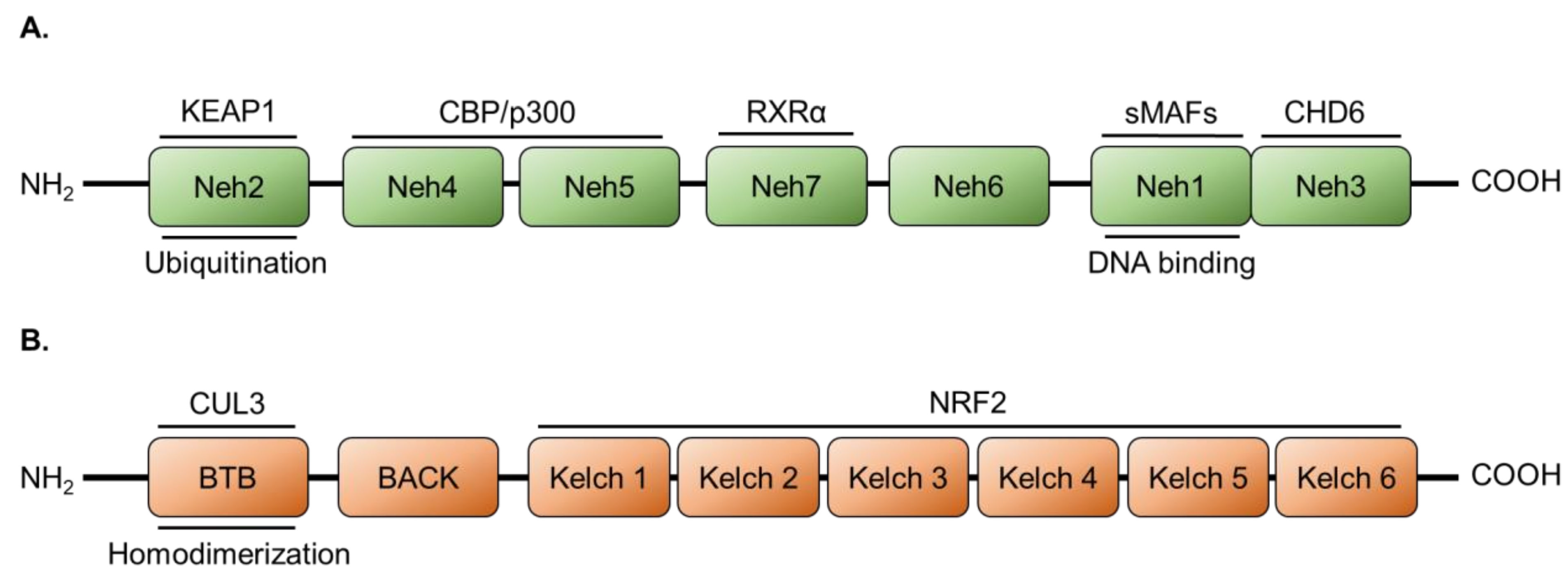
2.1. NRF2 Protein Structure

NRF2 is a cap’n’collar basic leucine zipper transcription factor comprised of seven NRF2-ECH homology (Neh) domains [63]. Each of these Neh domains has distinct functions that include DNA binding, transactivation activity, and binding sites for regulatory factors (Figure 1A). The foremost of the latter is the negative regulator, kelch-like ECH-associated protein 1 (KEAP1), which controls the subcellular localization and degradation of NRF2 through the Neh2 domain [64]. DNA binding is mediated by the Neh1 domain, which is also involved in NRF2 heterodimerization with transcription co-factors including small musculoaponeurotic fibrosarcoma (sMAF) [65,66]. Neh3 contributes towards NRF2 transactivation activity via the binding of chromodomain-helicase-DNA binding 6 (CHD6) to engage in chromatin remodeling necessary for transcription, and the other Neh domains bind assorted co-activators/co-repressors [67,68]. The nuclear receptor, retinoid X receptor α (RXRα), acts as a co-repressor binding to Neh7. Neh4/5 bind the histone acyltransferase and co-activator, cyclic-AMP response element-binding protein (CREB) binding protein (CBP, also CREBBP) [69,70]. Recently, another histone acyltransferase homologous to CBP, p300, has also been reported to bind the Neh4/5 domains and function similarly as a co-activator [71].

2.2. Regulation of NRF2 by KEAP1/CUL3

The KEAP1 protein consists of three functional domains: a broad-complex, tramtrack and bric a brac (BTB) domain; the BTB and c-terminal kelch (BACK) domain; and six kelch motifs (Figure 1B). These kelch motifs form a β-propeller structure and are responsible for NRF2 binding [72]. It is at the BTB domain that KEAP1 forms a homodimer, as well as acting as an adaptor for another key NRF2 regulator, E3 ubiquitin ligase cullin-3 (CUL3) [73]. Under normal physiological conditions, there are low levels of NRF2 localized to the cytosol, where it is bound and sequestered by KEAP1. Recruitment of CUL3 to the complex leads to the KEAP-1-mediated poly-ubiquitination of lysine residues within the Neh2 domain of NRF2, which is then followed by degradation through the ubiquitin proteasome system (Figure 2).
In the event of elevated oxidative stress, sensor cysteine residues located in the KEAP1 kelch domains act as a redox switch, which is flipped upon modification by oxidants. In this event, NRF2 is disassociated from the KEAP-CUL3 complex and accumulates in the cytosol [73]. It is then able to localize into the nucleus where it heterodimerizes with sMAF transcriptional co-factors and binds to genomic antioxidant response elements (AREs) [65]. NRF2 binding of ARE sequences located in the promoter regions of target genes leads to induced expression [74,75].

2.3. Activation of NRF2 in Melanomas

In melanomas, tumor transformation is often driven by the dysregulation of the mitogen-activated-protein-kinase (MAPK)-proliferation signaling pathway as a consequence of BRAF and NRAS mutations seen in 50% and 20% of cases, respectively [7,8]. Recently, a direct connection has been made between the activation of NRF2 and MAPK signaling in melanomas. Jessen and collaborators reported that the knockdown of NRF2 in melanoma cell lines led to decreased proliferation and EGFR expression [76]. Furthermore, the injection of NRF2-knockout mouse melanoma cells subcutaneously into C57BL/6 mice resulted in 40% of mice with no tumor formation as opposed to all mice injected with control cells. Mice with NRF2-knockout melanomas also exhibited a delayed tumor onset and prolonged tumor-free survival. They also demonstrated that NRF2 negatively regulated microphthalmia-associated transcription factor (MITF) activity, with NRF2 knockdown leading to induced melanogenesis gene expression associated with melanocyte differentiation. In a follow-up study, the same group reported that NRF2 regulates EGFR expression via binding to an ARE in its promoter region in melanoma cells [77]. MAPK signaling has also been shown to induce NRF2 activation in primary mouse cell lines with lentiviral transduction of oncogenic KRAS and BRAF [78]. The connection between NRF2 and EGFR signaling has been demonstrated in other cancer types as well [79,80].
The presence of elevated oxidative stress in melanomas is another cause of NRF2 activation during melanoma initiation and progression. As previously mentioned, the sources for this oxidative stress include UVR, melanogenesis, and reprogrammed cancer metabolism (Figure 2). Other important sources of ROS (e.g., superoxide and hydrogen peroxide) and RNS (e.g., nitric oxide) are associated with NADPH oxidase (NOX) and nitric oxide synthase (NOS) enzymes, respectively [50,81]. In melanomas, NOX1, NOX4, and NOX5 have been confirmed to be expressed, with NOX1 expression consistent throughout progression and NOX4 upregulated during metastasis [45,82,83]. As for NOX5, a recent study combined gene expression and flux balance analysis of BRAF-mutant melanomas to reveal the upregulation of NOX5 expression that is associated with the resistance to BRAF inhibitor treatment [84]. Isoforms of the NOS family have also been found to be upregulated in melanoma and contribute towards elevated proliferation and progression [85,86]. Interestingly, ROSs are also produced under circumstances of intermittent hypoxia, and NRF2 has been reported to contribute to melanoma metastasis in a mouse model of sleep apnea [87]. Another cause for NRF2 activation is alterations in its regulation at the transcriptional and posttranslational levels, as implicated by its expression during progression.

2.4. Expression and Regulation of NRF2 in Melanomas

A histological examination of melanomas for NRF2 expression indicates that NRF2 expression decreases early in melanomagenesis between benign and dysplastic nevi and maintains that lower expression between primary and metastatic stages [88]. At the same time, however, nuclear NRF2 expression is an indicator of a poor prognosis correlating with reduced survival [88,89]. Interestingly, a recent study investigating changes in the melanoma proteome following NRF2 modulation revealed that the inhibition of NRF2 expression led to changes in the expression of genes related to melanoma progression including stem cell marker CD44 [90]. It was hypothesized that reduced NRF2 expression could contribute to phenotype switching exhibited in melanomas in which a proliferative state changes to an invasive state or vice versa [91]. This switching occurs in part via changes in the expression of MITF, the master transcriptional regulator of melanocyte differentiation, where upregulation leads to a proliferative phenotype and downregulation causing an invasive phenotype [92]. It could be that NRF2 expression is needed during periods of rapid growth and is downregulated when an invasive phenotype is established, but more research is needed to confirm this potential relationship.
Despite reduced levels of NRF2 expression during melanoma progression, there is continued expression of NRF2 transcriptional targets. This continued transcription could be due to the contribution of other transcription factors, such as NF-κB, or the altered regulation of NRF2 allowing transactivation activity even with a reduced amount of the protein. In a subset of melanomas, this is explained by KEAP1 deactivating mutations that have been observed and cause elevated NRF2 expression following transformation to a primary tumor [93].
Other regulators include the homologous histone acyltransferases p300 and CBP that function as co-activators promoting the transactivation activity of NRF2. It was previously shown that p300/CBP acetylate NRF2 which enables NRF2 to bind to ARE sequences and promote expression of target genes [94]. The expression of the p300 gene, EP300, is often upregulated, as it is located in regions of chromosomal copy gain seen in melanomas [95]. Interestingly, a histological analysis of melanomas revealed that, throughout progression, p300 nuclear localization decreases and cytoplasmic localization increases [96]. Furthermore, the reduced nuclear expression of p300 correlated with a poorer prognosis in patients. This could be due to p300/CBP contributing to MITF transcriptional activity, and so these changes could be another result of phenotypic switching [97]. Evidence of this comes from a study into the pharmacologically inhibition of p300/CBP, which leads to reduced proliferation and MITF transcriptional activity [98]. It is likely that this occurs as melanomas progress to a more invasive phenotype as seen in the histological data. The knockdown of p300/CBP causes reduced growth, increased cell death, and an elevation in ROS, which further indicates their contribution towards a proliferative phenotype [99].
The RXRα nuclear receptor acts as a co-repressor of the binding of NRF2 to the Neh7 domain and has been shown to downregulate the expression of ARE-regulated genes [69]. A histological analysis of RXRα expression throughout melanoma progression indicates that its nuclear expression decreases between benign nevi and the primary tumor [100]. This lower expression continues during metastasis. We have shown, using a melanocyte-specific knockout mouse model, that the loss of RXRα and isoform RXRβ in melanocytes leads to reduced recruitment of IFN-γ secreting immune cells following UVR, due to the altered expression of intercellular signaling chemokines and cytokines [101]. It has also been reported that both NRF2 and ROS, in turn, negatively regulate RXRα expression, which might explain why it is reduced during melanoma progression. Possibly, this negative regulation could be connected to resistance to immunotherapy [102,103]. This is because IFN-γ positively modulates PD-L1 expression, and higher PD-L1 expression has been correlated with the improved efficacy of anti-PD-1 therapy [104,105]. NRF2 itself has also been reported to modulate PD-L1 expression directly, although this report conflicts with the previous studies by suggesting the combined inhibition of PD-1 and PD-L1 [106].
Overall, the regulation of NRF2 transactivation activity is highly complex, and further research is needed to understand how it contributes to the altered redox homeostasis seen in melanomas. It is likely that a combinatorial approach would be required if NRF2 itself were to be targeted, as it is likely that a monotherapy would be detrimental due to melanoma phenotype switching. It could be that any one of the ARE-regulated antioxidant gene products might prove a more effective therapeutic target due to the intricate web of interactions surrounding NRF2.

3. The Glutathione Antioxidant System

The glutathione (GSH) antioxidant system consists of the enzymes necessary to synthesize, utilize, and recycle GSH (the predominant non-protein thiol) to regulate the level of ROS present in the cell (Figure 3). GSH is a tripeptide comprised of sulfur-containing cysteine, glutamate, and glycine in the form of γ-L-glutamyl-L-cysteinylglycine [107]. The enzymes involved in its synthesis are the rate-limiting glutamate cysteine ligase (GCL), comprised of catalytic (GCLC) and modifier (GCLM) subunits, and GSH synthetase (GSS) [107]. The GSH synthesis enzyme genes GCLC, GCLM, and GSS have AREs in their promoter regions and are positively regulated by NRF2 [108,109,110].
GSH is largely localized to the cytosol but is also present to a lesser extent in the mitochondria and endoplasmic reticulum [111,112]. Key to the function of GSH as an antioxidant is that it provides electrons for selenium-containing glutathione-dependent peroxidases (GPXs), which go on to reduce both hydrogen peroxide and lipid peroxides [107]. In this process, GSH is oxidized to its disulfide form (GSSG). The recycling of GSSG back into its reduced form is catalyzed by glutathione reductase (GSR) and consumes NADPH produced in part by glucose-6-phosphate dehydrogenase (G6PD) in the pentose phosphate pathway [107]. Of the genes encoding these enzymes, GPX2, GSR, and G6PD are known to have promoter regions containing AREs and are regulated by NRF2 [48,113,114].
Another key family of enzymes in this system are glutathione-S-transferases (GSTs), which are split into three overarching categories: microsomal, mitochondrial, and cytosolic. Cytosolic GSTs are further divided into seven classes in humans, with pi and mu being the most studied [115]. GSTs are phase II enzymes responsible for detoxifying, via GSH conjugation, electrophilic molecules that can include anticancer drugs, which often implicates GSTs in chemoresistance [115]. GSTs are often upregulated in melanomas, as with many cancers [116]. Both GSTP1 and GSTM1 are known to be regulated by NRF2 through ARE binding [117].
Glutathione synthesis genes have been found to be upregulated in many cancers including melanoma [54,118]. However, it has also been reported that the expressions of GCLM and GCLC do not correlate with NRF2 expression in melanoma [118]. This is interesting, as NRF2 expression has been reported to decrease with melanoma progression, as previously discussed. It has also been determined that melanomas with higher expressions of GCLC correlate with a better prognosis in patients; however, GCLC expression does not necessarily indicate changes in GCL activity, as GCLM expression plays a larger role in controlling the function of GCL [119,120]. Melanoma cells do appear to benefit from elevated cellular GSH content, as it has been shown to contribute towards melanoma cell proliferation and metastasis [51]. Indeed, it appears that GSH levels are depleted as melanomas progress to metastasis, and this helps to explain the potential detrimental effects of an exogenous antioxidant treatment [121]. Inhibition of glutathione synthesis via the GCLC inhibitor buthionine sulfoximine has not been shown to be effective against melanomas in vitro or in vivo; however, in combination with cysteine transport or thioredoxin inhibition, greater efficacy is achieved [52,122].
Expressions of GPX2, GPX4, GSR, and G6PD have been found to be elevated in melanomas and other cancers [54]. Interestingly, GPX3 seems to be downregulated in both melanoma cell lines and tissues due to promoter region hypermethylation [123]. In that same study, it was also determined that GPX3 overexpression inhibits melanoma proliferation, and silencing GPX3 expression had the inverse effect on proliferation while promoting a more invasive phenotype. Furthermore, induced GPX3 expression in melanoma cells causes a metabolic shift towards oxidative phosphorylation and away from glycolysis, while also negatively regulating both HIF1-α and HIF2- α due to decreased stabilization in the relative absence of ROS [124]. GPX2 and GPX4 have been demonstrated to play a role in the resistance of melanoma to therapies [125,126]. There are some studies that have investigated blood GPX activity as a biomarker in melanoma patients, where increased activity is shown as compared to healthy volunteers or after surgical resection [127,128]. Pharmacological inhibition of GSR in melanoma cell lines has been shown to reduce proliferation, reduce migration, increase ROS, and decrease cellular GSH:GSSG ratio, suggesting the GSR function is co-opted by melanomas [129]. The authors of that study also found that inhibition of GSR induced oxidative stress that hindered melanoma cell metastasis, further indicating the co-option of endogenous antioxidant systems during disease progression. The role of G6PD in cancer redox biology, including for melanomas, has recently been reviewed [130]. Melanoma cell lines have been demonstrated to have elevated expressions and activities of G6PD compared with normal melanocytes, and the knockdown of G6PD in xenograft models show G6PD facilitates melanoma growth in vivo [83,131]. That same group also reported that G6PD works in tandem with NOX4 to activate STAT3 signaling to promote melanoma proliferation and avoid cell cycle arrest [83].
As the predominant cellular antioxidant, GSH and its associated enzyme effectors are co-opted during melanoma initiation, progression, and metastasis. A common theme seen by constituent enzymes in the GSH system is a contribution towards melanoma cell proliferation and its ability to metastasize. Furthermore, GPXs and GSTs are strongly associated with the development of drug resistance in melanomas and continue to hinder effective chemotherapeutic treatment. These factors highlight the altered redox homeostasis present in melanomas while providing potential targets for novel therapies.

4. The Thioredoxin Antioxidant System

The thioredoxin antioxidant system is comprised of thioredoxin (TRX or TXN), thioredoxin reductase (TR), and NADPH [132]. This system functions via the transfer of electrons from NADPH to TR, which can then go on to reduce TRX (Figure 3). Reduced TRX can then go on to modulate the redox status of a multitude of substrates that include peroxiredoxins, apoptosis signaling kinase 1, and DNA synthesis enzyme ribonucleotide reductase [133,134,135]. TRX has two isoforms, TR1 and TR2, which are expressed in the cytoplasm and mitochondria, respectively. Upon reducing a substrate, the two cysteine residues in the active site of TRX are reversibly oxidized to form a disulfide bond, and then, much like GSH and GSR, they can be recycled to a dithiol via TR to enable further antioxidant activity [132]. TR is a selenocysteine-containing, homodimeric flavoprotein. Two isoforms predominate in all tissues: cytosolic TR1 and mitochondrial TR2. A third isoform, TR3, is expressed in the testes [132]. In addition to reducing TRX, TR1 is also known to play a role in regulating the DNA-binding activity of the transcription factor, nuclear factor kappa B (NF-κB) [136,137]. As with the GSH antioxidant system, the enzymatic activity of G6PD in the pentose phosphate pathway is a source of NADPH that feeds the TRX antioxidant system [48]. Due to the elevated intracellular ROS present in cancer cells, the expression of the thioredoxin antioxidant system has been shown to be upregulated in many tumor types [138]. Both TXN and TXNRD1, encoding TRX and TR1, respectively, are known to contain AREs in their promoter regions and are directly regulated by NRF2 [139].
The expression of both TXN and TXNRD1 is known to be elevated in melanomas and other cancers [54]. TRX has specifically been shown to have increasing expression throughout disease progression, and its overexpression contributes towards enhanced metastasis [140]. Furthermore, TRX secretion by melanomas has been demonstrated to contribute to cancer immunoevasion by the recruitment and differentiation of T cells into immunosuppressive regulatory T cells [140]. The negative regulator of TRX, thioredoxin interacting protein (TXNIP), has been shown to have a reduced expression from benign nevi to primary melanoma and a further reduced expression in metastatic melanoma [141]. Interestingly in that same report, TXNIP expression was elevated following treatment with the BRAF inhibitor, PLX4032, and then decreased again with acquired drug-resistance, indicating a potential role for TRX in that process.
The expression of TR1 has been analyzed in melanoma patient-tissue microarrays revealing a continuous elevation in TR1 expression from benign nevi to primary melanoma and further into metastatic melanoma [53]. In that same study, it was also demonstrated that the knockdown of TXNRD1 induces glycolysis dependency in melanoma cell lines, and that the combination of TXNRD1 knockdown with a pharmacological blockade of glycolysis-hindered metastasis in a mouse xenograft model. TR1, acting through TRX, has also been directly implicated in cancer cell motility, where it has been shown to modify the Cys101 residue of L-plastin to promote migration. This would indicate a potential role for the TRX system in melanoma metastasis [142]. Indeed, in our study investigating the role of TR1 in normal melanocyte development, we observed a depigmentation phenotype in the bodily regions most distant from the developmental tissue of origin for the melanocyte lineage, the neural crest, which further supports a role for the TRX system in cellular migration [43]. The inhibition of TR1 via auranofin or the small molecule MJ25 has demonstrated some antimelanoma efficacy in vitro, where they exhibited cytotoxicity, and MJ25 weakly induced p53 expression and activity [143]. However, targeting TR1 for melanoma treatment alone is unlikely to lead to any therapeutic outcomes due to the ability of compensatory antioxidant mechanisms to be engaged via NRF2 activation [43,137]. As mentioned previously, the elevated nuclear localization of NRF2 has been an indicator of poor prognosis in melanomas, and so any treatment that might activate NRF2 could be a risk if the cancer persists after treatment. It is likely that TR1 inhibition would need be combined with approaches that involve the interruption of the GSH system or cysteine transport into the cell in addition to targeted or immunotherapies.
Similar to the GSH system, the function of TRX and TR1 are integral to normal melanocyte physiology, and their functions are co-opted by melanoma. It appears that the TRX system contributes towards progression by aiding in both proliferation and immunoevasion. The system also contributes to metastasis through involvement in cell motility and appears to be involved in drug resistance.

5. Cystine/Cysteine Cycling

Cysteine, a reduced form of the disulfide dimer cystine, is a crucial amino acid that is a constituent of many cellular antioxidant proteins due to its capability to undergo redox reactions [107,132]. Therefore, the ability of cells to import cystine is crucial for regulating oxidative stress. This is accomplished through the cystine/glutamate antiporter xCT (system xc) that functions via the exchange of intracellular glutamate 1:1 for extracellular cystine [144]. Imported cystine is then rapidly reduced in the cytosol to cysteine, where it can be utilized by the cell or exported back into the extracellular space to provide a reducing environment [145]. Two genes encode the subunits of system xc, SLC7A11 (xCT) and SLC3A2 (4F2) [146]. SLC7A11 encodes the functional, specific subunit of system xc, while SLC3A2 encodes a subunit that comprises multiple amino acid transporters. As an important source of cysteine necessary for intracellular and extracellular antioxidant activities, SLC7A11 is upregulated in many cancer types in order to alter normal redox homeostasis and mitigate the cytotoxic effects of elevated oxidative stress [147]. The promoter region for SLC7A11 is also known to contain an ARE and to be directly regulated by NRF2 [148].
SLC7A11 expression has been shown to be elevated in cancers such as melanoma, both before and after acquired drug resistance [54,84]. The cysteine cycle has recently been identified as a potential target for melanoma therapy, as inhibiting the function of xCT with (S)-4-carboxyphenylglycine has demonstrated melanoma cell cytotoxicity, especially in combination with inhibition of GSH synthesis using buthionine sulfoximine [52]. Furthermore, the xCT antiporter has been demonstrated to contribute towards both cell-line and mouse xenograft-melanoma proliferation, and the small molecule riluzole has been reported to be an inhibitor [149]. SLC7A11 CRISPR/Cas9 knockout inhibits proliferation, migration, and invasion in vitro, and mouse xenograft experiments demonstrated that cysteine transport also facilitates proliferation and metastasis in vivo [150]. SLC7A11 has also been shown to be upregulated in BRAF inhibitor-resistant melanoma cell lines, which themselves exhibited elevated oxidative stress as compared to drug naive cells [151]. It was also determined that vorinostat, a histone deacetylase inhibitor, treatment after MAPK inhibition suppressed SLC7A11 expression in patients with metastatic melanoma. Together, these studies highlight the potential for targeting SLC7A11 in combination with other therapies to counteract the altered redox homeostasis present in melanomas both before and after acquired drug resistance.

6. Peroxiredoxins and Sulfiredoxin

Peroxiredoxins (PRDXs) are a family of antioxidant enzymes key in regulating intracellular oxidative stress via the level of hydrogen peroxide in the cell [152]. There are six mammalian isoforms (PRDX1-6) that contain an active site with either one or two redox-active cysteine residues. PRDX isoforms also differ in subcellular localization with PRDX1 and PRDX2 in the cytosol and nucleus; PRDX3 in the mitochondria; PRDX4 in the ER (also exists extracellularly); PRDX5 in the mitochondria, peroxisomes, and cytosol; and PRDX 6 in the cytosol [153,154,155,156,157]. The TRX and GSH antioxidant systems are capable of recycling oxidized PRDX, which allows it to continue its redox activity [152]. PRDXs have been found to be upregulated in numerous cancer types; however, only PRDX1 and PRDX6 have confirmed AREs in their promoter regions [139,156,158,159].
Sulfiredoxin (SRXN) is another redox enzyme that assists PRDXs in their antioxidant function. Particularly, SRXN is responsible for reducing hyperoxidized PRDX that can occur if the TRX or GSH systems are not able to reduce oxidized PRDX in time [152]. This hyperoxidation only occurs in the PRDXs that contain two active site cysteines, and so they do not interact with PRDX5 and PRDX6 [160]. The SRXN1 isoform is known to have a promoter region ARE and to be regulated by NRF2 [161].
PRDXs 1-6 and SRXN1 have been reported to have elevated expression in melanomas and other cancers [54]. However, there are conflicting reports of the decreased expressions of PRDX1 and PRDX2 in at least a subset of melanomas. For PRDX1, histological staining revealed a loss of expression during the progression between benign nevi and malignant melanomas [162]. The two above contradictory reports could be due to the difference in expression level noted globally at the level of the transcript [54] vs. alteration in protein expression [162] noted in specific cell types within benign and advanced stage melanomas. Interestingly, mitochondrial PRDX1 expression appears to be upregulated in BRAF inhibitor-resistant cell lines, indicating a potential role in drug resistance [163]. Downregulation of PRDX2 expression is known to occur due to the methylation of its promoter region; however, the cause for PRDX1′s reduced expression is not as clear [164]. PRDX2 has been shown to inhibit melanoma proliferation and migration in cell lines and metastasis in a mouse xenograft model [165]. This activity occurred due to the increase E-cadherin expression normally lost during melanoma progression.
In uveal melanoma, the presence of cancers overexpressing PRDX3 has been an indicator of poor prognosis, with high expression correlated to metastasis and reduced patient survival [60]. PRDX6 has been associated with promoting melanocyte proliferation with expression either induced with EGFR stimulation or reduced with EGFR inhibition using erlotinib. SRXN has been found to have induced expression in mouse skin following tumor-promoting 12-O-tetradecanoylphorbol-13-acetate treatment and to also have elevated expression in human melanoma tissue microarrays [166]. It has also been shown to be upregulated after either TR1 inhibition via auranofin or siRNA knockdown of the melanocortin 1 receptor of melanoma cell lines [167,168]. Overall, the roles played by PRDXs in melanoma are complex, likely due to their distribution, and further research into the role they play is needed if they are to be targeted for melanoma therapy. SRXN1, on the other hand, appears to have a clearer role in promoting melanoma initiation; however, its exact role in melanoma progression and metastasis still needs to be elucidated.

7. NAD(P)H: Quinone Oxidoreductase 1

NAD(P)H: Quinone Oxidoreductase 1 (NQO1) is a phase II antioxidant flavoenzyme that detoxifies via the reduction of reactive quinones to hydroquinones in a manner that avoids generation of ROS [169]. It is largely localized to the cytosol and exhibits low and consistent expression under normal conditions [170]. In the skin, the expression of NQO1 can be induced by UVR, particularly the UVA wavelength, to protect against oxidative stress [41,171]. Other important functions of NQO1 include the stabilization of p53 during conditions of elevated DNA damage, as well as regulating the initiation of 20 s proteasomal degradation [172,173]. In cancer, NQO1 is often highly expressed relative to normal tissue and there is ongoing research into therapies involving NQO1 substrate pro-drugs used to selectively target cancer cells [169,174]. NQO1 is well known to be regulated by NRF2 and has a confirmed ARE sequence in its promoter region [175].
As with many other NRF2 antioxidant transcriptional targets, NQO1 has been shown to be upregulated in melanomas and other cancers [54]. A histology of melanoma tissue microarrays indicates that NQO1 is upregulated in melanoma but has no association with patients’ five-year survival [176]. It was hypothesized that NQO1 might play a role in initiation while not notably affecting progression and metastasis. In a clinicopathological study of melanomas, the same group analyzed the expression of six biomarkers, including NOQ1, which were elevated in early stages (I and II) and reduced at later stages (III and IV) [177]. In melanoma cell lines, NQO1 has been demonstrated to promote proliferation by promoting cell-cycle progression [57]. Induced NQO1 expression has also been shown to induce melanogenesis in cancer cell lines, with silenced expression having the opposite effect [178]. It is possible that NQO1 is directly or indirectly associated with MITF activity in melanomas and, therefore, might be involved with a proliferative phenotype over an invasive one.

8. Heme Oxygenase 1

Heme oxygenase 1 (HO-1) is an antioxidant enzyme that catalyzes the first rate-limiting step in the degradation of heme into the antioxidant biliverdin, carbon monoxide, and pro-oxidant ferrous iron [179]. The ferrous iron is then taken up by ferratin when is often upregulated at the same time as HO-1 [180]. Biliverdin is reduced by biliverdin reductase into bilirubin, which also engages in antioxidant activity. HO-1 expression is induced by elevated cellular oxidative stress, as seen in its induction following UVA radiation of the skin [41,171]. In cancer, elevated HO-1 expression upregulates proliferation, resistance to anticancer therapy, and immune evasion [181,182,183,184]. HO-1 expression also often favors cancer progression to metastasis [185]. As a phase II enzyme, HMOX1 (encoding HO-1) is regulated by NRF2 and contains an ARE in its promoter region [186].
The elevated expression of HMOX1 has been seen in multiple cancers that also include melanoma [54]. Overexpression of HO-1 in melanoma cell lines causes increased proliferation and increased resistance to oxidative stress, and xenograft experiments with overexpressing cell lines cause reduced survival in mice [187]. Together, these data indicate HO-1′s role in melanoma progression. HO-1 also has been shown to contribute towards dysregulated proliferation in BRAFV600E melanomas, through direct association with the BRAF protein [58]. Furthermore, it has been implicated in BRAF inhibitor resistance where HO-1 silencing or inhibition, using tin mesoporphyrin IX, increased sensitivity to vemurafenib, increased IL-15-activated natural killer cells, and upregulated natural killer-cell target expression enabling the improved activity of melanoma cells [188]. HO-1 has also been shown to mediate the differential stress response between normal tissue and melanomas seen in the efficacy of therapy combining short-term starving or fast-mimicking diet and doxorubicin [189]. This occurs at least in part due to enhancing anti-melanoma immune cell accumulation. From these studies, it is evident that HO-1 plays an important role in melanoma disease progression and provides a promising target for overcoming BRAF inhibitor resistance going forward.

9. Heat Shock Protein 70

Heat shock protein 70 (HSP70) is a member of the heat shock protein superfamily, which act as molecular chaperones that assist in the correct folding, transport, and degradation of cellular proteins [190]. Thirteen HSP70 family members haven been identified so far and are encoded collectively by 17 genes enabling diverse subcellular localization for their function [191]. HSP70 proteins have been considered to engage in antioxidant activity due to their ability to protect cells against oxidative stress, possibly through their ability to sequester damaged proteins and prevent aggregation [192,193,194]. The expression of HSP70 is upregulated in numerous cancer types and has been shown to limit apoptotic signaling through the PI3K-Akt pathway. HSP70 does so by binding and stabilizing Akt and inhibiting Bax activation [195,196]. An ARE has been shown to be located in the promoter region of HSP70, and so its expression can be regulated by NRF2 [197].
HSP70 has been shown to have elevated expression in melanoma tissue microarrays, when compared with benign nevi, and is also known to protect melanoma cells from oxidative stress, such as that following UV irradiation [198,199]. The inhibition of HSP70, via the 2-phenylethyne-sulfonamide derivative PET-16, resulted in melanomas that exhibited reduced migration and invasion through direct association of HSP70 with both FAK and BRAFV600E [198]. A natural product source for potential HSP70 inhibitors is the C-prenylated chalcone, isocordoin 1. Derivatives of isocordoin 1 have been demonstrated to be cytotoxic, induce apoptosis, and decrease expression of HSP70 in melanoma cells lines [200].
There is also a potential role for using melanoma “cell surface expressed HSP70” to improve immunotherapy by triggering natural killer cell targeting to induce cancer cell death [201]. One route that has been studied for presenting melanoma expressed HSP70 to the immune system is through extracellular vesicles. Incubation of treatment-naive B16 melanoma cells with either exogenous soluble HSP70 or exogenous extracellular vesicles loaded with EVs led to the increased melanoma-cell surface expression of HSP70 that led to immune response-mediated melanoma-cell death [202]. In that same study, mouse xenograft models using B16 cells treated with HSP70-loaded EVs also exhibited reduced tumor volume and increased survival, suggesting that the induction of melanoma-cell surface expression of HSP70 could aid immunotherapy.

10. Superoxide Dismutase (SOD)

The superoxide dismutase (SOD) family of enzymes regulate both extracellular and intracellular ROS-mediated oxidative stress [50]. They do so by converting superoxide into hydrogen peroxide, which can be further processed by other antioxidant enzymes. There are three human SOD genes: SOD1 (copper/zinc SOD), SOD2 (manganese SOD), and SOD3 [50]. The three SOD enzymes have distinct localizations, with SOD1 expressed in the cytosol, SOD2 in the mitochondria, and SOD3 expressed extracellularly [203,204,205]. Overall, the SOD enzymes do not tend of be overexpressed in cancer and often exhibit lower expressions than normal tissue [205]. All three SOD enzymes have been shown to be regulated by NRF2; however, only SOD1 and SOD2 have a confirmed ARE in the promoter region [206,207,208].
Gene expression analysis of melanomas and other cancers indicated an upregulation of SOD2 expression, while a histological examination of pigmented melanoma tissue samples revealed the upregulated expression of both SOD1 and SOD2 compared to healthy skin [54]. Interestingly, mimicking SOD2 with the mitochondrial superoxide scavenger (2-(2,2,6,6-tetramethylpiperidin-1-oxyl-4-ylamino)-2-oxoethyl) triphenylphosphonium chloride induced melanoma-cell line death and apoptosis and reduced tumor size in mouse xenografts [209]. However, SOD also appears to play a role in BRAF-inhibitor resistance in melanoma cell lines as BRAF inhibition of drug-naive melanoma cell lines induces elevated free radical formation. Supplementation of treated cells with SOD or inhibiting nitric oxide synthase counteracts the effectiveness of BRAF-inhibitor treatment and rescues the growth of BRAF-mutant melanoma cells [210]. After the development of BRAF inhibitor resistance, melanoma cell lines exhibit elevated levels of ROS and expression of SOD2 [211]. In that same study, the knockdown of SOD2 expression in resistant melanoma cells inhibited proliferation, while the administration of the exogenous antioxidant, N-acetyl cysteine, re-sensitized resistant cells to BRAF-inhibitor treatment. These studies indicate that SODs, especially SOD2, are promising targets for overcoming drug resistance by disrupting antioxidant protection of the mitochondria.

11. Clinical Applications of Exogenous Antioxidants

With the knowledge that melanomas are associated with elevated oxidative stress, clinical investigators have aimed to find therapeutic candidates that can pursue this feature as an avenue for a novel method of intervention against and/or treatment of melanoma. Naturally, this has led to investigation of dietary and topical antioxidants, such as vitamin A derivatives, vitamin C, vitamin E, nicotinamide, and selenium, for the prevention and treatment of melanoma. The results of these studies help highlight the complexity of the altered redox homeostasis present in melanomas that complicates their treatment with exogenous antioxidants.

11.1. Vitamin A

Clinical studies suggest that retinoid or carotenoid use in the context of melanoma prevention appears to have either a slightly protective effect or no effect on risk. In the Vitamins and Lifestyle (VITAL) study, a large prospective cohort of 69,635 participants taking retinol supplements showed a decreased risk of melanoma (HR = 0.60, 95% CI 0.41–0.90); however, they found that a dietary or total intake of vitamin A or carotenoids was not associated with melanoma risk [212]. Prospective data from the Nurses’ Health Study showed that increased retinol intake from foods and supplements appeared protective within a subgroup of women who were otherwise at low risk for melanoma based on several non-dietary factors, with RR = 0.39 and 95% CI of 0.22–0.71; in other groups, there was no significant effect [213]. On the other hand, the Women’s Health Study, a randomized, double-blind, placebo-controlled trial, reported that beta carotene use had no impact on melanoma risk, with a relative risk of 0.90 (95% CI 0.49–1.68) [214]. A Meta-Analysis by Zhang et al., which included a total of eight case-control studies and two prospective studies, suggests that the intake of retinol, rather than total vitamin A or beta-carotene, is significantly associated with a reduced risk of melanoma, with a summary odds ratio for the highest compared to lowest intake of retinol, total vitamin A, and beta-carotene found to be 0.80 (95% CI = 0.69–0.92), 0.86 (95% CI = 0.59–1.25), and 0.87 (95% CI = 0.62–1.20), respectively [215]. Additionally, several clinical trials have reported the histologic and clinical improvement of dysplastic nevi with the use of topical tretinoin (all trans-retinoic acid) [216,217].

11.2. Vitamin C

In pre-clinical studies, vitamin C, a well-known antioxidant, has been shown to induce the apoptosis of malignant melanoma cells at higher concentrations but may promote tumor growth at lower concentrations [218]. However, there is very little clinical evidence regarding vitamin C and its potential effects on the prevention and/or treatment of melanomas. Prospective data from the Nurses’ Health Study showed that, contrary to preconceived expectations, a higher risk of melanoma was observed in those with greater dietary intakes of vitamin C with RR = 1.43 and 95% CI 1.01–2.00 and a significant positive dose-response with the frequency of orange juice consumption (p = 0.008) [213]. However, it is possible that another component in vitamin-C containing foods such as orange juice may be contributing to an increase in risk. In contrast, Murray et al. found that the application of a topical formula containing 1% alpha-tocopherol, 15% L-ascorbic acid, and 0.5% ferulic acid at the dose of 2 g/cm daily for four days resulted in decreased UV-induced p53 induction, cyclobutane pyrimidine dimers, and cytokine expression in vivo [219].

11.3. Vitamin E

Vitamin E exhibits strong antioxidant effects with its ability to quench free radicals and inhibit lipid peroxidation; thus, it is thought to have potential anti-cancer applications [220]. However, clinical studies regarding vitamin E’s effects on melanoma have had varied results. Several case control studies examining the effects of vitamin E on melanoma incidence evaluated in a 2020 review in Cancer have shown inconsistent results [217]. Prospective data from the Nurses’ Health Study showed neither total nor dietary vitamin E was associated with melanoma risk (multivariate RRs 1.11 (95% CI = 0.66–1.85) and 0.88 (95% CI = 0.59–1.32), respectively) [213]. Whereas the SU.VI.MAX trial, a randomized, placebo-controlled trial investigating the health effects of antioxidant vitamins and minerals, found that women consuming antioxidant supplements, including dietary supplementation with vitamin E, were at an increased risk for developing melanoma [221]. Studies involving the use of topical vitamin E appear to have more favorable effects. A 2002 in vivo study showed that topical vitamin E and N-ac15ysteinestein treatment 24 h prior to UV exposure significantly decreased the expression of matrix metalloprotease MMP-12 in UV-irradiated skin, and thus, may provide a protective effect [222]. As described above in the section on vitamin C, a topical formula containing 1% alpha-tocopherol, 15% L-ascorbic acid, and 0.5% ferulic acid may confer anti-cancer effects [219].

11.4. Nicotinamide (Niacinamide)

Nicotinamide is a member of the vitamin B3 group and has been shown to enhance the repair of both oxidative and UV-induced DNA damage in human melanocytes in pre-clinical studies [223]. However, there is little clinical evidence regarding nicotinamide and melanoma. The ONTRAC study (Oral Nicotinamide To Reduce Actinic Cancer) was a multicenter, phase III, double-blinded placebo-controlled trial in which 386 participants were recruited [224]. The ONTRAC study revealed a decreased rate in the number of new non-melanoma skin cancers in the nicotinamide group vs. placebo group; and, while the study did not aim to assess the effect of nicotinamide on the development of melanomas, 10 melanomas arose during the intervention period. The melanomas were evenly distributed between the treatment and control groups and showed no significant differences in thickness and invasiveness between the two groups [224]. No conclusions can be drawn regarding the association between nicotinamide intake and melanoma development, as recruitment of patients in the ONTRAC study was not based on their risk for melanoma, and there were very few cases of melanomas among participants [223].

11.5. Selenium

Selenium is thought to play a role in several cellular pathways with downstream anti-tumor effects; these effects include reducing DNA damage and oxidative stress, decreasing inflammation, enhancing the immune response, altering DNA methylation, regulating the cell cycle, inducing cell death of malignant cells, and inhibiting angiogenesis [225,226,227,228,229,230,231,232,233,234]. Selenium is also critical to the function of thioredoxin reductase and glutathione peroxidases, making it an important micronutrient for the body’s endogenous antioxidant systems [235]. It would then be reasonable to conclude that it could be beneficial for the prevention and treatment of melanomas; however, the clinical data available regarding selenium and its effects on melanomas and other skin cancers vary.
One prospective study performed in Poland reported the 10-year-survival of 375 melanoma patients depending on their serum selenium levels [225]. Patients were assigned to one of four groups depending on a one-time serum selenium level collected prior to treatment (other than initial resection) and were followed up from the date of melanoma diagnosis until death or 2020. They found that the subgroup with low serum selenium (<76.44 μg/L) was associated with an increased mortality rate in the 10 years following diagnosis compared to patients with higher selenium levels, HR = 8.42; p = 0.005 and HR = 5.83 and p = 0.02, for uni- and multivariable models, respectively [225]. Additionally, a separate cohort study of 200 patients with non-metastatic melanomas showed that patients with stage I and II melanomas with recurrence within two years had significantly lower serum selenium levels [236]. In contrast, an Italian cohort with high environmental exposure to selenium was found to have a significant 3.9-fold increase in melanoma incidence as compared to an unexposed cohort [237]. A 2018 Cochrane review also suggests that selenium supplementation may increase melanoma risk [238]. They found three randomized controlled trials that investigated the risk of melanoma following selenium supplementation, but only two were deemed to have a low risk of bias [238,239,240]. When only including the low-risk-of-bias studies, they found a relative risk estimate of 1.35 with a 95% confidence interval between 0.41–4.52 [238]. However, it should be noted that there were only eight cases in the selenium groups and four cases in the placebo groups. Whereas a 2016 meta-analysis reports that high selenium exposure neither increased nor decreased the risk of skin cancer based on six estimates from four studies (pooled OR = 1.09, 95% CI: 0.98–1.21) [241]. However, they suggested that high selenium exposure may have different effects depending on the type of cancer, stating that it decreased the risk of breast, lung, esophageal, gastric, and prostate cancer. It needs to be remembered that the meta-analysis of multiple studies performed under different conditions with multiple variables is distinct from specific clinical study/studies performed on a bigger cohort with well-controlled variables and following the adjustment of the confounding factors.
Overall, exogenous antioxidants do not have a clear therapeutic role in the prevention or treatment of melanomas, as many of the available studies contradict one another, some even suggest that certain antioxidants could increase melanoma risk. These varying data further underscore the need to better understand the complex interplay between melanoma and both intracellular and microenvironmental oxidative stress.

12. Conclusions and Future Directions

The elevated oxidative stress seen in melanomas has been of consistent interest as a potential source for therapeutic interventions. However, it is becoming increasingly clear that the notion that therapies need only to reverse increased oxidative stress is an oversimplification of the complex altered redox homeostasis present in melanomas. Indeed, there are numerous studies that demonstrate that exogenous antioxidant treatment of melanomas might be detrimental [62]. This is the case, due to the fact that oxidative stress hampers the ability of melanoma cells to metastasize, and those cells that have already metastasized possibly upregulate antioxidant systems to favor their survival and protect against the cytotoxic effects of oxidative stress [121].
In this review we have highlighted how protective endogenous antioxidant systems are co-opted during melanoma initiation and progression. The net effect of this co-option is an increased antioxidant capacity within melanoma cells which allows for their unrestrained proliferation, metastasis, and drug resistance [84]. Fortunately, this reliance on endogenous antioxidant systems for progression and metastasis reveals potential targets for novel therapies. This is seen in the glutathione antioxidant system, where there is upregulation of GSH synthesis genes, increased recycling of GSH mediated by GSR, and increased expression of endogenous antioxidants associated with drug resistance, such as GPX2, GPX4, and GSTs [54]. Furthermore, NRF2 contributes to the elevated expression of G6PD, which is an important source for NADPH necessary for the activity of both glutathione and thioredoxin antioxidant systems [54]. With the thioredoxin antioxidant system, there is an upregulation of both TXN and TXNRD1 expression that contributes to PRDX function and the clearance of cellular free radicals [54]. Both of these systems are dependent on the amino acid cysteine, which is imported through the cystine/glutamate antiporter xCT (system xc) comprised in part by the NRF2 transcriptional target, SLC7A11, which also has upregulated expression in melanomas [54].
Three potential antimelanoma targets within these antioxidant systems could include (but are not limited to) glutathione synthesis, thioredoxin reductase enzymatic activity, and the cystine/cysteine cycle. A combination of these redox targets would then be included with conventional melanoma therapies. For example, it has previously been shown that the combined inhibition of GSH synthesis and cystine/cysteine cycling was more effective in killing melanoma cells than either treatment alone [52]. This blockade of co-opted antioxidant function could then be combined with MAPK inhibition or immunotherapy. The knockdown of thioredoxin reductase 1 alone in melanoma alone does not prevent metastasis but does induce a dependency on glycolysis, and a complete knockout of TXNRD1 in melanocytes increases the nuclear localization of NRF2 and the synthesis of GSH post-UVB irradiation [43,53]. Interestingly, acquired resistance to BRAF inhibition induces a shift to oxidative phosphorylation [242]. So, it would be interesting to determine the effect that combinatorial inhibition of MAPK signaling and thioredoxin reductase 1 would have on melanoma cells.
Another avenue of treatment through intracellular redox manipulation is through the targeting of mitochondria. We recently performed a high-throughput screen to identify compounds effective against drug-resistant melanoma and found that compounds that target the electron transport chain were the most potent and others have studied different compounds that act similarly [81,243]. Inhibition of the electron transport chain leads to the accumulation of oxidative stress, making it a pro-oxidant treatment [244]. Another promising avenue is compounds that are capable of disrupting mitochondrial membrane potential as exhibited by natural product polyphenols acting as proton shuttles that interfere with the electrochemical gradient across the inner mitochondrial membrane [245,246]. Additionally, polyphenols can also inhibit the electron transport chain and, thereby, further increase the oxidative stress present in cancer cells.
It is also important to examine NRF2 and its transcriptional targets in the context of the immune response. Melanoma cell NRF2 has an indirect connection with an immune response through its negative regulation of the transcription factor RXRα, where we have previously shown that knockout of RXRα and RXRβ in melanocytes alters the expression of the cytokines/chemokines that recruit IFN-γ secreting immune cells [101]. However, the targeting of NRF2 directly in an attempt to improve an antimelanoma immune response is not likely to be effective, as it has been previously shown that NRF2-null mice have an increased susceptibility to xenografts of B16-F10 melanoma cells, possibly due in part to an impaired immune system [247]. These data indicate that any intervention that might also compromise the NRF2 function in nonmelanoma cancer-adjacent cells could be detrimental, and, therefore, endogenous antioxidants downstream of NRF2/KEAP1/ARE signaling could be better targets of immunotherapy. These targets could include thioredoxin secretion, heme oxygenase 1, and cell-surface expression of heat shock protein 70, previously shown to play a role in the immune response to melanomas, as discussed above.
It should be noted that there has been some efficacy seen in clinical studies investigating the role of exogenous antioxidants in the prevention of melanoma, but its utility in treatment requires further detailed studies on intervention at different stages of melanoma progression and metastasis to better understand and subsequently avoid any unintended detrimental effects.

Author Contributions

Writing–original draft preparation, E.L.C. and A.L.B.; writing–review and editing, E.L.C., A.L.B. and A.K.I.; visualization, E.L.C.; supervision, A.K.I.; funding acquisition, A.K.I. All authors have read and agreed to the published version of the manuscript.

Funding

This work was supported in part by the National Institute of Environmental Health Sciences (NIEHS) of the National Institute of Health (NIH) under the award number 1R01 ES016629-01A1 (principal investigator: A.K.I.), the Oregon State University/Oregon Health & Science University College Pharmacy Pilot Project Grant (principal investigator: A.K.I.), and a training grant from the National Center for Complementary and Integrative Health (NCCIH) of the National Institutes of Health (NIH) under award number T32 AT010131.

Conflicts of Interest

The authors state no conflicts of interest.

References

  1. Lim, H.W.; Collins, S.A.B.; Resneck, J.S., Jr.; Bolognia, J.L.; Hodge, J.A.; Rohrer, T.A.; Van Beek, M.J.; Margolis, D.J.; Sober, A.J.; Weinstock, M.A.; et al. The burden of skin disease in the United States. J. Am. Acad. Dermatol. 2017, 76, 958–972. [Google Scholar] [CrossRef] [Green Version]
  2. Islami, F.; Ward, E.M.; Sung, H.; Cronin, K.A.; Tangka, F.K.L.; Sherman, R.L.; Zhao, J.; Anderson, R.N.; Henley, S.J.; Yabroff, K.R.; et al. Annual Report to the Nation on the Status of Cancer, Part 1: National Cancer Statistics. J. Natl. Cancer Inst. 2021, 113, 1648–1669. [Google Scholar] [CrossRef]
  3. Lin, J.Y.; Fisher, D.E. Melanocyte biology and skin pigmentation. Nature 2007, 445, 843–850. [Google Scholar] [CrossRef]
  4. Boissy, R.E. Melanosome transfer to and translocation in the keratinocyte. Exp. Dermatol. 2003, 12 (Suppl. 2), S5–S12. [Google Scholar] [CrossRef] [PubMed]
  5. Mullenders, L.H.F. Solar UV damage to cellular DNA: From mechanisms to biological effects. Photochem. Photobiol. Sci. 2018, 17, 1842–1852. [Google Scholar] [CrossRef] [PubMed]
  6. Davis, E.J.; Johnson, D.B.; Sosman, J.A.; Chandra, S. Melanoma: What do all the mutations mean? Cancer 2018, 124, 3490–3499. [Google Scholar] [CrossRef] [PubMed]
  7. Schadendorf, D.; van Akkooi, A.C.J.; Berking, C.; Griewank, K.G.; Gutzmer, R.; Hauschild, A.; Stang, A.; Roesch, A.; Ugurel, S. Melanoma. Lancet 2018, 392, 971–984. [Google Scholar] [CrossRef]
  8. Hodis, E.; Watson, I.R.; Kryukov, G.V.; Arold, S.T.; Imielinski, M.; Theurillat, J.P.; Nickerson, E.; Auclair, D.; Li, L.; Place, C.; et al. A landscape of driver mutations in melanoma. Cell 2012, 150, 251–263. [Google Scholar] [CrossRef] [PubMed] [Green Version]
  9. Miller, K.D.; Nogueira, L.; Mariotto, A.B.; Rowland, J.H.; Yabroff, K.R.; Alfano, C.M.; Jemal, A.; Kramer, J.L.; Siegel, R.L. Cancer treatment and survivorship statistics, 2019. CA Cancer J. Clin. 2019, 69, 363–385. [Google Scholar] [CrossRef] [Green Version]
  10. Domingues, B.; Lopes, J.M.; Soares, P.; Populo, H. Melanoma treatment in review. Immunotargets Ther. 2018, 7, 35–49. [Google Scholar] [CrossRef] [Green Version]
  11. Batus, M.; Waheed, S.; Ruby, C.; Petersen, L.; Bines, S.D.; Kaufman, H.L. Optimal management of metastatic melanoma: Current strategies and future directions. Am. J. Clin. Dermatol. 2013, 14, 179–194. [Google Scholar] [CrossRef] [Green Version]
  12. Van Zeijl, M.C.; van den Eertwegh, A.J.; Haanen, J.B.; Wouters, M.W. (Neo)adjuvant systemic therapy for melanoma. Eur. J. Surg. Oncol. 2017, 43, 534–543. [Google Scholar] [CrossRef]
  13. Garbe, C.; Peris, K.; Hauschild, A.; Saiag, P.; Middleton, M.; Bastholt, L.; Grob, J.J.; Malvehy, J.; Newton-Bishop, J.; Stratigos, A.J.; et al. Diagnosis and treatment of melanoma. European consensus-based interdisciplinary guideline—Update 2016. Eur. J. Cancer 2016, 63, 201–217. [Google Scholar] [CrossRef] [PubMed]
  14. Christofyllakis, K.; Pföhler, C.; Bewarder, M.; Müller, C.S.L.; Thurner, L.; Rixecker, T.; Vogt, T.; Stilgenbauer, S.; Yordanova, K.; Kaddu-Mulindwa, D. Adjuvant therapy of high-risk (stages IIC-IV) malignant melanoma in the post interferon-alpha era: A systematic review and meta-analysis. Front. Oncol. 2021, 10, 637161. [Google Scholar] [CrossRef] [PubMed]
  15. Wilson, M.A.; Schuchter, L.M. Chemotherapy for Melanoma. Cancer Treat. Res. 2016, 167, 209–229. [Google Scholar] [CrossRef]
  16. Soengas, M.S.; Lowe, S.W. Apoptosis and melanoma chemoresistance. Oncogene 2003, 22, 3138–3151. [Google Scholar] [CrossRef] [Green Version]
  17. Flaherty, K.T. Targeting metastatic melanoma. Annu. Rev. Med. 2012, 63, 171–183. [Google Scholar] [CrossRef]
  18. Chapman, P.B.; Hauschild, A.; Robert, C.; Haanen, J.B.; Ascierto, P.; Larkin, J.; Dummer, R.; Garbe, C.; Testori, A.; Maio, M.; et al. Improved survival with vemurafenib in melanoma with BRAF V600E mutation. N. Engl. J. Med. 2011, 364, 2507–2516. [Google Scholar] [CrossRef] [PubMed] [Green Version]
  19. Livingstone, E.; Zimmer, L.; Vaubel, J.; Schadendorf, D. BRAF, MEK and KIT inhibitors for melanoma: Adverse events and their management. Chin. Clin. Oncol. 2014, 3, 29. [Google Scholar] [CrossRef]
  20. Rizos, H.; Menzies, A.M.; Pupo, G.M.; Carlino, M.S.; Fung, C.; Hyman, J.; Haydu, L.E.; Mijatov, B.; Becker, T.M.; Boyd, S.C.; et al. BRAF inhibitor resistance mechanisms in metastatic melanoma: Spectrum and clinical impact. Clin. Cancer Res. 2014, 20, 1965–1977. [Google Scholar] [CrossRef] [Green Version]
  21. Eroglu, Z.; Ribas, A. Combination Therapy with BRAF and MEK Inhibitors for Melanoma: Latest Evidence and Place in Therapy. Ther. Adv. Med. Oncol. 2016, 8, 48–56. [Google Scholar] [CrossRef] [Green Version]
  22. Flaherty, K.T.; Robert, C.; Hersey, P.; Nathan, P.; Garbe, C.; Milhem, M.; Demidov, L.V.; Hassel, J.C.; Rutkowski, P.; Mohr, P.; et al. Improved survival with MEK inhibition in BRAF-mutated melanoma. N. Engl. J. Med. 2012, 367, 107–114. [Google Scholar] [CrossRef] [Green Version]
  23. Wright, C.J.M.; McCormack, P.L. Trametinib: First Global Approval. Drugs 2013, 73, 1245–1254. [Google Scholar] [CrossRef] [PubMed]
  24. Larkin, J.; Ascierto, P.A.; Dreno, B.; Atkinson, V.; Liszkay, G.; Maio, M.; Mandala, M.; Demidov, L.; Stroyakovskiy, D.; Thomas, L.; et al. Combined vemurafenib and cobimetinib in BRAF-mutated melanoma. N. Engl. J. Med. 2014, 371, 1867–1876. [Google Scholar] [CrossRef] [PubMed] [Green Version]
  25. Karimkhani, C.; Gonzalez, R.; Dellavalle, R.P. A review of novel therapies for melanoma. Am. J. Clin. Dermatol. 2014, 15, 323–337. [Google Scholar] [CrossRef]
  26. Long, G.V.; Stroyakovskiy, D.; Gogas, H.; Levchenko, E.; de Braud, F.; Larkin, J.; Garbe, C.; Jouary, T.; Hauschild, A.; Grob, J.J.; et al. Dabrafenib and trametinib versus dabrafenib and placebo for Val600 BRAF-mutant melanoma: A multicentre, double-blind, phase 3 randomised controlled trial. Lancet 2015, 386, 444–451. [Google Scholar] [CrossRef]
  27. Pelster, M.S.; Amaria, R.N. Combined Targeted Therapy and Immunotherapy in Melanoma: A Review of the Impact on the Tumor Microenvironment and Outcomes of Early Clinical Trials. Ther. Adv. Med. Oncol. 2019, 11, 1758835919830826. [Google Scholar] [CrossRef] [Green Version]
  28. Van Breeschoten, J.; Wouters, M.W.J.M.; Hilarius, D.L.; Haanen, J.B.; Blank, C.U.; Aarts, M.J.B.; van den Berkmortel, F.W.P.J.; de Groot, J.-W.B.; Hospers, G.A.P.; Kapiteijn, E.; et al. First-Line BRAF/MEK Inhibitors versus Anti-PD-1 Monotherapy in BRAFV600-Mutant Advanced Melanoma Patients: A Propensity-Matched Survival Analysis. Br. J. Cancer 2021, 124, 1222–1230. [Google Scholar] [CrossRef]
  29. Gata, V.A.; Lisencu, C.I.; Vlad, C.I.; Piciu, D.; Irimie, A.; Achimas-Cadariu, P. Tumor infiltrating lymphocytes as a prognostic factor in malignant melanoma. Review of the literature. J. Buon. 2017, 22, 592–598. [Google Scholar] [PubMed]
  30. Schreiber, R.D.; Old, L.J.; Smyth, M.J. Cancer immunoediting: Integrating immunity’s roles in cancer suppression and promotion. Science 2011, 331, 1565–1570. [Google Scholar] [CrossRef] [PubMed] [Green Version]
  31. Gasser, S.; Lim, L.H.K.; Cheung, F.S.G. The role of the tumour microenvironment in immunotherapy. Endocr. Relat. Cancer 2017, 24, T283–T295. [Google Scholar] [CrossRef] [PubMed]
  32. Khair, D.O.; Bax, H.J.; Mele, S.; Crescioli, S.; Pellizzari, G.; Khiabany, A.; Nakamura, M.; Harris, R.J.; French, E.; Hoffmann, R.M.; et al. Combining Immune Checkpoint Inhibitors: Established and Emerging Targets and Strategies to Improve Outcomes in Melanoma. Front. Immunol. 2019, 10, 453. [Google Scholar] [CrossRef] [PubMed] [Green Version]
  33. Larkin, J.; Chiarion-Sileni, V.; Gonzalez, R.; Grob, J.-J.; Rutkowski, P.; Lao, C.D.; Cowey, C.L.; Schadendorf, D.; Wagstaff, J.; Dummer, R.; et al. Five-Year Survival with Combined Nivolumab and Ipilimumab in Advanced Melanoma. N. Engl. J. Med. 2019, 381, 1535–1546. [Google Scholar] [CrossRef] [PubMed] [Green Version]
  34. Chen, D.S.; Mellman, I. Elements of cancer immunity and the cancer-immune set point. Nature 2017, 541, 321–330. [Google Scholar] [CrossRef] [PubMed]
  35. Gide, T.N.; Wilmott, J.S.; Scolyer, R.A.; Long, G.V. Primary and Acquired Resistance to Immune Checkpoint Inhibitors in Metastatic Melanoma. Clin. Cancer Res. 2018, 24, 1260–1270. [Google Scholar] [CrossRef] [PubMed] [Green Version]
  36. Sharma, P.; Hu-Lieskovan, S.; Wargo, J.A.; Ribas, A. Primary, Adaptive, and Acquired Resistance to Cancer Immunotherapy. Cell 2017, 168, 707–723. [Google Scholar] [CrossRef] [Green Version]
  37. D’Orazio, J.; Jarrett, S.; Amaro-Ortiz, A.; Scott, T. UV radiation and the skin. Int. J. Mol. Sci. 2013, 14, 12222–12248. [Google Scholar] [CrossRef] [PubMed] [Green Version]
  38. Handy, D.E.; Loscalzo, J. Redox regulation of mitochondrial function. Antioxid. Redox Signal. 2012, 16, 1323–1367. [Google Scholar] [CrossRef]
  39. Denat, L.; Kadekaro, A.L.; Marrot, L.; Leachman, S.A.; Abdel-Malek, Z.A. Melanocytes as instigators and victims of oxidative stress. J. Invest. Dermatol. 2014, 134, 1512–1518. [Google Scholar] [CrossRef] [Green Version]
  40. Ryoo, I.G.; Kwak, M.K. Regulatory crosstalk between the oxidative stress-related transcription factor Nfe2l2/Nrf2 and mitochondria. Toxicol. Appl. Pharmacol. 2018, 359, 24–33. [Google Scholar] [CrossRef]
  41. Marrot, L.; Jones, C.; Perez, P.; Meunier, J.R. The significance of Nrf2 pathway in (photo)-oxidative stress response in melanocytes and keratinocytes of the human epidermis. Pigment. Cell Melanoma Res. 2008, 21, 79–88. [Google Scholar] [CrossRef]
  42. Cassidy, P.B.; Liu, T.; Florell, S.R.; Honeggar, M.; Leachman, S.A.; Boucher, K.M.; Grossman, D. A Phase II Randomized Placebo-Controlled Trial of Oral N-acetylcysteine for Protection of Melanocytic Nevi against UV-Induced Oxidative Stress In Vivo. Cancer Prev. Res. 2017, 10, 36–44. [Google Scholar] [CrossRef] [PubMed] [Green Version]
  43. Carpenter, E.L.; Wyant, M.B.; Indra, A.; Ito, S.; Wakamatsu, K.; Merrill, G.F.; Moos, P.J.; Cassidy, P.B.; Leachman, S.A.; Ganguli-Indra, G.; et al. Thioredoxin Reductase 1 Modulates Pigmentation and Photobiology of Murine Melanocytes in vivo. J. Invest. Dermatol. 2022. [Google Scholar] [CrossRef] [PubMed]
  44. Meyskens, F.L., Jr.; McNulty, S.E.; Buckmeier, J.A.; Tohidian, N.B.; Spillane, T.J.; Kahlon, R.S.; Gonzalez, R.I. Aberrant redox regulation in human metastatic melanoma cells compared to normal melanocytes. Free Radic. Biol. Med. 2001, 31, 799–808. [Google Scholar] [CrossRef] [Green Version]
  45. Liu-Smith, F.; Dellinger, R.; Meyskens, F.L., Jr. Updates of reactive oxygen species in melanoma etiology and progression. Arch. Biochem. Biophys. 2014, 563, 51–55. [Google Scholar] [CrossRef] [Green Version]
  46. Warburg, O.; Wind, F.; Negelein, E. The Metabolism of Tumors in the Body. J. Gen. Physiol. 1927, 8, 519–530. [Google Scholar] [CrossRef] [Green Version]
  47. Abildgaard, C.; Guldberg, P. Molecular drivers of cellular metabolic reprogramming in melanoma. Trends Mol. Med. 2015, 21, 164–171. [Google Scholar] [CrossRef]
  48. Mitsuishi, Y.; Taguchi, K.; Kawatani, Y.; Shibata, T.; Nukiwa, T.; Aburatani, H.; Yamamoto, M.; Motohashi, H. Nrf2 redirects glucose and glutamine into anabolic pathways in metabolic reprogramming. Cancer Cell 2012, 22, 66–79. [Google Scholar] [CrossRef] [Green Version]
  49. Liou, G.Y.; Storz, P. Reactive oxygen species in cancer. Free Radic. Res. 2010, 44, 479–496. [Google Scholar] [CrossRef] [Green Version]
  50. Obrador, E.; Liu-Smith, F.; Dellinger, R.W.; Salvador, R.; Meyskens, F.L.; Estrela, J.M. Oxidative stress and antioxidants in the pathophysiology of malignant melanoma. Biol. Chem. 2019, 400, 589–612. [Google Scholar] [CrossRef] [Green Version]
  51. Carretero, J.; Obrador, E.; Anasagasti, M.J.; Martin, J.J.; Vidal-Vanaclocha, F.; Estrela, J.M. Growth-associated changes in glutathione content correlate with liver metastatic activity of B16 melanoma cells. Clin. Exp. Metastasis 1999, 17, 567–574. [Google Scholar] [CrossRef]
  52. Vene, R.; Castellani, P.; Delfino, L.; Lucibello, M.; Ciriolo, M.R.; Rubartelli, A. The cystine/cysteine cycle and GSH are independent and crucial antioxidant systems in malignant melanoma cells and represent druggable targets. Antioxid. Redox Signal. 2011, 15, 2439–2453. [Google Scholar] [CrossRef] [PubMed]
  53. Cassidy, P.B.; Honeggar, M.; Poerschke, R.L.; White, K.; Florell, S.R.; Andtbacka, R.H.; Tross, J.; Anderson, M.; Leachman, S.A.; Moos, P.J. The role of thioredoxin reductase 1 in melanoma metabolism and metastasis. Pigment. Cell Melanoma Res. 2015, 28, 685–695. [Google Scholar] [CrossRef]
  54. Rotblat, B.; Grunewald, T.G.; Leprivier, G.; Melino, G.; Knight, R.A. Anti-oxidative stress response genes: Bioinformatic analysis of their expression and relevance in multiple cancers. Oncotarget 2013, 4, 2577–2590. [Google Scholar] [CrossRef] [PubMed] [Green Version]
  55. Bisevac, J.P.; Djukic, M.; Stanojevic, I.; Stevanovic, I.; Mijuskovic, Z.; Djuric, A.; Gobeljic, B.; Banovic, T.; Vojvodic, D. Association Between Oxidative Stress and Melanoma Progression. J. Med. Biochem. 2018, 37, 12–20. [Google Scholar] [CrossRef]
  56. Sander, C.S.; Hamm, F.; Elsner, P.; Thiele, J.J. Oxidative stress in malignant melanoma and non-melanoma skin cancer. Br. J. Dermatol. 2003, 148, 913–922. [Google Scholar] [CrossRef] [PubMed]
  57. Garate, M.; Wani, A.A.; Li, G. The NAD(P)H:Quinone Oxidoreductase 1 induces cell cycle progression and proliferation of melanoma cells. Free Radic. Biol. Med. 2010, 48, 1601–1609. [Google Scholar] [CrossRef] [PubMed]
  58. Liu, L.; Wu, Y.; Bian, C.; Nisar, M.F.; Wang, M.; Hu, X.; Diao, Q.; Nian, W.; Wang, E.; Xu, W.; et al. Heme oxygenase 1 facilitates cell proliferation via the B-Raf-ERK signaling pathway in melanoma. Cell Commun. Signal. 2019, 17, 3. [Google Scholar] [CrossRef] [PubMed] [Green Version]
  59. Cannavo, S.P.; Tonacci, A.; Bertino, L.; Casciaro, M.; Borgia, F.; Gangemi, S. The role of oxidative stress in the biology of melanoma: A systematic review. Pathol. Res. Pract. 2019, 215, 21–28. [Google Scholar] [CrossRef]
  60. Ramasamy, P.; Larkin, A.M.; Linge, A.; Tiernan, D.; McAree, F.; Horgan, N.; Moriarty, P.; Beatty, S.; Murphy, C.C.; Clynes, M.; et al. PRDX3 is associated with metastasis and poor survival in uveal melanoma. J. Clin. Pathol. 2020, 73, 408–412. [Google Scholar] [CrossRef]
  61. Lokaj, K.; Meierjohann, S.; Schutz, C.; Teutschbein, J.; Schartl, M.; Sickmann, A. Quantitative differential proteome analysis in an animal model for human melanoma. J. Proteome Res. 2009, 8, 1818–1827. [Google Scholar] [CrossRef] [PubMed]
  62. Le Gal, K.; Ibrahim, M.X.; Wiel, C.; Sayin, V.I.; Akula, M.K.; Karlsson, C.; Dalin, M.G.; Akyurek, L.M.; Lindahl, P.; Nilsson, J.; et al. Antioxidants can increase melanoma metastasis in mice. Sci. Transl. Med. 2015, 7, 308re8. [Google Scholar] [CrossRef]
  63. Moi, P.; Chan, K.; Asunis, I.; Cao, A.; Kan, Y.W. Isolation of NF-E2-related factor 2 (Nrf2), a NF-E2-like basic leucine zipper transcriptional activator that binds to the tandem NF-E2/AP1 repeat of the beta-globin locus control region. Proc. Natl. Acad. Sci. USA 1994, 91, 9926–9930. [Google Scholar] [CrossRef] [PubMed] [Green Version]
  64. Itoh, K.; Wakabayashi, N.; Katoh, Y.; Ishii, T.; Igarashi, K.; Engel, J.D.; Yamamoto, M. Keap1 represses nuclear activation of antioxidant responsive elements by Nrf2 through binding to the amino-terminal Neh2 domain. Genes Dev. 1999, 13, 76–86. [Google Scholar] [CrossRef] [PubMed] [Green Version]
  65. Igarashi, K.; Kataoka, K.; Itoh, K.; Hayashi, N.; Nishizawa, M.; Yamamoto, M. Regulation of transcription by dimerization of erythroid factor NF-E2 p45 with small Maf proteins. Nature 1994, 367, 568–572. [Google Scholar] [CrossRef] [PubMed]
  66. Katsuoka, F.; Yamamoto, M. Small Maf proteins (MafF, MafG, MafK): History, structure and function. Gene 2016, 586, 197–205. [Google Scholar] [CrossRef] [Green Version]
  67. Nioi, P.; Nguyen, T.; Sherratt, P.J.; Pickett, C.B. The carboxy-terminal Neh3 domain of Nrf2 is required for transcriptional activation. Mol. Cell Biol. 2005, 25, 10895–10906. [Google Scholar] [CrossRef] [Green Version]
  68. Nam, L.B.; Keum, Y.S. Binding partners of NRF2: Functions and regulatory mechanisms. Arch. Biochem. Biophys. 2019, 678, 108184. [Google Scholar] [CrossRef]
  69. Wang, H.; Liu, K.; Geng, M.; Gao, P.; Wu, X.; Hai, Y.; Li, Y.; Li, Y.; Luo, L.; Hayes, J.D.; et al. RXRalpha inhibits the NRF2-ARE signaling pathway through a direct interaction with the Neh7 domain of NRF2. Cancer Res. 2013, 73, 3097–3108. [Google Scholar] [CrossRef] [Green Version]
  70. Katoh, Y.; Itoh, K.; Yoshida, E.; Miyagishi, M.; Fukamizu, A.; Yamamoto, M. Two domains of Nrf2 cooperatively bind CBP, a CREB binding protein, and synergistically activate transcription. Genes Cells 2001, 6, 857–868. [Google Scholar] [CrossRef]
  71. Ganner, A.; Pfeiffer, Z.C.; Wingendorf, L.; Kreis, S.; Klein, M.; Walz, G.; Neumann-Haefelin, E. The acetyltransferase p300 regulates NRF2 stability and localization. Biochem. Biophys. Res. Commun. 2020, 524, 895–902. [Google Scholar] [CrossRef] [PubMed]
  72. Canning, P.; Sorrell, F.J.; Bullock, A.N. Structural basis of Keap1 interactions with Nrf2. Free Radic. Biol. Med. 2015, 88, 101–107. [Google Scholar] [CrossRef] [Green Version]
  73. Kobayashi, A.; Kang, M.I.; Okawa, H.; Ohtsuji, M.; Zenke, Y.; Chiba, T.; Igarashi, K.; Yamamoto, M. Oxidative stress sensor Keap1 functions as an adaptor for Cul3-based E3 ligase to regulate proteasomal degradation of Nrf2. Mol. Cell Biol. 2004, 24, 7130–7139. [Google Scholar] [CrossRef] [PubMed] [Green Version]
  74. Raghunath, A.; Sundarraj, K.; Nagarajan, R.; Arfuso, F.; Bian, J.; Kumar, A.P.; Sethi, G.; Perumal, E. Antioxidant response elements: Discovery, classes, regulation and potential applications. Redox Biol. 2018, 17, 297–314. [Google Scholar] [CrossRef] [PubMed]
  75. Jung, K.A.; Kwak, M.K. The Nrf2 system as a potential target for the development of indirect antioxidants. Molecules 2010, 15, 7266–7291. [Google Scholar] [CrossRef] [PubMed] [Green Version]
  76. Jessen, C.; Kress, J.K.C.; Baluapuri, A.; Hufnagel, A.; Schmitz, W.; Kneitz, S.; Roth, S.; Marquardt, A.; Appenzeller, S.; Ade, C.P.; et al. The transcription factor NRF2 enhances melanoma malignancy by blocking differentiation and inducing COX2 expression. Oncogene 2020, 39, 6841–6855. [Google Scholar] [CrossRef]
  77. Kress, J.K.C.; Jessen, C.; Marquardt, A.; Hufnagel, A.; Meierjohann, S. NRF2 Enables EGFR Signaling in Melanoma Cells. Int. J. Mol. Sci. 2021, 22, 3803. [Google Scholar] [CrossRef]
  78. DeNicola, G.M.; Karreth, F.A.; Humpton, T.J.; Gopinathan, A.; Wei, C.; Frese, K.; Mangal, D.; Yu, K.H.; Yeo, C.J.; Calhoun, E.S.; et al. Oncogene-induced Nrf2 transcription promotes ROS detoxification and tumorigenesis. Nature 2011, 475, 106–109. [Google Scholar] [CrossRef]
  79. Ma, C.S.; Lv, Q.M.; Zhang, K.R.; Tang, Y.B.; Zhang, Y.F.; Shen, Y.; Lei, H.M.; Zhu, L. NRF2-GPX4/SOD2 axis imparts resistance to EGFR-tyrosine kinase inhibitors in non-small-cell lung cancer cells. Acta Pharmacol. Sin. 2021, 42, 613–623. [Google Scholar] [CrossRef]
  80. Choi, B.H.; Ryu, D.Y.; Ryoo, I.G.; Kwak, M.K. NFE2L2/NRF2 silencing-inducible miR-206 targets c-MET/EGFR and suppresses BCRP/ABCG2 in cancer cells. Oncotarget 2017, 8, 107188–107205. [Google Scholar] [CrossRef] [Green Version]
  81. Pizzimenti, S.; Ribero, S.; Cucci, M.A.; Grattarola, M.; Monge, C.; Dianzani, C.; Barrera, G.; Muzio, G. Oxidative Stress-Related Mechanisms in Melanoma and in the Acquired Resistance to Targeted Therapies. Antioxidants 2021, 10, 1942. [Google Scholar] [CrossRef]
  82. Liu, F.; Gomez Garcia, A.M.; Meyskens, F.L., Jr. NADPH oxidase 1 overexpression enhances invasion via matrix metalloproteinase-2 and epithelial-mesenchymal transition in melanoma cells. J. Invest. Dermatol. 2012, 132, 2033–2041. [Google Scholar] [CrossRef] [PubMed] [Green Version]
  83. Cai, T.; Kuang, Y.; Zhang, C.; Zhang, Z.; Chen, L.; Li, B.; Li, Y.; Wang, Y.; Yang, H.; Han, Q.; et al. Glucose-6-phosphate dehydrogenase and NADPH oxidase 4 control STAT3 activity in melanoma cells through a pathway involving reactive oxygen species, c-SRC and SHP2. Am. J. Cancer Res. 2015, 5, 1610–1620. [Google Scholar] [PubMed]
  84. Paudel, B.B.; Lewis, J.E.; Hardeman, K.N.; Hayford, C.E.; Robbins, C.J.; Stauffer, P.E.; Codreanu, S.G.; Sherrod, S.D.; McLean, J.A.; Kemp, M.L.; et al. An Integrative Gene Expression and Mathematical Flux Balance Analysis Identifies Targetable Redox Vulnerabilities in Melanoma Cells. Cancer Res. 2020, 80, 4565–4577. [Google Scholar] [CrossRef] [PubMed]
  85. Yang, Z.; Misner, B.; Ji, H.; Poulos, T.L.; Silverman, R.B.; Meyskens, F.L.; Yang, S. Targeting nitric oxide signaling with nNOS inhibitors as a novel strategy for the therapy and prevention of human melanoma. Antioxid. Redox Signal. 2013, 19, 433–447. [Google Scholar] [CrossRef] [Green Version]
  86. Lopez-Rivera, E.; Jayaraman, P.; Parikh, F.; Davies, M.A.; Ekmekcioglu, S.; Izadmehr, S.; Milton, D.R.; Chipuk, J.E.; Grimm, E.A.; Estrada, Y.; et al. Inducible nitric oxide synthase drives mTOR pathway activation and proliferation of human melanoma by reversible nitrosylation of TSC2. Cancer Res. 2014, 74, 1067–1078. [Google Scholar] [CrossRef] [Green Version]
  87. Li, L.; Ren, F.; Qi, C.; Xu, L.; Fang, Y.; Liang, M.; Feng, J.; Chen, B.; Ning, W.; Cao, J. Intermittent hypoxia promotes melanoma lung metastasis via oxidative stress and inflammation responses in a mouse model of obstructive sleep apnea. Respir. Res. 2018, 19, 28. [Google Scholar] [CrossRef] [Green Version]
  88. Hamalainen, M.; Teppo, H.R.; Skarp, S.; Haapasaari, K.M.; Porvari, K.; Vuopala, K.; Kietzmann, T.; Karihtala, P. NRF1 and NRF2 mRNA and Protein Expression Decrease Early during Melanoma Carcinogenesis: An Insight into Survival and MicroRNAs. Oxid. Med. Cell Longev. 2019, 2019, 2647068. [Google Scholar] [CrossRef] [Green Version]
  89. Hintsala, H.R.; Jokinen, E.; Haapasaari, K.M.; Moza, M.; Ristimaki, A.; Soini, Y.; Koivunen, J.; Karihtala, P. Nrf2/Keap1 Pathway and Expression of Oxidative Stress Lesions 8-hydroxy-2′-deoxyguanosine and Nitrotyrosine in Melanoma. Anticancer Res. 2016, 36, 1497–1506. [Google Scholar]
  90. Weitzenbock, H.P.; Gschwendtner, A.; Wiesner, C.; Depke, M.; Schmidt, F.; Trautinger, F.; Hengstschlager, M.; Hundsberger, H.; Mikula, M. Proteome analysis of NRF2 inhibition in melanoma reveals CD44 up-regulation and increased apoptosis resistance upon vemurafenib treatment. Cancer Med. 2022, 11, 956–967. [Google Scholar] [CrossRef]
  91. Granados, K.; Poelchen, J.; Novak, D.; Utikal, J. Cellular Reprogramming-A Model for Melanoma Cellular Plasticity. Int. J. Mol. Sci. 2020, 21, 8274. [Google Scholar] [CrossRef] [PubMed]
  92. Goding, C.R. Commentary. A picture of Mitf in melanoma immortality. Oncogene 2011, 30, 2304–2306. [Google Scholar] [CrossRef]
  93. Miura, S.; Shibazaki, M.; Kasai, S.; Yasuhira, S.; Watanabe, A.; Inoue, T.; Kageshita, Y.; Tsunoda, K.; Takahashi, K.; Akasaka, T.; et al. A somatic mutation of the KEAP1 gene in malignant melanoma is involved in aberrant NRF2 activation and an increase in intrinsic drug resistance. J. Invest. Dermatol. 2014, 134, 553–556. [Google Scholar] [CrossRef] [PubMed] [Green Version]
  94. Sun, Z.; Chin, Y.E.; Zhang, D.D. Acetylation of Nrf2 by p300/CBP augments promoter-specific DNA binding of Nrf2 during the antioxidant response. Mol. Cell Biol. 2009, 29, 2658–2672. [Google Scholar] [CrossRef] [PubMed] [Green Version]
  95. Lin, W.M.; Baker, A.C.; Beroukhim, R.; Winckler, W.; Feng, W.; Marmion, J.M.; Laine, E.; Greulich, H.; Tseng, H.; Gates, C.; et al. Modeling genomic diversity and tumor dependency in malignant melanoma. Cancer Res. 2008, 68, 664–673. [Google Scholar] [CrossRef] [Green Version]
  96. Rotte, A.; Bhandaru, M.; Cheng, Y.; Sjoestroem, C.; Martinka, M.; Li, G. Decreased expression of nuclear p300 is associated with disease progression and worse prognosis of melanoma patients. PLoS ONE 2013, 8, e75405. [Google Scholar] [CrossRef] [Green Version]
  97. Hartman, M.L.; Czyz, M. MITF in melanoma: Mechanisms behind its expression and activity. Cell Mol. Life Sci. 2015, 72, 1249–1260. [Google Scholar] [CrossRef] [Green Version]
  98. Wang, R.; He, Y.; Robinson, V.; Yang, Z.; Hessler, P.; Lasko, L.M.; Lu, X.; Bhathena, A.; Lai, A.; Uziel, T.; et al. Targeting Lineage-specific MITF Pathway in Human Melanoma Cell Lines by A-485, the Selective Small-molecule Inhibitor of p300/CBP. Mol. Cancer Ther. 2018, 17, 2543–2550. [Google Scholar] [CrossRef] [Green Version]
  99. Ding, L.; Wen, Y.; Zhang, X.; Zhao, F.; Lv, K.; Shi, J.H.; Shen, S.; Pan, X. Transcriptional network constituted of CBP, Ku70, NOX2, and BAX prevents the cell death of necrosis, paraptosis, and apoptosis in human melanoma. Cell Death Discov. 2021, 7, 40. [Google Scholar] [CrossRef]
  100. Chakravarti, N.; Lotan, R.; Diwan, A.H.; Warneke, C.L.; Johnson, M.M.; Prieto, V.G. Decreased expression of retinoid receptors in melanoma: Entailment in tumorigenesis and prognosis. Clin. Cancer Res. 2007, 13, 4817–4824. [Google Scholar] [CrossRef] [Green Version]
  101. Coleman, D.J.; Garcia, G.; Hyter, S.; Jang, H.S.; Chagani, S.; Liang, X.; Larue, L.; Ganguli-Indra, G.; Indra, A.K. Retinoid-X-receptors (alpha/beta) in melanocytes modulate innate immune responses and differentially regulate cell survival following UV irradiation. PLoS Genet. 2014, 10, e1004321. [Google Scholar] [CrossRef]
  102. Chorley, B.N.; Campbell, M.R.; Wang, X.; Karaca, M.; Sambandan, D.; Bangura, F.; Xue, P.; Pi, J.; Kleeberger, S.R.; Bell, D.A. Identification of novel NRF2-regulated genes by ChIP-Seq: Influence on retinoid X receptor alpha. Nucleic Acids Res. 2012, 40, 7416–7429. [Google Scholar] [CrossRef] [Green Version]
  103. Demary, K.; Wong, L.; Liou, J.S.; Faller, D.V.; Spanjaard, R.A. Redox control of retinoic acid receptor activity: A novel mechanism for retinoic acid resistance in melanoma cells. Endocrinology 2001, 142, 2600–2605. [Google Scholar] [CrossRef] [PubMed]
  104. Daud, A.I.; Wolchok, J.D.; Robert, C.; Hwu, W.J.; Weber, J.S.; Ribas, A.; Hodi, F.S.; Joshua, A.M.; Kefford, R.; Hersey, P.; et al. Programmed Death-Ligand 1 Expression and Response to the Anti-Programmed Death 1 Antibody Pembrolizumab in Melanoma. J. Clin. Oncol. 2016, 34, 4102–4109. [Google Scholar] [CrossRef] [PubMed]
  105. Thiem, A.; Hesbacher, S.; Kneitz, H.; di Primio, T.; Heppt, M.V.; Hermanns, H.M.; Goebeler, M.; Meierjohann, S.; Houben, R.; Schrama, D. IFN-gamma-induced PD-L1 expression in melanoma depends on p53 expression. J. Exp. Clin. Cancer Res. 2019, 38, 397. [Google Scholar] [CrossRef] [PubMed] [Green Version]
  106. Zhu, B.; Tang, L.; Chen, S.; Yin, C.; Peng, S.; Li, X.; Liu, T.; Liu, W.; Han, C.; Stawski, L.; et al. Targeting the upstream transcriptional activator of PD-L1 as an alternative strategy in melanoma therapy. Oncogene 2018, 37, 4941–4954. [Google Scholar] [CrossRef] [PubMed]
  107. Lu, S.C. Glutathione synthesis. Biochim. Biophys. Acta 2013, 1830, 3143–3153. [Google Scholar] [CrossRef] [Green Version]
  108. Mulcahy, R.T.; Wartman, M.A.; Bailey, H.H.; Gipp, J.J. Constitutive and beta-naphthoflavone-induced expression of the human gamma-glutamylcysteine synthetase heavy subunit gene is regulated by a distal antioxidant response element/TRE sequence. J. Biol. Chem. 1997, 272, 7445–7454. [Google Scholar] [CrossRef] [Green Version]
  109. Erickson, A.M.; Nevarea, Z.; Gipp, J.J.; Mulcahy, R.T. Identification of a variant antioxidant response element in the promoter of the human glutamate-cysteine ligase modifier subunit gene. Revision of the ARE consensus sequence. J. Biol. Chem. 2002, 277, 30730–30737. [Google Scholar] [CrossRef] [Green Version]
  110. Lee, T.D.; Yang, H.; Whang, J.; Lu, S.C. Cloning and characterization of the human glutathione synthetase 5′-flanking region. Biochem. J. 2005, 390, 521–528. [Google Scholar] [CrossRef] [Green Version]
  111. Meredith, M.J.; Reed, D.J. Status of the mitochondrial pool of glutathione in the isolated hepatocyte. J. Biol. Chem. 1982, 257, 3747–3753. [Google Scholar] [CrossRef]
  112. Hwang, C.; Sinskey, A.J.; Lodish, H.F. Oxidized redox state of glutathione in the endoplasmic reticulum. Science 1992, 257, 1496–1502. [Google Scholar] [CrossRef] [PubMed]
  113. Singh, A.; Rangasamy, T.; Thimmulappa, R.K.; Lee, H.; Osburn, W.O.; Brigelius-Flohe, R.; Kensler, T.W.; Yamamoto, M.; Biswal, S. Glutathione peroxidase 2, the major cigarette smoke-inducible isoform of GPX in lungs, is regulated by Nrf2. Am. J. Respir. Cell Mol. Biol. 2006, 35, 639–650. [Google Scholar] [CrossRef] [PubMed]
  114. Harvey, C.J.; Thimmulappa, R.K.; Singh, A.; Blake, D.J.; Ling, G.; Wakabayashi, N.; Fujii, J.; Myers, A.; Biswal, S. Nrf2-regulated glutathione recycling independent of biosynthesis is critical for cell survival during oxidative stress. Free Radic. Biol. Med. 2009, 46, 443–453. [Google Scholar] [CrossRef] [PubMed] [Green Version]
  115. Allocati, N.; Masulli, M.; Di Ilio, C.; Federici, L. Glutathione transferases: Substrates, inihibitors and pro-drugs in cancer and neurodegenerative diseases. Oncogenesis 2018, 7, 8. [Google Scholar] [CrossRef] [PubMed]
  116. Platz, A.; Jungnelius, U.; Grafstrom, E.; Lagerlof, B.; Mannervik, B.; Ringborg, U. Glutathione transferase P1-1 expression in human melanoma metastases: Correlation to N-RAS mutations and expression. Acta Oncol. 1995, 34, 759–765. [Google Scholar] [CrossRef]
  117. Suzuki, T.; Takagi, Y.; Osanai, H.; Li, L.; Takeuchi, M.; Katoh, Y.; Kobayashi, M.; Yamamoto, M. Pi class glutathione S-transferase genes are regulated by Nrf 2 through an evolutionarily conserved regulatory element in zebrafish. Biochem. J. 2005, 388, 65–73. [Google Scholar] [CrossRef] [Green Version]
  118. Funes, J.M.; Henderson, S.; Kaufman, R.; Flanagan, J.M.; Robson, M.; Pedley, B.; Moncada, S.; Boshoff, C. Oncogenic transformation of mesenchymal stem cells decreases Nrf2 expression favoring in vivo tumor growth and poorer survival. Mol. Cancer 2014, 13, 20. [Google Scholar] [CrossRef] [Green Version]
  119. Mougiakakos, D.; Okita, R.; Ando, T.; Durr, C.; Gadiot, J.; Ichikawa, J.; Zeiser, R.; Blank, C.; Johansson, C.C.; Kiessling, R. High expression of GCLC is associated with malignant melanoma of low oxidative phenotype and predicts a better prognosis. J. Mol. Med. 2012, 90, 935–944. [Google Scholar] [CrossRef]
  120. Franklin, C.C.; Backos, D.S.; Mohar, I.; White, C.C.; Forman, H.J.; Kavanagh, T.J. Structure, function, and post-translational regulation of the catalytic and modifier subunits of glutamate cysteine ligase. Mol. Aspects Med. 2009, 30, 86–98. [Google Scholar] [CrossRef] [Green Version]
  121. Piskounova, E.; Agathocleous, M.; Murphy, M.M.; Hu, Z.; Huddlestun, S.E.; Zhao, Z.; Leitch, A.M.; Johnson, T.M.; DeBerardinis, R.J.; Morrison, S.J. Oxidative stress inhibits distant metastasis by human melanoma cells. Nature 2015, 527, 186–191. [Google Scholar] [CrossRef] [Green Version]
  122. Rocha, C.R.; Kajitani, G.S.; Quinet, A.; Fortunato, R.S.; Menck, C.F. NRF2 and glutathione are key resistance mediators to temozolomide in glioma and melanoma cells. Oncotarget 2016, 7, 48081–48092. [Google Scholar] [CrossRef] [Green Version]
  123. Chen, H.; Zheng, Z.; Kim, K.Y.; Jin, X.; Roh, M.R.; Jin, Z. Hypermethylation and downregulation of glutathione peroxidase 3 are related to pathogenesis of melanoma. Oncol. Rep. 2016, 36, 2737–2744. [Google Scholar] [CrossRef] [Green Version]
  124. Yi, Z.; Jiang, L.; Zhao, L.; Zhou, M.; Ni, Y.; Yang, Y.; Yang, H.; Yang, L.; Zhang, Q.; Kuang, Y.; et al. Glutathione peroxidase 3 (GPX3) suppresses the growth of melanoma cells through reactive oxygen species (ROS)-dependent stabilization of hypoxia-inducible factor 1-alpha and 2-alpha. J. Cell Biochem. 2019, 120, 19124–19136. [Google Scholar] [CrossRef] [PubMed]
  125. Gao, K.; Lockwood, W.W.; Li, J.; Lam, W.; Li, G. Genomic analyses identify gene candidates for acquired irinotecan resistance in melanoma cells. Int. J. Oncol. 2008, 32, 1343–1349. [Google Scholar] [CrossRef] [PubMed] [Green Version]
  126. Viswanathan, V.S.; Ryan, M.J.; Dhruv, H.D.; Gill, S.; Eichhoff, O.M.; Seashore-Ludlow, B.; Kaffenberger, S.D.; Eaton, J.K.; Shimada, K.; Aguirre, A.J.; et al. Dependency of a therapy-resistant state of cancer cells on a lipid peroxidase pathway. Nature 2017, 547, 453–457. [Google Scholar] [CrossRef]
  127. Ros-Bullon, M.R.; Sanchez-Pedreno, P.; Martinez-Liarte, J.H. Whole blood glutathione peroxidase activity in melanoma patients. Cancer Lett. 1999, 144, 25–30. [Google Scholar] [CrossRef]
  128. Czajkowski, R.; Czajkowska, A.; Drewa, T.; Olszewska, D.; Zegarski, W.; Piech, J.; Kowaliszyn, B.; Zegarska, B. Activity of antioxidant enzymes in melanoma patients. Int. J. Dermatol. 2013, 52, 1454–1456. [Google Scholar] [CrossRef] [PubMed]
  129. Li, X.; Wu, J.; Zhang, X.; Chen, W. Glutathione reductase-mediated thiol oxidative stress suppresses metastasis of murine melanoma cells. Free Radic. Biol. Med. 2018, 129, 256–267. [Google Scholar] [CrossRef]
  130. Yang, H.C.; Wu, Y.H.; Yen, W.C.; Liu, H.Y.; Hwang, T.L.; Stern, A.; Chiu, D.T. The Redox Role of G6PD in Cell Growth, Cell Death, and Cancer. Cells 2019, 8, 1055. [Google Scholar] [CrossRef] [Green Version]
  131. Hu, T.; Zhang, C.; Tang, Q.; Su, Y.; Li, B.; Chen, L.; Zhang, Z.; Cai, T.; Zhu, Y. Variant G6PD levels promote tumor cell proliferation or apoptosis via the STAT3/5 pathway in the human melanoma xenograft mouse model. BMC Cancer 2013, 13, 251. [Google Scholar] [CrossRef] [Green Version]
  132. Lu, J.; Holmgren, A. The thioredoxin antioxidant system. Free Radic. Biol. Med. 2014, 66, 75–87. [Google Scholar] [CrossRef]
  133. Chae, H.Z.; Chung, S.J.; Rhee, S.G. Thioredoxin-dependent peroxide reductase from yeast. J. Biol. Chem. 1994, 269, 27670–27678. [Google Scholar] [CrossRef]
  134. Saitoh, M.; Nishitoh, H.; Fujii, M.; Takeda, K.; Tobiume, K.; Sawada, Y.; Kawabata, M.; Miyazono, K.; Ichijo, H. Mammalian thioredoxin is a direct inhibitor of apoptosis signal-regulating kinase (ASK) 1. EMBO J. 1998, 17, 2596–2606. [Google Scholar] [CrossRef] [Green Version]
  135. Holmgren, A.; Sengupta, R. The use of thiols by ribonucleotide reductase. Free Radic. Biol. Med. 2010, 49, 1617–1628. [Google Scholar] [CrossRef] [PubMed]
  136. Hirota, K.; Murata, M.; Sachi, Y.; Nakamura, H.; Takeuchi, J.; Mori, K.; Yodoi, J. Distinct roles of thioredoxin in the cytoplasm and in the nucleus. A two-step mechanism of redox regulation of transcription factor NF-kappaB. J. Biol. Chem. 1999, 274, 27891–27897. [Google Scholar] [CrossRef] [PubMed] [Green Version]
  137. Muri, J.; Thut, H.; Feng, Q.; Kopf, M. Thioredoxin-1 distinctly promotes NF-kappaB target DNA binding and NLRP3 inflammasome activation independently of Txnip. eLife 2020, 9, e53627. [Google Scholar] [CrossRef] [PubMed]
  138. Mohammadi, F.; Soltani, A.; Ghahremanloo, A.; Javid, H.; Hashemy, S.I. The thioredoxin system and cancer therapy: A review. Cancer Chemother. Pharmacol. 2019, 84, 925–935. [Google Scholar] [CrossRef] [PubMed]
  139. Hawkes, H.J.; Karlenius, T.C.; Tonissen, K.F. Regulation of the human thioredoxin gene promoter and its key substrates: A study of functional and putative regulatory elements. Biochim. Biophys. Acta 2014, 1840, 303–314. [Google Scholar] [CrossRef]
  140. Wang, X.; Dong, H.; Li, Q.; Li, Y.; Hong, A. Thioredoxin induces Tregs to generate an immunotolerant tumor microenvironment in metastatic melanoma. Oncoimmunology 2015, 4, e1027471. [Google Scholar] [CrossRef] [Green Version]
  141. Meylan, P.; Pich, C.; Winkler, C.; Ginster, S.; Mury, L.; Sgandurra, M.; Dreos, R.; Frederick, D.T.; Hammond, M.; Boland, G.M.; et al. Low expression of the PPARgamma-regulated gene thioredoxin-interacting protein accompanies human melanoma progression and promotes experimental lung metastases. Sci. Rep. 2021, 11, 7847. [Google Scholar] [CrossRef] [PubMed]
  142. Balta, E.; Hardt, R.; Liang, J.; Kirchgessner, H.; Orlik, C.; Jahraus, B.; Hillmer, S.; Meuer, S.; Hubner, K.; Wabnitz, G.H.; et al. Spatial oxidation of L-plastin downmodulates actin-based functions of tumor cells. Nat. Commun. 2019, 10, 4073. [Google Scholar] [CrossRef] [PubMed] [Green Version]
  143. Sachweh, M.C.; Stafford, W.C.; Drummond, C.J.; McCarthy, A.R.; Higgins, M.; Campbell, J.; Brodin, B.; Arner, E.S.; Lain, S. Redox effects and cytotoxic profiles of MJ25 and auranofin towards malignant melanoma cells. Oncotarget 2015, 6, 16488–16506. [Google Scholar] [CrossRef] [PubMed] [Green Version]
  144. Bannai, S. Exchange of cystine and glutamate across plasma membrane of human fibroblasts. J. Biol. Chem. 1986, 261, 2256–2263. [Google Scholar] [CrossRef]
  145. Conrad, M.; Sato, H. The oxidative stress-inducible cystine/glutamate antiporter, system x (c) (-): Cystine supplier and beyond. Amino Acids 2012, 42, 231–246. [Google Scholar] [CrossRef] [PubMed]
  146. Bassi, M.T.; Gasol, E.; Manzoni, M.; Pineda, M.; Riboni, M.; Martin, R.; Zorzano, A.; Borsani, G.; Palacin, M. Identification and characterisation of human xCT that co-expresses, with 4F2 heavy chain, the amino acid transport activity system xc. Pflugers. Arch. 2001, 442, 286–296. [Google Scholar] [CrossRef]
  147. Lin, W.; Wang, C.; Liu, G.; Bi, C.; Wang, X.; Zhou, Q.; Jin, H. SLC7A11/xCT in cancer: Biological functions and therapeutic implications. Am. J. Cancer Res. 2020, 10, 3106–3126. [Google Scholar]
  148. Sasaki, H.; Sato, H.; Kuriyama-Matsumura, K.; Sato, K.; Maebara, K.; Wang, H.; Tamba, M.; Itoh, K.; Yamamoto, M.; Bannai, S. Electrophile response element-mediated induction of the cystine/glutamate exchange transporter gene expression. J. Biol. Chem. 2002, 277, 44765–44771. [Google Scholar] [CrossRef] [Green Version]
  149. Shin, S.S.; Jeong, B.S.; Wall, B.A.; Li, J.; Shan, N.L.; Wen, Y.; Goydos, J.S.; Chen, S. Participation of xCT in melanoma cell proliferation in vitro and tumorigenesis in vivo. Oncogenesis 2018, 7, 86. [Google Scholar] [CrossRef] [Green Version]
  150. Sato, M.; Onuma, K.; Domon, M.; Hasegawa, S.; Suzuki, A.; Kusumi, R.; Hino, R.; Kakihara, N.; Kanda, Y.; Osaki, M.; et al. Loss of the cystine/glutamate antiporter in melanoma abrogates tumor metastasis and markedly increases survival rates of mice. Int. J. Cancer 2020, 147, 3224–3235. [Google Scholar] [CrossRef]
  151. Wang, L.; Leite de Oliveira, R.; Huijberts, S.; Bosdriesz, E.; Pencheva, N.; Brunen, D.; Bosma, A.; Song, J.Y.; Zevenhoven, J.; Los-de Vries, G.T.; et al. An Acquired Vulnerability of Drug-Resistant Melanoma with Therapeutic Potential. Cell 2018, 173, 1413–1425.e14. [Google Scholar] [CrossRef] [Green Version]
  152. Rhee, S.G.; Woo, H.A.; Kil, I.S.; Bae, S.H. Peroxiredoxin functions as a peroxidase and a regulator and sensor of local peroxides. J. Biol. Chem. 2012, 287, 4403–4410. [Google Scholar] [CrossRef] [PubMed] [Green Version]
  153. Park, J.; Lee, S.; Lee, S.; Kang, S.W. 2-cys peroxiredoxins: Emerging hubs determining redox dependency of Mammalian signaling networks. Int. J. Cell Biol. 2014, 2014, 715867. [Google Scholar] [CrossRef] [Green Version]
  154. Brown, K.K.; Cox, A.G.; Hampton, M.B. Mitochondrial respiratory chain involvement in peroxiredoxin 3 oxidation by phenethyl isothiocyanate and auranofin. FEBS Lett. 2010, 584, 1257–1262. [Google Scholar] [CrossRef] [PubMed] [Green Version]
  155. Zito, E.; Melo, E.P.; Yang, Y.; Wahlander, A.; Neubert, T.A.; Ron, D. Oxidative protein folding by an endoplasmic reticulum-localized peroxiredoxin. Mol. Cell 2010, 40, 787–797. [Google Scholar] [CrossRef] [Green Version]
  156. Kim, Y.; Jang, H.H. The Role of Peroxiredoxin Family in Cancer Signaling. J. Cancer Prev. 2019, 24, 65–71. [Google Scholar] [CrossRef] [PubMed] [Green Version]
  157. Manevich, Y.; Fisher, A.B. Peroxiredoxin 6, a 1-Cys peroxiredoxin, functions in antioxidant defense and lung phospholipid metabolism. Free Radic. Biol. Med. 2005, 38, 1422–1432. [Google Scholar] [CrossRef]
  158. Kim, Y.J.; Ahn, J.Y.; Liang, P.; Ip, C.; Zhang, Y.; Park, Y.M. Human prx1 gene is a target of Nrf2 and is up-regulated by hypoxia/reoxygenation: Implication to tumor biology. Cancer Res. 2007, 67, 546–554. [Google Scholar] [CrossRef] [Green Version]
  159. Chowdhury, I.; Mo, Y.; Gao, L.; Kazi, A.; Fisher, A.B.; Feinstein, S.I. Oxidant stress stimulates expression of the human peroxiredoxin 6 gene by a transcriptional mechanism involving an antioxidant response element. Free Radic. Biol. Med. 2009, 46, 146–153. [Google Scholar] [CrossRef]
  160. Woo, H.A.; Jeong, W.; Chang, T.S.; Park, K.J.; Park, S.J.; Yang, J.S.; Rhee, S.G. Reduction of cysteine sulfinic acid by sulfiredoxin is specific to 2-cys peroxiredoxins. J. Biol. Chem. 2005, 280, 3125–3128. [Google Scholar] [CrossRef] [Green Version]
  161. Soriano, F.X.; Leveille, F.; Papadia, S.; Higgins, L.G.; Varley, J.; Baxter, P.; Hayes, J.D.; Hardingham, G.E. Induction of sulfiredoxin expression and reduction of peroxiredoxin hyperoxidation by the neuroprotective Nrf2 activator 3H-1,2-dithiole-3-thione. J. Neurochem. 2008, 107, 533–543. [Google Scholar] [CrossRef] [PubMed] [Green Version]
  162. Hintsala, H.R.; Soini, Y.; Haapasaari, K.M.; Karihtala, P. Dysregulation of redox-state-regulating enzymes in melanocytic skin tumours and the surrounding microenvironment. Histopathology 2015, 67, 348–357. [Google Scholar] [CrossRef] [PubMed]
  163. Yuan, L.; Mishra, R.; Patel, H.; Abdulsalam, S.; Greis, K.D.; Kadekaro, A.L.; Merino, E.J.; Garrett, J.T. Utilization of Reactive Oxygen Species Targeted Therapy to Prolong the Efficacy of BRAF Inhibitors in Melanoma. J. Cancer 2018, 9, 4665–4676. [Google Scholar] [CrossRef]
  164. Furuta, J.; Nobeyama, Y.; Umebayashi, Y.; Otsuka, F.; Kikuchi, K.; Ushijima, T. Silencing of Peroxiredoxin 2 and aberrant methylation of 33 CpG islands in putative promoter regions in human malignant melanomas. Cancer Res. 2006, 66, 6080–6086. [Google Scholar] [CrossRef] [Green Version]
  165. Lee, D.J.; Kang, D.H.; Choi, M.; Choi, Y.J.; Lee, J.Y.; Park, J.H.; Park, Y.J.; Lee, K.W.; Kang, S.W. Peroxiredoxin-2 represses melanoma metastasis by increasing E-Cadherin/beta-Catenin complexes in adherens junctions. Cancer Res. 2013, 73, 4744–4757. [Google Scholar] [CrossRef] [PubMed] [Green Version]
  166. Wei, Q.; Jiang, H.; Matthews, C.P.; Colburn, N.H. Sulfiredoxin is an AP-1 target gene that is required for transformation and shows elevated expression in human skin malignancies. Proc. Natl. Acad. Sci. USA 2008, 105, 19738–19743. [Google Scholar] [CrossRef] [Green Version]
  167. Saei, A.A.; Gullberg, H.; Sabatier, P.; Beusch, C.M.; Johansson, K.; Lundgren, B.; Arvidsson, P.I.; Arner, E.S.J.; Zubarev, R.A. Comprehensive chemical proteomics for target deconvolution of the redox active drug auranofin. Redox Biol. 2020, 32, 101491. [Google Scholar] [CrossRef]
  168. Seong, I.; Kim, J. Gene expression regulation by agonist-independent constitutive signaling of melanocortin-1 receptor. Endocrinol. Metab. 2014, 29, 179–184. [Google Scholar] [CrossRef] [Green Version]
  169. Zhang, K.; Chen, D.; Ma, K.; Wu, X.; Hao, H.; Jiang, S. NAD(P)H:Quinone Oxidoreductase 1 (NQO1) as a Therapeutic and Diagnostic Target in Cancer. J. Med. Chem. 2018, 61, 6983–7003. [Google Scholar] [CrossRef] [PubMed]
  170. Siegel, D.; Kepa, J.K.; Ross, D. NAD(P)H:quinone oxidoreductase 1 (NQO1) localizes to the mitotic spindle in human cells. PLoS ONE 2012, 7, e44861. [Google Scholar] [CrossRef] [Green Version]
  171. Rysava, A.; Cizkova, K.; Frankova, J.; Roubalova, L.; Ulrichova, J.; Vostalova, J.; Vrba, J.; Zalesak, B.; Rajnochova Svobodova, A. Effect of UVA radiation on the Nrf2 signalling pathway in human skin cells. J. Photochem. Photobiol. B 2020, 209, 111948. [Google Scholar] [CrossRef] [PubMed]
  172. Asher, G.; Lotem, J.; Cohen, B.; Sachs, L.; Shaul, Y. Regulation of p53 stability and p53-dependent apoptosis by NADH quinone oxidoreductase 1. Proc. Natl. Acad. Sci. USA 2001, 98, 1188–1193. [Google Scholar] [CrossRef]
  173. Moscovitz, O.; Tsvetkov, P.; Hazan, N.; Michaelevski, I.; Keisar, H.; Ben-Nissan, G.; Shaul, Y.; Sharon, M. A mutually inhibitory feedback loop between the 20S proteasome and its regulator, NQO1. Mol. Cell 2012, 47, 76–86. [Google Scholar] [CrossRef] [Green Version]
  174. Shin, W.S.; Han, J.; Verwilst, P.; Kumar, R.; Kim, J.H.; Kim, J.S. Cancer Targeted Enzymatic Theranostic Prodrug: Precise Diagnosis and Chemotherapy. Bioconjug. Chem. 2016, 27, 1419–1426. [Google Scholar] [CrossRef]
  175. Favreau, L.V.; Pickett, C.B. Transcriptional regulation of the rat NAD(P)H:quinone reductase gene. Identification of regulatory elements controlling basal level expression and inducible expression by planar aromatic compounds and phenolic antioxidants. J. Biol. Chem. 1991, 266, 4556–4561. [Google Scholar] [CrossRef]
  176. Cheng, Y.; Li, J.; Martinka, M.; Li, G. The expression of NAD(P)H:quinone oxidoreductase 1 is increased along with NF-kappaB p105/p50 in human cutaneous melanomas. Oncol. Rep. 2010, 23, 973–979. [Google Scholar] [CrossRef] [PubMed]
  177. Li, J.; Zhang, Z.; Li, G. Patient outcome prediction using multiple biomarkers in human melanoma: A clinicopathological study of 118 cases. Exp. Ther. Med. 2011, 2, 131–135. [Google Scholar] [CrossRef] [Green Version]
  178. Choi, T.Y.; Sohn, K.C.; Kim, J.H.; Kim, S.M.; Kim, C.H.; Hwang, J.S.; Lee, J.H.; Kim, C.D.; Yoon, T.J. Impact of NAD(P)H:quinone oxidoreductase-1 on pigmentation. J. Invest. Dermatol. 2010, 130, 784–792. [Google Scholar] [CrossRef] [Green Version]
  179. Chiang, S.K.; Chen, S.E.; Chang, L.C. A Dual Role of Heme Oxygenase-1 in Cancer Cells. Int. J. Mol. Sci. 2018, 20, 39. [Google Scholar] [CrossRef] [PubMed] [Green Version]
  180. Nitti, M.; Piras, S.; Marinari, U.M.; Moretta, L.; Pronzato, M.A.; Furfaro, A.L. HO-1 Induction in Cancer Progression: A Matter of Cell Adaptation. Antioxidants 2017, 6, 29. [Google Scholar] [CrossRef]
  181. Maines, M.D.; Abrahamsson, P.A. Expression of heme oxygenase-1 (HSP32) in human prostate: Normal, hyperplastic, and tumor tissue distribution. Urology 1996, 47, 727–733. [Google Scholar] [CrossRef]
  182. Was, H.; Dulak, J.; Jozkowicz, A. Heme oxygenase-1 in tumor biology and therapy. Curr. Drug Targets 2010, 11, 1551–1570. [Google Scholar] [CrossRef]
  183. Tan, Q.; Wang, H.; Hu, Y.; Hu, M.; Li, X.; Aodengqimuge; Ma, Y.; Wei, C.; Song, L. Src/STAT3-dependent heme oxygenase-1 induction mediates chemoresistance of breast cancer cells to doxorubicin by promoting autophagy. Cancer Sci. 2015, 106, 1023–1032. [Google Scholar] [CrossRef]
  184. Chauveau, C.; Remy, S.; Royer, P.J.; Hill, M.; Tanguy-Royer, S.; Hubert, F.X.; Tesson, L.; Brion, R.; Beriou, G.; Gregoire, M.; et al. Heme oxygenase-1 expression inhibits dendritic cell maturation and proinflammatory function but conserves IL-10 expression. Blood 2005, 106, 1694–1702. [Google Scholar] [CrossRef] [Green Version]
  185. Tertil, M.; Skrzypek, K.; Florczyk, U.; Weglarczyk, K.; Was, H.; Collet, G.; Guichard, A.; Gil, T.; Kuzdzal, J.; Jozkowicz, A.; et al. Regulation and novel action of thymidine phosphorylase in non-small cell lung cancer: Crosstalk with Nrf2 and HO-1. PLoS ONE 2014, 9, e97070. [Google Scholar] [CrossRef] [PubMed]
  186. Prestera, T.; Talalay, P.; Alam, J.; Ahn, Y.I.; Lee, P.J.; Choi, A.M. Parallel induction of heme oxygenase-1 and chemoprotective phase 2 enzymes by electrophiles and antioxidants: Regulation by upstream antioxidant-responsive elements (ARE). Mol. Med. 1995, 1, 827–837. [Google Scholar] [CrossRef] [PubMed] [Green Version]
  187. Was, H.; Cichon, T.; Smolarczyk, R.; Rudnicka, D.; Stopa, M.; Chevalier, C.; Leger, J.J.; Lackowska, B.; Grochot, A.; Bojkowska, K.; et al. Overexpression of heme oxygenase-1 in murine melanoma: Increased proliferation and viability of tumor cells, decreased survival of mice. Am. J. Pathol. 2006, 169, 2181–2198. [Google Scholar] [CrossRef] [PubMed] [Green Version]
  188. Furfaro, A.L.; Ottonello, S.; Loi, G.; Cossu, I.; Piras, S.; Spagnolo, F.; Queirolo, P.; Marinari, U.M.; Moretta, L.; Pronzato, M.A.; et al. HO-1 downregulation favors BRAF(V600) melanoma cell death induced by Vemurafenib/PLX4032 and increases NK recognition. Int. J. Cancer 2020, 146, 1950–1962. [Google Scholar] [CrossRef] [PubMed]
  189. Di Biase, S.; Lee, C.; Brandhorst, S.; Manes, B.; Buonno, R.; Cheng, C.W.; Cacciottolo, M.; Martin-Montalvo, A.; de Cabo, R.; Wei, M.; et al. Fasting-Mimicking Diet Reduces HO-1 to Promote T Cell-Mediated Tumor Cytotoxicity. Cancer Cell 2016, 30, 136–146. [Google Scholar] [CrossRef] [Green Version]
  190. Richter, K.; Haslbeck, M.; Buchner, J. The heat shock response: Life on the verge of death. Mol. Cell 2010, 40, 253–266. [Google Scholar] [CrossRef]
  191. Vostakolaei, M.A.; Hatami-Baroogh, L.; Babaei, G.; Molavi, O.; Kordi, S.; Abdolalizadeh, J. Hsp70 in cancer: A double agent in the battle between survival and death. J. Cell Physiol. 2021, 236, 3420–3444. [Google Scholar] [CrossRef] [PubMed]
  192. Verbeke, P.; Clark, B.F.; Rattan, S.I. Reduced levels of oxidized and glycoxidized proteins in human fibroblasts exposed to repeated mild heat shock during serial passaging in vitro. Free Radic. Biol. Med. 2001, 31, 1593–1602. [Google Scholar] [CrossRef]
  193. Luo, Q.; Jiang, L.; Chen, G.; Feng, Y.; Lv, Q.; Zhang, C.; Qu, S.; Zhu, H.; Zhou, B.; Xiao, X. Constitutive heat shock protein 70 interacts with alpha-enolase and protects cardiomyocytes against oxidative stress. Free Radic. Res. 2011, 45, 1355–1365. [Google Scholar] [CrossRef] [PubMed]
  194. Lazarev, V.F.; Nikotina, A.D.; Mikhaylova, E.R.; Nudler, E.; Polonik, S.G.; Guzhova, I.V.; Margulis, B.A. Hsp70 chaperone rescues C6 rat glioblastoma cells from oxidative stress by sequestration of aggregating GAPDH. Biochem. Biophys. Res. Commun. 2016, 470, 766–771. [Google Scholar] [CrossRef]
  195. Gao, T.; Newton, A.C. The turn motif is a phosphorylation switch that regulates the binding of Hsp70 to protein kinase C. J. Biol. Chem. 2002, 277, 31585–31592. [Google Scholar] [CrossRef] [PubMed] [Green Version]
  196. Ruchalski, K.; Mao, H.; Li, Z.; Wang, Z.; Gillers, S.; Wang, Y.; Mosser, D.D.; Gabai, V.; Schwartz, J.H.; Borkan, S.C. Distinct hsp70 domains mediate apoptosis-inducing factor release and nuclear accumulation. J. Biol. Chem. 2006, 281, 7873–7880. [Google Scholar] [CrossRef] [PubMed] [Green Version]
  197. Almeida, D.V.; da Silva Nornberg, B.F.; Geracitano, L.A.; Barros, D.M.; Monserrat, J.M.; Marins, L.F. Induction of phase II enzymes and hsp70 genes by copper sulfate through the electrophile-responsive element (EpRE): Insights obtained from a transgenic zebrafish model carrying an orthologous EpRE sequence of mammalian origin. Fish. Physiol. Biochem. 2010, 36, 347–353. [Google Scholar] [CrossRef]
  198. Budina-Kolomets, A.; Webster, M.R.; Leu, J.I.; Jennis, M.; Krepler, C.; Guerrini, A.; Kossenkov, A.V.; Xu, W.; Karakousis, G.; Schuchter, L.; et al. HSP70 Inhibition Limits FAK-Dependent Invasion and Enhances the Response to Melanoma Treatment with BRAF Inhibitors. Cancer Res. 2016, 76, 2720–2730. [Google Scholar] [CrossRef] [PubMed] [Green Version]
  199. Park, K.C.; Kim, D.S.; Choi, H.O.; Kim, K.H.; Chung, J.H.; Eun, H.C.; Lee, J.S.; Seo, J.S. Overexpression of HSP70 prevents ultraviolet B-induced apoptosis of a human melanoma cell line. Arch. Dermatol. Res. 2000, 292, 482–487. [Google Scholar] [CrossRef]
  200. Russo, A.; Cardile, V.; Avola, R.; Graziano, A.; Montenegro, I.; Said, B.; Madrid, A. Isocordoin analogues promote apoptosis in human melanoma cells via Hsp70. Phytother. Res. 2019, 33, 3242–3250. [Google Scholar] [CrossRef]
  201. Juhasz, K.; Thuenauer, R.; Spachinger, A.; Duda, E.; Horvath, I.; Vigh, L.; Sonnleitner, A.; Balogi, Z. Lysosomal rerouting of Hsp70 trafficking as a potential immune activating tool for targeting melanoma. Curr. Pharm. Des. 2013, 19, 430–440. [Google Scholar] [CrossRef] [PubMed] [Green Version]
  202. Komarova, E.Y.; Suezov, R.V.; Nikotina, A.D.; Aksenov, N.D.; Garaeva, L.A.; Shtam, T.A.; Zhakhov, A.V.; Martynova, M.G.; Bystrova, O.A.; Istomina, M.S.; et al. Hsp70-containing extracellular vesicles are capable of activating of adaptive immunity in models of mouse melanoma and colon carcinoma. Sci. Rep. 2021, 11, 21314. [Google Scholar] [CrossRef] [PubMed]
  203. Altobelli, G.G.; Van Noorden, S.; Balato, A.; Cimini, V. Copper/Zinc Superoxide Dismutase in Human Skin: Current Knowledge. Front. Med. 2020, 7, 183. [Google Scholar] [CrossRef] [PubMed]
  204. Dhar, S.K.; St Clair, D.K. Manganese superoxide dismutase regulation and cancer. Free Radic. Biol. Med. 2012, 52, 2209–2222. [Google Scholar] [CrossRef]
  205. Griess, B.; Tom, E.; Domann, F.; Teoh-Fitzgerald, M. Extracellular superoxide dismutase and its role in cancer. Free Radic. Biol. Med. 2017, 112, 464–479. [Google Scholar] [CrossRef]
  206. Dong, J.; Sulik, K.K.; Chen, S.Y. Nrf2-mediated transcriptional induction of antioxidant response in mouse embryos exposed to ethanol in vivo: Implications for the prevention of fetal alcohol spectrum disorders. Antioxid. Redox Signal. 2008, 10, 2023–2033. [Google Scholar] [CrossRef] [Green Version]
  207. Dreger, H.; Westphal, K.; Weller, A.; Baumann, G.; Stangl, V.; Meiners, S.; Stangl, K. Nrf2-dependent upregulation of antioxidative enzymes: A novel pathway for proteasome inhibitor-mediated cardioprotection. Cardiovasc. Res. 2009, 83, 354–361. [Google Scholar] [CrossRef] [Green Version]
  208. Jung, J.S.; Lee, S.Y.; Kim, D.H.; Kim, H.S. Protopanaxatriol Ginsenoside Rh1 Upregulates Phase II Antioxidant Enzyme Gene Expression in Rat Primary Astrocytes: Involvement of MAP Kinases and Nrf2/ARE Signaling. Biomol. Ther. 2016, 24, 33–39. [Google Scholar] [CrossRef] [Green Version]
  209. Nazarewicz, R.R.; Dikalova, A.; Bikineyeva, A.; Ivanov, S.; Kirilyuk, I.A.; Grigor’ev, I.A.; Dikalov, S.I. Does scavenging of mitochondrial superoxide attenuate cancer prosurvival signaling pathways? Antioxid. Redox Signal. 2013, 19, 344–349. [Google Scholar] [CrossRef] [Green Version]
  210. Yu, L.; Gao, L.X.; Ma, X.Q.; Hu, F.X.; Li, C.M.; Lu, Z. Involvement of superoxide and nitric oxide in BRAF(V600E) inhibitor PLX4032-induced growth inhibition of melanoma cells. Integr. Biol. 2014, 6, 1211–1217. [Google Scholar] [CrossRef]
  211. Yuan, L.; Mishra, R.; Patel, H.; Alanazi, S.; Wei, X.; Ma, Z.; Garrett, J.T. BRAF Mutant Melanoma Adjusts to BRAF/MEK Inhibitors via Dependence on Increased Antioxidant SOD2 and Increased Reactive Oxygen Species Levels. Cancers 2020, 12, 1661. [Google Scholar] [CrossRef]
  212. Asgari, M.M.; Brasky, T.M.; White, E. Association of vitamin A and carotenoid intake with melanoma risk in a large prospective cohort. J. Invest. Dermatol. 2012, 132, 1573–1582. [Google Scholar] [CrossRef] [PubMed] [Green Version]
  213. Feskanich, D.; Willett, W.C.; Hunter, D.J.; Colditz, G.A. Dietary intakes of vitamins A, C, and E and risk of melanoma in two cohorts of women. Br. J. Cancer 2003, 88, 1381–1387. [Google Scholar] [CrossRef] [PubMed] [Green Version]
  214. Lee, I.M.; Cook, N.R.; Manson, J.E.; Buring, J.E.; Hennekens, C.H. Beta-carotene supplementation and incidence of cancer and cardiovascular disease: The Women’s Health Study. J. Natl. Cancer Inst. 1999, 91, 2102–2106. [Google Scholar] [CrossRef] [Green Version]
  215. Zhang, Y.P.; Chu, R.X.; Liu, H. Vitamin A intake and risk of melanoma: A meta-analysis. PLoS ONE 2014, 9, e102527. [Google Scholar] [CrossRef] [PubMed] [Green Version]
  216. Mounessa, J.; Buntinx-Krieg, T.; Qin, R.; Dunnick, C.A.; Dellavalle, R.P. Primary and Secondary Chemoprevention of Malignant Melanoma. Am. J. Clin. Dermatol. 2016, 17, 625–634. [Google Scholar] [CrossRef] [PubMed]
  217. Jeter, J.M.; Bowles, T.L.; Curiel-Lewandrowski, C.; Swetter, S.M.; Filipp, F.V.; Abdel-Malek, Z.A.; Geskin, L.J.; Brewer, J.D.; Arbiser, J.L.; Gershenwald, J.E.; et al. Chemoprevention agents for melanoma: A path forward into phase 3 clinical trials. Cancer 2019, 125, 18–44. [Google Scholar] [CrossRef] [Green Version]
  218. Yang, G.; Yan, Y.; Ma, Y.N.; Yang, Y.X. Vitamin C at high concentrations induces cytotoxicity in malignant melanoma but promotes tumor growth at low concentrations. Mol. Carcinogen 2017, 56, 1965–1976. [Google Scholar] [CrossRef]
  219. Murray, J.C.; Burch, J.A.; Streilein, R.D.; Iannacchione, M.A.; Hall, R.P.; Pinnell, S.R. A topical antioxidant solution containing vitamins C and E stabilized by ferulic acid provides protection for human skin against damage caused by ultraviolet irradiation. J. Am. Acad. Dermatol. 2008, 59, 418–425. [Google Scholar] [CrossRef]
  220. Chhabra, G.; Ndiaye, M.A.; Garcia-Peterson, L.M.; Ahmad, N. Melanoma Chemoprevention: Current Status and Future Prospects. Photochem. Photobiol. 2017, 93, 975–989. [Google Scholar] [CrossRef] [Green Version]
  221. Hercberg, S.; Galan, P.; Preziosi, P.; Bertrais, S.; Mennen, L.; Malvy, D.; Roussel, A.M.; Favier, A.; Briancon, S. The SU.VI.MAX Study: A randomized, placebo-controlled trial of the health effects of antioxidant vitamins and minerals. Arch. Intern. Med. 2004, 164, 2335–2342. [Google Scholar] [CrossRef] [PubMed] [Green Version]
  222. Chung, J.H.; Seo, J.Y.; Lee, M.K.; Eun, H.C.; Lee, J.H.; Kang, S.; Fisher, G.J.; Voorhees, J.J. Ultraviolet modulation of human macrophage metalloelastase in human skin in vivo. J. Invest. Dermatol. 2002, 119, 507–512. [Google Scholar] [CrossRef] [Green Version]
  223. Minocha, R.; Damian, D.L.; Halliday, G.M. Melanoma and nonmelanoma skin cancer chemoprevention: A role for nicotinamide? Photodermatol. Photoimmunol. Photomed. 2018, 34, 5–12. [Google Scholar] [CrossRef] [PubMed] [Green Version]
  224. Chen, A.C.; Martin, A.J.; Choy, B.; Fernandez-Penas, P.; Dalziell, R.A.; McKenzie, C.A.; Scolyer, R.A.; Dhillon, H.M.; Vardy, J.L.; Kricker, A.; et al. A Phase 3 Randomized Trial of Nicotinamide for Skin-Cancer Chemoprevention. N. Engl. J. Med. 2015, 373, 1618–1626. [Google Scholar] [CrossRef] [PubMed] [Green Version]
  225. Rogoza-Janiszewska, E.; Malinska, K.; Baszuk, P.; Marciniak, W.; Derkacz, R.; Lener, M.; Jakubowska, A.; Cybulski, C.; Huzarski, T.; Masojc, B.; et al. Serum Selenium Level and 10-Year Survival after Melanoma. Biomedicines 2021, 9, 991. [Google Scholar] [CrossRef]
  226. Chen, Y.C.; Prabhu, K.S.; Mastro, A.M. Is Selenium a Potential Treatment for Cancer Metastasis? Nutrients 2013, 5, 1149–1168. [Google Scholar] [CrossRef] [Green Version]
  227. Evans, S.O.; Khairuddin, P.F.; Jameson, M.B. Optimising Selenium for Modulation of Cancer Treatments. Anticancer Res. 2017, 37, 6497–6509. [Google Scholar] [CrossRef] [Green Version]
  228. Rayman, M.P. Selenium and human health. Lancet 2012, 379, 1256–1268. [Google Scholar] [CrossRef]
  229. Rayman, M.P. Selenium intake, status, and health: A complex relationship. Hormones 2020, 19, 9–14. [Google Scholar] [CrossRef] [Green Version]
  230. Schott, M.; de Jel, M.M.; Engelmann, J.C.; Renner, P.; Geissler, E.K.; Bosserhoff, A.K.; Kuphal, S. Selenium-binding protein 1 is down-regulated in malignant melanoma. Oncotarget 2018, 9, 10445–10456. [Google Scholar] [CrossRef] [Green Version]
  231. Chen, T.; Wong, Y.S. Selenocystine induces apoptosis of A375 human melanoma cells by activating ROS-mediated mitochondrial pathway and p53 phosphorylation. Cell Mol. Life Sci. 2008, 65, 2763–2775. [Google Scholar] [CrossRef] [PubMed]
  232. Poerschke, R.L.; Moos, P.J. Thioredoxin reductase 1 knockdown enhances selenazolidine cytotoxicity in human lung cancer cells via mitochondrial dysfunction. Biochem. Pharmacol. 2011, 81, 211–221. [Google Scholar] [CrossRef] [PubMed] [Green Version]
  233. Jonsson-Videsater, K.; Bjorkhem-Bergman, L.; Hossain, A.; Soderberg, A.; Eriksson, L.C.; Paul, C.; Rosen, A.; Bjornstedt, M. Selenite-induced apoptosis in doxorubicin-resistant cells and effects on the thioredoxin system. Biochem. Pharmacol. 2004, 67, 513–522. [Google Scholar] [CrossRef] [PubMed]
  234. Marciel, M.P.; Khadka, V.S.; Deng, Y.; Kilicaslan, P.; Pham, A.; Bertino, P.; Lee, K.; Chen, S.; Glibetic, N.; Hoffmann, F.W.; et al. Correction: Selenoprotein K deficiency inhibits melanoma by reducing calcium flux required for tumor growth and metastasis. Oncotarget 2018, 9, 30937. [Google Scholar] [CrossRef]
  235. Ecker, A.; Barbosa, N.V.; Ardisson-Araujo, D. Accessing the transcriptional status of selenoproteins in skin cancer-derived cell lines. J. Trace Elem. Med. Biol. 2020, 60, 126476. [Google Scholar] [CrossRef]
  236. Deffuant, C.; Celerier, P.; Boiteau, H.L.; Litoux, P.; Dreno, B. Serum selenium in melanoma and epidermotropic cutaneous T-cell lymphoma. Acta Derm. Venereol. 1994, 74, 90–92. [Google Scholar] [CrossRef] [PubMed]
  237. Vinceti, M.; Rothman, K.J.; Bergomi, M.; Borciani, N.; Serra, L.; Vivoli, G. Excess melanoma incidence in a cohort exposed to high levels of environmental selenium. Cancer Epidemiol. Biomarkers Prev. 1998, 7, 853–856. [Google Scholar]
  238. Vinceti, M.; Filippini, T.; Del Giovane, C.; Dennert, G.; Zwahlen, M.; Brinkman, M.; Zeegers, M.P.; Horneber, M.; D’Amico, R.; Crespi, C.M. Selenium for preventing cancer. Cochrane Database Syst. Rev. 2018, 1, CD005195. [Google Scholar] [CrossRef]
  239. Algotar, A.M.; Stratton, M.S.; Ahmann, F.R.; Ranger-Moore, J.; Nagle, R.B.; Thompson, P.A.; Slate, E.; Hsu, C.H.; Dalkin, B.L.; Sindhwani, P.; et al. Phase 3 clinical trial investigating the effect of selenium supplementation in men at high-risk for prostate cancer. Prostate 2013, 73, 328–335. [Google Scholar] [CrossRef] [PubMed] [Green Version]
  240. Karp, D.D.; Lee, S.J.; Keller, S.M.; Wright, G.S.; Aisner, S.; Belinsky, S.A.; Johnson, D.H.; Johnston, M.R.; Goodman, G.; Clamon, G.; et al. Randomized, double-blind, placebo-controlled, phase III chemoprevention trial of selenium supplementation in patients with resected stage I non-small-cell lung cancer: ECOG 5597. J. Clin. Oncol. 2013, 31, 4179–4187. [Google Scholar] [CrossRef] [Green Version]
  241. Cai, X.; Wang, C.; Yu, W.; Fan, W.; Wang, S.; Shen, N.; Wu, P.; Li, X.; Wang, F. Selenium Exposure and Cancer Risk: An Updated Meta-analysis and Meta-regression. Sci. Rep. 2016, 6, 19213. [Google Scholar] [CrossRef] [PubMed] [Green Version]
  242. Corazo-Rozas, P.; Guerreschi, P.; André, F.; Gabert, P.; Lancel, S.; Dekiouk, S.; Fontaine, D.; Tardivel, M.; Savina, A.; Quesnel, B.; et al. Mitochondrial oxidative phosphorylation controls cancer cell’s life and death descisions upon exposure to MAPK inhibitors. Oncotarget 2016, 7, 39473–39485. [Google Scholar] [CrossRef] [PubMed] [Green Version]
  243. Carpenter, E.L.; Chagani, S.; Nelson, D.; Cassidy, P.B.; Laws, M.; Ganguli-Indra, G.; Indra, A.K. Mitochondrial complex I inhibitor deguelin induces metabolic reprogramming and sensitizes vemurafenib-resistant BRAFV600E mutation bearing metastatic melanoma cells. Mol. Carcinog. 2019, 58, 1680–1690. [Google Scholar] [CrossRef] [Green Version]
  244. Vrijsen, S.; Besora-Casals, L.; van Veen, S.; Zielich, J.; van den Haute, C.; Hamouda, N.N.; Fischer, C.; Ghesquière, B.; Tournev, I.; Agostinis, P. ATP13A2-mediated endo-lysosomal polyamine export counters mitochondrial oxidative stress. Proc. Natl. Acad. Sci. USA 2020, 117, 31198–31207. [Google Scholar] [CrossRef]
  245. Diaconeasa, Z.; Ayvaz, H.; Ruginǎ, D.; Leopold, L.; Stǎnilǎ, A.; Socaciu, C.; Tăbăran, F.; Luput, L.; Mada, D.C.; Pintea, A.; et al. Melanoma inhibition by anthocyanins is associated with the reduction of oxidative stress biomarkers and changes in mitochondrial membrane potential. Plant. Foods Hum. Nutr. 2017, 72, 404–410. [Google Scholar] [CrossRef]
  246. Stevens, J.F.; Revel, J.S.; Maier, C.S. Mitochondira-centric review of polyphenol bioactivity in cancer models. Antioxid. Redox Signal. 2018, 29, 1589–1611. [Google Scholar] [CrossRef]
  247. Zhu, H.; Jia, Z.; Trush, M.A.; Li, Y.R. Nrf2 deficiency promotes melanoma growth and lung metastasis. React. Oxyg. Species 2016, 2, 308–314. [Google Scholar] [CrossRef] [PubMed] [Green Version]
Figure 1. Scheme of the domains present in (A) NRF2 and (B) KEAP1 and associated functions. Above each protein domain are referenced interactions and below are associated functions. CBP, CREB binding protein; CHD6, chromodomain-helicase-DNA binding 6; CUL3, E3 ubiquitin ligase cullin-3; KEAP1, kelch-like ECH-associated protein 1; Neh, NRF2-ECH homology; NRF2, nuclear factor erythroid 2-like 2; RXRα, retinoid x receptor α; and sMAF, small musculoaponeurotic fibrosarcoma.
Cancers 14 01531 g001
Figure 2. Sources of oxidative stress in melanocytes and subsequent activation of the NRF2/KEAP1/ARE-signaling pathway. Endogenous sources of oxidative stress include ROS produced by mitochondrial respiration, melanosomal melanogenesis, and the activity of NOX enzymes as well as RNS produced by NOS enzymes. It should be noted that NOX enzymes are expressed in several subcellular compartments in addition to the plasma membrane, as depicted here. Exogenous sources of oxidative stress include environmental stressors such as UVR and exposure of the skin to pro-oxidant chemical substances. Under normal conditions, there is a consistent low level of NRF2 present that is sequestered in the cytosol by KEAP1. CUL3 can then bind to the NRF2/KEAP1 complex leading to the ubiquitination and subsequent degradation of NRF2. However, under conditions of oxidative stress, there are redox-sensitive cysteine residues on KEAP1 that become modified leading to the release of NRF2. Released NRF2 can then enter the nucleus, bind to AREs, and promote the expression of antioxidant genes. ARE, antioxidant response element; CUL3, E3 ubiquitin ligase cullin-3; GSH, glutathione; KEAP1, kelch-like ECH-associated protein 1; NOS, nitric oxide synthase; NOX, NADPH oxidase; NRF2, nuclear factor erythroid 2-like 2; RNS, reactive nitrogen species; ROS, reactive oxygen species; TRX, thioredoxin; and UVR, ultraviolet radiation.
Cancers 14 01531 g002
Figure 3. Schematic overview of the cysteine-dependent glutathione (top) and thioredoxin (bottom) antioxidant systems. The glutathione antioxidant system is comprised of the enzymes necessary for the synthesis, utilization, and recycling of the tripeptide glutathione. The amino acid cysteine is essential to both the glutathione and thioredoxin antioxidant systems and is taken into the cell in the form of the disulfide dimer cystine by the cystine/glutamate antiporter xCT (system xc), the protein subunits of which are encoded by NRF2 transcriptional targets SLC7A11 and SLC3A2. Cystine is rapidly reduced into cysteine in cytosol to be further utilized by the cell. The thioredoxin antioxidant system is comprised of the enzymes necessary for the utilization and recycling of the thioredoxin enzyme. Proteins in grey shapes are encoded by genes directly regulated by NRF2 through AREs. The blue text in the figure indicates the oxidized form of a molecule/protein, and the red text indicates the reduced form. ARE, antioxidant response element; G6PD, glucose-6-phosphate dehydrogenase; GCL, glutamate cysteine ligase; GPX, glutathione peroxidase; GSH, glutathione GSR, glutathione reductase; GSS, glutathione synthetase; GST, glutathione-S-transferase; PRDX, peroxiredoxin; TR, thioredoxin reductase; and TRX, thioredoxin.
Cancers 14 01531 g003
Publisher’s Note: MDPI stays neutral with regard to jurisdictional claims in published maps and institutional affiliations.

