Next Issue
Volume 13, December-1
Previous Issue
Volume 13, November-1


Cancers, Volume 13, Issue 22 (November-2 2021) – 279 articles

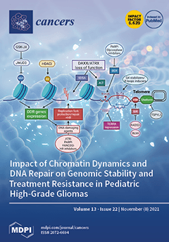
Cover Story (view full-size image): Recurrent mutations in the N-terminal tail of histones H3.3 and H3.1 act as key biological drivers of pediatric high-grade glioma (pHGG), leading to distinct epigenetic reprogramming, telomere maintenance mechanisms and oncogenesis scenarios, and defining pHGG subgroups. Notably, mutations in H3.3 are frequently associated with mutations affecting the H3.3 chaperone complex ATRX/DAXX. How the (epi)genetic alterations defining each pHGG subgroup provide novel therapeutic opportunities remains a challenge. Pinto et al. summarize the DNA repair pathways that drive resistance to current therapeutic strategies in pHGGs and review the functions of H3.3 and its chaperones in chromatin dynamics and DNA repair. They then discuss potential strategies targeting DNA repair and telomere maintenance mechanisms to improve the treatment of pHGG. View this paper
  • Issues are regarded as officially published after their release is announced to the table of contents alert mailing list.
  • You may sign up for e-mail alerts to receive table of contents of newly released issues.
  • PDF is the official format for papers published in both, html and pdf forms. To view the papers in pdf format, click on the "PDF Full-text" link, and use the free Adobe Reader to open them.
Order results
Result details
Section
Select all
Export citation of selected articles as:
19 pages, 9376 KiB  
Article
Identification of a Chemotherapeutic Lead Molecule for the Potential Disruption of the FAM72A-UNG2 Interaction to Interfere with Genome Stability, Centromere Formation, and Genome Editing
by Senthil Renganathan, Subrata Pramanik, Rajasekaran Ekambaram, Arne Kutzner, Pok-Son Kim and Klaus Heese
Cancers 2021, 13(22), 5870; https://doi.org/10.3390/cancers13225870 - 22 Nov 2021
Cited by 8 | Viewed by 2659
Abstract
Family with sequence similarity 72 A (FAM72A) is a pivotal mitosis-promoting factor that is highly expressed in various types of cancer. FAM72A interacts with the uracil-DNA glycosylase UNG2, the enzyme that prevents mutagenesis by eliminating uracil from DNA molecules through cleaving the N-glycosylic [...] Read more.
Family with sequence similarity 72 A (FAM72A) is a pivotal mitosis-promoting factor that is highly expressed in various types of cancer. FAM72A interacts with the uracil-DNA glycosylase UNG2, the enzyme that prevents mutagenesis by eliminating uracil from DNA molecules through cleaving the N-glycosylic bond and initiating the base excision repair pathway, thus maintaining genome integrity. In the present study, we determined a specific FAM72A-UNG2 heterodimer protein interaction using molecular docking and dynamics. In addition, through in silico screening, we identified withaferin B as a molecule that can specifically prevent the FAM72A-UNG2 interaction by blocking its cell signaling pathways. Our results provide an excellent basis for possible therapeutic approaches in the clinical treatment of cancer. Full article
(This article belongs to the Special Issue Targeting Signal Transduction Pathways in Cancer)
Show Figures

Figure 1

19 pages, 5795 KiB  
Article
Synergistic Anti-Tumor Effect of Simvastatin Combined to Chemotherapy in Osteosarcoma
by Adèle Mangelinck, Nadia Habel, Audrey Mohr, Nathalie Gaspar, Bojana Stefanovska and Olivia Fromigué
Cancers 2021, 13(22), 5869; https://doi.org/10.3390/cancers13225869 - 22 Nov 2021
Cited by 12 | Viewed by 2723
Abstract
Context: Osteosarcoma is the most common primary solid malignancy of the bone, mainly affecting pediatric patients. The main clinical issues are chemoresistance and metastatic spread, leading to a survival rate stagnating around 60% for four decades. Purpose: Here, we investigated the effect of [...] Read more.
Context: Osteosarcoma is the most common primary solid malignancy of the bone, mainly affecting pediatric patients. The main clinical issues are chemoresistance and metastatic spread, leading to a survival rate stagnating around 60% for four decades. Purpose: Here, we investigated the effect of simvastatin as adjuvant therapy on chemotherapy. Methods: Cell viability was assessed by the MTT test, and a combination index was evaluated by an isobologram approach. Cell motility was assessed by wound-healing assay. Cell-derived xenograft models were established in mice. FFPE tumor samples were assessed by immunohistochemistry. Results: In vitro experiments indicate that simvastatin synergized the conventional chemotherapy drugs’ inhibitory effect on cell viability. Functional assays reveal that simvastatin supplementation favored the anticancer mechanism of action of the tested chemotherapy drugs, such as DNA damage through intercalation or direct alkylation and disorganization of microtubules. Additionally, we show that even though simvastatin alone did not modify tumor behavior, it potentiated the inhibitory effect of doxorubicin on primary tumor growth (+50%, p < 0.05) and metastatic spread (+50%, p < 0.05). Our results provide evidence that simvastatin exerted an anti-tumor effect combined with chemotherapy in the preclinical murine model and represents valuable alternative adjuvant therapy that needs further investigation in clinical trials. Full article
Show Figures

Figure 1

5 pages, 181 KiB  
Editorial
Novel Treatment Strategies for Glioblastoma—A Summary
by Stanley S. Stylli
Cancers 2021, 13(22), 5868; https://doi.org/10.3390/cancers13225868 - 22 Nov 2021
Cited by 5 | Viewed by 1406
Abstract
Glioblastoma (GBM) is the most common primary central nervous system tumor in adults, accounting for approximately 80% of all brain-related malignancies [...] Full article
(This article belongs to the Special Issue Novel Treatment Strategies for Glioblastoma)
19 pages, 2972 KiB  
Review
Beyond the Usual Suspects: Hepatitis E Virus and Its Implications in Hepatocellular Carcinoma
by Mara Klöhn, Jil Alexandra Schrader, Yannick Brüggemann, Daniel Todt and Eike Steinmann
Cancers 2021, 13(22), 5867; https://doi.org/10.3390/cancers13225867 - 22 Nov 2021
Cited by 15 | Viewed by 2735
Abstract
Hepatitis E virus infections are the leading cause of viral hepatitis in humans, contributing to an estimated 3.3 million symptomatic cases and almost 44,000 deaths annually. Recently, HEV infections have been found to result in chronic liver infection and cirrhosis in severely immunocompromised [...] Read more.
Hepatitis E virus infections are the leading cause of viral hepatitis in humans, contributing to an estimated 3.3 million symptomatic cases and almost 44,000 deaths annually. Recently, HEV infections have been found to result in chronic liver infection and cirrhosis in severely immunocompromised patients, suggesting the possibility of HEV-induced hepatocarcinogenesis. While HEV-associated formation of HCC has rarely been reported, the expansion of HEV’s clinical spectrum and the increasing evidence of chronic HEV infections raise questions about the connection between HEV and HCC. The present review summarizes current clinical evidence of the relationship between HEV and HCC and discusses mechanisms of virus-induced HCC development with regard to HEV pathogenesis. We further elucidate why the development of HEV-induced hepatocellular carcinoma has so rarely been observed and provide an outlook on possible experimental set-ups to study the relationship between HEV and HCC formation. Full article
(This article belongs to the Special Issue Feature Paper from Journal Reviewers)
Show Figures

Figure 1

19 pages, 5094 KiB  
Article
Anti-Tumor Activity of Expanded PBMC-Derived NK Cells by Feeder-Free Protocol in Ovarian Cancer
by Minhua Chen, Yutong Li, Yu Wu, Siqi Xie, Jie Ma, Jingjing Yue, Rong Lv, Zhigang Tian, Fang Fang and Weihua Xiao
Cancers 2021, 13(22), 5866; https://doi.org/10.3390/cancers13225866 - 22 Nov 2021
Cited by 7 | Viewed by 3048
Abstract
Natural killer (NK) cells have shown great therapeutic potential against a wide range of cancers due to their pan-specific target recognition. Numerous reports indicate that NK cell immunotherapy is an effective therapeutic approach for treating hematological malignancies, but shows limited effects against solid [...] Read more.
Natural killer (NK) cells have shown great therapeutic potential against a wide range of cancers due to their pan-specific target recognition. Numerous reports indicate that NK cell immunotherapy is an effective therapeutic approach for treating hematological malignancies, but shows limited effects against solid tumors. In this study, several models of ovarian cancer (OC) were used to test the anti-cancer effects of NK cells derived from human peripheral blood mononuclear cells and expanded using a feeder cell-free expansion system (eNKs). The results show that eNKs exhibit potent inhibitory activity on tumor growth in different ovarian cancer xenograft mice (i.e., solid tumors, abdominal metastatic tumors, and ascites), importantly, in a dose-dependent manner. Moreover, adoptive transfer of eNKs resulted in significant reduction in ascites formation in OC peritoneal tumor models, and especially in reducing intraperitoneal ascites. We found that eNKs could migrate to the tumor site, retain their activity, and proliferate to maintain high cell counts in cutaneous xenograft mice. In addition, when increased the infusion with a high dose of 12 × 107 cells/mouse, Graft-versus-host disease could be induced by eNK. These data show that eNK cell immunotherapy could be a promising treatment strategy for ovarian cancers, including solid tumors and ascites. Full article
(This article belongs to the Topic Animal Model in Biomedical Research)
Show Figures

Figure 1

14 pages, 10446 KiB  
Review
Radiomics Models for Predicting Microvascular Invasion in Hepatocellular Carcinoma: A Systematic Review and Radiomics Quality Score Assessment
by Qiang Wang, Changfeng Li, Jiaxing Zhang, Xiaojun Hu, Yingfang Fan, Kuansheng Ma, Ernesto Sparrelid and Torkel B. Brismar
Cancers 2021, 13(22), 5864; https://doi.org/10.3390/cancers13225864 - 22 Nov 2021
Cited by 29 | Viewed by 3900
Abstract
Preoperative prediction of microvascular invasion (MVI) is of importance in hepatocellular carcinoma (HCC) patient treatment management. Plenty of radiomics models for MVI prediction have been proposed. This study aimed to elucidate the role of radiomics models in the prediction of MVI and to [...] Read more.
Preoperative prediction of microvascular invasion (MVI) is of importance in hepatocellular carcinoma (HCC) patient treatment management. Plenty of radiomics models for MVI prediction have been proposed. This study aimed to elucidate the role of radiomics models in the prediction of MVI and to evaluate their methodological quality. The methodological quality was assessed by the Radiomics Quality Score (RQS), and the risk of bias was evaluated by the Quality Assessment of Diagnostic Accuracy Studies (QUADAS-2). Twenty-two studies using CT, MRI, or PET/CT for MVI prediction were included. All were retrospective studies, and only two had an external validation cohort. The AUC values of the prediction models ranged from 0.69 to 0.94 in the test cohort. Substantial methodological heterogeneity existed, and the methodological quality was low, with an average RQS score of 10 (28% of the total). Most studies demonstrated a low or unclear risk of bias in the domains of QUADAS-2. In conclusion, a radiomics model could be an accurate and effective tool for MVI prediction in HCC patients, although the methodological quality has so far been insufficient. Future prospective studies with an external validation cohort in accordance with a standardized radiomics workflow are expected to supply a reliable model that translates into clinical utilization. Full article
(This article belongs to the Section Systematic Review or Meta-Analysis in Cancer Research)
Show Figures

Figure 1

19 pages, 3786 KiB  
Article
IGF1R/IR Mediates Resistance to BRAF and MEK Inhibitors in BRAF-Mutant Melanoma
by Hima Patel, Rosalin Mishra, Nour Yacoub, Samar Alanazi, Mary Kate Kilroy and Joan T. Garrett
Cancers 2021, 13(22), 5863; https://doi.org/10.3390/cancers13225863 - 22 Nov 2021
Cited by 18 | Viewed by 3254
Abstract
The use of BRAF and MEK inhibitors for patients with BRAF-mutant melanoma is limited as patients relapse on treatment as quickly as 6 months due to acquired resistance. We generated trametinib and dabrafenib resistant melanoma (TDR) cell lines to the MEK and BRAF [...] Read more.
The use of BRAF and MEK inhibitors for patients with BRAF-mutant melanoma is limited as patients relapse on treatment as quickly as 6 months due to acquired resistance. We generated trametinib and dabrafenib resistant melanoma (TDR) cell lines to the MEK and BRAF inhibitors, respectively. TDR cells exhibited increased viability and maintenance of downstream p-ERK and p-Akt as compared to parental cells. Receptor tyrosine kinase arrays revealed an increase in p-IGF1R and p-IR in the drug resistant cells versus drug sensitive cells. RNA-sequencing analysis identified IGF1R and INSR upregulated in resistant cell lines compared to parental cells. Analysis of TCGA PanCancer Atlas (skin cutaneous melanoma) showed that patients with a BRAF mutation and high levels of IGF1R and INSR had a worse overall survival. BMS-754807, an IGF1R/IR inhibitor, suppressed cell proliferation along with inhibition of intracellular p-Akt in TDR cells. Dual inhibition of IGF1R and INSR using siRNA reduced cell proliferation. The combination of dabrafenib, trametinib, and BMS-754807 treatment reduced in vivo xenograft tumor growth. Examining the role of IGF1R and IR in mediating resistance to BRAF and MEK inhibitors will expand possible treatment options to aid in long-term success for BRAF-mutant melanoma patients. Full article
(This article belongs to the Collection Drug Resistance and Novel Therapies in Cancers)
Show Figures

Figure 1

12 pages, 1017 KiB  
Article
Tylectomy Safety in Salvage of Eyes with Retinoblastoma
by Junyang Zhao, Qiyan Li, Zhao Xun Feng, Jianping Zhang, Songyi Wu, Liwen Jin and Brenda L. Gallie
Cancers 2021, 13(22), 5862; https://doi.org/10.3390/cancers13225862 - 22 Nov 2021
Cited by 7 | Viewed by 2395
Abstract
Intraocular surgery is tabooed in retinoblastoma management, due to the concern of lethal extraocular spread. We reviewed the outcomes of consecutive children with intraocular retinoblastoma diagnosed at 29 Chinese centers between 2012–2014. We compared the outcomes of three categories of treatment: eye salvage [...] Read more.
Intraocular surgery is tabooed in retinoblastoma management, due to the concern of lethal extraocular spread. We reviewed the outcomes of consecutive children with intraocular retinoblastoma diagnosed at 29 Chinese centers between 2012–2014. We compared the outcomes of three categories of treatment: eye salvage including tylectomy (Group I), eye salvage without tylectomy (Group II), and primary enucleation (Group III). A total of 960 patients (1243 eyes) were diagnosed: 256 in Group I, 370 in Group II, and 293 in Group III; 41 patients abandoned treatment upfront. The estimated 5-year overall survivals (OS) were, for Group I, 94%, for Group II 89%, and for Group III 95%. The estimated 5-year disease-specific survivals (DSS) were, for Group I, 96%, for Group II 90%, and for Group III 95%. Patients in Group I had a significantly higher 5-year DSS than patients in Group II (p = 0.003) and not significantly different than patients in Group III (p = 0.367). Overall survival was not compromised by the inclusion of tylectomy in eye salvage therapy compared to eye salvage without tylectomy or primary enucleation. Disease-specific survival was better when tylectomy was included in eye salvage treatments. Tylectomy as part of multimodal treatment may contribute to the care of retinoblastoma patients with chemotherapy-resistant tumor, eyes with concomitant ocular complications, or at the risk of treatment abandonment. Full article
(This article belongs to the Special Issue Detection and Treatment of Retinoblastoma)
Show Figures

Figure 1

18 pages, 2775 KiB  
Article
MAPK Inhibition Requires Active RAC1 Signaling to Effectively Improve Iodide Uptake by Thyroid Follicular Cells
by Márcia Faria, Rita Domingues, Maria João Bugalho, Paulo Matos and Ana Luísa Silva
Cancers 2021, 13(22), 5861; https://doi.org/10.3390/cancers13225861 - 22 Nov 2021
Cited by 3 | Viewed by 1915
Abstract
The Sodium/Iodide Symporter (NIS) is responsible for the active transport of iodide into thyroid follicular cells. Differentiated thyroid carcinomas (DTCs) usually preserve the functional expression of NIS, allowing the use of radioactive iodine (RAI) as the treatment of choice for metastatic disease. However, [...] Read more.
The Sodium/Iodide Symporter (NIS) is responsible for the active transport of iodide into thyroid follicular cells. Differentiated thyroid carcinomas (DTCs) usually preserve the functional expression of NIS, allowing the use of radioactive iodine (RAI) as the treatment of choice for metastatic disease. However, a significant proportion of patients with advanced forms of TC become refractory to RAI therapy and no effective therapeutic alternatives are available. Impaired iodide uptake is mainly caused by the defective functional expression of NIS, and this has been associated with several pathways linked to malignant transformation. MAPK signaling has emerged as one of the main pathways implicated in thyroid tumorigenesis, and its overactivation has been associated with the downregulation of NIS expression. Thus, several strategies have been developed to target the MAPK pathway attempting to increase iodide uptake in refractory DTC. However, MAPK inhibitors have had only partial success in restoring NIS expression and, in most cases, it remained insufficient to allow effective treatment with RAI. In a previous work, we have shown that the activity of the small GTPase RAC1 has a positive impact on TSH-induced NIS expression and iodide uptake in thyroid cells. RAC1 is a downstream effector of NRAS, but not of BRAF. Therefore, we hypothesized that the positive regulation induced by RAC1 on NIS could be a relevant signaling cue in the mechanism underlying the differential response to MEK inhibitors, observed between NRAS- and BRAF-mutant tumors. In the present study, we found that the recovery of NIS expression induced through MAPK pathway inhibition can be enhanced by potentiating RAC1 activity in thyroid cell systems. The negative impact on NIS expression induced by the MAPK-activating alterations, NRAS Q61R and BRAF V600E, was partially reversed by the presence of the MEK 1/2 inhibitors AZD6244 and CH5126766. Notably, the inhibition of RAC1 signaling partially blocked the positive impact of MEK inhibition on NIS expression in NRAS Q61R cells. Conversely, the presence of active RAC1 considerably improved the rescue of NIS expression in BRAF V600E thyroid cells treated with MEK inhibitors. Overall, our data support an important role for RAC1 signaling in enhancing MAPK inhibition in the context of RAI therapy in DTC, opening new opportunities for therapeutic intervention. Full article
(This article belongs to the Special Issue Role of Small GTPase Signaling in Tumorigenesis)
Show Figures

Figure 1

13 pages, 1648 KiB  
Review
Chromosomal Rearrangements and Altered Nuclear Organization: Recent Mechanistic Models in Cancer
by Concetta Federico, Francesca Bruno, Denise Ragusa, Craig S. Clements, Desiree Brancato, Marianne P. Henry, Joanna M. Bridger, Sabrina Tosi and Salvatore Saccone
Cancers 2021, 13(22), 5860; https://doi.org/10.3390/cancers13225860 - 22 Nov 2021
Cited by 7 | Viewed by 3481
Abstract
The last decade has seen significant progress in understanding how the genome is organized spatially within interphase nuclei. Recent analyses have confirmed earlier molecular cytogenetic studies on chromosome positioning within interphase nuclei and provided new information about the topologically associated domains (TADs). Examining [...] Read more.
The last decade has seen significant progress in understanding how the genome is organized spatially within interphase nuclei. Recent analyses have confirmed earlier molecular cytogenetic studies on chromosome positioning within interphase nuclei and provided new information about the topologically associated domains (TADs). Examining the nuances of how genomes are organized within interphase nuclei will provide information fundamental to understanding gene regulation and expression in health and disease. Indeed, the radial spatial positioning of individual gene loci within nuclei has been associated with up- and down-regulation of specific genes, and disruption of normal genome organization within nuclei will result in compromised cellular health. In cancer cells, where reorganization of the nuclear architecture may occur in the presence of chromosomal rearrangements such as translocations, inversions, or deletions, gene repositioning can change their expression. To date, very few studies have focused on radial gene positioning and the correlation to gene expression in cancers. Further investigations would improve our understanding of the biological mechanisms at the basis of cancer and, in particular, in leukemia initiation and progression, especially in those cases where the molecular consequences of chromosomal rearrangements are still unclear. In this review, we summarize the main milestones in the field of genome organization in the nucleus and the alterations to this organization that can lead to cancer diseases. Full article
(This article belongs to the Special Issue Feature Paper from Journal Reviewers)
Show Figures

Figure 1

13 pages, 277 KiB  
Review
Radiotherapy in the Treatment of Subcutaneous Melanoma
by Valentina Borzillo and Paolo Muto
Cancers 2021, 13(22), 5859; https://doi.org/10.3390/cancers13225859 - 22 Nov 2021
Cited by 5 | Viewed by 2090
Abstract
Malignant melanoma frequently develops cutaneous and/or subcutaneous metastases during the course of the disease. These may present as non-nodal locoregional metastases (microsatellite, satellite, or in-transit) included in stage III or as distant metastases in stage IV. Their presentation is heterogeneous and associated with [...] Read more.
Malignant melanoma frequently develops cutaneous and/or subcutaneous metastases during the course of the disease. These may present as non-nodal locoregional metastases (microsatellite, satellite, or in-transit) included in stage III or as distant metastases in stage IV. Their presentation is heterogeneous and associated with significant morbidity resulting from both disease-related functional damage and treatment side effects. The standard treatment is surgical excision, whereas local therapies or systemic therapies have a role when surgery is not indicated. Radiotherapy can be used in the local management of ITM, subcutaneous relapses, or distant metastases to provide symptom relief and prolong regional disease control. To increase the local response without increasing toxicity, the addition of hyperthermia and intralesional therapies to radiotherapy appear to be very promising. Boron neutron capture therapy, based on nuclear neutron capture and boron isotope fission reaction, could be an alternative to standard treatments, but its use in clinical practice is still limited. The potential benefit of combining radiotherapy with targeted therapies and immunotherapy has yet to be explored in this lesion setting. This review explores the role of radiotherapy in the treatment of cutaneous and subcutaneous lesions, its impact on outcomes, and its association with other treatment modalities. Full article
(This article belongs to the Special Issue Subcutaneous Melanoma)
27 pages, 4048 KiB  
Article
Evaluation of Hedgehog Pathway Inhibition on Nevoid Basal Cell Carcinoma Syndrome Fibroblasts and Basal Cell Carcinoma-Associated Fibroblasts: Are Vismodegib and Sonidegib Useful to Target Cancer-Prone Fibroblasts?
by Laura Eibenschutz, Silvia Caputo, Emanuela Camera, Anna Carbone, Vitaliano Silipo, Emilia Migliano, Caterina Aurizi, Carlo Cota, Pasquale Frascione and Barbara Bellei
Cancers 2021, 13(22), 5858; https://doi.org/10.3390/cancers13225858 - 22 Nov 2021
Cited by 3 | Viewed by 2246
Abstract
Activating mutations in the Hh pathway underlies the development of sporadic and familial skin BCC. For these oncogenic proliferations displaying ligand-independent activation of the intracellular pathway, two molecules have been approved for therapeutic purposes: vismodegib and sonidegib. Improper Hh signalling occurs in many [...] Read more.
Activating mutations in the Hh pathway underlies the development of sporadic and familial skin BCC. For these oncogenic proliferations displaying ligand-independent activation of the intracellular pathway, two molecules have been approved for therapeutic purposes: vismodegib and sonidegib. Improper Hh signalling occurs in many human tumours also via a paracrine mechanism (ligand-dependent) in which the secretion of Hh ligands by stromal cells support tumour growth. On the other hand, the mobilization of neoplastic stroma by cancer cells is sustained by the activation of Hh signalling in surrounding fibroblasts suggesting a central role of this bidirectional crosstalk in carcinogenesis. Additionally, loss-of-function mutations in the PTCH1 gene in the context of NBCCS, an autosomal dominant disorder predisposing to multiple BCCs, determine tumour permissive phenotypes in dermal fibroblasts. Here, profiling syndromic and BCC-associated fibroblasts unveiled an extraordinary similarity characterized by overexpression of several Hh target genes and a marked pro-inflammatory outline. Both cell types exposed to Hh inhibitors displayed reversion of the tumour-prone phenotype. Under vismodegib and sonidegib treatment, the Wnt/β-catenin pathway, frequently over-active in tumour stroma, resulted down-regulated by pAKT-GSK3β axis and consequent increase of β-catenin turnover. Overall, this study demonstrated that vismodegib and sonidegib impacting on fibroblast tumour supportive functions might be considered in therapy for BCC independently to the mutation status of Hh components in neoplastic cells. Full article
(This article belongs to the Special Issue Functional Genomics of Cancer)
Show Figures

Figure 1

18 pages, 2956 KiB  
Article
Human Breast Extracellular Matrix Microstructures and Protein Hydrogel 3D Cultures of Mammary Epithelial Cells
by Chandler R. Keller, Yang Hu, Kelsey F. Ruud, Anika E. VanDeen, Steve R. Martinez, Barry T. Kahn, Zhiwu Zhang, Roland K. Chen and Weimin Li
Cancers 2021, 13(22), 5857; https://doi.org/10.3390/cancers13225857 - 22 Nov 2021
Cited by 5 | Viewed by 2469
Abstract
Tissue extracellular matrix (ECM) is a structurally and compositionally unique microenvironment within which native cells can perform their natural biological activities. Cells grown on artificial substrata differ biologically and phenotypically from those grown within their native tissue microenvironment. Studies examining human tissue ECM [...] Read more.
Tissue extracellular matrix (ECM) is a structurally and compositionally unique microenvironment within which native cells can perform their natural biological activities. Cells grown on artificial substrata differ biologically and phenotypically from those grown within their native tissue microenvironment. Studies examining human tissue ECM structures and the biology of human tissue cells in their corresponding tissue ECM are lacking. Such investigations will improve our understanding about human pathophysiological conditions for better clinical care. We report here human normal breast tissue and invasive ductal carcinoma tissue ECM structural features. For the first time, a hydrogel was successfully fabricated using whole protein extracts of human normal breast ECM. Using immunofluorescence staining of type I collagen (Col I) and machine learning of its fibrous patterns in the polymerized human breast ECM hydrogel, we have defined the microstructural characteristics of the hydrogel and compared the microstructures with those of other native ECM hydrogels. Importantly, the ECM hydrogel supported 3D growth and cell-ECM interaction of both normal and cancerous mammary epithelial cells. This work represents further advancement toward full reconstitution of the human breast tissue microenvironment, an accomplishment that will accelerate the use of human pathophysiological tissue-derived matrices for individualized biomedical research and therapeutic development. Full article
(This article belongs to the Special Issue Organotypic 3D In Vitro Tumor Models: Bioengineering and Applications)
Show Figures

Figure 1

26 pages, 1568 KiB  
Review
Updates on Immunotherapy and Immune Landscape in Renal Clear Cell Carcinoma
by Myung-Chul Kim, Zeng Jin, Ryan Kolb, Nicholas Borcherding, Jonathan Alexander Chatzkel, Sara Moscovita Falzarano and Weizhou Zhang
Cancers 2021, 13(22), 5856; https://doi.org/10.3390/cancers13225856 - 22 Nov 2021
Cited by 41 | Viewed by 4165
Abstract
Several clinicopathological features of clear cell renal cell carcinomas (ccRCC) contribute to make an “atypical” cancer, including resistance to chemotherapy, sensitivity to anti-angiogenesis therapy and ICIs despite a low mutational burden, and CD8+ T cell infiltration being the predictor for poor prognosis–normally [...] Read more.
Several clinicopathological features of clear cell renal cell carcinomas (ccRCC) contribute to make an “atypical” cancer, including resistance to chemotherapy, sensitivity to anti-angiogenesis therapy and ICIs despite a low mutational burden, and CD8+ T cell infiltration being the predictor for poor prognosis–normally CD8+ T cell infiltration is a good prognostic factor in cancer patients. These “atypical” features have brought researchers to investigate the molecular and immunological mechanisms that lead to the increased T cell infiltrates despite relatively low molecular burdens, as well as to decipher the immune landscape that leads to better response to ICIs. In the present study, we summarize the past and ongoing pivotal clinical trials of immunotherapies for ccRCC, emphasizing the potential molecular and cellular mechanisms that lead to the success or failure of ICI therapy. Single-cell analysis of ccRCC has provided a more thorough and detailed understanding of the tumor immune microenvironment and has facilitated the discovery of molecular biomarkers from the tumor-infiltrating immune cells. We herein will focus on the discussion of some major immune cells, including T cells and tumor-associated macrophages (TAM) in ccRCC. We will further provide some perspectives of using molecular and cellular biomarkers derived from these immune cell types to potentially improve the response rate to ICIs in ccRCC patients. Full article
(This article belongs to the Special Issue Clear Cell Renal Cell Carcinoma 20212022)
Show Figures

Figure 1

24 pages, 6181 KiB  
Article
The Release of Inflammatory Mediators from Acid-Stimulated Mesenchymal Stromal Cells Favours Tumour Invasiveness and Metastasis in Osteosarcoma
by Sofia Avnet, Silvia Lemma, Margherita Cortini, Gemma Di Pompo, Francesca Perut, Maria Veronica Lipreri, Laura Roncuzzi, Marta Columbaro, Costantino Errani, Alessandra Longhi, Nicoletta Zini, Dominique Heymann, Massimo Dominici, Giulia Grisendi, Giulia Golinelli, Lorena Consolino, Dario Livio Longo, Cristina Nanni, Alberto Righi and Nicola Baldini
Cancers 2021, 13(22), 5855; https://doi.org/10.3390/cancers13225855 - 22 Nov 2021
Cited by 14 | Viewed by 2969
Abstract
Osteosarcoma is the most frequent primary malignant bone tumour with an impressive tendency to metastasise. Highly proliferative tumour cells release a remarkable amount of protons into the extracellular space that activates the NF-kB inflammatory pathway in adjacent stromal cells. In this study, we [...] Read more.
Osteosarcoma is the most frequent primary malignant bone tumour with an impressive tendency to metastasise. Highly proliferative tumour cells release a remarkable amount of protons into the extracellular space that activates the NF-kB inflammatory pathway in adjacent stromal cells. In this study, we further validated the correlation between tumour glycolysis/acidosis and its role in metastases. In patients, at diagnosis, we found high circulating levels of inflammatory mediators (IL6, IL8 and miR-136-5p-containing extracellular vesicles). IL6 serum levels significantly correlated with disease-free survival and 18F-FDG PET/CT uptake, an indirect measurement of tumour glycolysis and, hence, of acidosis. In vivo subcutaneous and orthotopic models, co-injected with mesenchymal stromal (MSC) and osteosarcoma cells, formed an acidic tumour microenvironment (mean pH 6.86, as assessed by in vivo MRI-CEST pH imaging). In these xenografts, we enlightened the expression of both IL6 and the NF-kB complex subunit in stromal cells infiltrating the tumour acidic area. The co-injection with MSC also significantly increased lung metastases. Finally, by using 3D microfluidic models, we directly showed the promotion of osteosarcoma invasiveness by acidosis via IL6 and MSC. In conclusion, osteosarcoma-associated MSC react to intratumoural acidosis by triggering an inflammatory response that, in turn, promotes tumour invasiveness at the primary site toward metastasis development. Full article
(This article belongs to the Special Issue Tumor Acidosis)
Show Figures

Figure 1

14 pages, 27722 KiB  
Article
Identification of a Distinct miRNA Regulatory Network in the Tumor Microenvironment of Transformed Mycosis Fungoides
by Cosimo Di Raimondo, Zhen Han, Chingyu Su, Xiwei Wu, Hanjun Qin, James F. Sanchez, Yate-Ching Yuan, Xochiquetzal Martinez, Farah Abdulla, Jasmine Zain, Chun-Wei Chen, Steven T. Rosen and Christiane Querfeld
Cancers 2021, 13(22), 5854; https://doi.org/10.3390/cancers13225854 - 22 Nov 2021
Cited by 8 | Viewed by 1920
Abstract
Large cell transformation of mycosis fungoides (LCT-MF) occurs in 20–50% of advanced MF and is generally associated with poor response and dismal prognosis. Although different mechanisms have been proposed to explain the pathogenesis, little is known about the role of microRNAs (miRs) in [...] Read more.
Large cell transformation of mycosis fungoides (LCT-MF) occurs in 20–50% of advanced MF and is generally associated with poor response and dismal prognosis. Although different mechanisms have been proposed to explain the pathogenesis, little is known about the role of microRNAs (miRs) in transcriptional regulation of LCT-MF. Here, we investigated the miR and mRNA expression profile in lesional skin samples of patients with LCT-MF and non-LCT MF using RNA-seq analysis. We found miR-146a and miR-21 to be significantly upregulated, and miR-708 the most significantly downregulated miR in LCT-MF. Integration of miR and mRNA expression profiles revealed the miR-regulated networks in LCT-MF. Ingenuity pathway analysis (IPA) demonstrated the involvement of genes for ICOS-ICOSL, PD1-PDL1, NF-κB, E2F transcription, and molecular mechanisms of cancer signaling pathways. Quantitative real time (qRT)-PCR results of target genes were consistent with the RNA-seq data. We further identified the immunosuppressive tumor microenvironment (TME) in LCT-MF. Moreover, our data indicated that miR-146a, -21 and -708 are associated with the immunosuppressive TME in LCT-MF. Collectively, our results suggest that the key LCT-MF associated miRs and their regulated networks may provide insights into its pathogenesis and identify promising targets for novel therapeutic strategies. Full article
Show Figures

Figure 1

22 pages, 5921 KiB  
Review
An Overview of Selected Rare B-Cell Lymphoproliferative Disorders: Imaging, Histopathologic, and Clinical Features
by Ahmed Ebada Salem, Yehia H. Zaki, Gamal El-Hussieny, Khaled I. ElNoueam, Akram M. Shaaban, Bhasker Rao Koppula, Mark Bustoros, Mohamed Salama, Khaled M. Elsayes, Kathryn Morton and Matthew F. Covington
Cancers 2021, 13(22), 5853; https://doi.org/10.3390/cancers13225853 - 22 Nov 2021
Cited by 3 | Viewed by 3063
Abstract
Lymphoproliferative disorders (LPD) are conditions characterized by the uncontrolled proliferation of B or T-cell lines. They encompass a wide spectrum of abnormalities, which may be broadly classified as reactive processes or malignant diseases, such as lymphoma, based on their cellular clonality and clinical [...] Read more.
Lymphoproliferative disorders (LPD) are conditions characterized by the uncontrolled proliferation of B or T-cell lines. They encompass a wide spectrum of abnormalities, which may be broadly classified as reactive processes or malignant diseases, such as lymphoma, based on their cellular clonality and clinical behavior. While some of these disorders are rare, they may be encountered sporadically in clinical practice, causing diagnostic dilemmas owing to overlap in their clinical and imaging features with more common disorders. The updated 4th edition WHO classification of lymphoid neoplasms was released in 2016 to incorporate the rapid clinical, pathological, molecular biology and cytogenetic advances of some of these disorders. Despite these updates, very little information is presented in the literature from the radiology perspective. The aim of this article is to familiarize radiologists and other physicians with certain rare variants of B-cell lymphoproliferative disorders with a focus on imaging features of these disorders, as well as to provide an overview of some important updates contained within the new WHO classification of lymphoid neoplasms. Full article
(This article belongs to the Special Issue Cancer Imaging: Current Practice and Future Perspectives)
Show Figures

Figure 1

18 pages, 3175 KiB  
Article
The Clinical Utility of the Geriatric Nutritional Risk Index in Predicting Postoperative Complications and Long-Term Survival in Elderly Patients with Colorectal Cancer after Curative Surgery
by Chun-Kai Liao, Yih-Jong Chern, Yu-Jen Hsu, Yueh-Chen Lin, Yen-Lin Yu, Jy-Ming Chiang, Chien-Yuh Yeh and Jeng-Fu You
Cancers 2021, 13(22), 5852; https://doi.org/10.3390/cancers13225852 - 22 Nov 2021
Cited by 18 | Viewed by 1969
Abstract
Research on the relationship between the geriatric nutritional risk index (GNRI) and postoperative complications/oncological outcomes in elderly colorectal cancer (CRC) patients is limited. This study investigated the prognostic value of the GNRI in aged CRC patients. We retrospectively analyzed 1206 consecutive CRC patients [...] Read more.
Research on the relationship between the geriatric nutritional risk index (GNRI) and postoperative complications/oncological outcomes in elderly colorectal cancer (CRC) patients is limited. This study investigated the prognostic value of the GNRI in aged CRC patients. We retrospectively analyzed 1206 consecutive CRC patients aged over 75 years who underwent curative-intent surgery from January 2008 to December 2015 and categorized them into high GNRI (≥98) and low GNRI (<98) groups according to a receiver operating characteristic (ROC) curve analysis. Uni- and multivariate logistic regression analysis were used to explore the association of the GNRI with postoperative complications. Kaplan–Meier survival analyses and the Cox proportional hazard model were used to explore the association between GNRI and survival. We discovered that GNRI is an independent risk factor for postoperative complications (HR: 1.774, p = 0.037). Surgical site infection, wound dehiscence and pneumonia were more common in patients with GNRI < 98. Survival analysis showed significantly worse overall survival and disease-free survival in the low GNRI group (both p < 0.001). In the multivariate analysis, GNRI < 98 was an independent risk factor for OS (HR: 1.329, p = 0.031) and DFS (HR: 1.312, p = 0.034). Thus, preoperative GNRI can be effectively used to predict postoperative complications and long-term survival in elderly CRC patients after curative surgery. Full article
Show Figures

Figure 1

10 pages, 983 KiB  
Article
Can the Six-Minute Walk Test Be Used to Individualize Physical Activity Intensity in Patients with Breast Cancer?
by Nicole Tubiana-Mathieu, Thibault Cornette, Stephane Mandigout, Sophie Leobon, François Vincent, Laurence Venat and Elise Deluche
Cancers 2021, 13(22), 5851; https://doi.org/10.3390/cancers13225851 - 22 Nov 2021
Cited by 3 | Viewed by 2112
Abstract
Background: Adapted physical activity (APA) aids breast cancer patients. It is necessary to use an adapted target heart rate (HR) when prescribing exercise intensity. Methods: In total, 138 patients previously included in two published randomized clinical trials underwent the CPET and 6MWT before [...] Read more.
Background: Adapted physical activity (APA) aids breast cancer patients. It is necessary to use an adapted target heart rate (HR) when prescribing exercise intensity. Methods: In total, 138 patients previously included in two published randomized clinical trials underwent the CPET and 6MWT before and after adjuvant therapy. Of these patients, 85 had performed APA, and 53 had received only the usual therapy. HRs were recorded during the two tests. Results: Before starting chemotherapy, good agreement (intraclass correlation (ICC) 0.69; confidence interval at 95% IC0.95 (0.591–0.769); p < 0.001) and a moderate correlation were evident between the 6MWT-HR and ventilatory threshold HR of the CPET (r = 0.70; p < 0.001). Good agreement and a high positive correlation were noted only in the group who engaged in APA (ICC 0.77; IC0.95 (0.659–0.848); p < 0.001; r = 0.8; p < 0.01); moderate agreement and a moderate positive correlation were apparent in the control group (ICC 0.57; IC0.95 (0.329–0.74); p < 0.001; r = 0.6; p < 0.01). The correlations were independent of age and body mass index. Conclusions: The 6MWT-HR can be used to prescribe exercise intensity for breast cancer patients both before and after specific treatment with concomitant APA. Full article
(This article belongs to the Special Issue Physical Activity and Cancer Care)
Show Figures

Figure 1

15 pages, 2557 KiB  
Article
miR-1227 Targets SEC23A to Regulate the Shedding of Large Extracellular Vesicles
by Andrew Chin, Javier Mariscal, Minhyung Kim, Giorgia Guerra, Blandine Victor, Chen Qian, Elisabetta Broseghini, Edwin Posadas, Michael R. Freeman, Shivani Sharma, Paolo Gandellini, Nadia Zaffaroni, Sungyong You, Keith Syson Chan, Jlenia Guarnerio, Muller Fabbri and Dolores Di Vizio
Cancers 2021, 13(22), 5850; https://doi.org/10.3390/cancers13225850 - 22 Nov 2021
Cited by 3 | Viewed by 2399
Abstract
Cancer cells shed a heterogenous mixture of extracellular vesicles (EVs), differing in both size and composition, which likely influence physiological processes in different manners. However, how cells differentially control the shedding of these EV populations is poorly understood. Here, we show that miR-1227, [...] Read more.
Cancer cells shed a heterogenous mixture of extracellular vesicles (EVs), differing in both size and composition, which likely influence physiological processes in different manners. However, how cells differentially control the shedding of these EV populations is poorly understood. Here, we show that miR-1227, which is enriched in prostate cancer EVs, compared to the cell of origin, but not in EVs derived from prostate benign epithelial cells, induces the shedding of large EVs (such as large oncosomes), while inhibiting the shedding of small EVs (such as exosomes). RNA sequencing from cells stably expressing miR-1227, a modified RISCTRAP assay that stabilizes and purifies mRNA-miR-1227 complexes for RNA sequencing, and in silico target prediction tools were used to identify miR-1227 targets that may mediate this alteration in EV shedding. The COPII vesicle protein SEC23A emerged and was validated by qPCR, WBlot, and luciferase assays as a direct target of miR-1227. The inhibition of SEC23A was sufficient to induce the shedding of large EVs. These results identify a novel mechanism of EV shedding, by which the inhibition of SEC23A by miR-1227 induces a shift in EV shedding, favoring the shedding of large EV over small EV. Full article
(This article belongs to the Special Issue Molecular Pharmacology of Anti-Cancer Drugs)
Show Figures

Figure 1

21 pages, 5668 KiB  
Article
AKF-D52, a Synthetic Phenoxypyrimidine-Urea Derivative, Triggers Extrinsic/Intrinsic Apoptosis and Cytoprotective Autophagy in Human Non-Small Cell Lung Cancer Cells
by Hyo-Sun Gil, Jeong-Hun Lee, Ahmed K. Farag, Ahmed H. E. Hassan, Kyung-Sook Chung, Jung-Hye Choi, Eun-Joo Roh and Kyung-Tae Lee
Cancers 2021, 13(22), 5849; https://doi.org/10.3390/cancers13225849 - 22 Nov 2021
Cited by 7 | Viewed by 2208
Abstract
Previously, we discovered that 1-(3,5-dimethoxyphenyl)-3-(4-(3-methoxyphenoxy)-2-((4-morpholinophenyl)amino)pyrimidin-5-yl)urea (AKF-D52), a synthetic phenoxypyrimidine urea derivative, acts as a growth inhibitor of various cancer cell types. In this study, we elucidated the antiproliferative properties of AFK-D52 and underlying mechanisms in non-small cell lung cancer (NSCLC) cells and an [...] Read more.
Previously, we discovered that 1-(3,5-dimethoxyphenyl)-3-(4-(3-methoxyphenoxy)-2-((4-morpholinophenyl)amino)pyrimidin-5-yl)urea (AKF-D52), a synthetic phenoxypyrimidine urea derivative, acts as a growth inhibitor of various cancer cell types. In this study, we elucidated the antiproliferative properties of AFK-D52 and underlying mechanisms in non-small cell lung cancer (NSCLC) cells and an A549 xenograft animal model. AKF-D52 was found to induce both caspase-dependent and -independent apoptotic cell death. Furthermore, the mitochondrial component of the AKF-D52-induced apoptosis mechanism involves a reduction in mitochondrial membrane potential and regulation in B cell lymphoma-2 family protein expression. Moreover, AKF-D52 activates the extrinsic pathway through up-regulated expression of death receptor 3 and Fas and then the formation of a death-inducing signaling complex. AKF-D52 also induced autophagy by increasing acidic vesicular organelle formation and microtubule-associated protein 1A/1B-light chain 3-II levels and reducing p62 levels. Notably, pretreatment with autophagy inhibitors enhanced AKF-D52-induced cell death, indicating that the induced autophagy is cytoprotective. AKF-D52 treatment also triggered reactive oxygen species (ROS) production in NSCLC cells, whereas the antioxidant α-tocopherol abolished AKF-D52-induced cell death. In a xenograft lung cancer mouse model, AKF-D52 administration attenuated tumor growth by inducing apoptosis and autophagy in tumor tissues. Collectively, our data indicate that AKF-D52-induced ROS production plays a role in mediating apoptosis and cytoprotective autophagy in NSCLC. Full article
(This article belongs to the Special Issue Role of Oxidative Stress in Cancer)
Show Figures

Figure 1

19 pages, 919 KiB  
Review
Risk Stratification of Endometrial Cancer Patients: FIGO Stage, Biomarkers and Molecular Classification
by Jenneke C. Kasius, Johanna M. A. Pijnenborg, Kristina Lindemann, David Forsse, Judith van Zwol, Gunnar B. Kristensen, Camilla Krakstad, Henrica M. J. Werner and Frédéric Amant
Cancers 2021, 13(22), 5848; https://doi.org/10.3390/cancers13225848 - 22 Nov 2021
Cited by 39 | Viewed by 12239
Abstract
Endometrial cancer (EC) is the most common gynaecologic malignancy in developed countries. The main challenge in EC management is to correctly estimate the risk of metastases at diagnosis and the risk to develop recurrences in the future. Risk stratification determines the need for [...] Read more.
Endometrial cancer (EC) is the most common gynaecologic malignancy in developed countries. The main challenge in EC management is to correctly estimate the risk of metastases at diagnosis and the risk to develop recurrences in the future. Risk stratification determines the need for surgical staging and adjuvant treatment. Detection of occult, microscopic metastases upstages patients, provides important prognostic information and guides adjuvant treatment. The molecular classification subdivides EC into four prognostic subgroups: POLE ultramutated; mismatch repair deficient (MMRd); nonspecific molecular profile (NSMP); and TP53 mutated (p53abn). How surgical staging should be adjusted based on preoperative molecular profiling is currently unknown. Moreover, little is known whether and how other known prognostic biomarkers affect prognosis prediction independent of or in addition to these molecular subgroups. This review summarizes the factors incorporated in surgical staging (i.e., peritoneal washing, lymph node dissection, omentectomy and peritoneal biopsies), and its impact on prognosis and adjuvant treatment decisions in an era of molecular classification of EC. Moreover, the relation between FIGO stage and molecular classification is evaluated including the current gaps in knowledge and future perspectives. Full article
(This article belongs to the Special Issue Diagnosis and Management of Endometrial Cancer)
Show Figures

Figure 1

31 pages, 1718 KiB  
Review
Melanoma Targeted Therapies beyond BRAF-Mutant Melanoma: Potential Druggable Mutations and Novel Treatment Approaches
by Karam Khaddour, Lucas Maahs, Ana Maria Avila-Rodriguez, Yazan Maamar, Sami Samaan and George Ansstas
Cancers 2021, 13(22), 5847; https://doi.org/10.3390/cancers13225847 - 22 Nov 2021
Cited by 14 | Viewed by 3907
Abstract
Melanomas exhibit the highest rate of somatic mutations among all different types of cancers (with the exception of BCC and SCC). The accumulation of a multimode of mutations in the driver oncogenes are responsible for the proliferative, invasive, and aggressive nature of melanomas. [...] Read more.
Melanomas exhibit the highest rate of somatic mutations among all different types of cancers (with the exception of BCC and SCC). The accumulation of a multimode of mutations in the driver oncogenes are responsible for the proliferative, invasive, and aggressive nature of melanomas. High-resolution and high-throughput technology has led to the identification of distinct mutational signatures and their downstream alterations in several key pathways that contribute to melanomagenesis. This has enabled the development of individualized treatments by targeting specific molecular alterations that are vital for cancer cell survival, which has resulted in improved outcomes in several cancers, including melanomas. To date, BRAF and MEK inhibitors remain the only approved targeted therapy with a high level of evidence in BRAFV600E/K mutant melanomas. The lack of approved precision drugs in melanomas, relative to other cancers, despite harboring one of the highest rates of somatic mutations, advocates for further research to unveil effective therapeutics. In this review, we will discuss potential druggable mutations and the ongoing research of novel individualized treatment approaches targeting non-BRAF mutations in melanomas. Full article
(This article belongs to the Collection Targeted Therapies and Immunotherapies in Metastatic Melanoma)
Show Figures

Figure 1

10 pages, 1810 KiB  
Article
Mutation in Genes Encoding Key Functional Groups Additively Increase Mortality in Patients with BRAFV600E-Mutant Advanced Papillary Thyroid Carcinoma
by Eyun Song, Meihua Jin, Ahreum Jang, Min Ji Jeon, Dong Eun Song, Hye Jin Yoo, Won Bae Kim, Young Kee Shong and Won Gu Kim
Cancers 2021, 13(22), 5846; https://doi.org/10.3390/cancers13225846 - 22 Nov 2021
Cited by 8 | Viewed by 1703
Abstract
The prognosis of BRAFV600E-mutant papillary thyroid carcinoma (PTC) ranges from indolent to highly aggressive courses. To better define the genetic diversity of this subtype, we evaluated the survival according to the presence of an additional mutation in genes encoding functional groups [...] Read more.
The prognosis of BRAFV600E-mutant papillary thyroid carcinoma (PTC) ranges from indolent to highly aggressive courses. To better define the genetic diversity of this subtype, we evaluated the survival according to the presence of an additional mutation in genes encoding functional groups (FGs) in BRAFV600E-mutant advanced PTC patients. Targeted next-generation sequencing was performed in primary tumors of 50 BRAFV600E-mutant PTCs with distant metastasis or aggressive variants. The mutation in genes encoding FGs included alterations in histone methyltransferases, SWI/SNF subunit, and the PI3K/AKT/mTOR pathway. The primary outcome was overall survival (OS). Fifteen patients only had the BRAFV600E-mutation (group 1), 22 had BRAFV600E and mutation other than FGs (group 2), and 13 had BRAFV600E and FG mutation (group 3). OS was significantly lower in patients with FG mutations (p = 0.001) than those without, and group 3 patients had the worst survival (p = 0.004). OS significantly varied among none, one, or two FG mutation sites (p = 0.005). Presence of FG mutation was independently associated with increased mortality (hazard ratio 11.65, 95% confidence interval 1.39–97.58, p = 0.024). Coexistence of mutations in BRAFV600E and genes encoding FGs was associated with high mortality. Identification of FG mutation in BRAFV600E-mutant PTCs may be valuable in risk stratifying this subtype. Full article
Show Figures

Figure 1

19 pages, 5873 KiB  
Article
1H-NMR Plasma Lipoproteins Profile Analysis Reveals Lipid Metabolism Alterations in HER2-Positive Breast Cancer Patients
by Giuseppe Corona, Emanuela Di Gregorio, Alessia Vignoli, Elena Muraro, Agostino Steffan and Gianmaria Miolo
Cancers 2021, 13(22), 5845; https://doi.org/10.3390/cancers13225845 - 21 Nov 2021
Cited by 6 | Viewed by 2356
Abstract
The lipid tumour demand may shape the host metabolism adapting the circulating lipids composition to its growth and progression needs. This study aims to exploit the straightforward 1H-NMR lipoproteins analysis to investigate the alterations of the circulating lipoproteins’ fractions in HER2-positive breast [...] Read more.
The lipid tumour demand may shape the host metabolism adapting the circulating lipids composition to its growth and progression needs. This study aims to exploit the straightforward 1H-NMR lipoproteins analysis to investigate the alterations of the circulating lipoproteins’ fractions in HER2-positive breast cancer and their modulations induced by treatments. The baseline 1H-NMR plasma lipoproteins profiles were measured in 43 HER2-positive breast cancer patients and compared with those of 28 healthy women. In a subset of 32 patients, longitudinal measurements were also performed along neoadjuvant chemotherapy, after surgery, adjuvant treatment, and during the two-year follow-up. Differences between groups were assessed by multivariate PLS-DA and by univariate analyses. The diagnostic power of lipoproteins subfractions was assessed by ROC curve, while lipoproteins time changes along interventions were investigated by ANOVA analysis. The PLS-DA model distinguished HER2-positive breast cancer patients from the control group with a sensitivity of 96.4% and specificity of 90.7%, mainly due to the differential levels of VLDLs subfractions that were significantly higher in the patients’ group. Neoadjuvant chemotherapy-induced a significant drop in the HDLs after the first three months of treatment and a specific decrease in the HDL-3 and HDL-4 subfractions were found significantly associated with the pathological complete response achievement. These results indicate that HER2-positive breast cancer is characterized by a significant host lipid mobilization that could be useful for diagnostic purposes. Moreover, the lipoproteins profiles alterations induced by the therapeutic interventions could predict the clinical outcome supporting the application of 1H-NMR lipoproteins profiles analysis for longitudinal monitoring of HER2-positive breast cancer in large clinical studies. Full article
Show Figures

Graphical abstract

20 pages, 772 KiB  
Review
Hepatocellular Carcinoma in Non-Alcoholic Fatty Liver Disease: From Epidemiology to Diagnostic Approach
by Ivica Grgurevic, Tonci Bozin, Mislav Mikus, Michal Kukla and James O’Beirne
Cancers 2021, 13(22), 5844; https://doi.org/10.3390/cancers13225844 - 21 Nov 2021
Cited by 26 | Viewed by 3575
Abstract
Non-alcoholic fatty liver disease (NAFLD) is becoming the leading cause of liver morbidity worldwide and, as such, represents the pathogenic background for the increasing incidence of hepatocellular carcinoma (HCC). The annual incidence of NAFLD-related HCC is expected to increase by 45–130% by 2030. [...] Read more.
Non-alcoholic fatty liver disease (NAFLD) is becoming the leading cause of liver morbidity worldwide and, as such, represents the pathogenic background for the increasing incidence of hepatocellular carcinoma (HCC). The annual incidence of NAFLD-related HCC is expected to increase by 45–130% by 2030. Diabetes mellitus is the most important risk factor for HCC development in NAFLD, with the risk further increased when associated with other metabolic traits, such as obesity, arterial hypertension and dyslipidemia. The highest risk of HCC exists in patients with advanced fibrosis or cirrhosis, although 20–50% of HCC cases arise in NAFLD patients with an absence of cirrhosis. This calls for further investigation of the pathogenic mechanisms that are involved in hepatocarcinogenesis, including genetics, metabolomics, the influence of the gut microbiota and immunological responses. Early identification of patients with or at risk of NAFLD is of utmost importance to improve outcomes. As NAFLD is highly prevalent in the community, the identification of cases should rely upon simple demographic and clinical characteristics. Once identified, these patients should then be evaluated for the presence of advanced fibrosis or cirrhosis and subsequently enter HCC surveillance programs if appropriate. A significant problem is the early recognition of non-cirrhotic NAFLD patients who will develop HCC, where new biomarkers and scores are potential solutions to tackle this issue. Full article
(This article belongs to the Special Issue Nonalcoholic Fatty Liver Disease and Hepatocellular Carcinoma)
Show Figures

Figure 1

12 pages, 1545 KiB  
Article
Surveillance for Patients with Oral Squamous Cell Carcinoma after Complete Surgical Resection as Primary Treatment: A Single-Center Retrospective Cohort Study
by Chonji Fukumoto, Ryo Oshima, Yuta Sawatani, Ryo Shiraishi, Toshiki Hyodo, Ryouta Kamimura, Tomonori Hasegawa, Yuske Komiyama, Sayaka Izumi, Atsushi Fujita, Takahiro Wakui and Hitoshi Kawamata
Cancers 2021, 13(22), 5843; https://doi.org/10.3390/cancers13225843 - 21 Nov 2021
Cited by 8 | Viewed by 1861
Abstract
Background: The surveillance methods oral squamous cell carcinoma (OSCC) patients may be chosen by considering the risk for recurrence, and it is important to establish appropriate methods during the period in which latent/dormant cancer cells become more apparent. To investigate the appropriate surveillance [...] Read more.
Background: The surveillance methods oral squamous cell carcinoma (OSCC) patients may be chosen by considering the risk for recurrence, and it is important to establish appropriate methods during the period in which latent/dormant cancer cells become more apparent. To investigate the appropriate surveillance of patients with OSCC based on the individual risk for recurrence and/or metastasis, we performed a retrospective cohort study after the complete surgical resection of OSCC as the primary treatment. Methods: The study was performed in 324 patients with OSCC who had been primarily treated with surgery from 2007 to 2020 at our hospital. We investigated the period, timing, and methods (visual examination, palpation and imaging using FDG-PET/CT or CECT) for surveillance in each case that comprised postsurgical treatment. Results: Regarding the time to occurrence of postsurgical events, we found that half of cases of local recurrence, cervical lymph node metastasis, and distant metastasis occurred within 200 days, and 75% of all of these events occurred within 400 days. However, the mean time for second primary cancer was 1589 days. The postsurgical events were detected earlier by imaging examinations than they were by visual examination and palpation. Conclusions: For the surveillance of patients with OSCC after primary surgery, it is desirable to perform FDG-PET/CT within 3–6 months and at 1 year after surgery and to consider CECT as an option in between FDG-PET/CT, while continuing history and physical examinations for about 5 years based on individual risk assessment. Full article
(This article belongs to the Special Issue Advanced Squamous Cell Carcinoma)
Show Figures

Figure 1

11 pages, 615 KiB  
Article
Metastatic Lymph Node Ratio for Predicting Recurrence in Medullary Thyroid Cancer
by Jinyoung Kim, Jun Park, Hyunju Park, Min Sun Choi, Hye Won Jang, Tae Hyuk Kim, Sun Wook Kim and Jae Hoon Chung
Cancers 2021, 13(22), 5842; https://doi.org/10.3390/cancers13225842 - 21 Nov 2021
Cited by 10 | Viewed by 2690
Abstract
The lymph node ratio (LNR) has been investigated as a prognostic factor in many different types of cancers, including differentiated thyroid cancer; however, reports regarding medullary thyroid cancer (MTC) are limited. Therefore, this study aims to evaluate LNR as a risk factor for [...] Read more.
The lymph node ratio (LNR) has been investigated as a prognostic factor in many different types of cancers, including differentiated thyroid cancer; however, reports regarding medullary thyroid cancer (MTC) are limited. Therefore, this study aims to evaluate LNR as a risk factor for structural recurrence in patients with MTC. Medical records of patients treated for MTC in a single tertiary center between 1995 and 2017 were retrospectively reviewed. LNR is defined as the number of metastatic lymph nodes or lymph node metastases (LNM) divided by the number of retrieved lymph nodes or lymph node yield (LNY). In the survival analysis, recurrence-free survival was defined as the time from the date of total thyroidectomy to recurrence or last follow-up. To identify risk factors influencing structural recurrence, univariable and multivariable Cox proportional hazard models were used. A total of 132 patients were enrolled. The mean age of study participants was 49.7 years, and 86 patients (65%) were women. Structural recurrence was identified in 39 patients at the end of the study period, and the median follow-up period was 8.7 years. In univariable analyses, gross extra thyroidal extension, N stage, postoperative serum calcitonin and carcinoembryonic antigen (CEA) levels, and LNR were significant (p < 0.05) predictors of structural recurrence. In multivariable analysis, postoperative serum calcitonin, postoperative serum CEA, and LNR were identified as a predictor of disease-free survival (p < 0.05). LNR can potentially predict structural recurrence as a quantitative evaluation tool for lymph node metastasis in patients with MTC. Full article
Show Figures

Figure 1

10 pages, 601 KiB  
Article
Soluble PD-L1 in Serum and Urine in Urinary Bladder Cancer Patients
by Anders Vikerfors, Sabina Davidsson, Janusz Frey, Tomas Jerlström and Jessica Carlsson
Cancers 2021, 13(22), 5841; https://doi.org/10.3390/cancers13225841 - 21 Nov 2021
Cited by 13 | Viewed by 1727
Abstract
Soluble PD-L1 (sPD-L1) levels have been identified as a potential biomarker for various cancers, but its diagnostic and prognostic value in urinary bladder cancer (BC) remains to be fully elucidated. In this study, we investigated sPD-L1 levels in serum and urine samples from [...] Read more.
Soluble PD-L1 (sPD-L1) levels have been identified as a potential biomarker for various cancers, but its diagnostic and prognostic value in urinary bladder cancer (BC) remains to be fully elucidated. In this study, we investigated sPD-L1 levels in serum and urine samples from 132 patients with BC and compared them to 51 patients with hematuria (controls). The levels of sPD-L1 in serum and urine were determined using ELISA. Soluble PD-L1 could be detected in 99.5% of the serum samples and 34.4% of the urine samples. Patients diagnosed with BC had significantly higher urinary levels of sPD-L1, compared to controls, however no difference were found in serum sPD-L1 levels (p = 0.038 and p = 0.61, respectively). Significantly higher serum sPD-L1 levels were found in patients with muscle invasive disease and metastatic disease, compared to patients with non-muscle invasive BC and non-metastatic disease (p < 0.05). There was also a trend for higher urine sPD-L1 levels in patients with metastatic disease, compared to patients with non-metastatic disease (p = 0.05). The results from this study suggest that sPD-L1 in serum, but not in urine, could be a potential prognostic biomarker for patients with BC. Full article
(This article belongs to the Special Issue Feature Paper from Journal Reviewers)
Show Figures

Figure 1

12 pages, 2002 KiB  
Article
Quantitative Analysis of Cell Aggregation Dynamics Identifies HDAC Inhibitors as Potential Regulators of Cancer Cell Clustering
by Fabien Gava, Julie Pignolet, Sébastien Déjean, Odile Mondésert, Renaud Morin, Joseph Agossa, Bernard Ducommun and Valérie Lobjois
Cancers 2021, 13(22), 5840; https://doi.org/10.3390/cancers13225840 - 21 Nov 2021
Cited by 1 | Viewed by 2071
Abstract
Characterization of the molecular mechanisms involved in tumor cell clustering could open the way to new therapeutic strategies. Towards this aim, we used an in vitro quantitative procedure to monitor the anchorage-independent cell aggregation kinetics in a panel of 25 cancer cell lines. [...] Read more.
Characterization of the molecular mechanisms involved in tumor cell clustering could open the way to new therapeutic strategies. Towards this aim, we used an in vitro quantitative procedure to monitor the anchorage-independent cell aggregation kinetics in a panel of 25 cancer cell lines. The analysis of the relationship between selected aggregation dynamic parameters and the gene expression data for these cell lines from the CCLE database allowed identifying genes with expression significantly associated with aggregation parameter variations. Comparison of these transcripts with the perturbagen signatures from the Connectivity Map resource highlighted that they were strongly correlated with the transcriptional signature of most histone deacetylase (HDAC) inhibitors. Experimental evaluation of two HDAC inhibitors (SAHA and ISOX) showed that they inhibited the initial step of in vitro tumor cell aggregation. This validates our findings and reinforces the potential interest of HDCA inhibitors to prevent metastasis spreading. Full article
(This article belongs to the Section Molecular Cancer Biology)
Show Figures

Figure 1

Previous Issue
Back to TopTop