Next Issue
Volume 12, October
Previous Issue
Volume 12, August


Cancers, Volume 12, Issue 9 (September 2020) – 389 articles

Cover Story (view full-size image): 
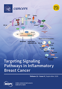
Inflammatory breast cancer (IBC) is the most lethal and aggressive form of breast cancer. Although it is rare—only 2% to 4% of breast cancer cases are classified as IBC—it represents 8% to 10% of breast cancer-related deaths due to the high rate of metastasis and poor prognosis. There is an urgent need to develop IBC-specific targeted therapies derived via understanding novel targets. This review summarizes the biological functions of critical signaling pathways in IBC progression and the latest preclinical and clinical studies of agents that target these pathways in IBC. It also discusses studies of crosstalk between several signaling pathways and the IBC tumor microenvironment. The findings described in this paper will help guide the development of effective therapies through preclinical and clinical research, eventually improving the treatment of patients with IBC. View this paper.
  • Issues are regarded as officially published after their release is announced to the table of contents alert mailing list.
  • You may sign up for e-mail alerts to receive table of contents of newly released issues.
  • PDF is the official format for papers published in both, html and pdf forms. To view the papers in pdf format, click on the "PDF Full-text" link, and use the free Adobe Reader to open them.
Order results
Result details
Section
Select all
Export citation of selected articles as:
10 pages, 240 KiB  
Commentary
Neoadjuvant Chemotherapy for Colon Cancer
by Marc T. Roth and Cathy Eng
Cancers 2020, 12(9), 2368; https://doi.org/10.3390/cancers12092368 - 26 Sep 2020
Cited by 14 | Viewed by 4761
Abstract
Early stage colon cancer is typically managed with surgical resection, although not all patients experience a durable remission. Adjuvant chemotherapy with a fluoropyrimidine, with or without oxaliplatin, is commonly utilized to increase the chance of cure, but its efficacy in the neoadjuvant setting [...] Read more.
Early stage colon cancer is typically managed with surgical resection, although not all patients experience a durable remission. Adjuvant chemotherapy with a fluoropyrimidine, with or without oxaliplatin, is commonly utilized to increase the chance of cure, but its efficacy in the neoadjuvant setting is not well established. Preoperative chemotherapy has demonstrated safety and efficacy in other gastrointestinal malignancies, but there is a paucity of data from large, prospective randomized trials, although multiple are ongoing. In this review, we will discuss the theoretical risks and benefits, logistical difficulties, and available safety and efficacy data pertaining to the use of chemotherapy in locally advanced colon cancer. Full article
(This article belongs to the Special Issue Adjuvant Chemotherapy for Colorectal Cancer)
11 pages, 5130 KiB  
Article
Epstein–Barr Virus Infection of Pseudostratified Nasopharyngeal Epithelium Disrupts Epithelial Integrity
by Fenggang Yu, Yanan Lu, Yingying Li, Yuji Uchio, Utomo Andi Pangnguriseng, Andy Visi Kartika, Hisashi Iizasa, Hironori Yoshiyama and Kwok Seng Loh
Cancers 2020, 12(9), 2722; https://doi.org/10.3390/cancers12092722 - 22 Sep 2020
Cited by 7 | Viewed by 3135
Abstract
Epstein–Barr virus (EBV) is a human oncogenic virus that causes several types of tumor, such as Burkitt’s lymphoma and nasopharyngeal carcinoma (NPC). NPC tumor cells are clonal expansions of latently EBV-infected epithelial cells. However, the mechanisms by which EBV transforms the nasopharyngeal epithelium [...] Read more.
Epstein–Barr virus (EBV) is a human oncogenic virus that causes several types of tumor, such as Burkitt’s lymphoma and nasopharyngeal carcinoma (NPC). NPC tumor cells are clonal expansions of latently EBV-infected epithelial cells. However, the mechanisms by which EBV transforms the nasopharyngeal epithelium is hampered, because of the lack of good in vitro model to pursue oncogenic process. Our primary nasopharyngeal epithelial cell cultures developed pseudostratified epithelium at the air-liquid interface, which was susceptible to EBV infection. Using the highly sensitive RNA in situ hybridization technique, we detected viral infection in diverse cell types, including ciliated cells, goblet cells, and basal cells. EBV-encoded small RNA-positive cells were more frequently detected in the suprabasal layer than in the basal layer. We established the most physiologically relevant EBV infection model of nasopharyngeal epithelial cells. This model will advance our understanding of EBV pathogenesis in the development of NPC. Full article
(This article belongs to the Special Issue Epstein-Barr Virus Infection in Cancer)
Show Figures

Graphical abstract

18 pages, 3474 KiB  
Article
Targeted-Alpha-Therapy Combining Astatine-211 and anti-CD138 Antibody in a Preclinical Syngeneic Mouse Model of Multiple Myeloma Minimal Residual Disease
by Sébastien Gouard, Catherine Maurel, Séverine Marionneau-Lambot, Delphine Dansette, Clément Bailly, François Guérard, Nicolas Chouin, Ferid Haddad, Cyril Alliot, Joëlle Gaschet, Romain Eychenne, Françoise Kraeber-Bodéré and Michel Chérel
Cancers 2020, 12(9), 2721; https://doi.org/10.3390/cancers12092721 - 22 Sep 2020
Cited by 10 | Viewed by 3646
Abstract
Despite therapeutic progress in recent years with the introduction of targeted therapies (daratumumab, elotuzumab), multiple myeloma remains an incurable cancer. The question is therefore to investigate the potential of targeted alpha therapy, combining an anti-CD138 antibody with astatine-211, to destroy the residual cells [...] Read more.
Despite therapeutic progress in recent years with the introduction of targeted therapies (daratumumab, elotuzumab), multiple myeloma remains an incurable cancer. The question is therefore to investigate the potential of targeted alpha therapy, combining an anti-CD138 antibody with astatine-211, to destroy the residual cells that cause relapses. A preclinical syngeneic mouse model, consisting of IV injection of 1 million of 5T33 cells in a KaLwRij C57/BL6 mouse, was treated 10 days later with an anti-mCD138 antibody, called 9E7.4, radiolabeled with astatine-211. Four activities of the 211At-9E7.4 radioimmunoconjugate were tested in two independent experiments: 370 kBq (n = 16), 555 kBq (n = 10), 740 kBq (n = 17) and 1100 kBq (n = 6). An isotype control was also tested at 555 kBq (n = 10). Biodistribution, survival rate, hematological parameters, enzymatic hepatic toxicity, histological examination and organ dosimetry were considered. The survival median of untreated mice was 45 days after engraftment. While the activity of 1100 kBq was highly toxic, the activity of 740 kBq offered the best efficacy with 65% of overall survival 150 days after the treatment with no evident sign of toxicity. This work demonstrates the pertinence of treating minimal residual disease of multiple myeloma with an anti-CD138 antibody coupled to astatine-211. Full article
(This article belongs to the Special Issue PET/CT in Multiple Myeloma Patients)
Show Figures

Graphical abstract

15 pages, 994 KiB  
Article
Development and Internal Validation of a Multivariable Prediction Model for Adrenocortical-Carcinoma-Specific Mortality
by Madeleine H. T. Ettaieb, Sander M. J. van Kuijk, Annelies de Wit-Pastoors, Richard A. Feelders, Eleonora P. M. Corssmit, Elisabeth M. W. Eekhoff, Paul van der Valk, Henri J. L. M. Timmers, Michiel N. Kerstens, Heinz-Josef Klümpen, Rachel S. van Leeuwaarde, Bas Havekes and Harm R. Haak
Cancers 2020, 12(9), 2720; https://doi.org/10.3390/cancers12092720 - 22 Sep 2020
Cited by 6 | Viewed by 2080
Abstract
Adrenocortical carcinoma (ACC) has an incidence of about 1.0 per million per year. In general, survival of patients with ACC is limited. Predicting survival outcome at time of diagnosis is a clinical challenge. The aim of this study was to develop and internally [...] Read more.
Adrenocortical carcinoma (ACC) has an incidence of about 1.0 per million per year. In general, survival of patients with ACC is limited. Predicting survival outcome at time of diagnosis is a clinical challenge. The aim of this study was to develop and internally validate a clinical prediction model for ACC-specific mortality. Data for this retrospective cohort study were obtained from the nine centers of the Dutch Adrenal Network (DAN). Patients who presented with ACC between 1 January 2004 and 31 October 2013 were included. We used multivariable Cox proportional hazards regression to compute the coefficients for the prediction model. Backward stepwise elimination was performed to derive a more parsimonious model. The performance of the initial prediction model was quantified by measures of model fit, discriminative ability, and calibration. We undertook an internal validation step to counteract the possible overfitting of our model. A total of 160 patients were included in the cohort. The median survival time was 35 months, and interquartile range (IQR) 50.7 months. The multivariable modeling yielded a prediction model that included age, modified European Network for the Study of Adrenal Tumors (mENSAT) stage, and radical resection. The c-statistic was 0.77 (95% Confidence Interval: 0.72, 0.81), indicating good predictive performance. We developed a clinical prediction model for ACC-specific mortality. ACC mortality can be estimated using a relatively simple clinical prediction model with good discriminative ability and calibration. Full article
(This article belongs to the Special Issue Adrenocortical Carcinoma)
Show Figures

Figure 1

19 pages, 4719 KiB  
Article
Establishment of Prostate Tumor Growth and Metastasis Is Supported by Bone Marrow Cells and Is Mediated by PIP5K1α Lipid Kinase
by Richard Karlsson, Per Larsson, Regina Miftakhova, Azharuddin Sajid Syed Khaja, Martuza Sarwar, Julius Semenas, Sa Chen, Andreas Hedblom, Tianyan Wang, Kristina Ekström-Holka, Athanasios Simoulis, Anjani Kumar, Niels Ødum, Thomas Grundström and Jenny L. Persson
Cancers 2020, 12(9), 2719; https://doi.org/10.3390/cancers12092719 - 22 Sep 2020
Cited by 4 | Viewed by 2882
Abstract
Cancer cells facilitate growth and metastasis by using multiple signals from the cancer-associated microenvironment. However, it remains poorly understood whether prostate cancer (PCa) cells may recruit and utilize bone marrow cells for their growth and survival. Furthermore, the regulatory mechanisms underlying interactions between [...] Read more.
Cancer cells facilitate growth and metastasis by using multiple signals from the cancer-associated microenvironment. However, it remains poorly understood whether prostate cancer (PCa) cells may recruit and utilize bone marrow cells for their growth and survival. Furthermore, the regulatory mechanisms underlying interactions between PCa cells and bone marrow cells are obscure. In this study, we isolated bone marrow cells that mainly constituted populations that were positive for CD11b and Gr1 antigens from xenograft PC-3 tumor tissues from athymic nu/nu mice. We found that the tumor-infiltrated cells alone were unable to form tumor spheroids, even with increased amounts and time. By contrast, the tumor-infiltrated cells together with PCa cells formed large numbers of tumor spheroids compared with PCa cells alone. We further utilized xenograft athymic nu/nu mice bearing bone metastatic lesions. We demonstrated that PCa cells were unable to survive and give rise to colony-forming units (CFUs) in media that were used for hematopoietic cell colony-formation unit (CFU) assays. By contrast, PC-3M cells survived when bone marrow cells were present and gave rise to CFUs. Our results showed that PCa cells required bone marrow cells to support their growth and survival and establish bone metastasis in the host environment. We showed that PCa cells that were treated with either siRNA for PIP5K1α or its specific inhibitor, ISA-2011B, were unable to survive and produce tumor spheroids, together with bone marrow cells. Given that the elevated expression of PIP5K1α was specific for PCa cells and was associated with the induced expression of VEGF receptor 2 in PCa cells, our findings suggest that cancer cells may utilize PIP5K1α-mediated receptor signaling to recruit growth factors and ligands from the bone marrow-derived cells. Taken together, our study suggests a new mechanism that enables PCa cells to gain proliferative and invasive advantages within their associated host microenvironment. Therapeutic interventions using PIP5K1α inhibitors may not only inhibit tumor invasion and metastasis but also enhance the host immune system. Full article
(This article belongs to the Section Tumor Microenvironment)
Show Figures

Figure 1

23 pages, 7553 KiB  
Article
SFMBT2-Mediated Infiltration of Preadipocytes and TAMs in Prostate Cancer
by Jungsug Gwak, Hayan Jeong, Kwanghyun Lee, Jee Yoon Shin, Taejun Sim, Jungtae Na, Jongchan Kim and Bong-Gun Ju
Cancers 2020, 12(9), 2718; https://doi.org/10.3390/cancers12092718 - 22 Sep 2020
Cited by 10 | Viewed by 3986
Abstract
Infiltration of diverse cell types into tumor microenvironment plays a critical role in cancer progression including metastasis. We previously reported that SFMBT2 (Scm-like with four mbt domains 2) regulates the expression of matrix metalloproteinases (MMPs) and migration and invasion of cancer cells in [...] Read more.
Infiltration of diverse cell types into tumor microenvironment plays a critical role in cancer progression including metastasis. We previously reported that SFMBT2 (Scm-like with four mbt domains 2) regulates the expression of matrix metalloproteinases (MMPs) and migration and invasion of cancer cells in prostate cancer. Here we investigated whether the down-regulation of SFMBT2 regulates the infiltration of preadipocytes and tumor-associated macrophages (TAMs) in prostate cancer. We found that the down-regulation of SFMBT2 promotes the infiltration of preadipocytes and TAMs through up-regulation of CXCL8, CCL2, CXCL10, and CCL20 expression in prostate cancer. Expression of CXCL8, CCL2, CXCL10, and CCL20 was also elevated in prostate cancer patients having a higher Gleason score (≥8), which had substantially lower SFMBT2 expression. We also found that the up-regulation of CXCL8, CCL2, CXCL10, and CCL20 expression is dependent on NF-κB activation in prostate cancer cells expressing a low level of SFMBT2. Moreover, increased IL-6 from infiltrated preadipocytes and TAMs promoted migration and invasion of prostate cancer cells expressing a low level of SFMBT2. Our study may suggest that SFMBT2 a critical regulator for the infiltration of preadipocytes and TAMs into the prostate tumor microenvironment. Thus, the regulation of SFMBT2 may provide a new therapeutic strategy to inhibit prostate cancer metastasis, and SFMBT2 could be used as a potential biomarker in prostate cancer metastasis. Full article
(This article belongs to the Section Molecular Cancer Biology)
Show Figures

Figure 1

22 pages, 3204 KiB  
Review
The Undervalued Avenue to Reinstate Tumor Suppressor Functionality of the p53 Protein Family for Improved Cancer Therapy-Drug Repurposing
by Joanna E. Zawacka-Pankau
Cancers 2020, 12(9), 2717; https://doi.org/10.3390/cancers12092717 - 22 Sep 2020
Cited by 8 | Viewed by 4881
Abstract
p53 and p73 are critical tumor suppressors that are often inactivated in human cancers through various mechanisms. Owing to their high structural homology, the proteins have many joined functions and recognize the same set of genes involved in apoptosis and cell cycle regulation. [...] Read more.
p53 and p73 are critical tumor suppressors that are often inactivated in human cancers through various mechanisms. Owing to their high structural homology, the proteins have many joined functions and recognize the same set of genes involved in apoptosis and cell cycle regulation. p53 is known as the ‘guardian of the genome’ and together with p73 forms a barrier against cancer development and progression. The TP53 is mutated in more than 50% of all human cancers and the germline mutations in TP53 predispose to the early onset of multiple tumors in Li–Fraumeni syndrome (LFS), the inherited cancer predisposition. In cancers where TP53 gene is intact, p53 is degraded. Despite the ongoing efforts, the treatment of cancers remains challenging. This is due to late diagnoses, the toxicity of the current standard of care and marginal benefit of newly approved therapies. Presently, the endeavors focus on reactivating p53 exclusively, neglecting the potential of the restoration of p73 protein for cancer eradication. Taken that several small molecules reactivating p53 failed in clinical trials, there is a need to develop new treatments targeting p53 proteins in cancer. This review outlines the most advanced strategies to reactivate p53 and p73 and describes drug repurposing approaches for the efficient reinstatement of the p53 proteins for cancer therapy. Full article
(This article belongs to the Special Issue The Role of p53 Family in Cancer)
Show Figures

Graphical abstract

14 pages, 1100 KiB  
Article
Radiation-Induced Hypothyroidism in Patients with Oropharyngeal Cancer Treated with IMRT: Independent and External Validation of Five Normal Tissue Complication Probability Models
by Zuzanna Nowicka, Bartłomiej Tomasik, Anna Papis-Ubych, Robert Bibik, Łukasz Graczyk, Tomasz Latusek, Tomasz Rutkowski, Krystyna Wyka, Jacek Fijuth, Jonathan D. Schoenfeld, Justyna Chałubińska-Fendler and Wojciech Fendler
Cancers 2020, 12(9), 2716; https://doi.org/10.3390/cancers12092716 - 22 Sep 2020
Cited by 7 | Viewed by 3029
Abstract
We aimed to externally validate five normal tissue complication probability (NTCP) models for radiation-induced hypothyroidism (RIHT) in a prospectively recruited cohort of 108 patients with oropharyngeal cancer (OPC). NTCP scores were calculated using original published formulas. Plasma thyrotropin (TSH) level was additionally assessed [...] Read more.
We aimed to externally validate five normal tissue complication probability (NTCP) models for radiation-induced hypothyroidism (RIHT) in a prospectively recruited cohort of 108 patients with oropharyngeal cancer (OPC). NTCP scores were calculated using original published formulas. Plasma thyrotropin (TSH) level was additionally assessed in the short-term after RT. After a median of 28 months of follow-up, thirty one (28.7%) patients developed RIHT. Thyroid mean dose and thyroid volume were significant predictors of RIHT: odds ratio equal to 1.11 (95% CI 1.03–1.19) for mean thyroid dose and 0.87 (95%CI 0.81–0.93) for thyroid volume in univariate analyses. Two of the evaluated NTCP models, published by Rønjom et al. and by Boomsma et al., had satisfactory performance with accuracies of 0.87 (95%CI 0.79–0.93) and 0.84 (95%CI: 0.76–0.91), respectively. Three remaining models, by Cella et al., Bakhshandeh et al. and Vogelius et al., performed significantly worse, overestimating the risk of RIHT in this patient cohort. A short-term TSH level change relative to baseline was not indicative of RIHT development in the follow-up (OR 0.96, 95%CI: 0.65–1.42, p = 0.825). In conclusion, the models by Rønjom et al. and by Boomsma et al. demonstrated external validity and feasibility for long-term prediction of RIHT in survivors of OPC treated with Intensity-Modulated Radiation Therapy (IMRT). Full article
(This article belongs to the Special Issue Symptoms and Side Effects in Cancer Survivors)
Show Figures

Figure 1

21 pages, 7044 KiB  
Article
Menin and Menin-Associated Proteins Coregulate Cancer Energy Metabolism
by Chih-Wei Chou, Xi Tan, Chia-Nung Hung, Brandon Lieberman, Meizhen Chen, Meena Kusi, Kohzoh Mitsuya, Chun-Lin Lin, Masahiro Morita, Zhijie Liu, Chun-Liang Chen and Tim Hui-Ming Huang
Cancers 2020, 12(9), 2715; https://doi.org/10.3390/cancers12092715 - 22 Sep 2020
Cited by 6 | Viewed by 3532
Abstract
The interplay between glycolysis and mitochondrial oxidative phosphorylation (OXPHOS) is central to maintain energy homeostasis. It remains to be determined whether there is a mechanism governing metabolic fluxes based on substrate availability in microenvironments. Here we show that menin is a key transcription [...] Read more.
The interplay between glycolysis and mitochondrial oxidative phosphorylation (OXPHOS) is central to maintain energy homeostasis. It remains to be determined whether there is a mechanism governing metabolic fluxes based on substrate availability in microenvironments. Here we show that menin is a key transcription factor regulating the expression of OXPHOS and glycolytic genes in cancer cells and primary tumors with poor prognosis. A group of menin-associated proteins (MAPs), including KMT2A, MED12, WAPL, and GATA3, is found to restrain menin’s full function in this transcription regulation. shRNA knockdowns of menin and MAPs result in reduced ATP production with proportional alterations of cellular energy generated through glycolysis and OXPHOS. When shRNA knockdown cells are exposed to metabolic stress, the dual functionality can clearly be distinguished among these metabolic regulators. A MAP can negatively counteract the regulatory mode of menin for OXPHOS while the same protein positively influences glycolysis. A close-proximity interaction between menin and MAPs allows transcriptional regulation for metabolic adjustment. This coordinate regulation by menin and MAPs is necessary for cells to rapidly adapt to fluctuating microenvironments and to maintain essential metabolic functions. Full article
(This article belongs to the Section Molecular Cancer Biology)
Show Figures

Figure 1

20 pages, 8382 KiB  
Article
Clinical, Genomic, and Pharmacological Study of MYCN-Amplified RB1 Wild-Type Metastatic Retinoblastoma
by Santiago Zugbi, Daiana Ganiewich, Arpita Bhattacharyya, Rosario Aschero, Daniela Ottaviani, Claudia Sampor, Eduardo G. Cafferata, Marcela Mena, Mariana Sgroi, Ursula Winter, Gabriela Lamas, Mariona Suñol, Manuel Daroqui, Edgardo Baialardo, Beatriz Salas, Anirban Das, Adriana Fandiño, Jasmine H. Francis, Fabiana Lubieniecki, Cinzia Lavarino, Ralph Garippa, Osvaldo L. Podhajcer, David H. Abramson, François Radvanyi, Guillermo Chantada, Andrea S. Llera and Paula Schaiquevichadd Show full author list remove Hide full author list
Cancers 2020, 12(9), 2714; https://doi.org/10.3390/cancers12092714 - 22 Sep 2020
Cited by 27 | Viewed by 4040
Abstract
An uncommon subgroup of unilateral retinoblastomas with highly aggressive histological features, lacking aberrations in RB1 gene with high-level amplification of MYCN (MCYNamplRB1+/+) has only been described as intra-ocular cases treated with initial enucleation. [...] Read more.
An uncommon subgroup of unilateral retinoblastomas with highly aggressive histological features, lacking aberrations in RB1 gene with high-level amplification of MYCN (MCYNamplRB1+/+) has only been described as intra-ocular cases treated with initial enucleation. Here, we present a comprehensive clinical, genomic, and pharmacological analysis of two cases of MCYNamplRB1+/+ with orbital and cervical lymph node involvement, but no central nervous system spread, rapidly progressing to fatal disease due to chemoresistance. Both patients showed in common MYCN high amplification and chromosome 16q and 17p loss. A somatic mutation in TP53, in homozygosis by LOH, and high chromosomal instability leading to aneuploidy was identified in the primary ocular tumor and sites of dissemination of one patient. High-throughput pharmacological screening was performed in a primary cell line derived from the lymph node dissemination of one case. This cell line showed resistance to broad spectrum chemotherapy consistent with the patient’s poor response but sensitivity to the synergistic effects of panobinostat–bortezomib and carboplatin–panobinostat associations. From these cells we established a cell line derived xenograft model that closely recapitulated the tumor dissemination pattern of the patient and served to evaluate whether triple chemotherapy significantly prolonged survival of the animals. We report novel genomic alterations in two cases of metastatic MCYNamplRB1+/+ that may be associated with chemotherapy resistance and in vitro/in vivo models that serve as basis for tailoring therapy in these cases. Full article
Show Figures

Graphical abstract

18 pages, 7700 KiB  
Article
Hormonal Receptor Status Determines Prognostic Significance of FGFR2 in Invasive Breast Carcinoma
by Marcin Braun, Dominika Piasecka, Bartlomiej Tomasik, Kamil Mieczkowski, Konrad Stawiski, Aleksandra Zielinska, Janusz Kopczynski, Dariusz Nejc, Radzislaw Kordek, Rafal Sadej and Hanna M. Romanska
Cancers 2020, 12(9), 2713; https://doi.org/10.3390/cancers12092713 - 22 Sep 2020
Cited by 8 | Viewed by 4193
Abstract
Interaction between fibroblast growth factor receptor 2 (FGFR2) and estrogen/progesterone receptors (ER/PR) affects resistance to anti-ER therapies, however the prognostic value of FGFR2 in breast cancer (BCa) remains largely unexplored. We have recently showed in vitro that FGFR2-mediated signaling alters PR activity and [...] Read more.
Interaction between fibroblast growth factor receptor 2 (FGFR2) and estrogen/progesterone receptors (ER/PR) affects resistance to anti-ER therapies, however the prognostic value of FGFR2 in breast cancer (BCa) remains largely unexplored. We have recently showed in vitro that FGFR2-mediated signaling alters PR activity and response to anti-ER treatment. Herein, prognostic significance of FGFR2 in BCa was evaluated in relation to both ER/PR protein status and a molecular signature designed to reflect PR transcriptional activity. FGFR2 was examined in 353 BCa cases using immunohistochemistry and Nanostring-based RNA quantification. FGFR2 expression was higher in ER+PR+ and ER+PR- compared to ER−PR− cases (p < 0.001). Low FGFR2 was associated with higher grade (p < 0.001), higher Ki67 proliferation index (p < 0.001), and worse overall and disease-free survival (HR = 2.34 (95% CI: 1.26–4.34), p = 0.007 and HR = 2.22 (95% CI: 1.25–3.93), p = 0.006, respectively). The poor prognostic value of low FGFR2 was apparent in ER+PR+, but not in ER+PR− patients, and it did not depend on the expression level of PR-dependent genes. Despite the functional link between FGFR2 and ER/PR revealed by preclinical studies, the data showed a link between FGFR2 expression and poor prognosis in BCa patients. Full article
(This article belongs to the Special Issue Hormone-Associated Cancers)
Show Figures

Graphical abstract

16 pages, 1304 KiB  
Review
The Unique Pharmacometrics of Small Molecule Therapeutic Drug Tracer Imaging for Clinical Oncology
by Mark P. S. Dunphy and Nagavarakishore Pillarsetty
Cancers 2020, 12(9), 2712; https://doi.org/10.3390/cancers12092712 - 22 Sep 2020
Cited by 5 | Viewed by 2625
Abstract
Translational development of radiolabeled analogues or isotopologues of small molecule therapeutic drugs as clinical imaging biomarkers for optimizing patient outcomes in targeted cancer therapy aims to address an urgent and recurring clinical need in therapeutic cancer drug development: drug- and target-specific biomarker assays [...] Read more.
Translational development of radiolabeled analogues or isotopologues of small molecule therapeutic drugs as clinical imaging biomarkers for optimizing patient outcomes in targeted cancer therapy aims to address an urgent and recurring clinical need in therapeutic cancer drug development: drug- and target-specific biomarker assays that can optimize patient selection, dosing strategy, and response assessment. Imaging the in vivo tumor pharmacokinetics and biomolecular pharmacodynamics of small molecule cancer drugs offers patient- and tumor-specific data which are not available from other pharmacometric modalities. This review article examines clinical research with a growing pharmacopoeia of investigational small molecule cancer drug tracers. Full article
(This article belongs to the Special Issue Cancer Molecular Imaging)
Show Figures

Figure 1

13 pages, 2142 KiB  
Article
Deregulated microRNAs Are Associated with Patient Survival and Predicted to Target Genes That Modulate Lung Cancer Signaling Pathways
by Cristiano P. Souza, Naiara C. Cinegaglia, Tainara F. Felix, Adriane F. Evangelista, Rogério A. Oliveira, Erica N. Hasimoto, Daniele C. Cataneo, Antônio J. M. Cataneo, Cristovam Scapulatempo Neto, Cristiano R. Viana, Flávia E. de Paula, Sandra A. Drigo, Robson F. Carvalho, Márcia M. C. Marques, Rui M. Reis and Patricia P. Reis
Cancers 2020, 12(9), 2711; https://doi.org/10.3390/cancers12092711 - 22 Sep 2020
Cited by 7 | Viewed by 2358
Abstract
(1) Background: Although the advances in diagnostic and treatment strategies, lung cancer remains the leading cause of cancer-related deaths, worldwide, with survival rates as low as 16% in developed countries. Low survival rates are mainly due to late diagnosis and the lack of [...] Read more.
(1) Background: Although the advances in diagnostic and treatment strategies, lung cancer remains the leading cause of cancer-related deaths, worldwide, with survival rates as low as 16% in developed countries. Low survival rates are mainly due to late diagnosis and the lack of effective treatment. Therefore, the identification of novel, clinically useful biomarkers is still needed for patients with advanced disease stage and poor survival. Micro(mi)RNAs are non-coding RNAs and potent regulators of gene expression with a possible role as diagnostic, prognostic and predictive biomarkers in cancer. (2) Methods: We applied global miRNA expression profiling analysis using TaqMan® arrays in paired tumor and normal lung tissues (n = 38) from treatment-naïve patients with lung adenocarcinoma (AD; n = 23) and lung squamous cell carcinoma (SCC; n = 15). miRNA target genes were validated using The Cancer Genome Atlas (TCGA) lung AD (n = 561) and lung SCC (n = 523) RNA-Seq datasets. (3) Results: We identified 33 significantly deregulated miRNAs (fold change, FC ≥ 2.0 and p < 0.05) in tumors relative to normal lung tissues, regardless of tumor histology. Enrichment analysis confirmed that genes targeted by the 33 miRNAs are aberrantly expressed in lung AD and SCC, and modulate known pathways in lung cancer. Additionally, high expression of miR-25-3p was significantly associated (p < 0.05) with poor patient survival, when considering both tumor histologies. (4) Conclusions: miR-25-3p may be a potential prognostic biomarker in non-small cell lung cancer. Genes targeted by miRNAs regulate EGFR and TGFβ signaling, among other known pathways relevant to lung tumorigenesis. Full article
(This article belongs to the Special Issue Non-small Cell Lung Cancer--Tumor Biology)
Show Figures

Figure 1

20 pages, 1835 KiB  
Article
Landscape of Genome-Wide DNA Methylation of Colorectal Cancer Metastasis
by Carmen Ili, Kurt Buchegger, Hannah Demond, Juan Castillo-Fernandez, Gavin Kelsey, Louise Zanella, Michel Abanto, Ismael Riquelme, Jaime López, Tamara Viscarra, Patricia García, Enrique Bellolio, David Saavedra and Priscilla Brebi
Cancers 2020, 12(9), 2710; https://doi.org/10.3390/cancers12092710 - 22 Sep 2020
Cited by 18 | Viewed by 4725
Abstract
Colorectal cancer is a heterogeneous disease caused by both genetic and epigenetics factors. Analysing DNA methylation changes occurring during colorectal cancer progression and metastasis formation is crucial for the identification of novel epigenetic markers of patient prognosis. Genome-wide methylation sequencing of paired samples [...] Read more.
Colorectal cancer is a heterogeneous disease caused by both genetic and epigenetics factors. Analysing DNA methylation changes occurring during colorectal cancer progression and metastasis formation is crucial for the identification of novel epigenetic markers of patient prognosis. Genome-wide methylation sequencing of paired samples of colon (normal adjacent, primary tumour and lymph node metastasis) showed global hypomethylation and CpG island (CGI) hypermethylation of primary tumours compared to normal. In metastasis we observed high global and non-CGI regions methylation, but lower CGI methylation, compared to primary tumours. Gene ontology analysis showed shared biological processes between hypermethylated CGIs in metastasis and primary tumours. After complementary analysis with The Cancer Genome Atlas (TCGA) cohort, FIGN, HTRA3, BDNF, HCN4 and STAC2 genes were found associated with poor survival. We mapped the methylation landscape of colon normal tissues, primary tumours and lymph node metastasis, being capable of identified methylation changes throughout the genome. Furthermore, we found five genes with potential for methylation biomarkers of poor prognosis in colorectal cancer patients. Full article
(This article belongs to the Special Issue Metastatic Colorectal Cancer)
Show Figures

Graphical abstract

19 pages, 3287 KiB  
Article
Bortezomib-Loaded Mesoporous Silica Nanoparticles Selectively Alter Metabolism and Induce Death in Multiple Myeloma Cells
by Alessandra Nigro, Luca Frattaruolo, Mariarosa Fava, Ilaria De Napoli, Marianna Greco, Alessandra Comandè, Marzia De Santo, Michele Pellegrino, Elena Ricci, Francesca Giordano, Ida Perrotta, Antonella Leggio, Luigi Pasqua, Diego Sisci, Anna Rita Cappello and Catia Morelli
Cancers 2020, 12(9), 2709; https://doi.org/10.3390/cancers12092709 - 21 Sep 2020
Cited by 15 | Viewed by 3148
Abstract
A mesoporous silica-based nanodevice bearing the antineoplastic drug bortezomib (BTZ), whose release is triggered in acidic environment and grafted with folic acid (FOL) as a targeting function (FOL-MSN-BTZ) was tested on folate receptor overexpressing (FR+) multiple myeloma (MM) cells and on FR negative [...] Read more.
A mesoporous silica-based nanodevice bearing the antineoplastic drug bortezomib (BTZ), whose release is triggered in acidic environment and grafted with folic acid (FOL) as a targeting function (FOL-MSN-BTZ) was tested on folate receptor overexpressing (FR+) multiple myeloma (MM) cells and on FR negative (FR−) normal cells. FOL-MSN-BTZ efficacy studies were conducted by means of growth experiments, TEM, TUNEL assay and Western Blotting analysis (WB). Metabolic investigations were performed to assess cells metabolic response to MSNs treatments. FOL-MSN-BTZ exclusively killed FR+ MM cells, leading to an apoptotic rate that was comparable to that induced by free BTZ, and the effect was accompanied by metabolic dysfunction and oxidative stress. Importantly, FOL-MSN-BTZ treated FR− normal cells did not show any significant sign of injury or metabolic perturbation, while free BTZ was still highly toxic. Notably, the vehicle alone (MSN-FOL) did not affect any biological process in both tested cell models. These data show the striking specificity of FOL-MSN-BTZ toward FR+ tumor cells and the outstanding safety of the MSN-FOL vehicle, paving the way for a future exploitation of FOL-MSN-BTZ in MM target therapy. Full article
(This article belongs to the Special Issue Recent Advances on the Pathobiology and Treatment of Multiple Myeloma)
Show Figures

Graphical abstract

11 pages, 1231 KiB  
Article
Computer Extracted Features from Initial H&E Tissue Biopsies Predict Disease Progression for Prostate Cancer Patients on Active Surveillance
by Sacheth Chandramouli, Patrick Leo, George Lee, Robin Elliott, Christine Davis, Guangjing Zhu, Pingfu Fu, Jonathan I. Epstein, Robert Veltri and Anant Madabhushi
Cancers 2020, 12(9), 2708; https://doi.org/10.3390/cancers12092708 - 21 Sep 2020
Cited by 15 | Viewed by 3306
Abstract
In this work, we assessed the ability of computerized features of nuclear morphology from diagnostic biopsy images to predict prostate cancer (CaP) progression in active surveillance (AS) patients. Improved risk characterization of AS patients could reduce over-testing of low-risk patients while directing high-risk [...] Read more.
In this work, we assessed the ability of computerized features of nuclear morphology from diagnostic biopsy images to predict prostate cancer (CaP) progression in active surveillance (AS) patients. Improved risk characterization of AS patients could reduce over-testing of low-risk patients while directing high-risk patients to therapy. A total of 191 (125 progressors, 66 non-progressors) AS patients from a single site were identified using The Johns Hopkins University’s (JHU) AS-eligibility criteria. Progression was determined by pathologists at JHU. 30 progressors and 30 non-progressors were randomly selected to create the training cohort D1 (n = 60). The remaining patients comprised the validation cohort D2 (n = 131). Digitized Hematoxylin & Eosin (H&E) biopsies were annotated by a pathologist for CaP regions. Nuclei within the cancer regions were segmented using a watershed method and 216 nuclear features describing position, shape, orientation, and clustering were extracted. Six features associated with disease progression were identified using D1 and then used to train a machine learning classifier. The classifier was validated on D2. The classifier was further compared on a subset of D2 (n = 47) against pro-PSA, an isoform of prostate specific antigen (PSA) more linked with CaP, in predicting progression. Performance was evaluated with area under the curve (AUC). A combination of nuclear spatial arrangement, shape, and disorder features were associated with progression. The classifier using these features yielded an AUC of 0.75 in D2. On the 47 patient subset with pro-PSA measurements, the classifier yielded an AUC of 0.79 compared to an AUC of 0.42 for pro-PSA. Nuclear morphometric features from digitized H&E biopsies predicted progression in AS patients. This may be useful for identifying AS-eligible patients who could benefit from immediate curative therapy. However, additional multi-site validation is needed. Full article
(This article belongs to the Special Issue Radiomics and Cancers)
Show Figures

Figure 1

15 pages, 3254 KiB  
Article
Glioblastoma Tissue Slice Tandem-Cultures for Quantitative Evaluation of Inhibitory Effects on Invasion and Growth
by Vasile Sidorcenco, Luisa Krahnen, Marion Schulz, Janina Remy, Donat Kögel, Achim Temme, Ute Krügel, Heike Franke and Achim Aigner
Cancers 2020, 12(9), 2707; https://doi.org/10.3390/cancers12092707 - 21 Sep 2020
Cited by 6 | Viewed by 2798
Abstract
Glioblastomas (GBMs) are the most malignant brain tumors and are essentially incurable even after extensive surgery, radiotherapy, and chemotherapy, mainly because of extensive infiltration of tumor cells into the adjacent normal tissue. Thus, the evaluation of novel drugs in malignant glioma treatment requires [...] Read more.
Glioblastomas (GBMs) are the most malignant brain tumors and are essentially incurable even after extensive surgery, radiotherapy, and chemotherapy, mainly because of extensive infiltration of tumor cells into the adjacent normal tissue. Thus, the evaluation of novel drugs in malignant glioma treatment requires sophisticated ex vivo models that approach the authentic interplay between tumor and host environment while avoiding extensive in vivo studies in animals. This paper describes the standardized setup of an organotypic brain tissue slice tandem-culture system, comprising of normal brain tissue from adult mice and tumor tissue from human glioblastoma xenografts, and explore its utility for assessing inhibitory effects of test drugs. The microscopic analysis of vertical sections of the slice tandem-cultures allows for the simultaneous assessment of (i) the invasive potential of single cells or cell aggregates and (ii) the space occupying growth of the bulk tumor mass, both contributing to malignant tumor progression. The comparison of tissue slice co-cultures with spheroids vs. tissue slice tandem-cultures using tumor xenograft slices demonstrates advantages of the xenograft tandem approach. The direct and facile application of test drugs is shown to exert inhibitory effects on bulk tumor growth and/or tumor cell invasion, and allows their precise quantitation. In conclusion, we describe a straightforward ex vivo system mimicking the in vivo situation of the tumor mass and the normal brain in GBM patients. It reduces animal studies and allows for the direct and reproducible application of test drugs and the precise quantitation of their effects on the bulk tumor mass and on the tumor’s invasive properties. Full article
Show Figures

Graphical abstract

15 pages, 2994 KiB  
Article
Machine Learning Model to Predict Pseudoprogression Versus Progression in Glioblastoma Using MRI: A Multi-Institutional Study (KROG 18-07)
by Bum-Sup Jang, Andrew J. Park, Seung Hyuck Jeon, Il Han Kim, Do Hoon Lim, Shin-Hyung Park, Ju Hye Lee, Ji Hyun Chang, Kwan Ho Cho, Jin Hee Kim, Leonard Sunwoo, Seung Hong Choi and In Ah Kim
Cancers 2020, 12(9), 2706; https://doi.org/10.3390/cancers12092706 - 21 Sep 2020
Cited by 19 | Viewed by 3520
Abstract
Some patients with glioblastoma show a worsening presentation in imaging after concurrent chemoradiation, even when they receive gross total resection. Previously, we showed the feasibility of a machine learning model to predict pseudoprogression (PsPD) versus progressive disease (PD) in glioblastoma patients. The previous [...] Read more.
Some patients with glioblastoma show a worsening presentation in imaging after concurrent chemoradiation, even when they receive gross total resection. Previously, we showed the feasibility of a machine learning model to predict pseudoprogression (PsPD) versus progressive disease (PD) in glioblastoma patients. The previous model was based on the dataset from two institutions (termed as the Seoul National University Hospital (SNUH) dataset, N = 78). To test this model in a larger dataset, we collected cases from multiple institutions that raised the problem of PsPD vs. PD diagnosis in clinics (Korean Radiation Oncology Group (KROG) dataset, N = 104). The dataset was composed of brain MR images and clinical information. We tested the previous model in the KROG dataset; however, that model showed limited performance. After hyperparameter optimization, we developed a deep learning model based on the whole dataset (N = 182). The 10-fold cross validation revealed that the micro-average area under the precision-recall curve (AUPRC) was 0.86. The calibration model was constructed to estimate the interpretable probability directly from the model output. After calibration, the final model offers clinical probability in a web-user interface. Full article
(This article belongs to the Special Issue Machine Learning Techniques in Cancer)
Show Figures

Figure 1

13 pages, 1080 KiB  
Article
Pathway-Based Integrative Analysis of Metabolome and Microbiome Data from Hepatocellular Carcinoma and Liver Cirrhosis Patients
by Boram Kim, Eun Ju Cho, Jung-Hwan Yoon, Soon Sun Kim, Jae Youn Cheong, Sung Won Cho and Taesung Park
Cancers 2020, 12(9), 2705; https://doi.org/10.3390/cancers12092705 - 21 Sep 2020
Cited by 7 | Viewed by 2613
Abstract
Aberrations of the human microbiome are associated with diverse liver diseases, including hepatocellular carcinoma (HCC). Even if we can associate specific microbes with particular diseases, it is difficult to know mechanistically how the microbe contributes to the pathophysiology. Here, we sought to reveal [...] Read more.
Aberrations of the human microbiome are associated with diverse liver diseases, including hepatocellular carcinoma (HCC). Even if we can associate specific microbes with particular diseases, it is difficult to know mechanistically how the microbe contributes to the pathophysiology. Here, we sought to reveal the functional potential of the HCC-associated microbiome with the human metabolome which is known to play a role in connecting host phenotype to microbiome function. To utilize both microbiome and metabolomic data sets, we propose an innovative, pathway-based analysis, Hierarchical structural Component Model for pathway analysis of Microbiome and Metabolome (HisCoM-MnM), for integrating microbiome and metabolomic data. In particular, we used pathway information to integrate these two omics data sets, thus providing insight into biological interactions between different biological layers, with regard to the host’s phenotype. The application of HisCoM-MnM to data sets from 103 and 97 patients with HCC and liver cirrhosis (LC), respectively, showed that this approach could identify HCC-related pathways related to cancer metabolic reprogramming, in addition to the significant metabolome and metagenome that make up those pathways. Full article
(This article belongs to the Special Issue Hepatocellular Carcinoma)
Show Figures

Graphical abstract

14 pages, 3216 KiB  
Article
Mass Spectrometry Imaging for Reliable and Fast Classification of Non-Small Cell Lung Cancer Subtypes
by Mark Kriegsmann, Christiane Zgorzelski, Rita Casadonte, Kristina Schwamborn, Thomas Muley, Hauke Winter, Martin Eichhorn, Florian Eichhorn, Arne Warth, Soeren-Oliver Deininger, Petros Christopoulos, Michael Thomas, Thomas Longerich, Albrecht Stenzinger, Wilko Weichert, Carsten Müller-Tidow, Jörg Kriegsmann, Peter Schirmacher and Katharina Kriegsmann
Cancers 2020, 12(9), 2704; https://doi.org/10.3390/cancers12092704 - 21 Sep 2020
Cited by 15 | Viewed by 4678
Abstract
Subtyping of non-small cell lung cancer (NSCLC) is paramount for therapy stratification. In this study, we analyzed the largest NSCLC cohort by mass spectrometry imaging (MSI) to date. We sought to test different classification algorithms and to validate results obtained in smaller patient [...] Read more.
Subtyping of non-small cell lung cancer (NSCLC) is paramount for therapy stratification. In this study, we analyzed the largest NSCLC cohort by mass spectrometry imaging (MSI) to date. We sought to test different classification algorithms and to validate results obtained in smaller patient cohorts. Tissue microarrays (TMAs) from including adenocarcinoma (ADC, n = 499) and squamous cell carcinoma (SqCC, n = 440), were analyzed. Linear discriminant analysis, support vector machine, and random forest (RF) were applied using samples randomly assigned for training (66%) and validation (33%). The m/z species most relevant for the classification were identified by on-tissue tandem mass spectrometry and validated by immunohistochemistry (IHC). Measurements from multiple TMAs were comparable using standardized protocols. RF yielded the best classification results. The classification accuracy decreased after including less than six of the most relevant m/z species. The sensitivity and specificity of MSI in the validation cohort were 92.9% and 89.3%, comparable to IHC. The most important protein for the discrimination of both tumors was cytokeratin 5. We investigated the largest NSCLC cohort by MSI to date and found that the classification of NSCLC into ADC and SqCC is possible with high accuracy using a limited set of m/z species. Full article
Show Figures

Figure 1

13 pages, 1538 KiB  
Review
Extraskeletal Myxoid Chondrosarcoma: State of the Art and Current Research on Biology and Clinical Management
by Silvia Stacchiotti, Giacomo Giulio Baldi, Carlo Morosi, Alessandro Gronchi and Roberta Maestro
Cancers 2020, 12(9), 2703; https://doi.org/10.3390/cancers12092703 - 21 Sep 2020
Cited by 35 | Viewed by 6545
Abstract
Extraskeletal myxoid chondrosarcoma (EMC) is an ultra-rare mesenchymal neoplasm with uncertain differentiation, which arises mostly in the deep soft tissue of proximal extremities and limb girdles. EMC is marked by a translocation involving the NR4A3 gene, which can be fused in-frame with different [...] Read more.
Extraskeletal myxoid chondrosarcoma (EMC) is an ultra-rare mesenchymal neoplasm with uncertain differentiation, which arises mostly in the deep soft tissue of proximal extremities and limb girdles. EMC is marked by a translocation involving the NR4A3 gene, which can be fused in-frame with different partners, most often EWSR1 or TAF1. Although EMC biology is still poorly defined, recent studies have started shedding light on the specific contribution of NR4A3 chimeric proteins to EMC pathogenesis and clinical outcome. Standard treatment for localized disease is surgery, plus or minus radiation therapy with an expected prolonged survival even though the risk of relapse is about 50%. In advanced cases, besides the standard chemotherapy currently used for soft tissue sarcoma, antiangiogenic agents have recently shown promising activity. The aim of this review is to provide the state of the art of treatment for localized and advanced disease, with a focus on pharmacological treatments available for EMC. The biological basis of current research and future perspectives will be also discussed. Full article
(This article belongs to the Special Issue New Therapeutic Advances in Rare Tumors)
Show Figures

Figure 1

2 pages, 146 KiB  
Editorial
Advances in Research on Human Meningiomas
by Sverre Helge Torp and David Scheie
Cancers 2020, 12(9), 2702; https://doi.org/10.3390/cancers12092702 - 21 Sep 2020
Cited by 1 | Viewed by 1307
Abstract
Meningiomas are the most common intracranial tumours in humans, constituting more than one third [...] Full article
(This article belongs to the Special Issue Advances in Research on Human Meningiomas)
21 pages, 1008 KiB  
Review
The Interplay between Long Noncoding RNAs and Proteins of the Epigenetic Machinery in Ovarian Cancer
by Naiade Calanca, Cecilie Abildgaard, Cláudia Aparecida Rainho and Silvia Regina Rogatto
Cancers 2020, 12(9), 2701; https://doi.org/10.3390/cancers12092701 - 21 Sep 2020
Cited by 6 | Viewed by 2964
Abstract
Comprehensive large-scale sequencing and bioinformatics analyses have uncovered a myriad of cancer-associated long noncoding RNAs (lncRNAs). Aberrant expression of lncRNAs is associated with epigenetic reprogramming during tumor development and progression, mainly due to their ability to interact with DNA, RNA, or proteins to [...] Read more.
Comprehensive large-scale sequencing and bioinformatics analyses have uncovered a myriad of cancer-associated long noncoding RNAs (lncRNAs). Aberrant expression of lncRNAs is associated with epigenetic reprogramming during tumor development and progression, mainly due to their ability to interact with DNA, RNA, or proteins to regulate gene expression. LncRNAs participate in the control of gene expression patterns during development and cell differentiation and can be cell and cancer type specific. In this review, we described the potential of lncRNAs for clinical applications in ovarian cancer (OC). OC is a complex and heterogeneous disease characterized by relapse, chemoresistance, and high mortality rates. Despite advances in diagnosis and treatment, no significant improvements in long-term survival were observed in OC patients. A set of lncRNAs was associated with survival and response to therapy in this malignancy. We manually curated databases and used bioinformatics tools to identify lncRNAs implicated in the epigenetic regulation, along with examples of direct interactions between the lncRNAs and proteins of the epigenetic machinery in OC. The resources and mechanisms presented herein can improve the understanding of OC biology and provide the basis for further investigations regarding the selection of novel biomarkers and therapeutic targets. Full article
(This article belongs to the Collection Regulatory and Non-Coding RNAs in Cancer Epigenetic Mechanisms)
Show Figures

Figure 1

14 pages, 715 KiB  
Article
Risk of Inflammatory Bowel Disease in Patients with Chronic Myeloproliferative Neoplasms: A Danish Nationwide Cohort Study
by Marie Bak, Tine Jess, Esben Meulengracht Flachs, Ann-Dorthe Zwisler, Knud Juel and Henrik Frederiksen
Cancers 2020, 12(9), 2700; https://doi.org/10.3390/cancers12092700 - 21 Sep 2020
Cited by 11 | Viewed by 3782
Abstract
An association between hematological cancers and inflammatory bowel disease (IBD) has previously been suggested, but the risk of IBD in patients with myeloproliferative neoplasms (MPNs) is unknown. We conducted a nationwide population-based cohort study using Danish registries, to estimate the risk of IBD [...] Read more.
An association between hematological cancers and inflammatory bowel disease (IBD) has previously been suggested, but the risk of IBD in patients with myeloproliferative neoplasms (MPNs) is unknown. We conducted a nationwide population-based cohort study using Danish registries, to estimate the risk of IBD in individuals diagnosed with essential thrombocythemia, polycythemia vera, myelofibrosis or unclassifiable MPN during 1994–2013. MPN patients were matched 1:10 with sex- and age-matched comparisons. Everyone was followed until a diagnosis of IBD, death/emigration, or 31 December 2013. The risk of IBD overall and according to MPN subtype was calculated using Cox regression and presented as hazard ratios (HRs) with 95% confidence intervals (CI). Of 8207 MPN patients followed for 45,232 person-years, 80 were diagnosed with IBD (61 ulcerative colitis, 19 Crohn’s disease). The rate of IBD per 1000 person-years was 1.8 (95% CI:1.4–2.2) in patients vs. 0.8 (95% CI:0.7–0.8) in comparisons, and the absolute 10-year risk of IBD was 0.8% (95% CI:0.6–1.0) in patients vs. 0.4% (95% CI:0.4–0.5) in comparisons. The HR of IBD was 2.4 (95% CI:2.1–2.9) with similar HRs for ulcerative colitis and Crohn’s disease. MPN subtype risks varied from 2.1 (95% CI:1.6–2.7) to 2.8 (95% CI:2.1–3.7). Our unselected cohort study showed a more than 2-fold increased risk of IBD in MPN patients. Full article
(This article belongs to the Special Issue New Insights into Myeloproliferative Neoplasms)
Show Figures

Figure 1

33 pages, 1830 KiB  
Review
RNA-Binding Proteins in Cancer: Functional and Therapeutic Perspectives
by Donghee Kang, Yerim Lee and Jae-Seon Lee
Cancers 2020, 12(9), 2699; https://doi.org/10.3390/cancers12092699 - 21 Sep 2020
Cited by 72 | Viewed by 8266
Abstract
RNA-binding proteins (RBPs) crucially regulate gene expression through post-transcriptional regulation, such as by modulating microRNA (miRNA) processing and the alternative splicing, alternative polyadenylation, subcellular localization, stability, and translation of RNAs. More than 1500 RBPs have been identified to date, and many of them [...] Read more.
RNA-binding proteins (RBPs) crucially regulate gene expression through post-transcriptional regulation, such as by modulating microRNA (miRNA) processing and the alternative splicing, alternative polyadenylation, subcellular localization, stability, and translation of RNAs. More than 1500 RBPs have been identified to date, and many of them are known to be deregulated in cancer. Alterations in the expression and localization of RBPs can influence the expression levels of oncogenes, tumor-suppressor genes, and genome stability-related genes. RBP-mediated gene regulation can lead to diverse cancer-related cellular phenotypes, such as proliferation, apoptosis, angiogenesis, senescence, and epithelial-mesenchymal transition (EMT)/invasion/metastasis. This regulation can also be associated with cancer prognosis. Thus, RBPs can be potential targets for the development of therapeutics for the cancer treatment. In this review, we describe the molecular functions of RBPs, their roles in cancer-related cellular phenotypes, and various approaches that may be used to target RBPs for cancer treatment. Full article
(This article belongs to the Special Issue Nucleic Acids in Cancer Diagnosis and Therapy)
Show Figures

Figure 1

40 pages, 1835 KiB  
Review
Connecting the Missing Dots: ncRNAs as Critical Regulators of Therapeutic Susceptibility in Breast Cancer
by Elena-Georgiana Dobre, Sorina Dinescu and Marieta Costache
Cancers 2020, 12(9), 2698; https://doi.org/10.3390/cancers12092698 - 21 Sep 2020
Cited by 9 | Viewed by 3829
Abstract
Whether acquired or de novo, drug resistance remains a significant hurdle in achieving therapeutic success in breast cancer (BC). Thus, there is an urge to find reliable biomarkers that will help in predicting the therapeutic response. Stable and easily accessible molecules such as [...] Read more.
Whether acquired or de novo, drug resistance remains a significant hurdle in achieving therapeutic success in breast cancer (BC). Thus, there is an urge to find reliable biomarkers that will help in predicting the therapeutic response. Stable and easily accessible molecules such as microRNAs (miRNAs) and long non-coding RNAs (lncRNAs) are regarded as valuable prognostic biomarkers and therapeutic targets since they act as crucial regulators of the various mechanisms involved in BC drug resistance. Here, we reviewed the current literature on ncRNAs as mediators of resistance to systemic therapies in BC. Interestingly, upon integrating data results from individual studies, we concluded that miR-221, miR-222, miR-451, Urothelial Carcinoma Associated 1 (UCA1), and Growth arrest-specific 5 (GAS5) are strong candidates as prognostic biomarkers and therapeutic targets since they are regulating multiple drug resistance phenotypes in BC. However, further research around their clinical implications is needed to validate and integrate them into therapeutic applications. Therefore, we believe that our review may provide relevant evidence for the selection of novel therapeutic targets and prognostic biomarkers for BC and will serve as a foundation for future translational research in the field. Full article
(This article belongs to the Special Issue Targeted Cancer Therapy)
Show Figures

Figure 1

19 pages, 4029 KiB  
Article
Integrating Phenotypic Search and Phosphoproteomic Profiling of Active Kinases for Optimization of Drug Mixtures for RCC Treatment
by Judy R. van Beijnum, Andrea Weiss, Robert H. Berndsen, Tse J. Wong, Louise C. Reckman, Sander R. Piersma, Marloes Zoetemelk, Richard de Haas, Olivier Dormond, Axel Bex, Alexander A. Henneman, Connie R. Jimenez, Arjan W. Griffioen and Patrycja Nowak-Sliwinska
Cancers 2020, 12(9), 2697; https://doi.org/10.3390/cancers12092697 - 21 Sep 2020
Cited by 11 | Viewed by 2997
Abstract
Combined application of multiple therapeutic agents presents the possibility of enhanced efficacy and reduced development of resistance. Definition of the most appropriate combination for any given disease phenotype is challenged by the vast number of theoretically possible combinations of drugs and doses, making [...] Read more.
Combined application of multiple therapeutic agents presents the possibility of enhanced efficacy and reduced development of resistance. Definition of the most appropriate combination for any given disease phenotype is challenged by the vast number of theoretically possible combinations of drugs and doses, making extensive empirical testing a virtually impossible task. We have used the streamlined-feedback system control (s-FSC) technique, a phenotypic approach, which converges to optimized drug combinations (ODC) within a few experimental steps. Phosphoproteomics analysis coupled to kinase activity analysis using the novel INKA (integrative inferred kinase activity) pipeline was performed to evaluate ODC mechanisms in a panel of renal cell carcinoma (RCC) cell lines. We identified different ODC with up to 95% effectivity for each RCC cell line, with low doses (ED5–25) of individual drugs. Global phosphoproteomics analysis demonstrated inhibition of relevant kinases, and targeting remaining active kinases with additional compounds improved efficacy. In addition, we identified a common RCC ODC, based on kinase activity data, to be effective in all RCC cell lines under study. Combining s-FSC with a phosphoproteomic profiling approach provides valuable insight in targetable kinase activity and allows for the identification of superior drug combinations for the treatment of RCC. Full article
(This article belongs to the Special Issue Combination Therapies in Cancers)
Show Figures

Figure 1

16 pages, 2410 KiB  
Article
Small Molecule Inhibitors of Microenvironmental Wnt/β-Catenin Signaling Enhance the Chemosensitivity of Acute Myeloid Leukemia
by Paul Takam Kamga, Giada Dal Collo, Adriana Cassaro, Riccardo Bazzoni, Pietro Delfino, Annalisa Adamo, Alice Bonato, Carmine Carbone, Ilaria Tanasi, Massimiliano Bonifacio and Mauro Krampera
Cancers 2020, 12(9), 2696; https://doi.org/10.3390/cancers12092696 - 21 Sep 2020
Cited by 14 | Viewed by 2797
Abstract
Wnt/β-catenin signaling has been reported in Acute Myeloid leukemia, but little is known about its significance as a prognostic biomarker and drug target. In this study, we first evaluated the correlation between expression levels of Wnt molecules and clinical outcome. Then, we studied—in [...] Read more.
Wnt/β-catenin signaling has been reported in Acute Myeloid leukemia, but little is known about its significance as a prognostic biomarker and drug target. In this study, we first evaluated the correlation between expression levels of Wnt molecules and clinical outcome. Then, we studied—in vitro and in vivo—the anti-leukemic value of combinatorial treatment between Wnt inhibitors and classic anti-leukemia drugs. Higher levels of β-catenin, Ser675-phospho-β-catenin and GSK-3α (total and Ser 9) were found in AML cells from intermediate or poor risk patients; nevertheless, patients presenting high activity of Wnt/β-catenin displayed shorter progression-free survival (PFS) according to univariate analysis. In vitro, many pharmacological inhibitors of Wnt signalling, i.e., LRP6 (Niclosamide), GSK-3 (LiCl, AR-A014418), and TCF/LEF (PNU-74654) but not Porcupine (IWP-2), significantly reduced proliferation and improved the drug sensitivity of AML cells cultured alone or in the presence of bone marrow stromal cells. In vivo, PNU-74654, Niclosamide and LiCl administration significantly reduced the bone marrow leukemic burden acting synergistically with Ara-C, thus improving mouse survival. Overall, our study demonstrates the antileukemic role of Wnt/β-catenin inhibition that may represent a potential new therapeutics strategy in AML. Full article
Show Figures

Figure 1

24 pages, 1872 KiB  
Article
Paradoxical Role of AT-rich Interactive Domain 1A in Restraining Pancreatic Carcinogenesis
by Sammy Ferri-Borgogno, Sugata Barui, Amberly M. McGee, Tamara Griffiths, Pankaj K. Singh, Cortt G. Piett, Bidyut Ghosh, Sanchari Bhattacharyya, Aatur Singhi, Kith Pradhan, Amit Verma, Zac Nagel, Anirban Maitra and Sonal Gupta
Cancers 2020, 12(9), 2695; https://doi.org/10.3390/cancers12092695 - 21 Sep 2020
Cited by 10 | Viewed by 3715
Abstract
Background & Aims: ARID1A is postulated to be a tumor suppressor gene owing to loss-of-function mutations in human pancreatic ductal adenocarcinomas (PDAC). However, its role in pancreatic pathogenesis is not clear despite recent studies using genetically engineered mouse (GEM) models. We aimed [...] Read more.
Background & Aims: ARID1A is postulated to be a tumor suppressor gene owing to loss-of-function mutations in human pancreatic ductal adenocarcinomas (PDAC). However, its role in pancreatic pathogenesis is not clear despite recent studies using genetically engineered mouse (GEM) models. We aimed at further understanding of its direct functional role in PDAC, using a combination of GEM model and PDAC cell lines. Methods: Pancreas-specific mutant Arid1a-driven GEM model (Ptf1a-Cre; KrasG12D; Arid1af/f or “KAC”) was generated by crossing Ptf1a-Cre; KrasG12D (“KC”) mice with Arid1af/f mice and characterized histologically with timed necropsies. Arid1a was also deleted using CRISPR-Cas9 system in established human and murine PDAC cell lines to study the immediate effects of Arid1a loss in isogenic models. Cell lines with or without Arid1a expression were developed from respective autochthonous PDAC GEM models, compared functionally using various culture assays, and subjected to RNA-sequencing for comparative gene expression analysis. DNA damage repair was analyzed in cultured cells using immunofluorescence and COMET assay. Results: Retention of Arid1a is critical for early progression of mutant Kras-driven pre-malignant lesions into PDAC, as evident by lower Ki-67 and higher apoptosis staining in “KAC” as compared to “KC” mice. Enforced deletion of Arid1a in established PDAC cell lines caused suppression of cellular growth and migration, accompanied by compromised DNA damage repair. Despite early development of relatively indolent cystic precursor lesions called intraductal papillary mucinous neoplasms (IPMNs), a subset of “KAC” mice developed aggressive PDAC in later ages. PDAC cells obtained from older autochthonous “KAC” mice revealed various compensatory (“escaper”) mechanisms to overcome the growth suppressive effects of Arid1a loss. Conclusions: Arid1a is an essential survival gene whose loss impairs cellular growth, and thus, its expression is critical during early stages of pancreatic tumorigenesis in mouse models. In tumors that arise in the setting of ARID1A loss, a multitude of “escaper” mechanisms drive progression. Full article
(This article belongs to the Section Molecular Cancer Biology)
Show Figures

Graphical abstract

25 pages, 5553 KiB  
Review
Applications of Genome-Wide Screening and Systems Biology Approaches in Drug Repositioning
by Elyas Mohammadi, Rui Benfeitas, Hasan Turkez, Jan Boren, Jens Nielsen, Mathias Uhlen and Adil Mardinoglu
Cancers 2020, 12(9), 2694; https://doi.org/10.3390/cancers12092694 - 21 Sep 2020
Cited by 12 | Viewed by 5746
Abstract
Modern drug discovery through de novo drug discovery entails high financial costs, low success rates, and lengthy trial periods. Drug repositioning presents a suitable approach for overcoming these issues by re-evaluating biological targets and modes of action of approved drugs. Coupling high-throughput technologies [...] Read more.
Modern drug discovery through de novo drug discovery entails high financial costs, low success rates, and lengthy trial periods. Drug repositioning presents a suitable approach for overcoming these issues by re-evaluating biological targets and modes of action of approved drugs. Coupling high-throughput technologies with genome-wide essentiality screens, network analysis, genome-scale metabolic modeling, and machine learning techniques enables the proposal of new drug–target signatures and uncovers unanticipated modes of action for available drugs. Here, we discuss the current issues associated with drug repositioning in light of curated high-throughput multi-omic databases, genome-wide screening technologies, and their application in systems biology/medicine approaches. Full article
(This article belongs to the Collection Application of Bioinformatics in Cancers)
Show Figures

Graphical abstract

Previous Issue
Next Issue
Back to TopTop