Next Issue
Volume 12, February
Previous Issue
Volume 11, December


Cancers, Volume 12, Issue 1 (January 2020) – 254 articles

Cover Story (view full-size image): 
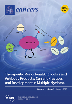
The introduction of immunotherapy marks a step forward in the quest for a cure for multiple myeloma. Monoclonal antibodies that target CD38 (daratumumab) and CS1 (elotuzumab) are highly effective and safe options for young and elderly patients. Daratumumab has been included in many regimens and moved from the relapsed to the frontline setting. Furthermore, antibody-drug conjugates such as belantamab mafodotin (GSK2857916) are also under clinical evaluation. Active immunotherapy, aimed at awakening patients’ immune systems against myeloma cells, is of great interest, with bispecific T cell engagers such as AMG420 proving to be effective in heavily pretreated patients in clinical trials. A number of challenges still remain, including the following: finding the correct timing for these agents during the disease course, understanding the mechanisms of resistance, and identifying the antigens to target [...] Read more.
  • Issues are regarded as officially published after their release is announced to the table of contents alert mailing list.
  • You may sign up for e-mail alerts to receive table of contents of newly released issues.
  • PDF is the official format for papers published in both, html and pdf forms. To view the papers in pdf format, click on the "PDF Full-text" link, and use the free Adobe Reader to open them.
Order results
Result details
Section
Select all
Export citation of selected articles as:
16 pages, 1355 KiB  
Review
The Tumor Suppressor Roles of MYBBP1A, a Major Contributor to Metabolism Plasticity and Stemness
by Blanca Felipe-Abrio and Amancio Carnero
Cancers 2020, 12(1), 254; https://doi.org/10.3390/cancers12010254 - 20 Jan 2020
Cited by 18 | Viewed by 6243
Abstract
The MYB binding protein 1A (MYBBP1A, also known as p160) acts as a co-repressor of multiple transcription factors involved in many physiological processes. Therefore, MYBBP1A acts as a tumor suppressor in multiple aspects related to cell physiology, most of them very relevant for [...] Read more.
The MYB binding protein 1A (MYBBP1A, also known as p160) acts as a co-repressor of multiple transcription factors involved in many physiological processes. Therefore, MYBBP1A acts as a tumor suppressor in multiple aspects related to cell physiology, most of them very relevant for tumorigenesis. We explored the different roles of MYBBP1A in different aspects of cancer, such as mitosis, cellular senescence, epigenetic regulation, cell cycle, metabolism plasticity and stemness. We especially reviewed the relationships between MYBBP1A, the inhibitory role it plays by binding and inactivating c-MYB and its regulation of PGC-1α, leading to an increase in the stemness and the tumor stem cell population. In addition, MYBBP1A causes the activation of PGC-1α directly and indirectly through c-MYB, inducing the metabolic change from glycolysis to oxidative phosphorylation (OXPHOS). Therefore, the combination of these two effects caused by the decreased expression of MYBBP1A provides a selective advantage to tumor cells. Interestingly, this only occurs in cells lacking pVHL. Finally, the loss of MYBBP1A occurs in 8%–9% of renal tumors. tumors, and this subpopulation could be studied as a possible target of therapies using inhibitors of mitochondrial respiration. Full article
Show Figures

Graphical abstract

17 pages, 3476 KiB  
Article
Survival-Associated Metabolic Genes in Human Papillomavirus-Positive Head and Neck Cancers
by Martin A. Prusinkiewicz, Steven F. Gameiro, Farhad Ghasemi, Mackenzie J. Dodge, Peter Y. F. Zeng, Hanna Maekebay, John W. Barrett, Anthony C. Nichols and Joe S. Mymryk
Cancers 2020, 12(1), 253; https://doi.org/10.3390/cancers12010253 - 20 Jan 2020
Cited by 33 | Viewed by 5662
Abstract
Human papillomavirus (HPV) causes an increasing number of head and neck squamous cell carcinomas (HNSCCs). Altered metabolism contributes to patient prognosis, but the impact of HPV status on HNSCC metabolism remains relatively uncharacterized. We hypothesize that metabolism-related gene expression differences unique to HPV-positive [...] Read more.
Human papillomavirus (HPV) causes an increasing number of head and neck squamous cell carcinomas (HNSCCs). Altered metabolism contributes to patient prognosis, but the impact of HPV status on HNSCC metabolism remains relatively uncharacterized. We hypothesize that metabolism-related gene expression differences unique to HPV-positive HNSCC influences patient survival. The Cancer Genome Atlas RNA-seq data from primary HNSCC patient samples were categorized as 73 HPV-positive, 442 HPV-negative, and 43 normal-adjacent control tissues. We analyzed 229 metabolic genes and identified numerous differentially expressed genes between HPV-positive and negative HNSCC patients. HPV-positive carcinomas exhibited lower expression levels of genes involved in glycolysis and higher levels of genes involved in the tricarboxylic acid cycle, oxidative phosphorylation, and β-oxidation than the HPV-negative carcinomas. Importantly, reduced expression of the metabolism-related genes SDHC, COX7A1, COX16, COX17, ELOVL6, GOT2, and SLC16A2 were correlated with improved patient survival only in the HPV-positive group. This work suggests that specific transcriptional alterations in metabolic genes may serve as predictive biomarkers of patient outcome and identifies potential targets for novel therapeutic intervention in HPV-positive head and neck cancers. Full article
(This article belongs to the Special Issue Human Papillomavirus and Cancers)
Show Figures

Figure 1

12 pages, 909 KiB  
Review
Role of Interleukin-34 in Cancer
by Eleonora Franzè, Carmine Stolfi, Edoardo Troncone, Patrizio Scarozza and Giovanni Monteleone
Cancers 2020, 12(1), 252; https://doi.org/10.3390/cancers12010252 - 20 Jan 2020
Cited by 32 | Viewed by 5004
Abstract
Cross-talk between cancer cells and the immune cells occurring in the tumor microenvironment is crucial in promoting signals that foster tumor growth and metastasis. Both cancer cells and immune cells secrete various interleukins (IL), which, either directly or indirectly, stimulate cancer-cell proliferation, survival, [...] Read more.
Cross-talk between cancer cells and the immune cells occurring in the tumor microenvironment is crucial in promoting signals that foster tumor growth and metastasis. Both cancer cells and immune cells secrete various interleukins (IL), which, either directly or indirectly, stimulate cancer-cell proliferation, survival, and diffusion, as well as contribute to sculpt the immune microenvironment, thereby amplifying tumorigenic stimuli. IL-34, a cytokine produced by a wide range of cells, has been initially involved in the control of differentiation, proliferation, and survival of myeloid cells. More recent studies documented the overexpression of IL-34 in several cancers, such as hepatocarcinoma, osteosarcoma, multiple myeloma, colon cancer, and lung cancer, and showed that tumor cells can produce and functionally respond to this cytokine. In this review, we summarize the multiple roles of IL-34 in various cancers, with the aim to better understand the relationship between the expression of this cytokine and cancer behavior and to provide new insights for exploring a new potential therapeutic target. Full article
(This article belongs to the Special Issue Interleukin in Cancer Pathogenesis and Treatment)
Show Figures

Figure 1

3 pages, 174 KiB  
Erratum
Erratum: Neuromedin U: A Small Peptide in the Big World of Cancer. Cancers 2019, 11, 1312
by Patrycja Przygodzka, Kamila Soboska, Ewelina Sochacka and Joanna Boncela
Cancers 2020, 12(1), 251; https://doi.org/10.3390/cancers12010251 - 20 Jan 2020
Cited by 4 | Viewed by 2004
Abstract
The authors wish to make the following corrections to this paper [...] Full article
(This article belongs to the Special Issue New Biomarkers in Cancers)
19 pages, 3901 KiB  
Article
Antiplatelet Drug Ticagrelor Enhances Chemotherapeutic Efficacy by Targeting the Novel P2Y12-AKT Pathway in Pancreatic Cancer Cells
by Omar Elaskalani, Alice Domenichini, Norbaini Binti Abdol Razak, Danielle E. Dye, Marco Falasca and Pat Metharom
Cancers 2020, 12(1), 250; https://doi.org/10.3390/cancers12010250 - 20 Jan 2020
Cited by 27 | Viewed by 5561
Abstract
Background: Extensive research has reported that extracellular ADP in the tumour microenvironment can stimulate platelets through interaction with the platelet receptor P2Y12. In turn, activated platelets release biological factors supporting cancer progression. Experimental data suggest that the tumour microenvironment components, of which platelets [...] Read more.
Background: Extensive research has reported that extracellular ADP in the tumour microenvironment can stimulate platelets through interaction with the platelet receptor P2Y12. In turn, activated platelets release biological factors supporting cancer progression. Experimental data suggest that the tumour microenvironment components, of which platelets are integral, can promote chemotherapy resistance in pancreatic ductal adenocarcinoma (PDAC). Thus, overcoming chemoresistance requires combining multiple inhibitors that simultaneously target intrinsic pathways in cancer cells and extrinsic factors related to the tumour microenvironment. We aimed to determine whether ticagrelor, an inhibitor of the ADP–P2Y12 axis and a well-known antiplatelet drug, could be a therapeutic option for PDAC. Methods: We investigated a functional P2Y12 receptor and its downstream signalling in a panel of PDAC cell lines and non-cancer pancreatic cells termed hTERT-HPNE. We tested the synergistic effect of ticagrelor, a P2Y12 inhibitor, in combination with chemotherapeutic drugs (gemcitabine, paclitaxel and cisplatin), in vitro and in vivo. Results: Knockdown studies revealed that P2Y12 contributed to epidermal growth factor receptor (EGFR) activation and the expression of SLUG and ZEB1, which are transcriptional factors implicated in metastasis and chemoresistance. Studies using genetic and pharmacological inhibitors showed that the P2Y12–EGFR crosstalk enhanced cancer cell proliferation. Inhibition of P2Y12 signalling significantly reduced EGF-dependent AKT activation and promoted the anticancer activity of anti-EGFR treatment. Importantly, ticagrelor significantly decreased the proliferative capacity of cancer but not normal pancreatic cells. In vitro, synergism was observed when ticagrelor was combined with several chemodrugs. In vivo, a combination of ticagrelor with gemcitabine significantly reduced tumour growth, whereas gemcitabine or ticagrelor alone had a minimal effect. Conclusions: These findings uncover a novel effect and mechanism of action of the antiplatelet drug ticagrelor in PDAC cells and suggest a multi-functional role for ADP-P2Y12 signalling in the tumour microenvironment. Full article
(This article belongs to the Special Issue Platelets and Cancer)
Show Figures

Figure 1

14 pages, 536 KiB  
Article
Germline Variants in Driver Genes of Breast Cancer and Their Association with Familial and Early-Onset Breast Cancer Risk in a Chilean Population
by Alejandro Fernandez-Moya, Sebastian Morales, Trinidad Arancibia, Patricio Gonzalez-Hormazabal, Julio C. Tapia, Raul Godoy-Herrera, Jose Miguel Reyes, Fernando Gomez, Enrique Waugh and Lilian Jara
Cancers 2020, 12(1), 249; https://doi.org/10.3390/cancers12010249 - 20 Jan 2020
Cited by 6 | Viewed by 3381
Abstract
The genetic variations responsible for tumorigenesis are called driver mutations. In breast cancer (BC), two studies have demonstrated that germline mutations in driver genes linked to sporadic tumors may also influence BC risk. The present study evaluates the association between SNPs and SNP-SNP [...] Read more.
The genetic variations responsible for tumorigenesis are called driver mutations. In breast cancer (BC), two studies have demonstrated that germline mutations in driver genes linked to sporadic tumors may also influence BC risk. The present study evaluates the association between SNPs and SNP-SNP interaction in driver genes TTN (rs10497520), TBX3 (rs2242442), KMT2D (rs11168827), and MAP3K1 (rs702688 and rs702689) with BC risk in BRCA1/2-negative Chilean families. The SNPs were genotyped in 489 BC cases and 1078 controls by TaqMan Assay. Our data do not support an association between rs702688: A>G or rs702689: G>A and BC risk. The rs10497520-T allele was associated with a decreased risk in patients with family history of BC or early-onset BC (OR = 0.6, p < 0.0001 and OR = 0.7, p = 0.05, respectively). rs2242442-G was associated with a protective effect and rs11168827-C was associated with increased BC risk in families with a strong history of BC (OR = 0.6, p = 0.02 and OR = 1.4, p = 0.05, respectively). As rs10497520-T and rs2242442-G seemed to protect against BC risk, we then evaluated their combined effect. Familial BC risk decreased in a dose-dependent manner with the protective allele count, reflecting an additive effect (p-trend < 10−4). To our knowledge, this is the first association study of BC driver gene germline variations in a Chilean population. Full article
(This article belongs to the Special Issue Molecular Genetics of Breast and Ovary Cancer)
Show Figures

Graphical abstract

9 pages, 1031 KiB  
Article
Evaluation of Vascular Patterns Using Contact Endoscopy and Narrow-Band Imaging (CE-NBI) for the Diagnosis of Vocal Fold Malignancy
by Nikolaos Davaris, Anke Lux, Nazila Esmaeili, Alfredo Illanes, Axel Boese, Michael Friebe and Christoph Arens
Cancers 2020, 12(1), 248; https://doi.org/10.3390/cancers12010248 - 20 Jan 2020
Cited by 14 | Viewed by 3908
Abstract
The endoscopic detection of perpendicular vascular changes (PVC) of the vocal folds has been associated with vocal fold cancer, dysplastic lesions, and papillomatosis, according to a classification proposed by the European Laryngological Society (ELS). The combination of contact endoscopy with narrow-band imaging (NBI-CE) [...] Read more.
The endoscopic detection of perpendicular vascular changes (PVC) of the vocal folds has been associated with vocal fold cancer, dysplastic lesions, and papillomatosis, according to a classification proposed by the European Laryngological Society (ELS). The combination of contact endoscopy with narrow-band imaging (NBI-CE) allows intraoperatively a highly contrasted, real-time visualization of vascular changes of the vocal folds. Aim of the present study was to determine the association of PVC to specific histological diagnoses, the level of interobserver agreement in the detection of PVC, and their diagnostic effectiveness in diagnosing laryngeal malignancy. The evaluation of our data confirmed the association of PVC to vocal fold cancer, dysplastic lesions, and papillomatosis. The level of agreement between the observers in the identification of PVC was moderate for the less-experienced observers and almost perfect for the experienced observers. The identification of PVC during NBI-CE proved to be a valuable indicator for diagnosing malignant and premalignant lesions. Full article
(This article belongs to the Special Issue Emerging Concepts in Treatment of Laryngeal Cancer)
Show Figures

Figure 1

12 pages, 1961 KiB  
Article
Sterol O-Acyl Transferase 1 as a Prognostic Marker of Adrenocortical Carcinoma
by Amanda Meneses Ferreira Lacombe, Iberê Cauduro Soares, Beatriz Marinho de Paula Mariani, Mirian Yumie Nishi, João Evangelista Bezerra-Neto, Helaine da Silva Charchar, Vania Balderrama Brondani, Fabio Tanno, Victor Srougi, José Luiz Chambo, Ricardo Miguel Costa de Freitas, Berenice Bilharinho Mendonca, Ana O. Hoff, Madson Q. Almeida, Isabel Weigand, Matthias Kroiss, Maria Claudia Nogueira Zerbini and Maria Candida Barisson Villares Fragoso
Cancers 2020, 12(1), 247; https://doi.org/10.3390/cancers12010247 - 19 Jan 2020
Cited by 20 | Viewed by 3212
Abstract
Adrenocortical carcinoma (ACC) is a rare endocrine malignancy with an unfavorable prognosis. Despite the poor prognosis in the majority of patients, no improvements in treatment strategies have been achieved. Therefore, the discovery of new prognostic biomarkers is of enormous interest. Sterol-O-acyl transferase 1 [...] Read more.
Adrenocortical carcinoma (ACC) is a rare endocrine malignancy with an unfavorable prognosis. Despite the poor prognosis in the majority of patients, no improvements in treatment strategies have been achieved. Therefore, the discovery of new prognostic biomarkers is of enormous interest. Sterol-O-acyl transferase 1 (SOAT1) is involved in cholesterol esterification and lipid droplet formation. Recently, it was demonstrated that SOAT1 inhibition leads to impaired steroidogenesis and cell viability in ACC. To date, no studies have addressed the impact of SOAT1 expression on ACC prognosis and clinical outcomes. We evaluated SOAT1 expression by quantitative real-time polymerase chain reaction and immunohistochemistry in a tissue microarray of 112 ACCs (Weiss score ≥ 3) from adults treated in a single tertiary center in Brazil. Two independent pathologists evaluated the immunohistochemistry results through a semiquantitative approach (0–4). We aimed to evaluate the correlation between SOAT1 expression and clinical, biochemical and anatomopathological parameters, recurrence-free survival (RFS), progression-free survival (PFS), and overall survival (OS). SOAT1 protein expression was heterogeneous in this cohort, 37.5% of the ACCs demonstrated a strong SOAT1 protein expression (score > 2), while 62.5% demonstrated a weak or absent protein expression (score ≤ 2). Strong SOAT1 protein expression correlated with features of high aggressiveness in ACC, such as excessive tumor cortisol secretion (p = 0.01), an advanced disease stage [European Network for the Study of Adrenal Tumors (ENSAT) staging system 3 and 4 (p = 0.011)] and a high Ki67 index (p = 0.002). In multivariate analysis, strong SOAT1 protein expression was an independent predictor of a reduced OS (hazard ratio (HR) 2.15, confidence interval (CI) 95% 1.26–3.66; p = 0.005) in all patients (n = 112), and a reduced RFS (HR 2.1, CI 95% 1.09–4.06; p = 0.027) in patients with localized disease at diagnosis (n = 83). Our findings demonstrated that SOAT1 protein expression has prognostic value in ACC and reinforced the importance of investigating SOAT1 as a possible therapeutic target for patients with ACC. Full article
(This article belongs to the Special Issue Adrenocortical Carcinoma)
Show Figures

Figure 1

18 pages, 4749 KiB  
Article
Does the Site of Origin of the Microcarcinoma with Respect to the Thyroid Surface Matter? A Multicenter Pathologic and Clinical Study for Risk Stratification
by Giovanni Tallini, Antonio De Leo, Andrea Repaci, Dario de Biase, Maria Letizia Bacchi Reggiani, Doriana Di Nanni, Francesca Ambrosi, Cira Di Gioia, Giorgio Grani, Kerry Jane Rhoden, Erica Solaroli, Fabio Monari, Sebastiano Filetti and Cosimo Durante
Cancers 2020, 12(1), 246; https://doi.org/10.3390/cancers12010246 - 19 Jan 2020
Cited by 17 | Viewed by 2998
Abstract
It is unclear whether the site of origin of papillary thyroid microcarcinoma (mPTC) with respect to the thyroid surface has an influence on clinicopathologic parameters. The objectives of the study were to: (i) Accurately measure the mPTC distance from the thyroid surface; (ii) [...] Read more.
It is unclear whether the site of origin of papillary thyroid microcarcinoma (mPTC) with respect to the thyroid surface has an influence on clinicopathologic parameters. The objectives of the study were to: (i) Accurately measure the mPTC distance from the thyroid surface; (ii) analyze whether this distance correlates with relevant clinicopathologic parameters; and (iii) investigate the impact of the site of origin of the mPTC on risk stratification. Clinicopathologic features and BRAF mutational status were analyzed and correlated with the site of origin of the mPTC in a multicenter cohort of 298 mPTCs from six Italian medical institutions. Tumors arise at a median distance of 3.5 mm below the surface of the thyroid gland. Statistical analysis identified four distinct clusters. Group A, mPTC: size ≥ 5 mm and distance of the edge of the tumor from the thyroid capsule = 0 mm; group B, mPTC: size ≥ 5 mm and distance of the edge of the tumor from the thyroid capsule > 0 mm; group C, mPTC: size < 5 mm and distance of the edge of the tumor from the thyroid capsule = 0 mm; and group D, mPTC: size < 5 mm and distance of the edge of the tumor from the thyroid capsule > 0 mm. Univariate analysis demonstrates significant differences between the groups: Group A shows the most aggressive features, and group D the most indolent ones. By multivariate analysis, group A tumors are characterized by tall cell histotype, BRAF V600E mutation, tumor fibrosis, aggressive growth with invasive features, vascular invasion, lymph node metastases, and intermediate ATA risk. The mPTC clinicopathologic features vary according to the tumor size and distance from the thyroid surface. A four-group model may be useful for risk stratification and to refine the selection of nodules to be targeted for fine needle aspiration. Full article
(This article belongs to the Special Issue Thyroid Cancer)
Show Figures

Figure 1

13 pages, 2239 KiB  
Article
Non-Thermal Atmospheric Pressure Bio-Compatible Plasma Stimulates Apoptosis via p38/MAPK Mechanism in U87 Malignant Glioblastoma
by Mahmuda Akter, Anshika Jangra, Seung Ah Choi, Eun Ha Choi and Ihn Han
Cancers 2020, 12(1), 245; https://doi.org/10.3390/cancers12010245 - 19 Jan 2020
Cited by 45 | Viewed by 4451
Abstract
Nonthermal plasma is a promising novel therapy for the alteration of biological and clinical functions of cells and tissues, including apoptosis and inhibition of tumor progression. This therapy generates reactive oxygen and nitrogen species (RONS), which play a major role in anticancer effects. [...] Read more.
Nonthermal plasma is a promising novel therapy for the alteration of biological and clinical functions of cells and tissues, including apoptosis and inhibition of tumor progression. This therapy generates reactive oxygen and nitrogen species (RONS), which play a major role in anticancer effects. Previous research has verified that plasma jets can selectively induce apoptosis in various cancer cells, suggesting that it could be a potentially effective novel therapy in combination with or as an alternative to conventional therapeutic methods. In this study, we determined the effects of nonthermal air soft plasma jets on a U87 MG brain cancer cell line, including the dose- and time-dependent effects and the physicochemical and biological correlation between the RONS cascade and p38/mitogen-activated protein kinase (MAPK) signaling pathway, which contribute to apoptosis. The results indicated that soft plasma jets efficiently inhibit cell proliferation and induce apoptosis in U87 MG cells but have minimal effects on astrocytes. These findings revealed that soft plasma jets produce a potent cytotoxic effect via the initiation of cell cycle arrest and apoptosis. The production of reactive oxygen species (ROS) in cells was tested, and an intracellular ROS scavenger, N-acetyl cysteine (NAC), was examined. Our results suggested that soft plasma jets could potentially be used as an effective approach for anticancer therapy. Full article
(This article belongs to the Special Issue Plasma in Cancer Treatment)
Show Figures

Figure 1

17 pages, 4909 KiB  
Article
Resistance to MET/VEGFR2 Inhibition by Cabozantinib Is Mediated by YAP/TBX5-Dependent Induction of FGFR1 in Castration-Resistant Prostate Cancer
by Filippos Koinis, Paul Corn, Nila Parikh, Jian Song, Ioulia Vardaki, Ioanna Mourkioti, Sue-Hwa Lin, Christopher Logothetis, Theocharis Panaretakis and Gary Gallick
Cancers 2020, 12(1), 244; https://doi.org/10.3390/cancers12010244 - 19 Jan 2020
Cited by 19 | Viewed by 4794
Abstract
The overall goal of this study was to elucidate the role of FGFR1 induction in acquired resistance to MET and VEGFR2 inhibition by cabozantinib in prostate cancer (PCa) and leverage this understanding to improve therapy outcomes. The response to cabozantinib was examined in [...] Read more.
The overall goal of this study was to elucidate the role of FGFR1 induction in acquired resistance to MET and VEGFR2 inhibition by cabozantinib in prostate cancer (PCa) and leverage this understanding to improve therapy outcomes. The response to cabozantinib was examined in mice bearing patient-derived xenografts in which FGFR1 was overexpressed. Using a variety of cell models that reflect different PCa disease states, the mechanism underpinning FGFR1 signaling activation by cabozantinib was investigated. We performed parallel investigations in specimens from cabozantinib-treated patients to confirm our in vitro and in vivo data. FGFR1 overexpression was sufficient to confer resistance to cabozantinib. Our results demonstrate transcriptional activation of FGF/FGFR1 expression in cabozantinib-resistant models. Further analysis of molecular pathways identified a YAP/TBX5-driven mechanism of FGFR1 and FGF overexpression induced by MET inhibition. Importantly, knockdown of YAP and TBX5 led to decreased FGFR1 protein expression and decreased mRNA levels of FGFR1, FGF1, and FGF2. This association was confirmed in a cohort of hormone-naïve patients with PCa receiving androgen deprivation therapy and cabozantinib, further validating our findings. These findings reveal that the molecular basis of resistance to MET inhibition in PCa is FGFR1 activation through a YAP/TBX5-dependent mechanism. YAP and its downstream target TBX5 represent a crucial mediator in acquired resistance to MET inhibitors. Thus, our studies provide insight into the mechanism of acquired resistance and will guide future development of clinical trials with MET inhibitors. Full article
(This article belongs to the Collection Drug Resistance and Novel Therapies in Cancers)
Show Figures

Figure 1

56 pages, 580 KiB  
Editorial
Acknowledgement to Reviewers of Cancers in 2019
by Cancers Editorial Office
Cancers 2020, 12(1), 243; https://doi.org/10.3390/cancers12010243 - 19 Jan 2020
Viewed by 7053
Abstract
The editorial team greatly appreciates the reviewers who have dedicated their considerable time and expertise to the journal’s rigorous editorial process over the past 12 months, regardless of whether the papers are finally published or not [...] Full article
23 pages, 1455 KiB  
Review
Nano-Therapies for Glioblastoma Treatment
by Edouard Alphandéry
Cancers 2020, 12(1), 242; https://doi.org/10.3390/cancers12010242 - 19 Jan 2020
Cited by 70 | Viewed by 8067
Abstract
Traditional anti-cancer treatments are inefficient against glioblastoma, which remains one of the deadliest and most aggressive cancers. Nano-drugs could help to improve this situation by enabling: (i) an increase of anti-glioblastoma multiforme (GBM) activity of chemo/gene therapeutic drugs, notably by an improved diffusion [...] Read more.
Traditional anti-cancer treatments are inefficient against glioblastoma, which remains one of the deadliest and most aggressive cancers. Nano-drugs could help to improve this situation by enabling: (i) an increase of anti-glioblastoma multiforme (GBM) activity of chemo/gene therapeutic drugs, notably by an improved diffusion of these drugs through the blood brain barrier (BBB), (ii) the sensibilization of radio-resistant GBM tumor cells to radiotherapy, (iii) the removal by surgery of infiltrating GBM tumor cells, (iv) the restoration of an apoptotic mechanism of GBM cellular death, (v) the destruction of angiogenic blood vessels, (vi) the stimulation of anti-tumor immune cells, e.g., T cells, NK cells, and the neutralization of pro-tumoral immune cells, e.g., Treg cells, (vii) the local production of heat or radical oxygen species (ROS), and (viii) the controlled release/activation of anti-GBM drugs following the application of a stimulus. This review covers these different aspects. Full article
Show Figures

Figure 1

14 pages, 1798 KiB  
Article
1H-NMR Based Serum Metabolomics Highlights Different Specific Biomarkers between Early and Advanced Hepatocellular Carcinoma Stages
by Andrea Casadei-Gardini, Laura Del Coco, Giorgia Marisi, Fabio Conti, Giulia Rovesti, Paola Ulivi, Matteo Canale, Giovanni Luca Frassineti, Francesco Giuseppe Foschi, Serena Longo, Francesco Paolo Fanizzi and Anna Maria Giudetti
Cancers 2020, 12(1), 241; https://doi.org/10.3390/cancers12010241 - 18 Jan 2020
Cited by 40 | Viewed by 5447
Abstract
The application of non-targeted serum metabolomics profiling represents a noninvasive tool to identify new clinical biomarkers and to provide early diagnostic differentiation, and insight into the pathological mechanisms underlying hepatocellular carcinoma (HCC) progression. In this study, we used proton Nuclear Magnetic Resonance ( [...] Read more.
The application of non-targeted serum metabolomics profiling represents a noninvasive tool to identify new clinical biomarkers and to provide early diagnostic differentiation, and insight into the pathological mechanisms underlying hepatocellular carcinoma (HCC) progression. In this study, we used proton Nuclear Magnetic Resonance (1H-NMR) Spectroscopy and multivariate data analysis to profile the serum metabolome of 64 HCC patients, in early (n = 28) and advanced (n = 36) disease stages. We found that 1H-NMR metabolomics profiling could discriminate early from advanced HCC patients with a cross-validated accuracy close to 100%. Orthogonal partial least squares discriminant analysis (OPLS-DA) showed significant changes in serum glucose, lactate, lipids and some amino acids, such as alanine, glutamine, 1-methylhistidine, lysine and valine levels between advanced and early HCC patients. Moreover, in early HCC patients, Kaplan–Meier analysis highlighted the serum tyrosine level as a predictor for overall survival (OS). Overall, our analysis identified a set of metabolites with possible clinical and biological implication in HCC pathophysiology. Full article
(This article belongs to the Special Issue Recent Research on Gastrointestinal Carcinoma)
Show Figures

Graphical abstract

48 pages, 2713 KiB  
Review
Pharmacological Inhibition of Oncogenic STAT3 and STAT5 Signaling in Hematopoietic Cancers
by Marie Brachet-Botineau, Marion Polomski, Heidi A. Neubauer, Ludovic Juen, Damien Hédou, Marie-Claude Viaud-Massuard, Gildas Prié and Fabrice Gouilleux
Cancers 2020, 12(1), 240; https://doi.org/10.3390/cancers12010240 - 18 Jan 2020
Cited by 47 | Viewed by 7687
Abstract
Signal Transducer and Activator of Transcription (STAT) 3 and 5 are important effectors of cellular transformation, and aberrant STAT3 and STAT5 signaling have been demonstrated in hematopoietic cancers. STAT3 and STAT5 are common targets for different tyrosine kinase oncogenes (TKOs). In addition, STAT3 [...] Read more.
Signal Transducer and Activator of Transcription (STAT) 3 and 5 are important effectors of cellular transformation, and aberrant STAT3 and STAT5 signaling have been demonstrated in hematopoietic cancers. STAT3 and STAT5 are common targets for different tyrosine kinase oncogenes (TKOs). In addition, STAT3 and STAT5 proteins were shown to contain activating mutations in some rare but aggressive leukemias/lymphomas. Both proteins also contribute to drug resistance in hematopoietic malignancies and are now well recognized as major targets in cancer treatment. The development of inhibitors targeting STAT3 and STAT5 has been the subject of intense investigations during the last decade. This review summarizes the current knowledge of oncogenic STAT3 and STAT5 functions in hematopoietic cancers as well as advances in preclinical and clinical development of pharmacological inhibitors. Full article
(This article belongs to the Special Issue Targeting STAT3 and STAT5 in Cancer)
Show Figures

Figure 1

18 pages, 1625 KiB  
Article
In-Depth Mapping of the Urinary N-Glycoproteome: Distinct Signatures of ccRCC-related Progression
by Lucia Santorelli, Giulia Capitoli, Clizia Chinello, Isabella Piga, Francesca Clerici, Vanna Denti, Andrew Smith, Angelica Grasso, Francesca Raimondo, Marco Grasso and Fulvio Magni
Cancers 2020, 12(1), 239; https://doi.org/10.3390/cancers12010239 - 18 Jan 2020
Cited by 14 | Viewed by 3708
Abstract
Protein N-glycosylation is one of the most important post-translational modifications and is involved in many biological processes, with aberrant changes in protein N-glycosylation patterns being closely associated with several diseases, including the progression and spreading of tumours. In light of this, [...] Read more.
Protein N-glycosylation is one of the most important post-translational modifications and is involved in many biological processes, with aberrant changes in protein N-glycosylation patterns being closely associated with several diseases, including the progression and spreading of tumours. In light of this, identifying these aberrant protein glycoforms in tumours could be useful for understanding the molecular mechanism of this multifactorial disease, developing specific biomarkers and finding novel therapeutic targets. We investigated the urinary N-glycoproteome of clear cell renal cell carcinoma (ccRCC) patients at different stages (n = 15 at pT1 and n = 15 at pT3), and of non-ccRCC subjects (n = 15), using an N-glyco-FASP-based method. Using label-free nLC-ESI MS/MS, we identified and quantified several N-glycoproteins with altered expression and abnormal changes affecting the occupancy of the glycosylation site in the urine of RCC patients compared to control. In particular, nine of them had a specific trend that was directly related to the stage progression: CD97, COCH and P3IP1 were up-expressed whilst APOB, FINC, CERU, CFAH, HPT and PLTP were down-expressed in ccRCC patients. Overall, these results expand our knowledge related to the role of this post-translational modification in ccRCC and translation of this information into pre-clinical studies could have a significant impact on the discovery of novel biomarkers and therapeutic target in kidney cancer. Full article
(This article belongs to the Special Issue Renal Cell Carcinoma)
Show Figures

Graphical abstract

24 pages, 1487 KiB  
Review
Cytoskeletal Proteins in Cancer and Intracellular Stress: A Therapeutic Perspective
by Mei Shan Ong, Shuo Deng, Clarissa Esmeralda Halim, Wanpei Cai, Tuan Zea Tan, Ruby Yun-Ju Huang, Gautam Sethi, Shing Chuan Hooi, Alan Prem Kumar and Celestial T. Yap
Cancers 2020, 12(1), 238; https://doi.org/10.3390/cancers12010238 - 18 Jan 2020
Cited by 63 | Viewed by 9530
Abstract
Cytoskeletal proteins, which consist of different sub-families of proteins including microtubules, actin and intermediate filaments, are essential for survival and cellular processes in both normal as well as cancer cells. However, in cancer cells, these mechanisms can be altered to promote tumour development [...] Read more.
Cytoskeletal proteins, which consist of different sub-families of proteins including microtubules, actin and intermediate filaments, are essential for survival and cellular processes in both normal as well as cancer cells. However, in cancer cells, these mechanisms can be altered to promote tumour development and progression, whereby the functions of cytoskeletal proteins are co-opted to facilitate increased migrative and invasive capabilities, proliferation, as well as resistance to cellular and environmental stresses. Herein, we discuss the cytoskeletal responses to important intracellular stresses (such as mitochondrial, endoplasmic reticulum and oxidative stresses), and delineate the consequences of these responses, including effects on oncogenic signalling. In addition, we elaborate how the cytoskeleton and its associated molecules present themselves as therapeutic targets. The potential and limitations of targeting new classes of cytoskeletal proteins are also explored, in the context of developing novel strategies that impact cancer progression. Full article
(This article belongs to the Special Issue Targeted Cancer Therapy)
Show Figures

Figure 1

17 pages, 4291 KiB  
Article
Loss of Rb1 Enhances Glycolytic Metabolism in Kras-Driven Lung Tumors In Vivo
by Lindsey R. Conroy, Susan Dougherty, Traci Kruer, Stephanie Metcalf, Pawel Lorkiewicz, Liqing He, Xinmin Yin, Xiang Zhang, Sengodagounder Arumugam, Lyndsay E.A. Young, Ramon C. Sun and Brian F. Clem
Cancers 2020, 12(1), 237; https://doi.org/10.3390/cancers12010237 - 17 Jan 2020
Cited by 12 | Viewed by 3873
Abstract
Dysregulated metabolism is a hallmark of cancer cells and is driven in part by specific genetic alterations in various oncogenes or tumor suppressors. The retinoblastoma protein (pRb) is a tumor suppressor that canonically regulates cell cycle progression; however, recent studies have highlighted a [...] Read more.
Dysregulated metabolism is a hallmark of cancer cells and is driven in part by specific genetic alterations in various oncogenes or tumor suppressors. The retinoblastoma protein (pRb) is a tumor suppressor that canonically regulates cell cycle progression; however, recent studies have highlighted a functional role for pRb in controlling cellular metabolism. Here, we report that loss of the gene encoding pRb (Rb1) in a transgenic mutant Kras-driven model of lung cancer results in metabolic reprogramming. Our tracer studies using bolus dosing of [U-13C]-glucose revealed an increase in glucose carbon incorporation into select glycolytic intermediates. Consistent with this result, Rb1-depleted tumors exhibited increased expression of key glycolytic enzymes. Interestingly, loss of Rb1 did not alter mitochondrial pyruvate oxidation compared to lung tumors with intact Rb1. Additional tracer studies using [U-13C,15N]-glutamine and [U-13C]-lactate demonstrated that loss of Rb1 did not alter glutaminolysis or utilization of circulating lactate within the tricarboxylic acid cycle (TCA) in vivo. Taken together, these data suggest that the loss of Rb1 promotes a glycolytic phenotype, while not altering pyruvate oxidative metabolism or glutamine anaplerosis in Kras-driven lung tumors. Full article
(This article belongs to the Collection Recent Advances in Tumor Suppressor)
Show Figures

Figure 1

20 pages, 1630 KiB  
Review
Does Direct and Indirect Exposure to Ionising Radiation Influence the Metastatic Potential of Breast Cancer Cells
by Munira A. Kadhim, Ammar Mayah and Susan A. Brooks
Cancers 2020, 12(1), 236; https://doi.org/10.3390/cancers12010236 - 17 Jan 2020
Cited by 6 | Viewed by 4335
Abstract
Ionising radiation (IR) is commonly used for cancer therapy; however, its potential influence on the metastatic ability of surviving cancer cells exposed directly or indirectly to IR remains controversial. Metastasis is a multistep process by which the cancer cells dissociate from the initial [...] Read more.
Ionising radiation (IR) is commonly used for cancer therapy; however, its potential influence on the metastatic ability of surviving cancer cells exposed directly or indirectly to IR remains controversial. Metastasis is a multistep process by which the cancer cells dissociate from the initial site, invade, travel through the blood stream or lymphatic system, and colonise distant sites. This complex process has been reported to require cancer cells to undergo epithelial-mesenchymal transition (EMT) by which the cancer cells convert from an adhesive, epithelial to motile, mesenchymal form and is also associated with changes in glycosylation of cell surface proteins, which may be functionally involved in metastasis. In this paper, we give an overview of metastatic mechanisms and of the fundamentals of cancer-associated glycosylation changes. While not attempting a comprehensive review of this wide and fast moving field, we highlight some of the accumulating evidence from in vitro and in vivo models for increased metastatic potential in cancer cells that survive IR, focusing on angiogenesis, cancer cell motility, invasion, and EMT and glycosylation. We also explore the indirect effects in cells exposed to exosomes released from irradiated cells. The results of such studies need to be interpreted with caution and there remains limited evidence that radiotherapy enhances the metastatic capacity of cancers in a clinical setting and undoubtedly has a very positive clinical benefit. However, there is potential that this therapeutic benefit may ultimately be enhanced through a better understanding of the direct and indirect effects of IR on cancer cell behaviour. Full article
Show Figures

Figure 1

18 pages, 4226 KiB  
Article
The Long Non-Coding RNA Prader Willi/Angelman Region RNA5 (PAR5) Is Downregulated in Anaplastic Thyroid Carcinomas Where It Acts as a Tumor Suppressor by Reducing EZH2 Activity
by Simona Pellecchia, Romina Sepe, Myriam Decaussin-Petrucci, Cristina Ivan, Masayoshi Shimizu, Carmela Coppola, Domenico Testa, George Adrian Calin, Alfredo Fusco and Pierlorenzo Pallante
Cancers 2020, 12(1), 235; https://doi.org/10.3390/cancers12010235 - 17 Jan 2020
Cited by 36 | Viewed by 3570
Abstract
Anaplastic thyroid carcinoma (ATC) represents one the most aggressive neoplasias in humans, and, nowadays, limited advances have been made to extend the survival and reduce the mortality of ATC. Thus, the identification of molecular mechanism underlying its progression is needed. Here, we evaluated [...] Read more.
Anaplastic thyroid carcinoma (ATC) represents one the most aggressive neoplasias in humans, and, nowadays, limited advances have been made to extend the survival and reduce the mortality of ATC. Thus, the identification of molecular mechanism underlying its progression is needed. Here, we evaluated the long non-coding RNA (lncRNA) expression profile of nine ATC in comparison with five normal thyroid tissues by a lncRNA microarray. By this analysis, we identified 19 upregulated and 28 downregulated lncRNAs with a fold change >1.1 or <−1.1 and p-value < 0.05, in ATC samples. Some of them were subsequently validated by qRT-PCR. Then, we investigated the role of the lncRNA Prader Willi/Angelman region RNA5 (PAR5), drastically and specifically downregulated in ATC. The restoration of PAR5 reduces proliferation and migration rates of ATC-derived cell lines indicating that its downregulation contributes to thyroid cancer progression. Our results suggest that PAR5 exerts its anti-oncogenic role by impairing Enhancer of Zeste Homolog 2 (EZH2) oncogenic activity since we demonstrated that PAR5 interacts with it in thyroid cancer cell lines, reducing EZH2 protein levels and its binding on the E-cadherin promoter, relieving E-cadherin from the negative regulation by EZH2. Consistently, EZH2 is overexpressed in ATC, but not in differentiated thyroid carcinomas. The results reported here define a tumor suppressor role for PAR5 in undifferentiated thyroid neoplasias, further highlighting the pivotal role of lncRNAs in thyroid carcinogenesis. Full article
(This article belongs to the Special Issue Thyroid Cancer)
Show Figures

Figure 1

10 pages, 218 KiB  
Article
Should Genetic Testing for Cancer Predisposition Be Standard-of-Care for Women with Invasive Breast Cancer? The Murtha Cancer Center Experience
by Seth K. Rummel, Leann A. Lovejoy, Clesson E. Turner, Craig D. Shriver and Rachel E. Ellsworth
Cancers 2020, 12(1), 234; https://doi.org/10.3390/cancers12010234 - 17 Jan 2020
Cited by 7 | Viewed by 3595
Abstract
Currently, genetic testing is offered only to women diagnosed with breast cancer who meet a defined set of criteria and is not included as standard-of-care treatment at the time of diagnosis. Thus, a significant number of women diagnosed with breast cancer may miss [...] Read more.
Currently, genetic testing is offered only to women diagnosed with breast cancer who meet a defined set of criteria and is not included as standard-of-care treatment at the time of diagnosis. Thus, a significant number of women diagnosed with breast cancer may miss the opportunity for precision medical treatment and risk management. The effects of eligibility, timing, and uptake of genetic testing were evaluated in a cohort of women with invasive breast cancer diagnosed between 2001–2018. Risk status was estimated using NCCN BRCA1/2 testing criteria and panel testing was performed for all women who had genomic DNA available. Of the 1231 women, 57.8% were eligible for genetic testing. Uptake of testing within high-risk women was 42.7% of which 6.6% pursued clinical testing only after a second tumor event. Mutation frequencies were 15.8%, 5.5%, and 4.0% in high-risk women with clinical testing, high-risk women without clinical testing, and low-risk women, respectively. More than 4% of all patients harbored pathogenic or likely pathogenic mutations detected only in the research setting. Inclusion of panel testing at the time of diagnosis would allow for appropriate surveillance and treatment strategies to be employed to reduce the risk of secondary tumors and improve patient outcome. Full article
(This article belongs to the Special Issue The Study of Cancer Susceptibility Genes)
Show Figures

Graphical abstract

16 pages, 2308 KiB  
Article
Elevated Expression of Lumican in Lung Cancer Cells Promotes Bone Metastasis through an Autocrine Regulatory Mechanism
by Kuan-Chung Hsiao, Pei-Yi Chu, Gee-Chen Chang and Ko-Jiunn Liu
Cancers 2020, 12(1), 233; https://doi.org/10.3390/cancers12010233 - 17 Jan 2020
Cited by 22 | Viewed by 3078
Abstract
Background: The microarray analysis of whole-genome expression indicated that the gene encoding the protein lumican, which is associated with extracellular matrix (ECM) interaction, was highly expressed in osteotropic lung cancer cell lines with an enhanced capacity of bone metastasis. Methods: The [...] Read more.
Background: The microarray analysis of whole-genome expression indicated that the gene encoding the protein lumican, which is associated with extracellular matrix (ECM) interaction, was highly expressed in osteotropic lung cancer cell lines with an enhanced capacity of bone metastasis. Methods: The expression of lumican in the osteotropic lung cancer cells was downregulated, and the in vitro migration, invasion, and adhesion of cancer cells to ECM components, and the in vivo bone metastasis capacity of these cells were examined. Exogenous lumican was provided to study the autocrine regulation mechanism of lumican in the bone metastasis of lung cancer cells. Results: Transfection with lumican-specific short hairpin RNA (shRNA) in the osteotropic lung cancer cells reduced the establishment of in vivo bone metastasis, but not lung metastasis. Reduction in the expression of lumican also decreased the attachment of lung osteotropic cancer cells to several components of the ECM and suppressed cell migration and invasion in vitro. Exogenous lumican restored these reduced capacities of lumican knockdown cells and promoted the seeding of lung cancer cells in the bone microenvironment. Conclusions: These results suggested that lumican promotes the metastasis of lung cancer cells to the bones via an autocrine regulatory mechanism, and blocking this interaction may provide a new therapeutic approach to reduce bone metastasis in cases of lung cancer. Full article
Show Figures

Figure 1

18 pages, 10204 KiB  
Article
Ex-Vivo Treatment of Tumor Tissue Slices as a Predictive Preclinical Method to Evaluate Targeted Therapies for Patients with Renal Carcinoma
by Caroline Roelants, Catherine Pillet, Quentin Franquet, Clément Sarrazin, Nicolas Peilleron, Sofia Giacosa, Laurent Guyon, Amina Fontanell, Gaëlle Fiard, Jean-Alexandre Long, Jean-Luc Descotes, Claude Cochet and Odile Filhol
Cancers 2020, 12(1), 232; https://doi.org/10.3390/cancers12010232 - 17 Jan 2020
Cited by 36 | Viewed by 5031
Abstract
Clear cell renal cell carcinoma (ccRCC) is the third type of urologic cancer. At time of diagnosis, 30% of cases are metastatic with no effect of chemotherapy or radiotherapy. Current targeted therapies lead to a high rate of relapse and resistance after a [...] Read more.
Clear cell renal cell carcinoma (ccRCC) is the third type of urologic cancer. At time of diagnosis, 30% of cases are metastatic with no effect of chemotherapy or radiotherapy. Current targeted therapies lead to a high rate of relapse and resistance after a short-term response. Thus, a major hurdle in the development and use of new treatments for ccRCC is the lack of good pre-clinical models that can accurately predict the efficacy of new drugs and allow the stratification of patients into the correct treatment regime. Here, we describe different 3D cultures models of ccRCC, emphasizing the feasibility and the advantage of ex-vivo treatment of fresh, surgically resected human tumor slice cultures of ccRCC as a robust preclinical model for identifying patient response to specific therapeutics. Moreover, this model based on precision-cut tissue slices enables histopathology measurements as tumor architecture is retained, including the spatial relationship between the tumor and tumor-infiltrating lymphocytes and the stromal components. Our data suggest that acute treatment of tumor tissue slices could represent a benchmark of further exploration as a companion diagnostic tool in ccRCC treatment and a model to develop new therapeutic drugs. Full article
(This article belongs to the Special Issue Renal Cell Carcinoma)
Show Figures

Figure 1

14 pages, 1430 KiB  
Article
Heterogeneous EGFR, CDK4, MDM4, and PDGFRA Gene Expression Profiles in Primary GBM: No Association with Patient Survival
by María González-Tablas, Daniel Arandia, María Jara-Acevedo, Álvaro Otero, Ana-Luisa Vital, Carlos Prieto, Nerea González-Garcia, Ana Belén Nieto-Librero, Herminio Tao, Daniel Pascual, Laura Ruiz, Pablo Sousa, Purificación Galindo-Villardón, Alberto Orfao and María Dolores Tabernero
Cancers 2020, 12(1), 231; https://doi.org/10.3390/cancers12010231 - 17 Jan 2020
Cited by 12 | Viewed by 3691
Abstract
Background: The prognostic impact of the expression profile of genes recurrently amplified in glioblastoma multiforme (GBM) remains controversial. Methods: We investigated the RNA gene expression profile of epidermal growth factor receptor (EGFR), cyclin-dependent kinase 4 (CDK4), murine doble minute [...] Read more.
Background: The prognostic impact of the expression profile of genes recurrently amplified in glioblastoma multiforme (GBM) remains controversial. Methods: We investigated the RNA gene expression profile of epidermal growth factor receptor (EGFR), cyclin-dependent kinase 4 (CDK4), murine doble minute 4 (MDM4), and platelet derived growth factor receptor alpha (PDGFRA) in 83 primary GBM tumors vs. 42 normal brain tissue samples. Interphase FISH (iFISH) analysis for the four genes, together with analysis of intragenic deletions in EGFR and PDGFRA, were evaluated in parallel at the DNA level. As validation cohort, publicly available RNA gene expression data on 293 samples from 10 different GBM patient series were also studied. Results: At the RNA level, CDK4 was the most frequently overexpressed gene (90%) followed by EGFR (58%) and PDGFRA (58%). Chromosome 7 copy number alterations, i.e., trisomy (49%) and polysomy (44%), showed no clear association with EGFR gene expression levels. In turn, intragenic EGFR deletions were found in 39 patients (47%), including EGFRvIII (46%) in association with EGFRvIVa (4%), EGFRvII (2%) or other EGFR deletions (3%) and PDGFRA deletion of exons 8–9 was found in only two tumors (2%). Conclusions: Overall, none of the gene expression profiles and/or intragenic EGFR deletions showed a significant impact on overall survival of GBM supporting the notion that other still unraveled features of the disease might play a more relevant prognostic role in GBM. Full article
(This article belongs to the Special Issue Brain Tumors)
Show Figures

Figure 1

14 pages, 996 KiB  
Article
Assessment of Tumor Mutational Burden in Pediatric Tumors by Real-Life Whole-Exome Sequencing and In Silico Simulation of Targeted Gene Panels: How the Choice of Method Could Affect the Clinical Decision?
by Hana Noskova, Michal Kyr, Karol Pal, Tomas Merta, Peter Mudry, Kristyna Polaskova, Tina Catela Ivkovic, Sona Adamcova, Tekla Hornakova, Marta Jezova, Leos Kren, Jaroslav Sterba and Ondrej Slaby
Cancers 2020, 12(1), 230; https://doi.org/10.3390/cancers12010230 - 17 Jan 2020
Cited by 8 | Viewed by 4651
Abstract
Background: Tumor mutational burden (TMB) is an emerging genomic biomarker in cancer that has been associated with improved response to immune checkpoint inhibitors (ICIs) in adult cancers. It was described that variability in TMB assessment is introduced by different laboratory techniques and various [...] Read more.
Background: Tumor mutational burden (TMB) is an emerging genomic biomarker in cancer that has been associated with improved response to immune checkpoint inhibitors (ICIs) in adult cancers. It was described that variability in TMB assessment is introduced by different laboratory techniques and various settings of bioinformatic pipelines. In pediatric oncology, no study has been published describing this variability so far. Methods: In our study, we performed whole exome sequencing (WES, both germline and somatic) and calculated TMB in 106 patients with high-risk/recurrent pediatric solid tumors of 28 distinct cancer types. Subsequently, we used WES data for TMB calculation using an in silico approach simulating two The Food and Drug Administration (FDA)-approved/authorized comprehensive genomic panels for cancer. Results: We describe a strong correlation between WES-based and panel-based TMBs; however, we show that this high correlation is significantly affected by inclusion of only a few hypermutated cases. In the series of nine cases, we determined TMB in two sequentially collected tumor tissue specimens and observed an increase in TMB along with tumor progression. Furthermore, we evaluated the extent to which potential ICI indication could be affected by variability in techniques and bioinformatic pipelines used for TMB assessment. We confirmed that this technological variability could significantly affect ICI indication in pediatric cancer patients; however, this significance decreases with the increasing cut-off values. Conclusions: For the first time in pediatric oncology, we assessed the reliability of TMB estimation across multiple pediatric cancer types using real-life WES and in silico analysis of two major targeted gene panels and confirmed a significant technological variability to be introduced by different laboratory techniques and various settings of bioinformatic pipelines. Full article
(This article belongs to the Special Issue Precision Medicine in Solid Tumors)
Show Figures

Figure 1

22 pages, 1452 KiB  
Review
RASSF1A Tumour Suppressor: Target the Network for Effective Cancer Therapy
by Lucía García-Gutiérrez, Stephanie McKenna, Walter Kolch and David Matallanas
Cancers 2020, 12(1), 229; https://doi.org/10.3390/cancers12010229 - 17 Jan 2020
Cited by 33 | Viewed by 5171
Abstract
The RASSF1A tumour suppressor is a scaffold protein that is involved in cell signalling. Increasing evidence shows that this protein sits at the crossroad of a complex signalling network, which includes key regulators of cellular homeostasis, such as Ras, MST2/Hippo, p53, and death [...] Read more.
The RASSF1A tumour suppressor is a scaffold protein that is involved in cell signalling. Increasing evidence shows that this protein sits at the crossroad of a complex signalling network, which includes key regulators of cellular homeostasis, such as Ras, MST2/Hippo, p53, and death receptor pathways. The loss of expression of RASSF1A is one of the most common events in solid tumours and is usually caused by gene silencing through DNA methylation. Thus, re-expression of RASSF1A or therapeutic targeting of effector modules of its complex signalling network, is a promising avenue for treating several tumour types. Here, we review the main modules of the RASSF1A signalling network and the evidence for the effects of network deregulation in different cancer types. In particular, we summarise the epigenetic mechanism that mediates RASSF1A promoter methylation and the Hippo and RAF1 signalling modules. Finally, we discuss different strategies that are described for re-establishing RASSF1A function and how a multitargeting pathway approach selecting druggable nodes in this network could lead to new cancer treatments. Full article
(This article belongs to the Special Issue Targeting Ras/RASSF in Cancer)
Show Figures

Figure 1

19 pages, 8522 KiB  
Article
Engineering and Characterization of Oncolytic Vaccinia Virus Expressing Truncated Herpes Simplex Virus Thymidine Kinase
by S. M. Bakhtiar Ul Islam, Bora Lee, Fen Jiang, Eung-Kyun Kim, Soon Cheol Ahn and Tae-Ho Hwang
Cancers 2020, 12(1), 228; https://doi.org/10.3390/cancers12010228 - 17 Jan 2020
Cited by 12 | Viewed by 4408
Abstract
Oncolytic viruses are a promising class of anti-tumor agents; however, concerns regarding uncontrolled viral replication have led to the development of a replication-controllable oncolytic vaccinia virus (OVV). The engineering involves replacing the native thymidine kinase (VV-tk) gene, in a Wyeth strain [...] Read more.
Oncolytic viruses are a promising class of anti-tumor agents; however, concerns regarding uncontrolled viral replication have led to the development of a replication-controllable oncolytic vaccinia virus (OVV). The engineering involves replacing the native thymidine kinase (VV-tk) gene, in a Wyeth strain vaccinia backbone, with the herpes simplex virus thymidine kinase (HSV-tk) gene, which allows for viral replication control via ganciclovir (GCV, an antiviral/cytotoxic pro-drug). Adding the wild-type HSV-tk gene might disrupt the tumor selectivity of VV-tk deleted OVVs; therefore, only engineered viruses that lacked tk activity were selected as candidates. Ultimately, OTS-412, which is an OVV containing a mutant HSV-tk, was chosen for characterization regarding tumor selectivity, sensitivity to GCV, and the influence of GCV on OTS-412 anti-tumor effects. OTS-412 demonstrated comparable replication and cytotoxicity to VVtk- (control, a VV-tk deleted OVV) in multiple cancer cell lines. In HCT 116 mouse models, OTS-412 replication in tumors was reduced by >50% by GCV (p = 0.004); additionally, combination use of GCV did not compromise the anti-tumor effects of OTS-412. This is the first report of OTS-412, a VV-tk deleted OVV containing a mutant HSV-tk transgene, which demonstrates tumor selectivity and sensitivity to GCV. The HSV-tk/GCV combination provides a safety mechanism for future clinical applications of OTS-412. Full article
(This article belongs to the Special Issue Oncolytic Virus Therapy Against Cancer)
Show Figures

Figure 1

19 pages, 1680 KiB  
Article
Cold Plasma-Treated Ringer’s Saline: A Weapon to Target Osteosarcoma
by Miguel Mateu-Sanz, Juan Tornín, Bénédicte Brulin, Anna Khlyustova, Maria-Pau Ginebra, Pierre Layrolle and Cristina Canal
Cancers 2020, 12(1), 227; https://doi.org/10.3390/cancers12010227 - 17 Jan 2020
Cited by 56 | Viewed by 6011
Abstract
Osteosarcoma (OS) is the main primary bone cancer, presenting poor prognosis and difficult treatment. An innovative therapy may be found in cold plasmas, which show anti-cancer effects related to the generation of reactive oxygen and nitrogen species in liquids. In vitro models are [...] Read more.
Osteosarcoma (OS) is the main primary bone cancer, presenting poor prognosis and difficult treatment. An innovative therapy may be found in cold plasmas, which show anti-cancer effects related to the generation of reactive oxygen and nitrogen species in liquids. In vitro models are based on the effects of plasma-treated culture media on cell cultures. However, effects of plasma-activated saline solutions with clinical application have not yet been explored in OS. The aim of this study is to obtain mechanistic insights on the action of plasma-activated Ringer’s saline (PAR) for OS therapy in cell and organotypic cultures. To that aim, cold atmospheric plasma jets were used to obtain PAR, which produced cytotoxic effects in human OS cells (SaOS-2, MG-63, and U2-OS), related to the increasing concentration of reactive oxygen and nitrogen species generated. Proof of selectivity was found in the sustained viability of hBM-MSCs with the same treatments. Organotypic cultures of murine OS confirmed the time-dependent cytotoxicity observed in 2D. Histological analysis showed a decrease in proliferating cells (lower Ki-67 expression). It is shown that the selectivity of PAR is highly dependent on the concentrations of reactive species, being the differential intracellular reactive oxygen species increase and DNA damage between OS cells and hBM-MSCs key mediators for cell apoptosis. Full article
(This article belongs to the Special Issue Plasma in Cancer Treatment)
Show Figures

Graphical abstract

14 pages, 1996 KiB  
Article
Human Medulloblastoma Cell Lines: Investigating on Cancer Stem Cell-Like Phenotype
by Arianna Casciati, Mirella Tanori, Rémi Manczak, Sofiane Saada, Barbara Tanno, Paola Giardullo, Elena Porcù, Elena Rampazzo, Luca Persano, Giampietro Viola, Claire Dalmay, Fabrice Lalloué, Arnaud Pothier, Caterina Merla and Mariateresa Mancuso
Cancers 2020, 12(1), 226; https://doi.org/10.3390/cancers12010226 - 17 Jan 2020
Cited by 23 | Viewed by 5417
Abstract
Medulloblastoma (MB) is the most common malignant pediatric brain tumor. Despite the progress of new treatments, the risk of recurrence, morbidity, and death remains significant and the long-term adverse effects in survivors are substantial. The fraction of cancer stem-like cells (CSCs) because of [...] Read more.
Medulloblastoma (MB) is the most common malignant pediatric brain tumor. Despite the progress of new treatments, the risk of recurrence, morbidity, and death remains significant and the long-term adverse effects in survivors are substantial. The fraction of cancer stem-like cells (CSCs) because of their self-renewal ability and multi-lineage differentiation potential is critical for tumor initiation, growth, and resistance to therapies. For the development of new CSC-targeted therapies, further in-depth studies are needed using enriched and stable MB-CSCs populations. This work, aimed at identifying the amount of CSCs in three available human cell lines (DAOY, D341, and D283), describes different approaches based on the expression of stemness markers. First, we explored potential differences in gene and protein expression patterns of specific stem cell markers. Then, in order to identify and discriminate undifferentiated from differentiated cells, MB cells were characterized using a physical characterization method based on a high-frequency dielectrophoresis approach. Finally, we compared their tumorigenic potential in vivo, through engrafting in nude mice. Concordantly, our findings identified the D283 human cell line as an ideal model of CSCs, providing important evidence on the use of a commercial human MB cell line for the development of new strategic CSC-targeting therapies. Full article
Show Figures

Figure 1

16 pages, 3652 KiB  
Article
Mutation Status and Epithelial Differentiation Stratify Recurrence Risk in Chordoid Meningioma—A Multicenter Study with High Prognostic Relevance
by Maria-Magdalena Georgescu, Anil Nanda, Yan Li, Bret C. Mobley, Phyllis L. Faust, Jack M. Raisanen and Adriana Olar
Cancers 2020, 12(1), 225; https://doi.org/10.3390/cancers12010225 - 17 Jan 2020
Cited by 14 | Viewed by 4333
Abstract
Chordoid meningioma is a rare WHO grade II histologic variant. Its molecular alterations or their impact on patient risk stratification have not been fully explored. We performed a multicenter, clinical, histological, and genomic analysis of chordoid meningiomas from 30 patients (34 tumors), representing [...] Read more.
Chordoid meningioma is a rare WHO grade II histologic variant. Its molecular alterations or their impact on patient risk stratification have not been fully explored. We performed a multicenter, clinical, histological, and genomic analysis of chordoid meningiomas from 30 patients (34 tumors), representing the largest integrated study to date. By NHERF1 microlumen immunohistochemical detection, three epithelial differentiation (ED) groups emerged: #1/fibroblastic-like, #2/epithelial-poorly-differentiated and #3/epithelial-well-differentiated. These ED groups correlated with tumor location and genetic profiling, with NF2 and chromatin remodeling gene mutations clustering in ED group #2, and TRAF7 mutations segregating in ED group #3. Mutations in LRP1B were found in the largest number of cases (36%) across ED groups #2 and #3. Pathogenic ATM and VHL germline mutations occurred in ED group #3 patients, conferring an aggressive or benign course, respectively. The recurrence rate significantly correlated with mutations in NF2, as single gene, and with mutations in chromatin remodeling and DNA damage response genes, as groups. The recurrence rate was very high in ED group #2, moderate in ED group #3, and absent in ED group #1. This study proposes guidelines for tumor recurrence risk stratification and practical considerations for patient management. Full article
Show Figures

Figure 1

Previous Issue
Next Issue
Back to TopTop