Next Issue
Volume 11, August
Previous Issue
Volume 11, June


Cancers, Volume 11, Issue 7 (July 2019) – 151 articles

Cover Story (view full-size image): 
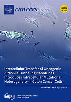
Oncogenic RAS is a major driver of cancer cell proliferation and dysregulation of cell signaling. The existing paradigm teaches us that it is propagated through cell division (vertical transmission). We proposed horizontal intercellular transport of oncogenic KRAS via tunneling nanotubes (TNTs) as a unique mechanism of inducing intracellular mutational heterogeneity in colon cancer cells. We discovered that TNTs are upregulated in colon cancer cells harboring mutant KRAS and deficits in mismatch repair. Furthermore, wild-type KRAS colon cancer cells that acquired mutant KRAS demonstrated increased formation of TNTs, which in turn promoted intercellular transfer of oncogenic KRAS. We propose that cell-to-cell transfer of mutant RAS is a novel form of communication that can potentially induce intracellular and, on a larger tumor microenvironmental scale, intratumoral heterogeneity. View this pa
Order results
Result details
Section
Select all
Export citation of selected articles as:
18 pages, 732 KiB  
Review
Strategies to Augment Natural Killer (NK) Cell Activity against Solid Tumors
by Ziqing Chen, Ying Yang, Lisa L. Liu and Andreas Lundqvist
Cancers 2019, 11(7), 1040; https://doi.org/10.3390/cancers11071040 - 23 Jul 2019
Cited by 34 | Viewed by 10891
Abstract
The immune system plays a crucial role to prevent local growth and dissemination of cancer. Therapies based on activating the immune system can result in beneficial responses in patients with metastatic disease. Treatment with antibodies targeting the immunological checkpoint axis PD-1 / PD-L1 [...] Read more.
The immune system plays a crucial role to prevent local growth and dissemination of cancer. Therapies based on activating the immune system can result in beneficial responses in patients with metastatic disease. Treatment with antibodies targeting the immunological checkpoint axis PD-1 / PD-L1 can result in the induction of anti-tumor T cell activation leading to meaningful long-lasting clinical responses. Still, many patients acquire resistance or develop dose-limiting toxicities to these therapies. Analysis of tumors from patients who progress on anti-PD-1 treatment reveal defective interferon-signaling and antigen presentation, resulting in immune escape from T cell-mediated attack. Natural killer (NK) cells are innate lymphocytes that can kill tumor cells without prior sensitization to antigens and can be activated to kill tumor cells that have an impaired antigen processing and presentation machinery. Thus, NK cells may serve as useful effectors against tumor cells that have become resistant to classical immune checkpoint therapy. Various approaches to activate NK cells are being increasingly explored in clinical trials against cancer. While clinical benefit has been demonstrated in patients with acute myeloid leukemia receiving haploidentical NK cells, responses in patients with solid tumors are so far less encouraging. Several hurdles need to be overcome to provide meaningful clinical responses in patients with solid tumors. Here we review the recent developments to augment NK cell responses against solid tumors with regards to cytokine therapy, adoptive infusion of NK cells, NK cell engagers, and NK cell immune checkpoints. Full article
(This article belongs to the Special Issue Signaling Pathways and Immune Checkpoint Regulation in Cancer)
Show Figures

Figure 1

13 pages, 2649 KiB  
Article
In Vivo Assessment of VCAM-1 Expression by SPECT/CT Imaging in Mice Models of Human Triple Negative Breast Cancer
by Christopher Montemagno, Laurent Dumas, Pierre Cavaillès, Mitra Ahmadi, Sandrine Bacot, Marlène Debiossat, Audrey Soubies, Loic Djaïleb, Julien Leenhardt, Nicolas De Leiris, Maeva Dufies, Gilles Pagès, Sophie Hernot, Nick Devoogdt, Pascale Perret, Laurent Riou, Daniel Fagret, Catherine Ghezzi and Alexis Broisat
Cancers 2019, 11(7), 1039; https://doi.org/10.3390/cancers11071039 - 23 Jul 2019
Cited by 4 | Viewed by 4694
Abstract
Recent progress in breast cancer research has led to the identification of Vascular Cell Adhesion Molecule-1 (VCAM-1) as a key actor of metastatic colonization. VCAM-1 promotes lung-metastases and is associated with clinical early recurrence and poor outcome in triple negative breast cancer (TNBC). [...] Read more.
Recent progress in breast cancer research has led to the identification of Vascular Cell Adhesion Molecule-1 (VCAM-1) as a key actor of metastatic colonization. VCAM-1 promotes lung-metastases and is associated with clinical early recurrence and poor outcome in triple negative breast cancer (TNBC). Our objective was to perform the in vivo imaging of VCAM-1 in mice models of TNBC. The Cancer Genomic Atlas (TCGA) database was analyzed to evaluate the prognostic role of VCAM-1 in TNBC. MDA-MB-231 (VCAM-1+) and control HCC70 (VCAM-1-) TNBC cells were subcutaneously xenografted in mice and VCAM-1 expression was assessed in vivo by single-photon emission computed tomography (SPECT) imaging using 99mTc-cAbVCAM1-5. Then, MDA-MB-231 cells were intravenously injected in mice and VCAM-1 expression in lung metastasis was assessed by SPECT imaging after 8 weeks. TCGA analysis showed that VCAM-1 is associated with a poor prognosis in TNBC patients. In subcutaneous tumor models, 99mTc-cAbVCAM1-5 uptake was 2-fold higher in MDA-MB-231 than in HCC70 (p < 0.01), and 4-fold higher than that of the irrelevant control (p < 0.01). Moreover, 99mTc-cAbVCAM1-5 uptake in MDA-MB-231 lung metastases was also higher than that of 99mTc-Ctl (p < 0.05). 99mTc-cAbVCAM1-5 is therefore a suitable tool to evaluate the role of VCAM-1 as a marker of tumor aggressiveness of TNBC. Full article
(This article belongs to the Special Issue Role of Medical Imaging in Cancers)
Show Figures

Graphical abstract

13 pages, 634 KiB  
Review
Emerging Roles of RAD52 in Genome Maintenance
by Manisha Jalan, Kyrie S. Olsen and Simon N. Powell
Cancers 2019, 11(7), 1038; https://doi.org/10.3390/cancers11071038 - 23 Jul 2019
Cited by 41 | Viewed by 7072
Abstract
The maintenance of genome integrity is critical for cell survival. Homologous recombination (HR) is considered the major error-free repair pathway in combatting endogenously generated double-stranded lesions in DNA. Nevertheless, a number of alternative repair pathways have been described as protectors of genome stability, [...] Read more.
The maintenance of genome integrity is critical for cell survival. Homologous recombination (HR) is considered the major error-free repair pathway in combatting endogenously generated double-stranded lesions in DNA. Nevertheless, a number of alternative repair pathways have been described as protectors of genome stability, especially in HR-deficient cells. One of the factors that appears to have a role in many of these pathways is human RAD52, a DNA repair protein that was previously considered to be dispensable due to a lack of an observable phenotype in knock-out mice. In later studies, RAD52 deficiency has been shown to be synthetically lethal with defects in BRCA genes, making RAD52 an attractive therapeutic target, particularly in the context of BRCA-deficient tumors. Full article
Show Figures

Figure 1

21 pages, 1267 KiB  
Review
AP-1 Transcription Factors as Regulators of Immune Responses in Cancer
by Vasileios Atsaves, Vasiliki Leventaki, George Z. Rassidakis and Francois X. Claret
Cancers 2019, 11(7), 1037; https://doi.org/10.3390/cancers11071037 - 23 Jul 2019
Cited by 142 | Viewed by 13666
Abstract
Immune check point blockade therapy has revolutionized the standard of cancer treatment and is credited with producing remarkable tumor remissions and increase in overall survival. This unprecedented clinical success however is feasible for a limited number of cancer patients due to resistance occurring [...] Read more.
Immune check point blockade therapy has revolutionized the standard of cancer treatment and is credited with producing remarkable tumor remissions and increase in overall survival. This unprecedented clinical success however is feasible for a limited number of cancer patients due to resistance occurring before or during a course of immunotherapy, which is often associated with activation of oncogenic signaling pathways, co-inhibitory checkpoints upregulation or expansion of immunosuppressive regulatory T-cells (Tregs) in the tumor microenviroment (TME). Targeted therapy aiming to inactivate a signaling pathway such as the Mitogen Activated Protein Kinases (MAPKs) has recently received a lot of attention due to emerging data from preclinical studies indicating synergy with immune checkpoint blockade therapy. The dimeric transcription factor complex Activator Protein-1 (AP-1) is a group of proteins involved in a wide array of cell processes and a critical regulator of nuclear gene expression during T-cell activation. It is also one of the downstream targets of the MAPK signaling cascade. In this review, we will attempt to unravel the roles of AP-1 in the regulation of anti-tumor immune responses, with a focus on the regulation of immune checkpoints and Tregs, seeking to extract useful insights for more efficacious immunotherapy. Full article
(This article belongs to the Special Issue Signaling Pathways and Immune Checkpoint Regulation in Cancer)
Show Figures

Figure 1

26 pages, 5551 KiB  
Review
Morphology and Molecular Features of Rare Colorectal Carcinoma Histotypes
by Andrea Remo, Matteo Fassan, Alessandro Vanoli, Luca Reggiani Bonetti, Valeria Barresi, Fabiana Tatangelo, Roberta Gafà, Guido Giordano, Massimo Pancione, Federica Grillo and Luca Mastracci
Cancers 2019, 11(7), 1036; https://doi.org/10.3390/cancers11071036 - 23 Jul 2019
Cited by 46 | Viewed by 12502
Abstract
Several histopathological variants of colorectal carcinoma can be distinguished, some associated with specific molecular profiles. However, in routine practice, ninety/ninety-five percent of all large bowel tumors are diagnosed as conventional adenocarcinoma, even though they are a heterogeneous group including rare histotypes, which are [...] Read more.
Several histopathological variants of colorectal carcinoma can be distinguished, some associated with specific molecular profiles. However, in routine practice, ninety/ninety-five percent of all large bowel tumors are diagnosed as conventional adenocarcinoma, even though they are a heterogeneous group including rare histotypes, which are often under-recognized. Indeed, colorectal cancer exhibits differences in incidence, location of tumor, pathogenesis, molecular pathways and outcome depending on histotype. The aim is therefore to review the morphological and molecular features of these rare variants of intestinal carcinomas which may hold the key to differences in prognosis and treatment. Full article
Show Figures

Figure 1

17 pages, 2357 KiB  
Article
Independent Negative Prognostic Role of TCF1 Expression within the Wnt/β-Catenin Signaling Pathway in Primary Breast Cancer Patients
by Concetta Saponaro, Emanuela Scarpi, Francesco Alfredo Zito, Francesco Giotta, Nicola Silvestris and Anita Mangia
Cancers 2019, 11(7), 1035; https://doi.org/10.3390/cancers11071035 - 22 Jul 2019
Cited by 12 | Viewed by 3391
Abstract
The Wnt pathway is involved in the progression of breast cancer (BC). We aimed to evaluate the expression of some components of the Wnt pathway (β-catenin, FZD4 (frizzled receptor 4), LRP5 (low-density lipoprotein receptor-related protein 5), LRP6, and TCF1 (T-cell factor 1)) to [...] Read more.
The Wnt pathway is involved in the progression of breast cancer (BC). We aimed to evaluate the expression of some components of the Wnt pathway (β-catenin, FZD4 (frizzled receptor 4), LRP5 (low-density lipoprotein receptor-related protein 5), LRP6, and TCF1 (T-cell factor 1)) to detect potential associations with NHERF1 (Na+/H+ exchanger regulatory factor 1) protein. Besides, we assessed their impact on patients’ clinical outcome. We evaluated 220 primary BC samples by immunohistochemistry (IHC) and protein localization by immunofluorescence. We found a significant correlation between NHERF1 and FZD4, LRP5, LRP6, and TCF1. Univariate analysis showed that the overexpression of β-catenin (p < 0.0001), FZD4 (p = 0.0001), LRP5, LRP6, and TCF1 (p < 0.0001 respectively) was related to poor disease-free survival (DFS). A Kaplan-Meier analysis confirmed univariate data and showed a poor DFS for cNHERF1+/FZD4+ (p = 0.0007), cNHERF1+/LRP5+ (p = 0.0002), cNHERF1+/LRP6+ (p < 0.0001), and cNHERF1+/TCF1+ phenotypes (p = 0.0034). In multivariate analysis, the expression of TCF1 and β-catenin was an independent prognostic variable of worse DFS (p = 0.009 and p = 0.027, respectively). In conclusion, we found that the overexpression of β-catenin, FZD4, LRP5, LRP6, and TCF1 was associated with poor prognosis. Furthermore, we first identified TCF1 as an independent prognostic factor of poor outcome, indicating it as a new potential biomarker for the management of BC patients. Also, the expression of Wnt pathway proteins, both alone and in association with NHERF1, suggests original associations of biological significance for new studies. Full article
Show Figures

Figure 1

15 pages, 5951 KiB  
Article
Flavopereirine Suppresses the Growth of Colorectal Cancer Cells through P53 Signaling Dependence
by Jhy-Ming Li, Yun-Ching Huang, Yi-Hung Kuo, Chih-Chung Cheng, Feng-Che Kuan, Shun-Fu Chang, Ying-Ray Lee, Chih-Chien Chin and Chung-Sheng Shi
Cancers 2019, 11(7), 1034; https://doi.org/10.3390/cancers11071034 - 22 Jul 2019
Cited by 13 | Viewed by 4271
Abstract
Colorectal cancer (CRC) is a significant cause of morbidity and mortality worldwide. The outcome of CRC patients remains poor. Thus, a new strategy for CRC treatment is urgently needed. Flavopereirine is a β-carboline alkaloid extracted from Geissospermum vellosii, which can reduce the [...] Read more.
Colorectal cancer (CRC) is a significant cause of morbidity and mortality worldwide. The outcome of CRC patients remains poor. Thus, a new strategy for CRC treatment is urgently needed. Flavopereirine is a β-carboline alkaloid extracted from Geissospermum vellosii, which can reduce the viability of various cancer cells through an unknown mode of action. The aim of the present study was to investigate the functional mechanism and therapeutic potential of flavopereirine on CRC cells in vitro and in vivo. Our data showed that flavopereirine significantly lowered cellular viability, caused intrinsic and extrinsic apoptosis, and induced G2/M-phase cell cycle arrest in CRC cells. Flavopereirine downregulated Janus kinases-signal transducers and activators of transcription (JAKs-STATs) and cellular myelocytomatosis (c-Myc) signaling in CRC cells. In contrast, the enforced expressions of constitutive active STAT3 and c-Myc could not restore flavopereirine-induced viability reduction. Moreover, flavopereirine enhanced P53 expression and phosphorylation in CRC cells. CRC cells with P53 knockout or loss-of-function mutation significantly diminished flavopereirine-mediated viability reduction, indicating that P53 activity plays a major role in flavopereirine-mediated CRC cell growth suppression. Flavopereirine also significantly repressed CRC cell xenograft growth in vivo by upregulating P53 and P21 and inducing apoptosis. In conclusion, flavopereirine-mediated growth suppression in CRC cells depended on the P53-P21, but not the JAKs-STATs-c-Myc signaling pathway. The present study suggests that flavopereirine may be efficacious in the clinical treatment of CRC harboring functional P53 signaling. Full article
(This article belongs to the Special Issue Colorectal Cancers)
Show Figures

Figure 1

25 pages, 567 KiB  
Review
PD-1/PD-L1 Targeting in Breast Cancer: The First Clinical Evidences are Emerging—A Literature Review
by Gabrielle Planes-Laine, Philippe Rochigneux, François Bertucci, Anne-Sophie Chrétien, Patrice Viens, Renaud Sabatier and Anthony Gonçalves
Cancers 2019, 11(7), 1033; https://doi.org/10.3390/cancers11071033 - 22 Jul 2019
Cited by 148 | Viewed by 16948
Abstract
Recently, the development of immunotherapy through the immune checkpoint blockade led to long-lasting responses in several types of cancers that are refractory to conventional treatments, such as melanoma or non-small cell lung cancer. Immunotherapy has also demonstrated significant improvements in various other types [...] Read more.
Recently, the development of immunotherapy through the immune checkpoint blockade led to long-lasting responses in several types of cancers that are refractory to conventional treatments, such as melanoma or non-small cell lung cancer. Immunotherapy has also demonstrated significant improvements in various other types of cancers. However, breast cancer remains one of the tumors that have not experienced the explosion of immunotherapy yet. Indeed, breast cancer was traditionally considered as being weakly immunogenic with a lower mutational load compared to other tumor types. In the last few years, anti-PD1/PD-L1 (Programmed death-ligand 1) agents have been evaluated in breast cancer, particularly in the triple negative subtype, with promising results observed when delivered as monotherapy or in combination with conventional treatments. In this review, we will report the results of the most recent studies evaluating immune checkpoint inhibitors in breast cancer. In addition, we will discuss the concomitant development of possible biomarkers, which is required for improving the selection of patients with the highest probability of benefiting from these agents. Full article
Show Figures

Figure 1

8 pages, 835 KiB  
Opinion
So Close, yet so Far: Discrepancies between Uveal and Other Melanomas. A Position Paper from UM Cure 2020
by Manuel Rodrigues, Leanne de Koning, Sarah E. Coupland, Aart G. Jochemsen, Richard Marais, Marc-Henri Stern, André Valente, Raymond Barnhill, Nathalie Cassoux, Andrew Evans, Iain Galloway, Martine J. Jager, Ellen Kapiteijn, Bozena Romanowska-Dixon, Bettina Ryll, Sergio Roman-Roman, Sophie Piperno-Neumann and the UM Cure 2020 Consortium
Cancers 2019, 11(7), 1032; https://doi.org/10.3390/cancers11071032 - 22 Jul 2019
Cited by 49 | Viewed by 5463
Abstract
Despite much progress in our understanding of uveal melanoma (UM) over the past decades, this rare tumour is still often misclassified. Although UM, like other melanomas, is very probably derived from melanocytes, it is drastically different from cutaneous melanoma and most other melanoma [...] Read more.
Despite much progress in our understanding of uveal melanoma (UM) over the past decades, this rare tumour is still often misclassified. Although UM, like other melanomas, is very probably derived from melanocytes, it is drastically different from cutaneous melanoma and most other melanoma subtypes in terms of epidemiology, aetiology, biology and clinical features, including an intriguing metastatic hepatotropism. UM carries distinctive prognostic chromosome alterations, somatic mutations and gene expression profiles, allowing an active tailored surveillance strategy and dedicated adjuvant clinical trials. There is no standard systemic treatment for disseminated UM at present. In contrast to cutaneous melanoma, UMs are not BRAF-mutated, thus curtailing the use of B-Raf inhibitors. Although these tumours are characterised by some immune infiltrates, immune checkpoint inhibitors are rarely effective, possibly due to a low mutation burden. UM patients across the world not only face rare cancer-related issues (e.g., specific management strategies, access to information and to expert centres), but also specific UM problems, which can be exacerbated by the common misconception that it is a subtype of cutaneous melanoma. As a European Consortium dedicated to research on UM and awareness on the disease, “UM Cure 2020” participants urge medical oncologists, pharmaceutical companies, and regulatory agencies to acknowledge UM as a melanoma with specific issues, in order to accelerate the development of new therapies for patients. Full article
(This article belongs to the Special Issue Uveal Melanoma)
Show Figures

Figure 1

11 pages, 1026 KiB  
Article
GNAQ Q209R Mutations Are Highly Specific for Circumscribed Choroidal Hemangioma
by Claudia Helga Dorothee Le Guin, Klaus Alfred Metz, Stefan Horst Kreis, Nikolaos Emmanouel Bechrakis, Norbert Bornfeld, Michael Zeschnigk and Dietmar Rudolf Lohmann
Cancers 2019, 11(7), 1031; https://doi.org/10.3390/cancers11071031 - 22 Jul 2019
Cited by 14 | Viewed by 3376
Abstract
Several tumors, including uveal melanoma, show somatic mutations of GNAQ/GNA11. Circumscribed choroidal hemangioma is a benign tumor that becomes symptomatic in adulthood. In some patients, morphologic examination of biopsies is required for differential diagnosis between amelanotic choroidal melanoma and circumscribed choroidal hemangioma. [...] Read more.
Several tumors, including uveal melanoma, show somatic mutations of GNAQ/GNA11. Circumscribed choroidal hemangioma is a benign tumor that becomes symptomatic in adulthood. In some patients, morphologic examination of biopsies is required for differential diagnosis between amelanotic choroidal melanoma and circumscribed choroidal hemangioma. Here, we report the results of GNAQ/GNA11 mutation analysis in samples from circumscribed choroidal hemangioma. Deep amplicon sequencing (Illumina MiSeq, San Diego, CA, USA) of positions R183 and Q209 of GNAQ and GNA11 in tissue samples from 33 patients with histologically diagnosed circumscribed choroidal hemangioma. All patients underwent biopsy or enucleation at our clinic between 2008 and 2018. To enable detection of variant alleles at low fractions, read depth exceeded 15,000-fold. DNA for genetic analysis was prepared from either snap-frozen (n = 22) or FFPE (n = 11) tissue samples. Samples from 28/33 patients (85%) showed a somatic missense mutation of GNAQ (c.626 A > G) predicted to result in p.Q209R. Variant allele fraction was variable (range 2.3% to 28%). Variants of GNAQ resulting in p.Q209 are characteristic for circumscribed choroidal hemangiomas. It appears that the GNAQ mutation spectrum in this tumor is narrow, possibly restricted to p.Q209R. Moreover, the spectrum is distinct from that of uveal melanoma, in which alterations resulting in p.Q209R are very rare. Full article
(This article belongs to the Special Issue Uveal Melanoma)
Show Figures

Figure 1

31 pages, 1889 KiB  
Review
Impact of ROS Generated by Chemical, Physical, and Plasma Techniques on Cancer Attenuation
by Sarmistha Mitra, Linh Nhat Nguyen, Mahmuda Akter, Gyungsoon Park, Eun Ha Choi and Nagendra Kumar Kaushik
Cancers 2019, 11(7), 1030; https://doi.org/10.3390/cancers11071030 - 22 Jul 2019
Cited by 115 | Viewed by 10479
Abstract
For the last few decades, while significant improvements have been achieved in cancer therapy, this family of diseases is still considered one of the deadliest threats to human health. Thus, there is an urgent need to find novel strategies in order to tackle [...] Read more.
For the last few decades, while significant improvements have been achieved in cancer therapy, this family of diseases is still considered one of the deadliest threats to human health. Thus, there is an urgent need to find novel strategies in order to tackle this vital medical issue. One of the most pivotal causes of cancer initiation is the presence of reactive oxygen species (ROS) inside the body. Interestingly, on the other hand, high doses of ROS possess the capability to damage malignant cells. Moreover, several important intracellular mechanisms occur during the production of ROS. For these reasons, inducing ROS inside the biological system by utilizing external physical or chemical methods is a promising approach to inhibit the growth of cancer cells. Beside conventional technologies, cold atmospheric plasmas are now receiving much attention as an emerging therapeutic tool for cancer treatment due to their unique biophysical behavior, including the ability to generate considerable amounts of ROS. This review summarizes the important mechanisms of ROS generated by chemical, physical, and plasma approaches. We also emphasize the biological effects and cancer inhibition capabilities of ROS. Full article
Show Figures

Figure 1

20 pages, 2673 KiB  
Article
CD56 Homodimerization and Participation in Anti-Tumor Immune Effector Cell Functioning: A Role for Interleukin-15
by Heleen H. Van Acker, Zoë P. Van Acker, Maarten Versteven, Peter Ponsaerts, Daniela Pende, Zwi N. Berneman, Sébastien Anguille, Viggo F. Van Tendeloo and Evelien L. Smits
Cancers 2019, 11(7), 1029; https://doi.org/10.3390/cancers11071029 - 22 Jul 2019
Cited by 6 | Viewed by 4383
Abstract
A particularly interesting marker to identify anti-tumor immune cells is the neural cell adhesion molecule (NCAM), also known as cluster of differentiation (CD)56. Namely, hematopoietic expression of CD56 seems to be confined to powerful effector immune cells. Here, we sought to elucidate its [...] Read more.
A particularly interesting marker to identify anti-tumor immune cells is the neural cell adhesion molecule (NCAM), also known as cluster of differentiation (CD)56. Namely, hematopoietic expression of CD56 seems to be confined to powerful effector immune cells. Here, we sought to elucidate its role on various killer immune cells. First, we identified the high motility NCAM-120 molecule to be the main isoform expressed by immune cells. Next, through neutralization of surface CD56, we were able to (1) demonstrate the direct involvement of CD56 in tumor cell lysis exerted by CD56-expressing killer cells, such as natural killer cells, gamma delta (γδ) T cells, and interleukin (IL)-15-cultured dendritic cells (DCs), and (2) reveal a putative crosstalk mechanism between IL-15 DCs and CD8 T cells, suggesting CD56 as a co-stimulatory molecule in their cell-to-cell contact. Moreover, by means of a proximity ligation assay, we visualized the CD56 homophilic interaction among cancer cells and between immune cells and cancer cells. Finally, by blocking the mitogen-activated protein kinase (MAPK) pathway and the phosphoinositide 3-kinase (PI3K)–Akt pathway, we showed that IL-15 stimulation directly led to CD56 upregulation. In conclusion, these results underscore the previously neglected importance of CD56 expression on immune cells, benefiting current and future immune therapeutic options. Full article
(This article belongs to the Special Issue Targeting Innate Immunity Cells in Cancer)
Show Figures

Figure 1

28 pages, 1438 KiB  
Review
The Central Contributions of Breast Cancer Stem Cells in Developing Resistance to Endocrine Therapy in Estrogen Receptor (ER)-Positive Breast Cancer
by David Rodriguez, Marc Ramkairsingh, Xiaozeng Lin, Anil Kapoor, Pierre Major and Damu Tang
Cancers 2019, 11(7), 1028; https://doi.org/10.3390/cancers11071028 - 22 Jul 2019
Cited by 51 | Viewed by 6693
Abstract
Breast cancer stem cells (BCSC) play critical roles in the acquisition of resistance to endocrine therapy in estrogen receptor (ER)-positive (ER + ve) breast cancer (BC). The resistance results from complex alterations involving ER, growth factor receptors, NOTCH, Wnt/β-catenin, hedgehog, YAP/TAZ, and the [...] Read more.
Breast cancer stem cells (BCSC) play critical roles in the acquisition of resistance to endocrine therapy in estrogen receptor (ER)-positive (ER + ve) breast cancer (BC). The resistance results from complex alterations involving ER, growth factor receptors, NOTCH, Wnt/β-catenin, hedgehog, YAP/TAZ, and the tumor microenvironment. These mechanisms are likely converged on regulating BCSCs, which then drive the development of endocrine therapy resistance. In this regard, hormone therapies enrich BCSCs in ER + ve BCs under both pre-clinical and clinical settings along with upregulation of the core components of “stemness” transcriptional factors including SOX2, NANOG, and OCT4. SOX2 initiates a set of reactions involving SOX9, Wnt, FXY3D, and Src tyrosine kinase; these reactions stimulate BCSCs and contribute to endocrine resistance. The central contributions of BCSCs to endocrine resistance regulated by complex mechanisms offer a unified strategy to counter the resistance. ER + ve BCs constitute approximately 75% of BCs to which hormone therapy is the major therapeutic approach. Likewise, resistance to endocrine therapy remains the major challenge in the management of patients with ER + ve BC. In this review we will discuss evidence supporting a central role of BCSCs in developing endocrine resistance and outline the strategy of targeting BCSCs to reduce hormone therapy resistance. Full article
Show Figures

Figure 1

20 pages, 7008 KiB  
Article
Mutations in the ND2 Subunit of Mitochondrial Complex I Are Sufficient to Confer Increased Tumorigenic and Metastatic Potential to Cancer Cells
by Joaquín Marco-Brualla, Sameer Al-Wasaby, Ruth Soler, Eduardo Romanos, Blanca Conde, Raquel Justo-Méndez, José A. Enríquez, Patricio Fernández-Silva, Luis Martínez-Lostao, Martín Villalba, Raquel Moreno-Loshuertos and Alberto Anel
Cancers 2019, 11(7), 1027; https://doi.org/10.3390/cancers11071027 - 21 Jul 2019
Cited by 17 | Viewed by 4521
Abstract
Multiprotein complexes of the mitochondrial electron transport chain form associations to generate supercomplexes. The relationship between tumor cell ability to assemble mitochondrial supercomplexes, tumorigenesis and metastasis has not been studied thoroughly. The mitochondrial and metabolic differences between L929dt cells, which lost matrix attachment [...] Read more.
Multiprotein complexes of the mitochondrial electron transport chain form associations to generate supercomplexes. The relationship between tumor cell ability to assemble mitochondrial supercomplexes, tumorigenesis and metastasis has not been studied thoroughly. The mitochondrial and metabolic differences between L929dt cells, which lost matrix attachment and MHC-I expression, and their parental cell line L929, were analyzed. L929dt cells have lower capacity to generate energy through OXPHOS and lower respiratory capacity than parental L929 cells. Most importantly, L929dt cells show defects in mitochondrial supercomplex assembly, especially in those that contain complex I. These defects correlate with mtDNA mutations in L929dt cells at the ND2 subunit of complex I and are accompanied by a glycolytic shift. In addition, L929dt cells show higher in vivo tumorigenic and metastatic potential than the parental cell line. Cybrids with L929dt mitochondria in L929 nuclear background reproduce all L929dt properties, demonstrating that mitochondrial mutations are responsible for the aggressive tumor phenotype. In spite of their higher tumorigenic potential, L929dt or mitochondrial L929dt cybrid cells are sensitive both in vitro and in vivo to the PDK1 inhibitor dichloroacetate, which favors OXPHOS, suggesting benefits for the use of metabolic inhibitors in the treatment of especially aggressive tumors. Full article
Show Figures

Figure 1

17 pages, 2239 KiB  
Review
Spatiotemporal pH Heterogeneity as a Promoter of Cancer Progression and Therapeutic Resistance
by David E. Korenchan and Robert R. Flavell
Cancers 2019, 11(7), 1026; https://doi.org/10.3390/cancers11071026 - 20 Jul 2019
Cited by 40 | Viewed by 7225
Abstract
Dysregulation of pH in solid tumors is a hallmark of cancer. In recent years, the role of altered pH heterogeneity in space, between benign and aggressive tissues, between individual cancer cells, and between subcellular compartments, has been steadily elucidated. Changes in temporal pH-related [...] Read more.
Dysregulation of pH in solid tumors is a hallmark of cancer. In recent years, the role of altered pH heterogeneity in space, between benign and aggressive tissues, between individual cancer cells, and between subcellular compartments, has been steadily elucidated. Changes in temporal pH-related processes on both fast and slow time scales, including altered kinetics of bicarbonate-CO2 exchange and its effects on pH buffering and gradual, progressive changes driven by changes in metabolism, are further implicated in phenotypic changes observed in cancers. These discoveries have been driven by advances in imaging technologies. This review provides an overview of intra- and extracellular pH alterations in time and space reflected in cancer cells, as well as the available technology to study pH spatiotemporal heterogeneity. Full article
(This article belongs to the Special Issue Metastatic Progression and Tumour Heterogeneity)
Show Figures

Figure 1

16 pages, 5775 KiB  
Article
Palbociclib Promotes Dephosphorylation of NPM/B23 at Threonine 199 and Inhibits Endometrial Cancer Cell Growth
by Chiao-Yun Lin, Li-Yu Lee, Tzu-Hao Wang, Cheng-Lung Hsu, Chia-Lung Tsai, Angel Chao and Chyong-Huey Lai
Cancers 2019, 11(7), 1025; https://doi.org/10.3390/cancers11071025 - 20 Jul 2019
Cited by 8 | Viewed by 3677
Abstract
Endometrial cancer incidence rates are growing, especially in countries with rapid socioeconomic transitions. Despite recent advances in chemotherapy, hormone therapy, and targeted therapy, advanced/recurrent disease remains a clinical challenge. Palbociclib—a selective inhibitor of cyclin-dependent kinases (CDK) 4/6—has therapeutic potential against estrogen receptor (ER)-positive [...] Read more.
Endometrial cancer incidence rates are growing, especially in countries with rapid socioeconomic transitions. Despite recent advances in chemotherapy, hormone therapy, and targeted therapy, advanced/recurrent disease remains a clinical challenge. Palbociclib—a selective inhibitor of cyclin-dependent kinases (CDK) 4/6—has therapeutic potential against estrogen receptor (ER)-positive and HER2-negative breast cancer. However, the question as to whether it can be clinically useful in endometrial cancer remains open. Here, we show that combined treatment with palbociclib and megesterol acetate exerts synergistic antiproliferative effects against endometrial cancer cells. Treatment of cancer cells with palbociclib suppressed NPM/B23 phosphorylation at threonine 199 (Thr199). We further demonstrated that CDK6 acts as a NPM/B23 kinase. Palbociclib-induced NPM/B23 dephosphorylation sensitized endometrial cancer cells to megesterol acetate through the upregulation of ERα expression. Immunohistochemistry revealed an overexpression of phospho-NPM/B23 (Thr199) in human endometrial cancer, and phospho-NPM/B23 (Thr199) expression levels were inversely associated with Erα in clinical specimen. In a xenograft tumor model, the combination of palbociclib and megesterol acetate successfully inhibited tumor growth. Taken together, our data indicate that palbociclib promoted NPM/B23 dephosphorylation at Thr199—an effect mediated by disruption of CDK6 kinase activity. We conclude that palbociclib holds promise for the treatment of endometrial cancer when used in combination with megesterol acetate. Full article
Show Figures

Figure 1

21 pages, 2099 KiB  
Article
Liver Cancer Cell Lines Treated with Doxorubicin under Normoxia and Hypoxia: Cell Viability and Oncologic Protein Profile
by Ilse R. Dubbelboer, Natasa Pavlovic, Femke Heindryckx, Erik Sjögren and Hans Lennernäs
Cancers 2019, 11(7), 1024; https://doi.org/10.3390/cancers11071024 - 20 Jul 2019
Cited by 40 | Viewed by 10137
Abstract
Hepatocellular carcinoma is often treated with a combination of doxorubicin and embolization, exposing it to high concentrations and hypoxia. Separation of the possible synergistic effect of this combination in vivo is difficult. Here, treatment with doxorubicin, under hypoxia or normoxia in different liver [...] Read more.
Hepatocellular carcinoma is often treated with a combination of doxorubicin and embolization, exposing it to high concentrations and hypoxia. Separation of the possible synergistic effect of this combination in vivo is difficult. Here, treatment with doxorubicin, under hypoxia or normoxia in different liver cancer cell lines, was evaluated. Liver cancer cells HepG2, Huh7, and SNU449 were exposed to doxorubicin, hypoxia, or doxorubicin + hypoxia with different duration. Treatment response was evaluated with cell viability, apoptosis, oxidative stress, and summarized with IC50. The protein profile of a 92-biomarker panel was analyzed on cells treated with 0 or 0.1 µM doxorubicin during 6 or 72 h, under normoxia or hypoxia. Hypoxia decreased viability of HepG2 and SNU499. HepG2 was least and SNU449 most tolerant to doxorubicin treatment. Cytotoxicity of doxorubicin increased over time in HepG2 and Huh7. The combination of doxorubicin + hypoxia affected the cells differently. Normalized protein expression was lower for HepG2 than Huh7 and SNU449. Hierarchical clustering separated HepG2 from Huh7 and SNU449. These three commonly used cell lines have critically different responses to chemotherapy and hypoxia, which was reflected in their different protein expression profile. These different responses suggest that tumors can respond differently to the combination of local chemotherapy and embolization. Full article
(This article belongs to the Special Issue Hepatocellular Cancer Treatment)
Show Figures

Graphical abstract

16 pages, 1591 KiB  
Article
ANGPT2 and NOS3 Polymorphisms and Clinical Outcome in Advanced Hepatocellular Carcinoma Patients Receiving Sorafenib
by Giorgia Marisi, Elisabetta Petracci, Francesco Raimondi, Luca Faloppi, Francesco Giuseppe Foschi, Gianfranco Lauletta, Massimo Iavarone, Matteo Canale, Martina Valgiusti, Luca Maria Neri, Paola Ulivi, Giulia Orsi, Giulia Rovesti, Ranka Vukotic, Fabio Conti, Alessandro Cucchetti, Giorgio Ercolani, Kalliopi Andrikou, Stefano Cascinu, Mario Scartozzi and Andrea Casadei-Gardiniadd Show full author list remove Hide full author list
Cancers 2019, 11(7), 1023; https://doi.org/10.3390/cancers11071023 - 20 Jul 2019
Cited by 26 | Viewed by 3552
Abstract
Sorafenib represents the standard of care for advanced hepatocellular carcinoma (HCC), even though a large number of patients have reported limited efficacy. The aim of the present study was to evaluate the prognostic value of single-nucleotide polymorphisms on angiopoietin-2 (ANGPT2) and [...] Read more.
Sorafenib represents the standard of care for advanced hepatocellular carcinoma (HCC), even though a large number of patients have reported limited efficacy. The aim of the present study was to evaluate the prognostic value of single-nucleotide polymorphisms on angiopoietin-2 (ANGPT2) and endothelial-derived nitric oxide synthase (NOS3) genes in 135 patients with advanced HCC receiving sorafenib. Eight ANGPT2 polymorphisms were analyzed by direct sequencing in relation to overall survival (OS) and progression-free survival (PFS). In univariate analysis, ANGPT2rs55633437 and NOS3 rs2070744 were associated with OS and PFS. In particular, patients with ANGPT2rs55633437 TT/GT genotypes had significantly lower median OS (4.66 vs. 15.5 months, hazard ratio (HR) 4.86, 95% CI 2.73–8.67, p < 0.001) and PFS (1.58 vs. 6.27 months, HR 4.79, 95% CI 2.73–8.35, p < 0.001) than those homozygous for the G allele. Moreover, patients with NOS3 rs2070744 TC/CC genotypes had significantly higher median OS (15.6 vs. 9.1 months, HR 0.65, 95% CI 0.44–0.97; p = 0.036) and PFS (7.03 vs. 3.5 months, HR 0.43, 95% CI 0.30–0.63; p < 0.001) than patients homozygous for the T allele. Multivariate analysis confirmed these polymorphisms as independent prognostic factors. Our results suggest that ANGPT2rs55633437 and NOS3 rs2070744 polymorphisms could identify a subset of HCC patients more resistant to sorafenib. Full article
Show Figures

Figure 1

17 pages, 812 KiB  
Review
Platelets as Key Factors in Hepatocellular Carcinoma
by Natasa Pavlovic, Bhavna Rani, Pär Gerwins and Femke Heindryckx
Cancers 2019, 11(7), 1022; https://doi.org/10.3390/cancers11071022 - 20 Jul 2019
Cited by 58 | Viewed by 6596
Abstract
Hepatocellular carcinoma (HCC) is a primary liver cancer that usually develops in the setting of chronic inflammation and liver damage. The hepatic microenvironment plays a crucial role in the disease development, as players such as hepatic stellate cells, resident liver macrophages (Kupffer cells), [...] Read more.
Hepatocellular carcinoma (HCC) is a primary liver cancer that usually develops in the setting of chronic inflammation and liver damage. The hepatic microenvironment plays a crucial role in the disease development, as players such as hepatic stellate cells, resident liver macrophages (Kupffer cells), endothelial cells, extracellular matrix, and a variety of immune cells interact in highly complex and intertwined signaling pathways. A key factor in these cross-talks are platelets, whose role in cancer has gained growing evidence in recent years. Platelets have been reported to promote HCC cell proliferation and invasion, but their involvement goes beyond the direct effect on tumor cells, as they are known to play a role in pro-fibrinogenic signaling and the hepatic immune response, as well as in mediating interactions between these factors in the stroma. Anti-platelet therapy has been shown to ameliorate liver injury and improve the disease outcome. However, platelets have also been shown to play a crucial role in liver regeneration after organ damage. Therefore, the timing and microenvironmental setting need to be kept in mind when assessing the potential effect and therapeutic value of platelets in the disease progression, while further studies are needed for understanding the role of platelets in patients with HCC. Full article
(This article belongs to the Special Issue Hepatocellular Cancer Treatment)
Show Figures

Figure 1

29 pages, 843 KiB  
Review
The Role of Breast Cancer Stem Cells as a Prognostic Marker and a Target to Improve the Efficacy of Breast Cancer Therapy
by Maria Giovanna Scioli, Gabriele Storti, Federico D’Amico, Pietro Gentile, Giulia Fabbri, Valerio Cervelli and Augusto Orlandi
Cancers 2019, 11(7), 1021; https://doi.org/10.3390/cancers11071021 - 20 Jul 2019
Cited by 50 | Viewed by 7909
Abstract
Breast cancer is the most common form of tumor in women and the leading cause of cancer-related mortality. Even though the major cellular burden in breast cancer is constituted by the so-called bulk tumor cells, another cell subpopulation named cancer stem cells (CSCs) [...] Read more.
Breast cancer is the most common form of tumor in women and the leading cause of cancer-related mortality. Even though the major cellular burden in breast cancer is constituted by the so-called bulk tumor cells, another cell subpopulation named cancer stem cells (CSCs) has been identified. The latter have stem features, a self-renewal capacity, and the ability to regenerate the bulk tumor cells. CSCs have been described in several cancer types but breast cancer stem cells (BCSCs) were among the first to be identified and characterized. Therefore, many efforts have been put into the phenotypic characterization of BCSCs and the study of their potential as prognostic indicators and therapeutic targets. Many dysregulated pathways in BCSCs are involved in the epithelial–mesenchymal transition (EMT) and are found up-regulated in circulating tumor cells (CTCs), another important cancer cell subpopulation, that shed into the vasculature and disseminate along the body to give metastases. Conventional therapies fail at eliminating BCSCs because of their quiescent state that gives them therapy resistance. Based on this evidence, preclinical studies and clinical trials have tried to establish novel therapeutic regimens aiming to eradicate BCSCs. Markers useful for BCSC identification could also be possible therapeutic methods against BCSCs. New approaches in drug delivery combined with gene targeting, immunomodulatory, and cell-based therapies could be promising tools for developing effective CSC-targeted drugs against breast cancer. Full article
Show Figures

Figure 1

27 pages, 1921 KiB  
Review
The Bone Extracellular Matrix as an Ideal Milieu for Cancer Cell Metastases
by Alexus D. Kolb and Karen M. Bussard
Cancers 2019, 11(7), 1020; https://doi.org/10.3390/cancers11071020 - 20 Jul 2019
Cited by 52 | Viewed by 14081
Abstract
Bone is a preferential site for cancer metastases, including multiple myeloma, prostate, and breast cancers.The composition of bone, especially the extracellular matrix (ECM), make it an attractive site for cancer cell colonization and survival. The bone ECM is composed of living cells embedded [...] Read more.
Bone is a preferential site for cancer metastases, including multiple myeloma, prostate, and breast cancers.The composition of bone, especially the extracellular matrix (ECM), make it an attractive site for cancer cell colonization and survival. The bone ECM is composed of living cells embedded within a matrix composed of both organic and inorganic components. Among the organic components, type I collagen provides the tensile strength of bone. Inorganic components, including hydroxyapatite crystals, are an integral component of bone and provide bone with its rigidity. Under normal circumstances, two of the main cell types in bone, the osteoblasts and osteoclasts, help to maintain bone homeostasis and remodeling through cellular communication and response to biophysical signals from the ECM. However, under pathological conditions, including osteoporosis and cancer, bone remodeling is dysregulated. Once in the bone matrix, disseminated tumor cells utilize normal products of bone remodeling, such as collagen type I, to fuel cancer cell proliferation and lesion outgrowth. Models to study the complex interactions between the bone matrix and metastatic cancer cells are limited. Advances in understanding the interactions between the bone ECM and bone metastatic cancer cells are necessary in order to both regulate and prevent metastatic cancer cell growth in bone. Full article
(This article belongs to the Special Issue Cancer Invasion and Metastasis)
Show Figures

Figure 1

17 pages, 1891 KiB  
Review
Emerging Therapeutic Opportunities Based on Current Knowledge of Uveal Melanoma Biology
by Raquel Vivet-Noguer, Malcy Tarin, Sergio Roman-Roman and Samar Alsafadi
Cancers 2019, 11(7), 1019; https://doi.org/10.3390/cancers11071019 - 20 Jul 2019
Cited by 30 | Viewed by 4898
Abstract
Uveal Melanoma (UM) is a rare and malignant intraocular tumor with dismal prognosis. Despite the efficient control of the primary tumor by radiation or surgery, up to 50% of patients subsequently develop metastasis, mainly in the liver. Once the tumor has spread from [...] Read more.
Uveal Melanoma (UM) is a rare and malignant intraocular tumor with dismal prognosis. Despite the efficient control of the primary tumor by radiation or surgery, up to 50% of patients subsequently develop metastasis, mainly in the liver. Once the tumor has spread from the eye, the treatment is challenging and the median survival is only nine months. UM represents an intriguing model of oncogenesis that is characterized by a relatively homogeneous histopathological architecture and a low burden of genetic alterations, in contrast to other melanomas. UM is driven by recurrent activating mutations in Gαq pathway, which are associated with a second mutation in BRCA1 associated protein 1 (BAP1), splicing factor 3b subunit 1 (SF3B1), or eukaryotic translation initiation factor 1A X-linked (EIF1AX), occurring in an almost mutually exclusive manner. The monosomy of chromosome 3 is also a recurrent feature that is associated with high metastatic risk. These events driving UM oncogenesis have been thoroughly investigated over the last decade. However, no efficient related therapeutic strategies are yet available and the metastatic disease remains mostly incurable. Here, we review current knowledge regarding the molecular biology and the genetics of uveal melanoma and highlight the related therapeutic applications and perspectives. Full article
(This article belongs to the Special Issue Uveal Melanoma)
Show Figures

Graphical abstract

20 pages, 7012 KiB  
Review
Targeted Radionuclide Therapy for Patients with Metastatic Pheochromocytoma and Paraganglioma: From Low-Specific-Activity to High-Specific-Activity Iodine-131 Metaiodobenzylguanidine
by Camilo Jimenez, William Erwin and Beth Chasen
Cancers 2019, 11(7), 1018; https://doi.org/10.3390/cancers11071018 - 20 Jul 2019
Cited by 50 | Viewed by 6131
Abstract
Low-specific-activity iodine-131–radiolabeled metaiodobenzylguanidine (I-131-MIBG) was introduced last century as a potential systemic therapy for patients with malignant pheochromocytomas and paragangliomas. Collective information derived from mainly retrospective studies has suggested that 30–40% of patients with these tumors benefit from this treatment. A low index [...] Read more.
Low-specific-activity iodine-131–radiolabeled metaiodobenzylguanidine (I-131-MIBG) was introduced last century as a potential systemic therapy for patients with malignant pheochromocytomas and paragangliomas. Collective information derived from mainly retrospective studies has suggested that 30–40% of patients with these tumors benefit from this treatment. A low index of radioactivity, lack of therapeutic standardization, and toxicity associated with intermediate to high activities (absorbed radiation doses) has prevented the implementation of I-131-MIBG’s in clinical practice. High-specific-activity, carrier-free I-131-MIBG has been developed over the past two decades as a novel therapy for patients with metastatic pheochromocytomas and paragangliomas that express the norepinephrine transporter. This drug allows for a high level of radioactivity, and as yet is not associated with cardiovascular toxicity. In a pivotal phase two clinical trial, more than 90% of patients achieved partial responses and disease stabilization with the improvement of hypertension. Furthermore, many patients exhibited long-term persistent antineoplastic effects. Currently, the high-specific-activity I-131-MIBG is the only approved therapy in the US for patients with metastatic pheochromocytomas and paragangliomas. This review will discuss the historical development of high-specific-activity I-131-MIBG, its benefits and adverse events, and future directions for clinical practice applicability and trial development. Full article
Show Figures

Figure 1

25 pages, 2084 KiB  
Review
The Molecular Hallmarks of the Serrated Pathway in Colorectal Cancer
by Fatima Domenica Elisa De Palma, Valeria D’Argenio, Jonathan Pol, Guido Kroemer, Maria Chiara Maiuri and Francesco Salvatore
Cancers 2019, 11(7), 1017; https://doi.org/10.3390/cancers11071017 - 20 Jul 2019
Cited by 115 | Viewed by 20671
Abstract
Colorectal cancer (CRC) is a leading cause of cancer death worldwide. It includes different subtypes that differ in their clinical and prognostic features. In the past decade, in addition to the conventional adenoma-carcinoma model, an alternative multistep mechanism of carcinogenesis, namely the “serrated [...] Read more.
Colorectal cancer (CRC) is a leading cause of cancer death worldwide. It includes different subtypes that differ in their clinical and prognostic features. In the past decade, in addition to the conventional adenoma-carcinoma model, an alternative multistep mechanism of carcinogenesis, namely the “serrated pathway”, has been described. Approximately, 15 to 30% of all CRCs arise from neoplastic serrated polyps, a heterogeneous group of lesions that are histologically classified into three morphologic categories: hyperplastic polyps, sessile serrated adenomas/polyps, and the traditional serrated adenomas/polyps. Serrated polyps are characterized by genetic (BRAF or KRAS mutations) and epigenetic (CpG island methylator phenotype (CIMP)) alterations that cooperate to initiate and drive malignant transformation from normal colon mucosa to polyps, and then to CRC. The high heterogeneity of the serrated lesions renders their diagnostic and pathological interpretation difficult. Hence, novel genetic and epigenetic biomarkers are required for better classification and management of CRCs. To date, several molecular alterations have been associated with the serrated polyp-CRC sequence. In addition, the gut microbiota is emerging as a contributor to/modulator of the serrated pathway. This review summarizes the state of the art of the genetic, epigenetic and microbiota signatures associated with serrated CRCs, together with their clinical implications. Full article
(This article belongs to the Special Issue Colorectal Cancers)
Show Figures

Graphical abstract

17 pages, 2379 KiB  
Article
LdrB Toxin with In Vitro and In Vivo Antitumor Activity as a Potential Tool for Cancer Gene Therapy
by Yaiza Jiménez-Martínez, Carmen Griñán-Lisón, Hoda Khaldy, Ana Martín, Alba Cambrils, Andrea Ibáñez Grau, Gema Jiménez, Juan A. Marchal and Houria Boulaiz
Cancers 2019, 11(7), 1016; https://doi.org/10.3390/cancers11071016 - 20 Jul 2019
Cited by 4 | Viewed by 4320
Abstract
Due to the high prevalence of cancer in recent years, it is necessary to develop new and more effective therapies that produce fewer side effects. Development of gene therapy for cancer based on the use of suicide genes that can damage the tumor [...] Read more.
Due to the high prevalence of cancer in recent years, it is necessary to develop new and more effective therapies that produce fewer side effects. Development of gene therapy for cancer based on the use of suicide genes that can damage the tumor cell, without requiring a prodrug for its lethal effect, is one of the recent foci of gene therapy strategies. We evaluated the cytotoxic impact of the LdrB toxin from Escherichia coli k12 as a possible tool for cancer gene therapy. For that, colorectal and breast cancer cells were transfected under the control of a TRE3G promoter inducible by doxycycline. Our results showed that ldrB gene expression induced a drastic inhibition of proliferation in vitro, in both 2D and 3D experimental models. Moreover, unlike conventional chemotherapy, the ldrB gene induced a severe loss of proliferation in vivo without any side effects in our animal model. This antitumor outcome was modulated by cell cycle arrest in the G0/G1 phase and apoptotic death. Scanning electronic microscopy demonstrates that the LdrB toxin conserves its pore-forming ability in HCT-116 cells as in E. coli k12. Taken together, our results provide, for the first time, a proof of concept of the antitumor capacity of the ldrB gene in colorectal and breast cancer. Full article
(This article belongs to the Special Issue Cell Death in Cancer)
Show Figures

Figure 1

17 pages, 2915 KiB  
Article
Comparative Assessment of Antitumor Effects and Autophagy Induction as a Resistance Mechanism by Cytotoxics and EZH2 Inhibition in INI1-Negative Epithelioid Sarcoma Patient-Derived Xenograft
by Silvia Stacchiotti, Valentina Zuco, Monica Tortoreto, Denis Cominetti, Anna Maria Frezza, Stefano Percio, Valentina Indio, Marta Barisella, Valentina Monti, Silvia Brich, Annalisa Astolfi, Chiara Colombo, Sandro Pasquali, Marco Folini, Mrinal M. Gounder, Maria A. Pantaleo, Paola Collini, Angelo Paolo Dei Tos, Paolo Giovanni Casali, Alessandro Gronchi and Nadia Zaffaroniadd Show full author list remove Hide full author list
Cancers 2019, 11(7), 1015; https://doi.org/10.3390/cancers11071015 - 19 Jul 2019
Cited by 23 | Viewed by 5207
Abstract
Epithelioid sarcoma (ES) is a rare mesenchymal malignancy marked by SMARCB1/INI1 deficiency. Retrospective clinical data report on the activity of anthracycline- and gemcitabine-based regimens. EZH2 inhibitors are currently being tested in clinical trials. Since comparisons of these agents are unlikely to be prospectively [...] Read more.
Epithelioid sarcoma (ES) is a rare mesenchymal malignancy marked by SMARCB1/INI1 deficiency. Retrospective clinical data report on the activity of anthracycline- and gemcitabine-based regimens. EZH2 inhibitors are currently being tested in clinical trials. Since comparisons of these agents are unlikely to be prospectively evaluated in the clinics, we took advantage of an INI1-deficient proximal-type ES patient-derived xenograft (PDX ES-1) to comparatively assess its preclinical antitumor activity. Mice were treated with doxorubicin and ifosfamide, singly or in combination, gemcitabine, and the EZH2 inhibitor EPZ-011989. Comparable antitumor activity (max tumor volume inhibition: ~90%) was caused by gemcitabine, EPZ-011989, and the doxorubicin–ifosfamide combination. The integration of RNAseq data, generated on tumors obtained from untreated and EPZ-011989-treated mice, and results from functional studies, carried out on the PDX-derived ES-1 cell line, revealed autophagy induction as a possible survival mechanism in residual tumor cells following EPZ-011989 treatment and identified HMGA2 as a main player in this process. Our data support the clinical use of gemcitabine and the doxorubicin–ifosfamide combination, confirm EZH2 as a therapeutic target in proximal-type ES, and suggest autophagy as a cytoprotective mechanism against EZH2 inhibition. Full article
Show Figures

Figure 1

39 pages, 2605 KiB  
Review
Helping the Released Guardian: Drug Combinations for Supporting the Anticancer Activity of HDM2 (MDM2) Antagonists
by Justyna Kocik, Monika Machula, Aneta Wisniewska, Ewa Surmiak, Tad A. Holak and Lukasz Skalniak
Cancers 2019, 11(7), 1014; https://doi.org/10.3390/cancers11071014 - 19 Jul 2019
Cited by 23 | Viewed by 8131
Abstract
The protein p53, known as the “Guardian of the Genome”, plays an important role in maintaining DNA integrity, providing protection against cancer-promoting mutations. Dysfunction of p53 is observed in almost every cancer, with 50% of cases bearing loss-of-function mutations/deletions in the TP53 gene. [...] Read more.
The protein p53, known as the “Guardian of the Genome”, plays an important role in maintaining DNA integrity, providing protection against cancer-promoting mutations. Dysfunction of p53 is observed in almost every cancer, with 50% of cases bearing loss-of-function mutations/deletions in the TP53 gene. In the remaining 50% of cases the overexpression of HDM2 (mouse double minute 2, human homolog) protein, which is a natural inhibitor of p53, is the most common way of keeping p53 inactive. Disruption of HDM2-p53 interaction with the use of HDM2 antagonists leads to the release of p53 and expression of its target genes, engaged in the induction of cell cycle arrest, DNA repair, senescence, and apoptosis. The induction of apoptosis, however, is restricted to only a handful of p53wt cells, and, generally, cancer cells treated with HDM2 antagonists are not efficiently eliminated. For this reason, HDM2 antagonists were tested in combinations with multiple other therapeutics in a search for synergy that would enhance the cancer eradication. This manuscript aims at reviewing the recent progress in developing strategies of combined cancer treatment with the use of HDM2 antagonists. Full article
(This article belongs to the Special Issue p21 – An Underestimated Driver for Cancers)
Show Figures

Figure 1

15 pages, 1969 KiB  
Review
WNT Signaling in Neuroblastoma
by Juergen Becker and Joerg Wilting
Cancers 2019, 11(7), 1013; https://doi.org/10.3390/cancers11071013 - 19 Jul 2019
Cited by 23 | Viewed by 5611
Abstract
The term WNT (wingless-type MMTV integration site family) signaling comprises a complex molecular pathway consisting of ligands, receptors, coreceptors, signal transducers and transcriptional modulators with crucial functions during embryonic development, including all aspects of proliferation, morphogenesis and differentiation. Its involvement in cancer biology [...] Read more.
The term WNT (wingless-type MMTV integration site family) signaling comprises a complex molecular pathway consisting of ligands, receptors, coreceptors, signal transducers and transcriptional modulators with crucial functions during embryonic development, including all aspects of proliferation, morphogenesis and differentiation. Its involvement in cancer biology is well documented. Even though WNT signaling has been divided into mainly three distinct branches in the past, increasing evidence shows that some molecular hubs can act in various branches by exchanging interaction partners. Here we discuss developmental and clinical aspects of WNT signaling in neuroblastoma (NB), an embryonic tumor with an extremely broad clinical spectrum, ranging from spontaneous differentiation to fatal outcome. We discuss implications of WNT molecules in NB onset, progression, and relapse due to chemoresistance. In the light of the still too high number of NB deaths, new pathways must be considered. Full article
(This article belongs to the Special Issue Targeting Wnt Signaling in Cancer)
Show Figures

Figure 1

20 pages, 2183 KiB  
Article
Thermal Liquid Biopsy (TLB): A Predictive Score Derived from Serum Thermograms as a Clinical Tool for Screening Lung Cancer Patients
by Alberto Rodrigo, Jorge L. Ojeda, Sonia Vega, Oscar Sanchez-Gracia, Angel Lanas, Dolores Isla, Adrian Velazquez-Campoy and Olga Abian
Cancers 2019, 11(7), 1012; https://doi.org/10.3390/cancers11071012 - 19 Jul 2019
Cited by 12 | Viewed by 4545
Abstract
Risk population screening programs are instrumental for advancing cancer management and reducing economic costs of therapeutic interventions and the burden of the disease, as well as increasing the survival rate and improving the quality of life for cancer patients. Lung cancer, with high [...] Read more.
Risk population screening programs are instrumental for advancing cancer management and reducing economic costs of therapeutic interventions and the burden of the disease, as well as increasing the survival rate and improving the quality of life for cancer patients. Lung cancer, with high incidence and mortality rates, is not excluded from this situation. The success of screening programs relies on many factors, with some of them being the appropriate definition of the risk population and the implementation of detection techniques with an optimal discrimination power and strong patient adherence. Liquid biopsy based on serum or plasma detection of circulating tumor cells or DNA/RNA is increasingly employed nowadays, but certain limitations constrain its wide application. In this work, we present a new implementation of thermal liquid biopsy (TLB) for lung cancer patients. TLB provides a prediction score based on the ability to detect plasma/serum proteome alterations through calorimetric thermograms that strongly correlates with the presence of lung cancer disease (91% accuracy rate, 90% sensitivity, 92% specificity, diagnostic odds ratio 104). TLB is a quick, minimally-invasive, low-risk technique that can be applied in clinical practice for evidencing lung cancer, and it can be used in screening and monitoring actions. Full article
(This article belongs to the Special Issue Liquid Biopsy for Cancer)
Show Figures

Graphical abstract

27 pages, 3523 KiB  
Article
Deciphering the Molecular Basis of Melatonin Protective Effects on Breast Cells Treated with Doxorubicin: TWIST1 a Transcription Factor Involved in EMT and Metastasis, a Novel Target of Melatonin
by Javier Menéndez-Menéndez, Francisco Hermida-Prado, Rocío Granda-Díaz, Alicia González, Juana María García-Pedrero, Nagore Del-Río-Ibisate, Alicia González-González, Samuel Cos, Carolina Alonso-González and Carlos Martínez-Campa
Cancers 2019, 11(7), 1011; https://doi.org/10.3390/cancers11071011 - 19 Jul 2019
Cited by 33 | Viewed by 5588
Abstract
Melatonin mitigates cancer initiation, progression and metastasis through inhibition of both the synthesis of estrogens and the transcriptional activity of the estradiol-ER (Estrogen receptor) complex in the estrogen-dependent breast cancer cell line MCF-7. Moreover, melatonin improves the sensitivity of MCF-7 to chemotherapeutic agents [...] Read more.
Melatonin mitigates cancer initiation, progression and metastasis through inhibition of both the synthesis of estrogens and the transcriptional activity of the estradiol-ER (Estrogen receptor) complex in the estrogen-dependent breast cancer cell line MCF-7. Moreover, melatonin improves the sensitivity of MCF-7 to chemotherapeutic agents and protects against their side effects. It has been described that melatonin potentiates the anti-proliferative effects of doxorubicin; however, the molecular changes involving gene expression and the activation/inhibition of intracellular signaling pathways remain largely unknown. Here we found that melatonin enhanced the anti-proliferative effect of doxorubicin in MCF-7 but not in MDA-MB-231 cells. Strikingly, doxorubicin treatment induced cell migration and invasion, and melatonin effectively counteracted these effects in MCF-7 but not in estrogen-independent MDA-MB-231 cells. Importantly, we describe for the first time the ability of melatonin to downregulate TWIST1 (Twist-related protein 1) in estrogen-dependent but not in estrogen-independent breast cancer cells. Combined with doxorubicin, melatonin inhibited the activation of p70S6K and modulated the expression of breast cancer, angiogenesis and clock genes. Moreover, melatonin regulates the levels of TWIST1-related microRNAs, such as miR-10a, miR-10b and miR-34a. Since TWIST1 plays a pivotal role in the epithelial to mesenchymal transition, acquisition of metastatic phenotype and angiogenesis, our results suggest that inhibition of TWIST1 by melatonin might be a crucial mechanism of overcoming resistance and improving the oncostatic potential of doxorubicin in estrogen-dependent breast cancer cells. Full article
Show Figures

Figure 1

Previous Issue
Next Issue
Back to TopTop