
Polymeric and Paper-Based Lab-on-a-Chip Devices in Food Safety: A Review


Abstract
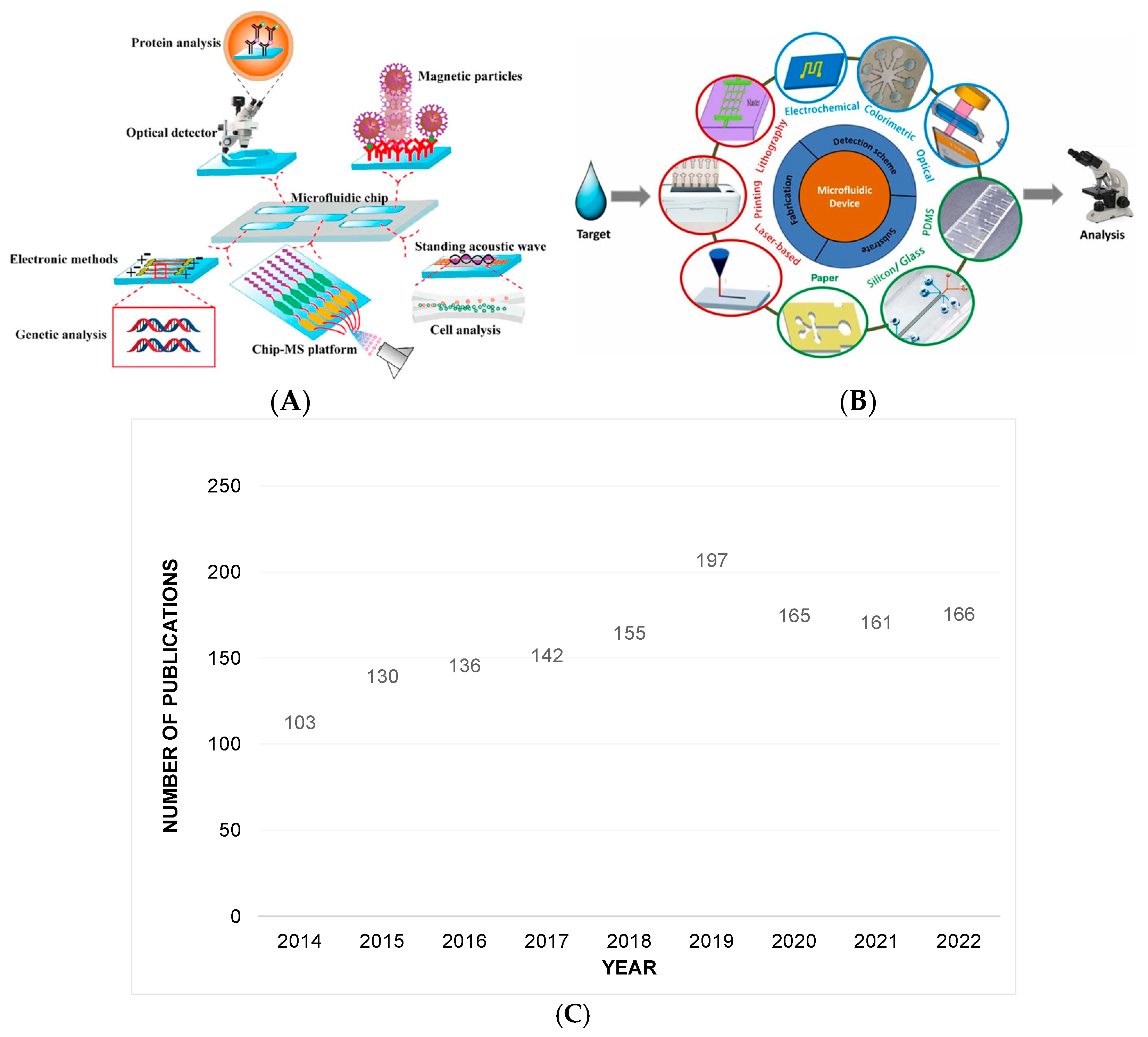
:1. Introduction
2. Polymer and Paper-Based Microfluidic Fabrication Methods
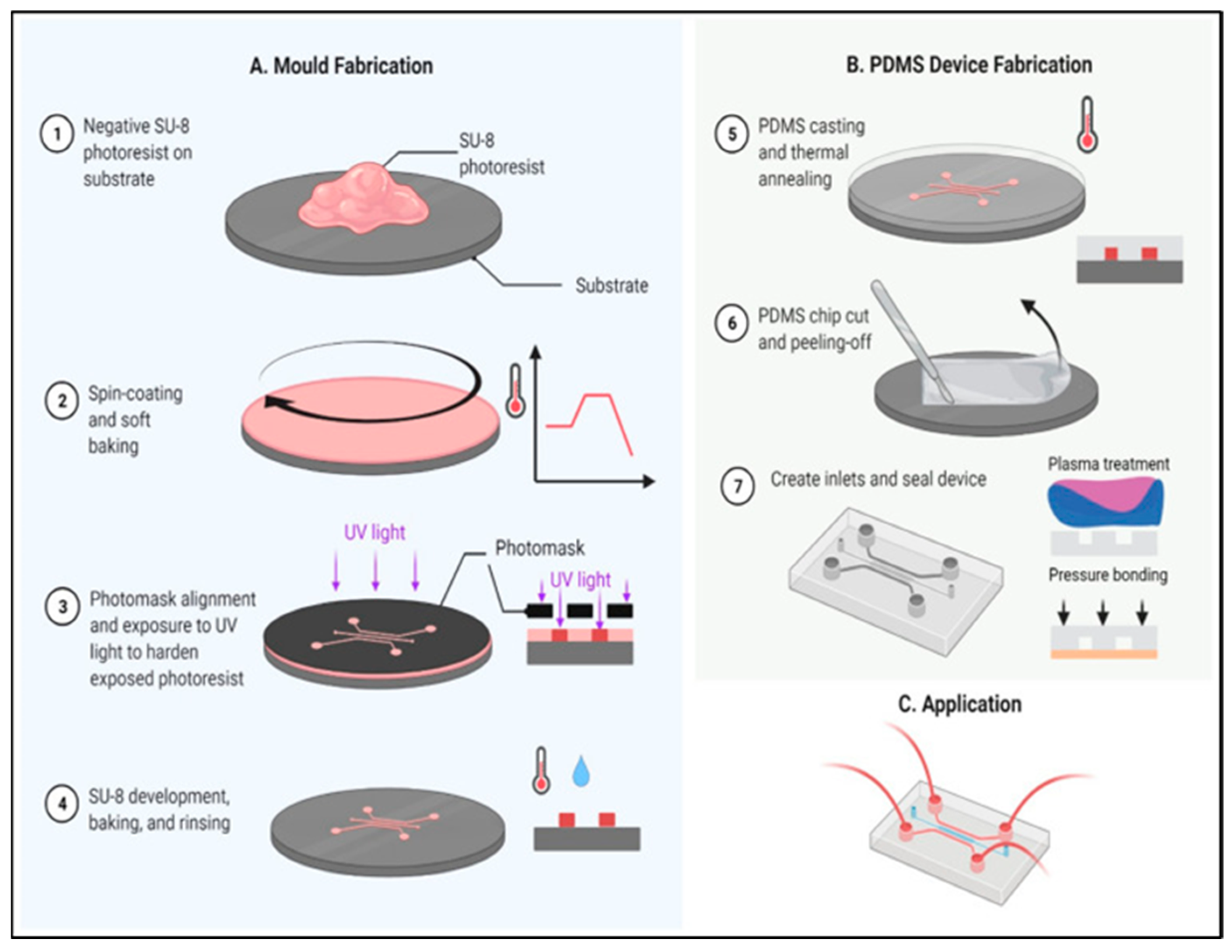
2.1. Polymer Mold-Based Techniques
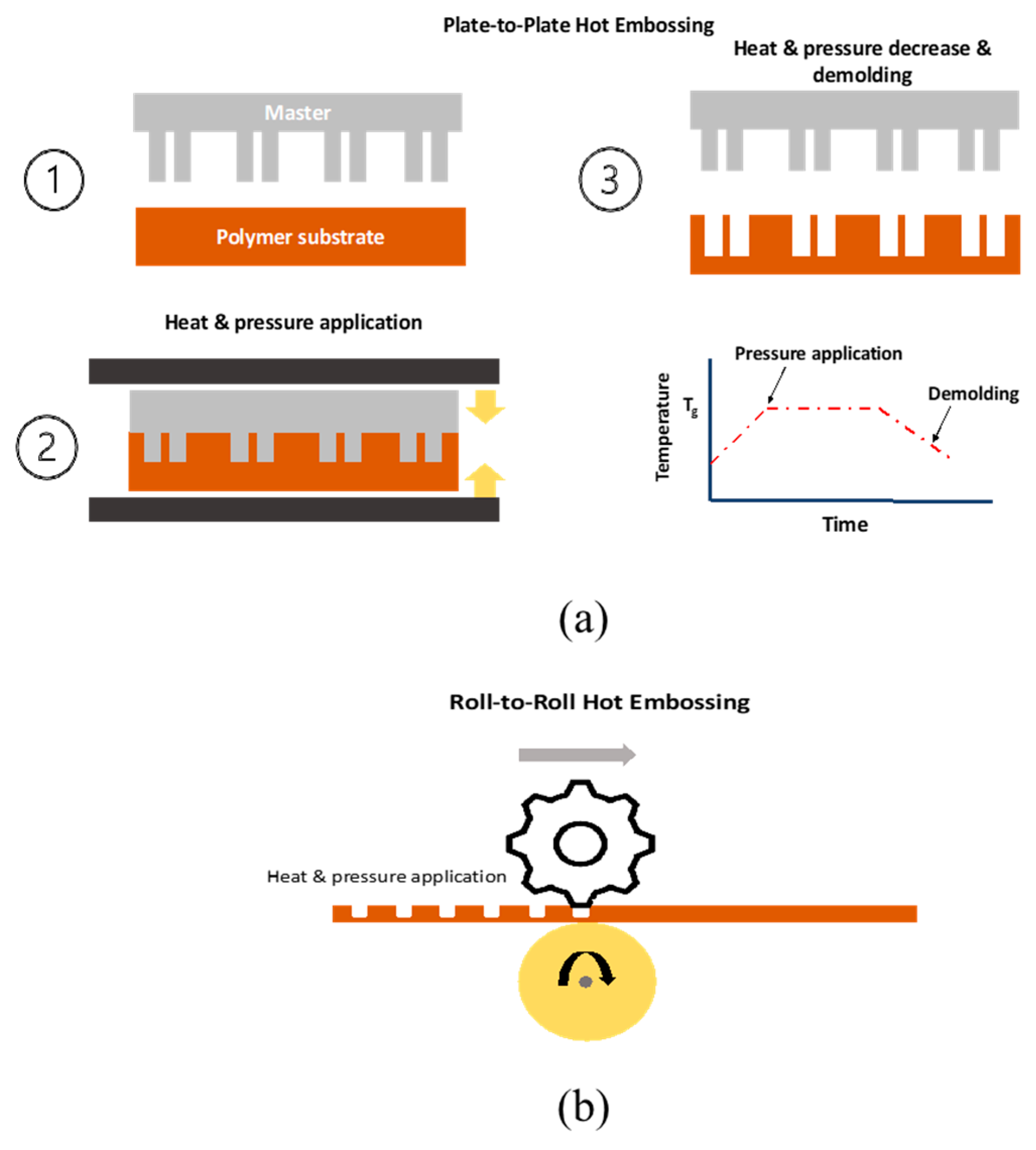
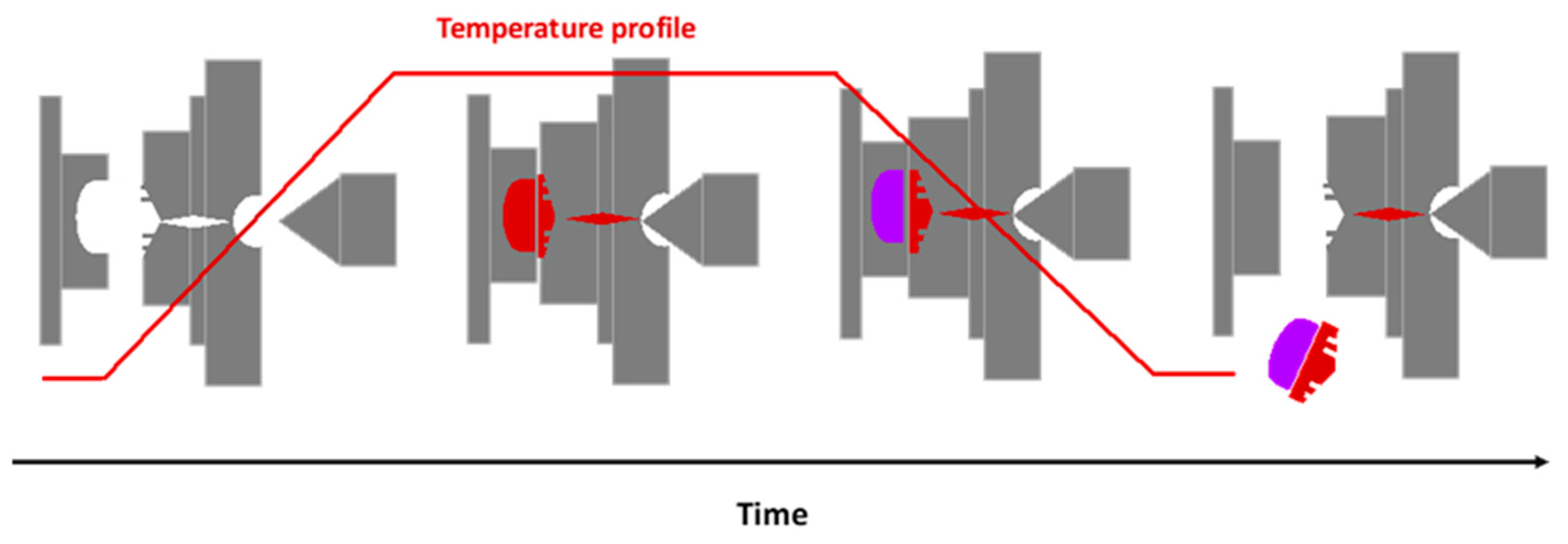
2.1.1. Hot Embossing
2.1.2. Injection Molding
2.1.3. Casting
2.2. Micromachining Techniques
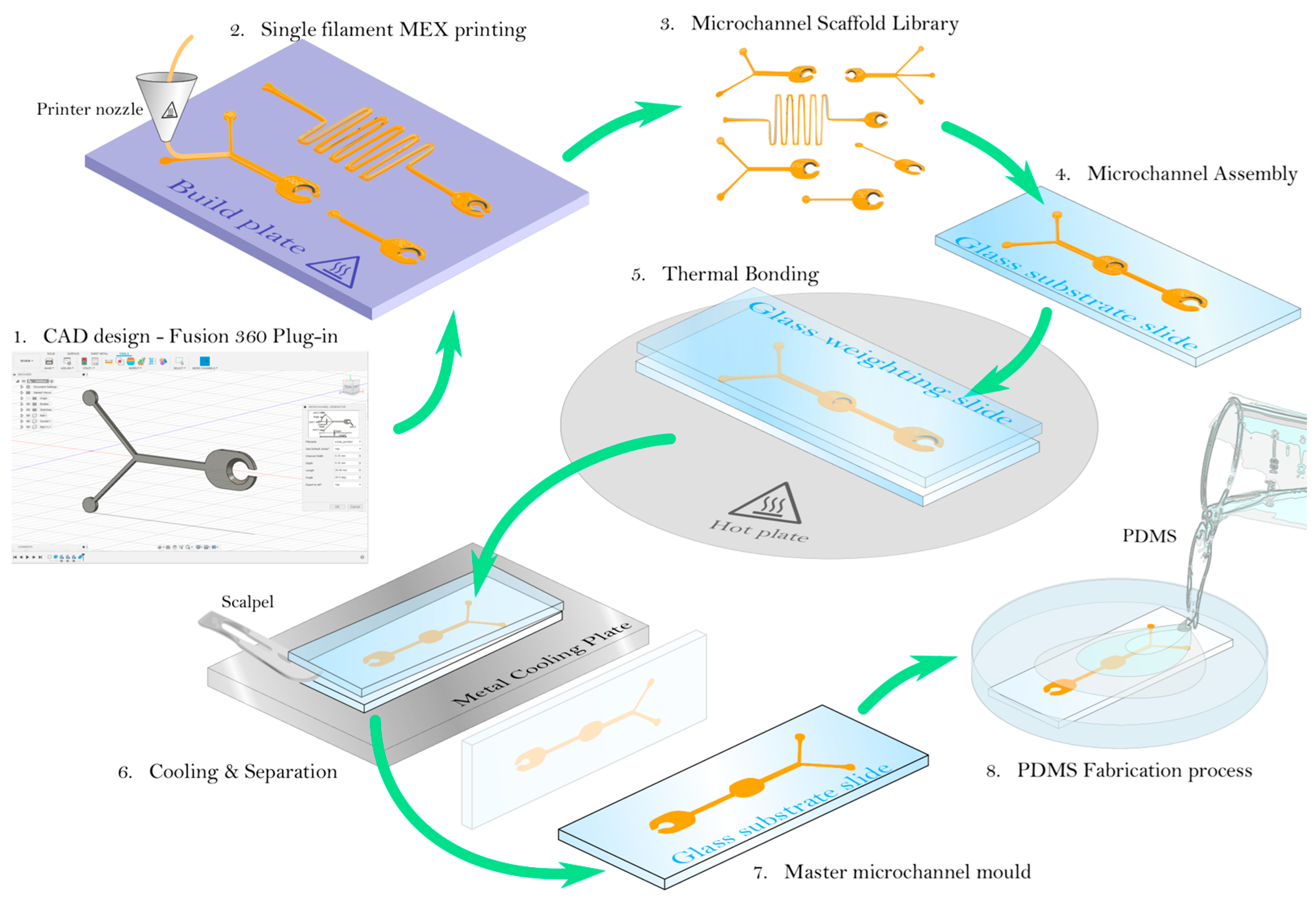
2.3. 3D Printing
2.4. Optical Lithography Techniques
2.5. Plasma Processing
2.6. Paper-Based Microfluidics
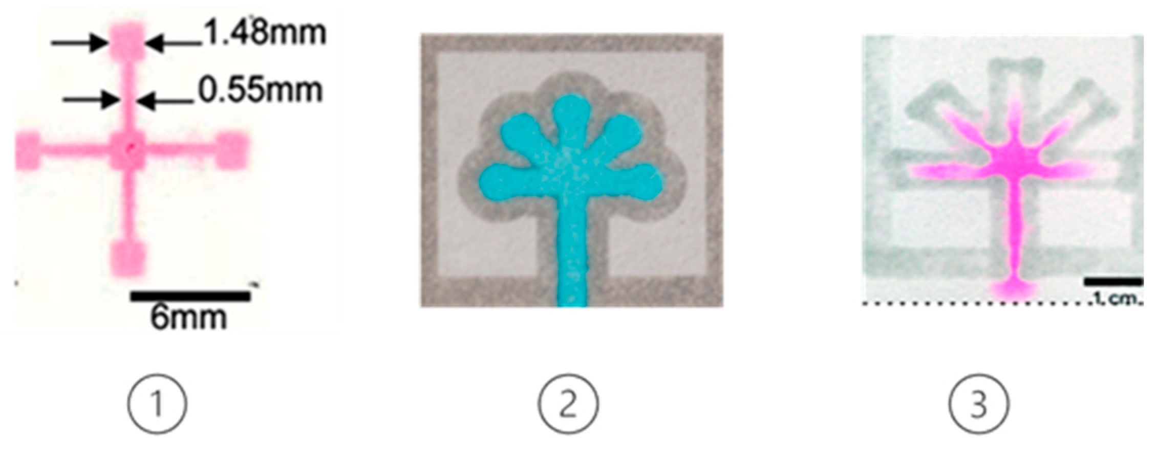
2.6.1. Wax Printing
2.6.2. Inkjet Printing
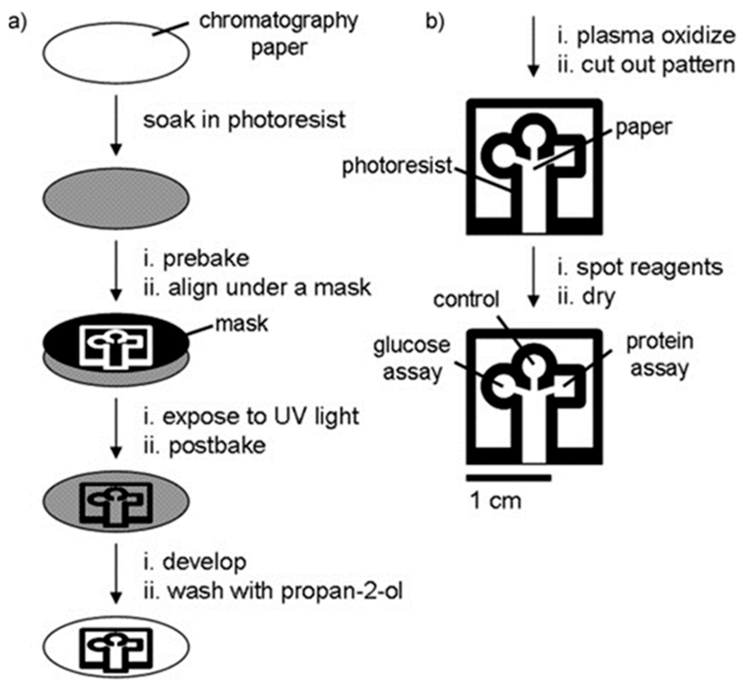
2.6.3. Optical Lithography
2.6.4. Screen Printing
2.6.5. Laser Processing Technology
2.6.6. Plasma Processing
2.6.7. 3D Printing/Lamination Methods
3. Microfluidics and LOCs in Food Safety Applications
3.1. Microfluidics for the Detection of Foodborne Pathogens
3.1.1. Escherichia coli Detection


3.1.2. Salmonella Strains Detection
3.1.3. Listeria Monocytogenes Detection
3.1.4. Staphylococcus aureus Detection
3.1.5. Campylobacter spp. Detection
4. Conclusions and Discussion
4.1. Research Outlook of Microfluidics and LOCs
4.2. Industrial Outlook of Microfluidics and LOCs
Author Contributions
Funding
Conflicts of Interest
References
- Sedighi, A.; Li, P.C. Challenges and Future Trends in DNA Microarray Analysis. Compr. Anal. Chem. 2014, 63, 25–46. [Google Scholar] [CrossRef]
- Escarpa, A. Lights and shadows on Food Microfluidics. Lab Chip 2014, 14, 3213–3224. [Google Scholar] [CrossRef] [PubMed]
- Ghallab, Y.; Badawy, W. Sensing methods for dielectrophoresis phenomenon: From bulky instruments to lab-on-a-chip. IEEE Circuits Syst. Mag. 2004, 4, 5–15. [Google Scholar] [CrossRef]
- Yager, P.; Edwards, T.; Fu, E.; Helton, K.; Nelson, K.; Tam, M.R.; Weigl, B.H. Microfluidic diagnostic technologies for global public health. Nature 2006, 442, 412–418. [Google Scholar] [CrossRef] [PubMed]
- Weng, X.; Neethirajan, S. Ensuring food safety: Quality monitoring using microfluidics. Trends Food Sci. Technol. 2017, 65, 10–22. [Google Scholar] [CrossRef]
- Livak-Dahl, E.; Sinn, I.; Burns, M. Microfluidic chemical analysis systems. Microelectron. Eng. 2015, 132, 156–175. [Google Scholar] [CrossRef]
- Temiz, Y.; Lovchik, R.D.; Kaigala, G.V.; Delamarche, E. Lab-on-a-chip devices: How to close and plug the lab. Microelectron. Eng. 2015, 132, 156–175. [Google Scholar] [CrossRef]
- Angell, J.B.; Terry, S.C.; Barth, P.W.; Terry, S.C. Silicon micromechanical devices. Sci. Am. 1983, 248, 44–55. [Google Scholar] [CrossRef]
- Guijt, R.; Manz, A. Miniaturised total chemical-analysis systems (ΜTAS) that periodically convert chemical into electronic information. Sens. Actuators B Chem. 2018, 273, 1334–1345. [Google Scholar] [CrossRef]
- Janasek, D.; Franzke, J.; Manz, A. Scaling and the design of miniaturized chemical-analysis systems. Nature 2006, 442, 374–380. [Google Scholar] [CrossRef]
- Nguyen, N.-T.; Wu, Z. Micromixers—A review. J. Micromechan. Microengin. 2004, 15, R1–R16. [Google Scholar] [CrossRef]
- Andersson, H.; van der Wijngaart, W.; Nilsson, P.; Enoksson, P.; Stemme, G. A valve-less diffuser micropump for microfluidic analytical systems. Sens. Actuators B Chem. 2001, 72, 259–265. [Google Scholar] [CrossRef]
- Au, A.K.; Lai, H.; Utela, B.R.; Folch, A. Microvalves and Micropumps for BioMEMS. Micromachines 2011, 2, 179–220. [Google Scholar] [CrossRef] [Green Version]
- Wu, J.; He, Z.; Chen, Q.; Lin, J.-M. Biochemical analysis on microfluidic chips. TrAC Trends Anal. Chem. 2016, 80, 213–231. [Google Scholar] [CrossRef]
- Rai, P.K.; Islam, M.; Gupta, A. Microfluidic devices for the detection of contamination in water samples: A review. Sens. Actuators A Phys. 2022, 347, 113926. [Google Scholar] [CrossRef]
- Ren, K.; Zhou, J.; Wu, H. Materials for microfluidic chip fabrication. Acc. Chem. Res. 2013, 46, 2396–2406. [Google Scholar] [CrossRef]
- FAkther, F.; Tran, H.D.; Zhang, J.; Nguyen, N.-T.; Ta, H.T. Lab-on-a-chip (lab-on-a-phone) for analysis of blood and diagnosis of blood diseases. In Nanotechnology for Hematology, Blood Transfusion, and Artificial Blood; Elsevier: Amsterdam, The Netherlands, 2021; pp. 237–264. [Google Scholar] [CrossRef]
- Li, J.; LeRiche, T.; Tremblay, T.-L.; Wang, C.; Bonneil, E.; Harrison, D.J.; Thibault, P. Application of Microfluidic Devices to Proteomics Research: Identification of Trace-level Protein Digests and Affinity Capture of Target Peptides. Mol. Cell. Proteom. 2002, 1, 157–168. [Google Scholar] [CrossRef] [Green Version]
- Neužil, P.; Giselbrecht, S.; Länge, K.; Huang, T.J.; Manz, A. Revisiting lab-on-a-chip technology for drug discovery. Nat. Rev. Drug Discov. 2012, 11, 620–632. [Google Scholar] [CrossRef]
- Yoon, J.-Y.; Kim, B. Lab-on-a-chip pathogen sensors for food safety. Sensors 2012, 12, 10713–10741. [Google Scholar] [CrossRef]
- Lee, W.G.; Kim, Y.-G.; Chung, B.G.; Demirci, U.; Khademhosseini, A. Nano/Microfluidics for diagnosis of infectious diseases in developing countries. Adv. Drug Deliv. Rev. 2010, 62, 449–457. [Google Scholar] [CrossRef] [Green Version]
- Faour-Klingbeil, D.; Todd, E.C.D. Prevention and Control of Foodborne Diseases in Middle-East North African Countries: Review of National Control Systems. Int. J. Environ. Res. Public Health 2019, 17, 70. [Google Scholar] [CrossRef] [PubMed] [Green Version]
- Waldbaur, A.; Rapp, H.; Länge, K.; Rapp, B.E. Let there be chip—Towards rapid prototyping of microfluidic devices: One-step manufacturing processes. Anal. Methods 2011, 3, 2681–2716. [Google Scholar] [CrossRef]
- Horie, K.; Barón, M.; Fox, R.B.; He, J.; Hess, M.; Kahovec, J.; Kitayama, T.; Kubisa, P.; Maréchal, E.; Mormann, W.; et al. Definitions of terms relating to reactions of polymers and to functional polymeric materials (IUPAC Recommendations 2003). Pure Appl. Chem. 2004, 76, 889–906. [Google Scholar] [CrossRef]
- Scott, S.M.; Ali, Z. Fabrication methods for microfluidic devices: An overview. Micromachines 2021, 12, 319. [Google Scholar] [CrossRef] [PubMed]
- Nielsen, J.B.; Hanson, R.L.; Almughamsi, H.M.; Pang, C.; Fish, T.R.; Woolley, A.T. Microfluidics: Innovations in materials and their fabrication and functionalization. Anal. Chem. 2019, 92, 150–168. [Google Scholar] [CrossRef]
- Duffy, D.C.; McDonald, J.C.; Schueller, O.J.A.; Whitesides, G.M. Rapid prototyping of microfluidic systems in poly(dimethylsiloxane). Anal. Chem. 1998, 70, 4974–4984. [Google Scholar] [CrossRef]
- van Midwoud, P.M.; Janse, A.; Merema, M.T.; Groothuis, G.M.M.; Verpoorte, E. Comparison of biocompatibility and adsorption properties of different plastics for advanced microfluidic cell and tissue culture models. Anal. Chem. 2012, 84, 3938–3944. [Google Scholar] [CrossRef]
- Credou, J.; Berthelot, T. Cellulose: From biocompatible to bioactive material. J. Mater. Chem. B 2014, 2, 4767–4788. [Google Scholar] [CrossRef] [Green Version]
- Akyazi, T.; Basabe-Desmonts, L.; Benito-Lopez, F. Review on microfluidic paper-based analytical devices towards commercialisation. Anal. Chim. Acta 2018, 1001, 1–17. [Google Scholar] [CrossRef]
- Martinez, A.W.; Phillips, S.T.; Butte, M.J.; Whitesides, G.M. Patterned Paper as a Platform for Inexpensive, Low-Volume, Portable Bioassays. Angew. Chem. 2007, 119, 1340–1342. [Google Scholar] [CrossRef]
- Heckele, M.; Bacher, W.; Müller, K.D. Hot Embossing-The Molding Technique for Plastic Microstructures; Springer: Berlin/Heidelberg, Germany, 1998. [Google Scholar]
- Kricka, L.J.; Fortina, P.; Panaro, N.J.; Wilding, P.; Alonso-Amigo, G.; Becker, H. Fabrication of plastic microchips by hot embossing. Lab Chip 2002, 2, 1–4. [Google Scholar] [CrossRef]
- Velten, T.; Bauerfeld, F.; Schuck, H.; Scherbaum, S.; Landesberger, C.; Bock, K. Roll-to-roll hot embossing of microstructures. Microsyst. Technol. 2010, 17, 619–627. [Google Scholar] [CrossRef]
- Ellinas, K.; Pliaka, V.; Kanakaris, G.; Tserepi, A.; Alexopoulos, L.; Gogolides, E. Micro-bead immunoassays for the detection of IL6 and PDGF-2 proteins on a microfluidic platform, incorporating superhydrophobic passive valves. Microelectron. Eng. 2017, 175, 73–80. [Google Scholar] [CrossRef]
- Kourmpetis, I.; Kastania, A.S.; Ellinas, K.; Tsougeni, K.; Baca, M.; De Malsche, W.; Gogolides, E. Gradient-temperature hot-embossing for dense micropillar array fabrication on thick cyclo-olefin polymeric plates: An example of a microfluidic chromatography column fabrication. Micro Nano Eng. 2019, 5, 100042. [Google Scholar] [CrossRef]
- Ng, S.H.; Wang, Z.F. Hot roller embossing for microfluidics: Process and challenges. Microsyst. Technol. 2008, 15, 1149–1156. [Google Scholar] [CrossRef]
- Deshmukh, S.S.; Goswami, A. Recent developments in hot embossing–A review. Mater. Manuf. Process. 2020, 36, 501–543. [Google Scholar] [CrossRef]
- Matlock-Colangelo, L.; Coon, B.; Pitner, C.L.; Frey, M.W.; Baeumner, A.J. Functionalized electrospun poly(vinyl alcohol) nanofibers for on-chip concentration of E. coli cells. Anal. Bioanal. Chem. 2015, 408, 1327–1334. [Google Scholar] [CrossRef]
- Schomburg, W.K.; Burlage, K.; Gerhardy, C. Ultrasonic hot embossing. Micromachines 2011, 2, 157–166. [Google Scholar] [CrossRef] [Green Version]
- Sucularli, F.; Arikan, M.S.; Yildirim, E. Investigation of process-affected zone in ultrasonic embossing of microchannels on thermoplastic substrates. J. Manuf. Process. 2020, 50, 394–402. [Google Scholar] [CrossRef]
- Chang, J.-H.; Yang, S.-Y. Development of fluid-based heating and pressing systems for micro hot embossing. Microsyst. Technol. 2005, 11, 396–403. [Google Scholar] [CrossRef]
- Juang, Y.-J.; Chiu, Y.-J. Fabrication of Polymer Microfluidics: An Overview. Polymers 2022, 14, 2028. [Google Scholar] [CrossRef] [PubMed]
- Li, X.; Liu, F.; Gong, N.; Huang, P.; Yang, C. Enhancing the joining strength of injection-molded polymer-metal hybrids by rapid heating and cooling. J. Mater. Process. Technol. 2017, 249, 386–393. [Google Scholar] [CrossRef]
- Su, Q.; Zhang, N.; Gilchrist, M.D. The use of variotherm systems for microinjection molding. J. Appl. Polym. Sci. 2015, 133, 42962. [Google Scholar] [CrossRef] [Green Version]
- Whiteside, B.; Babenko, M.; Tuinea-Bobe, C.; Brown, E.; Coates, P. Ultrasonic Injection Moulding: A Study of Thermal Behaviour and Nanofeature Replication. 2016. Available online: www.euspen.eu (accessed on 26 January 2023).
- Hansen, T.S.; Selmeczi, D.; Larsen, N.B. Fast prototyping of injection molded polymer microfluidic chips. J. Micromechan. Microengin. 2009, 20, 015020. [Google Scholar] [CrossRef]
- Yu, W.; Chen, Y.; Wang, Z.; Qiao, L.; Xie, R.; Zhang, J.; Bian, S.; Li, H.; Zhang, Y.; Chen, A. Multiple authentications of high-value milk by centrifugal microfluidic chip-based real-time fluorescent LAMP. Food Chem. 2021, 351, 129348. [Google Scholar] [CrossRef]
- Xia, Y.; Whitesides, G.M. Soft Lithography. 1998. Available online: www.annualreviews.org. (accessed on 26 January 2023).
- Aumiller, G.D.; Chandross, E.A.; Tomlinson, W.J.; Weber, H.P. Submicrometer resolution replication of relief patterns for integrated optics. J. Appl. Phys. 1974, 45, 4557–4562. [Google Scholar] [CrossRef]
- Xue, L.; Jin, N.; Guo, R.; Wang, S.; Qi, W.; Liu, Y.; Li, Y.; Lin, J. Microfluidic Colorimetric Biosensors Based on MnO2 Nanozymes and Convergence–Divergence Spiral Micromixers for Rapid and Sensitive Detection of Salmonella. ACS Sens. 2021, 6, 2883–2892. [Google Scholar] [CrossRef]
- Salih, N.M.; Sahdan, M.; Sahdan, Z.; Morsin, M.; Asmah, M.T. Fabrication and Integration of PDMS-Glass Based Microfluidic with Optical Absorbance Measurement Device for Coliform Bacteria Detection. In 6th International Conference on the Development of Biomedical Engineering in Vietnam (BME6); Vo Van, T., Nguyen Le, T.A., Nguyen Duc, T., Eds.; Springer: Singapore, 2018; pp. 75–81. [Google Scholar]
- Leclerc, C.A.; Williams, S.; Powe, C.; Zepp, N.; Lipworth, D.; Pensini, E.; Collier, C.M. Rapid design and prototyping of microfluidic chips via computer numerical control micromilling and anisotropic shrinking of stressed polystyrene sheets. Microfluid. Nanofluidics 2021, 25, 1–12. [Google Scholar] [CrossRef]
- Liu, S.; Fan, Y.; Gao, K.; Zhang, Y. Fabrication of Cyclo-olefin polymer-based microfluidic devices using CO2 laser ablation. Mater. Res. Express 2018, 5, 095305. [Google Scholar] [CrossRef]
- Gao, K.; Liu, J.; Fan, Y.; Zhang, Y. Ultra-low-cost fabrication of polymer-based microfluidic devices with diode laser ablation. Biomed. Microdevices 2019, 21, 83. [Google Scholar] [CrossRef]
- Gao, K.; Liu, J.; Fan, Y.; Zhang, Y. An effective method for fabricating microchannels on the polycarbonate (PC) substrate with CO2 laser. Int. J. Adv. Manuf. Technol. 2017, 92, 1365–1370. [Google Scholar] [CrossRef]
- Yin, Z. Rapid prototyping of PET microfluidic chips by laser ablation and water-soaking bonding method. Micro Nano Lett. 2018, 13, 1302–1305. [Google Scholar] [CrossRef]
- Min, K.; Lim, J.; Lim, J.H.; Hwang, E.; Kim, Y.; Lee, H.; Lee, H.; Hong, S. Fabrication of perforated PDMS microchannel by successive laser pyrolysis. Materials 2021, 14, 7275. [Google Scholar] [CrossRef]
- Hsieh, Y.-K.; Chen, S.-C.; Huang, W.-L.; Hsu, K.-P.; Gorday, K.A.V.; Wang, T.; Wang, J. Direct micromachining of microfluidic channels on biodegradable materials using laser ablation. Polymers 2017, 9, 242. [Google Scholar] [CrossRef] [Green Version]
- Bilican, I.; Guler, M.T. Assessment of PMMA and polystyrene based microfluidic chips fabricated using CO2 laser machining. Appl. Surf. Sci. 2020, 534, 147642. [Google Scholar] [CrossRef]
- Huang, J.; Qin, Q.; Wang, J. A review of stereolithography: Processes and systems. Processes 2020, 8, 1138. [Google Scholar] [CrossRef]
- Jasveer, S.; Jianbin, X. Comparison of Different Types of 3D Printing Technologies. Int. J. Sci. Res. Publ. 2018, 8, 7602. [Google Scholar] [CrossRef]
- Nelson, M.D.; Ramkumar, N.; Gale, B.K. Flexible, transparent, sub-100 μm microfluidic channels with fused deposition modeling 3D-printed thermoplastic polyurethane. Anal. Chim. Acta 2019, 1071, 36–43. [Google Scholar] [CrossRef]
- Duarte, L.C.; Figueredo, F.; Ribeiro, L.E.; Cortón, E.; Coltro, W.K. Label-free counting of Escherichia coli cells in nanoliter droplets using 3D printed microfluidic devices with integrated contactless conductivity detection. Sens. Actuators B Chem. 2017, 251, 427–432. [Google Scholar] [CrossRef]
- Duarte, L.C.; Chagas, C.L.; Ribeiro, L.E.; Coltro, W.K. 3D printing of microfluidic devices with embedded sensing electrodes for generating and measuring the size of microdroplets based on contactless conductivity detection. Sens. Actuators B Chem. 2017, 251, 427–432. [Google Scholar] [CrossRef]
- Kanitthamniyom, P.; Hon, P.Y.; Zhou, A.; Abdad, M.Y.; Leow, Z.Y.; Yazid, N.B.M.; Xun, V.L.W.; Vasoo, S.; Zhang, Y. A 3D-printed magnetic digital microfluidic diagnostic platform for rapid colorimetric sensing of carbapenemase-producing Enterobacteriaceae. Microsyst. Nanoeng. 2021, 7, 47. [Google Scholar] [CrossRef] [PubMed]
- Felton, H.; Hughes, R.; Diaz-Gaxiola, A. Negligible-cost microfluidic device fabrication using 3D-printed interconnecting channel scaffolds. PLoS ONE 2021, 16, e0245206. [Google Scholar] [CrossRef] [PubMed]
- Kim, A.A.; Kustanovich, K.; Baratian, D.; Ainla, A.; Shaali, M.; Jeffries, G.D.M.; Jesorka, A. SU-8 free-standing microfluidic probes. Biomicrofluidics 2017, 11, 014112. [Google Scholar] [CrossRef] [PubMed]
- Dy, A.J.; Cosmanescu, A.; Sluka, J.; A Glazier, J.; Stupack, D.; Amarie, D. Fabricating microfluidic valve master molds in SU-8 photoresist. J. Micromec. Microengin. 2014, 24, 57001. [Google Scholar] [CrossRef] [Green Version]
- Corredor, S.F.; Mayoussi, F.; Luitz, M.; Kick, A.; Goralczyk, A.; Böcherer, D.; Vera, G.; Helmer, D.; Kotz-Helmer, F.; Rapp, B.E. A Polystyrene Photoresin for Direct Lithography of Microfluidic Chips. Adv. Mater. Technol. 2022, 7, 84. [Google Scholar] [CrossRef]
- Ellinas, K.; Tsougeni, K.; Petrou, P.S.; Boulousis, G.; Tsoukleris, D.; Pavlatou, E.; Tserepi, A.; Kakabakos, S.E.; Gogolides, E. Three-dimensional plasma micro-nanotextured cyclo-olefin-polymer surfaces for biomolecule immobilization and environmentally stable superhydrophobic and superoleophobic behavior. Chem. Eng. J. 2016, 300, 394–403. [Google Scholar] [CrossRef]
- Rhee, S.W.; Taylor, A.M.; Tu, C.H.; Cribbs, D.H.; Cotman, C.W.; Jeon, N.L. Patterned cell culture inside microfluidic devices. Lab Chip 2004, 5, 102–107. [Google Scholar] [CrossRef]
- Tsao, C.-W.; DeVoe, D. Bonding of thermoplastic polymer microfluidics. Microfluid. Nanofluidics 2008, 6, 1–16. [Google Scholar] [CrossRef]
- Tsougeni, K.; Papageorgiou, D.; Tserepi, A.; Gogolides, E. ‘Smart’ polymeric microfluidics fabricated by plasma processing: Controlled wetting, capillary filling and hydrophobic valving. Lab Chip 2009, 10, 462–469. [Google Scholar] [CrossRef]
- Tsougeni, K.; Papadakis, G.; Gianneli, M.; Grammoustianou, A.; Constantoudis, V.; Dupuy, B.; Petrou, P.S.; Kakabakos, S.E.; Tserepi, A.; Gizeli, E.; et al. Plasma nanotextured polymeric lab-on-a-chip for highly efficient bacteria capture and lysis. Lab Chip 2015, 16, 120–131. [Google Scholar] [CrossRef]
- Geissler, M.; Brassard, D.; Clime, L.; Pilar, A.V.C.; Malic, L.; Daoud, J.; Barrère, V.; Luebbert, C.; Blais, B.W.; Corneau, N.; et al. Centrifugal microfluidic lab-on-a-chip system with automated sample lysis, DNA amplification and microarray hybridization for identification of enterohemorrhagic Escherichia coli culture isolates. Analyst 2020, 145, 6831–6845. [Google Scholar] [CrossRef]
- Gravel, J.-F.; Geissler, M.; Chapdelaine, S.; Boissinot, K.; Voisin, B.; Charlebois, I.; Poirier-Richard, H.-P.; Grégoire, A.; Boissinot, M.; Bergeron, M.G.; et al. Portable bead-based fluorescence detection system for multiplex nucleic acid testing: A case study with Bacillus anthracis. Microfluid. Nanofluidics 2013, 16, 1075–1087. [Google Scholar] [CrossRef]
- Gorkin, R.; Park, J.; Siegrist, J.; Amasia, M.; Lee, B.S.; Park, J.-M.; Kim, J.; Kim, H.; Madou, M.; Cho, Y.-K. Centrifugal microfluidics for biomedical applications. Lab Chip 2010, 10, 1758–1773. [Google Scholar] [CrossRef] [Green Version]
- Muller, H.; Clegg, D.L. Automatic Paper Chromatography. Available online: https://pubs.acs.org/sharingguidelines (accessed on 26 January 2023).
- Yamada, K.; Henares, T.G.; Suzuki, K.; Citterio, D. Papierbasierte tintenstrahlgedruckte Mikrofluidiksysteme für die Analytik. Angew. Chem. 2015, 127, 5384–5401. [Google Scholar] [CrossRef]
- Li, X.; Tian, J.; Garnier, G.; Shen, W. Fabrication of paper-based microfluidic sensors by printing. Colloids Surf. B: Biointerfaces 2010, 76, 564–570. [Google Scholar] [CrossRef]
- Martinez, A.W.; Phillips, S.T.; Wiley, B.; Gupta, M.; Whitesides, G.M. FLASH: A rapid method for prototyping paper-based microfluidic devices. Lab Chip 2008, 8, 2146–2150. [Google Scholar] [CrossRef]
- Olkkonen, J.; Lehtinen, K.; Erho, T. Flexographically printed fluidic structures in paper. Anal. Chem. 2010, 82, 10246–10250. [Google Scholar] [CrossRef]
- Chitnis, G.; Ding, Z.; Chang, C.-L.; Savran, C.A.; Ziaie, B. Laser-treated hydrophobic paper: An inexpensive microfluidic platform. Lab Chip 2011, 11, 1161–1165. [Google Scholar] [CrossRef]
- Abe, K.; Suzuki, K.; Citterio, D. Inkjet-printed microfluidic multianalyte chemical sensing paper. Anal. Chem. 2008, 80, 6928–6934. [Google Scholar] [CrossRef]
- Abe, K.; Kotera, K.; Suzuki, K.; Citterio, D. Inkjet-printed paperfluidic immuno-chemical sensing device. Anal. Bioanal. Chem. 2010, 398, 885–893. [Google Scholar] [CrossRef]
- Qin, X.; Liu, J.; Zhang, Z.; Li, J.; Yuan, L.; Zhang, Z.; Chen, L. Microfluidic paper-based chips in rapid detection: Current status, challenges, and perspectives. TrAC Trends Anal. Chem. 2021, 143, 116371. [Google Scholar] [CrossRef]
- Ruiz, R.A.; Gonzalez, J.L.; Vazquez-Alvarado, M.; Martinez, N.W.; Martinez, A.W. Beyond Wax Printing: Fabrication of Paper-Based Microfluidic Devices Using a Thermal Transfer Printer. Anal. Chem. 2022, 94, 8833–8837. [Google Scholar] [CrossRef] [PubMed]
- Carrilho, E.; Martinez, A.W.; Whitesides, G.M. Understanding wax printing: A simple micropatterning process for paper-based microfluidics. Anal. Chem. 2009, 81, 7091–7095. [Google Scholar] [CrossRef] [PubMed]
- Ng, J.S.; Hashimoto, M. Fabrication of paper microfluidic devices using a toner laser printer. RSC Adv. 2020, 10, 29797–29807. [Google Scholar] [CrossRef]
- Asif, M.; Awan, F.R.; Khan, Q.M.; Ngamsom, B.; Pamme, N. Paper-based analytical devices for colorimetric detection of: S. aureus and E. coli and their antibiotic resistant strains in milk. Analyst 2020, 145, 7320–7329. [Google Scholar] [CrossRef]
- Zhao, Y.; Zeng, D.; Yan, C.; Chen, W.; Ren, J.; Jiang, Y.; Jiang, L.; Xue, F.; Ji, D.; Tang, F.; et al. Rapid and accurate detection of: Escherichia coli O157:H7 in beef using microfluidic wax-printed paper-based ELISA. Analyst 2020, 145, 3106–3115. [Google Scholar] [CrossRef]
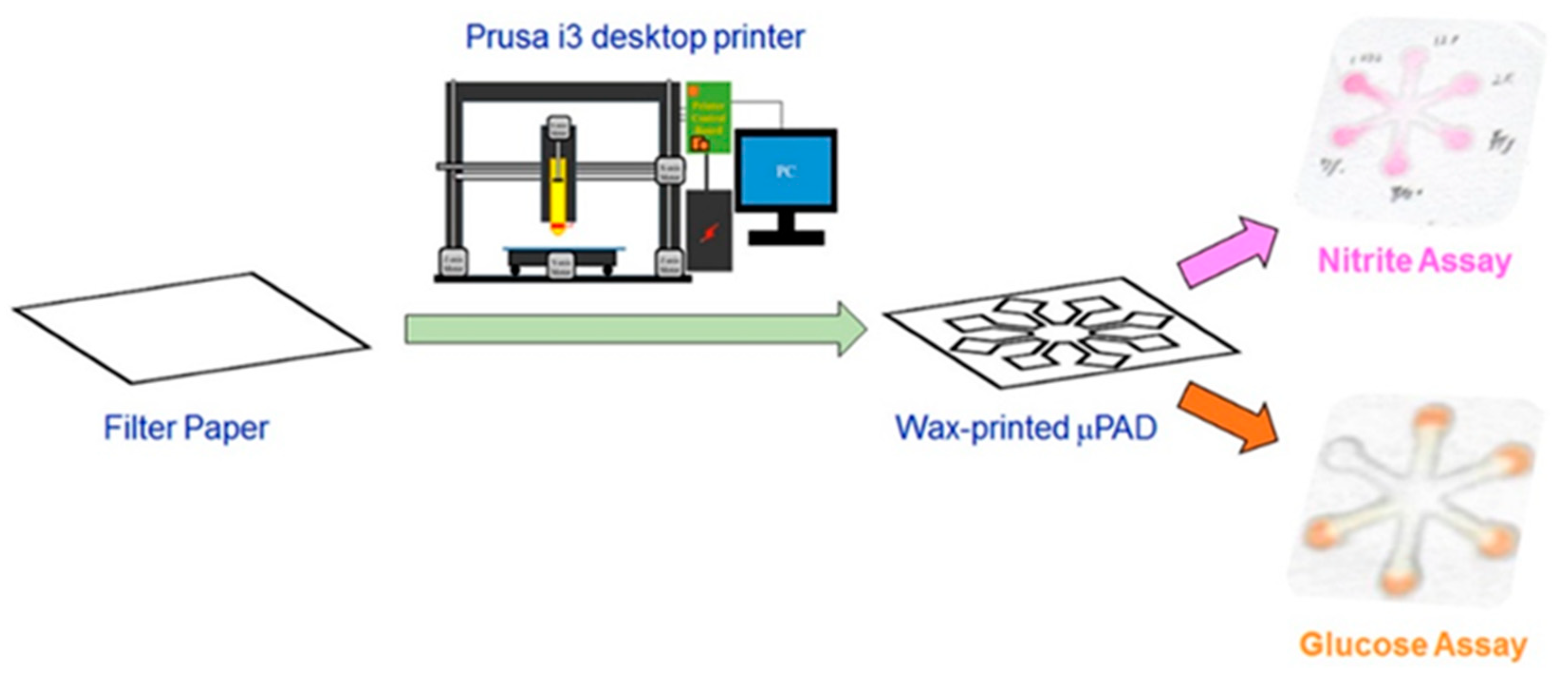
- Chiang, C.-K.; Kurniawan, A.; Kao, C.-Y.; Wang, M.-J. Single step and mask-free 3D wax printing of microfluidic paper-based analytical devices for glucose and nitrite assays. Talanta 2018, 194, 837–845. [Google Scholar] [CrossRef]
- Wang, J.; Monton, M.R.N.; Zhang, X.; Filipe, C.D.M.; Pelton, R.; Brennan, J.D. Hydrophobic sol-gel channel patterning strategies for paper-based microfluidics. Lab Chip 2013, 14, 691–695. [Google Scholar] [CrossRef]
- Rajendra, V.; Sicard, C.; Brennan, J.D.; Brook, M.A. Printing silicone-based hydrophobic barriers on paper for microfluidic assays using low-cost ink jet printers. Analyst 2014, 139, 6361–6365. [Google Scholar] [CrossRef]
- Maejima, K.; Tomikawa, S.; Suzuki, K.; Citterio, D. Inkjet printing: An integrated and green chemical approach to microfluidic paper-based analytical devices. RSC Adv. 2013, 3, 9258–9263. [Google Scholar] [CrossRef]
- Tekin, E.; Smith, P.J.; Schubert, U.S. Inkjet printing as a deposition and patterning tool for polymers and inorganic particles. Soft Matter 2008, 4, 703–713. [Google Scholar] [CrossRef]
- Gonzalez-Macia, L.; Morrin, A.; Smyth, M.R.; Killard, A.J. Advanced printing and deposition methodologies for the fabrication of biosensors and biodevices. Analyst 2010, 135, 845–867. [Google Scholar] [CrossRef] [Green Version]
- Komuro, N.; Takaki, S.; Suzuki, K.; Citterio, D. Inkjet printed (bio)chemical sensing devices. Anal. Bioanal. Chem. 2013, 405, 5785–5805. [Google Scholar] [CrossRef]
- Delaney, J.T.; Smith, P.J.; Schubert, U.S. Inkjet printing of proteins. Soft Matter 2009, 5, 4866–4877. [Google Scholar] [CrossRef]
- Hossain, S.M.Z.; Ozimok, C.; Sicard, C.; Aguirre, S.D.; Ali, M.M.; Li, Y.; Brennan, J.D. Multiplexed paper test strip for quantitative bacterial detection. Anal. Bioanal. Chem. 2012, 403, 1567–1576. [Google Scholar] [CrossRef]
- Snyder, S.A.; Boban, M.; Li, C.; VanEpps, J.S.; Mehta, G.; Tuteja, A. Lysis and direct detection of coliforms on printed paper-based microfluidic devices. Lab Chip 2020, 20, 4413–4419. [Google Scholar] [CrossRef]
- Bruzewicz, D.A.; Reches, M.; Whitesides, G.M. Low-Cost Printing of Poly(dimethylsiloxane) Barriers To Define Microchannels in Paper. Anal. Chem. 2008, 80, 3387–3392. [Google Scholar] [CrossRef] [Green Version]
- Haller, P.D.; Flowers, C.A.; Gupta, M. Three-dimensional patterning of porous materials using vapor phase polymerization. Soft Matter 2011, 7, 2428–2432. [Google Scholar] [CrossRef]
- He, Q.; Ma, C.; Hu, X.; Chen, H. Method for Fabrication of Paper-Based Microfluidic Devices by Alkylsilane Self-Assembling and UV/O3-Patterning. Anal. Chem. 2013, 85, 1327–1331. [Google Scholar] [CrossRef]
- Carrilho, E.; Phillips, S.T.; Vella, S.J.; Martinez, A.W.; Whitesides, G.M. Paper Microzone Plates. Anal. Chem. 2009, 81, 5990–5998. [Google Scholar] [CrossRef]
- Lin, D.; Li, B.; Qi, J.; Ji, X.; Yang, S.; Wang, W.; Chen, L. Low cost fabrication of microfluidic paper-based analytical devices with water-based polyurethane acrylate and their application for bacterial detection. Sens. Actuators B Chem. 2019, 303, 127213. [Google Scholar] [CrossRef]
- Nóbrega, L.N.; Magalhães, L.D.O.; Fonseca, A. A urethane-acrylate microflow-analyzer with an integrated cadmium column. Microchem. J. 2013, 110, 553–557. [Google Scholar] [CrossRef]
- Yu, L.; Shi, Z.Z. Microfluidic paper-based analytical devices fabricated by low-cost photolithography and embossing of Parafilm®. Lab Chip 2015, 15, 1642–1645. [Google Scholar] [CrossRef] [PubMed]
- Rengaraj, S.; Cruz-Izquierdo, Á.; Scott, J.L.; Di Lorenzo, M. Impedimetric paper-based biosensor for the detection of bacterial contamination in water. Sens. Actuators B Chem. 2018, 265, 50–58. [Google Scholar] [CrossRef]
- Mahmud, A.; Blondeel, E.J.M.; Kaddoura, M.; MacDonald, B.D. Creating compact and microscale features in paper-based devices by laser cutting. Analyst 2016, 141, 6449–6454. [Google Scholar] [CrossRef]
- Pebdeni, A.B.; Hosseini, M. Fast and selective whole cell detection of Staphylococcus aureus bacteria in food samples by paper based colorimetric nanobiosensor using peroxidase-like catalytic activity of DNA-Au/Pt bimetallic nanoclusters. Microchem. J. 2020, 159, 105475. [Google Scholar] [CrossRef]
- Sohrabpoor, H.; Issa, A.; Al Hamaoy, A.; Ahad, I.U.; Chikarakara, E.; Bagga, K.; Brabazon, D. Chapter 24—Development of Laser Processing Technologies via Experimental Design. In Advances in Laser Materials Processing, 2nd ed.; Lawrence, J., Ed.; Woodhead Publishing: Sawston, UK, 2018; pp. 707–729. [Google Scholar] [CrossRef]
- Li, X.; Tian, J.; Nguyen, T.; Shen, W. Paper-Based Microfluidic Devices by Plasma Treatment. Anal. Chem. 2008, 80, 9131–9134. [Google Scholar] [CrossRef]
- Wang, H.; Enders, A.; Preuss, J.-A.; Bahnemann, J.; Heisterkamp, A.; Torres-Mapa, M.L. 3D printed microfluidic lab-on-a-chip device for fiber-based dual beam optical manipulation. Sci. Rep. 2021, 11, 14584. [Google Scholar] [CrossRef]
- Rasooly, A.; Bruck, H.A.; Kostov, Y. An ELISA Lab-on-a-Chip (ELISA-LOC). Microfluid. Diagn. Methods Protoc. 2012, 949, 451–471. [Google Scholar] [CrossRef]
- Gaal, G.; Mendes, M.; de Almeida, T.P.; Piazzetta, M.H.; Gobbi, L.; Riul, A.; Rodrigues, V. Simplified fabrication of integrated microfluidic devices using fused deposition modeling 3D printing. Sens. Actuators B Chem. 2017, 242, 35–40. [Google Scholar] [CrossRef]
- Zäh, M.F.; Lutzmann, S. Modelling and simulation of electron beam melting. Prod. Eng. 2009, 4, 15–23. [Google Scholar] [CrossRef]
- Richard, C.; Neild, A.; Cadarso, V.J. The emerging role of microfluidics in multi-material 3D bioprinting. Lab Chip 2020, 20, 2044–2056. [Google Scholar] [CrossRef]
- Samson, A.A.S.; Lee, J.; Song, J.M. Paper-based inkjet bioprinting to detect fluorescence resonance energy transfer for the assessment of anti-inflammatory activity. Sci. Rep. 2018, 8, 591. [Google Scholar] [CrossRef] [Green Version]
- Fu, X.; Xia, B.; Ji, B.; Lei, S.; Zhou, Y. Flow controllable three-dimensional paper-based microfluidic analytical devices fabricated by 3D printing technology. Anal. Chim. Acta 2019, 1065, 64–70. [Google Scholar] [CrossRef]
- Galanis, P.; He, P.; Katis, I.; Iles, A.; Kumar, A.; Eason, R.; Sones, C. Local photo-polymer deposition-assisted fabrication of multilayer paper-based devices. Sens. Actuators B Chem. 2020, 322, 128574. [Google Scholar] [CrossRef]
- Schilling, K.M.; Jauregui, D.; Martinez, A.W. Paper and toner three-dimensional fluidic devices: Programming fluid flow to improve point-of-care diagnostics. Lab Chip 2013, 13, 628–631. [Google Scholar] [CrossRef] [Green Version]
- WHO EMRO. An Overview of Emerging Foodborne and Waterborne Diseases. 1996. Available online: https://www.emro.who.int/emhj-volume-2-1996/volume-2-issue-1/article7.html (accessed on 11 January 2023).
- Swaminathan, B.; Feng, P. Rapid detection of food-borne pathogenic bacteria. Annu. Rev. Microbiol. 1994, 48, 401. Available online: https://link.gale.com/apps/doc/A16379190/AONE?u=anon~76b7b0&sid=googleScholar&xid=e24a0c4c (accessed on 26 January 2023). [CrossRef]
- Food and Water Borne Diseases. Available online: https://www2.gnb.ca/content/gnb/en/departments/ocmoh/cdc/content/food_andwaterborne.html (accessed on 26 January 2023).
- Pesticide Residues in Food. Available online: https://www.who.int/news-room/fact-sheets/detail/pesticide-residues-in-food (accessed on 26 January 2023).
- PRai, P.K.; Lee, S.S.; Zhang, M.; Tsang, Y.F.; Kim, K.-H. Heavy metals in food crops: Health risks, fate, mechanisms, and management. Environ. Int. 2019, 125, 365–385. [Google Scholar] [CrossRef]
- Tauxe, R.V. Emerging foodborne pathogens. Int. J. Food Microbiol. 2002, 78, 31–41. [Google Scholar] [CrossRef]
- What Are Biosensors? Principle, Working, Types and Applications. Available online: https://www.electronicshub.org/types-of-biosensors/ (accessed on 26 January 2023).
- Waters, L.C.; Jacobson, S.C.; Kroutchinina, N.; Khandurina, J.; Foote, R.S.; Ramsey, J.M. Microchip Device for Cell Lysis, Multiplex PCR Amplification, and Electrophoretic Sizing. Anal. Chem. 1997, 70, 158–162. [Google Scholar] [CrossRef]
- Peña-Bahamonde, J.; Nguyen, H.N.; Fanourakis, S.K.; Rodrigues, D.F. Recent advances in graphene-based biosensor technology with applications in life sciences. J. Nanobiotechnol. 2018, 16, 1–17. [Google Scholar] [CrossRef] [PubMed] [Green Version]
- Heuner, K.; Swanson, M. Legionella: Molecular Microbiology; Caister Academic Press: Norwich, UK, 2008; p. 249. [Google Scholar]
- Legionnaires Disease and Pontiac Fever | CDC. Available online: https://www.cdc.gov/legionella/index.html (accessed on 26 January 2023).
- Legionellosis. Available online: https://www.who.int/news-room/fact-sheets/detail/legionellosis (accessed on 26 January 2023).
- Saad, M.; Castiello, F.R.; Faucher, S.P.; Tabrizian, M. Introducing an SPRi-based titration assay using aptamers for the detection of Legionella pneumophila. Sens. Actuators B Chem. 2021, 351, 130933. [Google Scholar] [CrossRef]
- Laribi, A.; Allegra, S.; Souiri, M.; Mzoughi, R.; Othmane, A.; Girardot, F. Legionella pneumophila sg1-sensing signal enhancement using a novel electrochemical immunosensor in dynamic detection mode. Talanta 2020, 215, 120904. [Google Scholar] [CrossRef] [PubMed]
- Ikeda, M.; Yamaguchi, N.; Tani, K.; Nasu, M. Rapid and simple detection of food poisoning bacteria by bead assay with a microfluidic chip-based system. J. Microbiol. Methods 2006, 67, 241–247. [Google Scholar] [CrossRef]
- Surface Acoustic Waves (SAWs) | Semiconductor Physics Group. Available online: https://www.sp.phy.cam.ac.uk/research/fundamentals-of-low-dimensional-semiconductor-systems/saw (accessed on 27 January 2023).
- Gagliardi, M.; Agostini, M.; Lunardelli, F.; Lamanna, L.; Miranda, A.; Bazzichi, A.; Luminare, A.G.; Cervelli, F.; Gambineri, F.; Totaro, M.; et al. Surface acoustic wave-based lab-on-a-chip for the fast detection of Legionella pneumophila in water. Sens. Actuators B Chem. 2023, 379, 133299. [Google Scholar] [CrossRef]
- Jaywant, S.A.; Arif, K.M. A Comprehensive Review of Microfluidic Water Quality Monitoring Sensors. Sensors 2019, 19, 4781. [Google Scholar] [CrossRef] [Green Version]
- Kim, U.; Ghanbari, S.; Ravikumar, A.; Seubert, J.; Figueira, S. Rapid, Affordable, and Point-of-Care Water Monitoring Via a Microfluidic DNA Sensor and a Mobile Interface for Global Health. IEEE J. Transl. Eng. Heal Med. 2013, 1, 3700207. [Google Scholar] [CrossRef]
- Kim, M.; Jung, T.; Kim, Y.; Lee, C.; Woo, K.; Seol, J.H.; Yang, S. A microfluidic device for label-free detection of Escherichia coli in drinking water using positive dielectrophoretic focusing, capturing, and impedance measurement. Biosens. Bioelectron. 2015, 74, 1011–1015. [Google Scholar] [CrossRef]
- Altintas, Z.; Akgun, M.; Kokturk, G.; Uludag, Y. A fully automated microfluidic-based electrochemical sensor for real-time bacteria detection. Biosens. Bioelectron. 2018, 100, 541–548. [Google Scholar] [CrossRef]
- Jiang, J.; Wang, X.; Chao, R.; Ren, Y.; Hu, C.; Xu, Z.; Liu, G.L. Smartphone based portable bacteria pre-concentrating microfluidic sensor and impedance sensing system. Sens. Actuators B Chem. 2014, 193, 653–659. [Google Scholar] [CrossRef]
- Maw, M.M.; Wang, J.S.; Yu, K.Z.; Wang, Y.J.; Dai, B.W.; Wu, X.D.; Pan, X.X. An Examination of the Validity of MRPS Method for the Detection of Label-free E. coli and Enterococci in Ships Ballast Water. IOP Conf. Ser. Earth Environ. Sci. 2018, 171, 012032. [Google Scholar] [CrossRef]
- Malec, A.; Kokkinis, G.; Haiden, C.; Giouroudi, I. Biosensing System for Concentration Quantification of Magnetically Labeled E. coli in Water Samples. Sensors 2018, 18, 2250. [Google Scholar] [CrossRef] [Green Version]
- Golberg, A.; Linshiz, G.; Kravets, I.; Stawski, N.; Hillson, N.J.; Yarmush, M.L.; Marks, R.; Konry, T. Cloud-Enabled Microscopy and Droplet Microfluidic Platform for Specific Detection of Escherichia coli in Water. PLoS ONE 2014, 9, e86341. [Google Scholar] [CrossRef]
- Park, T.S.; Yoon, J.-Y. Smartphone Detection of Escherichia coli From Field Water Samples on Paper Microfluidics. IEEE Sens. J. 2014, 15, 1902–1907. [Google Scholar] [CrossRef]
- Dharmasiri, U.; Witek, M.A.; Adams, A.A.; Osiri, J.K.; Hupert, M.L.; Bianchi, T.S.; Roelke, D.L.; Soper, S.A. Enrichment and detection of escherichia coli O157:H7 from water samples using an antibody modified microfluidic chip. Anal. Chem. 2010, 82, 2844–2849. [Google Scholar] [CrossRef] [Green Version]
- Wang, R.; Xu, Y.; Sors, T.J.; Ren, W.; Wang, R. Impedimetric detection of bacteria by using a microfluidic chip and silver nanoparticle based signal enhancement. Microchim. Acta 2018, 185, 1–8. [Google Scholar] [CrossRef]
- Alonzo, L.F.; Hinkley, T.C.; Miller, A.; Calderon, R.; Garing, S.; Williford, J.; Clute-Reinig, N.; Spencer, E.; Friend, M.; Madan, D.; et al. A microfluidic device and instrument prototypes for the detection of Escherichia coli in water samples using a phage-based bioluminescence assay. Lab Chip 2022, 22, 2155–2164. [Google Scholar] [CrossRef]
- Saez, J.; Catalan-Carrio, R.; Owens, R.M.; Basabe-Desmonts, L.; Benito-Lopez, F. Basabe-Desmonts, and F. Benito-Lopez, “Microfluidics and materials for smart water monitoring: A review. Anal. Chim. Acta 2021, 1186, 338392. [Google Scholar] [CrossRef]
- Microbiologically Safe Foods. Available online: https://books.google.gr/books?hl=el&lr=&id=q2Rfq1ZIWTMC&oi=fnd&pg=PA3&dq=foodborne+pathogens+financial+losses&ots=LF2nWyJ8Cm&sig=brS8EOE1kGh4geBHaZqVzRca2R0&redir_esc=y#v=onepage&q=foodborne%20pathogens%20financial%20losses&f=false (accessed on 27 January 2023).
- Nygren, B.L.; Schilling, K.A.; Blanton, E.M.; Silk, B.J.; Cole, D.J.; Mintz, E.D. Foodborne outbreaks of shigellosis in the USA, 1998–2008. Epidemiol. Infect. 2013, 141, 233–241. [Google Scholar] [CrossRef] [Green Version]
- Figdor, D.; Gulabivala, K. Survival against the odds: Microbiology of root canals associated with post-treatment disease. Endod. Top. 2008, 18, 62–77. [Google Scholar] [CrossRef]
- E. coli. Available online: https://www.who.int/news-room/fact-sheets/detail/e-coli (accessed on 20 December 2022).
- Different Types of Immunoassays. Available online: https://www.sepmag.eu/blog/different-types-of-immunoassays (accessed on 19 December 2022).
- Darwish, I.A. Immunoassay Methods and their Applications in Pharmaceutical Analysis: Basic Methodology and Recent Advances. Int. J. Biomed. Sci. IJBS 2006, 2, 217–235. [Google Scholar] [PubMed]
- Kim, Y.-J.; Hwang, E.-S.; Yoon, S.-I.; Park, S. MEMS-Based Biosensor. Encycl. Microfluid. Nanofluidics 2008, 1083–1091. [Google Scholar] [CrossRef]
- Bhalla, N.; Jolly, P.; Formisano, N.; Estrela, P. Introduction to biosensors. Essays Biochem. 2016, 60, 1–8. [Google Scholar] [CrossRef] [PubMed] [Green Version]
- Mujika, M.; Arana, S.; Castaño, E.; Tijero, M.; Vilares, R.; Ruano-López, J.; Cruz, A.; Sainz, L.; Berganza, J. Magnetoresistive immunosensor for the detection of Escherichia coli O157:H7 including a microfluidic network. Biosens. Bioelectron. 2009, 24, 1253–1258. [Google Scholar] [CrossRef] [PubMed]
- Dastider, S.G.; Abdullah, A.; Jasim, I.; Yuksek, N.S.; Dweik, M.; Almasri, M. Low concentration E. coli O157:H7 bacteria sensing using microfluidic MEMS biosensor. Rev. Sci. Instrum. 2018, 89, 125009. [Google Scholar] [CrossRef]
- Asgari, S.; Dhital, R.; Aghvami, S.A.; Mustapha, A.; Zhang, Y.; Lin, M. Separation and detection of E. coli O157:H7 using a SERS-based microfluidic immunosensor. Microchim. Acta 2022, 189, 1–10. [Google Scholar] [CrossRef]
- Zheng, L.; Cai, G.; Wang, S.; Liao, M.; Li, Y.; Lin, J. A microfluidic colorimetric biosensor for rapid detection of Escherichia coli O157:H7 using gold nanoparticle aggregation and smart phone imaging. Biosens. Bioelectron. 2018, 124, 143–149. [Google Scholar] [CrossRef]
- Tsougeni, K.; Kaprou, G.; Loukas, C.; Papadakis, G.; Hamiot, A.; Eck, M.; Rabus, D.; Kokkoris, G.; Chatzandroulis, S.; Papadopoulos, V.; et al. Lab-on-Chip platform and protocol for rapid foodborne pathogen detection comprising on-chip cell capture, lysis, DNA amplification and surface-acoustic-wave detection. Sens. Actuators B Chem. 2020, 320, 128345. [Google Scholar] [CrossRef]
- Garibyan, L.; Avashia, N. Polymerase Chain Reaction. J. Investig. Dermatol. 2013, 133, e6. [Google Scholar] [CrossRef] [Green Version]
- Zhang, H.; Huang, F.; Cai, G.; Li, Y.; Lin, J. Rapid and sensitive detection of Escherichia coli O157:H7 using coaxial channel-based DNA extraction and microfluidic PCR. J. Dairy Sci. 2018, 101, 9736–9746. [Google Scholar] [CrossRef]
- Wu, L.L.; Babikian, S.; Li, G.-P.; Bachman, M. Microfluidic printed circuit boards. In Proceedings of the 2011 IEEE 61st Electronic Components and Technology Conference (ECTC), Lake Buena Vista, FL, USA, 31 May–3 June 2011; pp. 1576–1581. [Google Scholar] [CrossRef]
- Georgoutsou-Spyridonos, M.; Filippidou, M.; Kaprou, G.D.; Mastellos, D.C.; Chatzandroulis, S.; Tserepi, A. Isothermal Recombinase Polymerase Amplification (RPA) of E. coli gDNA in Commercially Fabricated PCB-Based Microfluidic Platforms. Micromachines 2021, 12, 1387. [Google Scholar] [CrossRef]
- Song, B.; Yu, J.; Sun, Y.; Wang, Q.; Xu, S.; Jia, Y.; Lin, S.; Zhang, Y.; Wang, C.; Zhang, Y.; et al. Microfluidics for the rapid detection of Escherichia coli O157:H7 using antibody-coated microspheres. Bioengineered 2021, 12, 392–401. [Google Scholar] [CrossRef]
- Jiang, Y.; Qiu, Z.; Le, T.; Zou, S.; Cao, X. Developing a dual-RCA microfluidic platform for sensitive E. coli O157:H7 whole-cell detections. Anal. Chim. Acta 2020, 1127, 79–88. [Google Scholar] [CrossRef]
- Salmonella. EFSA. Available online: https://www.efsa.europa.eu/en/topics/topic/salmonella (accessed on 29 November 2022).
- Jasim, I.; Abdullha, A.; Shen, Z.; Zhang, S.; Alalem, M.; Dewik, M.; Almasri, M. An impedance biosensor for simultaneous detection of low concentration of Salmonella serogroups in poultry samples. Sci. Rep. 2017, 8, 726–729. [Google Scholar] [CrossRef]
- Hou, Y.; Cai, G.; Zheng, L.; Lin, J. A microfluidic signal-off biosensor for rapid and sensitive detection of Salmonella using magnetic separation and enzymatic catalysis. Food Control 2019, 103, 186–193. [Google Scholar] [CrossRef]
- Liu, J.; Jasim, I.; Shen, Z.; Zhao, L.; Dweik, M.; Zhang, S.; Almasri, M. A microfluidic based biosensor for rapid detection of Salmonella in food products. PLoS ONE 2019, 14, e0216873. [Google Scholar] [CrossRef] [Green Version]
- Kim, G.; Moon, J.-H.; Moh, C.-Y.; Lim, J.-G. A microfluidic nano-biosensor for the detection of pathogenic Salmonella. Biosens. Bioelectron. 2015, 67, 243–247. [Google Scholar] [CrossRef]
- Wang, S.; Zheng, L.; Cai, G.; Liu, N.; Liao, M.; Li, Y.; Zhang, X.; Lin, J. A microfluidic biosensor for online and sensitive detection of Salmonella typhimurium using fluorescence labeling and smartphone video processing. Biosens. Bioelectron. 2019, 140, 111333. [Google Scholar] [CrossRef]
- Kubo, I.; Kajiya, M.; Aramaki, N.; Furutani, S. Detection of Salmonella Enterica in Egg Yolk by PCR on a Microfluidic Disc Device Using Immunomagnetic Beads. Sensors 2020, 20, 1060. [Google Scholar] [CrossRef] [Green Version]
- Srisa-Art, M.; Boehle, K.E.; Geiss, B.J.; Henry, C.S. Highly Sensitive Detection of Salmonella typhimurium Using a Colorimetric Paper-Based Analytical Device Coupled with Immunomagnetic Separation. Anal. Chem. 2018, 90, 1035–1043. [Google Scholar] [CrossRef]
- Fronczek, C.F.; You, D.J.; Yoon, J.-Y. Single-pipetting microfluidic assay device for rapid detection of Salmonella from poultry package. Biosens. Bioelectron. 2013, 40, 342–349. [Google Scholar] [CrossRef] [PubMed]
- Park, T.S.; Li, W.; McCracken, K.E.; Yoon, J.-Y. Smartphone quantifies Salmonella from paper microfluidics. Lab Chip 2013, 13, 4832–4840. [Google Scholar] [CrossRef] [PubMed]
- Sun, Y.; Quyen, T.L.; Hung, T.Q.; Chin, W.H.; Wolff, A.; Bang, D.D. A lab-on-a-chip system with integrated sample preparation and loop-mediated isothermal amplification for rapid and quantitative detection of Salmonella spp. in food samples. Lab Chip 2015, 15, 1898–1904. [Google Scholar] [CrossRef] [PubMed] [Green Version]
- Carroll, K.C.; Hobden, J.A.; Miller, S.; Morse, S.A.; Mietzner, T.A.; Detrick, B.; Mitchell, T.G.; McKerrow, J.H.; Sakanari, J.A. Jawetz, Melnick & Adelberg’s Medical Microbiology, 27th ed. Available online: https://books.google.com/books/about/Jawetz_Melnick_Adelbergs_Medical_Microbi.html?hl=el&id=PumOCgAAQBAJ (accessed on 20 December 2022).
- Listeriosis—National Public Health Organization. Available online: https://eody.gov.gr/disease/listeriosi/ (accessed on 19 December 2022).
- Listeria (Listeriosis) | FDA. Available online: https://www.fda.gov/food/foodborne-pathogens/listeria-listeriosis (accessed on 19 December 2022).
- Listeria—Review of Epidemiology and Pathogenesis-PubMed. Available online: https://pubmed.ncbi.nlm.nih.gov/17332901/ (accessed on 20 December 2022).
- Bian, X.; Jing, F.; Li, G.; Fan, X.; Jia, C.; Zhou, H.; Jin, Q.; Zhao, J. A microfluidic droplet digital PCR for simultaneous detection of pathogenic Escherichia coli O157 and Listeria monocytogenes. Biosens. Bioelectron. 2015, 74, 770–777. [Google Scholar] [CrossRef]
- Azinheiro, S.; Kant, K.; Shahbazi, M.-A.; Garrido-Maestu, A.; Prado, M.; Dieguez, L. A smart microfluidic platform for rapid multiplexed detection of foodborne pathogens. Food Control 2020, 114, 107242. [Google Scholar] [CrossRef]
- Cady, N.C.; Stelick, S.; Kunnavakkam, M.V.; Batt, C.A. Real-time PCR detection of Listeria monocytogenes using an integrated microfluidics platform. Sens. Actuators B Chem. 2005, 107, 332–341. [Google Scholar] [CrossRef]
- Kanayeva, D.A.; Wang, R.; Rhoads, D.; Erf, G.F.; Slavik, M.F.; Tung, S.; Li, Y. Efficient Separation and Sensitive Detection of Listeria monocytogenes Using an Impedance Immunosensor Based on Magnetic Nanoparticles, a Microfluidic Chip, and an Interdigitated Microelectrode. J. Food Prot. 2012, 75, 1951–1959. [Google Scholar] [CrossRef]
- Liu, F.; Zhang, C. A novel paper-based microfluidic enhanced chemiluminescence biosensor for facile, reliable and highly-sensitive gene detection of Listeria monocytogenes. Sens. Actuators B Chem. 2015, 209, 399–406. [Google Scholar] [CrossRef]
- Kargar, M.; Ghasemi, A. Role of Listeria monocytogenes hlyA gene isolated from fresh cheese in human habitual abortion in Marvdasht. Iran. J. Clin. Infect. Dis. 2009, 4, 214–218. [Google Scholar]
- Malic, L.; Zhang, X.; Brassard, D.; Clime, L.; Daoud, J.; Luebbert, C.; Barrere, V.; Boutin, A.; Bidawid, S.; Farber, J.; et al. Polymer-based microfluidic chip for rapid and efficient immunomagnetic capture and release of Listeria monocytogenes. Lab Chip 2015, 15, 3994–4007. [Google Scholar] [CrossRef]
- Jin, Z.; Ding, G.; Li, G.; Yang, G.; Han, Y.; Hao, N.; Deng, J.; Zhang, Y.; Zhang, W.; Li, W. Rapid detection of foodborne bacterial pathogens using visual high-throughput microfluidic chip. J. Chem. Technol. Biotechnol. 2020, 95, 1460–1466. [Google Scholar] [CrossRef]
- Oh, S.J.; Park, B.H.; Choi, G.; Seo, J.H.; Jung, J.H.; Choi, J.S.; Kim, D.H.; Seo, T.S. Fully automated and colorimetric foodborne pathogen detection on an integrated centrifugal microfluidic device. Lab Chip 2016, 16, 1917–1926. [Google Scholar] [CrossRef]
- Rivas-Macho, A.; Eletxigerra, U.; Diez-Ahedo, R.; Merino, S.; Sanjuan, A.; Bou-Ali, M.M.; Ruiz-Rubio, L.; del Campo, J.; Vilas-Vilela, J.L.; Goñi-De-Cerio, F.; et al. Design and 3D printing of an electrochemical sensor for Listeria monocytogenes detection based on loop mediated isothermal amplification. Heliyon 2022, 9, e12637. [Google Scholar] [CrossRef]
- Jung, T.; Jung, Y.; Ahn, J.; Yang, S. Continuous, rapid concentration of foodborne bacteria (Staphylococcus aureus, Salmonella typhimurium, and Listeria monocytogenes) using magnetophoresis-based microfluidic device. Food Control 2020, 114, 107229. [Google Scholar] [CrossRef]
- Staphylococcal (Staph) Food Poisoning | Food Safety | CDC. Available online: https://www.cdc.gov/foodsafety/diseases/staphylococcal.html (accessed on 19 December 2022).
- Masalha, M.; Borovok, I.; Schreiber, R.; Aharonowitz, Y.; Cohen, G. Analysis of Transcription of the Staphylococcus aureus Aerobic Class Ib and Anaerobic Class III Ribonucleotide Reductase Genes in Response to Oxygen. J. Bacteriol. 2001, 183, 7260–7272. [Google Scholar] [CrossRef] [Green Version]
- Lu, X.; Samuelson, D.R.; Xu, Y.; Zhang, H.; Wang, S.; Rasco, B.A.; Xu, J.; Konkel, M.E. Detecting and Tracking Nosocomial Methicillin-Resistant Staphylococcus aureus Using a Microfluidic SERS Biosensor. Anal. Chem. 2013, 85, 2320–2327. [Google Scholar] [CrossRef] [Green Version]
- Tan, F.; Leung, P.H.M.; Liu, Z.-b.; Zhang, Y.; Xiao, L.; Ye, W.; Zhang, X.; Yi, L.; Yang, M. A PDMS microfluidic impedance immunosensor for E. coli O157:H7 and Staphylococcus aureus detection via antibody-immobilized nanoporous membrane. Sens. Actuators B Chem. 2011, 159, 328–335. [Google Scholar] [CrossRef]
- Zuo, P.; Li, X.; Dominguez, D.C.; Ye, B.-C. A PDMS/paper/glass hybrid microfluidic biochip integrated with aptamer-functionalized graphene oxide nano-biosensors for one-step multiplexed pathogen detection. Lab Chip 2013, 13, 3921–3928. [Google Scholar] [CrossRef] [Green Version]
- Lutz, S.; Weber, P.; Focke, M.; Faltin, B.; Hoffmann, J.; Müller, C.; Mark, D.; Roth, G.; Munday, P.; Armes, N.; et al. Microfluidic lab-on-a-foil for nucleic acid analysis based on isothermal recombinase polymerase amplification (RPA). Lab Chip 2010, 10, 887–893. [Google Scholar] [CrossRef]
- Meng, X.; Zhang, G.; Sun, B.; Liu, S.; Wang, Y.; Gao, M.; Fan, Y.; Zhang, G.; Shi, G.; Kang, X. Rapid Detection of mecA and femA Genes by Loop-Mediated Isothermal Amplification in a Microfluidic System for Discrimination of Different Staphylococcal Species and Prediction of Methicillin Resistance. Front. Microbiol. 2020, 11, 1487. [Google Scholar] [CrossRef]
- Ma, S.-Y.; Chiang, Y.-C.; Hsu, C.-H.; Chen, J.-J.; Hsu, C.-C.; Chao, A.-C.; Lin, Y.-S. Peanut Detection Using Droplet Microfluidic Polymerase Chain Reaction Device. J. Sens. 2019, 2019, 4712084. [Google Scholar] [CrossRef] [Green Version]
- Ouyang, Q.; Zeng, S.; Jiang, L.; Hong, L.; Xu, G.; Dinh, X.-Q.; Qian, J.; He, S.; Qu, J.; Coquet, P.; et al. Sensitivity Enhancement of Transition Metal Dichalcogenides/Silicon Nanostructure-based Surface Plasmon Resonance Biosensor. Sci. Rep. 2016, 6, 28190. [Google Scholar] [CrossRef] [PubMed] [Green Version]
- Tokel, O.; Yildiz, U.H.; Inci, F.; Durmus, N.G.; Ekiz, O.O.; Turker, B.; Cetin, C.; Rao, S.; Sridhar, K.; Natarajan, N.; et al. Portable Microfluidic Integrated Plasmonic Platform for Pathogen Detection. Sci. Rep. 2015, 5, 09152. [Google Scholar] [CrossRef] [PubMed] [Green Version]
- Song, B.; Wang, J.; Yan, Z.; Liu, Z.; Pan, X.; Zhang, Y.; Zhang, X. Microfluidics for the rapid detection of Staphylococcus aureus using antibody-coated microspheres. Bioengineered 2020, 11, 1137–1145. [Google Scholar] [CrossRef]
- Kaakoush, N.O.; Mitchell, H.M.; Man, S.M. Campylobacter. In Molecular Medical Microbiology; Academic Press: Cambridge, MA, USA, 2015; pp. 1187–1236. [Google Scholar] [CrossRef]
- Campylobacter. Available online: https://www.who.int/news-room/fact-sheets/detail/campylobacter (accessed on 19 December 2022).
- Pendleton, S.; D’souza, D.; Joshi, S.; Hanning, I. Current Perspectives on Campylobacter. In Food Safety; Academic Press: Cambridge, MA, USA, 2015; pp. 215–234. [Google Scholar] [CrossRef]
- Ma, L.; Petersen, M.; Lu, X. Identification and Antimicrobial Susceptibility Testing of Campylobacter Using a Microfluidic Lab-on-a-Chip Device. Appl. Environ. Microbiol. 2020, 86. [Google Scholar] [CrossRef]
- Jin, J.; Duan, L.; Fu, J.; Chai, F.; Zhou, Q.; Wang, Y.; Shao, X.; Wang, L.; Yan, M.; Su, X.; et al. A real-time LAMP-based dual-sample microfluidic chip for rapid and simultaneous detection of multiple waterborne pathogenic bacteria from coastal waters. Anal. Methods 2021, 13, 2710–2721. [Google Scholar] [CrossRef]
- Rajeswari, P.K.P.; Soderberg, L.M.; Yacoub, A.; Leijon, M.; Svahn, H.A.; Joensson, H.N. Multiple pathogen biomarker detection using an encoded bead array in droplet PCR. J. Microbiol. Methods 2017, 139, 22–28. [Google Scholar] [CrossRef]
- Strohmeier, O.; Marquart, N.; Mark, D.; Roth, G.; Zengerle, R.; von Stetten, F. Real-time PCR based detection of a panel of food-borne pathogens on a centrifugal microfluidic “LabDisk” with on-disk quality controls and standards for quantification. Anal. Methods 2014, 6, 2038–2046. [Google Scholar] [CrossRef]
- Pires, N.M.M.; Dong, T. Microfluidic Biosensor Array with Integrated Poly(2,7-Carbazole)/Fullerene-Based Photodiodes for Rapid Multiplexed Detection of Pathogens. Sensors 2013, 13, 15898–15911. [Google Scholar] [CrossRef] [Green Version]


| Fabrication Method | Material | Advantages | Disadvantages | Reference |
|---|---|---|---|---|
| Hot Embossing | PMMA COP |
|
| [35,36,39,40,41] |
| Injection molding | COC PMMA |
|
| [46,47,48] |
| Casting (Soft lithography) | PDMS |
|
| [51,52] |
| Micromachining techniques | COP PMMA PS/PC PET PDMS Biodegradable polymers |
|
| [53,54,55,56,57,58] |
| 3D printing | Polylactic acid (PLA) Acrylonitrile butadiene styrene (ABS) |
|
| [62,63,64,65] |
| Lithography techniques | SU-8 |
|
| [68,69,70] |
| Plasma processing | PMMA |
|
| [71,72,73,74,75,76,77,78] |
| Fabrication Method | Material | Advantages | Disadvantages | Reference |
|---|---|---|---|---|
| Wax printing | Wax |
|
| [91,92] |
| Inkjet printing | PS/hydrophobic sol-gel (MTMS)/Silicon/Alkyl ketene dimer/Polyacrylate/TiO2 nanoparticles in polyurethane |
|
| [94,95,96,101,102] |
| Photolithography | Poly (o-nitrobenzyl methacrylate) (PoNBMA)/ Octadecyl trichlorosilane (OTS)/SU-8/PMMA/cyclized poly (isoprene) derivative photoresist/ Propanediol methyl ether acetate (PGMEA)/Polyurethane acrylate |
|
| [31,102,105,106,107,108] |
| Screen printing | Conductive carbon-based ink Biodegradable polymers |
|
| [110] |
| Laser processing | Directly on paper |
|
| [111,112] |
| Plasma processing | Alkylketene dimer |
|
| [114] |
| 3D printing/Lamination methods | Resin |
|
| [109,115,116,118,119,120,121] |
| Detection Method | Material | Sample | LOD | Fabrication Method | Reference |
|---|---|---|---|---|---|
| Escherichia coli | |||||
| Biosensor magnetical changes | SU-8 photoresist | Food sample | 105 cfu/mL | 3D stereolithographic technique | [162] |
| MEMs biosensor | SU-8/glass | Bacteria samples | 39 cfu/mL | Photolithography/surface micromachining | [163] |
| SERS immunosensor | PDMS | Romain lettuce | 0.5 cfu/mL | Photolithography/Soft lithography | [164] |
| PDMS coated Biosensor Color | PDMS/glass | Chicken sample | 50 cfu/mL | 3D printing/Surface plasma treatment | [165] |
| SAW biosensor | Polymeric materials | Milk sample | 1–5 cells | Plasma treatment | [166] |
| micro-PCR | Polycarbonate | Bacterial sample | 1.2 × 10⁻¹ cfu/mL | Carving technique | [168] |
| PRA, isothermal amplification | PCB photosensitive dry film | Bacterial sample | DNA amplification achieved | [170] | |
| ATP assay | PDMS/Glass | Bacterial sample | 4.91 × 10 cfu/μl | Soft lithography | [171] |
| P-ELISA | Paper | Beef sample | 105 cfu/mL | Wax printing | [92] |
| Dual RCA | PDMS | Orange juice/milk | 80 cells/mL | Soft lithography | [172] |
| Salmonella spp. | |||||
| MEMs impedance biosensor | PDMS SU8 | poultry products | 7 cfu/mL | Surface micromachining Oxygen plasma | [174] |
| Impedance biosensor | SU-8 PDMS glass | ready-to-eat turkey sample | 300 cells/mL | Surface micromachining Oxygen plasma | [176] |
| Immuno-biosensor Fluorescence | Silicone PDMS/glass | Chicken extract Borate buffer | 103 cfu/mL | Photolithography Plasma treatment | [177] |
| Immune magnetic nanoparticles (MNPs) Fluorescent microspheres (FMSs) | PDMS glass | Apple juice | 58 cfu/mL | SU-8 photolithography Oxygen plasma treatment | [178] |
| PCR fluorescent factor | Silicon wafer glass | egg yolk | 5 × 10⁴ cells/mL | ion etching photolithography | [179] |
| Immunological reactions color change | paper | bird feces whole milk | 100 cells/mL 103 cfu/mL | Wax printing | [180] |
| Mie scattering | acrylonitrile-butadiene-styrene (ABS)/polycarbonate | poultry package | 10 cfu/mL | Micro-milling | [181] |
| Screen imaging Mie scattering | cellulose chromatographic paper | Bacterial sample | 1 cell | SU-8 negative photoresist | [182] |
| Biosensor | PDMS Glass acrylonitrile Butadiene Styrene (ABS) | Bacteria sample (E. coli, Salmonella, L. monocytogenes) | 33 cfu/mL | 3D printing | [175] |
| LAMP amplification | polymeric | Food sample | 55 cells/test | Injection molding | [183] |
| Listeria monocytogenes | |||||
| ddPCR | PDMS glass | Bacterial sample Drinking water | 10 cfu/mL | Soft lithography | [188] |
| PCR fluorescent dye | PDMS SU-8 | Bacterial sample | 104 cells | Soft lithography Oxygen plasma | [190] |
| qPCR | PDMS sponge Salt crystals | Bacterial culture | 104 cfu/mL | Oxygen plasma | [189] |
| impedance immunosensor, modified magnetic nanoparticles | PDMS glass | Lettuce Milk Ground beef | 103 cfu/mL | Hot embossing | [191] |
| Biosensor PCR | Paper | Bacterial sample | 6.3 × 10⁻² pmol/L | Wax screen printing | [192] |
| Immuno-magnetic nanoparticles, magnetic changes | Thermoplastic material | Bacterial sample | 10 cells per 1 μL | Hot embossing | [194] |
| LAMP visual detection | PMMA | Food sample (i.e., infant’s formula) | 3.8 × 10² cfu/mL | Hot embossing | [195] |
| LAMP colorimetric test | PMMA | Bacterial sample | 10 cells per pathogen | Hot embossing | [196] |
| LAMP electrochemical sensor | Plastic | Bacterial sample | 1.25 pg DNA per reaction | 3D printing technique | [197] |
| Magnetophoresis | Polyethylene PDMS | Milk Cabbage | 620 fg (femtograms) | Soft Lithography | [198] |
| Staphylococcus aureus | |||||
| Biosensor, Colorimetric | Whatman’s No.1 filter paper | Bacterial sample | 80 cfu/mL | CO2 laser treatment | [202] |
| (SERS) biosensor, PCR & MLST | SU-8 | Clinical samples | 17.400 SERS spectra (optofluidic system) | Masking UV exposure | [203] |
| Immunosensor | PDMS & aluminum foil | Bacterial sample | 102 cfu/mL | Oxygen plasma | [202] |
| (GO) nano biosensor | Paper/Glass/PDMS | Spiked bacterial samples | 5.2 × 10⁴ cfu/mL | Soft Lithography | [203] |
| RPA | PDMS | Bacterial samples | <10 copies | Hot embossing | [204] |
| LAMP | Polycarbonate (PC) | Clinical samples | 20 cfu/reaction | Injection molding | [205] |
| PCR | PDMS/glass | Food & bacterial sample | - | UV hot embossing | [206] |
| Plasmon resonance platform, Fluorescence imaging | PMMA | Bacterial sample | - | Laminated object manufacturing (LOM) | [208] |
| Fluorescence | PDMS SU8-3025 | Bacterial sample | 1.5 × 101 cfu/ μl | Soft Lithography-silane treatment | [209] |
| Campylobacter spp. | |||||
| Colorimetric | PDMs/glass/PVDF membrane | milk and poultry | 1 × 10² cfu/mL 1 × 10⁴ cfu/25 g | Photolithography | [213] |
| LAMP assay/Colorimetric | Lab on disk from PMMA | Coastal water | 7.92 × 10⁻³–9.54 × 10⁻¹ pg of genomic DNA/reaction | - | [214] |
| droplet PCR/color-coded beads | PDMS/SU-8/silicon wafer | Bacterial sample | 1 DNA target/droplet | Soft lithography | [215] |
| Optical biosensor | PMMA/PDMS/glass/OPD | real water samples | 1 × 10⁵ cells/mL for C. jejuni | Injection molding | [217] |
Disclaimer/Publisher’s Note: The statements, opinions and data contained in all publications are solely those of the individual author(s) and contributor(s) and not of MDPI and/or the editor(s). MDPI and/or the editor(s) disclaim responsibility for any injury to people or property resulting from any ideas, methods, instructions or products referred to in the content. |
© 2023 by the authors. Licensee MDPI, Basel, Switzerland. This article is an open access article distributed under the terms and conditions of the Creative Commons Attribution (CC BY) license (https://creativecommons.org/licenses/by/4.0/).
Share and Cite
Mitrogiannopoulou, A.-M.; Tselepi, V.; Ellinas, K. Polymeric and Paper-Based Lab-on-a-Chip Devices in Food Safety: A Review. Micromachines 2023, 14, 986. https://doi.org/10.3390/mi14050986
Mitrogiannopoulou A-M, Tselepi V, Ellinas K. Polymeric and Paper-Based Lab-on-a-Chip Devices in Food Safety: A Review. Micromachines. 2023; 14(5):986. https://doi.org/10.3390/mi14050986
Chicago/Turabian StyleMitrogiannopoulou, Athina-Marina, Vasiliki Tselepi, and Kosmas Ellinas. 2023. "Polymeric and Paper-Based Lab-on-a-Chip Devices in Food Safety: A Review" Micromachines 14, no. 5: 986. https://doi.org/10.3390/mi14050986