
Oscillatory Motion of Water Droplets Both in Oil and on Superhydrophobic Surface under Corona Discharge
 , ,
, , 
Abstract
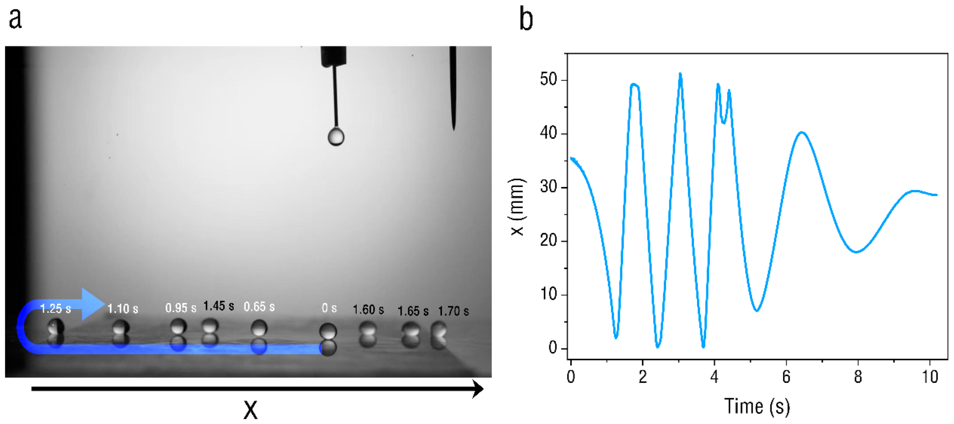
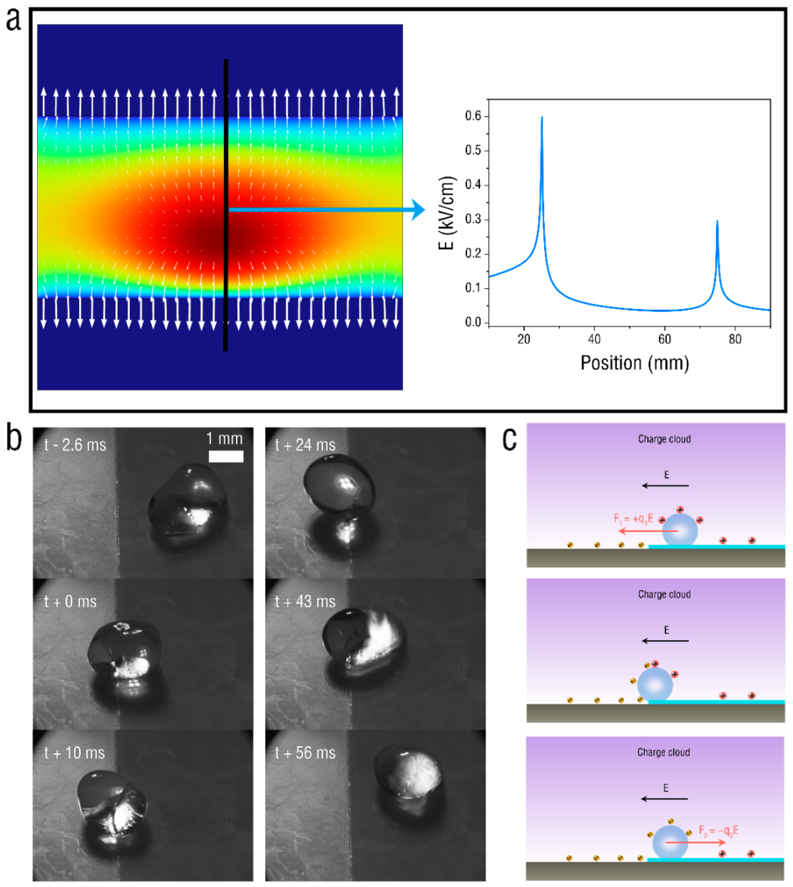
:1. Introduction
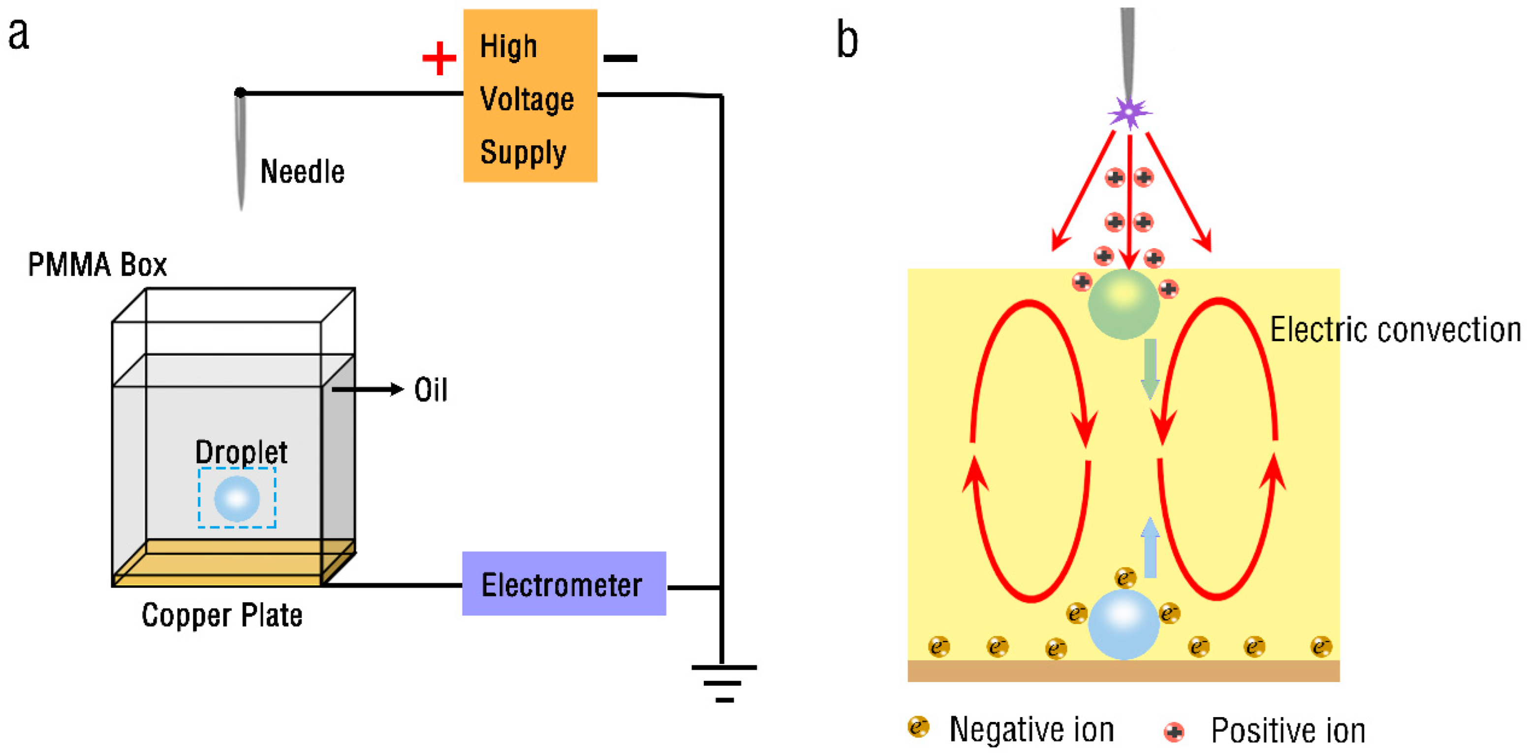
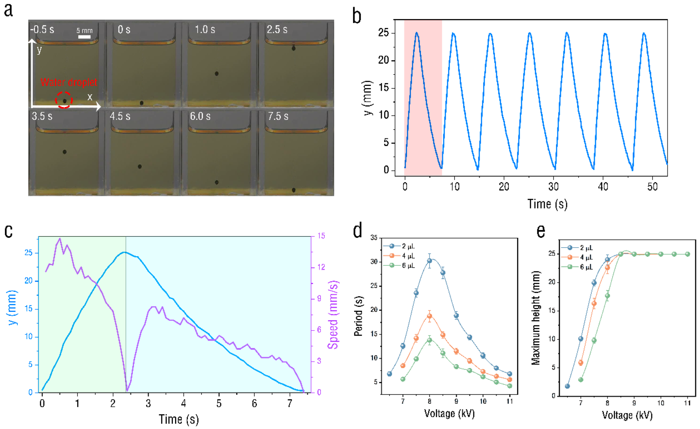
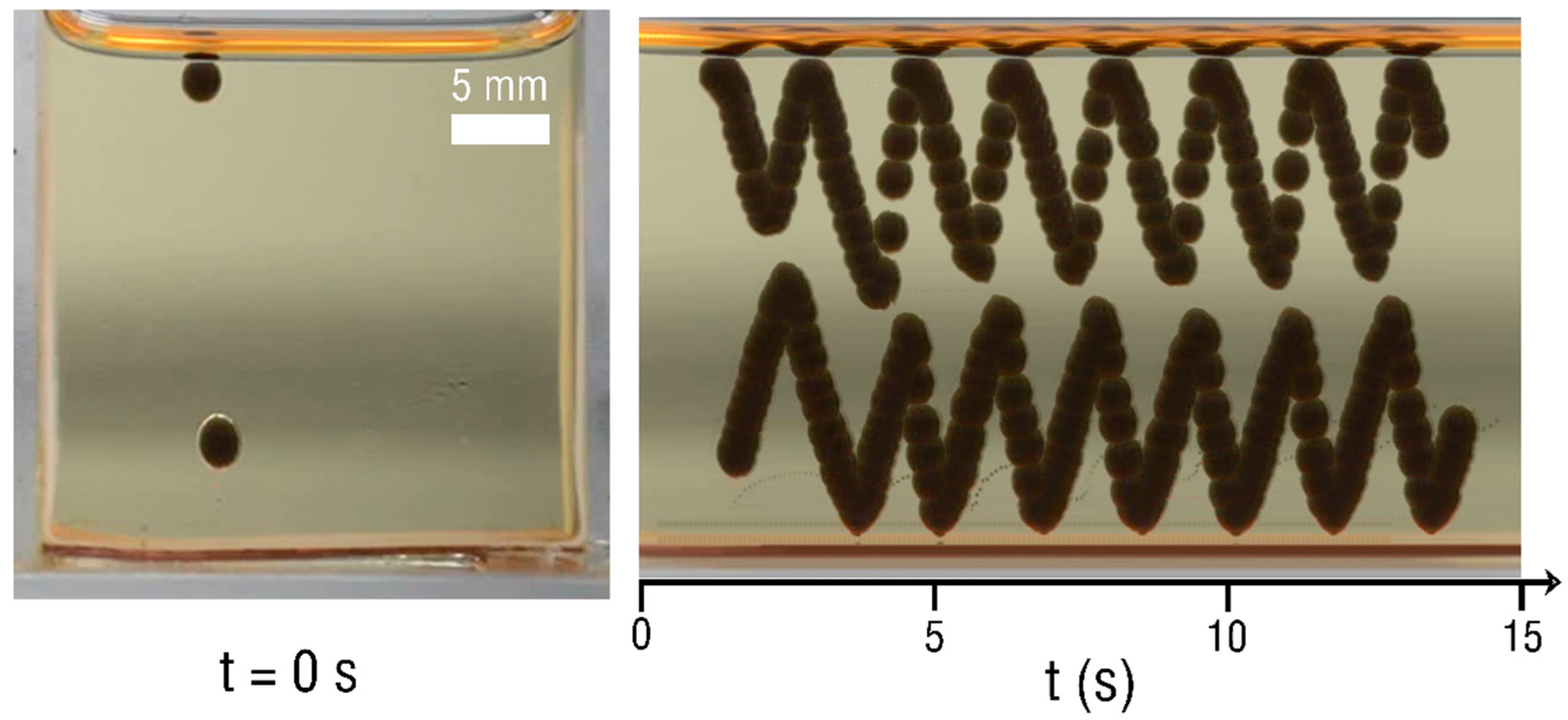
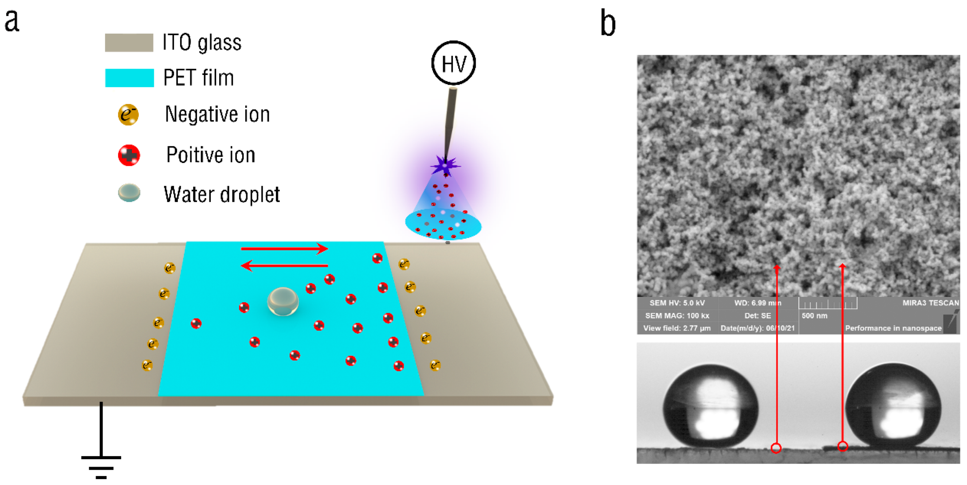
2. Experimental Section
3. Conclusions
Author Contributions
Funding
Institutional Review Board Statement
Informed Consent Statement
Data Availability Statement
Conflicts of Interest
References
- Teh, S.-Y.; Lin, R.; Hung, L.-H.; Lee, A.P. Droplet microfluidics. Lab Chip 2008, 8, 198–220. [Google Scholar] [CrossRef] [PubMed]
- Sohrabi, S.; Moraveji, M.K. Droplet microfluidics: Fundamentals and its advanced applications. RSC Adv. 2020, 10, 27560–27574. [Google Scholar] [CrossRef] [PubMed]
- Ju, W.; Wu, Y.; Lin, S.; Zhao, F.; Tan, S. Visual experimental study of droplet impinging on liquid film and analysis of droplet evolution characteristics. Exp. Comput. Multiph. Flow 2022, 4, 212–220. [Google Scholar] [CrossRef]
- Atencia, J.; Beebe, D.J. Controlled microfluidic interfaces. Nature 2005, 437, 648–655. [Google Scholar] [CrossRef]
- Capretto, L.; Cheng, W.; Hill, M.; Zhang, X. Micromixing within microfluidic devices. In Microfluidics; Springer: Berlin/Heidelberg, Germany, 2011; pp. 27–68. [Google Scholar]
- Simon, M.G.; Lee, A.P. Microfluidic droplet manipulations and their applications. In Microdroplet Technology; Springer: Berlin/Heidelberg, Germany, 2012; pp. 23–50. [Google Scholar]
- Vitorino, R.; Guedes, S.; da Costa, J.P.; Kašička, V. Microfluidics for peptidomics, proteomics, and cell analysis. Nanomaterials 2021, 11, 1118. [Google Scholar] [CrossRef] [PubMed]
- Franke, T.; Abate, A.R.; Weitz, D.A.; Wixforth, A. Surface acoustic wave (SAW) directed droplet flow in microfluidics for PDMS devices. Lab Chip 2009, 9, 2625–2627. [Google Scholar] [CrossRef]
- Basu, A.S.; Gianchandani, Y.B. A programmable array for contact-free manipulation of floating droplets on featureless substrates by the modulation of surface tension. J. Microelectromech. Syst. 2009, 18, 1163–1172. [Google Scholar] [CrossRef]
- Shi, L.T.; Jiang, C.G.; Ma, G.J.; Wu, C.W. Electric field assisted manipulation of microdroplets on a superhydrophobic surface. Biomicrofluidics 2010, 4, 041101. [Google Scholar] [CrossRef] [Green Version]
- Taniguchi, T.; Torii, T.; Higuchi, T. Chemical reactions in microdroplets by electrostatic manipulation of droplets in liquid media. Lab Chip 2002, 2, 19–23. [Google Scholar] [CrossRef]
- Lehmann, U.; Hadjidj, S.; Parashar, V.K.; Vandevyver, C.; Rida, A.; Gijs, M.A.M. Two-dimensional magnetic manipulation of microdroplets on a chip as a platform for bioanalytical applications. Sens. Actuators B Chem. 2006, 117, 457–463. [Google Scholar] [CrossRef]
- Zhan, Y.; Yu, S.; Amirfazli, A.; Siddiqui, A.R.; Li, W. Magnetically Responsive Superhydrophobic Surfaces for Microdroplet Manipulation. Adv. Mater. Interfaces 2022, 9, 2102010. [Google Scholar] [CrossRef]
- Yap, Y.-F.; Tan, S.-H.; Nguyen, N.-T.; Murshed, S.M.S.; Wong, T.-N.; Yobas, L. Thermally mediated control of liquid microdroplets at a bifurcation. J. Phys. D Appl. Phys. 2009, 42, 065503. [Google Scholar] [CrossRef]
- Fan, B.; Li, F.; Chen, L.; Shi, L.; Yan, W.; Zhang, Y.; Li, S.; Wang, X.; Wang, X.; Chen, H. Photovoltaic Manipulation of Water Microdroplets on a Hydrophobic LiNbO3 Substrate. Phys. Rev. Appl. 2017, 7, 064010. [Google Scholar] [CrossRef]
- Zhou, H.; Yao, S. Electrostatic charging and control of droplets in microfluidic devices. Lab Chip 2013, 13, 962–969. [Google Scholar] [CrossRef] [PubMed]
- Yang, S.H.; Im, D.J. Effect of Deformation on Droplet Contact Charge Electrophoresis. Langmuir 2020, 36, 10379–10386. [Google Scholar] [CrossRef] [PubMed]
- Bishop, K.J.M.; Drews, A.M.; Cartier, C.A.; Pandey, S.; Dou, Y. Contact Charge Electrophoresis: Fundamentals and Microfluidic Applications. Langmuir 2018, 34, 6315–6327. [Google Scholar] [CrossRef]
- Xu, L.; Peng, J.; Yan, M.; Zhang, D.; Shen, A.Q. Droplet synthesis of silver nanoparticles by a microfluidic device. Chem. Eng. Process. Process Intensif. 2016, 102, 186–193. [Google Scholar] [CrossRef]
- Hase, M.; Watanabe, S.N.; Yoshikawa, K. Rhythmic motion of a droplet under a dc electric field. Phys. Rev. E 2006, 74, 046301. [Google Scholar] [CrossRef] [Green Version]
- Drews, A.M.; Lee, H.-Y.; Bishop, K.J.M. Ratcheted electrophoresis for rapid particle transport. Lab Chip 2013, 13, 4295–4298. [Google Scholar] [CrossRef]
- Dou, Y.; Cartier, C.A.; Fei, W.; Pandey, S.; Razavi, S.; Kretzschmar, I.; Bishop, K.J.M. Directed motion of metallodielectric particles by contact charge electrophoresis. Langmuir 2016, 32, 13167–13173. [Google Scholar] [CrossRef]
- Drews, A.M.; Cartier, C.A.; Bishop, K.J.M. Contact Charge Electrophoresis: Experiment and Theory. Langmuir 2015, 31, 3808–3814. [Google Scholar] [CrossRef] [PubMed]
- Kim, J.G.; Im, D.J.; Jung, Y.M.; Kang, I.S. Deformation and motion of a charged conducting drop in a dielectric liquid under a nonuniform electric field. J. Colloid Interface Sci. 2007, 310, 599–606. [Google Scholar] [CrossRef] [PubMed]
- Zhang, J.-H.; Li, Z.; Xu, J.; Li, J.; Yan, K.; Cheng, W.; Xin, M.; Zhu, T.; Du, J.; Chen, S.; et al. Versatile self-assembled electrospun micropyramid arrays for high-performance on-skin devices with minimal sensory interference. Nat. Commun. 2022, 13, 5839. [Google Scholar] [CrossRef] [PubMed]
- Whitesides, G.M. The origins and the future of microfluidics. Nature 2006, 442, 368–373. [Google Scholar] [CrossRef]
- Beebe, D.J.; Mensing, G.A.; Walker, G.M. Physics and applications of microfluidics in biology. Annu. Rev. Biomed. Eng. 2002, 4, 261–286. [Google Scholar] [CrossRef] [PubMed]
- Ohyama, R.; Inoue, K.; Chang, J.S. Schlieren optical visualization for transient EHD induced flow in a stratified dielectric liquid under gas-phase ac corona discharges. J. Phys. D Appl. Phys. 2007, 40, 573. [Google Scholar] [CrossRef]
- Li, Y.; Jin, H.; Nie, S.; Zhang, P.; Gao, N. Dynamic behavior of water droplets and flashover characteristics on a superhydrophobic silicone rubber surface. Appl. Phys. Lett. 2017, 110, 201602. [Google Scholar] [CrossRef]


Publisher’s Note: MDPI stays neutral with regard to jurisdictional claims in published maps and institutional affiliations. |
© 2022 by the authors. Licensee MDPI, Basel, Switzerland. This article is an open access article distributed under the terms and conditions of the Creative Commons Attribution (CC BY) license (https://creativecommons.org/licenses/by/4.0/).
Share and Cite
Tang, Q.; Zhang, Z.; Zhang, J.-H.; Tang, F.; Wang, C.; Cui, X. Oscillatory Motion of Water Droplets Both in Oil and on Superhydrophobic Surface under Corona Discharge. Micromachines 2022, 13, 2229. https://doi.org/10.3390/mi13122229
Tang Q, Zhang Z, Zhang J-H, Tang F, Wang C, Cui X. Oscillatory Motion of Water Droplets Both in Oil and on Superhydrophobic Surface under Corona Discharge. Micromachines. 2022; 13(12):2229. https://doi.org/10.3390/mi13122229
Chicago/Turabian StyleTang, Qiang, Zongtang Zhang, Jia-Han Zhang, Feiran Tang, Chengjun Wang, and Xiaxia Cui. 2022. "Oscillatory Motion of Water Droplets Both in Oil and on Superhydrophobic Surface under Corona Discharge" Micromachines 13, no. 12: 2229. https://doi.org/10.3390/mi13122229