In 2006, the concept of deep learning again attracted the attention of researchers [
17]. From 2007, deep learning began to be applied in breast pathology image data. Then, under the efforts of many researchers, random gradient descent (SGD), Dropout, and other network optimization strategies were successively proposed, especially GPU parallel computing technology that solves the problem of multiple optimization times of deep network parameters for a long time, which has set off an upsurge of deep learning worldwide and continues to this day. Over the past 10 years, many classical deep learning architectures were proposed, such as AlexNet [
18], Recurrent Neural Networks (RNN), Long Short-Term Memory (LSTM) [
19], Generative Adversarial Network (GAN) [
20], and transformer [
21]. In this section, we investigate the application of deep learning to histopathological sections of breast cancer, including the most advanced and effective models available today, and we provide a summary of related work. The literature can be divided into three primary categories based on the direction of research reported in each article: breast cancer detection, breast cancer segmentation, and breast cancer classification [
22].
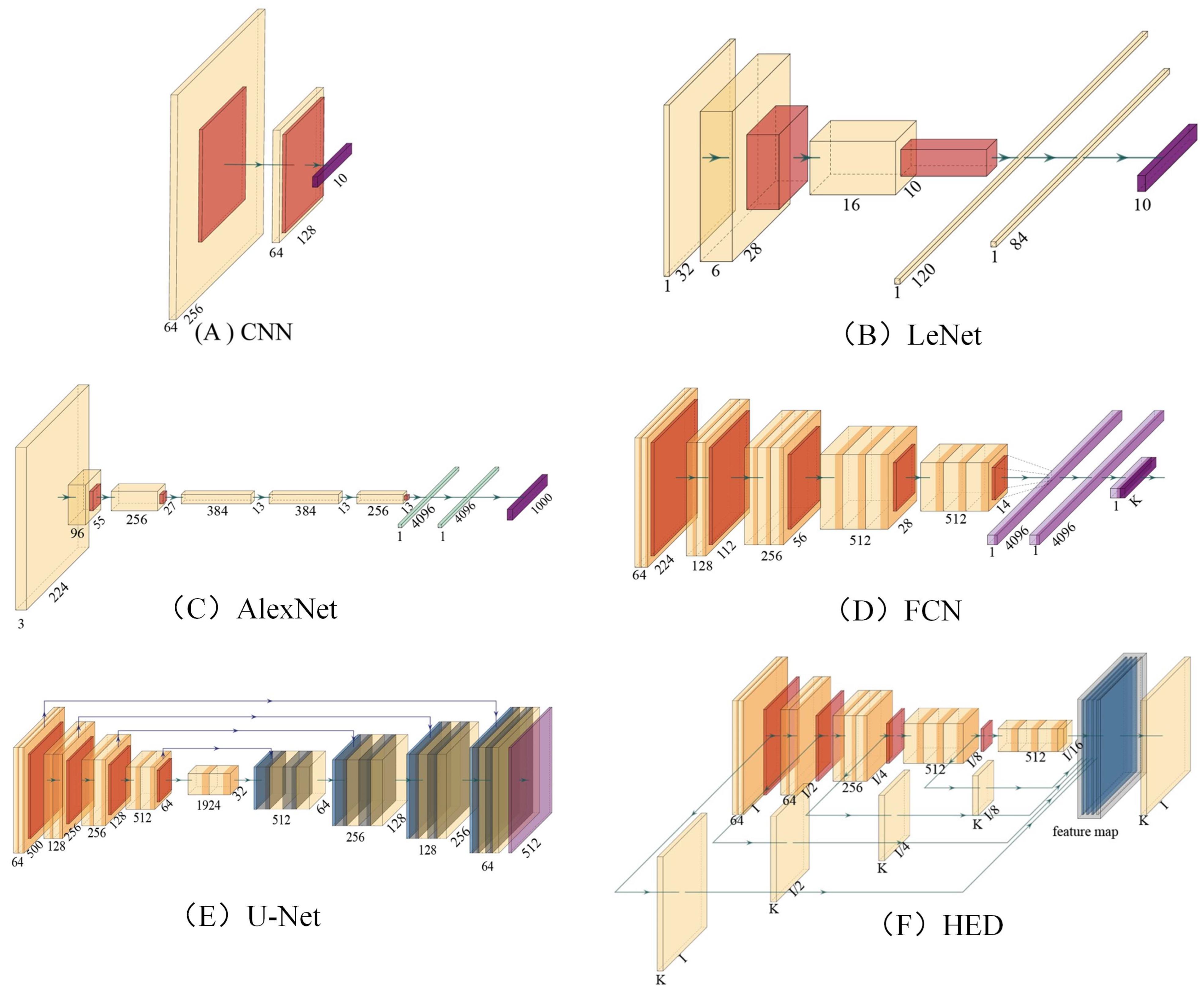
Figure 2 shows the common basic neural network structure, where yellow squares represent convolution layers, orange squares represent pooling layers, purple squares represent full-connected layers, and blue squares represent deconvolution layers. Among them,
Figure 2A is a simplified convolution neural network, which is composed of only two layers of convolution, and the rest of the depth convolution neural network can be composed by superimposing
Figure 2A. LeNet (
Figure 2B) [
23] is an early convolutional neural network, which was proposed by Yann LeCun et al., in 1990. AlexNet (
Figure 2C) is a deep convolutional neural network proposed by Hinton et al., in 2012 and won the championship in the ImageNet challenge that year.
Figure 2D is a full convolution neural network (FCN) used for semantic segmentation in the early stage [
24]. It is also widely used in the early research of breast cancer pathological image segmentation task. The appearance of U-Net [
25] has significantly improved the performance of FCN in medical image segmentation tasks. Holistically Nested Network (HED) [
26] has achieved better results than traditional edge detection algorithms in edge detection tasks, and this method has also been used to improve the performance of breast cancer segmentation tasks [
27].
3.1. Detection of Breast Lesions
In medical image analysis, detection aims to locate areas of interest in tissue slices [
28,
29,
30,
31,
32,
33,
34,
35,
36,
37,
38,
39,
40]. The detection system provides strong support for object segmentation, the distinction between malignant and benign tumors, or the detection of tumors or lesions. For example, nuclear or mitosis has important implications for cancer screening. Cell spatial distribution analysis and mitotic count provide support for differentiation. Automatic cell/nucleus detection is a prerequisite for a series of subsequent tasks, such as cell/nucleus instance segmentation, tracking, and morphological measurements [
41]. In recent years, many studies based on deep learning were performed in this field of study. Among existing deep learning detection algorithms, CNN-based networks perform better than other network structures in detection accuracy [
42]. In certain areas, CNN-based networks have achieved diagnostic standards that surpass pathologists [
43]. Next, we will introduce some typical models that work particularly well for accuracy and performance.
Table 2 shows the application of deep learning algorithm in histopathological detection of breast cancer. The application of deep learning algorithm in histopathological detection of breast cancer was classified according to the types of models, and the strategies were summarized.
George et al. [
44] proposed a low-complexity breast cancer detection convolutional neural network (CNN) called NucDeep, which includes a low-complexity CNN for feature extraction of non-overlapping nuclear plaques and converts local nuclear features into compact image-level features to improve classifier performance (
Figure 3A). Chen et al. [
45] proposed a novel deep cascaded neural network model (CasNN). CasNN greatly increased the speed of detection of mitosis (
Figure 3B). Liu et al. [
43] proposed InceptionV3 as a way to automatically detect and locate cancers in high-resolution images (
Figure 3C). The author uses InceptionV3 as an experimental framework, and the balance and expansion of data are achieved through data enhancement and balance to improve model accuracy. Bardou et al. [
46] proposed a method for automatic classification of breast cancer histological images based on a convolutional neural network (
Figure 3D). The linear rectification function (ReLU) layer is used for the convolution and fully connected layers to accelerate the convergence learning rate, introduce nonlinearity, and adjust the network weight to prevent overfitting.
Some classical detection algorithms [
47,
48,
49] in the field of natural images have also been used in the problems related to pathological images of breast cancer. Lu et al. [
50] proposed a model based on the latest yolo v4 structure that can quickly and accurately segment the lesion area in high-resolution breast cancer pathological slices. The ROI recognition accuracy is 0.936 and F1 score is 0.787, which is of great significance for improving the diagnostic efficiency and accuracy of pathologists on breast pathological images. Huang et al. [
51] proposed an algorithm for breast cancer pathological image nuclear detection based on mask RCNN. This method effectively combines feature pyramid network (FPN), ResNet, and other modules to achieve more accurate detection. Harrison et al. [
52] proposed an algorithm for tumor detection in breast pathological images based on Faster RCNN and found that patching the images can significantly improve the sensitivity of the model, from 1% to 60%, and the performance improvement brought by dye normalization is limited. Yamaguchi et al. [
53] proposed an automatic detection algorithm for breast cancer based on single-shot multibox detector (SSD) and achieved 88.3% and 90.5% diagnostic accuracy in two (benign or malignant) and three (benign, non-invasive carcinoma, or invasive carcinoma) classification tasks, respectively. Mitotic cell count is an important biomarker for grading and prognosis of breast cancer and is also a common application of pathological intelligence analysis of breast cancer. Zorgani et al. [
54] designed a method to detect breast cancer mitotic cells based on the deep yolo architecture and obtained 0.839 F1 measure on the ICPR2012 dataset.
Thus, by reading and analyzing the article, we found that the model proposed by [
44] is a low-complexity model with classification results comparable to existing technologies and uses nucleus patches alone rather than random patches. The proposed method of [
45] can obtain various multi-level and multi-scale features from breast cancer histopathological images, providing competitive performance in the classification of complex breast cancer histopathological images. However, the dataset collected is relatively small and contains only two types of images, and the dataset should be extended to include images for multiclass classification problems. The authors of [
43] made data enhancement for negative samples to solve the large gap between positive and negative samples and optimized the sampling process to remove the patch as the background. After the probability graph is obtained, the tumor region is continuously iterated according to the current maximum value to predict the tumor region based on the non-maximum suppression method. In [
46], compared with CNN, the performance of the manual feature-based method based on encoding model for local descriptors to construct image representation is low, and the performance of multiclass classification is lower than that of binary classification. Classical object detection algorithms can also achieve remarkable results in the field of pathological images, especially Faster RCNN series algorithms.
From the above results, it can be seen that the deep learning model has the advantages of direct learning characteristics on breast cancer pathological images, which can greatly reduce the manual investment and also reduce the artificial differences caused by manual reading. Higher accuracy also provides help for the development of precision medicine.
Table 2.
Summary of the application of deep learning algorithms in breast cancer histopathology for detection.
Table 2.
Summary of the application of deep learning algorithms in breast cancer histopathology for detection.
| Model | Strategy | Advantages | Publication |
|---|
| RNN | Development of decision support systems for pathology | RNN allows neurons in the hidden layer to communicate with each other, storing the previous output as information in the hidden layer | [55] |
| | Propose a SmallMitosis framework for the detection of mitotic cells from hematoxylin and eosin (H&E)-stained breast histological images | | [56] |
| Inception | Histologic identification of tumor cells in lymph nodes | Inception increases the width of the network by pooling each layer with a different convolution to extract features from the previous layer, and by adding a 1*1 convolution after the pooling layer before the 3*3 and 5*5 convolutions, which effectively avoids complex parameters and computational effort | [57] |
| | Improve the computer-aided diagnosis method based on deep learning | | [58] |
| ResNet | Detection of invasive ductal carcinoma in breast histological images and the classification of lymphoma subtypes | The main feature of ResNet is the residual block, the purpose of the residual block is to preserve the characteristics of the parameters before the current layer is trained and to pass these parameters into the subsequent layers together with the trained data | [59] |
| | Diagnostic breast cancer whole-slide tissue images | | [60] |
| | Propose an automatic detection method for invasive ductal carcinoma (IDC) based on deep transfer learning technology | | [61] |
| | Propose Mask RCNN, a multi-task deep learning framework for object detection and instance segmentation, to automatically detect mitosis | | [62] |
| DCNN | Propose an accurate method for detecting the mitotic cells from histopathological slides using a multi-stage deep learning framework | | [63] |
| | Present an SSAE for efficient nuclei detection on high-resolution histopathological images of breast cancer | | [64] |
| | Introduce deep learning as a technique to improve the objectivity and efficiency of histopathologic slide analysis | | [65,66,67,68,69,70] |
| Semi-Supervised Learning | Present a semi-supervised deep learning strategy for breast cancer diagnosis | Semi-supervised learning is to use a large number of unlabeled samples and a small number of labeled samples to train the classifier, solving the problem of insufficient labeled samples | [71,72] |
| YOLO | A fast lesion detection method based on yolo is proposed | Simple structure and fast speed | [50] |
| Faster RCNN | A fast detection method of breast tumor based on Faster RCNN is proposed | Faster RCNN realizes object detection performance with high accuracy through second-order network and Region Proposal Network | [52] |
| Single Shot multibox Detector (SSD) | An automatic detection method of breast cancer lesion based on SSD is proposed | One stage, good at detecting small objects | [53] |
3.2. Segmentation Method of Breast Pathological Image
Segmentation refers to dividing the input image into many specific areas with unique properties and extracting them, separating the content in a region of interest (ROI) from the image background. The ROI in a pathological image of breast cancer is part of a lesion. When using deep learning correlation methods, it is generally necessary to analyze and extract the characteristics of tumor lesions in ROI so as to detect and classify pathological images. Pathological image segmentation plays an important role in the field of pathological image processing and analysis, which is helpful to provide reliable basis for clinical auxiliary diagnosis and treatment. Despite the high complexity of pathological images and the lack of simple linear features, pathological image segmentation technology has made significant progress due to the effective application of deep learning algorithm in pathological image segmentation. In pathological image segmentation, deep learning algorithms have made remarkable achievements. Most pathological image segmentation uses supervised deep learning algorithms, such as FCN, RNN, and U-Net. Next, we will introduce some typical models that work particularly well for accuracy and performance.
Table 3 shows the application of deep learning algorithm in histopathological segmentation of breast cancer. The application of deep learning algorithm in histopathological segmentation of breast cancer was classified according to the types of models, and the strategies were summarized.
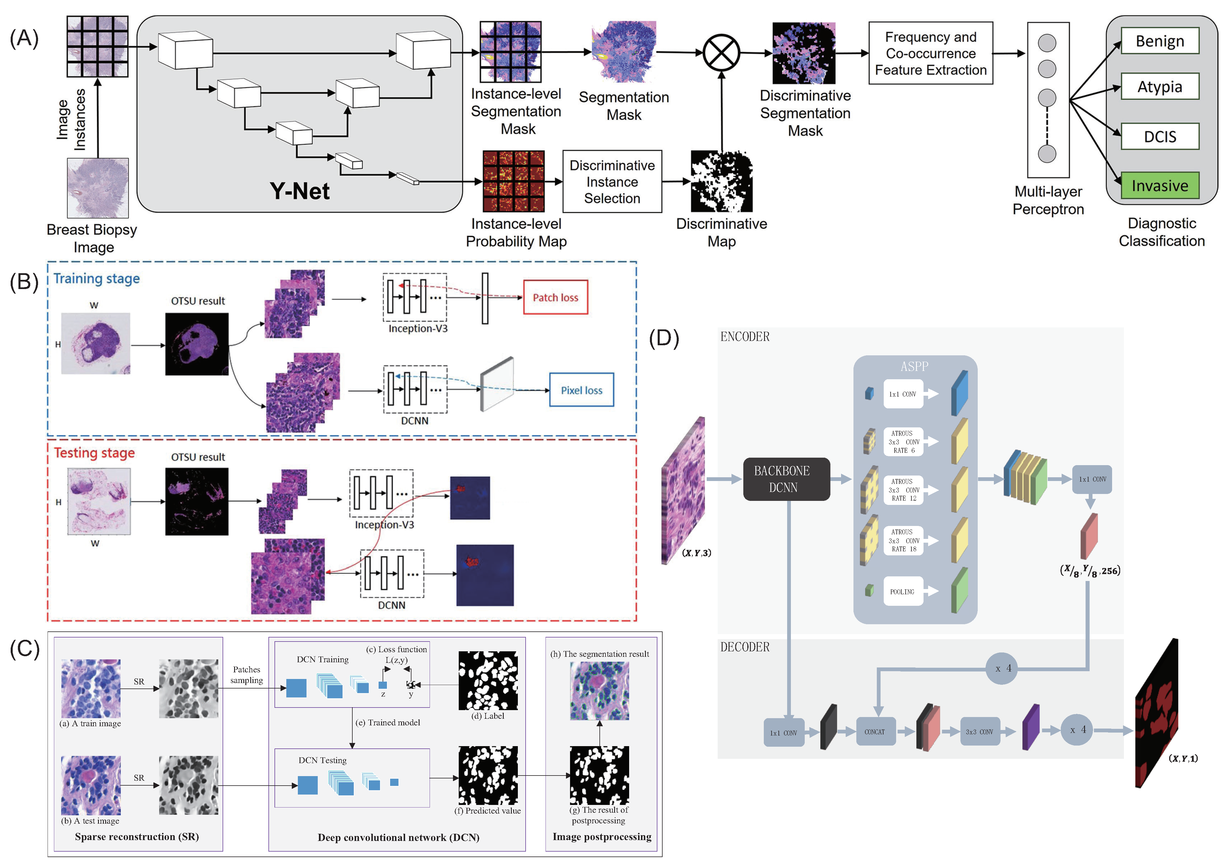
Mehta et al. [
73] introduced a method to generate distinguishable tissue-level segmentation masks for breast cancer diagnosis (
Figure 4A). This Y-Net network expands and generalizes U-Net, adds a parallel branch for the generation of discriminative maps, and supports modularization of convolution blocks. Guo et al. [
74] proposed v3-DCNN, a fast cancer region segmentation framework (
Figure 4B). The classification model Inception-V3 was used to preselect the tumor area, and then the semantic segmentation model DCNN was used to segment the 1280 × 1280 patch to reduce computation time and improve accuracy. Pan et al. [
75] proposed an automatic nuclear segmentation method for histopathological images of breast cancer stained with H&E. The sparse reconstruction method is used to roughly remove the background to emphasize the core of the pathological image, and then the deep convolutional network (DCN) of the multilayer convolutional network cascade is used to effectively segment the core from the background (
Figure 4C). Maria Priego-Torres et al. [
76] presented a processing pipeline for automatic segmentation of breast cancer images to present different types of histopathological patterns (
Figure 4D). The deep convolutional neural network (DCNN) and the encoder–decoder with separable convolution structure were used to complete the segmentation of each patch, and the local segmentation results were merged based on the effective full connection condition random field (CRF) to avoid discontinuity and inconsistency.
Transformer-based methods are also widely used in medical image segmentation [
21]. However, there are few studies about the segmentation task of breast pathological images. Therefore, we try to retrieve transformer-based segmentation methods in the fields related to breast pathological image segmentation, aiming to promote the development of transformer-based methods in this field. Cam et al. [
77] quantitatively evaluated the segmentation performance of six popular transformer-based segmentation networks on pathological images based on the PAIP liver histopathology dataset and compared the classical CNN-based segmentation networks. The results show that the transformer-based segmentation network is generally better than the CNN-based model, proving the effectiveness of the transformer architecture on pathological image segmentation tasks. Li et al. [
78] proposed a vision language medical image segmentation model, LViT (Language measures Vision Transformer), to solve the problem of insufficient annotation of medical images and verified the cell segmentation performance of this method on the MoNuSeg dataset. Diao et al. [
79] introduced transformer into the classic U-Net architecture to extract and encode global context information and achieved SOTA performance in the nasopharyngeal carcinoma pathological image dataset.
Semi-automatic segmentation algorithm also attracted much attention in the field of breast cancer image analysis, mainly applied to X-ray [
80], ultrasound [
81], MRI [
82], and other images, and there is less research on pathological images of breast cancer. In recent related research, Lai et al. [
83], in conjunction with semi-supervised and active learning, proposed a segmentation algorithm for brain tissue pathological images and achieved IoU scores competitive with fully supervised learning.
Thus, through research and analysis, we found that the features generated by the discriminant segmentation mask used by the authors in [
73] were able to achieve the same segmentation accuracy as the most advanced methods while learning fewer parameters. However, this paper only studied breast biopsy images and did not extend to other medical imaging tasks. The method proposed in [
74], based on the V3 DCNN model, achieved a higher FROC score of 83.5% than the champion method Camelyon16 80.7%, and further, the automatic heat map generation of WSI was achieved. However, the proposed model lacks dataset validation and should be tested on more breast histopathological images. In [
75], k-SVD and Batch-OMP algorithms were used for sparse reconstruction to enhance the nuclear region. In the segmentation stage, DCN trained by structural label was used to obtain the exact pixel of the nucleus, and morphological operation and some prior knowledge were introduced to improve the segmentation performance and reduce errors. The proposed algorithm is a general method and can be applied to many pathological applications. However, the number of datasets is too small, and the number of background pixels is far more than that of nuclei, so there is an imbalance between the number of nuclear pixels and background pixels. The proposed segmentation model in [
76] performed well on standard success rate and similarity segmentation metrics, especially considering that the dataset included WSI images with high tumor variability. Web-based viewers and annotation tools were developed to allow collaboration with pathologists and technologists to establish a way to create datasets. However, all images used in this working study were stained in the same laboratory and digitized using the same scanner, and images from other sources should be used to increase the heterogeneity of the training set. Transformer-based methods have achieved remarkable results in the field of computer vision and become popular in medical image analysis tasks. [
21] Transformer can well encode context characteristics. This is also used by most researchers to build the global features of breast cancer pathological images so as to improve the model performance. A large number of experimental results show the effectiveness of transformer architecture in this field [
77,
79,
80]. The semi-automatic segmentation algorithm is relatively less used in the breast cancer pathological image segmentation task. So far, this method is still a field worthy of researchers to explore.
Table 3.
Summary of the application of deep learning algorithms in breast cancer histopathology for segmentation.
Table 3.
Summary of the application of deep learning algorithms in breast cancer histopathology for segmentation.
| Model | Strategy | Advantages | Publication |
|---|
| ResNet | Propose segmentation of limited data using rough image-level tags with performance comparable to fully labeled datasets | The main feature of ResNet is the residual block, the purpose of the residual block is to preserve the characteristics of the parameters before the current layer is trained and to pass these parameters onto the subsequent layers together with the trained data | [84] |
| FCN | Propose a fast segmentation method for breast cancer metastases in pathological images | The FCN replaces the fully connected layer behind the traditional CNN with a convolutional layer so that the output of the network will be a heat map rather than a category; at the same time, the image size is recovered using upsampling in order to address the reduction in image size due to convolution and pooling | [85] |
| | Propose an automatic method for detecting mitosis | | [86] |
| | Describe a method to automatically segment nuclei from hematoxylin and eosin (H&E)-stained histopathology data with fully convolutional networks | | [87] |
| | Use annotated datasets to create accurate models | | [60] |
| | Propose a histopathological tissue analysis framework based on deep learning and verifies its universality and model generalization under different data distributions | | [88] |
| U-Net | Use histopathological images obtained with hematoxylin and eosin staining for biopsy samples for the diagnosis and segmentation of breast cancer | U-Net networks are able to use valid labeled data more effectively from a very small number of training images, relying on data augmentation | [89] |
| | Address the task of tissue-level segmentation in intermediate resolution of histopathological breast cancer images | | [90] |
| | Propose a deep learning framework consisting of high-resolution encoder paths, pyramidal pooled bottleneck modules in porous space, and decoders | | [91] |
| | Investigate whether it is possible to further improve the performance of the classifier model at the patch level by integrating multiple extracted histological features into the input image | | [92] |
| CNN | Improve the performance of current Simple Linear Iterative Clustering (SLIC) algorithm to achieve hyperpixel segmentation of high-dimensional features | | [93] |
| | Use a pretrained convolutional neural network (CNN) for segmentation and then another Hybrid-CNN for classification of mitoses | | [94] |
| | Identify a useful cell segmentation approach with histopathological images that uses prominent deep learning algorithms and spatial relationships | | [95] |
| | Propose a framework that combines the effectiveness of attention-based encoder–decoder architecture with an empty space pyramid pool with efficient dimensional convolution (kide-Segnet) | | [96] |
| | Propose a deep learning model for automatic segmentation of complex cores in tissue images by encoder-decoder structure | | [97,98] |
| Transformer | Transformer-encoded global features improve U-Net segmentation performance | Transformer model can be used to encode the global features of pathological images and can improve the performance of current algorithms in many fields | [79] |
3.3. Disease Classification Based on Breast Pathological Images
Classification of medical images by defining the anatomical or pathological features distinguishes certain anatomical structures or tissues. Classification tasks can include many applications in determining the presence of disease, including the identification of tumor types. Deep learning is often used with medical images to classify target lesions into two or more categories. Binary classification refers to distinguishing between breast cancer tissue slices and normal breast tissue slices in the pathological tissue slice dataset used. Multiclass classifications divide pathological tissue slices of the breast into multiple categories using deep learning algorithms based on requirements. Common classifications of breast tissue slices are normal, benign, in situ carcinoma, or invasive carcinoma. In general, the accuracy of the binary classification task is higher than that of the multiclass classification task. Among some existing deep learning classification algorithms, the classification accuracy has reached or even exceeded that of pathologists [
43]. Next, we will introduce some typical models that work particularly well for accuracy and performance.
Table 4 shows the application of deep learning algorithm in histopathological classification of breast cancer. The application of deep learning algorithm in histopathological classification of breast cancer was classified according to the types of models, and the strategies were summarized.
Convolution neural network is the most widely used method in breast cancer pathological image classification task. [
46,
99,
100,
101,
102,
103,
104,
105,
106,
107,
108,
109] Roy et al. [
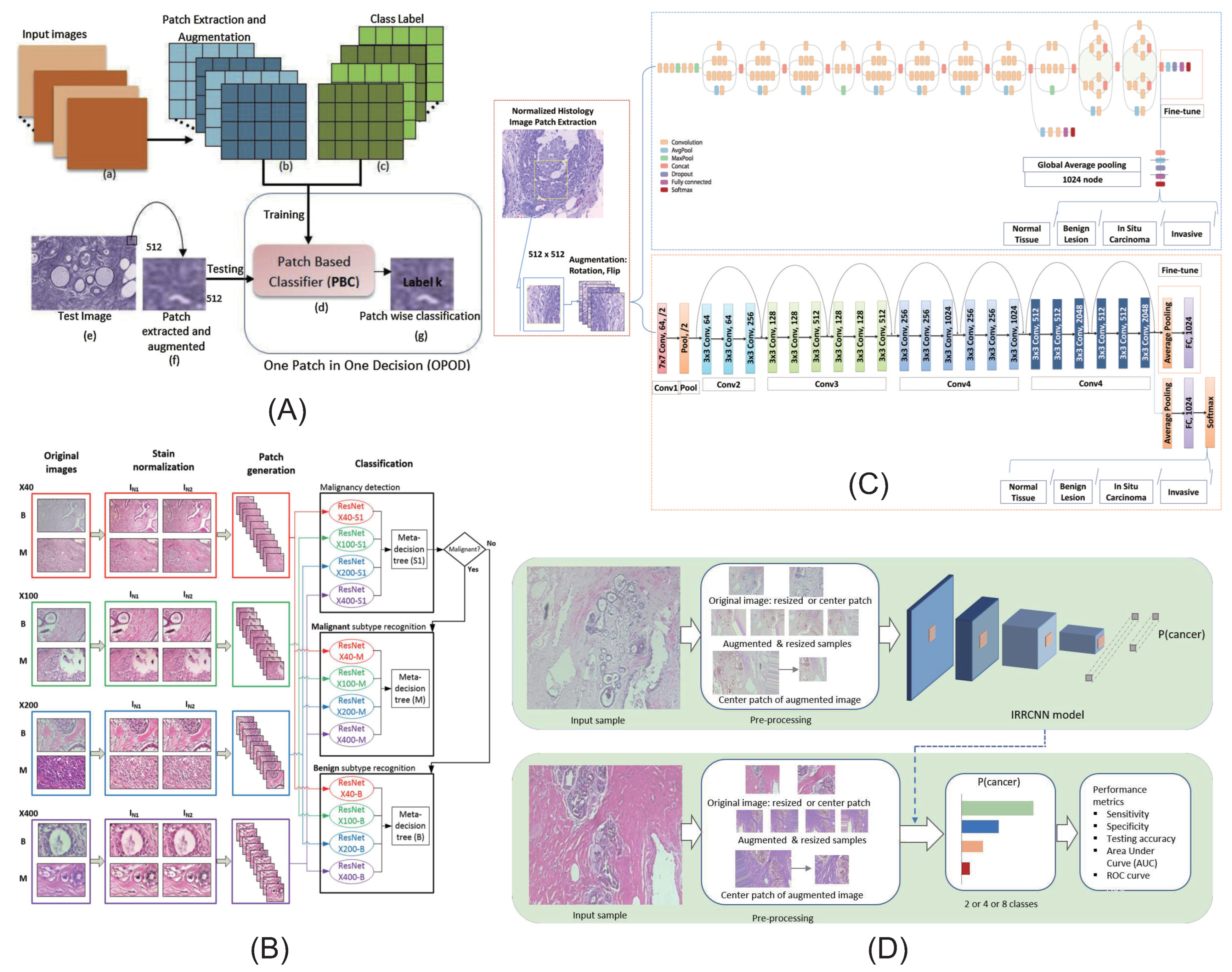
110] developed a patch-based classifier (PBC) through a convolutional neural network (CNN) for automatic classification of breast cancer histopathological images (
Figure 5A). They used two methods: one patch in one decision (OPOD), and all patches in one decision (APOD). Gandomkar et al. [
111] proposed a framework MuDeRN (multicategory classification of breast histopathological images using deep residual networks) (
Figure 5B). The 152-layer RsetNet is used to sort tasks to improve deeper network optimization for higher model accuracy. Vesal et al. [
112] proposed a method based on metastatic learning to divide histological images of breast cancer into four subtypes: normal, benign, carcinoma in situ, and invasive carcinoma(
Figure 5C). Migration learning migrates ImageNet’s pretrained parameters into InceptionV3 and RestNet50 and removes the last five layers of the network model to obtain global information. Alom et al. [
113] proposed a method for breast cancer classification using the Inception recurrent residual convolutional neural network (IRRCNN) model, which is a combination of the Inception network (Inception-v4), the residual network (ResNet), and a convolutional neural network (RCNN), which provides advantages to DCNN models that exhibit superior performance in object recognition tasks (
Figure 5D). Sudharshan et al. [
109] proposed a weakly supervised learning framework based on multi-instance learning for classification of breast pathological images. This method does not need to label each instance so it significantly alleviates the problem of difficult labeling of pathological images.
In the field of medical image analysis, transformer-based methods were first used to process disease classification tasks and produced significant results. [
114,
115,
116,
117,
118,
119,
120,
121,
122] There have also been many significant advances in the classification of pathological images of breast cancer. Alotaibi et al. [
119] designed an integrated model based on VIT and DeiT to classify pathological images of breast cancer tissues and achieved an accuracy of 98.17 on BreakHis public dataset. However, this method requires pretraining on large-scale datasets and model fine-tuning to alleviate the data hunger of the transformer, which will obviously increase the training cost of the model and limit the scope of use. Shao et al. [
120] used the global characteristics of the transformer to build the relationship between instances in order to capture the context information so as to improve the performance of multi-instance learning on the breast whole-slide image. They achieved 93.09% of AUC’s binary classification performance on CAMELYON16 dataset. Chen et al. [
121] proposed the Multimodal Co-Attendance Transformer (MCAT) architecture, which aims to build the relationship between WSI and genomic features and use it in survival analysis tasks. This method has proved effective on different cancer datasets, including breast cancer datasets. Chen et al. [
122] proposed a multi-scale vision transformer model (GasHis Transformer) for gastric cancer tissue image classification. The author also verified the effectiveness of this method on the breast pathology image dataset. He et al. [
123] proposed Deconv-Transformer (DecT), which incorporates the color deconvolution in the form of convolution layers, and uses a self-attention mechanism to match the independent properties of the HED channel information obtained by the color deconvolution. In [
124], DCET-Net (based on two backbone streams of CNN and transformer) was proposed, which utilizes CNN stream to focus on the local deep feature extraction of histopathological images, while through the Transformer stream, it enhances the global information representation of images.
The capsule network proposed by Geoffrey Hinton also has some valuable exploration in this field. Anupama et al. [
125] used capsule network with preprocessed histology images, which demonstrates that preprocessing data and tuning parameter can improve the performance of conventional architectures. Wang et al. [
126] used FE-BkCapsNet based on deep feature fusion and enhanced routing, which combines the advantages of CNN and CapsNet, and the classification performances are better than that of BkNet and CapsNet. However, it is a very time-consuming methodology to classify based on capsule features and convolution features extracted in two parallel channels. Iesmantas et al. [
127] developed convolutional capsule network for classification of four types of images of breast tissue biopsy when hematoxylin and eosin staining is applied, but regularization was not taken into consideration.
Table 4.
Summary of the application of deep learning algorithms in breast cancer histopathology for disease classification.
Table 4.
Summary of the application of deep learning algorithms in breast cancer histopathology for disease classification.
| Model | Strategy | Advantages | Publication |
|---|
| Deep Belief Network (DBN) | Propose a new patch-based deep learning method PA-DBN-BC for breast cancer detection and classification in histopathological images | | [128] |
| Deep Neural Network (DNN) | Propose a new feature extractor, Deep Manifold Reservation Autoencoder, for automatic classification of breast cancer histopathological images | Deep Belief Network is a probability generation model, which has important value in the early application of deep learning methods | [129] |
| Generative Adversarial Network (GAN) | Explore whether a deep learning algorithm can learn objective histologic H&E features | GAN can be used as a means of data enhancement to alleviate the problem of insufficient data of pathological images of breast cancer | [103] |
| Visual Geometry Group Network (VGG) | Discuss and compare the task of automatic amplification in breast cancer detection based on multiple classification | VGG is a deep neural network proposed in 2014, which provides rich deep features for early breast pathological image research | [130] |
| Recurrent Neural Network (RNN) | Present a deep learning model to classify hematoxylin¨Ceosin-stained breast biopsy images into four classes | RNN can construct the context between pathological image features and be used for prediction of slide-level diagnostic results | [131] |
| | Propose a second-order multi-instance learning approach that stacks adaptive aggregators by attentional mechanisms and recurrent neural networks (RNN) for histopathological image classification | | [132] |
| Inception | Compare different machine learning methods for classification and evaluation of breast cancer tumors | | [133] |
| | Propose a depth model based on computer-aided transfer learning as a binary classifier for breast cancer detection | | [134] |
| | Propose a method for diagnosing breast cancer as benign or malignant in magnification specific binary (MSB) classification | | [135] |
| Dynamic Convolution Neural Network (DCNN) | Propose an efficient deep convolutional neural network classification model for fast back propagation learning | DCNN can adaptively adjust the convolution kernel parameters according to the input data, enhance the feature expression ability of the model, and specifically solve the tasks related to breast pathological images | [136] |
| | Develop a deep learning model biopsy microscopic image cancer network (BMIC_Net) for multiple classification of BC | | [137] |
| | Propose two efficient models based on deep transfer learning to improve the binary and multiclassification systems | | [138,139] |
| Convolution Neural Network (CNN) | Propose a new deep architecture based on self-integration to leverage semantic information from annotated images and explore information hidden in unlabeled data | | [140] |
| | Propose an analysis and synthesis model learning method with novel algorithms and search strategies to classify images more effectively | | [141,142,143,144,145,146,147,148,149,150] |
| | Propose a set of training techniques and use image processing techniques to improve the performance of CNN-based models in breast cancer classification | | [143,151,152,153,154,155,156,157] |
| Deep residual network (ResNet) | Present a deep neural network which performs representation learning and cell nuclei recognition in an end-to-end manner | | [158] |
| | Propose an automatic multiclassification method for breast cancer histopathological images based on metastasis learning | | [159] |
| | Present a method that employs a convolutional neural network for detecting tumor on entire-slide images | | [59,130,136,160] |
| | Propose a breast cancer multiclassification method using a proposed deep learning model | | [106,113,137,161,162,163,164,165,166,167,168] |
Thus, through research and analysis, we found that the classifier proposed by [
110] first predicts the class label of each input patch by OPOD technique and then predicts the whole-image label by APOD technique. At the same time, the number of filters and kernel size of each layer are adjusted so that the number of trainable parameters is smaller than the number of samples and overfitting can be prevented. The authors’ proposed framework in [
111], MuDeRN, first trains a deep residual network (ResNet) to classify patches in images as benign or malignant. Images classified as malignant were then subdivided into four cancer subgroups, and images classified as benign were divided into four cancer subgroups. MuDeRN classified patients as benign or cancerous with 98.77% accuracy and achieved 96.25% patient-level accuracy across the eight categories. However, for some subtypes with too few cases, MuDeRN’s performance should be investigated on a larger database. In [
112], the conventional use of normalized means to deal with color differences, using a different normalized way, showing a good effect but should verify its effect. The author does not use a test set in the training, and the results produced by using only one partition in the training set are hardly convincing. At the same time, the author changed the structure of the end of the network without proving the correctness of the modification by experiment. The method proposed in [
113], the IRRCNN model, was used to successfully classify binary and multiple types of breast cancer with constant amplification coefficients. Image and patient-level data were evaluated using different magnifications on publicly available histopathological datasets for breast cancer. Compared with the existing breast cancer classification algorithm, it shows superior performance. Recently, there were also some studies based on attention mechanisms [
169] and the use of deep semantic features and image texture features [
170] for breast cancer classification. They all achieved good results and provided some references and directions for future research.
From the above results, it can be seen that deep learning has already achieved remarkable results in the field of pathological image classification of breast cancer. How to further improve the performance of deep learning-assisted diagnosis and better provide treatment recommendations based on existing results in pathological image analysis will be the focus of research in the following section. Eventually, we hope to realize a deep learning model that can integrate multimodal data (including medical images, gene sequences, diagnostic reports, drug molecular structure, and other related information) and truly exert the value of deep learning in clinical applications.
3.4. Genetic Prediction Based on Deep Learning
WSI is widely used in digital pathology to predict gene mutations, molecular subtypes, and clinical outcomes. Therefore, they are usually divided into patches for training neural networks and prediction models. However, because patch-level tags are usually unavailable, we cannot directly classify each patch. In the past few decades, with the help of the rapid development of high-throughput technologies of microarrays and gene expression analysis technologies, there have been many studies that use gene expression patterns to understand the molecular characteristics of breast cancer. Van de Vijver [
171] conducted a preliminary study to effectively predict the prognosis of breast cancer through gene expression profile. They clustered gene expression profile data and correlated them with prognostic values. The integration of gene expression profile data and clinical data may improve the accuracy of prognosis and diagnostic prediction models [
172]. In fact, microarray data are high-dimensional, and each patient contains about 25,000 genes. There may be potential relationships between different genes, which may improve the accuracy of prognosis prediction of breast cancer [
173]. Many genes related to breast cancer have been identified. Mutation and abnormal amplification of oncogenes and tumor suppressor genes play a key role in the occurrence and development of tumors. For example, two famous breast cancer risk anti-cancer genes, BRCA1 and BRCA2, and human epidermal growth factor receptor 2, also known as c-erbB-2, are important carcinogens in breast cancer, and so on.
Table 5 shows the application of deep learning algorithm in genetic prediction of breast cancer.
Khademi et al., proposed the probability graph model (PGM) [
172], which predicts and diagnoses breast cancer by integrating two independent microarray models and clinical data. They first applied principal component analysis (PCA) to reduce the dimensions of microarray data and built a depth confidence network to extract the feature representation of the data. At the same time, they also applied structural learning algorithms to clinical data. However, today, inspired by the successful application of deep learning methods in the cv field and the huge contribution of multidimensional data to cancer prognosis prediction, there is a lot of work to directly provide slide-level prediction through deep learning, and digital whole image (WSI) may provide a computationally effective and efficient method to quantitatively characterize the heterogeneity of cancer specimen cell level. Pathologists usually use WSIs to identify nuclear features, diagnose cancer status, and measure histopathological grading of cancer tissues. Preliminary evidence shows that the application of deep learning method can automatically predict the cancer subtypes of various cancers [
174], predict the mutations of lung cancer [
175] and liver cancer [
176], classify mesotheliomas [
172], detect DNA methylation patterns [
177], estimate the status of human epidermal growth factor receptor in breast cancer [
178], and predict the pan cancerous prognosis of patients [
179]. However, pan cancer research [
180] cannot provide an in-depth description of breast cancer histopathology, mutation, and pathway activity level. At present, DL based on cnn can predict the gene mutation status in H&E-stained WSIs, and it has the potential to improve the prognosis and treatment of cancer by using biomarkers that are currently undetectable to clinicians. Although artificial intelligence cannot completely replace human beings in practice, gene mutation prediction can be used as a prescreening to improve the cost efficiency before next-generation sequencing, thus improving the performance of precision medical treatment.
However, there is still a lack of research related to deep learning that links breast cancer WSI with genes [
181]. After our careful search, Wang, Xiaoxiao et al. [
182] developed a computer system (DL based on cnn) to predict the molecular marker (gBRCA mutation) of BC through tumor histomorphology analysis. To study whether gBRCA mutation can affect the tumor cell pattern on BCH&E-stained WSIs; Qu, Hui et al. [
181] used ResNet followed by a full connection layer with self-attention and maximum pooling to display the weight graph of each tumor tile so as to understand the decision of the classifier and highlight the area that contributes the most to the final prediction. It is proved that the key gene mutation results and biological pathway activities of breast cancer can be predicted through the deep learning classifier of full-slide images; He, Bryan et al. [
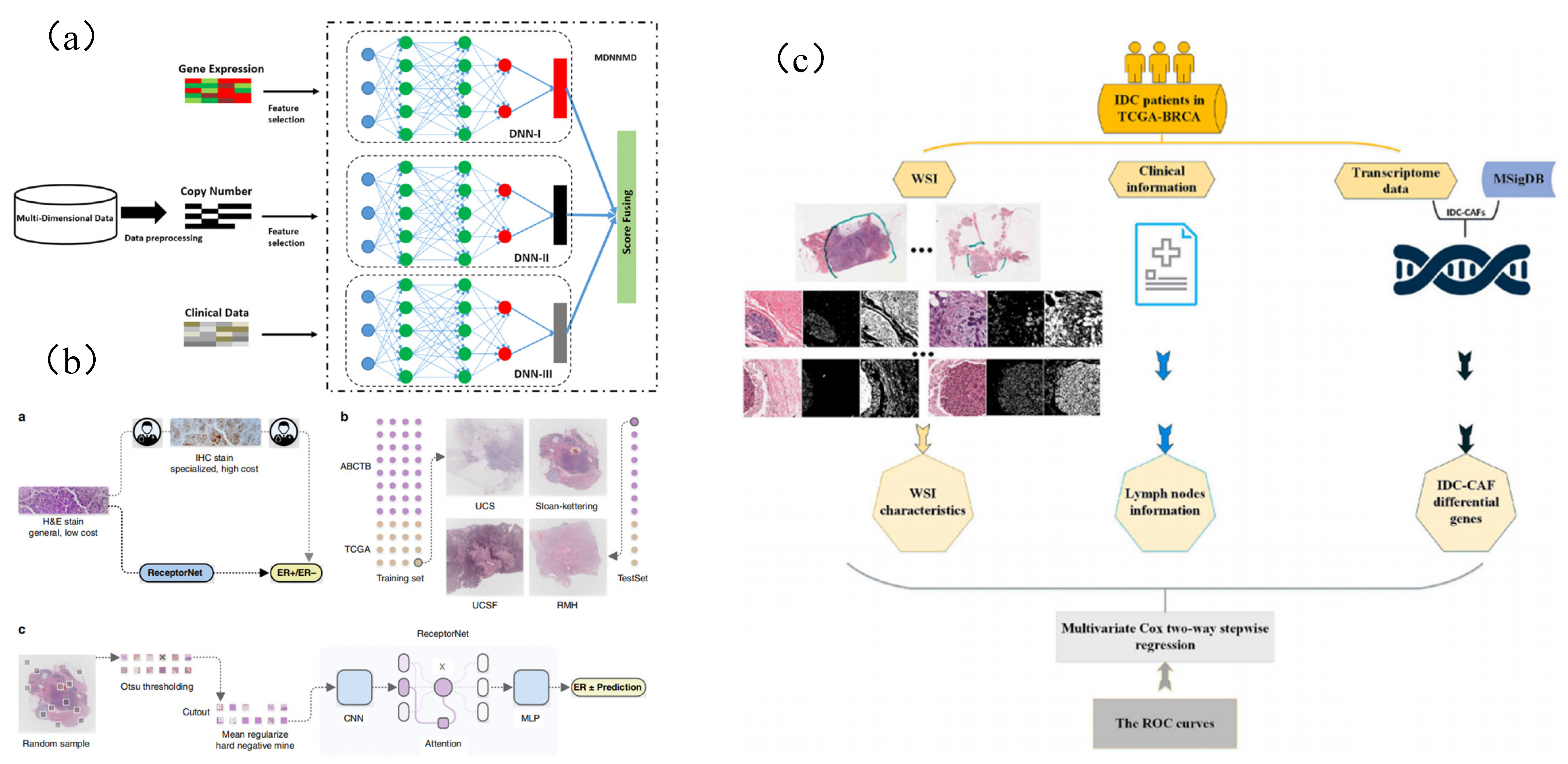
183] used a complete slide image combined with hematoxylin and eosin (H&E) staining, trained a large number of H&E and IHC-labeled image pair datasets using deep neural networks, and proved the accurate ER receptor state estimation from H&E staining (
Figure 6B).
In addition, considering the limitations of the method based on a single information source, such as a lack of nonuniversality, uniqueness, and noise data, multimodal learning is proposed to solve these problems and obtain a final decision by combining relevant information from multiple sources [
184,
185]. As a kind of multimodal learning, multimodal deep learning [
186] proposed a new multimodal deep neural network prognosis prediction for human breast cancer by integrating multidimensional data (MDNNMD). MDNNMD is an effective method to integrate multidimensional data, including gene expression profile, copy number change (CNA) profile, and clinical data with the score level of final prediction results. This method takes into account the heterogeneity between different data types and makes full use of the abstract high-level representation of each data source (
Figure 6A).
Petkov et al. [
187] accurately predict the prognosis of IDC, which is helpful to determine the individualized adjuvant treatment of breast cancer patients. Lin, Zhiquan et al. [
188] proposed and tested WSI preprocessing and feature extraction methods. Combining CAF gene, WSI characteristics, and lymph node status, a multigroup model was established to predict the prognosis of IDC breast cancer patients (
Figure 6C).
In the field of digital pathology, unsupervised clustering has been widely used to reduce the dimension of patches to facilitate multi-instance learning (for example, patches from WSI can be immediately installed on the graphics processing unit (GPU)) [
189]. This method is also used to derive additional cluster-based characteristics and identify rare events. Dooley et al. [
190] and Zhuet al. [
191] clustered the plaques and used the frequency of plaques in each cluster as a new feature to predict the rejection of heart transplantation. Similarly, see Abbet et al. [
189]. Although various unsupervised clustering applications have been developed in digital pathology, few studies have evaluated the use of unsupervised clustering to identify image patches related to gene mutation. Chen et al. [
192] proposed a multi-instance learning method based on unsupervised clustering and developed an in-depth learning model using WSIs of three common cancer types obtained from the Cancer Genome Map (TCGA) to optimize the prediction of genetic mutation.
Figure 6.
Typical application of deep learning in genetic prediction task. (
a) MDNNMD model uses multidimensional data to predict the prognosis of breast cancer [
186]. (
b) Estrogen receptor status (ERS) was predicted from the whole-slide image of H&E staining [
183]. (
c) A multi-omics signature to predict the prognosis of invasive ductal carcinoma of the breast [
188].
Table 5.
Summary of the application of deep learning algorithms in breast cancer histopathology for genetic prediction.
Table 5.
Summary of the application of deep learning algorithms in breast cancer histopathology for genetic prediction.
| Model | Strategy | Advantages | Publication |
|---|
| Attention mechanism | Weighting different pathological images based on attention mechanism to improve prediction results | Attention mechanism simulates human visual behavior by applying different weights to images. This method can highlight the key areas and non key areas in pathological images, thus improving the prediction results of the model | [181] |
| | Based on ResNet and attention mechanism, a method for predicting pathological gene subtypes of breast cancer is proposed | | [183] |
| KNN and K-means | Use unsupervised clustering method to reduce the workload of manual labeling by pathologists | Unsupervised | [192] |


