Article

Discovery of a High-Efficient Algicidal Bacterium against Microcystis aeruginosa Based on Examinations toward Culture Strains and Natural Bloom Samples

1
Zhejiang Provincial Key Laboratory for Subtropical Water Environment and Marine Biological Resources Protection, National and Local Joint Engineering Research Center of Ecological Treatment Technology for Urban Water Pollution, College of Life and Environmental Sciences, Wenzhou University, Wenzhou 325035, China
2
State Key Laboratory of Biocatalysis and Enzyme Engineering, School of Life Sciences, Hubei University, Wuhan 430062, China
3
Wenzhou Shanxi Hydro-junction Management Center, Wenzhou 325035, China
4
Institute for Eco-Environmental Research of Sanyang Wetland, Wenzhou University, Wenzhou 325035, China
*
Author to whom correspondence should be addressed.
These authors contributed equally to this work.
Toxins 2023, 15(3), 220; https://doi.org/10.3390/toxins15030220
Submission received: 16 February 2023 / Revised: 6 March 2023 / Accepted: 11 March 2023 / Published: 14 March 2023

Abstract

:
Harmful cyanobacterial blooms occur worldwide and pose a great threat to aquatic ecosystems and public health. The application of algicidal bacteria represents an eco-friendly strategy for controlling harmful cyanobacterial blooms; thus, searching for a high efficiency of algicidal bacteria has been becoming an important and continuous task in science. Herein, we identified a bacterial strain coded Streptomyces sp. HY with a highly algicidal activity, and investigated its algicidal efficiency and mechanism against Microcystis aeruginosa. The strain HY displayed high algicidal activity toward Microcystis aeruginosa cells, with a removal rate of 93.04% within 2 days via indirect attack. Streptomyces sp. HY also showed the ability to lyse several genera of cyanobacterial strains, including Dolichospermum, Pseudanabaena, Anabaena, and Synechocystis, whereas it showed a minor impact on the green alga Scenedesmus obliquus, demonstrating its selectivity specially for targeting cyanobacteria. Its algicidal mechanism involved damages to the photosynthesis system, morphological injury of algal cells, oxidative stress, and dysfunction of the DNA repair system. Furthermore, HY treatment reduced the expression levels of genes (mcyB and mcyD) related to microcystin biosynthesis and decreased the total content of microcystin-leucine-arginine by 79.18%. Collectively, these findings suggested that the algicidal bacteria HY is a promising candidate for harmful cyanobacterial bloom control.
Key Contribution: A bacterial strain (Streptomyces sp. HY) with a highly algicidal activity against Microcystis aeruginosa was identified, and the underlying algicidal mechanism was studied. Streptomyces sp. HY can selectively kill several bloom-forming cyanobacterial strains and strongly inhibit the biosynthesis of microcystin-leucine-arginine in Microcystis aeruginosa.

Graphical Abstract

1. Introduction

Harmful cyanobacterial blooms (HCBs) have become a worldwide concern for water eutrophication and global warming in recent years [1]. More than 60% of the lakes in China are eutrophic and suffer from HCBs, such as Lake Taihu [2]. HCBs can seriously damage aquatic ecosystems and pose a great threat to human health by producing various cyanotoxins. Microcystis aeruginosa (M. aeruginosa) is one of the most dominant harmful cyanobacterial species, as well as the major species producing and releasing microcystins (MCs) worldwide [1]. MCs are a group of cyclic heptapeptides comprising hundreds of variants, and MC-leucine-arginine (MC-LR) is one of the most toxic and abundant variants [3]. Accumulated reports have proved that MC-LR causes liver cancer and tumors via inhibiting the activities of protein phosphatases 1A and 2A in humans and animals [4]. Moreover, MC-LR also displays reproductive toxicity, neurotoxicity, immunotoxicity, and other toxicities [5].
Given that the guide concentration of MC-LR for drinking water is less than 1 μg/L [6], it is urgent to develop cost-effective and eco-friendly approaches to control HCBs caused by M. aeruginosa. Several strategies have been proposed to solve HCBs, including chemical, physical, and biological methods [7]. Mechanical removal, adsorption-flocculation, and ultra-sonication are frequently used physical methods. Although these methods are sometimes effective, they are limited by their high costs and low efficiency in large areas and volumes. Chemical methods have been successfully used to control HCBs, with CuSO4 [8] and hydrogen peroxide (H2O2) [9] once popular chemical algicides. However, these agents are non-specific and come with the potential risk of secondary pollution because of their toxicity and fast release of cyanotoxins. These disadvantages greatly limit their general application, and alternative biological tools, especially algicidal bacteria, have attracted considerable attention for their low-cost, eco-friendliness, and great potential [10]. Previous reports have found that many algicidal bacteria not only disrupt the photosystem of certain cyanobacteria, but also suppress the expression of several key genes related to photosystem, significantly decreasing the maximum quantum yield of photosystem II (PSII) and the photosynthetic electron transport rate (rETR) of cyanobacteria [11]. Furthermore, several algicidal strains can also inhibit the expression of genes related to MC biosynthesis [12], even degrading MCs [13]. To date, a number of algicidal bacterial species from about 50 genera have been identified [10,14], including Bacillus spp. [15], Pseudomonas spp. [16], Aeromonas spp. [17], Raoultella spp. [11], and Streptomyces spp. [18]. Streptomyces species are known for their ability to biosynthesize different kinds of secondary metabolites, including antibiotics and other active substances [19].
In general, algicidal bacteria achieve their activities through two mechanisms: (1) direct algicidal activities through cell-to-cell contact with cyanobacterial cells; and (2) indirect algicidal activities by secreting extracellular algicides, such as proteins, antibiotics, pigments, alkaloids, and other substances to kill cyanobacteria [20]. Approximately 30% of bacteria reported are suggested to be direct algicidal bacteria, whereas the remaining 70% are indirect ones [20]. Algicidal bacteria displaying both mechanisms have been rarely identified [21]. Both modes of algicidal bacteria and their active algicidal chemicals are considered as effective agents for controlling HCBs. Another enormous challenge comes from the fact that algicidal bacteria kill Microcystis cells and result in fast release of intracellular MCs into the water in a few hours. MCs can remain stable in water for several days, resulting in extensive exposure of all aquatic organisms to MCs before they are completely degraded [6]. Therefore, controlling Microcystis blooms and inhibiting MC biosynthesis at the same time through indirect algicidal bacteria is a promising strategy. The major objective of the present work was to isolate and identify novel algicidal bacteria against HCBs, hopefully with higher efficiency than those found in previous studies.
In this study, we investigated a newly isolated algicidal strain (Streptomyces sp. HY) from the Dongzhen Reservoir, Putian, Fujian Province. The algicidal process and underlying mechanisms of action against M. aeruginosa FACHB 905 were studied. The strain HY strongly inhibited the growth and biosynthesis of MC-LR in M. aeruginosa via secreting active compound(s). HY cell-free filtrate induced serious photosystem impairment by dramatically reducing photosynthetic efficiency (Fv/Fm) and inhibiting the maximum photosynthetic rate (ETRmax). After exposure to the HY filtrate, the level of reactive oxygen species (ROS) and content of malondialdehyde (MDA) increased obviously, whereas a number of key genes were downregulated. This study enriched our knowledge of interactions between Microcystis-bacteria, and contributed to the development of novel biocontrol approaches against HCBs.

2. Results

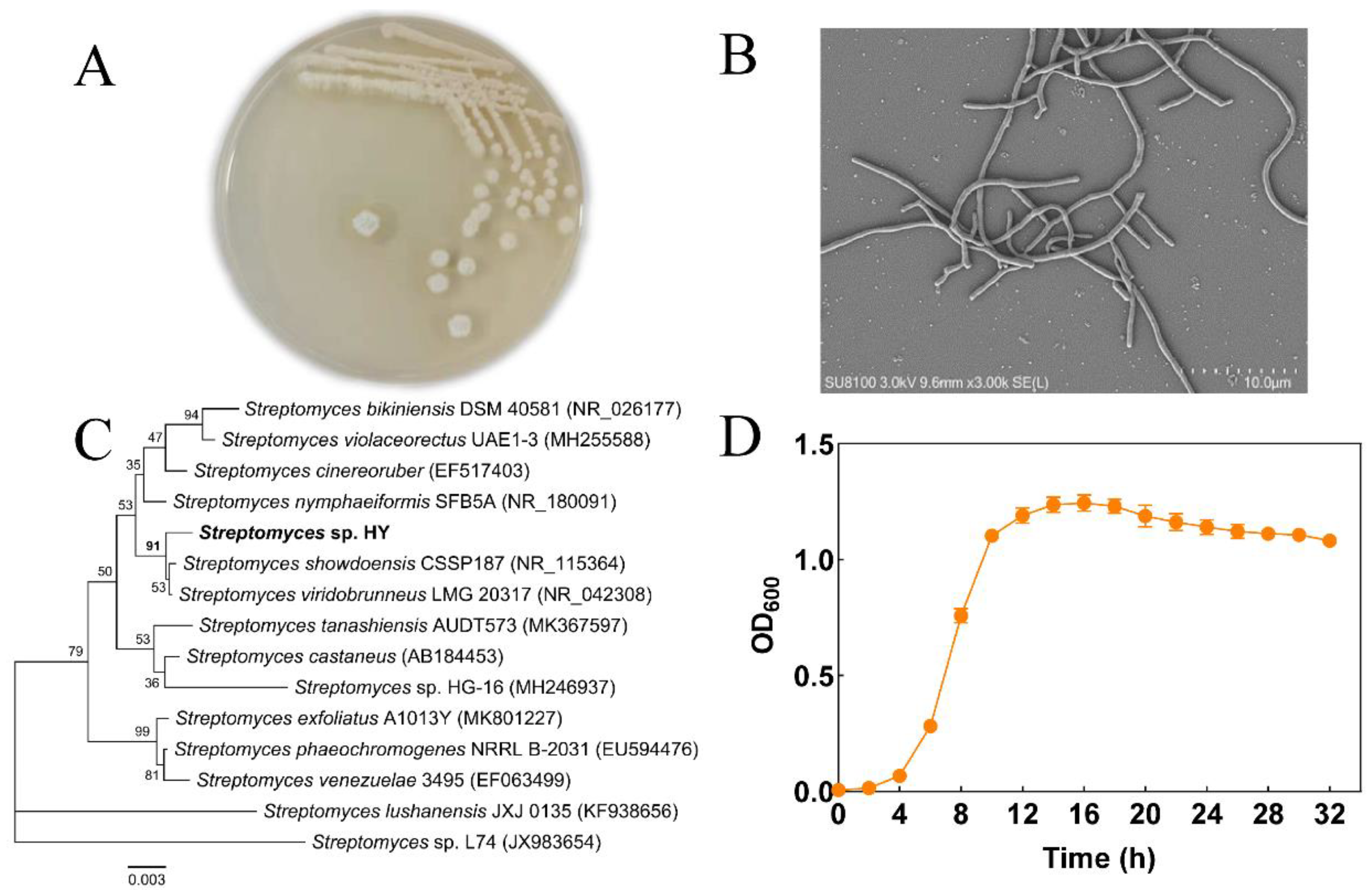
2.1. Isolation and Identification of Algicidal Bacteria

In total, 411 bacterial strains were isolated during October to December in 2021 from Fujian and Zhejiang Provinces in China, and subsequently tested for their algal activity against M. aeruginosa FCHAB 905. Strain #320 from Dongzhen Reservoir (Putian, Fujian, China) exhibited high algicidal activity (Figure 1) and was chosen for further study. The 16S rRNA sequence of this strain shared high similarity (>98.9%) to strains of Streptomyces spp. (Figure 1C). In a phylogenetic tree, strain #320 clustered closely with Streptomyces spp. strains, although it formed an independent branch. Streptomyces sp. #320 grew well on R2A agar medium (Figure 1), as well as on Gause No. 1 agar plates (data not shown), a selective medium for actinomycetes [22]. When incubated for more than 4 days on a plate, colonies were circular with white aerial mycelium, and a mound of grey, rectiflexibilis-type spores were formed (Figure 1A,B). Thus, based on the 16S rRNA sequence analysis and cellular morphology characteristics, this algicidal strain was identified as Streptomyces sp. and designated as strain Streptomyces sp. HY hereafter.

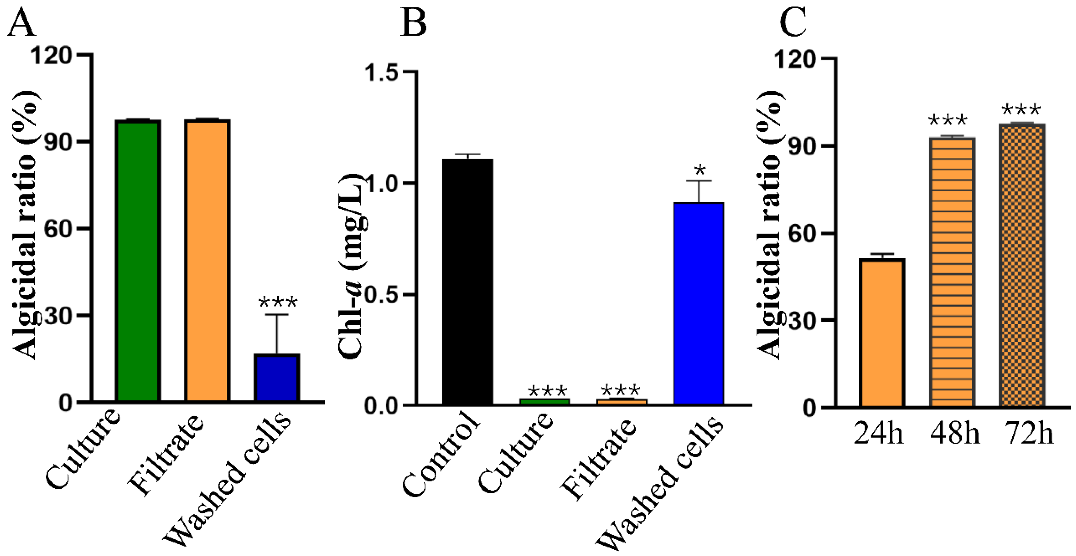
2.2. Algicidal Characteristics of Streptomyces sp. HY

As shown in Figure 1D, HY reached the exponential growth phase after 4 h of culture; this phase lasted for 10 h. Several Streptomyces spp. strains secrete active compounds to kill cyanobacteria [18,19,23], so we tested the algicidal activity of strain HY during the stationary growth phase at 24 h of fermentation. As indicated in Figure 2A, the algicidal ratio of bacterial culture, the cell-free filtrate, and the washed cells against M. aeruginosa FACHB 905 was 97.4%, 97.7%, and 16.97%, respectively, on the 3rd day. The M. aeruginosa FACHB 905 culture after treatment was yellow-to-white. Almost all cyanobacterial cells were lysed by HY bacterial culture or cell-free supernatant from HY in 3 days, and the content of the photosynthesis pigment Chlorophyll a (Chl-a) also decreased rapidly from 0.78 mg/L to 0.03 mg/L or 0.028 mg/L (Figure 2B), respectively. The Chl-a content of the washed-bacterial-cells-treated group and control group was 0.91 mg/L and 1.11 mg/L, respectively (Figure 2B). The cultures both remained green. These results showed that HY killed M. aeruginosa efficiently via an indirect mode by secreting algicidal compounds. Therefore, we used its cell-free filtrate in subsequent studies.

2.3. Algicidal Spectrum of HY

To explore the algicidal spectrum of HY, we tested several cyanobacterial strains, as shown in Table 1. HY displayed high algicidal activity against all single cell strains, with an algicidal ratio of 99.78% against M. aeruginosa FACHB 526, 95.20% against M. aeruginosa PCC 7806, and 88.74 % against Synechocystis sp. PCC 6803. HY also exhibited high algicidal activity against the filamentous cyanobacteria tested, with an algicidal ratio of 88.44% for Pseudoanabaena sp. WZU 1801, 97.5% for Dolichospermum sp. WZU 1811, and 64.97% for Anabaena sp. PCC 7120. However, HY showed much lower algicidal effects against the eukaryotic green algae Scenedesmus obliquus (S. obliquus) (25.74%). These data demonstrated that HY showed broad-spectrum algicidal activity against cyanobacteria, with few side effects on other non-target eukaryotic phytoplankton species. HY may have a great potential for controlling the threat of multiple HCBs, especially M. aeruginosa.

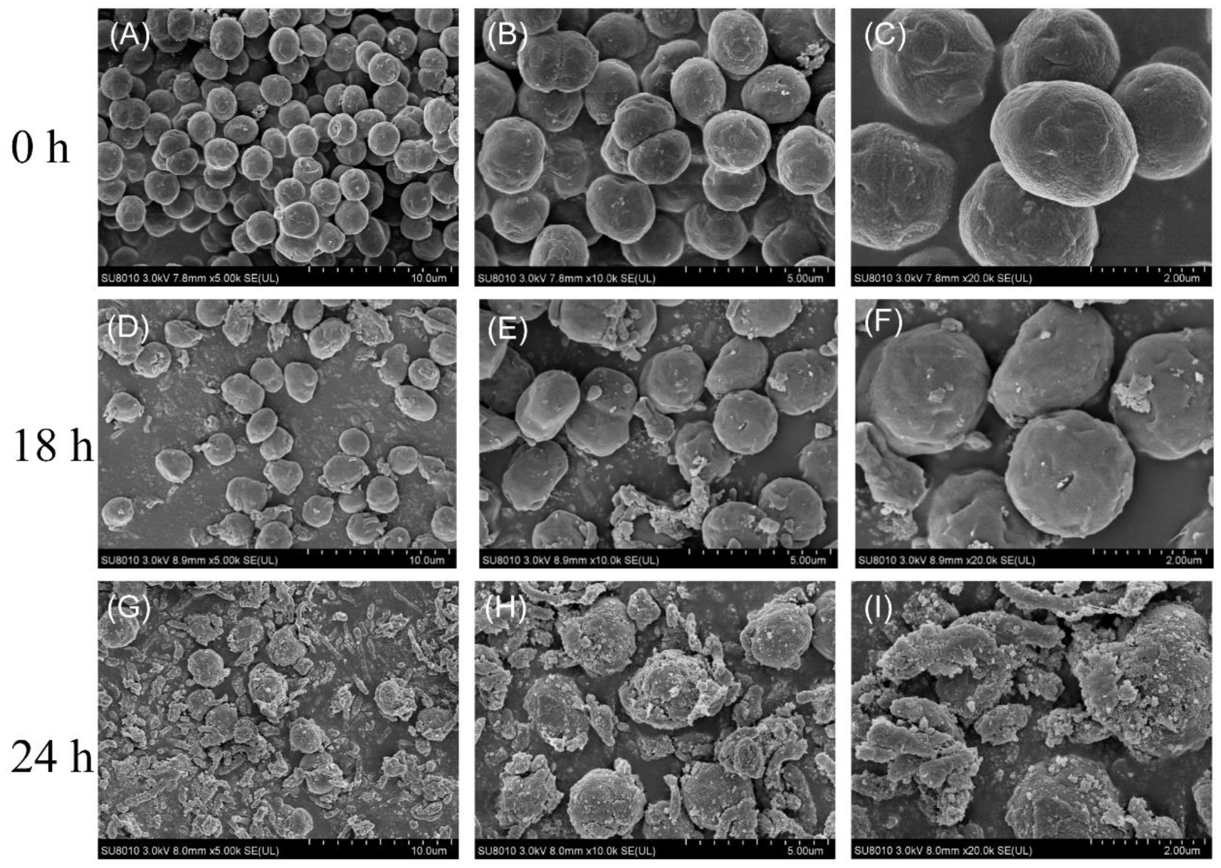
2.4. Effect of HY on Cell Morphology of M. aeruginosa

To further elucidate the algicidal effects of HY against cyanobacterial cells, we used the typical MC-producing M. aeruginosa strain FACHB 905 for further studies. M. aeruginosa FACHB 905 was exposed to the cell-free filtrate of HY for 18 h and 24 h, and cell morphological changes were observed by SEM. As Figure 3A–C shows, M. aeruginosa cells from the control group were round and retained an intact cell surface. In contrast, most of the algal cells became shrunken and deformed after 18 h of treatment with HY cell-free filtrate, and some algal cells began to break down (Figure 3D–F). As the treatment time lengthened, most cells were lysed, leaving just a few intact M. aeruginosa cells with a rough surface and lots of cell debris at 24 h (Figure 3G–I). The cyanobacterial cell walls were severely damaged, leading to the leakage of intracellular components. A similar phenomenon was observed using a light microscope. Our data clearly suggested that HY killed algal cells through indirect attack.

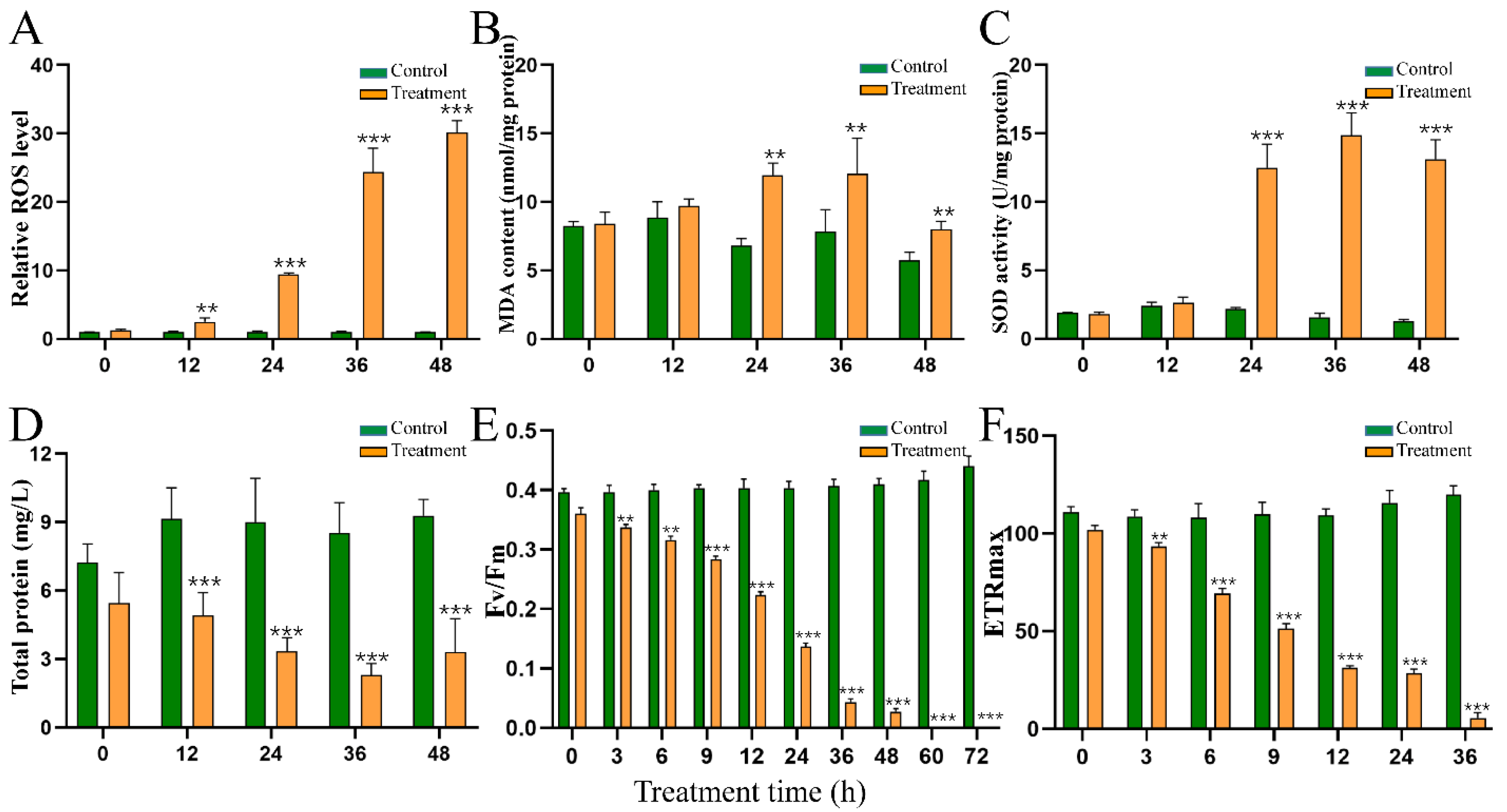
2.5. Effect of HY Filtrate on the Content of ROS and MDA and the Activity of SOD

To study the algicidal mechanisms of HY, we assessed changes in the antioxidant systems of M. aeruginosa cells at indicated time points. Intracellular ROS were significantly induced by the supernatant of HY (Figure 4A) when compared with those of the untreated group. Relative levels of ROS increased about 2.50, 9.40, 24.34, and 30.15 times after 12, 24, 36, and 48 h (Figure 4A), respectively, when compared with the control group. The initial MDA content was about 8.41 mol/mg protein in M. aeruginosa and remained stable in the control group. The MDA content increased by about 0.76, 0.54, and 0.39 times at 24, 36, and 48 h after HY treatment (Figure 4B), respectively. We also measured the activity of SOD, a typical antioxidant enzyme, to study the response of M. aeruginosa. As Figure 4 shows, SOD activity increased over time, and was about 5.78, 9.44, and 10.34 times higher than that of the control group at 24, 36, and 48 h (Figure 4C), respectively. It is worth noting that although SOD showed high activity, algicidal compounds in HY supernatant still induced massive increases in ROS and MDA, resulting in oxidative stress in M. aeruginosa cells. The content of total protein in M. aeruginosa declined obviously (Figure 4D).

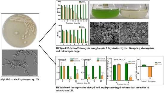
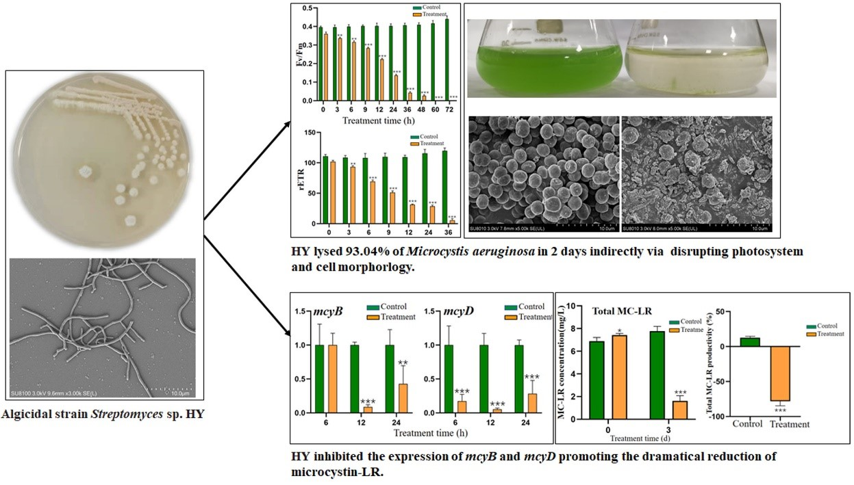
2.6. Effect of HY Filtrate on the Photosynthesis System of M. aeruginosa

To further explore the physiological response of M. aeruginosa to HY filtrate, we monitored Fv/Fm and ETRmax. The Fv/Fm (Figure 4E) and ETRmax (Figure 4F) of M. aeruginosa declined rapidly following treatment with HY supernatant. The Fv/Fm value decreased from 0.36 to 0.22, 0.14, 0.043 (12.04% of the initial value), and 0.026 (7.41% of the initial value) at 12 h, 24 h, 36 h, and 48 h, respectively. Finally, the Fv/Fm value declined to 0 at 60 h. Similarly, the ETRmax value of treated M. aeruginosa cells decreased dramatically when compared with that of the control group, from 102 to 69.2, 31.33, 28.53, and 5.5 at 6 h, 12 h, 24 h, and 36 h, respectively.

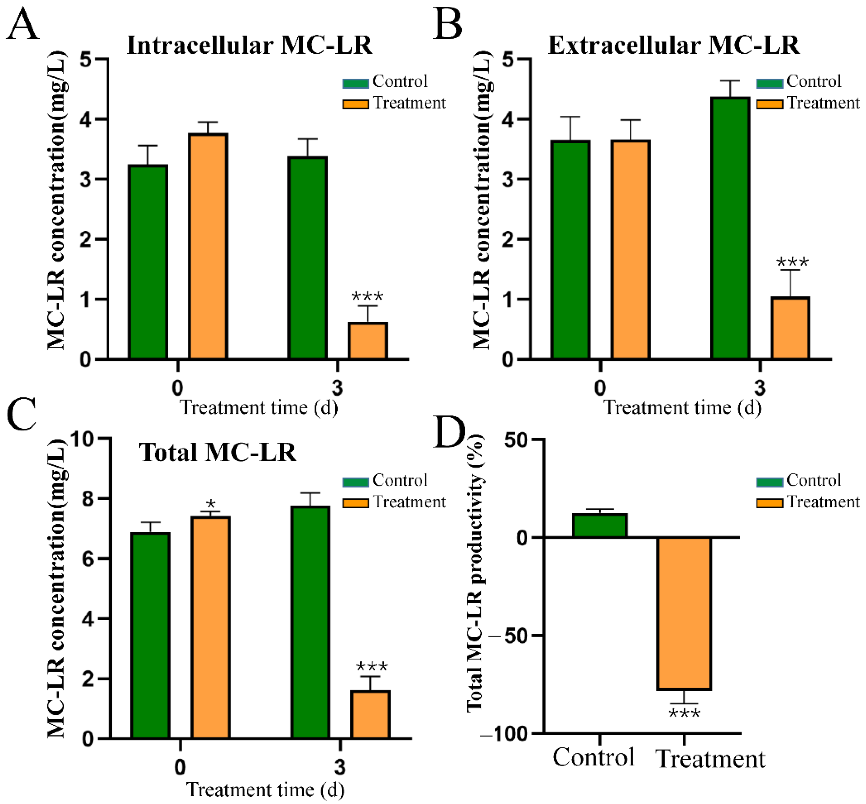
2.7. Effect of HY Culture on the Content and Distribution of MC-LR

Following exposure to HY culture for 3 days, the total content of MC-LR decreased significantly, from 7.77 ng/mL to 1.62 ng/mL (Figure 5C). The intracellular MC-LR concentration of M. aeruginosa also declined rapidly (from 3.39 ng/mL to 1.04 ng/mL), which was lower than that of the control group (3.39 ng/mL) (Figure 5A). In addition, the extracellular MC-LR concentration of M. aeruginosa cells (0.58 ng/mL) was also significantly lower than that of the control group (4.38 ng/mL) (Figure 5B). Similar to a previous report [23], we found that the MC-LR productivity of the control group increased by 12.67%, when compared with the initial concentration. The MC-LR productivity of the treated groups decreased by 78.22% (Figure 5D). This result suggested that HY not only strongly inhibited the biosynthesis of intracellular MC-LR, but might also be simultaneously involved in extracellular MC-LR degradation.

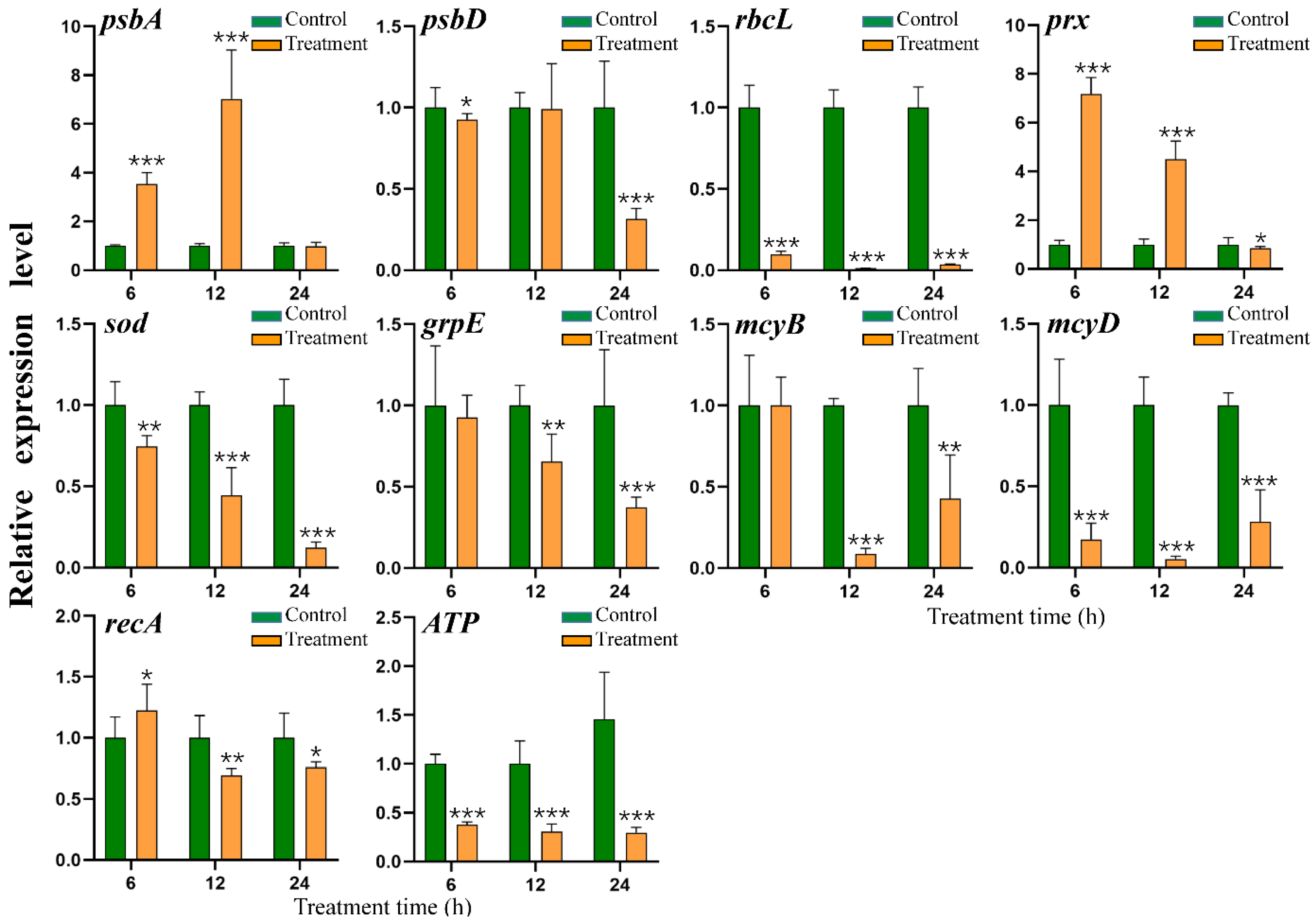
2.8. Effect of HY Filtrate on the Transcription of Key Genes in M. aeruginosa

To further study the mechanism by which HY damages M. aeruginosa, we assessed the transcription levels of several key genes (psbA, psbD, rbcL, prx, sod, grpE, mcyB, mcyD, recA, and ATP) by RT-qPCR. The psbA and psbD are important genes encoding the D1 and D2 proteins of photosystem II (PS II), respectively. The expression of psbA was increased by approximately 353% and 703% at 6 h and 12 h after treatment with HY supernatant, respectively, finally falling back to a similar level (98.1%) to that of the untreated group after 24 h (Figure 6). The expression of psbD did not change very much during the early period of treatment, but decreased by about 30% after 24 h (Figure 6). The rbcL gene encodes an important enzyme (RuBisCo) for carbon dioxide assimilation, and its expression pattern was very different from those of psbA and psbD. rbcL expression was significantly inhibited by HY filtrate over time, decreasing to 8.44%, 1.06%, and 3.22% that of the control group expression at 6 h, 12 h, and 24 h, respectively (Figure 6).
The expression patterns of two genes related to antioxidant defense, prx and sod, were also different from those of psbA and psbD. prx expression was upregulated by 9.98- and 4.5-fold at 6 h and 12 h, respectively, and downregulated 0.16-fold at 24 h. sod expression was totally suppressed, decreasing to 75.6%, 44.5%, and 12.3% of the level in the control at 6 h, 12 h, and 24 h, respectively. The transcription levels of two microcystin biosynthesis genes (mcyB, mcyD) were significantly downregulated at 12 and 24 h, with mcyD expression suppressed from the beginning of the treatment period to the end (Figure 6). This result further confirmed that HY efficiently inhibited the biosynthesis of MC-LR in M. aeruginosa. The expression of the recA gene related to DNA repair was slightly upregulated (22.5%) at 6 h, but then decreased to 30.7% and 24% of the control group at 12 h and 24 h, respectively. Another stress response gene, grpE, shared a similar expression tendency, with expression declining significantly from 12 h to 24 h. Moreover, the expression of the key energy metabolism gene ATP, encoding a subunit of ATP synthase, was strongly inhibited by HY filtrate at all timepoints tested, being reduced by approximately 62.14%, 69.29%, and 79.65% at 6 h, 12 h, and 24 h, respectively. This result suggested that HY adversely affected various biological processes of M. aeruginosa in a complex manner.

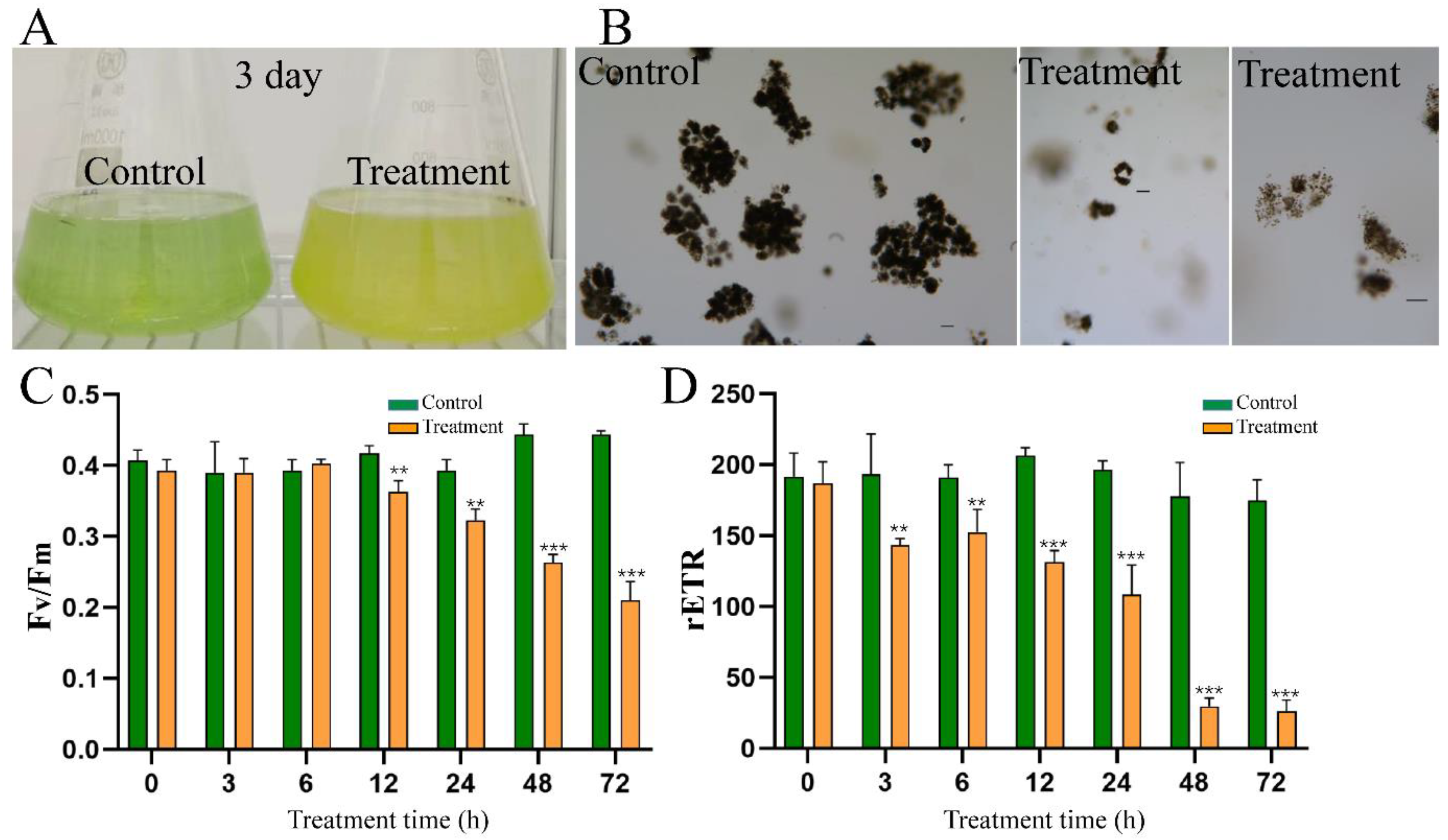
2.9. Effect of HY Filtrate on Colonies of M. aeruginosa from Natural Bloom Samples

Previous studies have demonstrated that algicidal bacteria could easily kill single cells of M. aeruginosa, but may not be active against colonial M. aeruginosa from natural waters [16,24]. We, therefore, investigated the algicidal activity of HY against M. aeruginosa colonies from natural bloom samples of the Xianju Reservoir. Treatment with HY filtrate efficiently flocculated colonial M. aeruginosa to the bottom of culture vessels and induced a fast decline of Fv/Fm and ETRmax (Figure 7C,D). However, the removal rate of HY filtrate was only 51.77% at 72 h, significantly lower than that observed in M. aeruginosa cultures (Figure 7A,B). These results suggested that algicidal compounds from HY could not efficiently lysate colonial M. aeruginosa and should be studied further before being used to control M. aeruginosa blooms in natural water bodies.

3. Discussion

Water eutrophication leads to the occurrence of HCBs worldwide. Microcystis is a widespread, bloom-forming, and toxic cyanobacterial group. Many efforts have been devoted to developing novel technologies to control HCBs, and the usage of algicidal bacteria has shown promising application potential. In this study, we aimed to isolate novel algicidal bacterial species and develop effective biological treatments against HCBs. Streptomyces sp. HY with high algicidal activity was successfully identified (Figure 1). Strain HY induced 93.04% lysis of Microcystis cells within 2 days and almost 100% lysis within 3 days (Figure 2). Several algicidal strains with activity against cells of Microcystis spp. have been reported, such as Streptomyces sp. L74 [19], Streptomyces globisporus G9 [24], and Streptomyces sp. HG-16 [23], which can lyse more than 70% M. aeruginosa cells within 5 days. However, the highest average algicidal activity was 94.6% by S. globisporus G9 in 5 days of treatment. The algicidal activity (97.72%) of Streptomyces sp. HY in 3 days was much higher than most of the bacterial strains reported previously. Therefore, Streptomyces sp. HY is a promising candidate as an algicidal bacterial strain for controlling HCBs.
Considering that most algicidal bacteria showed obvious host-specificity and only worked well against a few species [20], we tested the lytic spectrum of Streptomyces sp. HY. We first assessed its regional host susceptibility using various Microcystis spp. strains. For instance, M. aeruginosa FACHB 905 (MC-producing) and M. aeruginosa FACHB 526 (non-MC-producing) are from China, whereas M. aeruginosa PCC 7806 (MC-producing) is from Europe. HY could lyse more than 91% of cells of each M. aeruginosa strain within 3 days, regardless of their origin (Figure 2 and Table 1). Several previous studies suggested that MCs might protect M. aeruginosa against algicidal substances and diverse allelopathic materials [25,26]. However, the removal efficiency of HY for each M. aeruginosa strain was almost the same (>95%) in 3 days, and none of the strains could withstand the damage caused by HY (Table 1) in this study. Therefore, Streptomyces sp. HY had a broad host spectrum against M. aeruginosa and might work well at a large geographic scale. However, colonial Microcystis samples from naturally blooming water bodies seem more resistant to HY supernatant than pure cultures in the lab (Figure 7). Most other strains reported share this limitation [16], except for Xanthobacter autotrophicus HYS0201-SM02 [27]. Algicidal bacteria, including HY, should, therefore, be used with chemical reagents or physical equipment to disrupt the colonial state of Microcystis. Alternatively, increasing the inoculum ratio of algicidal bacteria might also work well against environmental strains, and further studies should be conducted.
We also tested the algacidal activity of Streptomyces sp. HY against other dominant species of bloom-forming cyanobacteria (Cyanophyta), including Dolichospermum, Anabaena, and Pseudoanabaena. Streptomyces sp. HY effectively lysed Dolichospermum (97.50%), Anabaena (64.97%), and Pseudoanabaena (88.44%) in 3 days, respectively (Table 1). Interestingly, HY had a minor impact on eukaryotic microalgae, such as S. obliquus (Chlorophyta) (Table 1) only killing about 25.74% within 3 days. Our results showed that HY was host (cyanobacteria)-specific and had few side effects on other non-target phytoplankton in the natural aquatic environment (Table 1), suggesting great potential for controlling HCBs. The lower sensitivity of S. obliquus to HY may be due to intrinsic differences in cellular structure between Chlorophyta and Cyanophyta.
Most algicidal bacterial strains (about 70%) work via indirect attack by secreting active algicidal compounds [20]. To study the algicidal mode of Streptomyces sp. HY, we analyzed the algicidal rate of its bacterial culture, cell-free supernatant, and washed cells, respectively. The filtrate of HY showed similar algicidal activity to the bacterial culture against M. aeruginosa (97.72% vs. 97.70%) within 3 days, whereas the washed cells exhibited much lower algicidal activity (16.97%) (Figure 2). This indicated that HY excreted substances into the medium to lyse M. aeruginosa cells via indirect attack. We also noted that algicidal substances from HY could eventually damage the cellular structure of M. aeruginosa cells (Figure 3). The result is in line with previous studies [23,28] The potential anti-cyanobacterial substances produced by HY are unknown, but we speculate that they are amino acid derivatives or other small molecules (unpublished data). These compounds produced by HY will be further isolated and characterized to identify their molecular structures, with the hope of mass-producing them for the ecofriendly control of HCBs.
Fv/Fm and ETRmax reflect the photosynthetic activity of cyanobacteria. Therefore, we monitored changes of these parameters during HY treatment. The value of Fv/Fm reduced significantly from 3 h after treatment and decreased to 0 at 60 h (Figure 4E). This decrease suggested that the photosystem of M. aeruginosa, especially PS II, was seriously damaged and led to the inhibition of photosynthesis. Additionally, the change in ETRmax showed a similar pattern. Its value was reduced significantly at 3 h after treatment and decreased to 3.9% that of the control group at 36 h (Figure 4F). This result suggested that the electron transport chain was seriously blocked. Moreover, the content of Chl-a was reduced from 1.11 mg/L to 0.003 mg/L after 72 h of treatment (Figure 2). We noted that the values of Fv/Fm and ETRmax were significantly downregulated at 12 h, much earlier than the death of cyanobacterial cells (Figure 5). Therefore, photosynthetic parameters may be important indicators during large-scale cyanobacteria culture.
We also detected the transcription levels of three key photosynthesis-related genes using qRT-PCR. The rbcL gene encodes the large subunit of Rubisco, a rate-limiting enzyme for carbon dioxide (CO2) fixation. The transcription of rbcL was strongly inhibited by HY treatment, decreasing to 3.22% of the control group after 24 h (Figure 6). The psbD gene encodes the D2 protein, a key subunit of the reaction center of PSII. Although psbD did not seem to be obviously influenced by substances from HY at 6 h and 12 h, its expression was finally reduced to 68.4% of that of the control at 24 h. The transcriptional suppression of rbcL and psbD was in line with the decline of photosynthetic parameters, in accordance with previous studies [11,28]. Hence, we speculated that the photosynthetic system was the early and primary target of HY algicidal substances. In our study, the expression of psbA (encoding the D1 subunit of PSII) increased 3.52-fold and 7.03-fold during the early phase of HY treatment (6 h and 12 h) and maintained a similar level to the control group after 24 h of treatment (Figure 6). Similar results were also described when M. aeruginosa was treatment with either 60 mg L−1 succinic acid [29] or algicidal supernatant [23]. The upregulation of psbA may be caused by M. aeruginosa cells trying to compensate for the inhibition of other photosynthesis-related genes.
Impairment of the photosynthesis system promotes the overproduction of ROS. Unfortunately, the photosynthesis system itself is very susceptible to ROS and oxidative stress [28,30]. This negative cascade loop strongly promotes the accumulation of cellular ROS, which react with biomolecules (including DNA, unsaturated fatty acids of membranes, and proteins) in M. aeruginosa cells. Therefore, we measured the contents of ROS and MDA during the algicidal process (Figure 4). We found a marked increase in ROS and MDA contents from 24 h to 48 h (Figure 4), clearly indicating that HY filtrate induces oxidative stress in M. aeruginosa cells. This trend was consistent with the increase in SOD activity (Figure 4).
The antioxidant enzymes peroxiredoxin (Prx) and SOD are encoded by prx and sod, respectively. They both play an important role in maintaining intracellular redox homeostasis by scavenging ROS [28,29]. We observed a marked increase in prx expression after treatment with HY supernatant (Figure 6). sod expression was totally inhibited, and prx and sod were downregulated as exposure time lengthened (Figure 6). This process would accelerate the shortage of antioxidant enzymes and trigger serious oxidative stress in Microcystis cells. To make matters worse, the transcription of other anti-stress genes, such as recA, and grpE, was also depressed. RecA plays a critical role in repairing damaged DNA [11,28], whereas GrpE promotes the folding of proteins as a chaperone [11,28]. After 24 h of exposure to HY filtrate, recA and grpE expression decreased to 76.08% and 37.14%, compared to the control group, respectively (Figure 6). The reduction of recA and grpE expression indicated that the DNA repair system and protein refolding system were both severely damaged in M. aeruginosa cells. Moreover, the transcription of a gene related to ATP biosynthesis was simultaneously suppressed (Figure 6). Transcriptional inhibition of recA, grpE, and ATP genes was similar to previous results [28]. Therefore, active substances produced by HY killed cyanobacterial cells by disrupting several key biological processes, including photosynthesis and protein, carbohydrate, and energy metabolism.
The collapse of Microcystis cell walls (Figure 3) resulted in the inevitable release of MCs into the medium. Therefore, we investigated the fate of MCs released from Microcystis cells treated with HY culture. Unlike the dramatic increase in total MC concentration after chemical treatment (such as CuSO4) [8], treatment with HY culture produced an obvious decrease in total MC-LR (Figure 5). Extracellular and intracellular MC-LR concentrations were reduced by 86.86% and 69.23% (Figure 5), respectively, when compared with the control group. More importantly, HY culture significantly suppressed total MC-LR productivity (−78.22%) (Figure 5), whereas Microcystis cells treated with algicidal strain HG16 still retained considerable (≈10%) MC-LR productivity [23]. Our data suggest that compound(s) produced by HY are involved in the inhibition of MC-LR biosynthesis. The results from qRT-PCR experiments were in agreement with this phenomenon. Treatment with HY filtrate significantly downregulated the expression levels of microcystin biosynthesis genes mycB and mcyD at 12 and 24 h (Figure 6). However, the residual concentration of MC-LR was still approximately 2 mg/L (about 20.8% of the control) in co-culture systems after 3 days (Figure 5), which is still much higher than the healthy guideline for MC-LR concentration suggested by the WHO [6]. Considering that MC-LR can persist for several days in the aquatic environment [31], it would pose a threat of secondary environmental pollution after bloom control. Novel strategies for eliminating Microcystis blooms and MC-LR at the same time are, therefore, urgently needed. A combination of algicidal bacteria and toxin-degrading bacteria seems simple and feasible [32]. We once isolated a MC toxin degrading strain Novosphingobium sp. THN1 with a mlr gene cluster from Taihu Lake [33]. Strains HY and THN1 could be used simultaneously against HCBs, and this method is worth further study. Moreover, as the pool of algicidal bacteria expands over time, the option of completely removing HCBs and cyanotoxins simultaneously with one isolate may be possible in the future.

4. Conclusions

In conclusion, Streptomyces sp. HY exhibited high algicidal activity against M. aeruginosa and other harmful cyanobacteria by secreting active compounds, and its algicidal activity was much higher those previously reported strains in the genus of Streptomyces. The underlying mechanisms involved impairment of photosynthetic systems, damage to the cellular membrane and cell walls, and oxidative stress. Streptomyces sp. HY also inhibited the biosynthesis and release of MC-LR. Our results collectively suggested that Streptomyces sp. HY is a promising algicidal strain for the control of HCBs.

5. Materials and Methods

5.1. Algal Strains and Culture Conditions

M. aeruginosa FACHB 905 (MC-producing), M. aeruginosa FACHB 7806 (MC-producing), M. aeruginosa FACHB 526 (non-MC-producing), Scenedesmus obliquus (S. obliquus) were supplied by the Freshwate Algal Culture Collection at the Institute of Hydrobiology, Chinese Academy of Sciences. Synechocystis sp. PCC 6803 and Anabaena sp. PCC 7120 were kindly provided by Prof. Xudong Xu (Institute of Hydrobiology, Chinese Academy of Sciences). Pseudoanabaena sp. WZU 1801 and Dolichospermum sp. WZU 1811 were maintained in our lab. All strains used in this study were cultivated in sterile BG11 medium (components are listed in Table S1) at 25 ± 0.5 °C with a 12 h/12 h (light and dark) cycle and 2500 lux of light intensity. Algae cultures were manually shaken every 12 h, and exponential cells with a starting density of 5 × 10⁶ cells/mL were used in this study.

5.2. Screening and Identification of Algicidal Bacteria

Soil samples were collected from Zhejiang and Fujian Provinces (China) in November 2021. Soil samples were placed into a sterilized bottle with 90 mL water and glass beads on a rotary shaker (28 °C, 180 rpm for 1 h). Following the screening method described by [28], a strain #332 (HY) with high algicidal activity was screened from 412 candidates and selected for subsequent investigation. Each single colony was obtained by re-streak on R2A medium (components are listed in Table S2) agar plates, and all strains were preserved at −80 °C in R2A medium containing 20% glycerol in our lab, respectively. Bacterial cell density was quantified by plating a dilution series onto R2A agar plates. To identify the algicidal bacteria strain #332, genomic DNA was extracted by using FastDNA Spin Kit for Soil (MP Biomedicals, Santa Ana, CA, USA), and the bacterial 16S ribosomal RNA gene was amplified by two universal primers, 27F (5′-AGAGTTTGATCATGGCTCAG-3′) and 1494R (5′-GGTTACCTTGTTACFACTT-3′). PCR products were sequenced in a biotechnology company. The sequence had been deposited in the GenBank/EMBL/DDBJ (accession number SUB12519787). The phylogenetic tree was constructed by a MAGE 11.0 software with the Neighbor-Joining method.

5.3. Determination of Algicidal Mode, Activity and Spectrum

To explore the algicidal activity and mode of HY, a single colony was inoculated into 50 mL R2A liquid medium, and incubated for 24 h (28 °C, 180 rpm) to the stationary period. Then, the culture was used to prepare supernatant and precipitated cells by centrifugation (12,000 rpm, 4 °C, 5 min). The supernatant was filtered through a 0.45 μm membrane (Millipore, Burlington, MA, USA) to prepare a cell-free filtrate. The remaining pellets were washed with BG11 medium twice, and lastly, re-suspended with the same volume of BG11 medium. Either the bacterial culture, washed cells, or cell-free filtrate were added with a dose of 5% (v/v) to the M. aeruginosa culture (5 × 10⁶ cells/mL), and an equal volume of R2A medium was added into the control group. Each flask was shaken every 12 h manually, and sampled (1.0 mL) every 24 h. Samples (1 mL) were fixed with 0.1 mL Lugol’s solution, and then M. aeruginosa cells were counted by a hemocytometer under an optical microscope (Leica, German). The algicidal rate of strain HY against M. aeruginosa was calculated with the following formula: algicidal rate (%) = (C0 − Ct)/C0 × 100% [28]. C0 represents the M. aeruginosa cell density of the control group, and Ct represents the M. aeruginosa cell density of the treated group. To study the algicidal spectrum of HY, cell-free filtrate of HY was added into the tested cyanobacterial cultures (5%, v/v), and R2A medium was used as the control. M. aeruginosa FACHB 905, FACHB 7806, FACHB 526, Synechocystis sp. PCC 6803, Anabaena sp. PCC 7120, Pseudoanabaena sp. WZU 1801, Dolichospermum sp. WZU 1811, and S. obliquus (initial cell density 5 × 10⁶ cells/mL) were tested. Cyanobacterial cell number was counted after 3 days.

5.4. Determination of Chlorophyll a Concentration

Samples were collected by centrifugation (4 °C, 8000 rpm, 5 min) and then immersed in acetone without light at 4 °C to extract Chl-a. The absorption of Chl-a was detected at 663 and 645 nm with a spectrophotometer and calculated with the following formula [11,34]:
Chl-a (mg/L) = 12.72 × OD663 − 2.7 × OD645

5.5. Determination of Fv/Fm and ETRmax

The relative maximum quantum yield (Fv/Fm) and the maximum photo synthetic rate (ETRmax) of Photosystem II (PS II) were measured at indicated time point by a pulse-amplitude-modulated fluorometer (PHYTO-PAM-II, WALZ, Effeltrich, Germany), as our previous study described [28]. Each sample was kept in dark conditions for 3–8 min before the detection.

5.6. Analyses of Intracellular ROS Level

The fluorescent dye 2′,7′-dichloro-dihydro-fluorescein diacetate (DCFH-DA) (Beyotime, Shanghai, China) was used to detect the intracellular ROS levels of M. aeruginosa cells, as with our previous studies [28,35]. Briefly, algal cells were treated with cell-free filtrate (5%, v/v) for 12, 24, 36, and 48 h. Then, cells were harvested at indicated time point and incubated with 10 μM DCFH-DA for 30 min at 25 °C in dark. M. aeruginosa cells were then washed with PBS three times to remove extracellular DCFH-DA probe. Finally, the fluorescence intensity (representing the ROS level) was detected using a microplate reader (Molecular Device, San Jose, CA SpectraMax iD3, USA). The excitation and emission wavelengths were 488 and 525 nm, respectively.

5.7. Content of MDA and Activity of SOD

M. aeruginosa was treated with HY cell-free filtrate (5%, v/v), and cells from 10 mL sample were collected at indicated time point day by centrifugation (4 °C, 5000 rpm, 5 min). Then, M. aeruginosa cells were disrupted by a cell disruptor (Life Real Biotechnology Co., Ltd., Hangzhou, China) for 10 min (6.5 m/s, ultrasonic time 1 min; interval time: 1 min) at 4 °C in PBS. The supernatant was obtained by centrifugation (12,000 rpm, 10 min, 4 °C), and was used for evaluation the activity of SOD and content of MDA with corresponding kits (Beyotime, Shanghai, China).

5.8. Observation of the Cellular Morphology by Scanning Electron Microscope (SEM)

M. aeruginosa cells were treated with 5% cell-free filtrate of HY for 0 h, 18 h, or 24 h, and collected by centrifugation (4 °C, 8000 rpm, 5 min). M. aeruginosa cells were fixed 12 h in 0.1 M phosphate-buffered saline (PBS) containing 2.5% glutaraldehyde solution (v/v). Next, cells were washed with 0.1 M PBS 3 times, and treated by 1% osmic acid solution for 1–2 h. Finally, M. aeruginosa cells were dehydrated in different concentration of ethanol solution, and were examined by SEM (JEOL, JSM-6510LV, Tokyo, Japan) after drying and coating with gold.

5.9. Analysis of MC-LR

M. aeruginosa was co-cultivated with HY (5%, v/v) for 3 days, and cells from 4 mL sample were collected at day 0 and the 3rd day by centrifugation (4 °C, 5000 rpm, 5 min). The cell-free filtrate was used for detection of the extracellular MC-LR, whereas the left cells were broken by a cell disruptor at 4 °C (Life Real Biotechnology Co., Ltd., Hangzhou, China) in PBS buffer. The supernatant after centrifugation (4 °C, 5000 rpm, 5 min) was used for measuring the content of intracellular MC-LR with an enzyme-linked immunosorbent assay (ELISA) kit from the Institute of Hydrobiology (Chinese Academy of Sciences, Wuhan, China). The MC-LR productivity was calculated as the previous study described [23].

5.10. Effect of HY on Filed Blooms Dominated by Microcystis spp.

Natural bloom samples, mainly dominated by colonial M. aeruginosa, and some colonies of M. novacikii and M. flos-aquae (about 99.47% in cyanobacterial population) were collected from the Xianju Reservoir, Wenzhou, Zhejiang, China (27°56′48.9″ N, 119°78′61.9″ E) on 25 June 2022. These bloom samples were transferred into flasks within 2 h, and diluted 4 times by sterile BG11 medium the next day. The cell-free filtrate of the strain HY culture was added to algal cultures at 5% (v/v). Samples were observed under a light microscope (DMi5, Leica Biosystems), and algacidal activity, Fv/Fm, and ETRmax were also evaluated as previously described [28].

5.11. Effect of Strain HY on the Key Gene Expression of M. aeruginosa

To probe the underlying algicidal mechanisms of HY against M. aeruginosa, the transcriptional levels of several key genes related to photosynthesis, MCs biosynthesis, anti-oxidation, and DNA repair were detected. M. aeruginosa samples were treated by cell-free supernatant of HY (5%, v/v) for 6, 12, and 24 h, and then harvested by centrifugation. Subsequently, total RNA was extracted by using Trizol reagent (Invitrogen, Carlsbad, CA, USA) [36]. The elimination of residual genomic DNA and reverse transcription used the PrimeScript™ RT reagent kit containing gDNA Eraser (Takara, Beijing, China). First-strand cDNA was generated from 1 g of total RNA, and quantitative real-time PCR (qRT-PCR) was performed with a Q1 Real-Time PCR System (Applied Biosystems, Waltham, MA, USA). The 15 μL of reaction mixture contained 7.5 μL of the Power SYBR®Green PCR master mixture (Life Technology, New York, NY, USA), 0.45 μL of each primer (final concentration is 10 pmol), 1.5 μL of cDNA template, and 5.1 μL nuclease-free water. The following amplification program was 1 cycle of 50 °C for 2 min, a secondary cycle of 95 °C for 10 min, following by 40 cycles of 95 °C for 15 s, and 60 °C for 1 min. The primers for target genes used in this study are listed in Table 2. The 2−△△Ct method was used to determine the transcription level of each target gene [37], and 16S rRNA was chosen as the internal reference gene.

5.12. Statistical Analysis

Each group in this study has three independent repetitions, and all data are presented as means ± SD unless stated. SPSS 18.0 software (SPSS Inc., Chicago, IL, USA) was used for statistical analysis. Significant difference level was considered when p < 0.05.

Supplementary Materials

The following supporting information can be downloaded at: https://www.mdpi.com/article/10.3390/toxins15030220/s1, Table S1: Components of BG11 medium; Table S2: Components of R2A liquid medium.

Author Contributions

Conceived and designed the experiments: R.L. and H.Z. Perform the experiments: Y.X., R.Z., Z.Z. and Y.C. Analyzed the data: R.L., H.Z., Y.X., R.Z., R.G., Z.M. and X.H. Wrote and revised the manuscript: H.Z., R.L. and Y.X. All authors have read and agreed to the published version of the manuscript.

Funding

This study was supported by the Zhejiang Provincial Natural Science Foundation of China (No. LD21C030001), Key Research and Development Program of National Natural Science Foundation of China (Grant No. 2021YFE0112000), and National Natural Science Foundation of China (Grant No. 31970219) to R.L. Open Funding Project of the State Key Laboratory of Biocatalysis and Enzyme Engineering, (Grant No. SKLBEE2021006) to H.Z.

Institutional Review Board Statement

Not applicable.

Informed Consent Statement

Not applicable.

Data Availability Statement

The data presented in this study are available in the article.

Acknowledgments

We thank Xudong Xu (Institute of Hydrobiology, Chinese Academy of Sciences, Wuhan, Hubei, China) for kindly providing Synechocystis sp. PCC 6803 and Anabaena sp. PCC 7120 strains.

Conflicts of Interest

The authors declare that they have no known competing financial interests or personal relationships that could have appeared to influence the work reported in this paper.

References

  1. Huisman, J.; Codd, G.A.; Paerl, H.W.; Ibelings, B.W.; Verspagen, J.M.H.; Visser, P.M. Cyanobacterial blooms. Nat. Rev. Microbiol. 2018, 16, 471–483. [Google Scholar] [CrossRef]
  2. Huo, D.; Gan, N.; Geng, R.; Cao, Q.; Song, L.; Yu, G.; Li, R. Cyanobacterial blooms in China: Diversity, distribution, and cyanotoxins. Harmful Algae 2021, 109, 102106. [Google Scholar] [CrossRef] [PubMed]
  3. Bouaïcha, N.; Miles, C.O.; Beach, D.G.; Labidi, Z.; Djabri, A.; Benayache, N.Y.; Nguyen-Quang, T. Structural Diversity, Characterization and Toxicology of Microcystins. Toxins 2019, 11, 714. [Google Scholar] [CrossRef] [PubMed] [Green Version]
  4. Chen, L.; Xie, P. Mechanisms of Microcystin-induced Cytotoxicity and Apoptosis. Mini-Rev. Med. Chem. 2016, 16, 1018–1031. [Google Scholar] [CrossRef]
  5. McLellan, N.L.; Manderville, R.A. Toxic mechanisms of microcystins in mammals. Toxicol. Res. 2017, 6, 391–405. [Google Scholar] [CrossRef] [Green Version]
  6. World Health Organization. Guidelines for Drinking-Water Quality: Volume 1, Recommendations. Guidel. Drink. Water Qual. 2004, 38, 104–108. [Google Scholar]
  7. Paerl, H.W.; Otten, T.G. Harmful Cyanobacterial Blooms: Causes, Consequences, and Controls. Microb. Ecol. 2013, 65, 995–1010. [Google Scholar] [CrossRef]
  8. Zhou, S.; Shao, Y.; Gao, N.; Deng, Y.; Qiao, J.; Ou, H.; Deng, J. Effects of different algaecides on the photosynthetic capacity, cell integrity and microcystin-LR release of Microcystis aeruginosa. Sci. Total. Environ. 2013, 463–464, 111–119. [Google Scholar] [CrossRef] [PubMed]
  9. Wang, J.; Chen, Z.; Chen, H.; Wen, Y. Effect of hydrogen peroxide on Microcystic aeruginosa: Role of cytochromes P450. Sci. Total. Environ. 2018, 626, 211–218. [Google Scholar] [CrossRef]
  10. Coyne, K.J.; Wang, Y.; Johnson, G. Algicidal Bacteria: A Review of Current Knowledge and Applications to Control Harmful Algal Blooms. Front. Microbiol. 2022, 13, 871177. [Google Scholar] [CrossRef]
  11. Li, D.; Kang, X.; Chu, L.; Wang, Y.; Song, X.; Zhao, X.; Cao, X. Algicidal mechanism of Raoultella ornithinolytica against Microcystis aeruginosa: Antioxidant response, photosynthetic system damage and microcystin degradation. Environ. Pollut. 2021, 287, 117644. [Google Scholar] [CrossRef] [PubMed]
  12. Wei, J.; Xie, X.; Huang, F.; Xiang, L.; Wang, Y.; Han, T.; Massey, I.Y.; Liang, G.; Pu, Y.; Yang, F. Simultaneous Microcystis algicidal and microcystin synthesis inhibition by a red pigment prodigiosin. Environ. Pollut. 2020, 256, 113444. [Google Scholar] [CrossRef] [PubMed]
  13. Li, H.; Ai, H.; Kang, L.; Sun, X.; He, Q. Simultaneous Microcystis Algicidal and Microcystin Degrading Capability by a Single Acinetobacter Bacterial Strain. Environ. Sci. Technol. 2016, 50, 11903–11911. [Google Scholar] [CrossRef] [PubMed]
  14. Yang, C.; Hou, X.; Wu, D.; Chang, W.; Zhang, X.; Dai, X.; Du, H.; Zhang, X.; Igarashi, Y.; Luo, F. The characteristics and algicidal mechanisms of cyanobactericidal bacteria, a review. World J. Microbiol. Biotechnol. 2020, 36, 188. [Google Scholar] [CrossRef] [PubMed]
  15. Liu, J.; Yang, C.; Chi, Y.; Wu, D.; Dai, X.; Zhang, X.; Igarashi, Y.; Luo, F. Algicidal characterization and mechanism of Bacillus licheniformis Sp34 against Microcystis aeruginosa in Dianchi Lake. J. Basic Microbiol. 2019, 59, 1112–1124. [Google Scholar] [CrossRef] [PubMed]
  16. Wang, X.; Xie, M.; Wu, W.; Shi, L.; Luo, L.; Li, P. Differential sensitivity of colonial and unicellular Microcystis strains to an algicidal bacterium Pseudomonas aeruginosa. J. Plankton Res. 2013, 35, 1172–1176. [Google Scholar] [CrossRef]
  17. Guo, X.; Liu, X.; Wu, L.; Pan, J.; Yang, H. The algicidal activity of Aeromonas sp. strain GLY-2107 against bloom-forming Microcystis aeruginosa is regulated by N-acyl homoserine lactone-mediated quorum sensing. Environ. Microbiol. 2016, 18, 3867–3883. [Google Scholar] [CrossRef]
  18. Zhang, B.-H.; Ding, Z.-G.; Li, H.-Q.; Mou, X.-Z.; Zhang, Y.-Q.; Yang, J.-Y.; Zhou, E.-M.; Li, W.-J. Algicidal Activity of Streptomyces eurocidicus JXJ-0089 Metabolites and Their Effects on Microcystis Physiology. Appl. Environ. Microbiol. 2016, 82, 5132–5143. [Google Scholar] [CrossRef] [Green Version]
  19. Luo, J.; Wang, Y.; Tang, S.; Liang, J.; Lin, W.; Luo, L. Isolation and Identification of Algicidal Compound from Streptomyces and Algicidal Mechanism to Microcystis aeruginosa. PLoS ONE 2013, 8, e76444. [Google Scholar] [CrossRef] [PubMed]
  20. Sun, R.; Sun, P.; Zhang, J.; Esquivel-Elizondo, S.; Wu, Y. Microorganisms-based methods for harmful algal blooms control: A review. Bioresour. Technol. 2018, 248, 12–20. [Google Scholar] [CrossRef]
  21. van Le, V.; Ko, S.-R.; Kang, M.; Lee, S.-A.; Oh, H.-M.; Ahn, C.-Y. Algicide capacity of Paucibacter aquatile DH15 on Microcystis aeruginosa by attachment and non-attachment effects. Environ. Pollut. 2022, 302, 119079. [Google Scholar] [CrossRef]
  22. Zuo, Y.; Li, L.; Zhang, T.; Zheng, L.; Dai, G.; Liu, L.; Song, L. Contribution of Streptomyces in sediment to earthy odor in the overlying water in Xionghe Reservoir, China. Water Res. 2010, 44, 6085–6094. [Google Scholar] [CrossRef]
  23. Yu, Y.; Zeng, Y.; Li, J.; Yang, C.; Zhang, X.; Luo, F.; Dai, X. An algicidal Streptomyces amritsarensis strain against Microcystis aeruginosa strongly inhibits microcystin synthesis simultaneously. Sci. Total. Environ. 2019, 650, 34–43. [Google Scholar] [CrossRef]
  24. Zeng, Y.; Wang, J.; Yang, C.; Ding, M.; Hamilton, P.B.; Zhang, X.; Yang, C.; Zhnag, L.; Dai, X. A Streptomyces globisporus strain kills Microcystis aeruginosa via cell-to-cell contact. Sci. Total. Environ. 2021, 769, 144489. [Google Scholar] [CrossRef]
  25. Paerl, H.W.; Otten, T.G. Blooms Bite the Hand That Feeds Them. Science 2013, 342, 433–434. [Google Scholar] [CrossRef]
  26. Malanga, G.; Giannuzzi, L.; Hernando, M. The possible role of microcystin (D-Leu1 MC-LR) as an antioxidant on Microcystis aeruginosa (Cyanophyceae). In vitro and in vivo evidence. Comp. Biochem. Physiol. Part C Toxicol. Pharmacol. 2019, 225, 108575. [Google Scholar] [CrossRef] [PubMed]
  27. Kim, B.-H.; Sang, M.; Hwang, S.-J.; Han, M.-S. In situ bacterial mitigation of the toxic cyanobacterium Microcystis aeruginosa: implications for biological bloom control. Limnol. Oceanogr. Methods 2008, 6, 513–522. [Google Scholar] [CrossRef]
  28. Shao, J.; Jiang, Y.; Wang, Z.; Peng, L.; Luo, S.; Gu, J.; Li, R. Interactions between algicidal bacteria and the cyanobacterium Microcystis aeruginosa: Lytic characteristics and physiological responses in the cyanobacteria. Int. J. Environ. Sci. Technol. 2014, 11, 469–476. [Google Scholar] [CrossRef] [Green Version]
  29. Chen, Y.-D.; Zhu, Y.; Xin, J.-P.; Zhao, C.; Tian, R.-N. Succinic acid inhibits photosynthesis of Microcystis aeruginosa via damaging PSII oxygen-evolving complex and reaction center. Environ. Sci. Pollut. Res. Int. 2021, 28, 58470–58479. [Google Scholar] [CrossRef] [PubMed]
  30. Nishiyama, Y.; Allakhverdiev, S.I.; Murata, N. A new paradigm for the action of reactive oxygen species in the photoinhibition of photosystem II. Biochim. Biophys. Acta 2006, 1757, 742–749. [Google Scholar] [CrossRef] [Green Version]
  31. Chorus, I. Toxic Cyanobacteria in Water—A guide to Their Public Health Consequences, Monitoring and Management; CRC Press: Boca Raton, FL, USA, 1999. [Google Scholar]
  32. Lee, C.; Jeon, M.S.; Vo, T.-T.; Park, C.; Choi, J.-S.; Kwon, J.; Roh, S.W.; Choi, Y.-E. Establishment of a new strategy against Microcystis bloom using newly isolated lytic and toxin-degrading bacteria. J. Appl. Phycol. 2018, 30, 1795–1806. [Google Scholar] [CrossRef]
  33. Jiang, Y.; Shao, J.; Wu, X.; Xu, Y.; Li, R. Active and silent members in the mlr gene cluster of a microcystin-degrading bacterium isolated from Lake Taihu, China. FEMS Microbiol. Lett. 2011, 322, 108–114. [Google Scholar] [CrossRef] [Green Version]
  34. Richards, F.A.; Thompson, T.G. The estimation and characterization of plankton populations by pigment analysis. ii. a spectrophotometric method for the estimation of plankton pigments. J. Mar. Res. 1952, 11, 156–172. [Google Scholar]
  35. Lu, J.; Zhang, H.; Pan, L.; Guan, W.; Lou, Y. Environmentally relevant concentrations of triclosan exposure promote the horizontal transfer of antibiotic resistance genes mediated by Edwardsiella piscicida. Environ. Sci. Pollut. Res Int. 2022, 29, 64622–64632. [Google Scholar] [CrossRef] [PubMed]
  36. Zhang, H.; Wang, S.; Wang, Y.; Xu, X. Functional Overlap of hetP and hetZ in Regulation of Heterocyst Differentiation in Anabaena sp. Strain PCC 7120. J. Bacteriol. 2018, 200, 707–717. [Google Scholar] [CrossRef] [Green Version]
  37. Livak, K.J.; Schmittgen, T.D. Analysis of relative gene expression data using real-time quantitative PCR and the 2−ΔΔCT Method. Methods 2001, 25, 402–408. [Google Scholar] [CrossRef]
Figure 1. Identification of strain #332 (HY) by morphology and 16S rRNA gene. Colonies on R2A plates at 4 d observed by inverted optical microscope (A) and mycelia observed by SEM (B). Phylogenetic tree of strain HY based on its 16S ribosomal RNA gene sequence (C). The growth curve of HY in R2A liquid medium (D).
Toxins 15 00220 g001
Figure 2. Algicidal modes and ratios of HY against M. aeruginosa FACHB-905. Algicidal ratios of bacterial culture, cell-free filtrate, and washed cells of HY against M. aeruginosa FACHB 905 at 72 h (A). Dynamics of Chl-a content (B) and algicidal ratios (C) of M. aeruginosa FACHB 905 after exposure to cell-free filtrate of HY at given time. * p < 0.05. *** p < 0.001.
Toxins 15 00220 g002
Figure 3. Scanning electron micrographs of M. aeruginosa FACHB 905 cells after exposure to Streptomyces sp. HY culture filtrate. (AC) M. aeruginosa cells without HY filtrate treatment at ×5000, ×10,000, and ×20,000, magnification, respectively. (DF) and (GI) M. aeruginosa cells with 18 h and 24 h HY filtrate treatment shown at above magnification, respectively.
Toxins 15 00220 g003
Figure 4. Dynamics of intracellular ROS level (A), MDA content (B), SOD activity (C), total protein (D), Fv/Fm (E), and ETRmax (F) of M.aeruginosa FACHB 905 cells under the treatment of HY filtrate. ** p < 0.01. *** p < 0.001.
Toxins 15 00220 g004
Figure 5. Effect of HY culture on the intracellular (A), extracellular (B), total MC-LR concentration (C), and MC-LR productivity (D) of M. aeruginosa FACHB 905 during the algicidal process. * p < 0.05. *** p < 0.001.
Toxins 15 00220 g005
Figure 6. Relative expression level of psbA, psbD, rbcL, prx, sod, grpE, mcyB, mcyD, recA, ATP of M. aeruginosa FACHB 905 after exposure to the cell-free filtrate of HY for 6 h, 12 h, and 24 h. * p < 0.05. ** p < 0.01. *** p < 0.001.
Toxins 15 00220 g006
Figure 7. Effect of HY cell-free filtrate on the colonial M. aeruginosa from natural bloom body (A) and samples observed by light microscope after treated with cell-free filtrate of HY at an inoculum of 5% (v/v) for 3 days (B). Dynamics of Fv/Fm (C) and ETRmax (D) of colonial M. aeruginosa cells under the treatment of HY filtrate. ** p < 0.01. *** p < 0.001. Bar = 50 μm.
Toxins 15 00220 g007
Table 1. Algicidal effect of strain HY against various cyanobacterial strains.
StrainAlgicidal Ratio (%)
Microcystis aeruginosa FACHB 52699.78 ± 0.00
Microcystis aeruginosa PCC 780695.20 ± 0.01
Synechocystis sp. PCC 680388.74 ± 0.01
Pseudoanabaena sp. WZU 180188.44 ± 0.00
Dolichospermum sp. WZU 181197.50 ± 0.00
Anabaena sp. PCC 712064.97 ± 0.06
Scenedesmus obliquuso25.74 ± 0.02
Table 2. Primer sequences for real-time quantitative PCR.
GeneForward Primer (5′-3′)Reverse Primer (5′-3′)
psbAGATCCAAGGACGCATTCCTACAACGGTGGTCCTTACCAG
psbD1TCGCAGTGACCATGGAGTAGTTGAAGACGGTGAAGGTT
rbcLCGTTTCCCCGTCGCTTTCCGAGTTTGGGTTTGATGGT
prxGCGAATTTAGCAGTATCAACACCGCGGTGCTGATTTCTTTTTTC
sodGAACCAACCAAGCCCAACCCAACAATGCCGCCCAAG
grpECGCAAACGCACAGCCAAGGAAGTGAATACCCATCTCGCCATC
mcyBCCTACCGAGCGCTTGGGGAAAATCCCCTAAAGATTCCTGAGT
mcyDACCCGGAACGGTCATAAATTGGCGGCTAATCTCTCCAAAACATTGC
recAGCCGAACAAACTAACGTGGTGGAACCGAACCCATGTC
ATPGTATGGATATCGTGGACACCGAGATCGACTAATTTGGGAGCG
16SGGACGGGTGAGTAACGCGTACCCATTGCGGAAAATTCCCC
Disclaimer/Publisher’s Note: The statements, opinions and data contained in all publications are solely those of the individual author(s) and contributor(s) and not of MDPI and/or the editor(s). MDPI and/or the editor(s) disclaim responsibility for any injury to people or property resulting from any ideas, methods, instructions or products referred to in the content.

