Article

Associations of Homocysteine, Folate, and Vitamin B12 with Osteoarthritis: A Mendelian Randomization Study

1
Department of Orthopaedics, Peking University Third Hospital, Beijing 100191, China
2
Engineering Research Center of Bone and Joint Precision Medicine, Ministry of Education Lisbon Portugal, Beijing 100191, China
3
Beijing Key Lab of Spine Diseases, Beijing 100191, China
*
Author to whom correspondence should be addressed.
These authors contributed equally to this work.
Nutrients 2023, 15(7), 1636; https://doi.org/10.3390/nu15071636
Submission received: 11 February 2023 / Revised: 21 March 2023 / Accepted: 26 March 2023 / Published: 28 March 2023
(This article belongs to the Section Micronutrients and Human Health)

Abstract

:
Homocysteine, inversely related to folate and vitamin B12, is an independent risk factor for several age-related disorders. However, little is known about the association of homocysteine and related vitamins with osteoarthritis (OA). This study aimed to elucidate the potential causal effects of homocysteine, folate, and vitamin B12 on site- and gender-specific OA by applying the two-sample Mendelian randomization (MR) approach. Genetically predicted homocysteine showed adverse effects on overall OA (95% confidence interval (CI): 1.044–1.155), knee OA (95% CI: 1.000–1.167), hip OA (95% CI: 1.057–1.297), and spine OA (95% CI: 1.017–1.216). Genetically predicted folate showed protective effects on overall OA (95% CI: 0.783–0.961) and spine OA (95% CI: 0.609–0.954). Folate (95% CI: 0.887–1.004) and vitamin B12 (95% CI: 0.886–1.009) showed a protective trend against knee OA. The patterns of associations were site and gender specific. In conclusion, homocysteine had adverse effects on OA, especially on OA at weight-bearing joints and in females. Folate and vitamin B12 had protective effects on OA. Homocysteine-lowering interventions may be a potential option in the treatment and prevention of OA.

1. Introduction

Osteoarthritis (OA) is a common joint disease characterized by cartilage damage and degradation of other tissues, such as subchondral bone within the joint [1]. OA is an age- and sex-related disorder with stiffness, pain, and impaired joint movement as the main symptoms. Studies have shown that the risk of OA increases rapidly between the ages of 50 years and 75 years and that it is more prevalent in women than in men [1,2]. OA can occur at all joints in the body. Notably, different patterns of OA have been demonstrated in different sites and genders in humans [2]. Although some symptomatic treatments have been recommended to reduce pain and improve joint movement, effective conservative treatments to halt or cure cartilage damage in OA are still currently unavailable, partly due to the limited understanding of the pathogenesis of OA [3].
Homocysteine, an intermediate metabolite within the methionine cycle, can be elevated in blood by deficiency of the related B vitamins, mainly folate and vitamin B12 [4]. A meta-analysis in China with 60,754 subjects aged 3–97 years demonstrated that the prevalence of hyperhomocysteinemia (HHcy, total homocysteine concentrations more than 15 μmol/L in blood) increased with age and was the highest in the group of people older than 65 years [4]. Higher blood concentrations of homocysteine have been identified as a risk factor for several age-related disorders, including cardiovascular disease (CVD) and osteoporosis [5,6]. Recently, in vitro studies revealed that homocysteine can stimulate osteoclast differentiation and cause chondrocyte dysfunction [7,8]. Considering the changes with aging for both OA and homocysteine and the potential relation between homocysteine and joint metabolism, a possible causal association of homocysteine with OA could be inferred. However, to date, no randomized controlled trials (RCTs) have been reported on the causal relationship between blood homocysteine and OA. Moreover, only a few observational studies have focused on the correlation between blood homocysteine levels and OA in humans, and the limited data provided inconsistent conclusions [9,10,11,12]. The Framingham OA study showed that homocysteine levels were not associated with the radiological incidence and progression of knee OA in women [10]. A cross-sectional study in Japan observed that homocysteine levels were related to the prevalence rate of spinal OA in postmenopausal women [9]. Another study in China observed a significantly elevated serum homocysteine level in patients with severe OA [12]. These inconsistent results are probably due to residue bias, confounding factors, and reverse causality. Thus, the exact causal relationship between blood homocysteine and OA is still unclear and deserves further investigation.
The Mendelian randomization (MR) approach is able to circumvent the disadvantages of the observational studies mentioned above by applying genetic variants as instrumental variables (IVs) and has been widely applied to evaluate the causal association of exposures with outcomes [13,14]. The present study aimed to examine the causal association of homocysteine and B vitamins with site- and gender-specific OA using the MR approach.

2. Materials and Methods
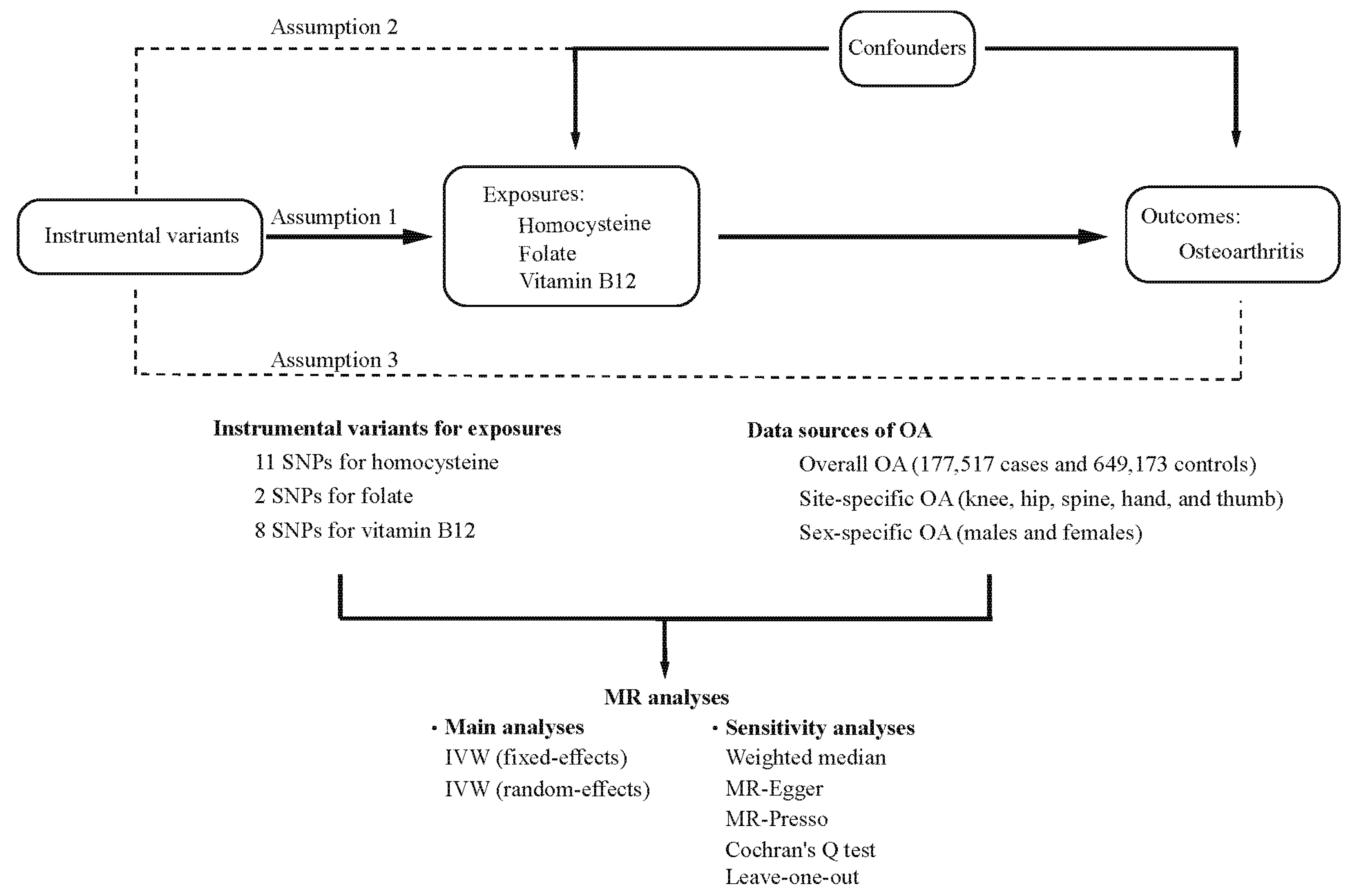
2.1. Study Design

MR analyses have 3 key assumptions: (1) IVs are strongly related to the exposure; (2) IVs should not be related to potential confounders; and (3) IVs should affect the outcome exclusively through the exposure. A sketch of the study design is illustrated in Figure 1.

2.2. Data Sources of Outcomes

Genetic data for OA were derived from the largest GWAS (genome-wide association study) to date by Boer’s group [15], including overall OA (177,517 cases and 649,173 controls) and 5 different sites of OA (knee OA, hip OA, spine OA, hand OA, and thumb OA) (Table S1). In addition, sex-specific analysis was conducted in this GWAS meta-analysis. Given that sex is related to both OA and homocysteine, genetic data for OA in each sex were also extracted from these GWAS.

2.3. Selection of Instrumental Variables

SNPs related to homocysteine, folate, and vitamin B12 were extracted from two previous GWASs, which included 44,147, 37,465, and 45,576 individuals of European ancestry [16,17].
IVs were selected according to the following criteria: (1) independent SNPs (r2 = 0.1, KB = 10,000) with genome-wide significance (p < 5 × 10⁻⁸) [18]; (2) nonrare SNPs (MAF ≥ 0.01); (3) SNPs unrelated to potential confounders by checking each of the SNPs in the PhenoScanner database (http://www.phenoscanner.medschl.cam.ac.uk/, accessed on 4 December 2022, Table S2) [19]; and (4) SNPs without reverse causality by performing the MR Steiger filtering test (Table S3) [20]. Finally, 11 SNPs for homocysteine, 2 SNPs for folate, and 8 SNPs for vitamin B12 were selected in MR analyses. Summary statistics of the selected SNPs are shown in Table 1 and Table S4.

2.4. MR Analyses

All analyses were performed in R software (version 4.1.3) using the R package “TwoSampleMR” [21]. The inverse-variance weighted (IVW) method based on the fixed-effects and random-effects model was utilized as the main analysis [22,23]. The weighted median and MR-Egger methods, which make diverse assumptions about horizontal pleiotropy, were performed as complementary methods to test the robustness of the main analysis [24,25]. Cochrane’s Q value was used to assess the heterogeneity among estimates of SNPs [26]. The intercept in MR-Egger regression and MR pleiotropy residual sum and outlier (MR-PRESSO) test were applied to identify pleiotropy [27]. The leave-one-out method was implemented by sequentially excluding each SNP to determine whether the estimates were driven by any single SNP.

3. Results

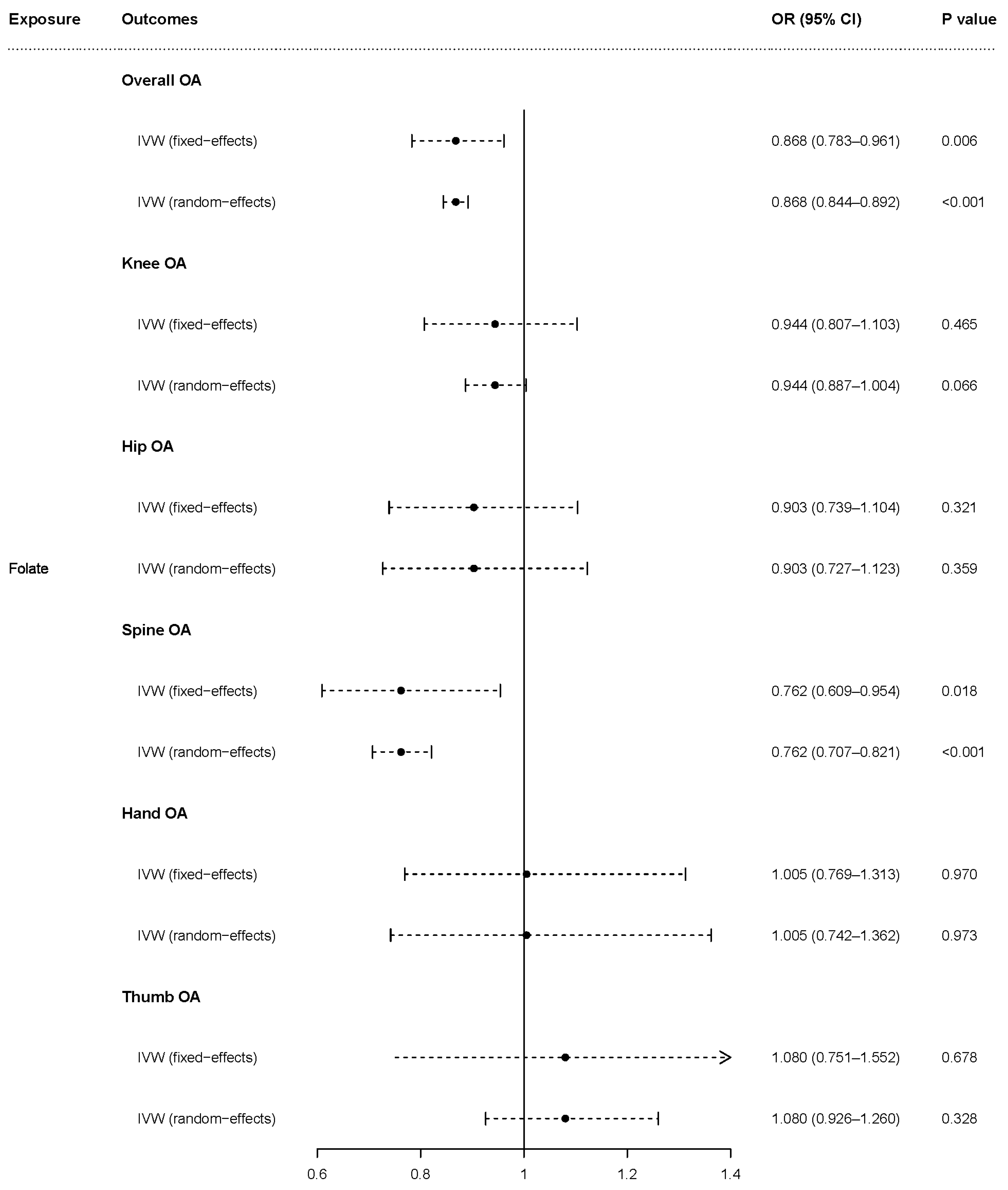
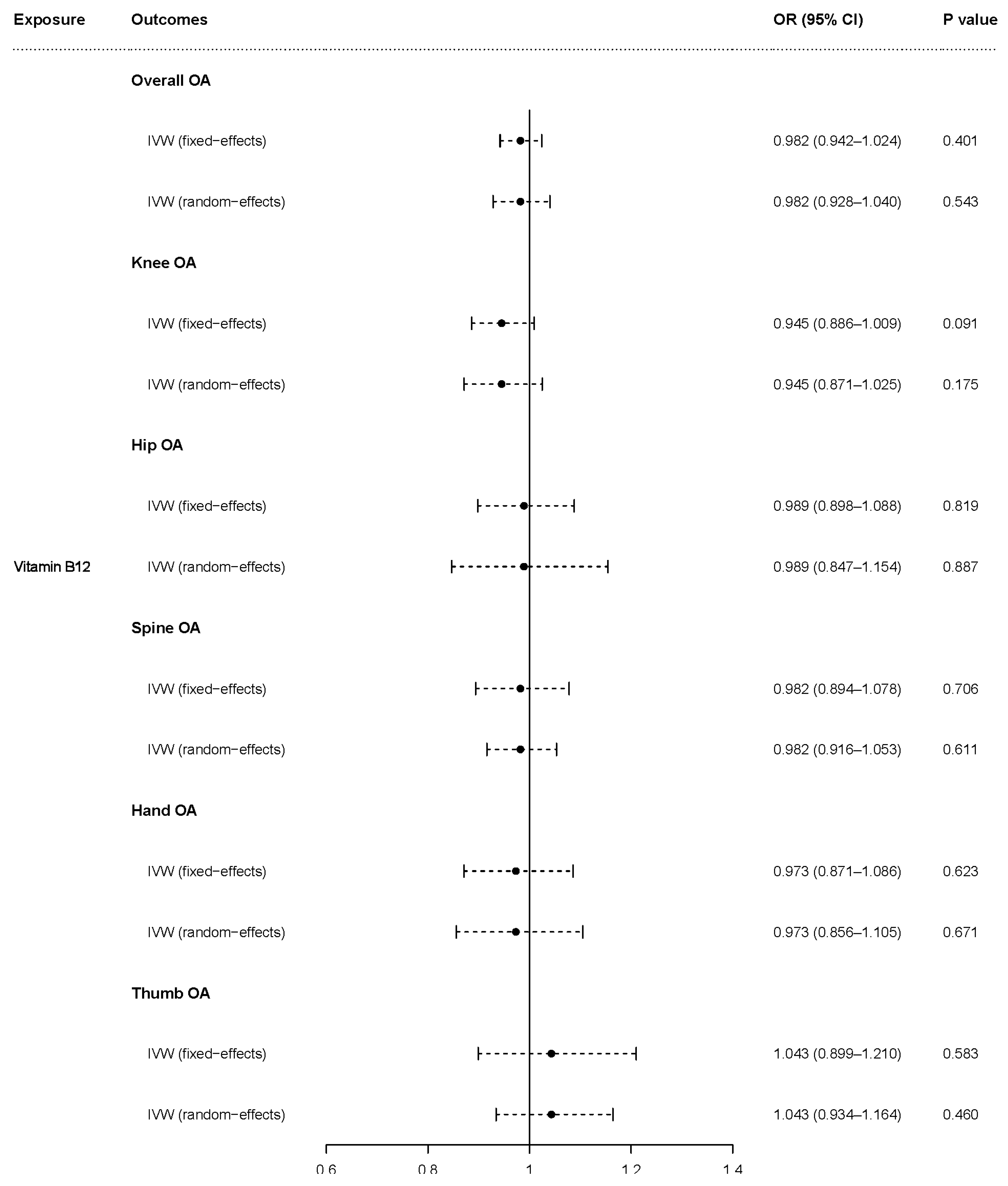
3.1. Causal Effects of Homocysteine, Folate, and Vitamin B12 on OA

The fixed-effects model of IVW analyses showed that genetically predicted homocysteine was causally associated with overall OA [odds ratio (OR) = 1.098, 95% (CI) = 1.044–1.155, p < 0.001], knee OA (OR = 1.080, 95% CI = 1.000–1.167, p = 0.050), and hip OA (OR = 1.171, 95% CI = 1.057–1.297, p = 0.002) but was not associated with hand OA (OR = 1.040, 95% CI = 0.907–1.193, p = 0.574) or thumb OA (OR = 1.057, 95% CI = 0.874–1.278, p = 0.571). The causal association of homocysteine with spine OA closely approached statistical significance (OR = 1.112, 95% CI = 0.995–1.243, p = 0.062) based on the fixed-effects model. The random-effects model, which demonstrated similar results to the fixed-effects model, showed that genetically predicted homocysteine was causally associated with overall OA (OR = 1.098, 95% CI = 1.039–1.161, p = 0.001), hip OA (OR = 1.171, 95% CI = 1.031–1.330, p = 0.015), and spine OA (OR = 1.112, 95% CI = 1.017–1.216, p = 0.020) but was not associated with hand OA (OR = 1.040, 95% CI = 0.874–1.237, p = 0.657) or thumb OA (OR = 1.057, 95% CI = 0.864–1.292, p = 0.592). Higher homocysteine levels tended to increase the risk of knee OA (OR = 1.080, 95% CI = 0.993–1.176, p = 0.074) based on the random-effects model (Figure 2).
The fixed-effects model of IVW analyses showed that genetically predicted folate was causally associated with overall OA (OR = 0.868, 95% CI = 0.783–0.961, p = 0.006) and spine OA (OR = 0.762, 95% CI = 0.609–0.954, p = 0.018) but was not associated with knee OA (OR = 0.944, 95% CI = 0.807–1.103, p = 0.465), hip OA (OR = 0.903, 95% CI = 0.739–1.104, p = 0.321), hand OA (OR = 1.005, 95% CI = 0.769–1.313, p = 0.970), or thumb OA (OR = 1.080, 95% CI = 0.751–1.552, p = 0.678). The random-effects model showed that genetically predicted folate was causally associated with overall OA (OR = 0.868, 95% CI = 0.844–0.892, p < 0.001) and spine OA (OR = 0.762, 95% CI = 0.707–0.821, p < 0.001) but not was associated with hip OA (OR = 0.903, 95% CI = 0.727–1.123, p = 0.359), hand OA (OR = 1.005, 95% CI = 0.742–1.362, p = 0.973), or thumb OA (OR = 1.080, 95% CI = 0.926–1.260, p = 0.328). The causal association of folate with knee OA closely approached statistical significance (OR = 0.944, 95% CI = 0.887–1.004, p = 0.066) based on the random-effects model (Figure 3).
The fixed-effects model of IVW analyses showed that genetically predicted vitamin B12 tended to be protective against knee OA (OR = 0.945, 95% CI = 0.886–1.009, p = 0.091) and genetically predicted vitamin B12 was not associated with overall OA (OR = 0.982, 95% CI = 0.942–1.024, p = 0.401), hip OA (OR = 0.989, 95% CI = 0.898–1.088, p = 0.819), spine OA (OR = 0.982, 95% CI = 0.894–1.078, p = 0.706), hand OA (OR = 0.973, 95% CI = 0.871–1.086, p = 0.623), or thumb OA (OR = 1.043, 95% CI = 0.899–1.210, p = 0.583). The random-effects model demonstrated that there was no association of genetically predicted vitamin B12 with overall OA (OR = 0.982, 95% CI = 0.928–1.040, p = 0.543), knee OA (OR = 0.945, 95% CI = 0.871–1.025, p = 0.175), hip OA (OR = 0.989, 95% CI = 0.847–1.154, p = 0.887), spine OA (OR = 0.982, 95% CI = 0.916–1.053, p = 0.611), hand OA (OR = 0.973, 95% CI = 0.856–1.105, p = 0.671), or thumb OA (OR = 1.043, 95% CI = 0.934–1.164, p = 0.460) (Figure 4).
Cochran’s Q (PCochran’s Q > 0.05), the MR-Egger intercept test (Pintercept > 0.05), and the MR-PRESSO global test (Pglobal test > 0.05) demonstrated no heterogeneity and no substantial horizontal pleiotropy, except for the association of vitamin B12 with hip OA (Tables S5 and S6). The results of the weighted median method, scatter plots, and leave-one-out analysis are shown in Table S7 and Figures S1–S4.

3.2. Causal Effects of Homocysteine, Folate, and Vitamin B12 on OA in Females

In females, the fixed-effects model of IVW analyses showed that genetically predicted homocysteine was associated with overall OA (OR = 1.090, 95% CI = 1.016–1.169, p = 0.016) and knee OA (OR = 1.137, 95% CI = 1.022–1.264, p = 0.018) but was not associated with hand OA (OR = 0.982, 95% CI = 0.807–1.193, p = 0.852) or thumb OA (OR = 1.003, 95% CI = 0.736–1.367, p = 0.983). The causal association of homocysteine with hip OA (OR = 1.137, 95% CI = 0.992–1.303, p = 0.064), as well as spine OA (OR = 1.156, 95% CI = 0.997–1.341, p = 0.055), closely approached statistical significance based on the fixed-effects model. The random-effects model showed that genetically predicted homocysteine was associated with overall OA (OR = 1.090, 95% CI = 1.039–1.143, p < 0.001), knee OA (OR = 1.137, 95% CI = 1.024–1.262, p = 0.016), hip OA (OR = 1.137, 95% CI = 1.027–1.258, p = 0.013), and spine OA (OR = 1.156, 95% CI = 1.048–1.275, p = 0.004) but was not associated with hand OA (OR = 0.982, 95% CI = 0.761–1.266, p = 0.886) or thumb OA (OR = 1.003, 95% CI = 0.804–1.252, p = 0.977) (Figure S5).
The fixed-effects model of IVW analyses demonstrated that there was no evidence for associations of folate with overall OA (OR = 0.902, 95% CI = 0.782–1.041, p = 0.159), knee OA (OR = 0.933, 95% CI = 0.752–1.159, p = 0.532), hip OA (OR = 0.953, 95% CI = 0.722–1.258, p = 0.734), spine OA (OR = 0.783, 95% CI = 0.580–1.059, p = 0.112), hand OA (OR = 0.961, 95% CI = 0.646–1.429, p = 0.844), and thumb OA (OR = 1.259, 95% CI = 0.671–2.361, p = 0.474) in females. The random-effects model showed that genetically predicted folate was inversely associated with overall OA (OR = 0.902, 95% CI = 0.821–0.991, p = 0.032) and knee OA (OR = 0.933, 95% CI = 0.929–0.938, p < 0.001), and the causal association of folate with spine OA (OR = 0.783, 95% CI = 0.598–1.026, p = 0.076) closely approached statistical significance. There was no evidence for associations of folate with hip OA (OR = 0.953, 95% CI = 0.711–1.276, p = 0.746), hand OA (OR = 0.961, 95% CI = 0.706–1.309, p = 0.801), and thumb OA (OR = 1.259, 95% CI = 0.826–1.917, p = 0.284) based on the random-effects model (Figure S6).
The fixed-effects model of IVW analyses showed that the causal association of vitamin B12 with knee OA in females (OR = 0.901, 95% CI = 0.808–1.004, p = 0.060) closely approached statistical significance, and there was no association of genetically predicted vitamin B12 with overall OA (OR = 0.972, 95% CI = 0.906–1.044, p = 0.437), hip OA (OR = 0.998, 95% CI = 0.868–1.147, p = 0.974), spine OA (OR = 1.012, 95% CI = 0.869–1.178, p = 0.880), hand OA (OR = 0.878, 95% CI = 0.719–1.074, p = 0.206), and thumb OA (OR = 0.973, 95% CI = 0.708–1.339, p = 0.868). The random-effects model showed that genetically predicted vitamin B12 was associated with knee OA (OR = 0.901, 95% CI = 0.822–0.987, p = 0.025) but was not associated with overall OA (OR = 0.972, 95% CI = 0.919–1.028, p = 0.325), hip OA (OR = 0.998, 95% CI = 0.864–1.152, p = 0.974), spine OA (OR = 1.012, 95% CI = 0.876–1.169, p = 0.873), hand OA (OR = 0.878, 95% CI = 0.721–1.070, p = 0.198), or thumb OA (OR = 0.973, 95% CI = 0.742–1.277, p = 0.846) (Figure S7).

3.3. Causal Effects of Homocysteine, Folate, and Vitamin B12 on OA in Males

In males, the fixed-effects model of IVW analyses showed that the causal association of homocysteine was related to overall OA (OR = 1.132, 95% CI = 1.042–1.229, p = 0.003), and the causal association of homocysteine with hip OA (OR = 1.171, 95% CI = 0.993–1.380, p = 0.061) closely approached statistical significance. There was no evidence for associations of homocysteine with knee OA (OR = 1.042, 95% CI = 0.921–1.180, p = 0.512), spine OA (OR = 1.098, 95% CI = 0.919–1.312, p = 0.305), hand OA (OR = 0.978, 95% CI = 0.716–1.336, p = 0.888), and thumb OA (OR = 1.073, 95% CI = 0.634–1.816, p = 0.792) based on the fixed-effects model. The random-effects model showed that genetically predicted homocysteine was associated with overall OA (OR = 1.132, 95% CI = 1.040–1.232, p = 0.004) but was not associated with knee OA (OR = 1.042, 95% CI = 0.914–1.189, p = 0.536), hip OA (OR = 1.171, 95% CI = 0.938–1.460, p = 0.163), spine OA (OR = 1.098, 95% CI = 0.907–1.329, p = 0.338), hand OA (OR = 0.978, 95% CI = 0.694–1.377, p = 0.898), or thumb OA (OR = 1.073, 95% CI = 0.538–2.143, p = 0.841) (Figure S8).
The fixed-effects model of IVW analyses showed that the causal association of folate was related to overall OA (OR = 0.792, 95% CI = 0.668–0.939, p = 0.007), and the causal association of folate with spine OA (OR = 0.737, 95% CI = 0.512–1.061, p = 0.100) closely approached statistical significance in males. No association was observed between folate and knee OA (OR = 0.886, 95% CI = 0.687–1.143, p = 0.351), hip OA (OR = 0.828, 95% CI = 0.591–1.160, p = 0.272), hand OA (OR = 1.221, 95% CI = 0.644–2.316, p = 0.541), or thumb OA (OR = 0.537, 95% CI = 0.180–1.606, p = 0.266) based on the fixed-effects model. The random-effects model demonstrated that genetically predicted folate was inversely associated with overall OA (OR = 0.792, 95% CI = 0.769–0.815, p < 0.001), knee OA (OR = 0.886, 95% CI = 0.788–0.996, p = 0.042), and spine OA (OR = 0.737, 95% CI = 0.569–0.954, p = 0.021), and the causal association of folate with hip OA (OR = 0.828, 95% CI = 0.661–1.037, p = 0.100) closely approached statistical significance. No association was observed between folate and hand OA (OR = 1.221, 95% CI = 0.881–1.692, p = 0.230) or thumb OA (OR = 0.537, 95% CI = 0.115–2.514, p = 0.430) based on the random-effects model (Figure S9).
The fixed-effects model of IVW analyses showed that the causal association of vitamin B12 with knee OA in males (OR = 0.891, 95% CI = 0.785–1.011, p = 0.073) closely approached statistical significance, and genetically predicted vitamin B12 was not associated with overall OA (OR = 0.971, 95% CI = 0.894–1.055, p = 0.489), hip OA (OR = 1.058, 95% CI = 0.894–1.251, p = 0.511), spine OA (OR = 0.922, 95% CI = 0.770–1.104, p = 0.378), hand OA (OR = 0.960, 95% CI = 0.699–1.318, p = 0.799), or thumb OA (OR = 0.918, 95% CI = 0.540–1.561, p = 0.752). The random-effects model showed that vitamin B12 was associated with knee OA (OR = 0.891, 95% CI = 0.818–0.971, p = 0.008) but not with overall OA (OR = 0.971, 95% CI = 0.879–1.073, p = 0.566), hip OA (OR = 1.058, 95% CI = 0.768–1.458, p = 0.731), spine OA (OR = 0.922, 95% CI = 0.818–1.040, p = 0.187), hand OA (OR = 0.960, 95% CI = 0.675–1.364, p = 0.818), or thumb OA (OR = 0.918, 95% CI = 0.663–1.270, p = 0.605) (Figure S10).

4. Discussion

In this MR study, we investigated the causal effects of homocysteine, folate, and vitamin B12 on the risk of overall and site- and gender-specific OA. The results revealed suggestive associations of higher genetically predicted homocysteine levels with increased risk of overall OA and OA at weight-bearing joints (knee, hip, and spine), especially in females. Folate and vitamin B12 had protective effects on knee OA in both males and females. Furthermore, folate was also associated with a lower risk of overall OA and spine OA in both males and females.
The present study shows that higher homocysteine levels could increase the risk of overall OA. The reason for this result is still unclear due to the lack of studies investigating the pathogenic mechanisms of homocysteine in OA. Based on the reported adverse effects of homocysteine in the literature, a possible association of homocysteine with OA could be inferred. A recent in vitro study reported that homocysteine could induce several detrimental changes in chondrocytes including mitochondrial dysfunction, apoptosis, and oxidative stress accumulation [8]. Moreover, accumulated evidence has suggested that homocysteine could affect bone metabolism and reduce bone strength [7]. Homocysteine in bone was not only bound to collagen of the extracellular matrix but also disturbed the osteoblast-mediated calcification process and stimulated osteoclast differentiation. In diet-induced HHcy in rats, homocysteine accumulated in bone was accompanied by bone loss and bone strength reduction [28]. It is well known that subchondral bone and cartilage form a functional complex called the bone–cartilage unit, which is involved in the pathophysiology of OA biochemically and mechanically [1]. Subchondral bone is an important aspect of the mechanical environment for cartilage, and its microstructure has close relationship with cartilage metabolism [29,30]. Although there are different microstructural changes in subchondral bone at different stages of OA (enhanced subchondral bone turnover in early OA and subchondral bone sclerosis in the advanced stage), subchondral bone in OA undergoes an uncoupling process at different stages during OA progression [1]. The equilibrium between osteoclast-mediated bone resorption and osteoblast-mediated bone formation is crucial for bone homeostasis, and spatiotemporal uncoupling is responsible for the degradation of subchondral bone and progression of OA [30]. These findings suggest that homocysteine plays adverse roles in collagen metabolism, chondrocyte function, and bone formation and may be involved in OA pathophysiology.
Notably, the causal effects of homocysteine on OA were site specific. Homocysteine was associated with OA in weight-bearing joints (knee, hip and spine) but not with hand OA and thumb OA. OA itself is site specific with heterogeneity in terms of prevalence, epidemiological risk factors, genetics, and etiology [1,31,32]. Mechanical factors causing the wear of cartilage and affecting the integrity of subchondral bone have been considered one of the most important factors ascribed to the site specificity of OA [33]. There are three main patterns of hand OA: the symmetrical pattern (same joint involvement from left and right hands), the row pattern (several distal interphalangeal (DIP) or proximal interphalangeal (PIP) joint involvement), and the ray pattern (DIP/PIP joint involvement of the same finger). The progression of hand OA demonstrated in a symmetrical pattern, which mainly resulted from systemic factors rather than mechanical factors [33]. However, unlike the hand joint, which is thought to be influenced mostly by systemic factors, weight-bearing joints (knee, hip, and spine) present a distinct biomechanical environment and are more susceptible to mechanical changes [34]. Mechanical factors may explain our MR results regarding homocysteine’s prominent effects in weight-bearing joints. Among the periarticular tissues, subchondral bone contributes significantly to the mechanical environment and plays different roles in the onset and progression of OA at different sites. Thus, the differential effects of homocysteine on OA in weight-bearing and non-weight-bearing joints suggest that blood homocysteine may be involved in OA pathogenesis not only through directly disturbing chondrocyte functions but also through affecting subchondral bone.
The sex-stratified MR analyses demonstrated that the causal effects of homocysteine on OA were gender specific. For females, homocysteine was associated with overall OA, knee OA, hip OA, and spine OA but not with hand OA and thumb OA. For males, homocysteine was only associated with overall OA and hip OA. Previous studies also reported sex specificity regarding the association between homocysteine and OA [9,10]. Both HHcy and OA are well-known sex-related disorders. Epidemiological studies have reported an increased prevalence of OA and HHcy in postmenopausal women. Estrogen deficiency could increase blood levels of homocysteine and induce or worsen OA [35,36,37]. Among the periarticular tissues, the subchondral bone responds most significantly to estrogen deficiency, exhibiting increased bone turnover accompanied by reduced stiffness and altered mechanical characteristics [37]. The sex-specific pattern of association seems to provide another clue that blood homocysteine levels influence OA development through bone structural alteration. Taking the site- and sex-specific patterns of homocysteine’s effects on OA together, subchondral bone is nonnegligible during OA development stimulated by homocysteine. The exact mechanisms need to be further investigated.
Since folate and vitamin B12 supplementation are well-recognized methods to reduce the risk of diseases by lowering the blood level of homocysteine, the causal effects of folate and vitamin B12 on OA were evaluated in the present study. The protective and site-specific effects of folate and vitamin B12 on OA were manifested. Sex-stratified analyses showed similar patterns in males and females. In line with our findings, previous studies also demonstrated an inverse effect of folate and vitamin B12 on OA. Folate deficiency was reported to be associated with increased radiographic severity of knee OA [38]. Knee OA patients had lower intakes of dietary folate than controls [39]. Dietary folate and vitamin B12 supplementation can improve joint functions in OA patients [40]. Rationally, folate and vitamin B12 supplementation may be an optional intervention to prevent OA.
People remain unconvinced about whether and how homocysteine and human OA are related. To date, no related RCTs have been conducted. Traditional observational and cohort studies have demonstrated inconsistent results, probably due to some confounding factors. The Framingham osteoarthritis study concluded that homocysteine was not associated with the progression of radiographic knee OA [10], whereas cross-sectional studies in Japan and China observed the correlation of circulating homocysteine levels with spinal OA and knee OA, respectively [9,12]. A recent MR study indicated that hip OA is not correlated with higher homocysteine concentrations and the related B vitamins have no causal effects on OA [41]. These results seem different from ours, which might be attributed to the choice of outcome data sources and selection of SNPs. Our MR study used the largest GWAS for OA to date, which contains the data of site- and gender-specific OA, and selected instrumental variables complying with three key assumptions in MR design. The present design might provide more comprehensive results about the potential association of homocysteine and the related B vitamins with OA.
There are several limitations in the present study. First, sensitivity analyses of the association of folate with outcomes could not be performed since only two SNPs after screening were available for folate. Second, since the individuals included in our study were mainly European individuals, our findings may not be directly generalizable to other populations. Third, since the GWAS meta-analysis of exposures did not include sex-specific genetic data, only genetic data for OA in each sex were used to conduct sex-specific analysis in our MR analyses. The sex-specific patterns of association between exposures and OA provide interesting clues and may provoke more subsequent investigations. Furthermore, GWAS meta-analysis is warranted to conduct sex-stratified analyses, especially when sex plays a role in the disorder.

5. Conclusions

This MR study provided evidence that genetically predicted homocysteine was causally associated with an increased risk of OA, especially at weight-bearing joints and in females. Folate and vitamin B12 had suggestively protective effects on OA. Homocysteine-lowering interventions, for example, through folate and vitamin B12 supplementation, may be a potential option for the treatment and prevention of OA, especially OA at weight-bearing joints and in females.

Supplementary Materials

The following supporting information can be downloaded at: https://www.mdpi.com/article/10.3390/nu15071636/s1, Figure S1: Scatter plot of the MR estimates for the association of homocysteine with the risk of OA and subtypes. (a) Overall OA; (b) knee OA; (c) hip OA; (d) spine OA; (e) hand OA; (f) thumb OA. The x-axis represents the effect size of SNPs on homocysteine; the y-axis represents the effect size of SNPs on OA and subtypes. Colors of the fitted line represent the four approaches used in univariable MR analyses. Figure S2: Leave-one-out analysis for MR analysis of the causal effect of homocysteine on OA and subtypes. (a) Overall OA; (b) knee OA; (c) hip OA; (d) spine OA; (e) hand OA; (f) thumb OA. Figure S3: Scatter plot of the MR estimates for the association of vitamin B12 with the risk of OA and subtypes. (a) Overall OA; (b) knee OA; (c) hip OA; (d) spine OA; (e) hand OA; (f) thumb OA. The x-axis represents the effect size of SNPs on vitamin B12; the y-axis represents the effect size of SNPs on OA and subtypes. Colors of the fitted line represent the four approaches used in univariable MR analyses. Figure S4: Leave-one-out analysis for MR analysis of the causal effect of vitamin B12 on OA and subtypes. (a) Overall OA; (b) knee OA; (c) hip OA; (d) spine OA; (e) hand OA; (f) thumb OA. Figure S5: Causal effect of homocysteine in OA in females in fixed-effects and random-effects IVW analyses. OR: odds ratio; CI: confidence interval; p value: p value of the causal estimate. Figure S6: Causal effect of folate in OA in females in fixed-effects and random-effects IVW analyses. OR: odds ratio; CI: confidence interval; p value: p value of the causal estimate. Figure S7: Causal effect of vitamin B12 in OA in females in fixed-effects and random-effects IVW analyses. OR: odds ratio; CI: confidence interval; p value: p value of the causal estimate. Figure S8: Causal effect of homocysteine in OA in males in fixed-effects and random-effects IVW analyses. OR: odds ratio; CI: confidence interval; p value: p value of the causal estimate. Figure S9: Causal effect of folate in OA in males in fixed-effects and random-effects IVW analyses. OR: odds ratio; CI: confidence interval; p value: p value of the causal estimate. Figure S10: Causal effect of vitamin B12 in OA in males in fixed-effects and random-effects IVW analyses. OR: odds ratio; CI: confidence interval; p value: p value of the causal estimate. Table S1: Detailed information on the genome-wide association study of OA. Table S2: Association (p < 5 × 10⁻⁸) of SNPs used as candidate genetic instruments for OA at any site with confounders or OA. Table S3: Results of overall MR Steiger direction test. Table S4: The summary information for instrumental variables of outcomes. Table S5: Cochran’s Q test for heterogeneity (MR-IVW). Table S6: Assessing directional pleiotropy through MR-Egger intercept and MR-PRESSO test. Table S7: Weighted median and MR-Egger methods for genetic associations between exposures and outcomes.

Author Contributions

Conception and design of the study—H.H., H.L. and L.C. Acquisition and analysis of data—H.H., L.C. and Y.Z. Visualization and validation—H.H. and Z.Y. Interpretation of data—H.L., C.S. and W.L. Article preparation—H.H., L.C., Y.Z. and Z.Y. Critical revision of manuscript—H.L., C.S. and W.L. Obtaining of funding—H.L. All authors have read and agreed to the published version of the manuscript.

Funding

This work was supported by the National Natural Science Foundation of China (grants Nos. 12172011, 11872076).

Institutional Review Board Statement

Not applicable.

Informed Consent Statement

Since this MR study is based on publicly available GWAS summary statistics, additional ethical approval was not needed.

Data Availability Statement

The GWAS summary data of OA was downloaded from the ‘Downloads’ page of the Musculoskeletal Knowledge Portal (https://mskkp.org, accessed on 4 December 2022), provided by Boer’s group. The GWAS summary data of folate and vitamin B12 were downloaded from GWAS Catalog website (https://www.ebi.ac.uk/gwas/, accessed on 4 December 2022).

Acknowledgments

The authors would like to express our sincere thanks to the original GWAS and the relevant consortia for providing the summary statistics.

Conflicts of Interest

All authors have no conflict of interest to disclose.

References

  1. Hunter, D.J.; Bierma-Zeinstra, S. Osteoarthritis. Lancet 2019, 393, 1745–1759. [Google Scholar] [CrossRef] [PubMed]
  2. Prieto-Alhambra, D.; Judge, A.; Javaid, M.K.; Cooper, C.; Diez-Perez, A.; Arden, N.K. Incidence and risk factors for clinically diagnosed knee, hip and hand osteoarthritis: Influences of age, gender and osteoarthritis affecting other joints. Ann. Rheum. Dis. 2014, 73, 1659–1664. [Google Scholar] [CrossRef] [PubMed] [Green Version]
  3. Martel-Pelletier, J.; Barr, A.J.; Cicuttini, F.M.; Conaghan, P.G.; Cooper, C.; Goldring, M.B.; Goldring, S.R.; Jones, G.; Teichtahl, A.J.; Pelletier, J.P. Osteoarthritis. Nat. Rev. Dis. Prim. 2016, 2, 16072. [Google Scholar] [CrossRef] [PubMed] [Green Version]
  4. Yang, B.; Fan, S.; Zhi, X.; Wang, Y.; Wang, Y.; Zheng, Q.; Sun, G. Prevalence of Hyperhomocysteinemia in China: A Systematic Review and Meta-Analysis. Nutrients 2015, 7, 74–90. [Google Scholar] [CrossRef] [Green Version]
  5. McLean, R.R.; Jacques, P.F.; Selhub, J.; Tucker, K.L.; Samelson, E.J.; Broe, K.E.; Hannan, M.T.; Cupples, L.A.; Kiel, D.P. Homocysteine as a Predictive Factor for Hip Fracture in Older Persons. N. Engl. J. Med. 2004, 350, 2042–2049. [Google Scholar] [CrossRef] [Green Version]
  6. Boushey, C.J.; Beresford, S.A.A.; Omenn, G.S.; Motulsky, A.G. A Quantitative Assessment of Plasma Homocysteine as a Risk Factor for Vascular Disease. Probable benefits of increasing folic acid intakes. JAMA 1995, 274, 1049–1057. [Google Scholar] [CrossRef]
  7. Koh, J.-M.; Lee, Y.-S.; Kim, Y.S.; Kim, D.J.; Kim, H.-H.; Park, J.-Y.; Lee, K.-U.; Kim, G.S. Homocysteine Enhances Bone Resorption by Stimulation of Osteoclast Formation and Activity through Increased Intracellular ROS Generation. J. Bone Miner. Res. 2006, 21, 1003–1011. [Google Scholar] [CrossRef]
  8. Ma, C.-H.; Chiua, Y.C.; Wu, C.-H.; Jou, I.-M.; Tu, Y.-K.; Hung, C.-H.; Hsieh, P.-L.; Tsai, K.-L. Homocysteine causes dysfunction of chondrocytes and oxidative stress through repression of SIRT1/AMPK pathway: A possible link between hyperhomocysteinemia and osteoarthritis. Redox Biol. 2018, 15, 504–512. [Google Scholar] [CrossRef]
  9. Nakano, M.; Nakamura, Y.; Urano, T.; Miyazaki, A.; Suzuki, T.; Watanabe, K.; Takahashi, J.; Shiraki, M. Associations of Homocysteine Metabolism with the Risk of Spinal Osteoarthritis Progression in Postmenopausal Women. J. Clin. Endocrinol. Metab. 2021, 106, 3428–3438. [Google Scholar] [CrossRef]
  10. Fayfman, M.; Niu, J.; Zhang, Y.; Felson, D.; Sack, B.; Aliabadi, P.; Selhub, J.; Hunter, D. The relation of plasma homocysteine to radiographic knee osteoarthritis. Osteoarthr. Cartil. 2009, 17, 766–771. [Google Scholar] [CrossRef] [Green Version]
  11. Shu, J.; Sun, X.; Li, J.; Li, F.; Tang, J.; Shi, L. Serum homocysteine levels and their association with clinical characteristics of inflammatory arthritis. Clin. Rheumatol. 2020, 39, 3295–3302. [Google Scholar] [CrossRef] [PubMed]
  12. Zhang, Q.; Li, H.; Zhang, Z.; Yang, F.; Chen, J. Serum Metabolites as Potential Biomarkers for Diagnosis of Knee Osteoarthritis. Dis. Markers 2015, 2015, 684794. [Google Scholar] [CrossRef] [PubMed] [Green Version]
  13. Smith, G.D.; Hemani, G. Mendelian randomization: Genetic anchors for causal inference in epidemiological studies. Hum. Mol. Genet. 2014, 23, R89–R98. [Google Scholar] [CrossRef] [PubMed] [Green Version]
  14. Smith, G.D.; Ebrahim, S. “Mendelian randomization”: Can genetic epidemiology contribute to understanding environmental determinants of disease? Int. J. Epidemiol. 2003, 32, 1–22. [Google Scholar] [CrossRef] [PubMed] [Green Version]
  15. Boer, C.G.; Hatzikotoulas, K.; Southam, L.; Stefánsdóttir, L.; Zhang, Y.; de Almeida, R.C.; Wu, T.T.; Zheng, J.; Hartley, A.; Teder-Laving, M.; et al. Deciphering osteoarthritis genetics across 826,690 individuals from 9 populations. Cell 2021, 184, 4784–4818.e17. [Google Scholar] [CrossRef]
  16. Van Meurs, J.B.; Pare, G.; Schwartz, S.M.; Hazra, A.; Tanaka, T.; Vermeulen, S.H.; Cotlarciuc, I.; Yuan, X.; Mälarstig, A.; Bandinelli, S.; et al. Common genetic loci influencing plasma homocysteine concentrations and their effect on risk of coronary artery disease. Am. J. Clin. Nutr. 2013, 98, 668–676. [Google Scholar] [CrossRef] [Green Version]
  17. Grarup, N.; Sulem, P.; Sandholt, C.H.; Thorleifsson, G.; Ahluwalia, T.S.; Steinthorsdottir, V.; Bjarnason, H.; Gudbjartsson, D.; Magnusson, O.T.; Sparsø, T.; et al. Genetic Architecture of Vitamin B12 and Folate Levels Uncovered Applying Deeply Sequenced Large Datasets. PLoS Genet. 2013, 9, e1003530. [Google Scholar] [CrossRef] [Green Version]
  18. Genomes Project Consortium. A map of human genome variation from population-scale sequencing. Nature 2010, 467, 1061–1073. [Google Scholar] [CrossRef] [Green Version]
  19. Kamat, M.A.; Blackshaw, J.A.; Young, R.; Surendran, P.; Burgess, S.; Danesh, J.; Butterworth, A.S.; Staley, J.R. PhenoScanner V2: An expanded tool for searching human genotype-phenotype associations. Bioinform. Oxf. Engl. 2019, 35, 4851–4853. [Google Scholar] [CrossRef] [Green Version]
  20. Hemani, G.; Tilling, K.; Davey Smith, G. Orienting the causal relationship between imprecisely measured traits using GWAS summary data. PLoS Genet. 2017, 13, e1007081. [Google Scholar]
  21. Hemani, G.; Zheng, J.; Elsworth, B.; Wade, K.H.; Haberland, V.; Baird, D.; Laurin, C.; Burgess, S.; Bowden, J.; Langdon, R.; et al. The MR-Base platform supports systematic causal inference across the human phenome. eLife 2018, 7, e34408. [Google Scholar] [CrossRef] [PubMed]
  22. Burgess, S.; Foley, C.N.; Zuber, V. Inferring Causal Relationships between Risk Factors and Outcomes from Genome-Wide Association Study Data. Annu. Rev. Genom. Hum. Genet. 2018, 19, 303–327. [Google Scholar] [CrossRef] [PubMed] [Green Version]
  23. Hartwig, F.P.; Davies, N.M.; Hemani, G.; Smith, G.D. Two-sample Mendelian randomization: Avoiding the downsides of a powerful, widely applicable but potentially fallible technique. Int. J. Epidemiol. 2016, 45, 1717–1726. [Google Scholar] [CrossRef] [PubMed] [Green Version]
  24. Bowden, J.; Davey Smith, G.; Burgess, S. Mendelian randomization with invalid instruments: Effect estimation and bias detection through Egger regression. Int. J. Epidemiol. 2015, 44, 512–525. [Google Scholar] [CrossRef] [Green Version]
  25. Bowden, J.; Smith, G.D.; Haycock, P.C.; Burgess, S. Consistent Estimation in Mendelian Randomization with Some Invalid Instruments Using a Weighted Median Estimator. Genet. Epidemiol. 2016, 40, 304–314. [Google Scholar] [CrossRef] [Green Version]
  26. Higgins, J.P.T.; Thompson, S.G.; Deeks, J.J.; Altman, D.G. Measuring inconsistency in meta-analyses. BMJ 2003, 327, 557–560. [Google Scholar] [CrossRef] [PubMed] [Green Version]
  27. Verbanck, M.; Chen, C.-Y.; Neale, B.; Do, R. Detection of widespread horizontal pleiotropy in causal relationships inferred from Mendelian randomization between complex traits and diseases. Nat. Genet. 2018, 50, 693–698. [Google Scholar] [CrossRef]
  28. Herrmann, M.; Tami, A.; Wildemann, B.; Wolny, M.; Wagner, A.; Schorr, H.; Taban-Shomal, O.; Umanskaya, N.; Ross, S.; Garcia, P.; et al. Hyperhomocysteinemia induces a tissue specific accumulation of homocysteine in bone by collagen binding and adversely affects bone. Bone 2009, 44, 467–475. [Google Scholar] [CrossRef]
  29. Zhao, Z.; Tan, Q.; Jiang, A.; Sun, S.; Liu, Z.; Li, W.; Song, C.; Leng, H. Evidence of subchondral bone’s effects on articular cartilage damage in OVX-OA rat. Eng. Fract. Mech. 2020, 233, 107081. [Google Scholar] [CrossRef]
  30. Jiang, A.; Xu, P.; Yang, Z.; Zhao, Z.; Tan, Q.; Li, W.; Song, C.; Dai, H.; Leng, H. Increased Sparc release from subchondral osteoblasts promotes articular chondrocyte degeneration under estrogen withdrawal. Osteoarthr. Cartil. 2022, 31, 26–38. [Google Scholar] [CrossRef]
  31. Hosnijeh, F.S.; Kavousi, M.; Boer, C.; Uitterlinden, A.; Hofman, A.; Reijman, M.; Oei, E.; Bierma-Zeinstra, S.; van Meurs, J. Development of a prediction model for future risk of radiographic hip osteoarthritis. Osteoarthr. Cartil. 2018, 26, 540–546. [Google Scholar] [CrossRef] [PubMed] [Green Version]
  32. MacGregor, A.J.; Li, Q.; Spector, T.D.; Williams, F. The genetic influence on radiographic osteoarthritis is site specific at the hand, hip and knee. Rheumatology 2009, 48, 277–280. [Google Scholar] [CrossRef] [Green Version]
  33. Bijsterbosch, J.; Meulenbelt, I.; Watt, I.; Rosendaal, F.R.; Huizinga, T.W.J.; Kloppenburg, M. Clustering of hand osteoarthritis progression and its relationship to progression of osteoarthritis at the knee. Ann. Rheum. Dis. 2014, 73, 567–572. [Google Scholar] [CrossRef]
  34. Stürmer, T.; Günther, K.-P.; Brenner, H. Obesity, overweight and patterns of osteoarthritis: The Ulm Osteoarthritis Study. J. Clin. Epidemiol. 2000, 53, 307–313. [Google Scholar] [CrossRef] [PubMed]
  35. Mijatovic, V.; Kenemans, P.; Jakobs, C.; van Baal, W.M.; Peters-Muller, E.R.; van der Mooren, M.J. A randomized controlled study of the effects of 17beta-estradiol-dydrogesterone on plasma homocysteine in postmenopausal women. Obstet. Gynecol. 1998, 91, 432–436. [Google Scholar] [CrossRef] [PubMed]
  36. Gaikwad, N.W. Mass spectrometry evidence for formation of estrogen–homocysteine conjugates: Estrogens can regulate homocysteine levels. Free. Radic. Biol. Med. 2013, 65, 1447–1454. [Google Scholar] [CrossRef]
  37. Castañeda, S.; Vicente-Rabaneda, E.F. Disentangling the molecular interplays between subchondral bone and articular cartilage in estrogen deficiency-induced osteoarthritis. Osteoarthr. Cartil. 2023, 31, 6–8. [Google Scholar] [CrossRef]
  38. Xu, H.; Shin, M.-H.; Kang, J.-H.; Choi, S.-E.; Park, D.-J.; Kweon, S.-S.; Lee, Y.-H.; Kim, H.-Y.; Lee, J.-K.; Lee, S.-S. Folate deficiency is associated with increased radiographic severity of osteoarthritis in knee joints but not in hand joints. Clin. Exp. Rheumatol. 2022; ahead of print. [Google Scholar] [CrossRef]
  39. Amirkhizi, F.; Ghoreishy, S.M.; Hamedi-Shahraki, S.; Asghari, S. Higher dietary phytochemical index is associated with lower odds of knee osteoarthritis. Sci. Rep. 2022, 12, 9059. [Google Scholar] [CrossRef]
  40. Flynn, M.A.; Irvin, W.; Krause, G. The effect of folate and cobalamin on osteoarthritic hands. J. Am. Coll. Nutr. 1994, 13, 351–356. [Google Scholar] [CrossRef]
  41. Fu, L.; Wang, Y.; Hu, Y.-Q. Causal effects of B vitamins and homocysteine on obesity and musculoskeletal diseases: A Mendelian randomization study. Front. Nutr. 2022, 9, 2741. [Google Scholar] [CrossRef] [PubMed]
Figure 1. An overview of the study design. SNP: single nucleotide polymorphism; OA: osteoarthritis; MR: Mendelian randomization; IVW: inverse-variance weighted.
Nutrients 15 01636 g001
Figure 2. Causal effect of homocysteine on OA and subtypes in fixed-effects and random-effects IVW analyses. OR: odds ratio; CI: confidence interval; p value: p value of the causal estimate.
Nutrients 15 01636 g002
Figure 3. Causal effect of folate on OA and subtypes in fixed-effects and random-effects IVW analyses. OR: odds ratio; CI: confidence interval; p value: p value of the causal estimate.
Nutrients 15 01636 g003
Figure 4. Causal effect of vitamin B12 on OA and subtypes in fixed-effects and random-effects IVW analyses. OR: odds ratio; CI: confidence interval; p value: p value of the causal estimate.
Nutrients 15 01636 g004
Table 1. Characteristics of SNPs for homocysteine, folate, and vitamin B12.
TraitSNPChromosome:PositionGeneEAOAEAFBetaSEp
Homocysteiners95714011:89201627NOX4AG0.45−0.0450.0082.43 × 10−10
rs93698986:49382193MUTAG0.620.04490.0072.17 × 10−10
rs713028411:89148372NOX4TC0.07−0.12420.0131.88 × 10−20
rs46603061:45978675MMACHCTC0.330.04350.0072.33 × 10−9
rs426487:89977760GTPBP10AG0.40−0.03950.0071.97 × 10−8
rs285139121:44487404CBSTC0.470.0560.0081.70 × 10−12
rs23470921:44486964CBSTC0.45−0.07180.0073.90 × 10−24
rs22755651:237048676MTRTG0.21−0.05420.0091.96 × 10−10
rs180122210:17156151CUBNAG0.340.04530.0078.43 × 10−10
rs1278084510:17223244CUBNAG0.650.05290.0097.80 × 10−10
rs121346631:11838646MTHFRAC0.80−0.1010.0112.54 × 10−21
Folaters174215111:11857788MTHFRGA0.8270.0980.0121.80 × 10−15
rs65219711:71849741FOLR3CT0.1790.0690.0112.50 × 10−10
Vitamin B12rs575323122:31003069TCN2CT0.7900.0640.0107.50 × 10−10
rs5607712210:17207015TRDMT1AC0.3350.0870.0094.80 × 10−21
rs4128111213:100518634CLYBLCT0.9480.170.0169.60 × 10−27
rs3452891211:59631535TCN1TC0.0360.170.0212.10 × 10−15
rs233657319:8367709CD320TC0.0310.320.0211.10 × 10−51
rs180122210:17156151CUBNGA0.5930.110.0071.10 × 10−52
rs11745605311:59616831TCN1GA0.9760.160.0271.90 × 10−9
rs11413216:49412433MUTCT0.6270.0610.0071.40 × 10−16
Gene = nearest gene to the SNP; EA = effect allele; EAF, effect allele frequency; beta = per allele effect on exposures; SE = standard error; p = p value for the genetic association.
Disclaimer/Publisher’s Note: The statements, opinions and data contained in all publications are solely those of the individual author(s) and contributor(s) and not of MDPI and/or the editor(s). MDPI and/or the editor(s) disclaim responsibility for any injury to people or property resulting from any ideas, methods, instructions or products referred to in the content.

