Article

Malnutrition Prevention after Allogeneic Hematopoietic Stem Cell Transplantation (alloHSCT): A Prospective Explorative Interventional Study with an Oral Polymeric Formulation Enriched with Transforming Growth Factor Beta 2 (TGF-β2)

1
Department of Clinical and Experimental Sciences, University of Brescia, Bone Marrow Transplant Unit, ASST-Spedali Civili di Brescia, 25123 Brescia, Italy
2
Dietetics and Clinical Nutrition Unit, ASST-Spedali Civili Brescia, 25123 Brescia, Italy
3
Gastroenterology Unit, ASST-Spedali Civili Brescia—University of Brescia, 25123 Brescia, Italy
4
Central Laboratory, ASST-Spedali Civili Brescia, 25123 Brescia, Italy
*
Authors to whom correspondence should be addressed.
Nutrients 2022, 14(17), 3589; https://doi.org/10.3390/nu14173589
Submission received: 27 July 2022 / Revised: 24 August 2022 / Accepted: 25 August 2022 / Published: 31 August 2022

Abstract

:
Malnutrition is common after allogeneic Hematopoietic Stem Cell Transplantation (alloHSCT), and interventions directed to correct nutritional status are warranted to improve transplant outcomes. In this prospective study, an oral polymeric formulation enriched with TGF-β2 (TE-OPF) was explored to correct malnutrition according to Patient-Generated Subjective Global Assessment (PG-SGA). TE-OPF was proposed to 51 consecutive patients who received transplants at our institution for hematological malignancies, and sufficient dose intake was established per protocol as at least 50% of the prescribed dose of TE-OPF: group A received adequate nutritional support; group B, inadequate. The study met the primary outcomes in terms of safety (no adverse events reported during TE-OPF intake except for its disgusting taste) and malnutrition (PG-SGA C 28 days after transplant): severely malnourished patients (PG-SGA C) accounted for 13% in group A and 88.9% in group B (p = 0.000). At the end of the study, after a median follow-up of 416 days, the estimated median Overall Survival (OS) was 734 days for well or moderately nourished patients (PG-SGA A/B) in comparison to 424 for malnourished patients (p = 0.03). Inadequate TE-OPF intake was associated with an increase in acute gastrointestinal Graft Versus Host Disease (GVHD) cumulative incidence (38% vs. 0% p = 0.006). A higher incidence of pneumonia was reported in group B (p = 0.006). IGF-1 levels at 14 and 28 days after transplant were significantly higher in group A and were associated with a lower incidence of acute GVHD (aGVHD). Higher subsets of B, T, and NK cells were found in group A, and a higher number of CD16+ NK cells was associated with a lower incidence of acute GVHD (p = 0.005) and increased survival at the end of the study (p = 0.023). Artificial neural network analysis suggested that inadequate TE-OPF intake, pneumonia, and sepsis significantly affected malnutrition 28 days after alloHSCT and survival 365 days after alloHSCT (normalized importance 100%, 82%, and 68%, respectively). In this exploratory and preliminary study, the use of TE-OPF appeared to reduce the incidence of malnutrition after alloHSCT, but larger and controlled studies are required.

1. Introduction

The symptomatic burden of allogeneic hematopoietic stem cell transplantation (allo-HSCT) can severely affect food intake and nutritional status [1,2,3,4]. Severe malnutrition puts transplant outcomes at risk, prolonging the time to engraftment and increasing the risk of infection, as well as the duration of hospitalization and mortality [5,6,7,8].
Nutritional assessment and nutritional support are therefore crucial to manage transplant patients and improve the transplant outcome [7,8,9,10]. This aspect is even more important if we consider that over the last ten years, the age limit for transplantation has risen to the age of 75, and patients older than 60 years old who have received transplants exceed 30% [11].
The Patient-Generated Subjective Global Assessment (PG-SGA) questionnaire is recommended as a standard tool for nutritional screening, assessment, monitoring, and triaging for nutritional interventions in patients with cancer [12].
Regarding nutritional support, enteral nutrition (EN) is generally considered the best approach in oncology, but, if that cannot be pursued, parenteral nutrition (PN) is recommended [13]. In HSCT, PN is generally reserved in the case of severe mucositis (Grade 3–4), ileus, or intractable emesis [13] and should be continued until the resolution of the complications [14]. Although ASPEN and ESPEN guidelines recommend EN as the best approach in nutritional support in HSCT [14,15], parenteral nutrition is still most widely used in HSCT settings [16]. Furthermore, it is well known that PN may be associated with gut mucosal atrophy, metabolic and hepatic complications, central venous catheter infections, fluid overload, and hyperglycemia, which also increase the risk of systemic infections and inflammation [10,16,17,18,19].
GVHD is the main cause of transplant-related morbidity and mortality after disease relapse [20]. From a clinical and pathogenetic point of view, gastrointestinal (GI) acute GVHD has many aspects in common with chronic inflammatory bowel disease (IBD), such as the loss of the intestinal epithelial barrier, alterations in the intestinal microbiota, and the use of immunosuppressive therapies for clinical interventions [20,21,22,23,24,25,26,27]. In an IBD setting, TGF-β2 is the most studied bioactive peptide for nutritional support [28]. TGF-β2 is an anti-inflammatory cytokine and a key modulator of the microbiota. It also has a relevant role in host immune cell crosstalk, it controls the differentiation, proliferation, and activation state of lymphocytes, macrophages, and dendritic cells, and it plays a critical role in the mechanisms of tolerance, prevention of autoimmunity, and in anti-inflammatory processes in the gut [28,29].
In this prospective interventional study, 51 patients who consecutively underwent allo-HSCT were evaluated from a nutritional point of view and supported with an Oral Polymeric Formulation Enriched with TGF-β2 (TE-OPF), commonly used in IBD settings.

2. Materials and Methods

The study was approved by the local ethical committee in March 2020. Patients signed informed consent and underwent nutritional evaluation by a dietitian. PG-SGA score nutritional support with an oral polymeric formulation enriched with TGF-β2 (MODULEN-IBD®) was proposed to all enrolled patients. Patients older than 18 years submitted to alloHSCT were included in the study, while exclusion criteria were due to unsigned informed consent, gastrointestinal perforations and fistulas, and intractable vomiting. The primary outcome of the study was to reduce the incidence of severe malnutrition (PG-SGA C, see below) 28 days after allo-HSCT to less than 50% in comparison to a historical control (in which the incidence of severe malnutrition at +28 days after allo-HSCT was 75% [30]. The study was closed when at least 24 subjects had taken at least 50% of the prescribed TE-OPF (sample size calculation with a power of 75% and an alpha level of 0.05). The baseline characteristics of the 51 enrolled patients are displayed in Table 1.
Malnutrition impact and clinical outcomes were explored according to TE-OPF intake in percentage based on the prescribed dose (treatment ratio—TR). No minimal dose was defined as clinically effective, but in the protocol design, 50% of the prescribed TE-OPF was considered adequate (Group A, Treatment Ratio TR > 50% of the prescribed dose); Group B was comprised of patients who did not take an adequate dose of TE-OPF. The Mann–Whitney test was used for statistical analysis regarding the correlations among TR and malnutrition, acute GVHD onset, clinical events (sepsis or pneumonia), and biomarker studies; for biomarker studies, Kaplan–Meier plots were defined according to the 25th percentile rounded to the nearest whole number. Overall survival 365 days after transplant was estimated according to Kaplan–Meier curves, and the log-rank test was used for univariate analysis. The cumulative incidence of acute GVHD was calculated months months after transplantation. In the log-rank and Fisher tests, patients were grouped according to the treatment ratio (TR) (TR> or <50% based on the study protocol, Group A and Group B, respectively). No differences were found in patients’ characteristics at admission for transplant between the groups (Table 2).
Nutritional status was assessed through the PG-SGA questionnaire [12,14] at admission and on day 0, +7, +14, +21, and +28 from transplant (day 0 was the transplant day). PG-SGA is composed of an objective section and a patient-reported one. The first reports the overall nutritional status with an alphabetical score (A = good nutritional status; B = moderate malnutrition; C = severe malnutrition). The second one is a numeric score that is calculated from four items reported by patients (weight loss, food intake, symptoms with a nutritional impact, and physical activity). Integration with Parenteral Nutrition (PN) was permitted when oral food intake was less than 60% of hospital meals for at least three days and/or oral mucositis grade 3–4, intractable diarrhea, and emesis were present, according to the criteria reported in ESPEN Guidelines 2009 [13].
Full blood counts, flow cytometric lymphocyte subpopulations’ absolute count (28 days after transplant), and serum IGF-1 levels with Diasorin “Liaison IGF-1” chemoimmunoluminescence analysis (14 and 28 days after transplant) were explored as potential biomarkers according to preliminary data [31]. Continuous variables were categorized at the 25th percentile for log-rank and Chi-square test analyses.
Multivariable analysis was performed with artificial neural network analysis in order to explore the relative importance of the several variables for clinical outcomes (malnutrition and survival at 365 days after transplantation [32].

TE-OPF Treatment Plan

The TE-OPF was diluted in a bottle of mineral water and proposed one or more times per day if necessary. The nutritional support started at admission and was carried out until discharge from allo-HSCT. Refusal and reasons for refusal of the proposed treatment were recorded. The amount of calories derived by oral supplement was calculated at admission based on on BMI and total daily energy expenditure (TDEE) by multiplying the basal metabolic rate (the Mifflin St. Jeor BMR estimation formula was used) by 1.3 (Ppysical activity level). From admission to the day of transplant, the amount of calories derived from oral formulation enriched with Transforming Growth Factor beta 2 was as follows:
-
BMI less than 22: 20% of TDEE;
-
BMI between 22.1 and 24.9: 12% of TDEE;
-
BMI between 25 and 29.9: 10% of TDEE;
-
BMI between 30 and 34.9: 8% of TDEE;
-
BMI higher than 35: 5% of TDEE.
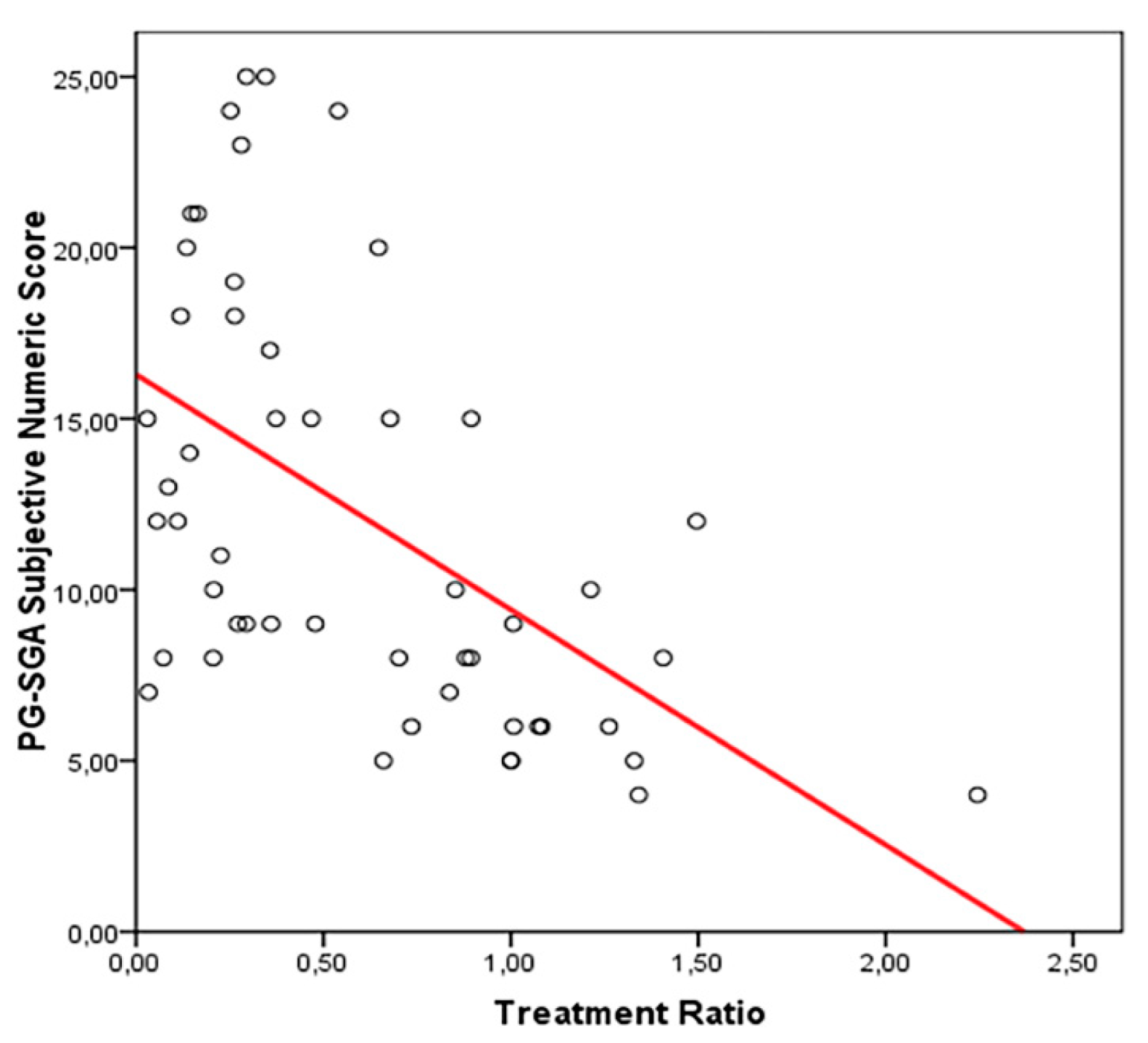
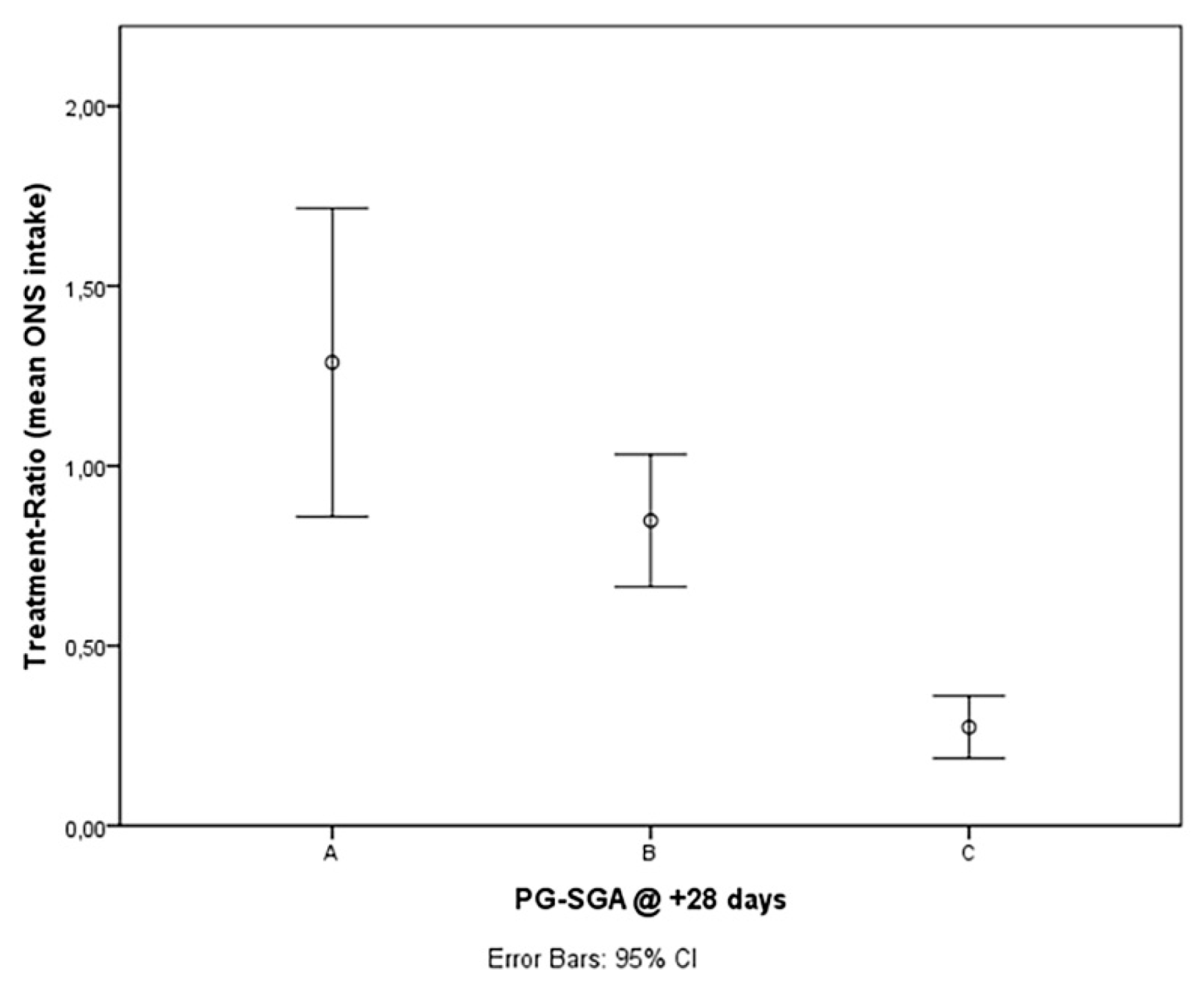
From day 1 to day +28 from transplantation, the quantity of supplement was increased by 10% compared to the initial dose if the patient showed a weight loss of less than 5% during hospitalization; with a weight loss of more than 5%, the dose of oral formulation was increased by 20% compared to the starting dose. Once reconstituted by the staff, the supplement could be flavored according to the patient’s taste with barley coffee or decaffeinated coffee. Compliance with TE-OPF intake was investigated with a specific questionnaire, and the percentage of administered dose compared to the prescribed dose was registered daily (Treatment Ratio—TR). Adverse events were reported according to good clinical practice (GCP).

3. Results

Primary declared outcomes were safety of TE-OPF and malnutrition 28 days after transplantation defined as PG-SGA C 28 days after transplantation.
No adverse events were attributed to TE-OPF. Patients refused the preparation due to a disgusting sensation in 19/27 cases (70%), mucositis in 10/27 (37%) or respiratory complications involving the use of non-invasive ventilation in 5/27 (18.5%). The mean assumption according to the prescribed dose (at least 50%), was 60%, median 46%, minimum 3% to maximum 224%.
The percentage of severely malnourished patients (PG-SGA C) after 28 days was significantly lower in the group with a treatment ratio (TR) higher than 50% (GROUP A) in comparison to the group with insufficient TE-OPF intake (GROUP B—TR < 50%) (12.5% vs. 88.9% p = 0.000) (Table 1). The study met the outcome criteria and was stopped.
In group A, 7 out 20 patients (30.4%) were well nourished (PG-SGA A) in comparison to 0 in the group with an inadequate intake of TE-OPF (Group B). No statistical differences were found in baseline patients’ characteristics at admission in both groups (Table 2). No statistically significant correlation was found between the type of conditioning and nutritional status at day +28 from allo-HSCT.
The percentage of the prescribed dose ranged from 3% to 224%, with a median of 46%. Higher TR, less subjective PG-SGA values (patient-reported numeric score) were registered 28 days after alloHSCT (correlation R2 0.288, p = 0.000) (Figure 1).
The mean percentage of TR (TE-OPF assumption as a percentage of the prescribed dose) in the group with PG-SGA scores A, B, and C at 28 days after transplantation was 129%, 85%, and 27% respectively. The difference was proved to be statistically significant (between A and B, p = 0.045 and between both A + B and C, p = 0.000; Mann–Whitney test) (Figure 2).
Study analysis was performed one year after the last patient enrolment with a median follow-up of 416 days (29–784). The estimated median OS of the whole population was 649 days (456–841), whereas patients in group A experienced an estimated median survival of 734 days in comparison to 424 in group B (p = NS). The estimated median OS was 734 days for patients achieving the primary study outcome (well or moderately nourished 28 days after transplant—PG-SGA A/B) in comparison to 424 for malnourished patients (p = 0.03) (Figure 3).

3.1. Secondary Outcomes

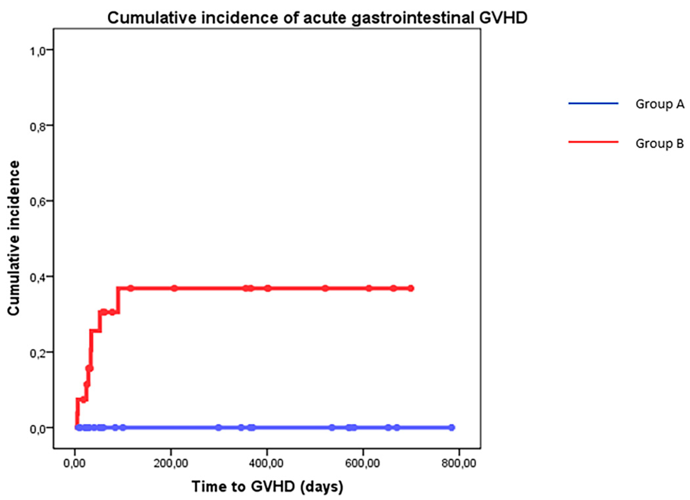
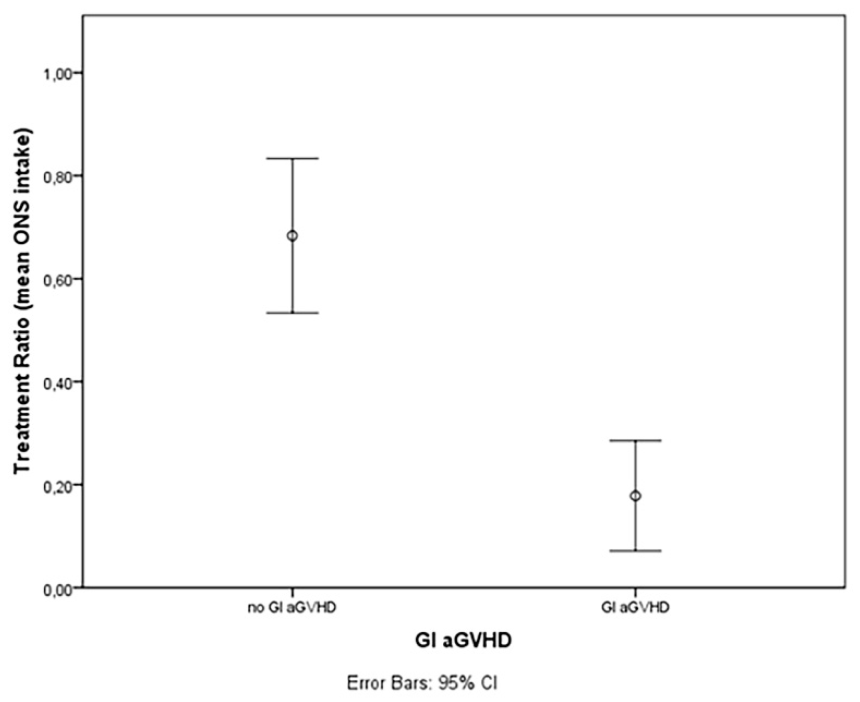
3.1.1. TE-OPF Assumption and GVHD

The estimated cumulative incidence of aGVHD Magic B or higher according to the MAGIC Consortium Criteria was similar in patients treated with adequate TE-OPF (Group A) in comparison to patients who received less than 50% of the prescribed dose (19% vs. 44% p = NS). Gastrointestinal GVHD (all grade) was reported only in patients belonging to group B (cumulative incidence of 38% vs. 0% p = 0.006) (Figure 4). Mean TE-OPF assumption was 68.3% of the prescribed dose in patients who did not experience acute GVHD and 17.8% in patients with aGVHD (Figure 5, p = 0.002).
Chronic GVHD cumulative incidence was 20%, and no differences were reported in the different treatment groups.

3.1.2. TE-OPF Assumption and Infectious Complications

Pneumonia was more frequent in group B patients (p = 0.006, 48.1% vs. 12.5%). Sepsis and enteritis were more frequent in group B, but the difference was not significant (55.5% vs. 29.1%, 37% vs. 16.6%, respectively) (Table 3).

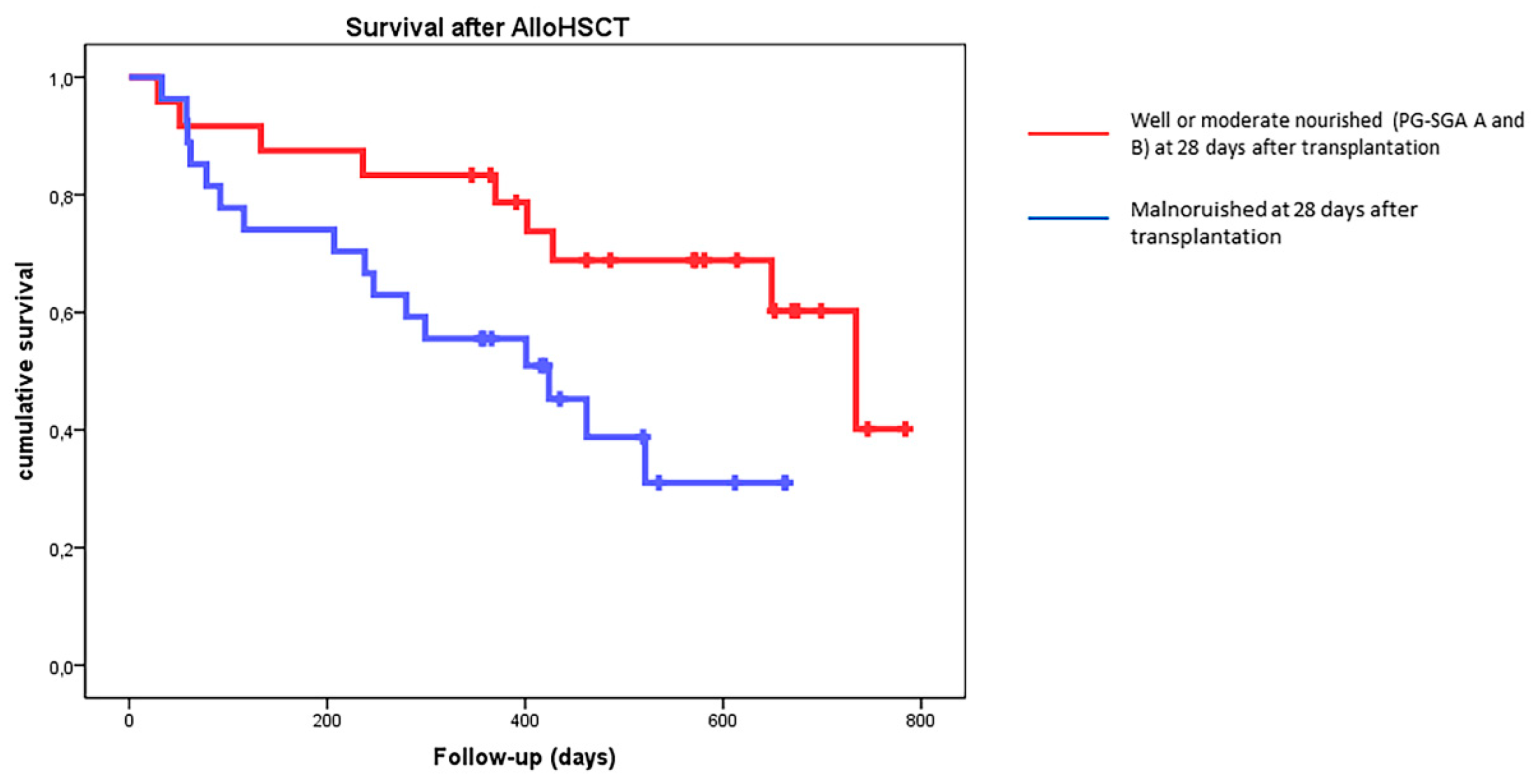
3.1.3. TE-OPF Assumption and Relapse-Free Survival

The relapse incidence was 25.9% in group A and 29.1% in group B (p = NS).

3.2. Biomarker Studies

3.2.1. TE-OPF and IGF-1

The mean IGF-1 was higher in group B at 14 and 28 days after transplantation (109 vs. 165 and 109 vs. 153, p = 0.024 and 0.013, respectively; normal range 99–364 ng/mL). Cumulative incidence of aGVHD was lower if values of IGF-1 at 14 days after transplant were higher than 100 ng/mL (p = 0.045). Cumulative incidence of acute gastrointestinal GVHD was lower in patients with higher values of IGF-1 at 14 and 28 days (p = 0.021 and 0.032, respectively).

3.2.2. TE-OPF, Malnutrition, and Lymphocyte Subsets

In group A, levels of total lymphocytes, B, T, and NK cells (mature and immature) 28 days after alloHSCT were higher than those in group B as reported in Table 4. The difference was significant (T-test) for CD16+ NK cells (p = 0.03) and CD3+ /CD4+ T cells (p = 0.025). Well-nourished patients (PG-SGA A and B) experienced a significantly higher number of CD16+ NK cells (p = 0.016), and a higher number of CD16+ NK cells (>100/mcL) was associated with a reduction in the cumulative incidence of acute GVHD (20% vs. 69% at 120 days after transplantation, p = 0.005) and an increased survival (median not reached vs. 734 days, p = 0.023).
Multivariable analysis was performed according to the method described by Caocci et al. [32] through artificial neural network analysis, and the normalized importance of several variables is reported: group A, pneumonia and sepsis significantly affected malnutrition 28 days and survival after one year (normalized importance: 100%, 82%, and 68%, respectively).

4. Discussion

The main objective of this prospective interventional study was to evaluate the incidence of severe malnutrition (PG-SGA C) at 28 days after alloHSCT in a cohort of 51 patients supported, from admission to day +28, with an oral polymeric nutritional supplement enriched with TGF-β2.
The use of this support is approved by the ACBS and NICE for “Crohn’s Disease active phase, and in remission if malnourished”, and this is the first prospective study to test it in the setting of malnutrition after alloHSCT.
One randomized clinical study showed that exclusive enteral nutrition with MODULEN-IBD® is more efficient to achieve mucosal healing than corticosteroids, proving in addition that this TE-OPF has trophic, anti-inflammatory, and microbiota-modulating effects on the gastrointestinal tract [33]. The anti-inflammatory effect of MODULEN-IBD® is attributable to the high levels of TGF-β: an immunosuppressive cytokine involved in the development and functions of immune cells, including T and B cells and also dendritic cells (DCs) [29].
In this prospective study, early assumption of TE-OPF was feasible and safe. Patients who took at least the half of the prescribed protocol dose achieve a reduced incidence of severe malnutrition 28 days after transplant according to the study design. The mean percentage of the Treatment Ratio (TR) in the group with PG-SGA score A at +28 days from allo-HSCT (well nourished) was 129%, in the group with PG-SGA score B (moderate malnutrition), was 85%; and in the group with PG-SGA C (severe malnutrition), 27%. The difference was proved to be statistically significant (between PG-SGA A and PG-SGA B, p = 0.045 and between both PGSGA A + B and PG-SGA C, p = 0.000; Mann–Whitney Test) and the effect on nutritional status may be dose dependent (Figure 1). In the subjective load of gastrointestinal symptoms, the higher the intake of TE-OPF, the lower the PG-SGA numeric score (Figure 2); the higher the score, the worse the nutritional status of the patients.
Acute GVHD was reported more frequently in patients with an inadequate assumption of TE-OPF, especially gastrointestinal GVHD that was absent in patients who took at least 50% of the prescribed dose. These preliminary results seem to confirm that the effect of this TE-OPF could be similar to that observed in IBD due to microbiota-modulating, anti-inflammatory, and trophic effects. As explained in the introduction, in both IBD and alloHSCT settings, malnutrition and the immunity response of the gastrointestinal tract are relevant. From both a clinical and pathogenetic point of view, gastrointestinal acute GVHD shares many aspects with chronic inflammatory bowel diseases (IBD), such as the loss of the intestinal epithelial barrier; alterations in the intestinal microbiota; cascade of T cell activation, proliferation, and cytotoxic activity; and the use of immunosuppressive therapies [22,25]. In bone marrow transplantation, acute GVHD may appear in 20–40% of patients with matched related donors, and in more than 50% of haploidentical stem cell transplants. Regarding gastrointestinal acute GVHD, up to 50% of patients with acute GVHD experience gastrointestinal symptoms [34]. All of these observations allow us to hypothesize that MODULEN-IBD® may also reduce the risk of GI GVHD thanks to its trophic, anti-inflammatory, and microbiota-modulating effects on the gastrointestinal tract. Reducing GI GVHD cumulative incidence was not the primary goal of the study, but patients who took more than 50% of the prescribed TE-OPF did not experience GI aGVHD (Figure 4), and the effect seemed to be dose dependent (Figure 5). Explaining the reasons with these data is not easy. It may be essential to evaluate the role of a prophylactic and earlier oral intake of MODULEN-IBD® from admission to +28 days from admission. This choice probably avoided or reduced microbiota dysbiosis: an important risk factor for GI GVHD [35,36,37]. Furthermore, the loss of microbiota diversity and the reduction of short chain fatty acid production have been observed in patients with GVHD [35,36,37]. Acetate, propionate, and butyrate are metabolites produced by microbiota and play an important role in the interaction between the microbiota and host immune cells, influencing systemic autoimmune responses and participating in different steps of inflammation processes [35,38,39]. MODULEN-IBD® administration in patients submitted to allo-HSCT may reduce the risk of GI GVHD due to its microbiota-modulating, anti-inflammatory, and trophic effects. Regarding intestinal trophism, patients with mucositis often refuse food and this further worsens intestinal dysbiosis. However, thanks to the consistency of MODULEN-IBD®, patients often continue to take an oral nutritional supplement despite oral pain, avoiding fasting. This may allow patients to maintain microbiota diversity.
Biomarkers’ data seemed to confirm the role of IGF-1 [31] as potential biomarker in this setting, suggesting that higher values (more frequent in well-nourished patients) seem to be protective for acute gastrointestinal GVHD, as reported for other chronic illness [40,41,42,43]. An increase in lymphocytes was reported according to TE-OPF consumption, which was more evident for NK subpopulations. Expansion of mature CD16+ NK in this population seemed to correlate with clinical outcomes such as acute GVHD and survival one year after transplant: recently, a model of NK expansion after UCB transplantation after TGF-β and IGF-1 [44] was reported, and the increase in CD16 cells after transplantation should be further explored.
Artificial neural network analysis suggests that consuming more than 50% of the prescribed dose seems to be an independent protective factor for malnutrition and one-year survival after alloHSCT.

5. Conclusions

In this exploratory and prelminary study, an early and prophylactic oral nutritional support, started at admission to day 28 from transplantation, reduced the incidence of severe malnutrition after four weeks (PG-SGA C) in patients submitted to allo-HSCT without side effects. TE-OPF seems to have a protective role also in the prevention of gastrointestinal GVHD and infections, possibly due to anti-inflammatory, immunomodulating, and trophic effects on the gastrointestinal tract and microbiota, but due to the investigative nature of the study, larger randomized studies are warranted.

Author Contributions

Conceptualization, E.M., F.A., M.M., M.A., C.R. and D.R.; methodology, E.M.; software, E.M., L.D., D.L. and F.A.; investigation, E.M., A.B., E.S., F.A., S.B., M.F., N.P., E.C., F.C., T.Z., E.A.B. and A.L.; data curation, E.M., F.A., A.B., E.S., F.A., S.B., M.F., N.P., E.C., F.C., T.Z., E.A.B. and A.L.; writing—original draft preparation, E.M., F.A., S.B. and D.R.; visualization, E.M., S.B. and D.R.; supervision, E.M. and D.R.; project administration, E.M. and D.R.; funding acquisition, E.M. and D.R. All authors have read and agreed to the published version of the manuscript.

Funding

This research was funded by Nestlè Health Science (Italy) with a study grant.

Institutional Review Board Statement

The study was conducted in accordance with the Declaration of Helsinki and was approved by the Institutional Ethics Committee of ASST-Spedali Civili di Brescia (protocol code NP3832 and date of approval 27 February 2020).

Informed Consent Statement

Informed consent was obtained from all subjects involved in the study.

Data Availability Statement

Raw data are available from the corresponding authors upon request.

Conflicts of Interest

This research was partially funded by Nestlè Health Science (Italy) and Associazione Italiana Leucemie (AIL-Sezione di Brescia) with a study grant for a dietitian support for the department.

References

  1. Espinoza, M.; Perelli, J.; Olmos, R.; Bertin, P.; Jara, V.; Ramirez, P. Nutritional Assessment as Predictor of Complications after Hematopoietic Stem Cell Transplantation. Rev. Bras. Hematol. Hemoter. 2016, 38, 7–14. [Google Scholar] [CrossRef]
  2. Sommacal, H.M.; Gazal, C.H.A.; Jochims, A.M.K.; Beghetto, M.; Paz, A.; da Rocha Silla, L.M.; de Mello, E.D. Clinical Impact of Systematic Nutritional Care in Adults Submitted to Allogeneic Hematopoietic Stem Cell Transplantation. Rev. Bras. Hematol. Hemoter. 2012, 34, 334–338. [Google Scholar] [CrossRef]
  3. Imataki, O.; Nakatani, S.; Hasegawa, T.; Kondo, M.; Ichihashi, K.; Araki, M.; Ishida, T.; Kim, S.-W.; Mori, S.; Fukuda, T.; et al. Nutritional Support for Patients Suffering from Intestinal Graft-versus-Host Disease after Allogeneic Hematopoietic Stem Cell Transplantation. Am. J. Hematol. 2006, 81, 747–752. [Google Scholar] [CrossRef]
  4. Gyurkocza, B.; Rezvani, A.; Storb, R.F. Allogeneic Hematopoietic Cell Transplantation: The State of the Art. Expert Rev. Hematol. 2010, 3, 285–299. [Google Scholar] [CrossRef]
  5. Brotelle, T.; Lemal, R.; Cabrespine, A.; Combal, C.; Hermet, E.; Ravinet, A.; Bay, J.-O.; Bouteloup, C. Prevalence of Malnutrition in Adult Patients Previously Treated with Allogeneic Hematopoietic Stem-Cell Transplantation. Clin. Nutr. 2018, 37, 739–745. [Google Scholar] [CrossRef]
  6. Rzepecki, P.; Barzal, J.; Sarosiek, T.; Szczylik, C. Biochemical Indices for the Assessment of Nutritional Status during Hematopoietic Stem Cell Transplantation: Are They Worth Using? A Single Center Experience. Bone Marrow Transpl. 2007, 40, 567–572. [Google Scholar] [CrossRef]
  7. Wang, B.; Yan, X.; Cai, J.; Wang, Y.; Liu, P. Nutritional Assessment with Different Tools in Leukemia Patients after Hematopoietic Stem Cell Transplantation. Chin. J. Cancer Res. 2013, 25, 762–769. [Google Scholar] [CrossRef]
  8. Liu, P.; Wang, B.; Yan, X.; Cai, J.; Wang, Y. Comprehensive Evaluation of Nutritional Status before and after Hematopoietic Stem Cell Transplantation in 170 Patients with Hematological Diseases. Chin. J. Cancer Res. 2016, 28, 626–633. [Google Scholar] [CrossRef]
  9. Hadjibabaie, M.; Iravani, M.; Taghizadeh, M.; Ataie-Jafari, A.; Shamshiri, A.R.; Mousavi, S.A.; Alimoghaddam, K.; Hosseini, S.; Ghavamzadeh, A. Evaluation of Nutritional Status in Patients Undergoing Hematopoietic SCT. Bone Marrow Transpl. 2008, 42, 469–473. [Google Scholar] [CrossRef]
  10. Morello, E.; Malagola, M.; Bernardi, S.; Pristipino, C.; Russo, D. The Role of Allogeneic Hematopoietic Stem Cell Transplantation in the Four P Medicine Era. Blood Res. 2018, 53, 3–6. [Google Scholar] [CrossRef] [Green Version]
  11. Falda, M.; Busca, A.; Baldi, I.; Mordini, N.; Bruno, B.; Allione, B.; Rambaldi, A.; Morello, E.; Narni, F.; Santarone, S.; et al. Nonmyeloablative Allogeneic Stem Cell Transplantation in Elderly Patients with Hematological Malignancies: Results from the GITMO (Gruppo Italiano Trapianto Midollo Osseo) Multicenter Prospective Clinical Trial. Am. J. Hematol. 2007, 82, 863–866. [Google Scholar] [CrossRef]
  12. Jager-Wittenaar, H.; Ottery, F.D. Assessing Nutritional Status in Cancer: Role of the Patient-Generated Subjective Global Assessment. Curr. Opin. Clin. Nutr. Metab. Care 2017, 20, 322–329. [Google Scholar] [CrossRef]
  13. Bozzetti, F.; Arends, J.; Lundholm, K.; Micklewright, A.; Zurcher, G.; Muscaritoli, M. ESPEN Guidelines on Parenteral Nutrition: Non-Surgical Oncology. Clin. Nutr. 2009, 28, 445–454. [Google Scholar] [CrossRef]
  14. August, D.A.; Huhmann, M.B.; American Society for Parenteral and Enteral Nutrition. Clinical Guidelines: Nutrition Support Therapy during Adult Anticancer Treatment and in Hematopoietic Cell Transplantation. JPEN J. Parenter. Enteral. Nutr. 2009, 33, 472–500. [Google Scholar] [CrossRef]
  15. Arends, J.; Baracos, V.; Bertz, H.; Bozzetti, F.; Calder, P.C.; Deutz, N.E.P.; Erickson, N.; Laviano, A.; Lisanti, M.P.; Lobo, D.N.; et al. ESPEN Expert Group Recommendations for Action against Cancer-Related Malnutrition. Clin. Nutr. 2017, 36, 1187–1196. [Google Scholar] [CrossRef]
  16. Arfons, L.M.; Lazarus, H.M. Total Parenteral Nutrition and Hematopoietic Stem Cell Transplantation: An Expensive Placebo? Bone Marrow Transpl. 2005, 36, 281–288. [Google Scholar] [CrossRef]
  17. Verdi Schumacher, M.; Moreira Faulhaber, G.A. Nutritional Status and Hyperglycemia in the Peritransplant Period: A Review of Associations with Parenteral Nutrition and Clinical Outcomes. Rev. Bras. Hematol. Hemoter. 2017, 39, 155–162. [Google Scholar] [CrossRef]
  18. Azarnoush, S.; Bruno, B.; Beghin, L.; Guimber, D.; Nelken, B.; Yakoub-Agha, I.; Seguy, D. Enteral Nutrition: A First Option for Nutritional Support of Children Following Allo-SCT? Bone Marrow Transpl. 2012, 47, 1191–1195. [Google Scholar] [CrossRef]
  19. Mattsson, J.; Westin, S.; Edlund, S.; Remberger, M. Poor Oral Nutrition after Allogeneic Stem Cell Transplantation Correlates Significantly with Severe Graft-versus-Host Disease. Bone Marrow Transpl. 2006, 38, 629–633. [Google Scholar] [CrossRef]
  20. Couriel, D.; Caldera, H.; Champlin, R.; Komanduri, K. Acute Graft-versus-Host Disease: Pathophysiology, Clinical Manifestations, and Management. Cancer 2004, 101, 1936–1946. [Google Scholar] [CrossRef]
  21. Sheean, P.; Braunschweig, C. The Incidence and Impact of Dextrose Dose on Hyperglycemia from Parenteral Nutrition (PN) Exposure in Hematopoietic Stem Cell Transplant (HSCT) Recipients. JPEN J Parenter Enteral Nutr 2006, 30, 345–350. [Google Scholar] [CrossRef] [PubMed]
  22. Fuji, S.; Einsele, H.; Savani, B.N.; Kapp, M. Systematic Nutritional Support in Allogeneic Hematopoietic Stem Cell Transplant Recipients. Biol Blood Marrow Transpl. 2015, 21, 1707–1713. [Google Scholar] [CrossRef] [PubMed]
  23. Fredricks, D.N. The Gut Microbiota and Graft-versus-Host Disease. J. Clin. Investig. 2019, 129, 1808–1817. [Google Scholar] [CrossRef]
  24. Nalle, S.C.; Turner, J.R. Intestinal Barrier Loss as a Critical Pathogenic Link between Inflammatory Bowel Disease and Graft-versus-Host Disease. Mucosal. Immunol. 2015, 8, 720–730. [Google Scholar] [CrossRef] [PubMed]
  25. Bassim, C.W.; Fassil, H.; Dobbin, M.; Steinberg, S.M.; Baird, K.; Cole, K.; Joe, G.; Comis, L.E.; Mitchell, S.A.; Grkovic, L.; et al. Malnutrition in Patients with Chronic GVHD. Bone Marrow Transpl. 2014, 49, 1300–1306. [Google Scholar] [CrossRef] [PubMed]
  26. Limketkai, B.N.; Wolf, A.; Parian, A.M. Nutritional Interventions in the Patient with Inflammatory Bowel Disease. Gastroenterol. Clin. N. Am. 2018, 47, 155–177. [Google Scholar] [CrossRef]
  27. Naymagon, S.; Naymagon, L.; Wong, S.-Y.; Ko, H.M.; Renteria, A.; Levine, J.; Colombel, J.-F.; Ferrara, J. Acute Graft-versus-Host Disease of the Gut: Considerations for the Gastroenterologist. Nat. Rev. Gastroenterol. Hepatol. 2017, 14, 711–726. [Google Scholar] [CrossRef]
  28. Hartman, C.; Berkowitz, D.; Weiss, B.; Shaoul, R.; Levine, A.; Adiv, O.E.; Shapira, R.; Fradkin, A.; Wilschanski, M.; Tamir, A.; et al. Nutritional Supplementation with Polymeric Diet Enriched with Transforming Growth Factor-Beta 2 for Children with Crohn’s Disease. Isr. Med. Assoc. J. 2008, 10, 503–507. [Google Scholar]
  29. Bauche, D.; Marie, J.C. Transforming Growth Factor Beta: A Master Regulator of the Gut Microbiota and Immune Cell Interactions. Clin. Transl. Immunol. 2017, 6, e136. [Google Scholar] [CrossRef]
  30. Morello, E.; Arena, F.; Malagola, M.; Turra, A.; Polverelli, N.; Zollner, T.; Cavagna, E.; Farina, M.; Cancelli, V.; Gandolfi, L. Malnutrition and gvhd in allogeneic bone marrow transplantation: A prospective study. In Haematologica; Ferrata Storti Foundation: Pavia, Italy, 2019; Volume 104, p. 97. [Google Scholar]
  31. Morello, E.; Guarinoni, M.G.; Arena, F.; Andreoli, M.; Bernardi, S.; Malagola, M.; Turra, A.; Polverelli, N.; Russo, D. A Systematic Review of the Literature and Perspectives on the Role of Biomarkers in the Management of Malnutrition After Allogeneic Hematopoietic Stem Cell Transplantation. Front. Immunol. 2020, 11, 535890. [Google Scholar] [CrossRef]
  32. Caocci, G.; Baccoli, R.; Vacca, A.; Mastronuzzi, A.; Bertaina, A.; Piras, E.; Littera, R.; Locatelli, F.; Carcassi, C.; La Nasa, G. Comparison between an Artificial Neural Network and Logistic Regression in Predicting Acute Graft-vs-Host Disease after Unrelated Donor Hematopoietic Stem Cell Transplantation in Thalassemia Patients. Exp. Hematol. 2010, 38, 426–433. [Google Scholar] [CrossRef] [PubMed]
  33. Pigneur, B.; Ruemmele, F.M. Nutritional Interventions for the Treatment of IBD: Current Evidence and Controversies. Therap Adv. Gastroenterol. 2019, 12, 1756284819890534. [Google Scholar] [CrossRef] [PubMed]
  34. Moreno, D.F.; Cid, J. Graft-versus-host disease. Med. Clin. (Barc.) 2019, 152, 22–28. [Google Scholar] [CrossRef] [PubMed]
  35. Shono, Y.; van den Brink, M.R.M. Gut Microbiota Injury in Allogeneic Haematopoietic Stem Cell Transplantation. Nat. Rev. Cancer 2018, 18, 283–295. [Google Scholar] [CrossRef]
  36. Romick-Rosendale, L.E.; Haslam, D.B.; Lane, A.; Denson, L.; Lake, K.; Wilkey, A.; Watanabe, M.; Bauer, S.; Litts, B.; Luebbering, N.; et al. Antibiotic Exposure and Reduced Short Chain Fatty Acid Production after Hematopoietic Stem Cell Transplant. Biol. Blood Marrow Transpl. 2018, 24, 2418–2424. [Google Scholar] [CrossRef]
  37. Whangbo, J.; Ritz, J.; Bhatt, A. Antibiotic-Mediated Modification of the Intestinal Microbiome in Allogeneic Hematopoietic Stem Cell Transplantation. Bone Marrow Transpl. 2017, 52, 183–190. [Google Scholar] [CrossRef]
  38. Sun, M.; Wu, W.; Liu, Z.; Cong, Y. Microbiota Metabolite Short Chain Fatty Acids, GPCR, and Inflammatory Bowel Diseases. J Gastroenterol. 2017, 52, 1–8. [Google Scholar] [CrossRef]
  39. Yoshioka, K.; Kakihana, K.; Doki, N.; Ohashi, K. Gut Microbiota and Acute Graft-versus-Host Disease. Pharmacol. Res. 2017, 122, 90–95. [Google Scholar] [CrossRef]
  40. Chen, Z.; Nilsson, E.; Lindholm, B.; Heimburger, O.; Barany, P.; Stenvinkel, P.; Qureshi, A.R.; Chen, J. Low Plasma Insulin-like Growth Factor-1 Associates with Increased Mortality in Chronic Kidney Disease Patients with Reduced Muscle Strength. J. Ren. Nutr. 2022; in press. [Google Scholar] [CrossRef]
  41. Guijarro, L.G.; Cano-Martinez, D.; Toledo-Lobo, M.V.; Salinas, P.S.; Chaparro, M.; Gomez-Lahoz, A.M.; Zoullas, S.; Rodriguez-Torres, R.; Roman, I.D.; Monasor, L.S.; et al. Relationship between IGF-1 and Body Weight in Inflammatory Bowel Diseases: Cellular and Molecular Mechanisms Involved. Biomed. Pharmacother. 2021, 144, 112239. [Google Scholar] [CrossRef]
  42. Papadopoulou, S.K.; Voulgaridou, G.; Kondyli, F.S.; Drakaki, M.; Sianidou, K.; Andrianopoulou, R.; Rodopaios, N.; Pritsa, A. Nutritional and Nutrition-Related Biomarkers as Prognostic Factors of Sarcopenia, and Their Role in Disease Progression. Diseases 2022, 10, 42. [Google Scholar] [CrossRef] [PubMed]
  43. Shimabukuro, M. IGF-1 and Cardiovascular and Non-Cardiovascular Mortality Risk in Patients with Chronic Kidney Disease: A Model of “Malnutrition- Inflammation-Atherosclerosis Syndrome”. J. Atheroscler. Thromb. 2022, 29, 1138–1139. [Google Scholar] [CrossRef] [PubMed]
  44. Shokouhifar, A.; Anani Sarab, G.; Yazdanifar, M.; Fereidouni, M.; Nouri, M.; Ebrahimi, M. Overcoming the UCB HSCs -Derived NK Cells Dysfunction through Harnessing RAS/MAPK, IGF-1R and TGF-Beta Signaling Pathways. Cancer Cell Int. 2021, 21, 298. [Google Scholar] [CrossRef] [PubMed]
Figure 1. Linear correlation between PG-SGA subjective numeric score and TE-OPF intake.
Nutrients 14 03589 g001
Figure 2. Mean TE-OPF assumption and malnutrition categories according to PG-SGA at 28 days after transplant (A = 129%, B = 85%, C = 27%, difference between A and B, p = 0.045, difference between A + B and C, p = 0.000; Mann–Whitney Test).
Nutrients 14 03589 g002
Figure 3. Estimated overall survival (OS) of the whole population according to PG-SGA status at 28 days after transplantation. p = 0.03.
Nutrients 14 03589 g003
Figure 4. Cumulative incidence of acute GVHD according to treatment group (Group A = blue line > 50% prescribed dose of TE-OPF—Group B = red line < 50% prescribed dose of TE-OPF); p = 0.006.
Nutrients 14 03589 g004
Figure 5. Mean TE-OPF assumption in patients with gastrointestinal GVHD compared with patients without GVHD (17.8% vs. 68.3%, p = 0.002, Mann–Whitney test).
Nutrients 14 03589 g005
Table 1. Patients’ characteristics.
Sex (n; Female/Male) 22/29
(43.1%/56.9%)
Age (Median) 55 (range 20–72)
Median follow-up (days) 279 (29–564)
Diagnosis
(Acute leukemias, Myelodysplastic syndrome or Myeloproliferative disease vs. Lymphoma or Myeloma)
40/11
(78.5%/21.5%)
Disease status at admission (Complete remission/Minimal residual disease/Advanced disease)22/21/8
(43.2%/41.1%/15.7%)
Donor type (Match-related donor/MUD/Haploidentical)21/20/10
(41.2%/39.2%/15.7%)
Stem cell source
(Peripheral blood /Bone marrow)
48/3
(94.1%/5.9%)
Nutritional status at admission following PG-SGA
(Score A/B/C)
38 A/12 B/1 C
(74.5%/23.5%/2%)
Table 2. Basal characteristics according to the Treatment Ratio (TR).
GROUP AGroup Bp Value
Total (n)24/51 (47%)27/51 (53%)
Sex (F/M)9/1513/14ns
Age (median)5454ns
Diagnosis (AL-MDS-MPD/LPD)19/521/6ns
Disease status (AD/CR)10/1412/15ns
Donor (MRD/MUD/Haplo)10/11/311/9/7ns
Source (PB/BM)22/226/1ns
Conditioning (MA/RIC)18/622/5ns
PG-SGA (A/B/C)19/5/019/7/1ns
F = female, M = male, AL = Acute Leukemia, MDS = Myelodysplastic Syndrome, MPD = Myeloproliferative Disease, LPD = Lymphoproliferative Disease, AD = Advanced Disease, CR = Complete Remission, MRD = Matched Related Donor, MUD = Matched Unrelated Donor, Haplo = Haploidentical Related Donor, PB = Peripheral Blood, BM = Bone Marrow, MA = Myeloablative Conditioning, RIC = Reduced Intensity Conditioning, PG-SGA = Patient-Generated Subjective Global Assessment.
Table 3. Outcomes following treatment (A > 50% TR, B < 50% TR).
GROUP AGROUP Bp Value
Total (n)24/51 (47%)27/51 (53%)
PG-SGA Score A + B at +28 days (n) (%)21/24 (87.5%)3/27 (11.1%)0.000
PG-SGA Score C at +28 days (n) (%)3/24 (12.5%)24/27 (88.9%)0.000
Prevalence of aGVHD (%)7/24 (29.1%)14/27 (51.8%)Ns
Prevalence of Gastrointestinal aGVHD (n) (%)0/24 8/27 (29.6%)0.005
Incidence of Sepsis (%)7/24 (29.1%)15/27 (55.5%)Ns
Incidence of Pneumonia (%)3/24 (12.5%)13/27 (48.1%)0.006
Survival after alloHSCT (median, days)734 (580–881)424 (347–501)Ns
Table 4. Levels of IGF-1 14 and 18 days after alloHSCT (ng/mL) and total lymphocytes and B, T, and NK cells (mature CD16+ and immature CD56+) 28 days after alloHSCT (cells/microliter).
BiomarkerGroup A
Mean (C.I.)
Group B
Mean (C.I.)
IGF1_14165.56 (45–300)109.16 (36–228)0.021
IGF1_28153.64 (80.2–256)109.52 (34–211)0.032
Lymphocytes_28820 (70–2160)490 (10–1970)NS
CD3+/CD4+_28121.23 (3–424)63.08 (1–260)0.025
CD19+_28197.86 (0–199)136.17 (0–418)NS
CD56+_28230.74 (37–1431)138.22 (0–446)NS
CD16+_28155.61 (30–501)80.87 (0–218)0.03
Publisher’s Note: MDPI stays neutral with regard to jurisdictional claims in published maps and institutional affiliations.

