Article

Oxidative Stress Protection by Canary Seed (Phalaris canariensis L.) Peptides in Caco-2 Cells and Caenorhabditis elegans

by
Uriel Urbizo-Reyes
1,2,
Kee-Hong Kim
2,
Lavanya Reddivari
2,
Joseph M. Anderson
3 and
Andrea M. Liceaga
1,2,*
1
Protein Chemistry and Bioactive Peptides Laboratory, Purdue University, 745 Agriculture Mall Drive, West Lafayette, IN 47907, USA
2
Department of Food Science, Purdue University, 745 Agriculture Mall Drive, West Lafayette, IN 47907, USA
3
Department of Agronomy, Purdue University, 915 W. State St., West Lafayette, IN 47907, USA
*
Author to whom correspondence should be addressed.
Nutrients 2022, 14(12), 2415; https://doi.org/10.3390/nu14122415
Submission received: 13 May 2022 / Revised: 6 June 2022 / Accepted: 8 June 2022 / Published: 10 June 2022
(This article belongs to the Collection Bioactive Peptides: Challenges and Opportunities)

Abstract

:
During oxidative stress, degenerative diseases such as atherosclerosis, Alzheimer’s, and certain cancers are likely to develop. Recent research on canary seed (Phalaris canariensis) peptides has demonstrated the high in vitro antioxidant potential. Thus, this study aimed to assess the cellular and in vivo antioxidant capacity of a low-molecular-weight (<3 kDa) canary seed peptide fraction (CSPF) using Caco-2 cells and the Caenorhabditis elegans model. The results show that the CSPF had no cytotoxicity effect on Caco-2 cells at any tested concentration (0.3–2.5 mg/mL). Additionally, the cellular antioxidant activity (CAA) of the CSPF was concentration-dependent, and the highest activity achieved was 80% by the CSPF at 2.5 mg/mL. Similarly, incubation with the CSPF significantly mitigated the acute and chronic oxidative damage, extending the lifespan of the nematodes by 88 and 61%, respectively. Furthermore, it was demonstrated that the CSPF reduced the accumulation of reactive oxygen species (ROS) to safe levels after sub-lethal doses of pro-oxidant paraquat. Quantitative real-time PCR revealed that the CSPF increased the expression of oxidative-stress-response-related gene GST-4. Overall, these results show that the CSPFs relied on GST-4 upregulation and scavenging of free radicals to confer oxidative stress protection and suggest that a CSPF can be used as a natural antioxidant in foods for health applications.

Graphical Abstract

1. Introduction

Hairless canary seed (Phalaris canariensis L.) is a novel cereal grain that has gained the attention of scientists due to its high protein content (23%), unique starch chemistry, and potential application as a functional ingredient [1,2,3,4]. Typical canary seeds are not safe for human consumption due to the presence of toxic siliceous hairs on the surface of the seeds’ kernel; however, crossbreeding techniques allowed the development of novel hairless varieties that are safe and approved for human consumption [5]. Previous studies demonstrated the high in vitro antioxidant activity of canary seed extracts, and it was believed that phenolic compounds were responsible for their antioxidant activity. However, recent research efforts have linked the antioxidant activity of canary seeds to the release of hydrophobic peptides from prolamin proteins during gastrointestinal digestion [6,7]. In accordance with these findings, various studies have also shown that protein hydrolysis is associated with a rise in antioxidant properties [8,9,10,11]. Moreover, it has been shown that a peptides’ rise in antioxidant activity is dependent on their amino acid composition, structure, hydrophobicity, and position in the peptide sequence [12]. For instance, amino acids such as tyrosine, tryptophan, methionine, lysine, cysteine, and histidine are known for their high antioxidant activity attributed to their hydrogen-donating, peroxyl-radical-trapping, and/or metal-ion-chelating abilities [13,14]. Furthermore, regarding the amino acid residue position, tyrosine in the C- and N-termini are important motifs of antioxidant peptides of rice (Oryza sativa) bran protein [15]. Likewise, the presence of non-polar residues such as valine, leucine, isoleucine, alanine, and phenylalanine at the N-terminal was a common characteristic of antioxidant peptides from ostrich (Struthio camelus) egg whites [16]. In this context, it is imperative to study antioxidant peptides derived from novel protein sources with high nutritional and biological value to satisfy the rising population’s food demand and preference for naturally sourced ingredients.
Under normal physiological conditions, DNA and human cells undergo exposure to reactive oxygen species (ROS) derived from essential metabolic processes in the human body or compounds commonly present in our environment, such as X-rays, ozone, smoke, environmental pollutants, and industrial chemicals [17]. This natural process repeats regularly; nevertheless, this balance becomes unfavorable as we age. Its progression contributes to aging and the development of several chronic complications, such as neurodegenerative disorders, atherosclerosis, inflammation, and certain cancers [18]. Generating canary seed peptides with commercial proteases could be an excellent strategy for developing bioactive agents that could mitigate the damage caused by ROS in the body [6,7]. Yet, the understanding of the health benefits of canary seed peptides remains limited, and no evidence has shown that their antioxidant activities could be translated in vivo. Thus, using an appropriate in vivo model organism to study oxidative stress is critical to recapitulate any metabolic implication of canary seed peptide supplementation.
In this respect, the nematode Caenorhabditis elegans is widely used as a cost-effective animal model to study pharmacological strategies to treat or prevent various human diseases and their effects at metabolic or genomic levels. Recently, C. elegans was used as a model to elucidate genetic pathways related to the antioxidative and anti-aging properties of phenolic compounds from mulberry (Morus alba) [19], and bioactive peptides from saltwater clam (Meretrix meretrix) [20]. In addition, it has been estimated that around 50% of C. elegans genes are homologous to genes implicated in human diseases, and studies have demonstrated that C. elegans is a valuable in vivo system for studying oxidative stress response pathways [21]. Therefore, this study aimed to evaluate the cellular (using Caco-2 cells) and in vivo (using a C. elegans model) antioxidant effect of canary seed peptides to generate new knowledge on the potential molecular mechanisms of these bioactive peptides involved in lowering oxidative stress.

2. Materials and Methods

2.1. Materials

USDA-GRAS hairless canary seeds (CDC Cibo) were purchased from a commercial vendor (Canpulse Foods LTD, Saskatoon, SK, Canada). Alcalase® (protease from Bacillus licheniformis, P2.4 U/g) was purchased from Novozymes (Bagsvaerd, Denmark). Nematode N2 (wild-type) C. elegans and bacterial strain Escherichia coli OP50 were purchased from the Caenorhabditis Genetics Center (University of Minnesota, Minneapolis, MN, USA). Ascorbic acid, 3-(4,5-dimethylthiazolyl-2)-2,5-diphenyltetrazolium bromide (MTT), tert-butyl hydrogen peroxide (t-BOOH), methyl viologen dichloride hydrate (paraquat), Eagle’s Minimum Essential Medium (EMEM), and Caco-2 cells were all purchased from Millipore Sigma (St. Louis, MO, USA). Chemicals and materials not specified above were purchased from companies VWR International (Radnor, PA, USA), Millipore Sigma (St. Louis, MO, USA), and Thermo Fisher Scientific (Waltham, MA, USA).

2.2. Preparation of Canary Seed Peptide Fraction (CSPF)

Canary seeds were prepared as previously reported [6]. Briefly, a canary seed protein solution (22.5 mg of protein/mL at pH 8) was hydrolyzed at 50 ± 3 °C for four hours with 3% (w/w) Alcalase®. Proteolysis was stopped by pasteurization (95 ± 3 °C) for 15 min. Then, the solution was cooled and centrifuged (17,636× g for 15 min) (Avanti J-26S Centrifuge, Beckman-Coulter INC. Brea, CA, USA). The supernatant was collected as a whole protein hydrolysate (WPH) and stored at −80 °C until use. The WPH was subject to simulated gastrointestinal digestion (SGD) using the method described by You, Zhao [22]. A WPH solution (10 mg of protein/mL, at pH 2) was prepared using 1 M HCl and then incubated with pepsin (4% weight/weight of protein) at 37 °C for two hours. The pH was increased to 5.3 using a 0.9 M NaHCO3 solution and increased further to 7.5 using a 1.0 M NaOH solution. Pancreatin was then added (4% w/w of protein), and the mixture was incubated again at 37 °C for two hours. SGD was terminated by pasteurization (95 ± 3 °C) for 15 min. Subsequently, the solution was centrifuged at 11,000× g for 15 min. The supernatant was ultrafiltered using a <3 kDa cutoff membrane to collect the peptide fraction identified in our previous study as being the most bioactive [6], which was referred to as canary seed peptide fraction (CSPF). CSPF was frozen at −80 °C for 12 h and freeze-dried using a Labconco FreeZone Plus 2.5 L cascade benchtop freeze dry system (Labconco Corp., Kansas City, MO, USA). Lastly, CSPF concentrations used in this study were based on previous investigations [6], preliminary antioxidant assays in Caco-2 cells, and C. elegans model.

2.3. Proximate Composition

CSPF was analyzed for moisture, ash, lipid, and protein content following the AOAC methods 950.46(b), 920.153, 960.39, and 984.13 (A-D), respectively (AOAC 2016), through a commercial analytical laboratory (A&L Great Lakes, Fort Wayne, IN, USA).

2.4. Total Amino Acid Analysis

The total amino acid analysis of CSPF digesta was performed by the University of Missouri Agriculture Experiment Station Chemical Laboratories (University of Missouri, Columbia, MO, USA) as previously described [23]. Briefly, two hundred milligrams of CSPF were subjected to complete hydrolysis with 6 N HCI at 155 °C for 16 h. The amino acids were analyzed using high-performance, cation exchange resin column in the Beckman 6300 Amino acid Analyzer (Beckman Instruments, Fullerton, CA, USA).

2.5. Cellular Viability Test

The cytotoxicity of CSPF was tested using the MTT assay as proposed by Er, Koparal [24]. Briefly, Caco-2 cells (passage 18) were seeded on a 96-well culture plate at 1 × 10⁴ cells/well and grown for 24 h using Dulbecco’s Modified Eagle Medium (DMEM). The DMEM was removed, and a 100 µL aliquot of DMEM containing 0.31, 0.62, 1.25, or 2.50 mg/mL of CSPF was added to the cells and incubated for 24 h at 37 °C in 5% CO2 conditions. Then, CSPF medium was removed, and 100 µL of an MTT solution (0.5 mg/mL) in EMEM was added to the cells. After two hours of incubation in dark conditions at 37 °C in 5% CO2, 100 µL of dimethylsulfoxide (DMSO) was added to dissolve blue crystals. Finally, the optical density was measured at 570 nm using Multiskan™ FC Microplate Photometer (Waltham, MA, USA). Cell viability was expressed as a percentage of viable cells relative to control cells (untreated cells).

2.6. Cellular Antioxidant Activity (CAA)

The CAA was evaluated following a modified methodology by Malaypally, Liceaga [25]. Briefly, 100 µL of Caco-2 cells (passage 18) were placed in a black 96-well plate (2.5 × 10⁵ cells/mL) and incubated for 6 h under 5% CO2 at 37 °C. Then, the growth medium was removed, and the cells were washed using 1× phosphate-buffered saline (PBS). Next, the cells were incubated with 100 µL of DMEM with 60 µM dichlorodihydrofluorescein diacetate (DCFH-DA) for one hour, followed by a washing step, and then exposed to a solution of CSPF (0.31, 0.62, 1.25, or 2.50 mg/mL) in DMEM for an additional hour. The CSPF solution was removed from each well, followed by a final wash with 1x PBS. Then, the cells were exposed to an oxidizing environment by adding 100 µL of 500 µM 2,2′-azobis (2-amidinopropane) dihydrochloride (AAPH) solution to each well. Dichlorodihydrofluorescein (DCFH) production was measured every 5 min using the fluorescent reader Spectra Max Gemini EM spectrofluorometer (Molecular Devices, Sunnyvale, CA, USA) with an excitation wavelength of 485 nm and an emission wavelength of 538 nm. The sample blank contained DMEM and DCFH-DA without AAPH; negative control wells contained cells with DCFH-DA and AAPH, and the positive control wells contained cells treated with L-ascorbic acid (50 µM), DCFH-DA, and AAPH. The cellular antioxidant activity was calculated by measuring the area under the curve based on Equation (1).
CAA unit = 100 − (ʃSample A/ʃblank A) × 100

2.7. Caenorhabditis Elegans Growth and Maintenance

C. elegans was grown and maintained according to the methodology proposed by Bai, Farias-Pereira [26] and Solis and Petrascheck [27]. Briefly, 0.7 mL of E. coli (OP50) grown overnight in lysogeny broth (LB) medium was plated on nematode growth medium (NGM) agar plates. Immediately after, wild-type (N2) strains of C. elegans were seeded, and the plates were incubated at 25 °C. After 2–3 days, enough nematode concentrations were achieved, and the worms were collected by washing with M9 buffer. The OP50 was removed by three washing steps, in which the supernatant was discarded after 10 min of sedimentation. Consequently, the worms were synchronized by suspending them in 700 µL of M9 buffer with 300 µL of household bleach, and 200 µL of 5 M NaOH, followed by 5 min of vigorous vortexing. The eggs were then collected by centrifugation 3000× g for 2 min and washed three times with M9 buffer. After the third wash step, eggs were suspended in 10 mL and hatched in a sterile test tube. Furthermore, L1-stage worms were grown to early L4 stage by incubating them in an NGM plate with live E. coli (OP50) for 44–46 h. Finally, L4 nematodes were collected and utilized for further experimentation.

2.8. Chronic and Acute Oxidative Stress Using C. elegans Model

The effect of CSPF on chronic and acute oxidative stress of C. elegans was determined following the methodology proposed by Bai, Farias-Pereira [26] and Zhao, Cheng [28]. Briefly, nematodes in the L4 stage were incubated in S-complete medium solution with CSPF (1, 2, or 3 mg/mL), 5-Fluoro-2′-deoxyuridine (FUdR) (120 μM), carbenicillin (2.8 μg/mL), and amphotericin (0.40 μg/mL) for 24 h at 25 °C. After incubation with CSPF, chronic or acute oxidative stress assessment was carried out. For chronic stress, a solution of S-complete-containing paraquat (25 mM), FUdR (120 μM), carbenicillin (2.8 μg/mL), amphotericin (0.40 μg/mL), and 20% of LB broth with E. coli (OP50) was added to the upper (200 μL), and lower (600 μL) compartment of a 24-transwell plate, and the media were changed every 70 h. The chronic stress analysis was initiated by adding 25 worms/well. For acute oxidative stress, 100 μL of a solution of S-complete-containing t-BOOH (5 mM) and 20% of LB broth with E. coli (OP50) was added to wells containing 25 worms/well using a 96-well plate. A total of 100 worms per treatment were scored microscopically based on their movement every 30 min for acute oxidative stress and 12 h for chronic oxidative stress, counting live and dead nematodes to determine their resistance to oxidative stress.

2.9. Quantitative Analysis of the Intracellular Reactive Oxygen Species (ROS)

The quantification of intracellular ROS accumulation in animals was carried out using the method by Sarasija and Norman [29]. Briefly, L4 nematodes were pre-treated with CSPF (1, 2, or 3 mg/mL) for 24 h as described in Section 2.8. Then, the worms were washed three times with M9 buffer and incubated in a solution of S-complete-containing paraquat (50 mM), FUdR (120 μM), carbenicillin (2.8 μg/mL), amphotericin (0.40 μg/mL), and 20% of LB broth with E. coli (OP50) for a period of 48 h at 25 °C. After that, L4 nematodes were washed three additional times with M9 buffer and then suspended in PBS at a pH 7 with 0.05% Tween®20. Next, worms were lysed by pulsed sonication for 40 s in an iced bath using a QSonica Q500 Sonicator (Qsonica LLC, Newton, MA, USA). An aliquot (50 μL) of worm extract (200 μg of protein/mL) was pipetted into a black 96-well plate with 50 μL of DCFH-DA (500 μM). The plate was incubated at 37 °C, and the ROS levels were quantified after 45 min using the fluorescent reader Spectra Max Gemini EM spectrofluorometer (Molecular Devices, Sunnyvale, CA, USA) at an excitation wavelength of 485 nm and an emission wavelength of 538 nm.

2.10. Gene Expression by Quantitative Real-Time Polymerase Chain Reaction (qPCR)

RNA was extracted following the methodology proposed by Green and Sambrook [30], and the qRT-PCR analysis followed the methodology proposed by Ogawa, Kodera [31]. Briefly, L4-stage nematodes were treated for 24 h with CSPF (3 mg/mL) at 20 °C as indicated in Section 2.5. Subsequently, the RNA was extracted by homogenizing nematodes in 1 mL of TRIzol (Invitrogen) for 5 min. RNA was then purified by adding 150 μL of chloroform followed by centrifugation at 12,000× g for 15 min at 4 °C. The supernatant was recovered as a source of RNA and was further washed and centrifuged three times with 0.5 mL of 75% ethanol at 12,000× g for 3 min. Finally, the pellet was resuspended with 0.5 mL of DPEC water, 0.5 mL of isopropanol, and 50 µL of a 3 M sodium acetate solution at pH 5.5 followed by centrifugation at 12,000× g for 15 min at 4 °C. The precipitated RNA was then suspended in DEPC water, and complementary DNA (cDNA) was subsequently synthesized using the superscript II kit according to the manufacturer’s protocol. cDNA was amplificated using SYBR premix Plus (SYBR Green) (Bio-Rad Laboratories, Hercules, CA, USA). The reaction was carried out using StepOne Real-Time PCR System (Applied Biosystems, Foster City, CA, USA), and the expression was normalized to housekeeping gene β-actin. Lastly, the data were analyzed using the Delta Delta CT (2−∆∆Ct) method to calculate the fold gene expression change relative to the control (no peptide exposure). Primers used to quantify each transcript are specified in Supplementary Table S1.

2.11. Statistical Analysis

The results in this study were analyzed using a complete randomized design by a one way-ANOVA followed by a Tukey’s post hoc test (p < 0.05 and p < 0.01). In addition, a log-rank methodology was applied for survival estimates at (p < 0.01 and p < 0.0001). The statistical analysis was carried out using the statistical software JMP® PRO version 15.1.0 (SAS Institute, Inc., Cary, NC, USA). At least three independent experiments were performed for each assay, and in the case of C. elegans studies, a minimum of three different populations, with at least 100 nematodes per treatment, were utilized. Finally, the results are reported as mean ± standard deviation (SD) of triplicate determinations.

3. Results and Discussion

3.1. Proximal Composition and Amino Acid Analysis

The CSPF was composed of carbohydrate 74% (by difference), protein 22%, ash 3%, and fat 1% (w/w, dry basis) content. Compared to previously reported raw canary seed powder consisting of carbohydrate 71% (by difference), protein 18%, ash 6%, and fat 5% (w/w, dry basis) [6], the CSPF was higher in carbohydrate and protein but lower in ash and fat. The amino acid composition of the CSPF is shown in Table 1. In particular, the CSPF had a higher content of glutamic acid 30.7%, isoleucine 4.3%, and histidine 2.0% compared to raw unhydrolyzed canary seed protein containing 26.0, 3.9, and 1.6%, respectively [5]. Concerning other cereal proteins, glutamic acid in the CSPF was also higher than that of rice (19%), maize (18%), barley (23%), oat (21%), and rye (24%) [1,32]. Furthermore, the concentration of aromatic amino acids (phenylalanine, tyrosine, and tryptophan) in the CSPF (13%) was also superior to that of maize (9%), rice (10%), wheat (8%), barley (10%), oat (10%), and rye (8%) [32]. In addition, the CSPF had a higher concentration of arginine (6%) and isoleucine (4%) than those present in maize, wheat, barley, and oat [32]. Although the CSPF was deficient in methionine and lysine, the level of essential amino acids was 34%, comparable to that reported for other cereal sources, which ranged between 29 and 35%, and remained constant when compared to unhydrolyzed canary seed protein [32]. While no specific antioxidant peptide sequences were identified in the present study, we previously identified and reported peptides in this CSPF that were rich in polypeptides (4–8 amino acids long) and contained a high content of glutamine, proline, and cationic residues (histidine and arginine) [33]. In this respect, proteolysis with commercial proteases such as Alcalase could contribute to the exposure of radical scavenging residues (e.g., aromatic, cationic, and non-polar amino acids) that increase the antioxidant activity and promote the rise in the glutamic acid content observed in this study. Nevertheless, future research should focus on developing a detailed identification of amino acid sequences in the CSPF involved in these antioxidant properties.

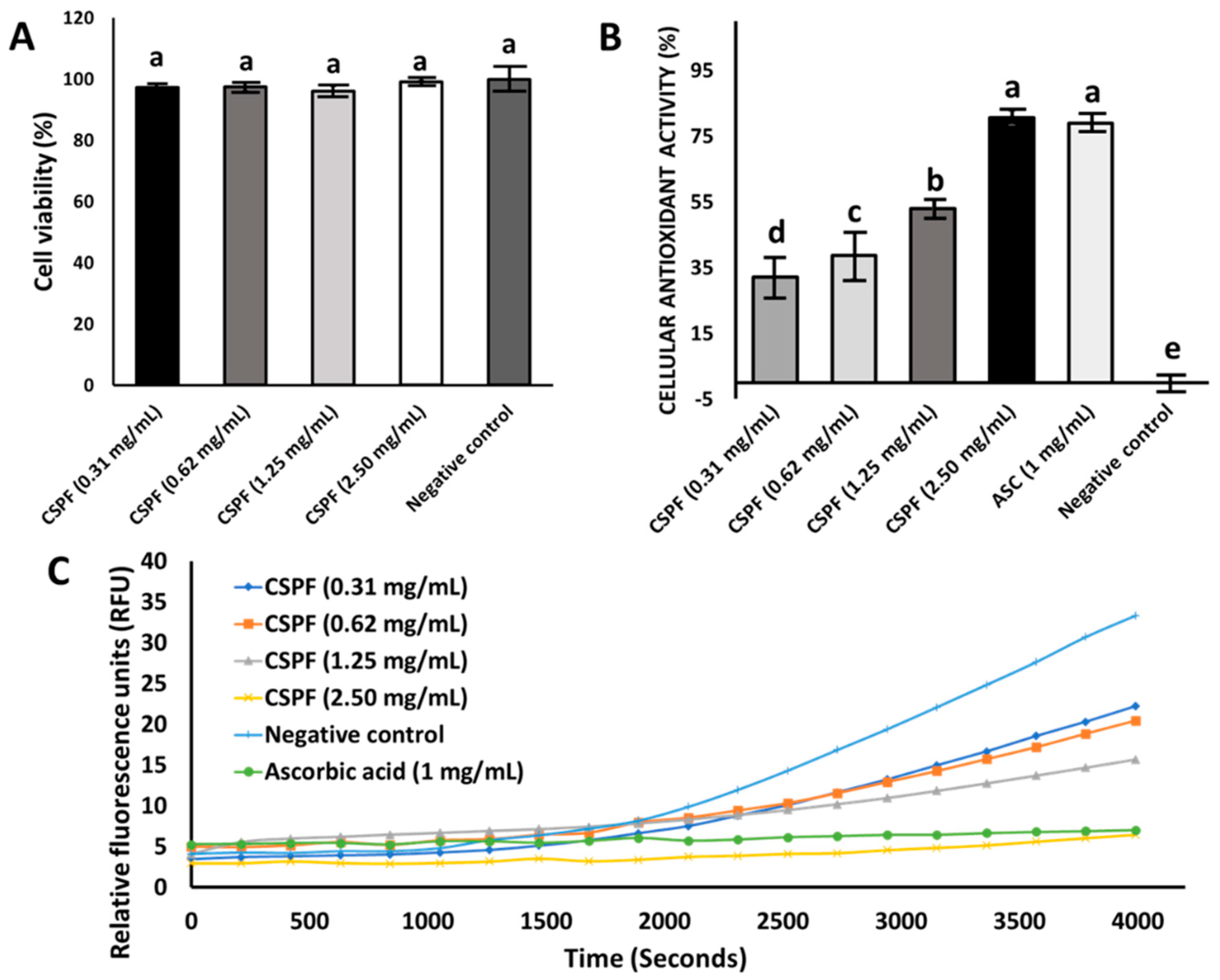
3.2. Effect of Canary Seed Peptides on Cellular Viability and Oxidative Stress

Caco-2 cells were selected in this study for their ability to provide a more robust association with active membrane transport (bioavailability) in the intestinal cell wall compared to other cell types (e.g., L-929, HepG2) [34,35]. Cellular viability assessment by the MTT assay is a well-recognized methodology and used on a wide range of molecules (e.g., drugs, topical ingredients, proteins, and phytochemicals) to assess the toxicity of an agent [36]. In this study, we evaluated the viability of Caco-2 cells after 24 h of exposure to the peptide fraction (CSPF) at various concentrations (Figure 1A). Results show that the CSPF was not cytotoxic at any of the tested concentrations (1–3 mg/mL), where no significant difference (p > 0.05) was found among tested concentration levels, and the cell viability ranged between 96 and 100%. Additionally, cell confluency remained unaffected, and cell loss was not observed in this study. Even though no information was available regarding CSPF cellular toxicity, these results align with observations reported in other toxicological studies, where no adverse effects were seen when feeding rats with a 50% hairless canary seed diet [37]. Subsequently, we analyzed the cellular antioxidant activity of the CSPF (Figure 1B). We monitored the oxidation of the intracellular probe DCFH-DA to its fluorescent counterpart DCF after exposure to the CSPF for one hour (Figure 1C). The probe DFH-DA is introduced to the cell and subject to deacetylation by cellular esterases; this leads to the production of the oxidable form of DCFH to DCF. Thus, for an antioxidant to prevent oxidation of DCFH to DCF, it must permeate the cell and compete intracellularly with free radicals from AAPH. Our results show that the CSPF confers a homogenous and gradual cellular antioxidant protection that increases significantly with concentration (p < 0.05), offering cellular antioxidant activity (CAA) of up to 80% for the CSPF at 2.5 mg/mL. There were no significant differences between the CSPF (2.5 mg/mL) and the positive control ascorbic acid (1 mg/mL). The high CAA observed in this study could be linked with the low molecular weight (<3 kDa) and the high content of aromatic amino acids 13% (w/w of protein) present in the CSPF (Table 1). Aromatic amino acids such as tyrosine, phenylalanine, and tryptophan are well-known for their excellent proton donation capabilities, stabilizing electron-deficient radicals, and preventing oxidative damage [14,38]. From a cellular transport perspective, we previously reported that CSPF had a high cellular transport capacity (>10%); this property could also contribute to their ability to scavenge radicals at intracellular levels [33].

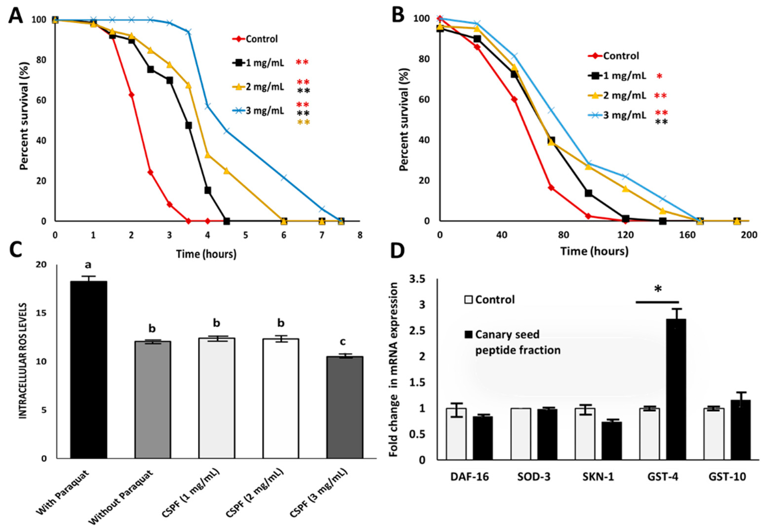
3.3. Antioxidant Properties of Canary Seed Peptides against Chronic and Acute Oxidative Stress

This study explored if CSPF have molecular implications for oxidative stress using a C. elegans model. Based on the cellular toxicity analysis, CSPF (1, 2, and 3 mg/mL) were used to assess their in vivo antioxidant activity. When pre-exposed to the CSPF, followed by induction of acute oxidative stress using t-BOOH, CSPF treatments were shown to significantly (p < 0.001) increase C. elegans’s lifespan compared to nematodes exposed to S-complete buffer alone (Figure 2A). The antioxidant protection was shown to be concentration-dependent, and the lifespan was extended significantly (p < 0.001) by 45, 63, and 88% at 1, 2, and 3 mg/mL peptide concentrations, respectively. These results are similar to those observed for the cellular antioxidant activity (Figure 1B), indicating an equally high capacity to permeate and protect the cells from acute oxidative damage in whole organisms. In the case of chronic oxidative stress (Figure 2B), CSPF pre-exposure was successful in significantly (p < 0.05) extending the lifespan of C. elegans relatively to the control by 23, 42, and 61% for the 1, 2, and 3 mg/mL peptide concentrations, respectively. The impact on the survival rate (%) derived from CSPF pre-exposure was lower chronic oxidative stress compared to acute oxidative stress; this could be due to the half-life of the antioxidant peptides, whose effects tend to diminish over time due to protein turnover [39]. Notably, on the chronic stress survival analysis, a significant difference between CSPF samples was only detected between 1 and 3 mg/mL. In this regard, these results are in agreement with those observed for ROS quantification (Figure 2C), where the treatment with 3 mg/mL decreased ROS levels (p < 0.01) compared to the 1 and 2 mg/mL treatments. Similarly, Zhang, Jiang [40] showed that supplementation of green alga (Chlorella vulgaris) protein hydrolysates at a concentration of 4 mg/mL successfully lowered the ROS in C. elegans compared to the untreated nematodes or lower peptide concentrations (1–2 mg/mL). We previously demonstrated the in vitro antioxidant capacity of peptides derived from commercial hydrolysis of canary seed protein [6]; yet, to the best of our knowledge, this is the first time the antioxidant activity of canary seed peptides obtained from simulated gastrointestinal digestion has been tested in vivo, indicating their nutraceutical potential. From these results, we confirm that the antioxidant capacity of a CSPF relies on the scavenging of free radicals as one of the mechanisms by which it prevents acute and chronic oxidative stress at both cellular (Caco-2) and live-organism (C. elegans) levels.

3.4. Expression of Antioxidant-Related Genes

The expression of various antioxidant-related genes (DAF-16, SOD-3, SKN-1, GST-4, and GST-10) was monitored by quantitative real-time PCR (qPCR) after 24 h of exposure to a high dose (3 mg/mL) of a CSPF in order to determine which molecular mechanisms could be tied to the antioxidant potential of a CSPF applied to C. elegans. The results for gene expression fold change are shown in Figure 2D. After pre-treatment with the CSPF, there was no significant difference in gene expression for DAF-16, SOD-3, SKN-1, or GST10. Nevertheless, significant upregulation was found for the GST-4 gene. GST-4 encodes the antioxidant enzyme glutathione S-transferase, which is involved in suppressing ROS formation and confers longer survival time to nematodes [41]. Research has demonstrated that glutathione-S-transferases (GSTs) act in the second detoxification phase, reducing ROS into less toxic compounds [42]. It has been reported that some plant metabolites have induced the expression of GST-4 and other enzymes involved in glutathione biosynthesis and metabolism. For instance, Pohl, Teixeira-Castro [43] and Ma, Cui [44] showed that GST-4 was upregulated after incubation of C. elegans with rapeseed extract and sesame peptides, respectively. In another study, tyrosol, a simple phenol from olive oil, was an effective inducer of GST-4 and promoted a drop in ROS levels of treated C. elegans nematodes [45]. Finally, other plant-derived compounds have shown that flavonoids such as baicalein, chrysin, and 6-hydroxyflavone were effective in extending the C. elegans longevity via GST-4 induction [46]. Interestingly, the GST-4 upregulation by CSPF was shown to be independent of the SKN-1 signaling pathway, an upstream regulator of antioxidant and detoxification genes such as GST-4. It is possible that GST-4 induction could be happening via the epidermal growth factor (EGF) signaling pathway and is promoted by EOR-1; this could also explain the lack of transcriptional activity from other GSTs such as GST-10 [47]. In this context, others have reported that royalactin, a honeybee protein, increased the expression of GST-4 via the EPG signaling pathway with no impact on the SKN-1 gene [48]. It is hypothesized that CSPFs could increase EGF expression, resulting in EOR-1 translocation to the nucleus, and promote the transcription of the detoxification gene GST-4, which might help decrease oxidation and extend C. elegans’s survival to oxidative stress [49]. Nevertheless, further investigations applying EGF- or SKN-lacking mutants as well as targeted analysis of protein expression levels are necessary to confirm this hypothesis. Overall, the results of this study suggest that GST-4-mediated antioxidant activity in coordination with the scavenging of free radicals could be the reason for the extended nematode survival times observed in this study.

4. Conclusions

In this study, we demonstrated that a canary seed peptide fraction produced by commercial enzymatic proteolysis with Alcalase followed by simulated gastrointestinal digestion showed no cytotoxic effect on Caco-2 cells at any of the tested concentrations (1–3 mg/mL). In addition, pre-exposure to a CSPF significantly lowered oxidative damage in Caco-2 cells in a concentration-dependent matter. Furthermore, we showed that pre-exposure to the canary seed peptide fraction substantially increased the resistance to acute and chronic oxidative stress by extending the lifespan of a live organism (C. elegans). The results from this study reveal that the CSPF relies on radical scavenging and up-regulation of the anti-oxidative-related gene (GST-4) to confer antioxidant protection to C. elegans, demonstrating the feasibility of using this simple in vivo model to establish preliminary determination of the antioxidant activity of peptides. Finally, GST-4 modulation was shown to be independent of upstream signaling pathways such as SKN-1 and suggests an alternative antioxidant route may be involved. Further research will be needed to elucidate this possibility and the biological activity that peptide fractions from canary seed might offer to more complex model organisms. Taken all together, the results in this study demonstrate that canary seed peptides can confer antioxidant protection in vivo and can be valuable as a nutraceutical ingredient for the food and pharmaceutical industries. Future steps should be focused on developing a more detailed understanding of specific peptide sequences responsible for the antioxidant activity observed in this study.

Supplementary Materials

The following supporting information can be downloaded at: https://www.mdpi.com/article/10.3390/nu14122415/s1. Table S1: Primer sequences used.

Author Contributions

U.U.-R.: Conceptualization, Methodology, Investigation, Data curation, Writing—original draft, Writing—review and editing. A.M.L.: Conceptualization, Methodology, Data curation, Writing, review and editing, Supervision, Funding acquisition. K.-H.K.: Methodology, Conceptualization, Writing—review and editing. L.R.: Conceptualization, Writing—review and editing. J.M.A.: Conceptualization, Writing—review and editing. All authors have read and agreed to the published version of the manuscript.

Funding

This work was supported by the USDA National Institute of Food and Agriculture, Hatch Act formula funds project 1019794.

Institutional Review Board Statement

Not applicable.

Informed Consent Statement

Not applicable.

Data Availability Statement

Not applicable.

Acknowledgments

Some strains were provided by the Caenorhabditis Genetics Center (CGC), which is funded by NIH Office of Research Infrastructure Programs (P40 OD010440).

Conflicts of Interest

The authors declare no conflict of interest. The funders had no role in the design of the study; in the collection, analyses, or interpretation of data; in the writing of the manuscript; or in the decision to publish the results.

References

  1. Mason, E.; L’Hocine, L.; Achouri, A.; Karboune, S. Hairless Canaryseed: A Novel Cereal with Health Promoting Potential. Nutrients 2018, 10, 1327. [Google Scholar] [CrossRef] [PubMed] [Green Version]
  2. Abdel-Aal, E.-S.M. Nutritional and functional attributes of hairless canary seed groats and components and their potential as functional ingredients. Trends Food Sci. Technol. 2021, 111, 680–687. [Google Scholar] [CrossRef]
  3. Novas, M.; Jiménez, A.; Asuero, A. Determination of Antioxidant Activity of Canary Seed Infusions by Chemiluminescence1. J. Anal. Chem. 2004, 59, 75–77. [Google Scholar] [CrossRef]
  4. Chen, Z.; Yu, L.; Wang, X.; Gu, Z.; Beta, T. Changes of phenolic profiles and antioxidant activity in canaryseed (Phalaris canariensis L.) during germination. Food Chem. 2016, 194, 608–618. [Google Scholar] [CrossRef]
  5. Mason, E.; L’Hocine, L.; Achouri, A.; Pitre, M.; Karboune, S. Health Promoting Bioactive Properties of Novel Hairless Canary Seed Flour after In Vitro Gastrointestinal Digestion. Foods 2020, 9, 932. [Google Scholar] [CrossRef]
  6. Urbizo-Reyes, U.; Aguilar-Toalá, J.; Liceaga, A. Hairless canary seeds (Phalaris canariensis L.) as a potential source of antioxidant, antihypertensive, antidiabetic, and antiobesity biopeptides. Food Prod. Process. Nutr. 2021, 3, 1–12. [Google Scholar] [CrossRef]
  7. Valverde, M.E.; Orona-Tamayo, D.; Nieto-Rendón, B.; Paredes-López, O. Antioxidant and antihypertensive potential of protein fractions from flour and milk substitutes from canary seeds (Phalaris canariensis L.). Plant Foods Hum. Nutr. 2017, 72, 20–25. [Google Scholar] [CrossRef] [PubMed]
  8. Ketnawa, S.; Liceaga, A.M. Effect of microwave treatments on antioxidant activity and antigenicity of fish frame protein hydrolysates. Food Bioprocess Technol. 2017, 10, 582–591. [Google Scholar] [CrossRef]
  9. Urbizo-Reyes, U.; San Martin-González, M.F.; Garcia-Bravo, J.; López-Malo Vigil, A.; Liceaga, A.M. Physicochemical characteristics of chia seed (Salvia hispanica) protein hydrolysates produced using ultrasonication followed by microwave-assisted hydrolysis. Food Hydrocoll. 2019, 97, 105187. [Google Scholar] [CrossRef]
  10. Mares-Mares, E.; Aguilar-Bravo, C.; Herrera-Castillo, F.L.M.; Sosa-Morales, M.C.E.; Del Rincón-Castro, M.C.; León-Galván, M.F. Antihypertensive and antioxidant capacity of a high protein beverage (walnut-sesame seeds- oat-soybean). In Proceedings of the 2017 ASABE Annual International Meeting, Spokane, WA, USA, 16–19 July 2017. [Google Scholar]
  11. Hall, F.; Liceaga, A. Effect of microwave-assisted enzymatic hydrolysis of cricket (Gryllodes sigillatus) protein on ACE and DPP-IV inhibition and tropomyosin-IgG binding. J. Funct. Foods 2020, 64, 103634. [Google Scholar] [CrossRef]
  12. Elias, R.J.; Kellerby, S.S.; Decker, E.A. Antioxidant activity of proteins and peptides. Crit. Rev. Food Sci. Nutr. 2008, 48, 430–441. [Google Scholar] [CrossRef] [PubMed]
  13. Rajapakse, N.; Mendis, E.; Jung, W.-K.; Je, J.-Y.; Kim, S.-K. Purification of a radical scavenging peptide from fermented mussel sauce and its antioxidant properties. Food Res. Int. 2005, 38, 175–182. [Google Scholar] [CrossRef]
  14. Sarmadi, B.H.; Ismail, A.J.P. Antioxidative peptides from food proteins: A review. Peptides 2010, 31, 1949–1956. [Google Scholar] [CrossRef]
  15. Adebiyi, A.P.; Adebiyi, A.O.; Yamashita, J.; Ogawa, T.; Muramoto, K. Purification and characterization of antioxidative peptides derived from rice bran protein hydrolysates. Eur. Food Res. Technol. 2009, 228, 553–563. [Google Scholar] [CrossRef]
  16. Tanzadehpanah, H.; Asoodeh, A.; Chamani, J. An antioxidant peptide derived from Ostrich (Struthio camelus) egg white protein hydrolysates. Food Res. Int. 2012, 49, 105–111. [Google Scholar] [CrossRef]
  17. Lobo, V.; Patil, A.; Phatak, A.; Chandra, N. Free radicals, antioxidants and functional foods: Impact on human health. Pharmacogn. Rev. 2010, 4, 118–126. [Google Scholar] [CrossRef] [Green Version]
  18. Song, W.; Kong, X.; Hua, Y.; Li, X.; Zhang, C.; Chen, Y. Antioxidant and antibacterial activity and in vitro digestion stability of cottonseed protein hydrolysates. LWT 2020, 118, 108724. [Google Scholar] [CrossRef]
  19. Yan, F.; Zheng, X. Protective effect of mulberry fruit anthocyanin on human hepatocyte cells (LO2) and Caenorhabditis elegans under hyperglycemic conditions. Food Res. Int. 2017, 102, 213–224. [Google Scholar] [CrossRef]
  20. Jia, W.; Peng, Q.; Su, L.; Yu, X.; Ma, C.W.; Liang, M.; Yin, X.; Zou, Y.; Huang, Z. Novel bioactive peptides from Meretrix meretrix protect Caenorhabditis elegans against free radical-induced oxidative stress through the stress response factor DAF-16/FOXO. Mar. Drugs 2018, 16, 444. [Google Scholar] [CrossRef] [Green Version]
  21. Moreno-Arriola, E.; Cárdenas-Rodríguez, N.; Coballase-Urrutia, E.; Pedraza-Chaverri, J.; Carmona-Aparicio, L.; Ortega-Cuellar, D. Caenorhabditis elegans: A useful model for studying metabolic disorders in which oxidative stress is a contributing factor. Oxidative Med. Cell. Longev. 2014, 2014, 705253. [Google Scholar] [CrossRef] [Green Version]
  22. You, L.; Zhao, M.; Regenstein, J.M.; Ren, J. Changes in the antioxidant activity of loach (Misgurnus anguillicaudatus) protein hydrolysates during a simulated gastrointestinal digestion. Food Chem. 2010, 120, 810–816. [Google Scholar] [CrossRef]
  23. Alaswad, A.A.; Song, B.; Oehrle, N.W.; Wiebold, W.J.; Mawhinney, T.P.; Krishnan, H.B. Development of soybean experimental lines with enhanced protein and sulfur amino acid content. Plant Sci. 2021, 308, 110912. [Google Scholar] [CrossRef] [PubMed]
  24. Er, S.; Koparal, A.T.; Kivanc, M. Cytotoxic effects of various lactic acid bacteria on Caco-2 cells. Turk. J. Biol. 2015, 39, 23–30. [Google Scholar] [CrossRef]
  25. Malaypally, S.P.; Liceaga, A.M.; Kim, K.-H.; Ferruzzi, M.; Martin, F.S.; Goforth, R.R. Influence of molecular weight on intracellular antioxidant activity of invasive silver carp (Hypophthalmichthys molitrix) protein hydrolysates. J. Funct. Foods 2015, 18, 1158–1166. [Google Scholar] [CrossRef]
  26. Bai, J.; Farias-Pereira, R.; Zhang, Y.; Jang, M.; Park, Y.; Kim, K.-H. C. elegans ACAT regulates lipolysis and its related lifespan in fasting through modulation of the genes in lipolysis and insulin/IGF-1 signaling. BioFactors 2020, 46, 754–765. [Google Scholar] [CrossRef] [PubMed]
  27. Solis, G.M.; Petrascheck, M. Measuring Caenorhabditis elegans life span in 96 well microtiter plates. J. Vis. Exp. 2011, 49, e2496. [Google Scholar] [CrossRef] [Green Version]
  28. Zhao, S.; Cheng, Q.; Peng, Q.; Yu, X.; Yin, X.; Liang, M.; Ma, C.W.; Huang, Z.; Jia, W. Antioxidant peptides derived from the hydrolyzate of purple sea urchin (Strongylocentrotus nudus) gonad alleviate oxidative stress in Caenorhabditis elegans. J. Funct. Foods 2018, 48, 594–604. [Google Scholar] [CrossRef]
  29. Sarasija, S.; Norman, K.R. Measurement of ROS in Caenorhabditis elegans using a reduced form of fluorescein. Bio-Protocol 2018, 8, e2800. [Google Scholar] [CrossRef] [Green Version]
  30. Green, M.R.; Sambrook, J. Total RNA extraction from Caenorhabditis elegans. Cold Spring Harb. Protoc. 2020. [Google Scholar] [CrossRef]
  31. Ogawa, T.; Kodera, Y.; Hirata, D.; Blackwell, T.K.; Mizunuma, M. Natural thioallyl compounds increase oxidative stress resistance and lifespan in Caenorhabditis elegans by modulating SKN-1/Nrf. Sci. Rep. 2016, 6, 21611. [Google Scholar] [CrossRef] [Green Version]
  32. Price, R.K.; Welch, R.W. Cereal Grains, in Encyclopedia of Human Nutrition, 3rd ed.; Academic Press: Cambridge, MA, USA, 2013; pp. 307–316. [Google Scholar]
  33. Urbizo-Reyes, U.; Liceaga, A.M.; Reddivari, L.; Kim, K.-H.; Anderson, J.M. Enzyme kinetics, molecular docking, and in silico characterization of canary seed (Phalaris canariensis L.) peptides with ACE and pancreatic lipase inhibitory activity. J. Funct. Foods 2022, 88, 104892. [Google Scholar] [CrossRef]
  34. Kellett, M.E.; Greenspan, P.; Pegg, R.B. Modification of the cellular antioxidant activity (CAA) assay to study phenolic antioxidants in a Caco-2 cell line. Food Chem. 2018, 244, 359–363. [Google Scholar] [CrossRef] [PubMed]
  35. Wan, H.; Liu, D.; Yu, X.; Sun, H.; Li, Y. A Caco-2 cell-based quantitative antioxidant activity assay for antioxidants. Food Chem. 2015, 175, 601–608. [Google Scholar] [CrossRef] [PubMed]
  36. Kumar, P.; Nagarajan, A.; Uchil, P.D. Analysis of cell viability by the MTT assay. Cold Spring Harb. Protoc. 2018. [Google Scholar] [CrossRef]
  37. Magnuson, B.A.; Patterson, C.A.; Hucl, P.; Newkirk, R.W.; Ram, J.I.; Classen, H.L. Safety assessment of consumption of glabrous canary seed (Phalaris canariensis L.) in rats. Food Chem. Toxicol. 2014, 63, 91–103. [Google Scholar] [CrossRef]
  38. Liu, W.-Y.; Zhang, J.-T.; Miyakawa, T.; Li, G.-M.; Gu, R.-Z.; Tanokura, M. Antioxidant properties and inhibition of angiotensin-converting enzyme by highly active peptides from wheat gluten. Sci. Rep. 2021, 11, 5206. [Google Scholar] [CrossRef]
  39. Gülseren, İ.; Vahapoglu, B. The Stability of Food Bioactive Peptides in Blood: An Overview. Int. J. Pept. Res. Ther. 2022, 28, 1–7. [Google Scholar] [CrossRef]
  40. Zhang, Y.; Jiang, W.; Hao, X.; Tan, J.; Wang, W.; Yu, M.; Zhang, G.; Zhang, Y. Preparation of the Enzymatic hydrolysates from Chlorella vulgaris protein and assessment of their antioxidant potential using Caenorhabditis elegans. Mol. Biotechnol. 2021, 63, 1040–1048. [Google Scholar] [CrossRef]
  41. Tao, J.; Wu, Q.-Y.; Ma, Y.-C.; Chen, Y.-L.; Zou, C.-G. Antioxidant response is a protective mechanism against nutrient deprivation in C. elegans. Sci. Rep. 2017, 7, 43547. [Google Scholar] [CrossRef] [Green Version]
  42. Masella, R.; Di Benedetto, R.; Varì, R.; Filesi, C.; Giovannini, C. Novel mechanisms of natural antioxidant compounds in biological systems: Involvement of glutathione and glutathione-related enzymes. J. Nutr. Biochem. 2005, 16, 577–586. [Google Scholar] [CrossRef]
  43. Pohl, F.; Teixeira-Castro, A.; Costa, M.D.; Lindsay, V.; Fiúza-Fernandes, J.; Goua, M.; Bermano, G.; Russell, W.; Maciel, P.; Kong Thoo Lin, P. GST-4-dependent suppression of neurodegeneration in C. elegans models of Parkinson’s and Machado-Joseph disease by rapeseed pomace extract supplementation. Front. Neurosci. 2019, 13, 1091. [Google Scholar] [CrossRef]
  44. Ma, X.; Cui, X.; Li, J.; Li, C.; Wang, Z. Peptides from sesame cake reduce oxidative stress and amyloid-β-induced toxicity by upregulation of SKN-1 in a transgenic Caenorhabditis elegans model of Alzheimer’s disease. J. Funct. Foods 2017, 39, 287–298. [Google Scholar] [CrossRef]
  45. Calahorra Garcia-Moreno, J.; Porta de la Riva, M.; Martínez-Lara, E.; Siles, E.; Cañuelo, A. Tyrosol, a simple phenol from EVOO, targets multiple pathogenic mechanisms of neurodegeneration in a C. elegans model of Parkinson’s disease. Neurobiol. Aging 2019, 82, 60–68. [Google Scholar] [CrossRef] [PubMed]
  46. Guerrero-Rubio, M.A.; Hernández-García, S.; García-Carmona, F.; Gandía-Herrero, F. Flavonoids’ Effects on Caenorhabditis elegans’ Longevity, Fat Accumulation, Stress Resistance and Gene Modulation Involve mTOR, SKN-1 and DAF-16. Antioxidants 2021, 10, 438. [Google Scholar] [CrossRef] [PubMed]
  47. Ijomone, O.M.; Miah, M.R.; Akingbade, G.T.; Bucinca, H.; Aschner, M. Nickel-induced developmental neurotoxicity in C. elegans includes cholinergic, dopaminergic and GABAergic degeneration, altered behaviour, and increased SKN-1 activity. Neurotox. Res. 2020, 37, 1018–1028. [Google Scholar] [CrossRef]
  48. Detienne, G.; Van de Walle, P.; De Haes, W.; Schoofs, L.; Temmerman, L. SKN-1-independent transcriptional activation of glutathione S-transferase 4 (GST-4) by EGF signaling. In Worm; Taylor & Francis: Abingdon, UK, 2016. [Google Scholar]
  49. Mohankumar, A.; Kalaiselvi, D.; Thiruppathi, G.; Muthusaravanan, S.; Nivitha, S.; Levenson, C.; Tawata, S.; Sundararaj, P. α-and β-santalols delay aging in Caenorhabditis elegans via preventing oxidative stress and protein aggregation. ACS Omega 2020, 5, 32641–32654. [Google Scholar] [CrossRef]
Figure 1. (A) Cell viability (%): MTT assay over canary seed peptide fraction (CSPF) at different protein concentrations, (B) intracellular antioxidant activity (%) of CSPF and ascorbic acid (ASC), and (C) inhibition of peroxyl-radical-induced DCFH oxidation to DCF by CSPF and ASC. Negative control: untreated cells. Bars and lines represent mean values of triplicate determinations ± standard deviation. Different letters (a–e) indicate statistical differences (p < 0.05) between samples.
Nutrients 14 02415 g001
Figure 2. In vivo antioxidant activity of canary seed peptide fraction (CSPF), using a C. elegans model. (A) Survival analysis of C. elegans pre-exposed to CSPF for 24 h followed by acute oxidative stress induction with tert-butyl hydrogen peroxide (t-BOOH). (B) Survival analysis of C. elegans pre-exposed to CSPF for 24 h followed by chronic oxidative stress induction with paraquat. (C) Intracellular accumulation of reactive oxygen species (ROS) in C. elegans after pre-exposure to CSPF for 24 h followed by paraquat for 48 h. (D) Relative mRNA levels of expression of DAF-16, SOD-3, SKN-1, GST-4, and GST-10 after 24 h exposure with 3 mg/mL of CSPF using ACT-1 as an internal control. Bars and lines represent mean values ± standard deviation. Three independent experiments were performed with at least 100 nematodes per treatment. Statistical differences are indicated as * p < 0.01, and ** p < 0.001 or different letters (a–c) p < 0.01.
Nutrients 14 02415 g002
Table 1. Total amino acid composition of canary seed peptide obtained from simulated gastrointestinal digestion.
Amino AcidsRelative Content (g/100 g)
Taurine §0.10
Aspartic Acid4.79
Threonine2.50
Serine3.98
Glutamic Acid30.72
Proline6.34
Lanthionine §0.19
Glycine3.21
Alanine4.44
Cysteine2.50
Valine4.55
Methionine1.37
Isoleucine4.31
Leucine7.56
Tyrosine3.46
Phenylalanine6.34
Hydroxylysine0.30
Ornithine §0.07
Lysine2.00
Histidine2.02
Arginine6.27
Tryptophan2.98
AAA12.78
PCAA10.29
SAA3.88
HAA41.08
EAA/NEAA0.51
Results are expressed on a dry basis. §: non-proteinogenic amino acids. AAA: aromatic amino acids. PCAA: positively charged amino acids. SAA: sulfur-containing amino acids. HAA: hydrophobic amino acids. EAA/NEAA: essential to non-essential amino acid ratio.
Publisher’s Note: MDPI stays neutral with regard to jurisdictional claims in published maps and institutional affiliations.

