Article

Phytochemical Analysis of the Aerial Parts of Campanula pelviformis Lam. (Campanulaceae): Documenting the Dietary Value of a Local Endemic Plant of Crete (Greece) Traditionally Used as Wild Edible Green

by
Olga S. Tsiftsoglou
1,
George Lagogiannis
1,
Antonia Psaroudaki
2,
Aikaterina Vantsioti
1,
Milan N. Mitić
3,
Jelena M. Mrmošanin
3 and
Diamanto Lazari
1,*
1
Laboratory of Pharmacognosy, Faculty of Health Sciences, School of Pharmacy, Aristotle University of Thessaloniki, 54124 Thessaloniki, Greece
2
Department of Nutrition and Dietetics, Hellenic Mediterranean University, Tripitos Area (2nd km Sitia–Palekastro), 72300 Sitia, Greece
3
Department of Chemistry, Faculty of Sciences and Mathematics, University of Niš, 18000 Niš, Serbia
*
Author to whom correspondence should be addressed.
Sustainability 2023, 15(9), 7404; https://doi.org/10.3390/su15097404
Submission received: 28 February 2023 / Revised: 23 April 2023 / Accepted: 25 April 2023 / Published: 29 April 2023

Abstract

:
Native wild edible greens usually include plants with widespread geographical ranges and represent a long tradition associated with well-documented health effects, especially in the frame of the Mediterranean diet. Although consuming local endemic and range-restricted plants as wild edible greens is rare, in some areas of Crete this is a long ethnobotanical tradition. The present study is focused on the phytochemical and nutritional element analyses of the edible parts of the wild-growing green Campanula pelviformis. To date, nine secondary metabolites have been isolated: lobetyolin (1), calaliukiuenoside (2), demethylsyrrigin (3), wahlenoside A (4), chlorogenic acid methyl (5) and butyl ester (6), nicotiflorin (7), rutin (8) and corchoionoside A (9). This first-time research on the phytochemical composition of this local endemic plant of Crete is a basic step in attempts to document its nutritional value, also allowing an exploration of its beneficial properties. The nutritional value of the Mediterranean diet owes much to the inclusion of native edible wild plants, which are abundant in mineral elements and bioactive compounds known to promote human health. Among these plants, the local Cretan endemic species C. pelviformis stands out as a rare and valuable source of wild edibles with traditional dietary significance in eastern Crete. This plant’s rich content of mineral elements and bioactive compounds makes it an intriguing subject for further research into the potential health benefits of wild plant consumption.

1. Introduction

The enrichment of the diet with wild edible plants in eastern Crete is a daily habit during the year, and this trend peaks during the rainy months from late October until early March [1,2,3]. The great variety of ways in which the natural grasses or greens (commonly known as horta) are consumed and the ingenuity in the traditional combinations with different types of food and food groups are related to the culinary preferences of the area’s inhabitants that are nowadays strengthened. Campanula pelviformis, known as ‘loutes’ (plural of luta) in the local area, is a narrow endemic species of eastern Crete [1,2]. It is widely used, among other wild edible plants, in the typical traditional local food “tsigarolachana” (meaning cooked in the casserole with a small quantity of olive oil). It grows wild in the same area. The edible part of this species is the young shoots and leaves. The “tsigarolachana” in eastern Crete can be served with various main dishes such as fish, seafood and meat but also cooked with them along with eggs and other vegetables. They are also the filling for delicious pies with other horta. The frequent use of C. pelviformis as well as other similar herbs shows the importance of preserving and promoting the traditional diet of eastern Crete as an alternative and interesting choice for proper nutrition. Traditional nutrition has often been referred to as the “diet of necessity” or “food of the poor”, that is, a dietary pattern that was associated with wars, economic misery and lack of other food groups. Although nowadays the socioeconomic conditions have improved on the island, the use and consumption of wild ‘horta’ (or chorta) is still one of the main characteristics of the traditional Cretan diet. Especially in eastern Crete, the phenomenon of marketing of wild herbs is observed and at very high prices. High demand of these products can lead to excessive collection with the risk of degrading genetic pools and biodiversity in the area. On the other hand, the utilization of edible native plants as food and potentially as financial resources through their cultivation acquires greater importance [1,2]. Habits such as these align with the preservation of the Mediterranean diet by connecting traditional food culture with current values related to biodiversity preservation, good agricultural practices and high nutritional value products in the diet. One of the most important steps for the utilization of a plant species is the investigation of its agronomic value [1,2]. Part of this study is the analysis of the secondary metabolites biosynthesized from this plant which are phytonutrients and provide therapeutic value. Moreover, in present study, the nutritional value of the young leaves of the plant was examined and the analysis of the content in macro and trace elements has been discussed.
Campanula pelviformis Lam. is not the only edible species of this genus which is part of Mediterranean diet. In Italy, many traditional and local recipes have the young aerial parts of C. rapunculus L. and C. trachelium L. [4,5] as ingredients. In Turkey, roots of C. glomerata L. subsp. hispida (Witasek) Hayek [6] and the young leaves of C. lyrata Lam. [7] are consumed as vegetables. According to ethnobotanical studies in Bosnia-Herzegovina, young shoots of C. rapunculoides L., C. trachelium L., C. glomerata L. and C. pyrimidalis L. are used as cooked vegetables [8]. This is not the only research concerning the nutritional value of the plants belonging to this genus. There are many other studies which provide information about the culinary use of Campanula spp. and indicate that, apart from ornamental plants, they are a source of many phytonutrients, fibers and significant amounts of minerals (macro and trace-elements) which play an important role in maintaining human health [9,10]. Recently, the medicinal, agro-food and ornamental potential of this species was also studied, along with other neglected local endemic plants of Crete [11,12,13].
Campanula pelviformis Lam. is a narrowly endemic species consumed as food mainly by the inhabitants of the Lassithi region of Crete [1,2]. Its culinary use is well established. In the present study, an attempt is made to scientifically document its nutritional value through an analysis of the main nutrients (carbohydrates, proteins, lipids) as well as macro and trace elements. Additionally, an investigation of the presence of phytonutrients in the edible part of the plant, which will add value to the plant, is sought. So far, the results are encouraging. The consumption of the plant could be expanded if it can be cultivated. Cultivation may alleviate the collection pressure for the wild-growing species, facilitate sustainable exploitation strategies and offer a backup solution for reintroduction purposes of the threatened species. Cultivation of the plant will promote its conservation and, consequently, the preservation of biodiversity.

2. Materials and Methods

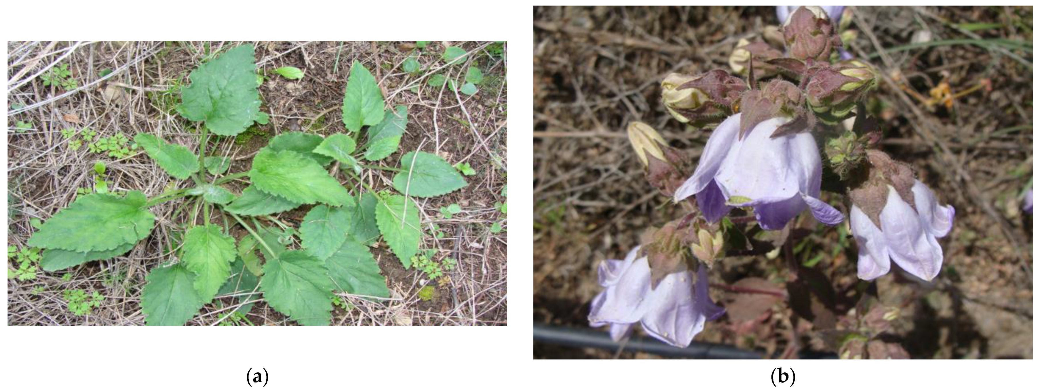
2.1. Plant Material Collection, Botanical Identification

Collection of the examined material was carried out in the following area: prefecture of Lassithi, Municipality of Sitia, Municipal Department of Armeni (Alt. 690 m; Latitude 35°02′50″ N; Longitude 26°04′00″ E). The microclimate of the area is characterized by cool summers and a wetter autumn, winter, and spring compared to the rest of the Sitia region. This region includes both cultivated and grassland areas affected by extensive animal grazing. The plant was identified taxonomically during the flowering period (May 2016) by Dr. A. Psaroudaki (see Figure 1). Harvesting of the plant material took place during the period when it is traditionally collected for consumption (from November to February). The following literature was studied for botanical identification: Flora Hellenica [14,15] and Flora of the Cretan Area [16]. A voucher specimen was deposited at the Herbarium of Aristotle University of Thessaloniki, Laboratory of Pharmacognosy, under No. Lazari D. 7350.

2.2. General Experimental Procedures

Column chromatography (CC) was carried out on silica gel 60 (Merck Art. 9385) and the gradient elution with the solvent mixtures indicated in each case. Thin Liquid Chromatography (TLC) was carried out on silica gel plates (Kieselgel F254, Merck, Art. 5554) and on cellulose plates (Cellulose, Merck, Art. 5552). The detection on TLC plates used UV light (absorbance: 254 and 366 nm). Vanillin-H2SO4 (1:1) spray reagent was used for the visualization of the chromatograms on silica gel plates; Neu reagent spray (ethanolamine diphenylborate) was used for the visualization of the chromatograms on cellulose plates. The high performance liquid chromatography (HPLC) analysis was conducted using a Lab Alliance Series III pump equipped with Clarity software and a Shodex RI detector. The analysis utilized a C18 ODS1 Spherisorb 10 μm column with dimensions of 250 mm × 10 mm (Waters).
Spectroscopic NMR data: The 1H-NMR and 13C-NMR spectra were recorded in CD3OD using AGILENT DD2 500 (500.1 MHz for 1H-NMR and 125.5 MHz for 13C-NMR) spectrometer. The chemical shifts are reported in δ (ppm) values relative to TMS (3.31 ppm for 1H-NMR and 49.05 ppm for 13C-NMR).

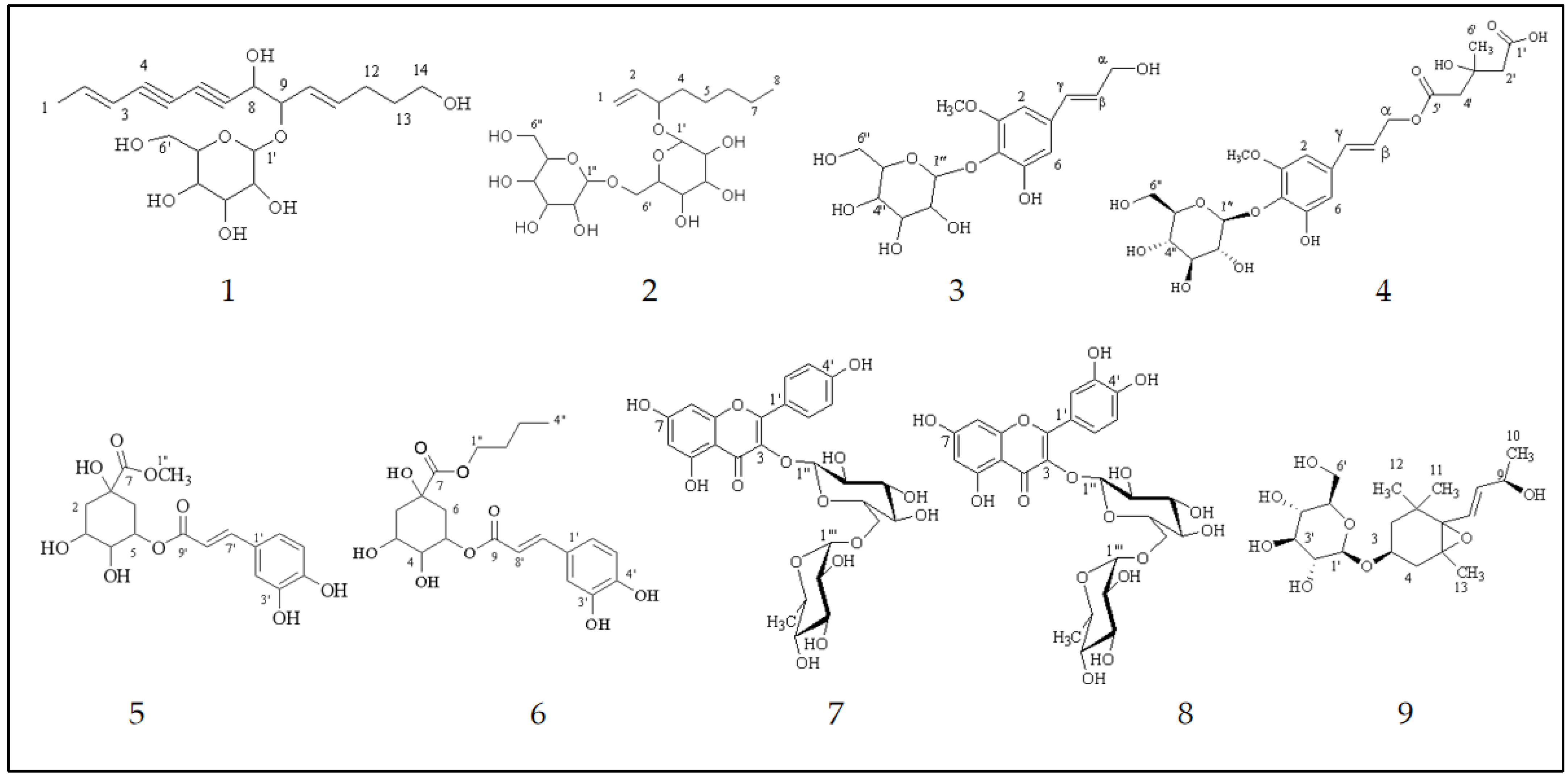
2.3. Extraction, Isolation and Identification of Secondary Metabolites

The naturally air-dried (in a shady place) aerial edible parts of the plant (186.48 g) were finely ground and exhaustively extracted at room temperature with petroleum ether (40°–60°), dichloromethane (DM) and a methanol (MeOH). The methanolic extract (55.95 g) was concentrated and the residue redissolved in boiling water (H2O). The water-soluble fraction was filtered and fractionated with a liquid–liquid extraction (distribution) using four different solvents of increasing polarity and extracted successively with n-hexane, ethyl acetate (EtOAc) and n-butanol (n-BuOH). The ethyl acetate residue (1.712 g) was subjected to CC (column chromatography) on silica gel (28.0 cm × 3.5 cm) using dichloromethane and methanol mixtures of increasing polarity as eluents to yield twenty-eight fractions (A-ZC). Further purification by CC on Sephadex LH-20 (63.00 cm × 2.5 cm) of fraction U (132.9 mg) (eluted with CH2Cl2-MeOH-H2O 82:18:1.8) using methanol as eluent resulted in the isolation of compound (7) (14.9 mg), (nicotiflorin). The butanol residue (6.71 g) was subjected to VLC (vacuum liquid chromatography) on silica gel (10.0 cm × 7.0 cm) using hexane-ethyl acetate and ethyl acetate-methanol mixtures of increasing polarity as eluents to yield twelve fractions of 300 mL each (A–M). Fractions A (207.5 mg), C (48.4 mg), F (527.9 mg) and G (744.9 mg) were subjected to further chromatographic separations as described below.
Further purification by CC on Sephadex LH-20 (62.00 cm × 2.5 cm) of fraction A (eluted with EtOAc, 100%) using methanol as eluent yielded nine fractions (AA-AI). Fraction AE (26.4 mg) was further fractionated using semipreparative HPLC (ACN-H2O, 1:1); this allowed the isolation of compounds (3) (demethylsyrrigin, tR = 10.63 min, 2.8 mg) and (6) (chlorogenic acid butyl ester, tR = 15.66 min, 3.8 mg). Fraction AG (24.3 mg) was further fractionated by semipreparative HPLC (ACN-H2O, 1:1); this allowed the isolation of compound (7) (nicotiflorin, tR = 12.12 min, 1.4 mg). Fraction C (48.4 mg, eluted with EtOAc:MeOH, 90:10) was further fractionated by semipreparative HPLC (MeOH-H2O, 1:1); this allowed the isolation of compounds (3) (demethylsyrrigin, tR = 12.30 min, 3.6 mg), (5) (chlorogenic acid methyl ester) (tR = 14.8 min, 2.7 mg) and (6) (chlorogenic acid butyl ester) (tR = 43.1 min, 1.4 mg). Further purification by CC on Sephadex LH-20 (60.00 cm × 2.5 cm) of fraction F (527.9 mg, eluted with EtOAc:MeOH, 75:25) using methanol as eluent yielded nine fractions (FA-FI). Fraction FB (52.8 mg) was further fractionated by semipreparative HPLC (MeOH-H2O, 1:1); this allowed the isolation of compounds (9) (corchoionoside A, tR = 13.9 min, 3.5 mg), (1) (lobetyolin, tR = 29.4 min, 1.6 mg) and (2) (calaliukiuenoside, tR = 43.5 min, 1.3 mg). A small quantity (49.8 mg) of fraction FF (184.7 mg) was further fractionated by semipreparative HPLC (MeOH-H2O, 1:1); this allowed the isolation of compounds (7) (nicotiflorin, tR = 28.3 min, 10.7 mg) and (8) (rutin, tR = 37.7 min, 9.5 mg). Fraction G (744.9 g) was subjected to CC on silica gel (11.0 cm × 3.5 cm) using DM-MeOH-H2O mixtures of increasing polarity as eluents to yield six fractions (GA-GF). Fraction GE (eluted with CH2Cl2-MeOH, 70:30, 438.5 mg) was subjected to CC on Sephadex LH-20 (80.0 cm × 2.5 cm) using MeOH as eluent to yield fourteen fractions (GEA-GEN). One of these fractions, GEF (22.5 mg), was identified as compound (4) (wahlenoside A).

2.4. Inductively Coupled Plasma-Optical Emission Spectrometry Analysis

The wet digestion technique was employed for the preparation of the sample in liquid form. For the analysis of the medicinal plant samples using ICP-OES, about 1.0236 g of the powder of the edible parts of the plant were measured in 150 mL Erlenmeyer flasks. Twenty milliliters of 65% of HNO3 was added and then heated for about 1 h at 120 °C to achieve complete dissolution of the samples or, more precisely, to pair to a small volume. Then, the content was cooled to room temperature. In the final phase, small volumes of hydrogen peroxide were added (2–3 mL) and the content was heated again in order to concentrate the sample to a smaller volume. After filtering the samples using blue filter paper (AHLSTROM MUNKSJÖ, Germany), the volume was adjusted to 25 mL for each extract.
The following operating conditions were used during the operation of the ICP-OES instrument series iCAP 6300 DUO: RF generator power 1150 W; rinse pump speed 100 rpm; pump speed for analysis 50 rpm; spray gas flow 0.7 L/min; cooling gas flow 12 L/min; auxiliary gas flow 0.5 L/min; the plasma observation directions were axial and radial; rinsing time was 30 s. The above described protocol was used in order to detect the content of chemical elements, such as aluminum (Al), arsenic (As), boron (B), barium (Ba), beryllium (Be), calcium (Ca), cadmium (Cd), cobalt (Co), chromium (Cr), copper (Co), iron (Fe), potassium (K), magnesium (Mg), manganese (Mn), sodium (Na), nickel (Ni), phosphorus (P), lead (Pd), silicon (Si) and zinc (Zn).

2.5. Examination of the Nutrient Content of Plant Material

Spectrophotometric analysis of edible aerial parts of C. pelviformis Lam. was carried out using a Perten DA 7250 NIR Analyzer, Perten–PerkinElmer (New York, NY, USA), precalibrated for a wide range of applications. It is possible to determinate moisture, protein, fat crude fiber, NDF, ADF, starch, sugars, calcium and phosphorus using this measurement method.
To calculate the percentage of carbohydrates and the nutritive value of the edible aerial parts of C. pelviformis Lam., we used the results of Perten and Equations (1) and (2) according to Shivraj and Khobragade [17].
The percentage of carbohydrate was given by = 100 − (percentage of ash + percentage of moisture + percentage of fat + percentage of protein).
Nutritive value = 4 × percentage of protein + 9 × percentage of fat + 4 × percentage of carbohydrate.

3. Results

The chemical structures of nine isolated compounds were established (Figure 2) on the basis of 1H, 13C and 2D NMR (gDQCOSY, gHSQCAD, gHMBCAD) spectroscopic analysis and through the comparison of literature data. These compounds are as follows: a polyacetylene: lobetyolin (compound 1) [18]; an alcohol glycoside: calaliukiuenoside (compound 2) [19]; two phenylpropanoids: demethylsyrrigin (compound 3) and wahlenoside A (compound 4) [20]; two chlorogenic acid esters: chlorogenic acid methyl ester (compound 5) and chlorogenic acid butyl ester (compound 6) [21]; two flavonoids: nicotiflorin (compound 7) [22] and rutin (compound 8) [23]; and one megastigmane: corchoionoside A (compound 9) [24]. The structures of the isolates are given in Figure 2 and were elucidated based on 1D and 2D NMR spectral analyses (see Supplementary Materials Tables S1–S9 and Figures S1–S45).

4. Discussion

Fresh fruits, vegetables, herbs, nuts, cereals, legumes, natural grasses and edible greens are major sources of nutrients (proteins, carbohydrates, fatty acids, vitamins, minerals) in the human diet and are also necessary for proper growth and development of the body. Along with those essential nutrients for life, edible plants also contain many secondary metabolites. Most of these phytochemicals are useful for the body because they can act as antioxidants, anti-inflammatory agents and antimicrobial agents. These natural compounds are known as phytonutrients and their relationship to maintaining human health and preventing diseases, as discussed below, is scientifically documented.
According to the literature, the main compounds of the plants belonging to the genus Campanula are flavonoids, phenolic acids, phenylpropanoids and polyacetylenes [25,26,27,28,29,30,31,32,33,34]. These results are in accordance with our research because of the isolation of compounds 1, 3, 4, 5, 6, 7 and 8. It is known that the daily consumption of foods rich in phytonutrients, such as flavonoids and other polyphenols, protects against the development of chronic non-communicable diseases (NCDs), including osteoporosis, diabetes and neurodegenerative and cardiovascular diseases. They also contribute to the management of these diseases [35].
Lobetyolin is a polyacetylene. The first isolation of lobetyolin was from the plant Lobelia inflata L. [36]. It has also been reported in other species of the Campanulaceae family, such as Pratia nummularia Lam. [37], Codonopsis pilosula (Franch.) Nannf. [38], C. tangshen Oliv. [39], C. cordifolioidea P. C. Tsoong [40], Campanula alliariifolia [29], C. medium L. [33], C. glomerata [32] and C. lactiflora M. Bieb. [31] apart from the studied plant. It is clear that lobetyolin is one of the most common compounds isolated from the species of the genus Codonopsis. Moreover, lobetyolin can be used as a chemotaxonomic marker for plants belonging to the genus Campanula as it is detected in significant percentages in three examined Korean species (C. punctata Lam., C. takesimana Nakai and C. glomerata var. Dahurica) [41].
Lobetyolin is one of the most common compounds isolated from Codonopsis pilosula (Franch.) Nannf. Many ethnopharmacological uses of this plant in China led to the intensive study of lobetyolin and its effects on human health. In a recent study, lobetyolin also showed cytotoxic activity against tumor cells in the lung [42]. According to the literature, lobetyolin shows anti-cancer, antiviral, anti-inflammatory and antioxidant properties, as well as mucosal protective effects. Moreover, this compound induces apoptosis in colon cancer cells by interfering with the metabolism of glutamine [43]. Lobetyolin has also been shown to promote the expression of apoptotic proteins, which is the natural process of cell death occurring in cancer cells. At the same time, it reduced ASCT2 (Alanine-Serine-Cysteine Transporter 2) levels and suppressed intracellular accumulation of glutamine, resulting in apoptotic cell death in breast cancer in humans [43]. It has also been found to weakly inhibit xanthine oxidase (XO) activity. Note that xanthine oxidase (XO) catalyzes the formation of uric acid by xanthine, a critical metabolic pathway associated with hyperuricaemia and gout [44].
Compound 2 has been isolated for the first time from the species Calanthe discolor Lindl. and C. liukiuensis Schltr. (Orchidaceae); it is known as “calaliukiuenoside” [18]. It has also been identified in Ginkgo biloba L. male flowers [45]. This is the first report of gentiobioside of 1-octen-ol in the plants of the Campanulaceae family.
Rutin (8) is a very abundant structural analog of quercetin. This flavonoid has also been reported in other species of the genus Campanula, such as C. oblongifolia Kharadze [30], C. maleevii Fed., C. glomerata L., C. oblongifolia (C. Koch) [46], C. alliariifolia Willd. [29] and C. alata Desf. [28]. In a recent study, rutin was found to exhibit antiviral properties, including its reported activity against the SARS CoV 2 virus [47,48] and as an inhibitor of the major protease and other protein targets of the SARS CoV 2 virus [49,50,51]. Rutin is also involved in inhibiting α-glucosidase in the gut, helping to maintain normal blood glucose levels [52]. Moreover, compound 8 has antimicrobial, anti-inflammatory and anti-cancer properties [53,54]. Rutin inhibits the enzyme called protein disulfide isomerase (PDI), which is involved in blood clot formation, strengthens capillaries and brings relief to people suffering from phlebitis [55].
Despite the high content of flavonoids in plants of the genus Campanula, nicotiflorin (7) is not a common compound: until now it has only been isolated from C. barbata [25]. According to a recent review article, nicotiflorin exhibits many biological activities against coronavirus, ischemia, renal impairment, hepatic complication, memory dysfunction, myocardial infarction, multiple myeloma and it also modulates insulin secretion. Moreover, nicotiflorin has biological potential against many enzymes, such as α-glucosidase and α-amylase [56].
Structural analogues of compounds 3, 4, 5 and 6 have been isolated from species of the genus Campanula. Both compounds 3 and 4 have been reported in the species Wahlenbergia marginata (Thunb.) A.DC. [19]. Moreover, some polyglycoside derivatives of wahlenoside A have been isolated from C. barbata L. [25]. Compounds 5 and 6, which are both esters of chlorogenic acid (methyl and butyl esters, respectively), have been reported and isolated for the first time in species of the genus Campanula. It must be mentioned that chlorogenic acid has been reported only in C. glomerata L. [57] and C. rapunculoides [58].
Megastigmanes (C13 isonorterpenoids) are a terpene class of compounds [59]. The genus Codonopsis of the Campanulaceae family is known to contain megastigmanes [60]. Until now, according to our knowledge, there have been no reports about this category of natural products in the genus Campanula. This is the first report about the presence of corchoionoside A (9) in this genus. The first isolation of corchoionoside A, which is a rare natural product, came from the leaves of Corchorus olitorius L. (Tiliaceae) [23].
In the current study, ash, crude protein, fats and carbohydrate contents in the analyzed plant material were found to be 7.92%, 12.90%, 2.46% and 64.19%, respectively. One of the signs of plant high digestibility is the amount of ash. The percentage in our case indicates the good digestibility of C. pelviformis′ young shoots. All the macronutrients were found to be within the range of the daily recommended quantities per 100 g of plant origin food. Moreover, the importance of crude fiber (15.65%) for appropriate digestive activity and waste removal [61] must be mentioned.
Macrominerals and trace elements are essential inorganic food nutrients. These elements, along with other nutrients (vitamins), are necessary for maintaining life. The human body is not able to synthesize them, and their total intake comes from foods. This is the reason they called “essential nutrients”. They play a key role in the maintenance of human health, and their presence in the body’s fluids and tissues is important for carrying out biochemical reactions [62]. The macrominerals include calcium, phosphorus, sodium, chloride, potassium and magnesium, while the trace elements include iron, copper, cobalt, iodine, boron zinc, manganese, molybdenum, fluoride, selenium and sulfur [63]. There are also other minerals, such as silicon, arsenic and nickel, which are known as ultra-trace elements [62]. Although the amount of calcium found in the human body’s fluids is low, it is adequate and acts as an intracellular messenger in cells and tissues. It is responsible for the proper functioning of the heart, muscles and nerves. About 99% of the human body’s calcium (calcium hydroxyapatite) and 85% of its phosphorus is found in bones and teeth. Phosphorus is involved in processes such as regulation of the acid–base balance of the body, the structural integrity of cells and the energy cycle. Moreover, this chemical element has an important role in the distribution of water inside and outside the cell. Magnesium is a co-factor of almost 350 enzymatic reactions. Sometimes magnesium acts directly on the enzyme as a structural or catalytic component, and sometimes it acts in the substrate (especially for reactions involving ATP). These functions make magnesium necessary in intermediate metabolism for the synthesis of primary metabolites (carbohydrates, lipids, nucleic acids and proteins) and for specific actions in various organs of the neuromuscular and cardiovascular systems. Iron is contained in erythrocyte hemoglobin and muscle myoglobin, liver ferritin and haem and non-haem enzymes. This macroelement is required for oxygen transport, electron transfer, oxidase activities and energy metabolism. Zinc, copper and magnesium have a wide array of vital physiological functions, and the most important of them is their catalytic role in many enzymes [64]. According to the analysis of minerals from edible parts of the studied species, C. pelviformis, 100 g of the dehydrated edible plant material contains appropriate amounts of potassium, calcium, phosphorus, magnesium, manganese, iron and cooper and has good nutritive value at 330.5 cal/100 g (Table 1 and Table 2), thus supporting its use as food. Furthermore, the sample shows a low percentage of crude fat (2.46%) and it appears to be a valuable source of fiber (15.65%) (Table 2). It is also important that As values for selected plant samples were well below the detection limit of the method. In addition, elements such as Pb (lead), Co (cobalt), Cd (cadmium) and Cr (chromium), which are considered toxins at high doses (heavy metals), are found in a very low quantity (Table 1).

5. Conclusions

It is known that consumption of fresh fruits, vegetables, grains, legumes, herbs, cereals, unrefined grains, nuts and olive oil is the base of the Mediterranean diet. The Greek diet, with Crete as its most prominent example, is the quintessential Mediterranean diet. Wild edible greens are also part of the traditional Greek and Cretan diet, and their health benefits are inextricably linked to their content in phytonutrients, fibers, macroelements and trace elements. The local Cretan endemic C. pelviformis is herein established for the first time as an interesting case of a rare wild edible green with traditional dietary value in the eastern Cretan diet. In the current study, the phytochemical analysis of C. pelviformis leads to the isolation of nine secondary metabolites. Almost all of the isolated compounds have been previously reported for their medicinal properties. This is the first time that megastigmanes (compound 9) are reported in the species of Campanula genus. This is also the first report of compounds 2, 5 and 6 in the genus Campanula. Furthermore, this is the first report of a mineral analysis of this particular species. Nutritive analysis shows sufficient amounts of carbohydrates, protein and fibers, as well as low fat content and suitable mineral elements. C. pelviformis exhibits good nutritive value, which supports its use as food, and is also a good source of various phytonutrients. The presence of phytonutrients in this plant gives additional nutritional value to this local endemic and range-restricted plant. Future steps will focus on developing species-specific effective propagation and cultivation methods in an attempt to pave the road for sustainable exploitation in the agro-alimentary sector.

Supplementary Materials

The following supporting information can be downloaded at: https://www.mdpi.com/article/10.3390/su15097404/s1, Supplementary material Figure S1: 1H-NMR spectrum of compound 1 (CD3OH 500 MHz); Figure S2: 13C-NMR spectrum of compound 1 (CD3OD, 125 MHz); Figure S3: gDQCOSY spectrum of compound 1 (CD3OD, 500 MHz); Figure S4: gHSQCAD spectrum of compound 1 (CD3OD, 500 MHz); Figure S5: gHMBCAD spectrum of compound 1 (CD3OD, 500 MHz); Figure S6: 1H-NMR spectrum of compound 2 (CD3OH, 500 MHz); Figure S7: 13C-NMR spectrum of compound 2 (CD3OD, 125 MHz); Figure S8: gDQCOSY spectrum of compound 2 (CD3OD, 500 MHz); Figure S9: gHSQCAD spectrum of compound 2 (CD3OD, 500 MHz); Figure S10: gHMBCAD spectrum of compound 2 (CD3OD, 500 MHz); Figure S11: 1H-NMR spectrum of compound 3 (CD3OD, 500 MHz); Figure S12: 13C-NMR spectrum of compound 3 (CD3OD, 125 MHz); Figure S13: gDQCOSY spectrum of compound 3 (CD3OD, 500 MHz); Figure S14: gHSQCAD spectrum of compound 3 (CD3OD, 500 MHz); Figure S15: gHMBCAD spectrum of compound 3 (CD3OD, 500 MHz); Figure S16: 1H-NMR spectrum of compound 4 (CD3OD, 500 MHz); Figure S17: 13C-NMR spectrum of compound 4 (CD3OD, 125 MHz); Figure S18: gDQCOSY spectrum of compound 4 (CD3OD, 500 MHz); Figure S19: gHSQCAD spectrum of compound 4 (CD3OD, 500 MHz); Figure S20: gHMBCAD spectrum of compound 4 (CD3OD, 500 MHz); Figure S21: 1H-NMR spectrum of compound 5 (CD3OD, 500 MHz); Figure S22: 13C-NMR spectrum of compound 5 (CD3OD, 125 MHz); Figure S23: gDQCOSY spectrum of compound 5 (CD3OD, 500 MHz); Figure S24: gHSQCAD spectrum of compound 5 (CD3OD, 500 MHz); Figure S25: gHMBCAD spectrum of compound 5 (CD3OD, 500 MHz); Figure S26: 1H-NMR spectrum of compound 6 (CD3OD, 500 MHz); Figure S27: 13C-NMR spectrum of compound 6 (CD3OD, 125 MHz); Figure S28: gDQCOSY spectrum of compound 6 (CD3OD, 500 MHz); Figure S29: gHSQCAD spectrum of compound 6 (CD3OD, 500 MHz); Figure S30: gHMBCAD spectrum of compound 6 (CD3OD, 500 MHz); Figure S31: 1H-NMR spectrum of compound 7 (CD3OD, 500 MHz); Figure S32: 13C-NMR spectrum of compound 7 (CD3OD, 125 MHz); Figure S33: gDQCOSY spectrum of compound 7 (CD3OD, 500 MHz); Figure S34: gHSQCAD spectrum of compound 7 (CD3OD, 500 MHz); Figure S35: gHMBCAD spectrum of compound 7 (CD3OD, 500 MHz); Figure S36: 1H-NMR spectrum of compound 8 (CD3OD, 500 MHz); Figure S37: 13C-NMR spectrum of compound 8 (CD3OD, 125 MHz); Figure S38: gDQCOSY spectrum of compound 8 (CD3OD, 500 MHz); Figure S39: gHSQCAD spectrum of compound 8 (CD3OD, 500 MHz); Figure S40: gHMBCAD spectrum of compound 8 (CD3OD, 500 MHz); Figure S41: 1H-NMR spectrum of compound 9 (CD3OD, 500 MHz); Figure S42: 13C-NMR spectrum of compound 9 (CD3OD, 125 MHz); Figure S43: gDQCOSY spectrum of compound 9 (CD3OD, 500 MHz); Figure S44: gHSQCAD spectrum of compound 9 (CD3OD, 500 MHz); Figure S45: gHMBCAD spectrum of compound 9 (CD3OD, 500 MHz); Table S1: 1H and 13C NMR of compound 1 (CD3OD, 500 MHz); Table S2: 1H and 13C NMR of compound 2 (CD3OD, 500 MHz); Table S3: 1H and 13C NMR of compound 3 (CD3OD, 500 MHz); Table S4: 1H and 13C NMR of compound 4 (CD3OD, 500 MHz); Table S5: 1H and 13C NMR of compound 5 (CD3OD, 500 MHz); Table S6: 1H and 13C NMR of compound 6 (CD3OD, 500 MHz); Table S7: 1H and 13C NMR of compound 7 (CD3OD, 500 MHz); Table S8: 1H and 13C NMR of compound 8 (CD3OD, 500 MHz); Table S9: 1H and 13C NMR of compound 9 (CD3OD, 500 MHz).

Author Contributions

Conceptualization, D.L.; methodology, D.L., O.S.T., G.L. and A.P.; software, O.S.T. and D.L; validation, O.S.T., M.N.M., J.M.M. and D.L.; formal analysis, A.P., O.S.T., G.L., A.V. and D.L.; investigation, G.L., A.V., O.S.T., M.N.M., J.M.M. and A.P.; resources, O.S.T. and D.L.; data curation, O.S.T., D.L. and A.P.; writing—original draft preparation, D.L., O.S.T. and A.P.; writing—review and editing, O.S.T. and D.L.; visualization, O.S.T. and D.L.; supervision, O.S.T., A.P. and D.L.; project administration, A.P. and D.L. All authors have read and agreed to the published version of the manuscript.

Funding

This research received no external funding.

Institutional Review Board Statement

Not applicable.

Informed Consent Statement

Not applicable.

Data Availability Statement

All data supporting the results of this study are included in the manuscript and/or Supplementary Material, and datasets are available upon request.

Acknowledgments

All the authors are grateful to Ilias Giannenas (Laboratory of Nutrition, School of Veterinary Medicine, Faculty of Health Sciences, Aristotle University of Thessaloniki) for permitting the use of PERTEN for the chemical–nutrient analysis of the examined wild edible green.

Conflicts of Interest

The authors declare no conflict of interest.

References

  1. Psaroudaki, A.; Dimitropoulakis, P.; Constantinidis, T.; Katsiotis, A.; Skaracis, G. Ten indigenous edible plants and their participation in the diet of the inhabitants of Eastern Crete nowadays. Cult. Agric. Food Environ. 2012, 34, 172–177. [Google Scholar] [CrossRef]
  2. Psaroudaki, A. Native Greens and Vegetables of Eastern Crete, 1st ed.; Publication of the Municipality of Siteia: Siteia-Crete, Greece, 2018. [Google Scholar]
  3. Vardavas, C.I.; Majchrzak, D.; Wagner, K.H.; Elmadfa, I.; Kafatos, A. Lipid concentrations of wild edible greens in Crete. Food Chem. 2006, 99, 822–834. [Google Scholar] [CrossRef]
  4. Guarrera, P.M.; Savo, V. Wild food plants used in traditional vegetable mixtures in Italy. J. Ethnopharmacol. 2016, 185, 202–234. [Google Scholar] [CrossRef]
  5. Ranfa, A.; Bodesmo, M. An Ethnobotanical investigation of traditional knowledge and uses of edible wild plants in the Umbria Region, Central Italy. J. Appl. Bot. Food Qual. 2017, 90, 246–258. [Google Scholar] [CrossRef]
  6. Karik, U.; Eryigitz, T.; Tunçtürk, R.; Tunçtürk, M. The mineral and nutrient contents of some edible wild plants grown in rural environment of Eastern Anatolia, Turkey. Fresenius Environ. Bull. 2018, 27, 9076–9082. [Google Scholar]
  7. Taskin, T.; Bitis, L. In vitro antioxidant activity of eight wild edible plants in Bursa province of Turkey. Farmacia 2016, 64, 706–711. [Google Scholar]
  8. Redzic, S.J. Wild Edible Plants and Their Traditional Use in the Human Nutrition in Bosnia-Herzegovina. Ecol. Food Nutr. 2006, 45, 189–232. [Google Scholar] [CrossRef]
  9. Targan, Ş.; Yelboğa, E.; Cittan, M. Macro and trace element contents of some wild plants consumed as vegetable in Manisa District. Turkey. J. Turkish Chem. Soc. 2018, 5, 751–762. [Google Scholar] [CrossRef]
  10. Fomina, T.; Kukushkina, T. Flowers of Campanula species as a source of biologically active substances. BIO Web Conf. 2021, 38, 00033. [Google Scholar] [CrossRef]
  11. Libiad, M.; Khabbach, A.; El Haissoufi, M.; Anestis, I.; Lamchouri, F.; Bourgou, S.; Megdiche-Ksouri, W.; Ghrabi-Gammar, Z.; Greveniotis, V.; Tsiripidis, I.; et al. Agro-Alimentary Potential of the Neglected and Underutilized Local Endemic Plants of Crete (Greece), Rif-Mediterranean Coast of Morocco and Tunisia: Perspectives and Challenges. Plants 2021, 10, 1770. [Google Scholar] [CrossRef] [PubMed]
  12. Bourgou, S.; Ben Haj Jilani, I.; Karous, O.; Megdiche-Ksouri, W.; Ghrabi-Gammar, Z.; Libiad, M.; Khabbach, A.; El Haissoufi, M.; Lamchouri, F.; Greveniotis, V.; et al. Medicinal-Cosmetic Potential of the Local Endemic Plants of Crete (Greece), Northern Morocco and Tunisia: Priorities for Conservation and Sustainable Exploitation of Neglected and Underutilized Phytogenetic Resources. Biology 2021, 10, 1344. [Google Scholar] [CrossRef]
  13. Krigas, N.; Tsoktouridis, G.; Anestis, I.; Khabbach, A.; Libiad, M.; Megdiche-Ksouri, W.; Ghrabi-Gammar, Z.; Lamchouri, F.; Tsiripidis, I.; Tsiafouli, M.A.; et al. Exploring the Potential of Neglected Local Endemic Plants of Three Mediterranean Regions in the Ornamental Sector: Value Chain Feasibility and Readiness Timescale for Their Sustainable Exploitation. Sustainability 2021, 13, 2539. [Google Scholar] [CrossRef]
  14. Strid, A.; Tan, K. Flora Hellenica. Vol. 1; Koeltz Scientific Books: Königstein, Germany, 1997. [Google Scholar]
  15. Strid, A.; Tan, K. Flora Hellenica. Vol. 2; Gantner Verlag: Ruggell, Liechtenstein, 2002. [Google Scholar]
  16. Turland, N.J.; Chilton, L. Flora of the Cretan Area; Press, R.J., Ed.; The Natural History Museum London: London, UK, 1993; pp. 70–252. [Google Scholar]
  17. Shivraj, H.N.; Khobragade, C.N.N. Determination of nutritive value and mineral elements of some important medicinal plants from Western part of India. J. Med. Plants. 2009, 8, 79–88. [Google Scholar]
  18. Xie, M.; Liu, J.; Yan, Z.; Li, X.; Yang, X.; Jin, H.; Suc, A.; Qin, B. Bio-guided isolation of plant growth regulators from allelopathic plant-Codonopsis pilosula: Phyto-selective activities and mechanisms. RSC Adv. 2018, 8, 13649–13655. [Google Scholar] [CrossRef]
  19. Yoshikawa, M.; Murakami, T.; Kishi, A.; Sakurama, T.; Matsuda, H.; Nomura, M.; Matsuda, H.; Kubo, M. Novel indole S,O-bisdesmoside, calanthoside, the precursor glycoside of tryptanthrin, indirubin, and isatin, with increasing skin blood flow promoting effects, from two Calanthe species (Orchidaceae). Chem. Pharm. Bull. 1998, 46, 886–888. [Google Scholar] [CrossRef] [PubMed]
  20. Tan, R.X.; Ma, W.G.; Wei, H.X.; Zhang, L.X. Glycosides from Wahlenbergia marginata. Phytochemistry 1998, 48, 1245–1250. [Google Scholar] [CrossRef]
  21. Ni, J.C.; Shi, J.T.; Tan, Q.W.; Chen, Q.J. Two new compounds from the fruit of Ailanthus altissima. Nat. Prod. Res. 2019, 33, 101–107. [Google Scholar] [CrossRef] [PubMed]
  22. Tsiftsoglou, O.S.; Lazari, D.; Stefanakis, M.K.; Kokkalou, E. Flavonoids and phenolic acids from the aerial parts of Alyssum alyssoides L. (Brassicaceae). Biochem. Syst. Ecol. 2019, 83, 51–53. [Google Scholar] [CrossRef]
  23. Pardede, A.; Adfa, M.; Kusnanda, A.J.; Ninomiya, M.; Koketsu, M. Flavonoid rutinosides from Cinnamomum parthenoxylon leaves and their hepatoprotective and antioxidant activity. Med. Chem. Res. 2017, 26, 2074–2079. [Google Scholar] [CrossRef]
  24. Yoshikawa, M.; Shimada, H.; Saka, M.; Yoshizumi, S.; Yamahara, J.; Matsuda, H. Medicinal Foodstuffs. V Moroheya (1): Absolute stereostructures of corchoionosides A, B and C, histamine inhibitors from the leaves of Vietnamese Corchorus olitorius L. (Tiliaceae). Chem. Pharm. Bull. 1997, 45, 464–469. [Google Scholar] [CrossRef]
  25. Cuendet, M.; Potterat, O.; Hostettmann, K. Flavonoids and phenylpropanoid derivatives from Campanula barbata. Phytochemistry 2001, 56, 631–636. [Google Scholar] [CrossRef]
  26. Teslov, L.S.; Koretskaya, L.N.; Tsareva, G.I. Phenolic compounds of Campanula rotundifolia and C. persicifolia. Chem. Nat. Compd. 1983, 19, 367. [Google Scholar] [CrossRef]
  27. Teslov, L.S. Flavonoid glycosides of Campanula persicifolia. Chem. Nat. Compd. 1990, 26, 223. [Google Scholar] [CrossRef]
  28. Touafek, O.; Kabouche, Z.; Brouard, I.; Bermejo, J.B. Flavonoids of Campanula alata and their antioxidant activity. Chem. Nat. Compd. 2011, 46, 968–970. [Google Scholar] [CrossRef]
  29. Dumlu, M.U.; Gurkan, E.; Tuzlaci, E. Chemical composition and antioxidant activity of Campanula alliariifolia. Nat. Prod. Res. 2008, 22, 477–482. [Google Scholar] [CrossRef] [PubMed]
  30. Dzhumyrko, S.F. Rutin from Campanula oblongifolia. Chem. Nat. Compd. 1970, 6, 655. [Google Scholar] [CrossRef]
  31. Lin, T.C.; Ando, M.; Lin, F.L.; Ishimaru, K. Polyacetylenes in hairy root cultures of Campanula lactiflora. J. Chin. Pharm. Sci. 2002, 54, 53–56. [Google Scholar]
  32. Tanaka, N.; Matsuura, E.; Terahara, N.; Ishimaru, K. Secondary metabolites in transformed root cultures of Campanula glomerata. J. Plant Physiol. 1999, 155, 251–254. [Google Scholar] [CrossRef]
  33. Tada, H.; Nakashima, T.; Kunitake, H.; Mori, K.; Tanaka, M.; Ishimaru, K. Polyacetylenes in Hairy Root Cultures of Campanula medium L. J. Plant Physiol. 1996, 147, 617–619. [Google Scholar] [CrossRef]
  34. Qi, Y.; Choi, S.-I.; Son, S.-R.; Han, H.-S.; Ahn, H.S.; Shin, Y.-K.; Lee, S.H.; Lee, K.-T.; Kwon, H.C.; Jang, D.S. Chemical Constituents of the Leaves of Campanula takesimana (Korean Bellflower) and Their Inhibitory Effects on LPS-induced PGE2 Production. Plants 2020, 9, 1232. [Google Scholar] [CrossRef] [PubMed]
  35. Mutha, R.E.; Tatiya, A.U.; Surana, S.J. Flavonoids as natural phenolic compounds and their role in therapeutics: An overview. Futur. J. Pharm. Sci. 2021, 7, 25. [Google Scholar] [CrossRef]
  36. Ishimaru, K.; Yonemitsu, H.; Shimomura, K. Lobetyolin and lobetyol from hairy root culture of Lobelia inflata. Phytochemistry 1991, 30, 2255–2257. [Google Scholar] [CrossRef]
  37. Ishimaru, K.; Osabe, M.; Yan, L.; Fujioka, T.; Mihashi, K.; Tanaka, N. Polyacetylene glycosides from Pratia nummularia cultures. Phytochemistry 2003, 62, 643–646. [Google Scholar] [CrossRef] [PubMed]
  38. Jiang, Y.P.; Liu, Y.F.; Guo, Q.L.; Shi, J.G. C14-polyacetylenol glycosides from the roots of Codonopsis pilosula. J. Asian Nat. Prod. Res. 2015, 17, 1166–1179. [Google Scholar] [CrossRef] [PubMed]
  39. Sun, J.; Wang, L.; Wang, M.; Wang, Z.; Li, F. Two new polyacetylene glycosides from the roots of Codonopsis tangshen Oliv. Nat. Prod. Res. 2016, 30, 2338–2343. [Google Scholar] [CrossRef]
  40. Mei, R.Q.; Lu, Q.; Hu, Y.F.; Liu, H.Y.; Bao, F.K.; Zhang, Y.; Cheng, Y.X. Three new polyyne (=polyacetylene) glucosides from the edible roots of Codonopsis cordifolioidea. Helv. Chim. Acta 2008, 91, 90–96. [Google Scholar] [CrossRef]
  41. Kim, H.J.; Son, C.D.; Kim, H.J.; Choi, K.; Oh, S.H.; Kang, S.H. The chemotaxonomic classification of Korean Campanulaceae based on triterpene, sterol, and polyacetylene contents. Biochem. Syst. Ecol. 2017, 74, 11–18. [Google Scholar] [CrossRef]
  42. Bailly, C. Anticancer Properties of lobetyolin, an essential component of radix Codonopsis (Dangshen). Nat. Prod. Bioprospect. 2021, 11, 143–153. [Google Scholar] [CrossRef]
  43. He, W.; Tao, W.; Zhang, F.; Jie, Q.; He, Y.; Zhu, W.; Tan, J.; Shen, W.; Li, L.; Yang, Y.; et al. Lobetyolin induces apoptosis of colon cancer cells by inhibiting glutamine metabolism. J. Cell Mol. Med. 2020, 24, 3359–3369. [Google Scholar] [CrossRef]
  44. Yoon, I.S.; Cho, S.S. Effects of lobetyolin on xanthine oxidase activity in vitro and in vivo: Weak and mixed inhibition. Nat. Prod. Res. 2021, 35, 1667–1670. [Google Scholar] [CrossRef]
  45. Li, M.; Li, B.; Hou, Y.; Tian, Y.; Chen, L.; Liu, S.; Zhang, N.; Dong, J. Anti-inflammatory effects of chemical components from Ginkgo biloba L. male flowers on lipopolysaccharide-stimulated RAW264.7 macrophages. Phytother Res. 2019, 33, 989–997. [Google Scholar] [CrossRef] [PubMed]
  46. Teslov, L.S.; Podushkin, V.Y. Flavonoids of Campanula maleeviim. Chem. Nat. Compd. 1988, 24, 255. [Google Scholar] [CrossRef]
  47. Shawky, E.; Nada, A.A.; Ibrahim, R.S. Potential role of medicinal plants and their constituents in the mitigation of Sars-Cov-2: Identifying related therapeutic targets using network pharmacology and molecular docking analyses. RSC Adv. 2020, 10, 27961–27983. [Google Scholar] [CrossRef]
  48. Das, P.; Majumder, R.; Mandal, M.; Basak, P. In-silico approach for identification of effective and stable inhibitors for Covid-19 main protease (Mpro) from flavonoid based phytochemical constituents of Calendula officinalis. J. Biomol. Struct. Dyn. 2021, 39, 6265–6280. [Google Scholar] [CrossRef] [PubMed]
  49. Al-Zahrani, A.A. Rutin as a promising inhibitor of main protease and other protein targets of COVID-19: In Silico Study. Nat. Prod. Commun. 2020, 15, 1934578X20953951. [Google Scholar] [CrossRef]
  50. Bhowmik, D.; Nandi, R.; Jagadeesan, R.; Kumar, N.; Prakash, A.; Kumar, D. Identification of potential inhibitors against Sars-CoV-2 by targeting proteins responsible for envelope formation and virion assembly using docking based virtual screening, and pharmacokinetics approaches. Infect. Genet. Evol. 2020, 84, 104451. [Google Scholar] [CrossRef]
  51. Shivanika, C.; Kumar, S.D.; Ragunathan, V.; Tiwari, P.; Sumitha, A.; Devi, P.B. Molecular docking, validation, dynamics simulations, and pharmacokinetic prediction of natural compounds against the Sars-Cov-2 main-protease. J. Biomol. Struct. Dyn. 2020, 83, 585–611. [Google Scholar] [CrossRef]
  52. Luthar, Z.; Germ, M.; Likar, M.; Golob, A.; Vogel-Mikuš, K.; Pongrac, P.; Kušar, A.; Pravst, I.; Kreft, I. Breeding Buckwheat for increased levels of rutin, quercetin and other bioactive compounds with potential antiviral effects. Plants 2020, 9, 1638. [Google Scholar] [CrossRef] [PubMed]
  53. Gullon, B.; Lu-Chau, T.A.; Moreira, M.T.; Lema, J.M.; Eibes, G. Rutin: A review on extraction, identification and purification methods, biological activities and approaches to enhance its bioavailability. Trends Food Sci. Technol. 2017, 67, 220–235. [Google Scholar] [CrossRef]
  54. Kawabata, K.; Mukai, R.; Ishisaka, A. Quercetin and related polyphenols: New insights and implications for their bioactivity and bioavailability. Food Funct. 2015, 6, 1399–1417. [Google Scholar] [CrossRef]
  55. Dar, M.A.; Tabassum, N. Rutin- potent natural thrombolytic agent. Int. Curr. Pharm. J. 2012, 1, 431–435. [Google Scholar] [CrossRef]
  56. Patel, D.K. Medicinal importance, pharmacological activities and analytical aspects of a flavonoid glycoside ‘Nicotiflorin’ in the medicine. Drug Metab. Bioanal. Lett. Former. 2022, 15, 2–11. [Google Scholar] [CrossRef] [PubMed]
  57. Teslov, L.S.; Blinova, K.F. Phenolic compounds of Campanula glomerata. Chem. Nat. Compd. 1974, 10, 392. [Google Scholar] [CrossRef]
  58. Teslov, L.S. Phenolic compounds of the above-ground part of Campanula rapunculoides L. Rastit’ Resur. 1996, 32, 87–92. [Google Scholar]
  59. Rao, A.S. Isolation, absolute configuration and bioactivities of megastigmanes or C13 isonorterpinoide. Chem. Int. 2017, 3, 69–91. [Google Scholar] [CrossRef]
  60. He, J.Y.; Ma, N.; Zhu, S.; Komatsu, K.; Li, Z.Y.; Fu, W.M. The genus Codonopsis (Campanulaceae): A review of phytochemistry, bioactivity and quality control. J Nat. Med. 2015, 69, 1–21. [Google Scholar] [CrossRef]
  61. Radha Kumar, M.; Puri, S.; Pundir, A.; Bangar, S.P.; Changan, S.; Choudhary, P.; Parameswari, E.; Alhariri, A.; Samota, M.K.; Damale, R.D.; et al. Evaluation of Nutritional, Phytochemical, and Mineral Composition of Selected Medicinal Plants for Therapeutic Uses from Cold Desert of Western Himalaya. Plants 2021, 10, 1429. [Google Scholar] [CrossRef]
  62. Soetan, K.O.; Olaiya, C.O.; Oyewole, O.E. The importance of mineral elements for humans, domestic animals and plants—A review. Afr. J. Food Sci. 2010, 4, 200–222. [Google Scholar] [CrossRef]
  63. Siddiqui, K.; Bawazeer, N.; Joy, S.S. Variation in macro and trace elements in progression of type 2 diabetes. Sci. World J. 2014, 2014, 461591. [Google Scholar] [CrossRef]
  64. EFSA (European Food Safety Authority). Dietary reference values for nutrients: Summary report. EFSA Support. Publ. 2017, 14, e15121. [Google Scholar] [CrossRef]
Figure 1. (a) Edible parts of the plant Campanula pelviformis Lam. during vegetation period (photo by A. Psaroudaki); (b) Inflorescence of the plant during flowering period (photo by A. Psaroudaki).
Sustainability 15 07404 g001
Figure 2. Isolated compounds from edible aerial parts of C. pelvivormis. [lobetyolin (1), calaliukiuenoside (2), demethylsyrrigin (3), wahlenoside A (4), chlorogenic acid methyl (5) and butyl ester (6), nicotiflorin (7), rutin (8) and corchoionoside A (9)].
Sustainability 15 07404 g002
Table 1. The content of essential macroelements and trace elements ±SD (µg/g) in edible parts of Campanula pelviformis.
ElementWavelengthrLOD (ppm)LOQ (ppm)μg/g
Al396.1520.9999890.0021170.00705882.0503 ± 1.1724
As189.0420.9999990.0029670.0098910
B249.7730.9998740.000680.00226618.6060 ± 0.2760
Ba455.4030.9998490.0000660.0002277.6779 ± 0.7084
Be313.0420.9999610.0000240.0000810.0049 ± 0
Ca396.8470.9997140.0004440.00137913,472.0594 ± 90.3673
Cd228.8020.9999950.0001720.0005740.0244 ± 0.0024
Co238.8920.9995630.001030.0034320.2345 ± 0
Cr284.3250.9998880.0008990.0029970.3737 ± 0.0412
Cu324.7540.9999360.0006910.00230212.4358 ± 0.1050
Fe238.2040.9994440.0006670.002222128.4372 ± 0.5862
K766.490.9999990.0973270.32442419,563.5843 ± 90.3799
Mg279.5530.9999960.000380.0012651959.0454 ± 22.2286
Mn259.3730.9995260.0001610.00053523.1470 ± 0.3053
Na588.9950.9999740.0006410.002136163.2456 ± 1.5144
Ni231.6040.9999830.0004440.0014816.7956 ± 0.0049
P213.6180.9999970.0042440.0141472559.9496 ± 7.3281
Pb216.9990.9999850.0081440.0271460.8867 ± 0.1392
Si251.6110.9994560.0017110.0057026.0506 ± 0.0390
Zn202.540.9999630.0001160.00038645.9228 ± 0.2442
LOD-limit of detection; LOQ-limit of determination; all the samples were analyzed in triplicate and the results were reported as mean values ± standard deviation.
Table 2. Nutrient content of edible parts of Campanula pelviformis.
Nutrient Content %
Ash content7.92
NDF As39.94
ADF As21.15
Moisture content12.53
Crude fat2.46
Crude protein12.90
Crude carbohydrate64.19
Crude fiber15.65
Nutritive 330.5 cal/100 g
NDF = neutral detergent fiber, ADF = acid detergent fiber.
Disclaimer/Publisher’s Note: The statements, opinions and data contained in all publications are solely those of the individual author(s) and contributor(s) and not of MDPI and/or the editor(s). MDPI and/or the editor(s) disclaim responsibility for any injury to people or property resulting from any ideas, methods, instructions or products referred to in the content.

