Communication

Increasing Risk of Tick-Borne Disease through Growth Stages in Ticks

Department of Dermatology, Mie University Graduate School of Medicine, Mie 514-8507, Japan
*
Author to whom correspondence should be addressed.
Clin. Pract. 2023, 13(1), 246-250; https://doi.org/10.3390/clinpract13010022
Submission received: 7 December 2022 / Revised: 30 January 2023 / Accepted: 3 February 2023 / Published: 6 February 2023

Abstract

:
Rickettsia and Coxiella spp. are pathogens transmitted by ticks to humans. However, the developmental stage of the tick carrying the greatest risk of infection is unknown. Detection of pathogen-specific genes proves that ticks carrying Rickettsia or Coxiella spp. constitute a reservoir of infection. However, conventional PCR methods are unable to quantitate the pathogens within ticks. In the present study, we collected ticks in the endemic area of Japanese spotted fever, caused by Rickettsia japonica, and determined the rate of tick-borne pathogens carried by the ticks. As a method of evaluation, next-generation sequencing was used to estimate the proportion of pathogens in 10 adult and 10 larval ticks. Ticks were identified Haemaphysalis longicornis (H.L) from the results of the sequencing of PCR products amplified using tick identification-specific primers. The gene detection rates were 10/10 for Rickettsia sp. and 10/10 for Coxiella sp. among the adult ticks. For the larval ticks, the ratios were 7/10 and 5/10 for Rickettsia sp. and Coxiella sp., respectively. The largest proportion of Coxiella sp.-specific DNA reached 96% in one adult tick. The proportion of Rickettsia sp. genes ranged from 1.76% to 41.81% (mean, 15.56%) in the adult ticks. The proportions of Coxiella and Rickettsia spp. genes in the larvae ranged from 0% to 27.4% (mean 5.86%) and from 0% to 14.6% (mean 3.38%), respectively. When the percentage of Rickettsia sp., out of all pathogens detected via next-generation sequencing, was analyzed between the adult and larval stages of the ticks, a significant difference was observed at p = 0.0254. For Coxiella sp., a highly significant difference (p < 0.0001) was found between the adult and larval stages of the ticks. In conclusion, the detection rates and proportions of Rickettsia and Coxiella spp. genes were highest in adult H.L ticks. The risk of contracting tick-borne infections may increase with bites from adult ticks, especially those harboring Coxiella sp.

1. Introduction

The ticks suck blood by biting. The tick’s mouthparts are shaped like scissors, which cut through the skin. The tick then inserts its jagged teeth, which connect with the host and feed from a pool of blood that forms under the skin. During blood sucking, they transmit a variety of pathogens into the host. For example, Rickettsia and Coxiella are bacterial pathogens carried by ticks that cause serious diseases in humans. Delayed diagnosis results in life-threatening conditions such as disseminated intravascular coagulation and multiple organ failure [1,2,3,4]. There are numerous reports on the prevalence of genes specific for these pathogens in ticks [5,6,7,8,9]. However, the growth stage during which bacterial pathogens develop and replicate within the tick has not been determined. Several bacterial pathogens, such as Borrelia [10] and Francisella [11], along with Rickettsia and Coxiella, are known to be transmitted by ticks to humans. However, the stage of the tick during which the transmissibility of disease-associated bacteria is most likely remains unknown. This is because, to our knowledge, there have been no reports of ticks being attached to patients who have developed tick-borne diseases. This information will be helpful for disease prevention. In this study, ten adult and ten larval ticks were collected, and next-generation sequencing (NGS) was performed to determine the growth stages of ticks during which tick-borne diseases are most transmissible.

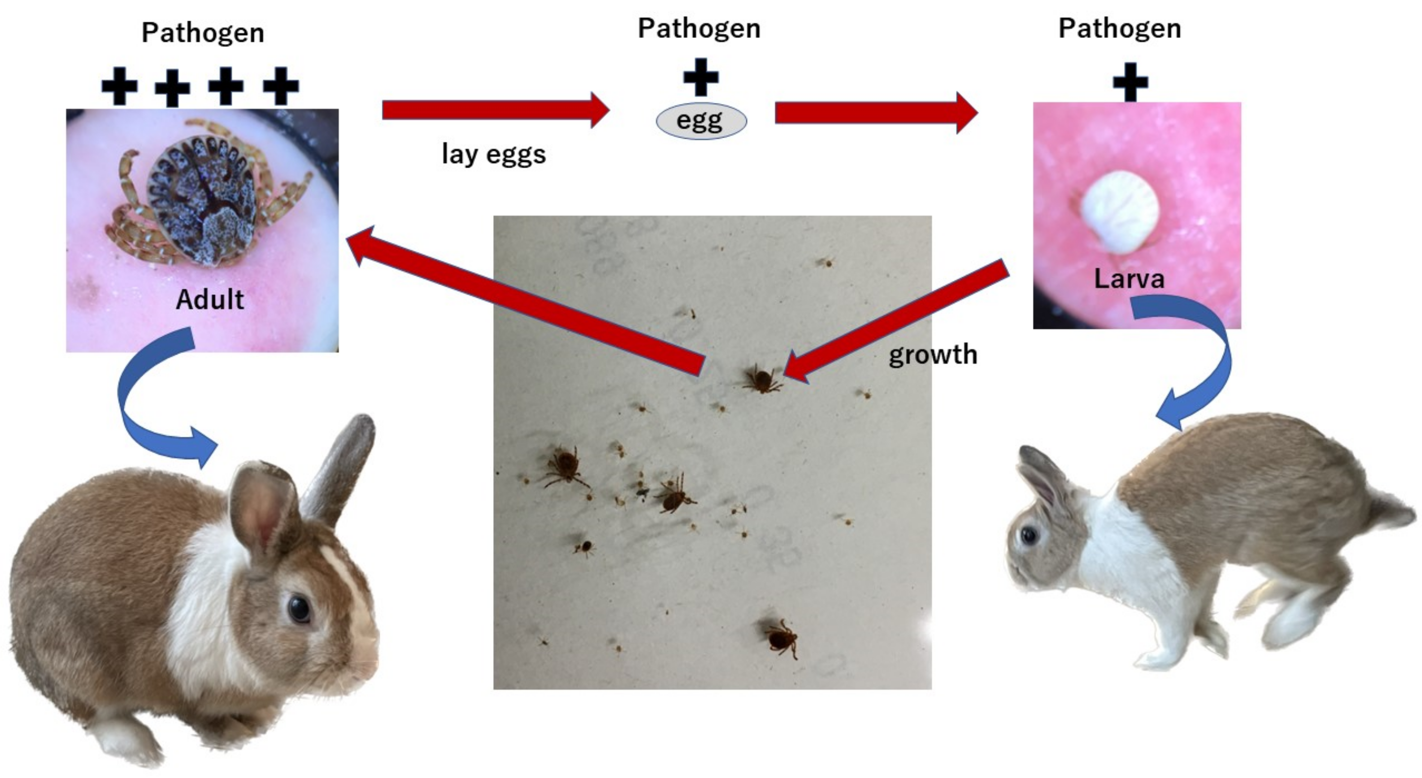
2. Materials and Methods
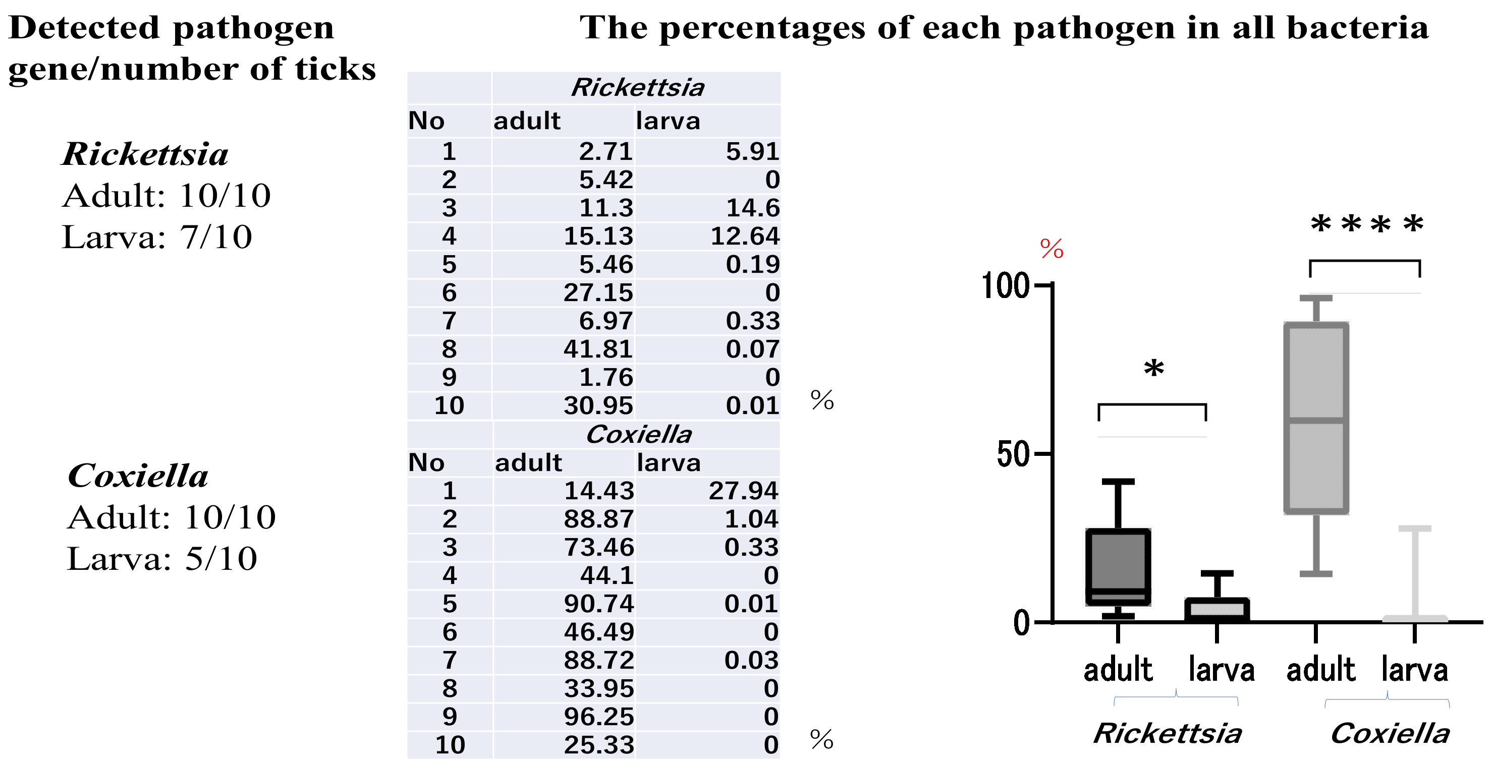
The types of bacteria in ticks collected from Minami-Ise, Mie Prefecture, Japan, were determined by analyzing the bacterial 16S rRNA regions using NGS. The area where larvae and adult ticks were collected in approximately the same numbers as in the Minami-Ise area, where the distribution of ticks was previously surveyed in summer, was selected as the tick survey area for this survey. This area was known to be an endemic area for Japanese spotted fever (JSF), with a large number of ticks harboring Rickettsia japonica (R. japonica). Furthermore, in this survey area, the reason for which Haemophysalis longicornis (H.L) was collected in large numbers during the summer season was that it was thought to reduce variation in the prevalence of tick-borne pathogens among tick species. Ten adult and ten larval ticks were captured in widely dispersed locations using the flagging method during the summer season. The ticks were placed in bottles and soaked overnight in 70% ethanol. The ticks were then removed using tweezers, crushed separately using a BioMasher II (Nippi, Tokyo, Japan), and stored in new tubes. DNA from the tick was extracted using a QIAamp DNA mini kit (Qiagen, German town, MD, USA) according to the manufacturer’s protocol. To identify the bacterial species, NGS analysis of the bacterial 16S rRNA lesions using DNA extracted from the ticks was performed by Macrogen Japan Corp. (Tokyo, Japan). In addition, DNA extracted from the ticks was used to perform polymerase chain reaction (PCR) using primers widely used for tick species identification [12]. The reaction mixture was placed in a thermal cycler (ASTEL GeneAtlas 485, Fukuoka, Japan), where it underwent one cycle of preheating (94 °C, 10 s), 35 cycles of denaturation (94 °C, 10 s), annealing (55 °C, 30 s), extension (72 °C, 30 s), and one cycle of delay (72 °C, 5 min). Sequences of the amplified PCR products were analyzed using Eurofins Genomics (Tokyo, Japan). The number of Rickettsia sp. and Coxiella sp. genes detected in the DNA extracted from ten adult and ten larval ticks was calculated. To make a comparative estimate of the number of pathogens in the adult and larval ticks, the percentages of Rickettsia sp. (KJ619629) and Coxiella sp. (KC776318) gene sequences identified were measured when the number of sequences of all pathogens was 100, based on the results of NGS analysis of DNA extracted from the ticks. Two independent groups (adult vs. larva) were compared using Student’s t-test.

3. Results

The ten adult and ten larval ticks were identified as H.L from the results of the sequencing of PCR products amplified using tick identification-specific primers. The numbers of Rickettsia and Coxiella spp. genes detected in the ten adult and ten larval ticks and the percentages of Rickettsia and Coxiella among all detected pathogen sequences are shown in Figure 1. Rickettsia sp. and Coxiella sp. genes were each detected in 10/10 of the adult ticks. Conversely, Rickettsia sp. was found in 7/10 and Coxiella sp. was found in 5/10 of the larval ticks. Anaplasma sp. was detected in only 1/10 of the adult ticks and was not detected in the larval ticks. Borrelia sp. and Francisella sp. were not detected in either life stage of the ticks. Coxiella sp. was the most common species among all the adult ticks (mean, 58.15%). The highest percentage of Coxiella sp. genes was approximately 96%, in one adult tick. The percentage of Rickettsia sp. genes found in the adult ticks ranged from 1.76% to 41.81% (mean, 15.56%). However, in the larval ticks, Coxiella sp. accounted for 0%–27% (mean 5.86%) of the genes detected, while Rickettsia sp. accounted for 0%–14.6% (mean 3.38%). Statistically significant differences were observed between adult ticks and larvae for both Rickettsia sp. and Coxiella sp. (Figure 1).
Diplorickettsia sp., which has not been previously reported in Japan, was detected in an adult tick. Moreover, Aeromonas sp. was detected in two larval ticks [13].

4. Discussion

Identifying pathogens within a tick does not qualify it as a reservoir of infection or prove infectivity, if the tick cannot transmit the pathogen. Furthermore, the NGS method used in the current study could only analyze the genus level. Within the genera Rickettsia and Coxiella, the gene sequences are extremely similar; therefore, it is difficult to identify them as individual species, even by quantitative PCR. The area where the ticks were surveyed is one of the most endemic areas in Japan for JSF caused by R. japonica. R. japonica has been detected in the blood of JSF-infected patients, and ticks in this region can transmit Rickettsia to the human body. We recognize the presence of residents with positive anti-IgG antibodies against Coxiella burnetti in the area studied (unpublished data). Hence, we considered it worthy of recognition, even at the genus level. Several studies have reported the percentage of Rickettsia in ticks at different growth stages [5,6,7,8]. The parentage of Coxiella sp. specific DNA detected in ticks has also been reported [8,9]. However, the growth stages of ticks during which the risk of transmission is the greatest remain unknown. There are no reports on the development of tick-borne infections caused by ticks still attached to the human body. PCR was used to confirm the presence of pathogens in the ticks. Gene detection indicates that ticks are carrying Rickettsia sp. or Coxiella sp.; however, evaluating the risk of infection is not feasible because PCR methods are not useful for determining the number of pathogens. This study investigated the detection rates of tick-borne pathogen genes and the relative percentages of these pathogens by performing NGS on ticks at different stages (adults and larvae). NGS results showed that adult ticks contained significantly higher amounts of Rickettsia sp. and Coxiella sp., whereas larval ticks contained few of these bacteria. The present study was based on a small number of ticks from a limited area. However, the area surveyed had the highest reported cases of JSF in Japan [1,14]. Most reported ticks in this area have been identified as H.L [15]. H.L harbors R. japonica, which causes JSF [16]. Therefore, the results regarding Rickettsia sp. in this study, which compared 10 adult H.L and 10 larval H.L ticks, should provide credible data on the risk of JSF infection from a bite by an adult H.L tick. Coxiella sp. were detected with NGS using DNA extracted from the ticks. This proved that ticks with Coxiella sp. that sucked human blood lacked sufficient infection in non-endemic areas of JSF in Mie Prefecture [17]. However, the Coxiella sp. in the surveyed area in Mie Prefecture likely had the potential to infect humans because residents in the area had antibody titers against Coxiella burnetti. As shown in Figure 2, ticks suck blood from wild animals, grow into adults, and lay eggs. The larvae analyzed in this study contained small amounts of tick-borne pathogens, and it is likely that Rickettsia sp. and Coxiella sp. multiply in the tick body as it grows. If the wildlife is the source of the pathogens detected in the ticks, then all adult ticks feeding from the same local wildlife likely contain the same types of pathogens and to the same degree. The present study was a comparative assessment of the risk of infection during the adult and larval stages of H.L ticks. In the future, it will be necessary to continue the survey by identifying areas where adult, nymph, and larval ticks can be captured in larger but equal numbers.

5. Conclusions

Wild animals serve as reservoirs for tick-borne diseases, and the number of these pathogens in ticks increases as they feed and grow. The number of pathogens transferred from adult ticks in the eggs is low. Larval ticks have not yet sucked blood from wild animals. Thus, the risk of disease pathogen transmission depends on the growth stage of the tick. Adult ticks are more likely to transmit pathogens than larvae.

Author Contributions

Conceptualization, M.K. Methodology, Y.M., T.N. and S.I. Writing—original draft preparation, M.K. Writing—review and editing, H.K. and K.Y. All authors have read and agreed to the published version of the manuscript.

Funding

This research received no external funding.

Institutional Review Board Statement

Not applicable.

Informed Consent Statement

Not applicable.

Data Availability Statement

All data supporting the findings of this study have been included in this article.

Conflicts of Interest

The authors declare no conflict to interest.

References

  1. Kondo, M.; Nishii, M.; Gabazza, E.C.; Kurokawa, I.; Akachi, S. Nine cases of Japan spotted fever diagnosed at our hospital in 2008. Int. J. Dermatol. 2010, 49, 430–434. [Google Scholar] [CrossRef] [PubMed]
  2. Nakata, R.; Motomura, M.; Tokuda, M.; Nakajima, H.; Masuda, T.; Fukuda, T.; Tsujino, A.; Yoshimura, T.; Kawakami, A. A case of Japanese spotted fever complicated with central nervous system involvement and multiple organ failure. Intern Med. 2012, 51, 783–786. [Google Scholar] [CrossRef] [PubMed]
  3. Angelakis, E.; Raoult, D. Q fever. Vet. Microbiol. 2010, 140, 297–309. [Google Scholar] [CrossRef] [PubMed]
  4. Graves, S.R.; Gerrard, J.; Coghill, S. Q fever following a tick bite. Aust. J. Gen. Pract. 2020, 49, 823–825. [Google Scholar] [CrossRef] [PubMed]
  5. Thu, M.J.; Qiu, Y.; Matsuno, K.; Kajihara, M.; Mori-Kajihara, A.; Omori, R.; Monma, N.; Chiba, K.; Seto, J.; Gokuden, M.; et al. Diversity of spotted fever group rickettsiae and their association with host ticks in Japan. Sci. Rep. 2019, 9, 1500. [Google Scholar] [CrossRef] [PubMed]
  6. Krishnamoorthy, P.; Sudhagar, S.; Goudar, A.L.; Jacob, S.S.; Suresh, K.P. Molecular survey and phylogenetic analysis of tick-borne pathogens in ticks infesting cattle from two South Indian states. Vet. Parasitol. Reg. Stud. Rep. 2021, 25, 100595. [Google Scholar] [CrossRef] [PubMed]
  7. Sato, M.; Ikeda, S.; Arai, R.; Kato, M.; Aoki, J.; Nishida, A.; Watanabe, K.; Hirokawa, C.; Watanabe, K.; Regilme, M.A.F.; et al. Diversity and distribution of ticks in Niigata Prefecture, Japan (2016–2018): Changes since 1950. Ticks Tick Borne Dis. 2021, 12, 101683. [Google Scholar] [CrossRef] [PubMed]
  8. Chalada, M.J.; Stenos, J.; Vincent, G.; Barker, D.; Bradbury, R.S. A molecular survey of tick-borne pathogens from ticks collected in central Queensland, Australia. Vector Borne Zoonotic Dis. 2018, 18, 151–163. [Google Scholar] [CrossRef] [PubMed]
  9. Watanabe, M.; Nakao, R.; Amin-Babjee, S.M.; Maizatul, A.M.; Youn, J.H.; Qiu, Y.; Sugimoto, C.; Watanabe, M. Molecular screening for Rickettsia, Anaplasmataceae and Coxiella burnetii in Rhipicephalus sanguineus ticks from Malaysia. Trop. Biomed. 2015, 32, 390–398. [Google Scholar] [PubMed]
  10. Morshed, M.G.; Lee, M.K.; Boyd, E.; Mak, S.; Fraser, E.; Nguyen, J.; Dykstra, E.A.; Otterstatter, M.; Henry, B.; Galanis, E. Passive Tick Surveillance and Detection of Borrelia Species in Ticks from British Columbia, Canada: 2002–2018. Vector Borne Zoonotic Dis. 2021, 21, 490–497. [Google Scholar] [CrossRef] [PubMed]
  11. Suzuki, J.; Hashino, M.; Matsumoto, S.; Takano, A.; Kawabata, H.; Takada, N.; Andoh, M.; Oikawa, Y.; Kajita, H.; Uda, A.; et al. Detection of Francisella tularensis and analysis of bacterial growth in ticks in Japan. Lett. Appl. Microbiol. 2016, 63, 240–246. [Google Scholar] [CrossRef] [PubMed]
  12. Ushijima, Y.; Oliver , J.H., Jr.; Keirans, J.E.; Tsurumi, M.; Kawabata, H.; Watanabe, H.; Fukunaga, M. Mitochondrial sequence variation in Carlos capensis (Neumann), a parasite of seabirds, collected on Torishima Island in Japan. J. Parasitol. 2003, 89, 196–198. [Google Scholar] [CrossRef] [PubMed]
  13. Kondo, M.; Matsushima, Y.; Nakanishi, T.; Iida, S.; Habe, K.; Yamanaka, K. A novel transmission of Aeromonas hydrophila via tick bites. J. Dermatol. 2022, 49, e243–e245. [Google Scholar] [CrossRef] [PubMed]
  14. Noguchi, M.; Oshita, S.; Yamazoe, N.; Miyazaki, M.; Takemura, Y.C. Important clinical features of Japanese spotted fever. Am. J. Trop. Med. Hyg. 2018, 99, 466–469. [Google Scholar] [CrossRef] [PubMed]
  15. Kondo, M.; Ando, K.; Yamanaka, K.; Mizutani, H. Rickettsia species AMONG ticks in an area of Japan endemic for Japanese spotted fever. Southeast Asian J. Trop. Med. Public Health 2017, 48, 585–589. [Google Scholar]
  16. Uchida, T.; Yan, Y.; Kitaoka, S. Detection of Rickettsia japonica in Haemaphysalis longicornis ticks by restriction fragment length polymorphism of PCR product. J. Clin. Microbiol. 1995, 33, 824–828. [Google Scholar] [CrossRef] [PubMed]
  17. Kondo, M.; Matsushima, Y.; Nakanishi, T.; Habe, K.; Yamanaka, K. Investigation of pathogens injected into the human body by ticks. Exp. Dermatol. 2022, 31, 1014–1015. [Google Scholar] [CrossRef] [PubMed]
Figure 1. The gene detection rates in the adult ticks were 10/10 for Rickettsia sp. and 10/10 for Coxiella sp. Conversely, the detection rates for larval ticks were 7/10 for Rickettsia sp. and 5/10 for Coxiella sp. * When the percentage of Rickettsia sp., out of all pathogens detected via next-generation sequencing, was analyzed between the adult and larval stages of the ticks, a significant difference was observed at p = 0.0254. **** For Coxiella sp., a highly significant difference (p < 0.0001) was found between the adult and larval stages of the ticks.
Clinpract 13 00022 g001
Figure 2. As ticks grow, pathogens multiply inside them. Adult ticks suck blood from wild animals and lay eggs, and tick-borne pathogens are present in the eggs. A few tick-borne pathogens are also present in larval ticks that have not yet sucked blood from a wild animal.
Clinpract 13 00022 g002
Disclaimer/Publisher’s Note: The statements, opinions and data contained in all publications are solely those of the individual author(s) and contributor(s) and not of MDPI and/or the editor(s). MDPI and/or the editor(s) disclaim responsibility for any injury to people or property resulting from any ideas, methods, instructions or products referred to in the content.

