Case Report

HbAdrian (α1:c.251del, p.Leu84Argfs*19)—A Novel Pathogenic Variant in the α1-Globin Gene Associated with Microcytosis from the North of Iran

1
Thalassemia Research Center, Hemoglobinopathies Institute, Mazandaran University of Medical Sciences, Sari 48175866, Iran
2
Sinaye Mehr Research Center, Mazandaran University of Medical Sciences, Sari 4817993375, Iran
*
Author to whom correspondence should be addressed.
Thalass. Rep. 2023, 13(2), 152-156; https://doi.org/10.3390/thalassrep13020014
Submission received: 1 March 2023 / Revised: 30 April 2023 / Accepted: 18 May 2023 / Published: 1 June 2023

Abstract

:
Background: Alpha thalassemia is one of the most common human genetic abnormalities. More than 400 different variations of the α-globin protein have been introduced, most of which are not associated with noticeable clinical manifestations. The identification of all variants of Hb in different regions helps in acquiring comprehensive knowledge concerning thalassemia disease, and it can be used in preventive programs as well as prenatal diagnosis (PND). Aims: In the present study, we describe a new α1 gene mutation that leads to a frameshift after codon 83. Methods: As a plan for a national screening program of thalassemia, routine cell blood count (CBC) and Hb capillary electrophoresis tests were applied. After taking written informed consent, genomic DNA was extracted, and, for identifying common Mediterranean α-Globin gene deletion, multiplex Gap-PCR was performed; for detecting other mutations on α- and β-Globin genes, a DNA sequencing method was used. Results: The results of CBC and capillary electrophoresis tests showed microcytosis in a female subject. The sequencing of the α-Globin gene showed that the case is heterozygote for a single-nucleotide deletion at codon 83 of the α1-Globin Gene. We named this mutation Hb Adrian (α1: c.251–T), which is a novel mutation. The mentioned mutation was also detected in the subject’s mother. Conclusions: The introduced mutation (Hb Adrian) leads to a frameshift change that produces a protein with 100 amino acids, which in comparison to a normal α-chain is shorter, and its amino acids are altered after codon 83. This hemoglobin is undetectable via the use of electrophoresis. Although no major hematological abnormalities were observed in the carriers, Hb Adrian should be considered in screening programs to help prevent Hb H disease in high-risk couples.

1. Introduction

Thalassemia is the most common monogenic genetic disease, with an estimated global carrier rate of 1–5% [1,2]. Approximately, 70,000 severely affected infants are born annually [3]. The disease is a form of hemoglobinopathy characterized by the absence or decreased expression of α- and β-globin genes. Thalassemia was initially restricted to the tropical and subtropical regions, including the Mediterranean, Sub-Saharan Africa, the Middle East, and Southern as well as Eastern Asia; however, regional migrations have increased its incidence rate in various parts of Europe and even in northern as well as southern parts of America [4].
Alpha thalassemia is one of the most common human genetic abnormalities throughout the world and Iran (especially in its northern regions). Depending on the copies of the defected genes, the clinical presentation of the disease is varied, ranging from being almost asymptomatic to lethal hemolytic anemia [5,6,7]. So far, more than 400 different variations of the α-globin gene have been identified [8].
The deletion of one (−α/αα) or two α-globin (−−/αα, −α/−α) genes leads to asymptomatic forms of α-thalassemia known as silent carrier and thalassemia trait, respectively. These deletions lead to an imbalance between α- and non-α-globin chain production. The compound heterozygosity of α+ and α0 mutations (−−/−α) results in HbH disease, which is mostly observed in Southeast Asia, as well as the Mediterranean region. The clinical manifestations of HbH disease are described by a considerable variable extent of anemia. These patients may indicate hepatosplenomegaly, variable forms of jaundice, gallstones, and hemolytic crisis [9]. The complete absence of α-globin chain production results in Hb Bart’s hydrops fetalis; the extent of anemia, cardiovascular disorders, and other pronounced complications of this condition, such as hemochromatosis, usually lead to intrauterine death [10].
National programs for preventing children being born with thalassemia began in Iran since 1997 [11]. As a part of this program, premarital screening for carriers is compulsory for each couple. The identification of all variants of Hb in different regions helps in obtaining comprehensive knowledge about thalassemia disease, and it can be used in preventive programs as well as prenatal diagnosis (PND). In the present study, we describe a new α1 gene mutation that leads to a frameshift after codon 83 (α1: c.251del T).

2. Materials and Methods
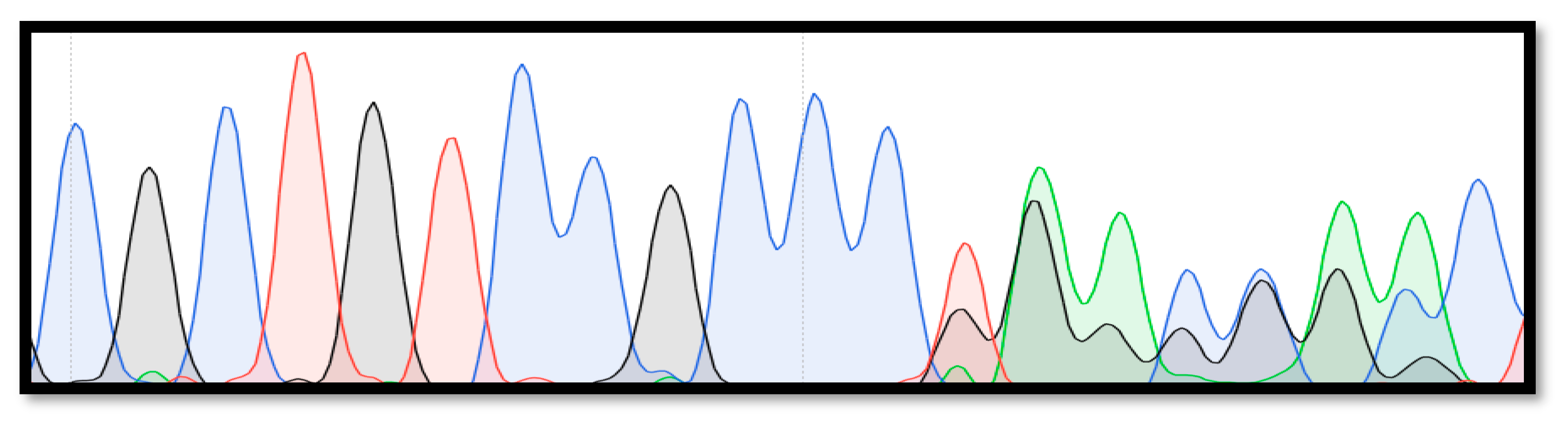
As a part of the national program for the premarital screening of thalassemia, a 29-year-old female was referred to the Fajr Medical Genetics Laboratory (Sari, Iran) for a routine hematological analysis. At first, CBC (complete blood count) and Hb capillary electrophoresis (Sebia, France) tests were carried out. Since the subject had reduced hematological indices, further molecular analyses were applied for the detection of pathogenic variants of the α- and β-globin genes. After taking written informed consent, a molecular analysis was conducted on genomic DNA extracted from peripheral blood via the use of a QIAamp DNA Mini Kit (Qiagen, Germany). For identifying common Mediterranean α-Globin gene deletions, multiplex Gap-PCR was performed, which identifies the presence of −α.3.7 and −α.4.2 single-gene deletions, the —SEA and —FIL Southeast Asian double-gene deletions, the —MED and −α.20.5 Mediterranean double-gene deletions, and αααAnti7 triplication [12]. For the detection of point mutations on the α- and β-Globin genes, the entire regions of these genes were first amplified with specific primers, and subsequently a Sanger sequencing method was used (3130xl genetic analyzer, ABI, Sab Fransisco, CA, USA). An in silico analysis, using VarSome, a search engine for human genomic variation, Mutation Taster, and SIFT (Sorting Intolerant from Tolerant) predicting tools, was applied to analyze the pathogenicity of the novel variant.

3. Results

The results of the CBC and capillary electrophoresis tests showed that the case and her mother had microcitosis, while her father had normal hematological indices (Table 1). Based on the multiplex Gap-PCR test results, the case did not carry the investigated common α-Globin gene deletions and αααAnti7 triplication. The sequencing of the α-Globin gene indicated that the subject carried a single-nucleotide deletion at codon 83 of the α1-Globin gene (Figure 1). The mentioned mutation was also detected in subject’s mother. The VarSome search engine predicted this variant as likely pathogenic. The Mutation Taster and SIFT online software categorized this frameshift alteration as disease-causing and damaging, respectively. We named this mutation Hb Adrian (α1: c.251del T), which is a novel variant.

4. Discussion

Thalassemia is a common hereditary genetic disorder in the north of Iran, and our previous studies indicated that around 15% of the population carry α-globin gene mutations [6]. Based on our previous report of the 412 neonates that were randomly selected, the -3.7 deletion had the highest frequency, 9.7%, and -4.2 deletion, ααα anti3.7 triplication, and α−5nt mutations had frequencies of 4.1%, 2.2%, and 0.49%, respectively, while none of the neonates had a --Med double-gene deletion. Tamaddoni et al. investigated the mutations among two hundred and fifty-five patients from Mazandaran, and 21 different mutations were observed among the investigated patients. According to the results of their study, the -3.7 deletion (44.9%), polyadenylation signal 2 (α Poly A2) (AATAAA > AATGAA) (18.2%), -4.2 deletion (9.1%), α-5nt (6.5%), - -MED (4.3%), and α codon 19 (-G) (4%) were the most frequent mutations observed in the region [13]. Moreover, several variants of hemoglobin, such as Hb D [14], Hb J-Toronto [15], Hb Setif [16], Hb Fontainebleau [17], Hb Daneshgah-Tehran [18], and Hb S [19], have also been reported from that region. Since Mazandran is located in the south of the Caspean Sea, which has a subtropical climate as well as a high prevalence of malaria having been observed in the region in the past, a high frequency of thalassemia is reported from there. In the present study, we report a novel pathogenic variant in the α1-globin gene, Hb Adrian (α1: c.251del T), detected in a family from Mazandaran, in the north of Iran. In our previous work we have also reported another new variant of α-globin (Hb Mazandaran) and the coinheritance of α- and β-globin gene mutations from Mazandaran province [20,21], which indicate the diversity of Hb variants in the region. The presented case was originally from Mazandaran province. There were several migrations of minor ethnic groups, such as Kurds, Turks, Georgians, and small groups of Indian origin, to Mazandaran province throughout history [17] that may have resulted in a high diversity of α-globin gene variants in the region.
Hb H disease usually occurs due to the coinheritance of α0-thalassemia due to deletions that remove both linked α-globin genes on chromosome 16 and deletional α+-thalassemia from single α-globin gene deletions (−−/−α); however, Hb H disease may occur from interactions between α0-thalassemia with non-deletional mutations or with abnormal hemoglobin, such as Hb Constant Spring [22]. It has been indicated that the clinical manifestations of Hb H disease caused by non-deletional types are usually more severe than those resulting from deletional types [23]. Non-deletional mutations can result in unstable hemoglobin that usually precipitates in red blood cells, forming insoluble inclusion bodies and eventually damaging or destroying red blood cells’ membranes [24].
The introduced variant (Hb Adrian) leads to a frameshift change that produces a truncated protein with 100 amino acids, which in comparison to a normal α-chain is shorter, and its amino acids are altered after codon 83. This hemoglobin is undetectable by electrophoresis and causes microcytosis. Although no severe hematological abnormalities were observed in the carriers, Hb Adrian should be considered in screening programs, especially in combination with double-gene CIS deletions, in order to prevent Hb H disease in couples at risk for α-thal.

Author Contributions

M.M. and H.J.; methodology, M.M.; software, H.K.; formal analysis, M.M.; data curation, H.J. and H.K.; writing—original draft preparation, M.R.M.; writing—review and editing, M.R.M.; project administration. All authors have read and agreed to the published version of the manuscript.

Funding

This research received no external funding.

Institutional Review Board Statement

Not applicable.

Informed Consent Statement

Informed consent was obtained from all subjects involved in the study.

Data Availability Statement

Data can be made available upon request to the corresponding author.

Acknowledgments

The authors are grateful to Bita Talebi, Maryam Rahimi, and Mohammad Amirzadegan for their assistance in the sampling, as well as in the writing of the manuscript. This case report was coordinated by the Fajr Genetics and Pathobiology Laboratory.

Conflicts of Interest

The authors declare no conflict of interest.

References

  1. Higgs, D.R.; Engel, J.D.; Stamatoyannopoulos, G. Thalassaemia. Lancet 2012, 379, 373–383. [Google Scholar] [CrossRef] [PubMed]
  2. Taher, A.T.; Weatherall, D.J.; Cappellini, M.D. Thalassaemia. Lancet 2018, 391, 155–167. [Google Scholar] [CrossRef] [PubMed]
  3. Mettananda, S.; Higgs, D.R. Molecular basis and genetic modifiers of thalassemia. Hematol./Oncol. Clin. 2018, 32, 177–191. [Google Scholar] [CrossRef]
  4. Mettananda, S.; Gibbons, R.J.; Higgs, D.R. α-Globin as a molecular target in the treatment of β-thalassemia. Blood J. Am. Soc. Hematol. 2015, 125, 3694–3701. [Google Scholar] [CrossRef]
  5. Galanello, R.; Cao, A. Alpha-thalassemia. Genet. Med. 2011, 13, 83–88. [Google Scholar] [CrossRef] [PubMed]
  6. Jalali, H.; Mahdavi, M.R.; Roshan, P.; Kosaryan, M.; Karami, H.; Mahdavi, M. Alpha thalassemia gene mutations in neonates from Mazandaran, Iran, 2012. Hematology 2014, 19, 192–195. [Google Scholar] [CrossRef]
  7. Farashi, S.; Harteveld, C.L. Molecular basis of α-thalassemia. Blood Cells Mol. Dis. 2018, 70, 43–53. [Google Scholar] [CrossRef]
  8. Giardine, B.; van Baal, S.; Kaimakis, P.; Riemer, C.; Miller, W.; Samara, M.; Kollia, P.; Anagnou, N.P.; Chui, D.H.; Wajcman, H.; et al. HbVar database of human hemoglobin variants and thalassemia mutations: 2007 update. Hum. Mutat. 2007, 28, 206. [Google Scholar] [CrossRef]
  9. Grosso, M.; Sessa, R.; Puzone, S.; Storino, M.R.; Izzo, P. Molecular basis of Thalassemia. Anemia 2012, 2012, 341–358. [Google Scholar]
  10. Weatherall, D.J.; Clegg, J.B. The Thalassaemia Syndromes; John Wiley & Sons: Hoboken, NJ, USA, 2008. [Google Scholar]
  11. Hashemieh, M.; Sessa, R.; Puzone, S.; Storino, M.R.; Izzo, P. The Iran thalassemia prevention program: Success or failure? Iran. J. Pediatr. Hematol. Oncol. 2015, 5, 161. [Google Scholar]
  12. Chong, S.S.; Boehm, C.D.; Higgs, D.R.; Cutting, G.R. Single-tube multiplex-PCR screen for common deletional determinants of α-thalassemia. Blood J. Am. Soc. Hematol. 2000, 95, 360–362. [Google Scholar]
  13. Tamaddoni, A.; Hadavi, V.; Nejad, N.H.; Khosh-Ain, A.; Siami, R.; Aghai-Meibodi, J.; Almadani, N.; Oberkanins, C.; Law, H.Y.; Najmabadi, H. α-Thalassemia mutation analyses in Mazandaran province, North Iran. Hemoglobin 2009, 33, 115–123. [Google Scholar] [CrossRef] [PubMed]
  14. Mahdavi, M.R.; Jalali, H.; Kosaryan, M.; Roshan, P.; Mahdavi, M. β-Globin gene cluster haplotypes of Hb D-Los Angeles in Mazandaran province, Iran. Genes Genet. Syst. 2015, 90, 55–57. [Google Scholar] [CrossRef] [PubMed]
  15. Mahdavi, M.R.; Bayat, N.; Hadavi, V.; Karami, H.; Roshan, P.; Najmabadi, H.; Rohanizadeh, H. Report of haemoglobin J-Toronto and alpha thalassemia in a family from North of Iran. JPMA-J. Pak. Med. Assoc. 2012, 62, 396. [Google Scholar] [PubMed]
  16. Mahdavi, M.R.; Karimi, M.; Yavarian, M.; Roshan, P.; Kosaryan, M.; Siami, R. Detection of Hb Setif in north Iran and the question of its origin: Iranian or multiethnic? Hemoglobin 2011, 35, 152–156. [Google Scholar] [CrossRef] [PubMed]
  17. Jalali, H.; Rasouli, S.; Najafi, M.; Karami, H.; Mahdavi, M. A report of Hb Fontainebleau [α21 (B2) Ala> Pro] as a result of founder effect phenomenon. Gene Rep. 2020, 19, 100587. [Google Scholar] [CrossRef]
  18. Jalali, H.; Mahdavi, M.R.; Karami, H. Hemoglobin Daneshgah-Tehran (HBA1: c. 218A> G p. His72Arg): A Rare α1-Globin Variant from Iran. Iran. J. Pediatr. Hematol. Oncol. 2020, 10, 200–202. [Google Scholar]
  19. Aghajani, F.; Mahdavi, M.R.; Kosaryan, M.; Mahdavi, M.; Hamidi, M.; Jalali, H. Identification of β-globin haplotypes linked to sickle hemoglobin (Hb S) alleles in Mazandaran province, Iran. Genes Genet. Syst. 2016, 91, 311–313. [Google Scholar] [CrossRef]
  20. Jalali, H.; Mahdavi, M.R.; Mahdavi, M.; Abbasi, A. Hb Mazandaran (α1) α51 Gly> Cys (CE9), c. 154 GGC> TGC: A Novel Haemoglobin Variant of α1-Globin Gene. Thalass. Rep. 2022, 12, 51–54. [Google Scholar]
  21. Jalali, H.; Karami, H.; Mahdavi, M.R.; Mahdavi, M. Co-Inheritance of Heterozygous β0-Thalassemia with Single Functional α-Globin Gene: Challenges of Carrier Detection in Pre-Marital Screening Program for Thalassemia. Thalass. Rep. 2022, 12, 101–104. [Google Scholar] [CrossRef]
  22. Fucharoen, S.; Viprakasit, V. Hb H disease: Clinical course and disease modifiers. ASH Educ. Program Book 2009, 2009, 26–34. [Google Scholar] [CrossRef] [PubMed]
  23. Papassotiriou, I.; Traeger-Synodinos, J.; Kanavakis, E.; Karagiorga, M.; Stamoulakatou, A.; Kattamis, C. Erythroid marrow activity and hemoglobin H levels in hemoglobin H disease. J. Pediatr. Hematol./Oncol. 1998, 20, 539–544. [Google Scholar] [CrossRef] [PubMed]
  24. Kalle Kwaifa, I.; Lai, M.I.; Md Noor, S. Non-deletional alpha thalassaemia: A review. Orphanet J. Rare Dis. 2020, 15, 1–12. [Google Scholar] [CrossRef] [PubMed]
Figure 1. Direct sequencing results of the α1-globin gene in a case with Hb Adrian (α1: c.251del T).
Thalassrep 13 00014 g001
Table 1. Hematological indices of the subject with Hb Adrian (α1: c.251del T), as well as those of her parents.
Age (y)RBC
(×106/µL)
Hb
(g/dL)
MCV
(fl)
MCH
(pg)
Hb-A
(%)
Hb-A2
(%)
Hb-F
(%)
Subject295.1111.97423.397.42.6<0.5
Mother513.8310742397.22.8<0.5
Father544.931390.126.597.32.7<0.5
Disclaimer/Publisher’s Note: The statements, opinions and data contained in all publications are solely those of the individual author(s) and contributor(s) and not of MDPI and/or the editor(s). MDPI and/or the editor(s) disclaim responsibility for any injury to people or property resulting from any ideas, methods, instructions or products referred to in the content.

