Article

Alpha-Thalassemia: Diversity of Clinical Phenotypes and Update on the Treatment

by
Duantida Songdej
1 and
Suthat Fucharoen
2,*
1
Pediatric Hematology-Oncology, Department of Pediatrics, Faculty of Medicine Ramathibodi Hospital, Mahidol University, Bangkok 10400, Thailand
2
Thalassemia Research Center, Institute of Molecular Biosciences, Mahidol University, Nakhon Pathom 73170, Thailand
*
Author to whom correspondence should be addressed.
Thalass. Rep. 2022, 12(4), 157-172; https://doi.org/10.3390/thalassrep12040020
Submission received: 16 September 2022 / Revised: 15 November 2022 / Accepted: 19 November 2022 / Published: 22 November 2022

Abstract

:
One of the more common single-gene disorders worldwide is α-thalassemia, carriers of which are found at variable frequencies (>1%) across all tropical and subtropical countries. Two linked α-globin genes on each allele of chromosome 16 regulate α-globin chain production. Deletion of one or more α-globin genes is the most frequent molecular defect found in α-thalassemia, whereas non-deletional mutations also occur, leading to unstable α-globin chains. HbH is the most common clinically important α-thalassemia disease and occurs when three α-globin genes are deleted/mutated, leaving only one copy of the gene intact. HbH can be divided into deletional (--/-α) and non-deletional genotypes (--/αTα). Whereas clinical phenotypes of the former are usually homogenously mild to moderate, those of the latter can be diverse. As HbH disease is particularly prevalent in Southeast Asia and some parts of the Mediterranean region, where β-thalassemia is also prevalent, affected patients are sometimes left undertreated. Therefore, hematologists and general physicians need to be educated to provide optimal disease monitoring and early identification of those with more severe phenotypes. Some issues regarding transfusion and iron chelation management differ from those of β-thalassemia, and these need to be recognized. Hb Bart’s hydrops fetalis syndrome (BHFS) is the most severe form of α-thalassemia; affected patients lack production of α-globin chains. Recent advances in fetal medicine and neonatal intensive care have made it possible for BHFS to no longer constitute a universally fatal disorder. Transfusion and chelation strategies for rare survivors are distinct and require updating.

1. Introduction

α-thalassemia occurs as a result of decreased or absent α-globin chain production and is characterized by a microcytic hypochromic anemia. Whereas affected patients with mild forms of α-thalassemia may only be detected incidentally as having microcytosis, those affected with moderate-to-severe forms present a wide range of clinical phenotypes, from asymptomatic anemia to hydrops fetalis. The former group is said to possess the α-thalassemia trait, whereas the two most clinically important diseases of the latter group are called Hemoglobin (Hb) H disease and Hb Bart’s hydrops fetalis syndrome (BHFS). HbH is most frequently caused by deletion of two duplicated α-globin genes on one allele of chromosome 16 and another one of the two α-globin genes on the other (--/-α), resulting in reduced α-globin synthesis to usually less than 30% [1]. This condition is also known as deletional HbH. As a consequence, excess β-like globin chains form tetramers called Hb Bart’s (γ4) in fetal life and HbH (β4) in postnatal life. Less commonly, HbH results from interactions between both α-globin genes deleted on one allele and a point mutation or insertion of one of the two genes on the other (--/αTα). This condition is termed non-deletional HbH, clinical signs and symptoms of which are generally more severe than those of deletional HbH. Anemia and tissue hypoxia experienced by affected patients with HbH and BHFS mainly result from reduced or absent main functional oxygen-carrying molecules, HbA (α2β2), as well as ineffective erythropoiesis and peripheral hemolysis caused by precipitated unstable HbH and Hb Bart’s. Additionally, the existence of non-functional high-oxygen affinity HbH (ranging from 0.8 to 40% in HbH disease) or Hb Bart’s (up to 100% in BHFS) in the blood stream [1] worsens the degree of tissue hypoxia.
As carriers of α-thalassemia are known to possess a selective advantage against falciparum malaria [2,3], undoubtedly, α+-thalassemias (-α) spread at a high frequency across tropical and subtropical areas of the world, including Mediterranean countries, Southeast Asia, Africa, the Middle East and the Indian subcontinent [4,5]. However, α0-thalassemia (--) is less common and occurs at a high frequency particularly in Southeast Asia and some parts of the Mediterranean region [4]. For example, a 1990’s Thai national survey revealed average carrier frequencies of α0- and α+-thalassemia of 20 to 30% [6], with the frequency going up to 40% in some areas of the northern part of the country [7]. This made HbH the most common thalassemia, accounting for ~75% of all thalassemia diseases found in Thailand [8]. The national policy for thalassemia carrier screening at antenatal care settings has been established since 1995, and it incorporated screening of α0-thalassemia carriers to prevent the birth of neonates with BHFS. A more recent study employed geostatistical modelling methods to estimate a continuous map of α-thalassemia mutations based on the geodatabase of α-thalassemia prevalence and genetic diversity surveys [9]. It has been demonstrated that allele frequencies of α+-, α0- and non-deletional α-thalassemia (αT) across all regions of Thailand were 2.43 to 15.03%, 0.57 to 4.46% and 1.57 to 1.65%, respectively. This gave rise to estimated numbers of newborns affected with BHFS, deletional and non-deletional HbH in 2020 of >400, >2600 and ~500 cases [9]. These data show α-thalassemia has remained an important public health problem in Thailand, where its population in 2019 totaled >65 million and that of live births >590,000 (data from the Ministry of Public Health, Thailand).
Estimated carrier frequencies of α+- and α0-thalassemia in other countries in Southeast Asia were reported to be ~12% in Vietnam, ~32% in Myanmar, ~36% in Lao PDR and ~37% in Cambodia, which were comparable to those observed in Thai populations [10]. Over the past few decades, owing to the effect of migration from countries with high carrier frequency, α-thalassemia has been increasingly observed outside tropical regions. A good example of this is California, USA, where the incidence of α-thalassemia—including HbH, HbH with Hb Constant Spring (CS) and BHFS—was reported to be 1 in 9000 births. This led to incorporating newborn screening for HbH disorders in the statewide hemoglobinopathy screening program since 1999 [11,12,13,14]. Prenatal diagnosis of BHFS in the region has also been offered. Similarly increasing α-thalassemia carrier frequency in previously non-endemic areas has led to developing carrier screening programs in Europe, such as in the Netherlands [15].
HbH disease was generally thought to be a mild form of thalassemia. More recently, its phenotypic variability, mostly related to α-thalassemia genotypes, has increasingly been recognized. As a result, the diagnosis, disease monitoring and treatments have been improved. BHFS has arisen as a hot topic in the α-thalassemia community over the last few years. This was owing to the emergence of the possibility of survival for this previously fatal disorder. In this article, phenotypic diversity, diagnosis, new issues on clinical course monitoring and treatments of HbH and BHFS will be covered.

2. Phenotypic Diversity of HbH Disease

2.1. Effects of α-Globin Genotypes on Severity of HbH

All affected individuals with HbH disease encounter a variable extent of anemia. However, as compared to deletional-HbH, those affected with non-deletional HbH generally present with more severe clinical signs and symptoms. Many patients with deletional HbH were diagnosed initially on the basis of a routine full blood count checkup or during an episode of infection/inflammation-induced hemolysis. Jaundice due to an acute hemolytic episode among these patients is uncommon; however, ~38% of patients with HbH have asymptomatic gallstones [16]. Growth retardation among deletional HbH is relatively rare. HbH with Hb CS (HbH-HbCS; --/αCSα) is the most common genotype of non-deletional HbH found worldwide, including Thailand [12,16,17]. Examples of other regionally common or recurring non-deletional HbH are HbH with Hb Pakse (HbH-HbPS) [18], HbH with Hb Quong Sze (HbH-HbQS) [19], HbH with Hb Pak Num Po (HbH-HbPNP) [20,21], HbH with Hb Adana (HbH-HbAdana) [22], HbH with Poly-adenylation signal mutation (HbH-PolyA) [23] and HbH with Hb Suan-Dok (HbH-HbSuan-Dok) [24,25]. Data comparing clinical manifestations between deletional and non-deletional HbH, derived from previous studies [16,17,26,27,28,29,30,31] and 246 Thai pediatric patients with HbH (Songdej D, manuscript in preparation), are summarized in Table 1. Clinical significances of regionally recurring non-deletional HbH are shown in Table 2.
While the clinical phenotype of affected patients with deletional HbH is generally uniform, the clinical presentation and natural history of those affected with non-deletional HbH are markedly diverse. Good examples of this are patients with HbH-HbCS. Even with the same α-globin genotype, some patients never require medical attention and were identified as having the disease only after investigations for other reasons, whereas a very small number of the patients exhibited severe fetal anemia, leading to hydropic features in utero [32,33]. This latter clinical syndrome is termed, “HbH hydrops fetalis”, which actually occurred more frequently among patients with rare non-deletional HbH genotypes, such as HbH-HbQS [34] and HbH-HbAdana [35], than among those with common HbH-HbCS. Transfusion requirements among HbH-HbCS and HbH-HbH-PS were also diverse. Data from the Hematology Clinic, Department of Pediatrics, Faculty of Medicine Ramathibodi Hospital, Mahidol University, Thailand, showed that 21.5% of such patients developed transfusion-dependent thalassemia (TDT) (Songdej d, manuscript in preparation). Similar proportions of patients with HbH-HbCS/HbH-HbPS requiring regular transfusion were also reported in two other related studies [16,17]. Prenatal diagnosis of pregnancies at risk of HbH-HbCS/HbH-HbPS is not generally recommended, as the majority of affected patients live with transfusion-independency into late adulthood. Information regarding the clinical course of the diseases, however, should be provided to the couples. Prenatal diagnosis should be offered to couples with a previous history of severely affected offspring with uncommon non-deletional HbH.

2.2. Effects of Co-Inheritance of β-Thalassemia

As HbE is very common in Southeast Asia and southern China, with its carrier frequency of up to 50% in some part of Thailand [10], interactions between HbH disease and HbE are commonly identified. Co-inheritance of HbH and heterozygous HbE is known as AE Bart’s disease, whereas co-inheritance of HbH with homozygous HbE is known as EF Bart’s disease. Clinical presentations of affected patients with AE Bart’s and EF Bart’s diseases were similar to those of HbH with the same α-globin genotypes. However, less chance of infection/inflammation-induced hemolysis was observed among AE Bart’s and EF Bart’s diseases compared to HbH [26,36]. Co-inheritance of HbH with heterozygous β-thalassemia usually resulted in a more severe degree of anemia compared with β-thalassemia heterozygote alone. However, the severity of anemia is less than common HbH disease [28].

2.3. Other Genetic Modifiers of HbH

To date, it remains difficult to predict clinical severity of HbH once initially diagnosed, especially among those with non-deletional genotypes. Studies on genetic modifiers underlying phenotypic heterogeneity of the disease continue far beyond those of β-thalassemia and remain an open field for the scientific community. Apart from co-inheritance of HbE and β-thalassemia heterozygote, two possible disease modifiers, α-thalassemia/mental retardation (ATRX) and α-hemoglobin stabilizing proteins (AHSP) have been explored [37,38,39]. Mutations in the ATRX gene on chromosome Xq13.3 cause a rare ATRX syndrome. The ATRX protein interacts with other genetic factors to bring about normal expression of many genes, including α- but not β-globin genes [28,38]. Affected males with ATRX syndrome, despite having four intact α-globin genes, encountered α-globin chain deficiency and HbH production, similar to that found in typical HbH disease. The mechanism by which AHSP modifies the severity of HbH is unclear. A recent study demonstrated that expression of AHSP negatively correlated with the Hb level of patients with HbH but positively correlated with disease severity [39]. Expression of AHSP was also found at higher levels in non-deletional compared with deletional HbH, with the highest levels found in HbH-HbCS and HbH-Hb-PS [39]. However, the usefulness of AHSP expression as a biomarker among patients with the same non-deletional HbH genotypes presenting diverse clinical severity remains unknown.

3. Diagnosis of HbH Disease

3.1. Hb Levels and Red Cell Indices

Most affected individuals with HbH disease have anemia with variable Hb levels. In both pediatric and adult patients, Hb generally ranges between ~9 and 11 g/dL in deletional and 8 and 9 g/dL in non-deletional HbH [17,28,31]. Mean corpuscular volume (MCV) appears to be lower than normal since the neonatal period, and it is usually found between ~55 and 65 fL among affected children and adults with deletional HbH [17,28]. Red cells containing Hb-CS become overhydrated; this in turn minimizes the expected microcytosis [12,40]. As such, the MCV of affected patients with HbH-HbCS is typically higher than that of deletional HbH, ranging from ~65 to 75 fL [17,28]. Low mean corpuscular hemoglobin (MCH) of ~17 to 19 pg and ~18 to 21 pg are also found in deletional and non-deletional HbH, respectively. Those affected with AE Bart’s and EF Bart’s disease exhibited a lower MCV but similar Hb level compared with their HbH counterparts. Such patients’ MCV can be as low as ~45 to 50 fL in deletional and ~50 to 60 fL in non-deletional AE Bart’s diseases [17]. Apart from microcytosis and hypochromia, peripheral blood smear (PBS) of patients with HbH also showed marked anisocytosis and poikilocytosis, similar to β-thalassemia blood pictures.

3.2. Inclusion Bodies
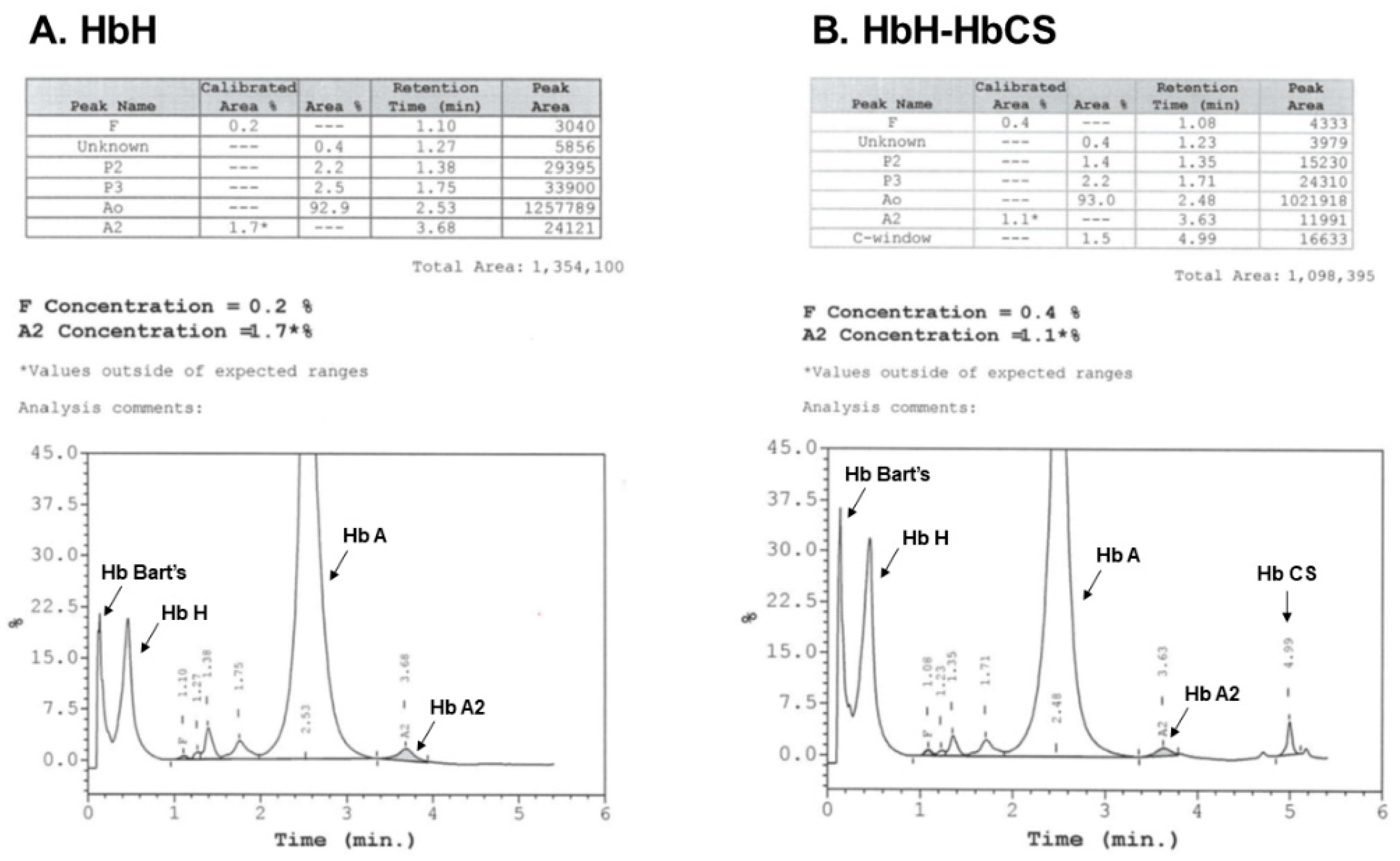
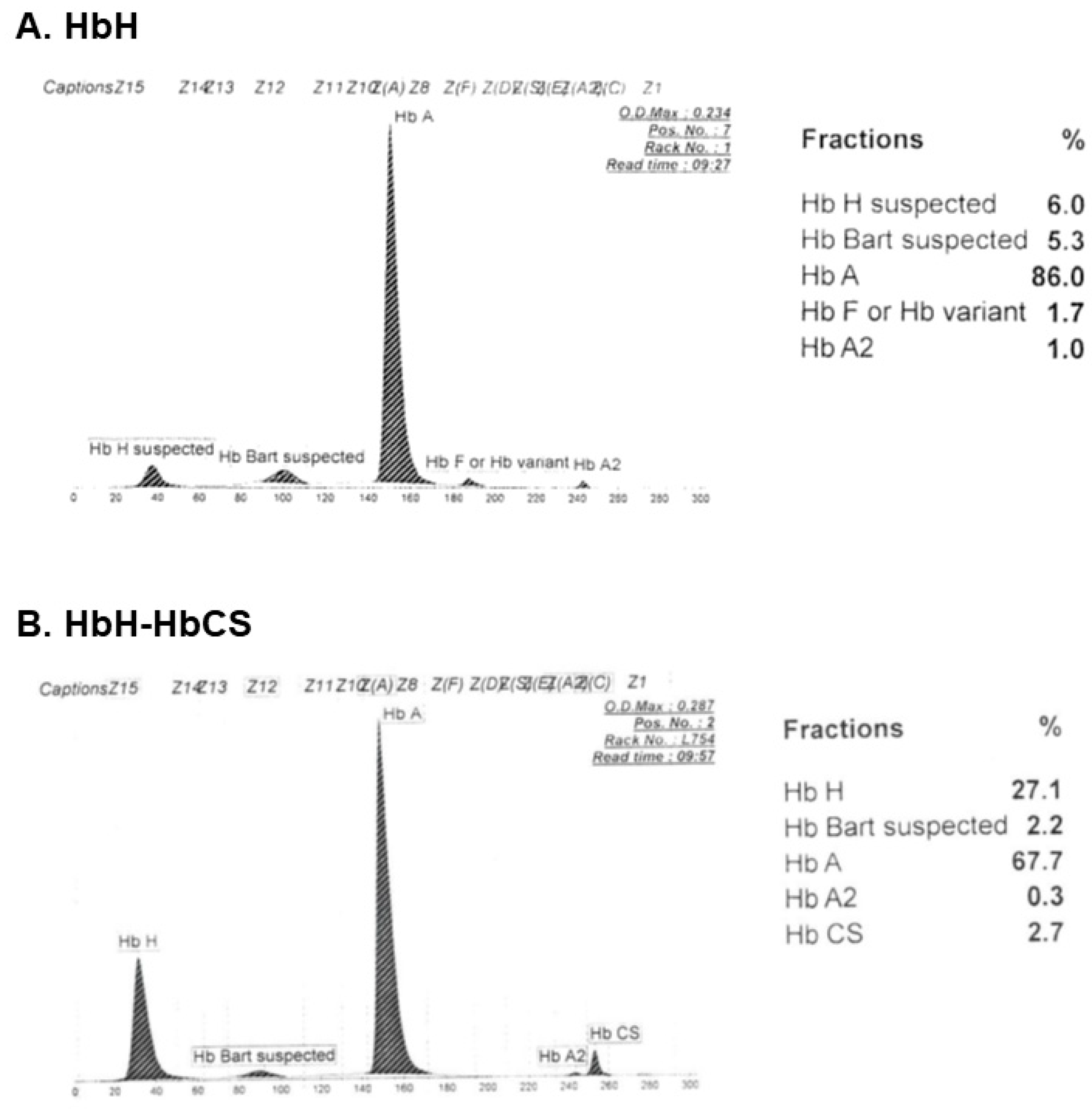
Inclusion bodies (IBs), representing precipitates of HbH (β4), can be visualized as the golf-ball-like appearance of red cells when PBS is exposed to supravital staining dye, such as methylene blue or brilliant cresyl blue. These IBs may decrease or be absent among affected neonates, in AE Bart’s and EF Bart’s diseases, and among patients co-inheriting β-thalassemia heterozygote, as the amount of β-globin chain production among these patients is reduced. As visualization of IBs is an operator-dependent process and its appearance may be confounded in many circumstances, Hb analysis and molecular analysis are required to diagnose HbH.

3.3. Hb Analysis

Both high performance liquid chromatography (HPLC) and capillary electrophoresis (CE) can be used for Hb analysis. The difference is that HPLC reports the presence/absence of HbH, Hb Bart’s and HbCS (or HbPS), whereas CE also quantifies the existing amount. Typical hemoglobin analysis of patients with HbH and HbH-HbCS by HPLC and CE systems are shown in Figure 1 and Figure 2. While the majority of Hb found in Hb analysis is HbA, HbH is typically found between 5% and 15% in both deletional and non-deletional HbH. Additional small amounts of HbCS (or HbPS) (usually ~2% to 5%) are observed among patients with HbH-HbCS (or HbH-HbPS) [41,42,43]. Hb Bart’s is detected between ~17 and 30% among affected neonates, and its different cutoff values have been employed for newborn screening of HbH in some regions, such as Southern China and California [14,43,44]. HbBart’s proportion decreases with age but remains present into adulthood in a minute amount among affected individuals [43]. Low HbA2 (usually < 2%) is a distinct feature of Hb analysis among HbH patients [1,43], with an exception among those co-inheriting the β-thalassemia heterozygote [45,46]. As HbCS (or HbPS) is present in such small amounts and is sometimes undetectable, it would be important for patients to receive molecular diagnosis.

3.4. Molecular Diagnosis

Molecular study is also mandatory for those patients recently transfused to receive a diagnosis. Multiplex gap-polymerase chain reaction (PCR) has been developed and widely used to detect the seven most prevalent α-globin deletions in many laboratories [47,48,49,50]. These include 2 α+-thalassemia; -α3.7 and -α4.2 and 5 α0-thalassemia; --SEA, --MED, --THAI, --FIL and -(α)20.5. Molecular analysis of HbCS and HbPS is commonly performed in clinical practice using allele-specific PCR [43,51]. For those suspected of having unknown α-globin deletions, multiple ligation-dependent probe amplification (MLPA) is the next investigation of choice [52,53]. For those suspected of having uncommon or unknown non-deletional α-globin mutations, Sanger sequencing of the αglobin genes is a practical alternative investigation, as the genes are relatively small (~1.2 kb).

4. Current General Management of HbH Disease

In countries where both α- and β-thalassemia are very prevalent, such as in Thailand, patients with HbH are often overlooked, as health care providers are already overwhelmed with transfusion-dependent β-thalassemia. Frequently, affected patients with HbH are cared for by non-hematologist practitioners, who do not recognize the phenotypic variability of the disease, leading to suboptimal disease monitoring and treatments for patients requiring them. Therefore, it remains important for the Ministry of Public Health, as well as thalassemia specialists in large tertiary care centers, to continuously provide updated knowledge on HbH disease management to medical staff, including general physicians, pediatricians and nurses, nationwide.
In general, all patients with HbH should receive folic acid supplementation at a dose of 2–5 mg/day, as this is required to increase erythropoietic activity [31]. This is especially important for those whose dietary folate might be inadequate and those affected women during their periconceptional period. Although vitamin D deficiency and osteoporosis were well-evidenced in both transfusion-dependent and non-transfusion-dependent β-thalassemia [54,55,56], especially as they advance in age, related data in HbH disease were limited. Therefore, biannual monitoring of vitamin D level and supplementation of vitamin D and calcium as needed is recommended, but with careful monitoring of renal function. Other management strategies for HbH patients with mild-to-moderate anemia are mostly preventive measures to avoid acute hemolytic episodes. These include complete immunization of vaccine-preventable diseases, prompt treatment of fever and infections and alertness to acute anemic symptoms. During hemolytic episodes, red cell transfusion, at a volume of 5 to 12 mg/kg/dose depending on levels of anemia, should be provided to restore Hb to 8 to 9 g/dL [31]. Additionally, adequate hydration with intravenous fluid should be provided to maintain circulation and avoid renal complication from excessive hemoglobinuria.

5. Challenges in Transfusion Management of HbH Disease

Although the majority of patients with HbH are non-transfusion-dependent, it would be important for physicians to realize that their transfusion requirements can be dynamic. History-taking on well-being, physical examination and monitoring of growth (especially height) among pediatric patients and monitoring of baseline Hb level should be routinely performed at least every three months. Physicians should be alert to the presence of at least one of the following signs and symptoms; declining baseline Hb, rapid enlargement of the spleen, failure of growth or secondary sexual development, signs of bone changes, poor quality of life and frequent hemolytic crisis [57]. When these symptoms persist or become progressive over a period of three to six months, regular red cell transfusion should then be initiated. It is currently not possible to predict whether those who have once shifted from non-TDT (NTDT) to TDT would require lifelong transfusion or would be able to stop transfusion when their symptoms improve after years. Clinical observations among Thai patients showed that some pediatric patients with non-deletional HbH (mostly HbH-HbCS) initially requiring frequent transfusions to promote growth spurts and entrance into puberty tolerated only occasional transfusions after they reached their maximal heights. In contrast, a number of them became TDT lifelong. Some patients, especially those with non-deletional HbH, developed severe fatigue in older age and became TDT [31]. Therefore, longitudinal studies concerning the clinical course of HbH are required to possibly identify clinical or genetic factors predicting the disease outcomes. Patients with transfusion-dependent HbH should be provided a transfusion regimen to maintain pretransfusion Hb 9.5–10.5 g/dL, similar to that arranged for transfusion-dependent β-thalassemia [58]. However, a more intensive transfusion therapy to maintain pre-transfusional “functional Hb” [total Hb (g/dL) × (1 − (HbH% + HbBart’s%))/100], instead of total Hb level, of ~9 to 10 g/dL should be considered for patients with HbH not responding well to the standard transfusion regimen. These may frequently occur in affected patients with rare types of non-deletional HbH, such as HbH-HbPNP [20,21] and HbHpolyA [23], surviving severe neonatal anemia and becoming transfusion-dependent from very early life. These patients usually have a higher proportion of non-functional HbH and Hb Bart’s compared with that found among patients with deletional or common non-deletional HbH, leading to more severe tissue hypoxia. The standard transfusion regimen often failed to promote growth and development, as functional Hbs of the transfused red cells were insufficient to replace all non-functional ones in the circulation. The above-mentioned intensive transfusion regimen was adapted from the regimen recently proposed for survivors with BHFS [59] (see section below).

6. Impact of HbH on Older Patients and Challenges in Management of Iron Overload

In non-transfusion-dependent HbH, iron overload usually begins in the second half of the second decade of life and becomes progressive with age, similar to that observed in non-transfusion-dependent β-thalassemia [57,60]. Despite receiving rare or only occasional transfusions, iron accumulation occurs mainly as a result of increased gastrointestinal iron absorption and release of iron from the reticuloendothelial system [60]. The resulting iron overload can reach the level observed in TDT and can lead to destruction of hepatocytes as well as many endocrine organs, such as the thyroid, parathyroid and pancreas [56]. Liver fibrosis and cirrhosis occurred in some patients with heavy iron overload. However, unlike TDT, iron overload cardiomyopathy is relatively rare [61]. The challenge involving iron monitoring in non-transfusion-dependent HbH is that serum ferritin, the most widely used marker for iron overload globally, underestimates the degree of tissue iron overload, especially liver iron concentration (LIC). Affected patients with HbH-HbCS even present lower serum ferritin relative to LIC compared with those with deletional HbH [62]. The LIC, currently assessed using MRIT2*, of >5 mg Fe/g dry weight (DW), was found to be associated with increased morbidity in NTDT [63]. MRIT2*, performed every 1 to 2 years from the age ≥15 years in all non-transfusion-dependent HbH is recommended, when possible, and the LIC of ≥ 5 mg Fe/g DW becomes an indication to start iron chelation therapy (ICT) [57]. In countries with limited access to MRIT2*, serum ferritin ≥ 800 ng/mL was shown to be the most accurate predictor of LIC ≥ 5 mg Fe/g DW [64]. Therefore, it can also be used as a cutoff to start ICT, but with a special caution for delayed treatment among patients with HbH-HbCS [62]. Deferasirox starting from the dose of 10 mg/kg/day is the chelator of choice for NTDT [60,65]. Affected patients with transfusion-dependent HbH should be chelated according to the thalassemia international federation (TIF) guidelines for TDT [58], in which ICT is indicated when serum ferritin >1000 ng/mL. Special attention should be provided to patients with very severe non-deletional HbH, among whom the standard transfusion regimen given cannot adequately replace HbH and Hb Bart’s in the circulation. These patients encounter excessive ineffective erythropoiesis, leading to continuously increased gastrointestinal iron absorption, and their serum ferritin levels can remarkedly underestimate LIC. This is dissimilar to most of TDTs, in which serum ferritin and LIC tend to correlate well but might be similar to the survivors with BHFS who were inadequately transfused [66]. Therefore, at least periodic MRIT2* should be performed for optimal adjustment of ICT among these patients.

7. Current and Ongoing Natural History-Modifying Treatments of HbH Disease

Splenectomy was shown to improve baseline Hb levels (~2 g/dL) among patients with HbH with marked splenomegaly and hypersplenism, specifically among those with HbH-HbCS [26,27,28,31,67]. Well-designed studies on the efficacy of splenectomy among patients affected with other genotypes of non-deletional HbH are lacking. Nevertheless, splenectomy is not generally recommended, especially among pediatric patients <5 years. This is owing to well-known risks of post-splenectomy overwhelming infection [68] and thromboembolic events, including deep vein thrombosis and pulmonary embolism [69,70]. Extensive discussion regarding the risks should be provided to affected individuals and their families before the procedure. Moreover, essential vaccines to prevent relevant infections and aspirin to prevent thromboembolic events should be prescribed to post-splenectomized patients [58]. The role of hematopoietic stem cell transplantation (HSCT) [20,23] and other novel therapies for HbH are summarized in Table 3.

8. The Paradigm Changes for BHFS

BHFS mostly resulted from homozygosity of the Southeast Asian type of α0-thalassemia deletion (--SEA/--SEA). This deletion spans both α-globin genes, but leaves the ζ-globin (embryonic α-like globin) gene intact on the chromosome 16 α-globin cluster. Minute amounts of the remaining embryonic Hb Portland I (ζ2γ2) allow survival of affected fetuses up until mid-to-late gestation, when, in the past, almost all of them died in utero. Some babies affected with BHFS were born prematurely and died shortly after birth. Diagnosis can be made upon clinical presentation of marked anemia and typical hydropic features. Numerous nucleated red cells were often observed in PBS, and Hb analysis demonstrated Hb Bart’s of up to 90 to 100%. Over the last few decades, because of remarkable improvement of prenatal management and neonatal intensive care, there have been increasing reports of survivors with BHFS. In 2017, the first international registry of survivors with BHFS (28 survived naturally, 41 received intrauterine treatments; IUTs) reported clinical courses and outcomes of the affected individuals [71], the key messages of which are summarized below.
  • Mothers carrying BHFS fetuses often experience obstetric complications, such as preterm delivery, polyhydramnios and preeclampsia. This occurred irrespective of whether the fetuses received IUTs (intrauterine transfusion, exchange transfusion and/or in utero stem cell transplantation).
  • Most survivors experienced stormy neonatal periods. However, IUTs significantly resulted in decreased fetal growth restriction, better Apgar scores and decreased length of mechanical ventilator required.
  • IUTs significantly decreased the chance of the survivors being born with hydropic features.
  • Up to 64% of the survivors had associated congenital abnormalities, urogenital abnormalities, including hypospadias and ambiguous genitalia, being the most common, followed by limb abnormalities.
  • Approximately one half of the survivors had growth impairment, regardless of IUTs received.
  • Up to 80% of the survivors had normal or only mildly delayed neurodevelopmental outcomes.
  • All of the survivors became transfusion-dependent shortly after birth and this continued lifelong, unless receiving successful HSCT.
At the most optimistic end, it would be currently possible to rescue affected fetuses with BHFS to yield surviving individuals with reasonable neurodevelopmental outcomes and in some occasions with considerably minor urogenital abnormalities. Therefore, in 2021, an expert consensus declaring that BHFS should no longer be considered a universally fatal disorder was published [72]. However, at the other end, all of these affected survivors relied heavily on the country health care system long term, as with TDTs. As such, it would also be crucial for health care providers to learn optimal management strategies for this distinct group of patients with α-thalassemia.

9. Treatment Strategies for Survivors of BHFS

9.1. Blood Transfusion

Related studies in Canada, where a number of patients with BHFS survived, as they were rescued by IUTs, recently proposed an “aggressive transfusion regimen” for long- term care of the affected patients [59,66]. Under the standard transfusion regimen, these survivors incurred a very high proportion of HbH, ranging from 24 to 64%. This resulted in actual remaining pre-transfusion functional Hb of 4 to 8 g/dL. Consequently, these patients experienced severe tissue hypoxia, as demonstrated by remarkably increased erythropoietin levels, severe growth retardation, marked splenomegaly, many endocrinopathies and even silent ischemic infarct of the brain [59]. One year after being administered an aggressive transfusion regimen, in which targeted pre-transfusion functional Hb was used for monitoring instead of total Hb, the patients’ spleen sizes and erythropoietin levels markedly decreased. In addition, biological markers indicating hemolysis and ineffective erythropoiesis, including indirect bilirubin, were significantly reduced over the period of time. The optimal threshold for pre-transfusion functional Hb to provide favorable outcomes in surviving BHFS was estimated at ~10.5 g/dL [73]. Those patients, whose circulating HbH exceeded 25%, may require a periodic exchange transfusion to maintain HbH ≤16% [59,73].
Survivors with BHFS most likely required red cell products in higher amounts compared with those required by patients with β-thalassemia. Therefore, it is important to have adequate blood supplies in the regions where rescuing the affected fetuses is performed. Moreover, public health policymakers need to understand the special needs of these patients.

9.2. Iron Overload Monitoring and Chelation Therapy

The other inevitable consequence of the aggressive transfusion regimen is early iron overload. These patients received red cell transfusion from in utero; therefore, unsurprisingly, serum ferritin may reach >1000 ng/mL by as early as 12 months of age [66]. Iron chelators among patients below two years should be used extremely cautiously, and chelators should not be prescribed, unless extremely necessary. Future studies on safety and efficacy of different types of iron chelators among these affected infants are required, although deferoxamine might seem to be the safest to begin with [66]. Serum ferritin should not be a single marker to optimize ICT among affected patients with BHFS. Survivors with BHFS, transfused with the standard transfusion regimen, exhibited a lower ferritin-to-LIC ratio compared with their transfusion-dependent β-thalassemia counterparts. This indicated that serum ferritin among these BHFS survivors underestimated LIC, similarly to that observed in NTDT. However, initiation of an aggressive transfusion regimen resulted in an increase in ferritin-to-LIC ratio to the level identified in β-thalassemia. Thus, periodic MRIT2* is recommended for accurate long-term monitoring of the iron burden among affected survivors with BHFS, especially those receiving the standard transfusion regimen.

9.3. HSCT

HSCT is the only curative treatment for surviving BHFS to date. At least 15 of such patients treated with HSCT have been reported, the majority of which (66%) underwent the process at ≤24 months [74]. The most frequently used conditioning regimen was myeloablative conditioning, using busulfan combined with fludarabine or cyclophosphamide. HSC sources were highly variable from human leukocyte antigen matched- and mismatched-related or unrelated donor stem cells. All except two of the reported cases were successfully transplanted and became transfusion-independent. Hb levels in a few patients were maintained upon stable mixed myeloid engraftment (chimerism >20%) [74]. This finding brought up the possible future concept that it may not be necessary to achieve full engraftment to transform patients to transfusion-independent status. By this means, reduced-toxicity conditioning can be employed to minimize the transplant-related mortalities and morbidities.

10. Recently Proposed Perinatal Management and Intrauterine HSCT for Affected Fetuses with BHFS

Clearly, for affected fetuses with BHFS, IUTs led to favorable perinatal outcomes [71]. Accordingly, intrauterine red cell transfusions should be given to all such cases whose parents choose to continue the pregnancies, starting as soon as they were prenatally diagnosed (usually at gestational age; GA ~20 weeks) [74]. Subsequent in utero transfusions can be repeated every three weeks to maintain optimal Hb levels up until birth. This can be adjusted using a non-invasive monitoring parameter, such as middle cerebral artery-peak systolic velocity (MCV-PSV) as a surrogate for fetal hemodynamic change [74]. In North America, a new ongoing international patient registry for BHFS was recently initiated, with the main aims to evaluate the outcomes of rescuing IUTs and to improve the clinical management of BHFS (NCT04872179).
The most recent novel therapeutic trial for affected individuals with BHFS is in utero HSCT (IUHSCT) (NCT02986698). This study is recruiting affected fetuses—GA between 18 and 26 weeks—not having major congenital anomalies and using maternal CD34+ cells as a stem cell source for infusion via the umbilical vein. The therapy is based on the rationale that the fetal immune system remains underdeveloped, allowing possible engraftment of the donor stem cells without providing a transplant conditioning regimen. Moreover, natural trafficking of some maternal cells in fetal blood likely results in fetal tolerance to maternal stem cells. However, at this stage, interim data demonstrated that only minute levels of maternal chimerism were observed in the patients’ blood at the 12 months’ post-natal follow-up, although sustained maternal cells tolerance was identified [75]. The studied patients continued to rely on four to five IUTs up until birth and became transfusion-dependent afterward. Therefore, currently, the most viable concept is that IUHSCT be performed to initiate sustained maternal cell tolerance and the affected patients require the second step of post-natal HSCT “boost” using the identical stem cell source with possibly minimal conditional regimen.
Emerging advanced IUTs are, of course, a huge step toward the management of BHFS. Nevertheless, in resource-limited countries where thalassemia is highly prevalent and β-thalassemia is already a major burden of the health care system, such as in Thailand, it remains unclear whether intentional rescuing of affected fetuses with BHFS is appropriate. Evidence-based counseling concerning the natural history of the disease can be provided and rescuing may be considered for families with recurrent loss of affected fetuses.

11. Conclusions

HbH disease is not always a benign disorder. The direction of care is to appropriately monitor progression of the disease and to early identify those with more severe phenotypes. Transfusion should be provided on an individual basis as required and longitudinal monitoring of the clinical course is essential. BHFS is no longer a universally fatal disorder, owing to availability of advanced IUTs. The survivors became transfusion-dependent; therefore, extensive evidence-based discussion with the affected families should be conducted before making the decision to continue the pregnancies or not.

Author Contributions

Conceptualization, D.S. and S.F.; literature review, D.S.; writing-original draft preparation, D.S.; writing-review and editing, D.S. and S.F. All authors have read and agreed to the published version of the manuscript.

Funding

This research received no external funding.

Institutional Review Board Statement

Not applicable.

Conflicts of Interest

The authors declare no conflict of interest.

References

  1. Harteveld, C.L.; Higgs, D.R. α-thalassaemia. Orphanet J. Rare Dis. 2010, 5, 13. [Google Scholar] [CrossRef] [PubMed] [Green Version]
  2. Enevold, A.; Alifrangis, M.; Sanchez, J.J.; Carneiro, I.; Roper, C.; Børsting, C.; Lusingu, J.; Vestergaard, L.S.; Lemnge, M.M.; Morling, N.; et al. Associations between alpha+-thalassemia and Plasmodium falciparum malarial infection in northeastern Tanzania. J. Infect. Dis. 2007, 196, 451–459. [Google Scholar] [CrossRef] [Green Version]
  3. Kariuki, S.N.; Williams, T.N. Human genetics and malaria resistance. Hum. Genet. 2020, 139, 801–811. [Google Scholar] [CrossRef] [Green Version]
  4. Weatherall, D.J. Phenotype—Genotype relationships in monogenic disease: Lessons from the thalassaemias. Nat. Rev. Genet. 2001, 2, 245–255. [Google Scholar] [CrossRef]
  5. Weatherall, D.J. The Evolving Spectrum of the Epidemiology of Thalassemia. Hematol. Oncol. Clin. N. Am. 2018, 32, 165–175. [Google Scholar] [CrossRef] [PubMed]
  6. Fucharoen, S.; Winichagoon, P. Thalassemia in SouthEast Asia: Problems and strategy for prevention and control. Southeast Asian J. Trop. Med. Public Health 1992, 23, 647–655. [Google Scholar]
  7. Kulaphisit, M.; Kampuansai, J.; Leecharoenkiat, K.; Wathikthinnakon, M.; Kangwanpong, D.; Munkongdee, T.; Svasti, S.; Fucharoen, S.; Smith, D.R.; Lithanatudom, P. A comprehensive ethnic-based analysis of alpha thalassaemia allelle frequency in northern Thailand. Sci. Rep. 2017, 7, 4690. [Google Scholar] [CrossRef] [PubMed]
  8. Fucharoen, S.; Winichagoon, P. Problems of thalassemia in Thailand. ICMR Ann. 1988, 8, 29–33. [Google Scholar]
  9. Hockham, C.; Ekwattanakit, S.; Bhatt, S.; Penman, B.S.; Gupta, S.; Viprakasit, V.; Piel, F.B. Estimating the burden of α-thalassaemia in Thailand using a comprehensive prevalence database for Southeast Asia. eLife 2019, 8, e40580. [Google Scholar] [CrossRef] [PubMed]
  10. Fucharoen, S.; Weatherall, D.J. Progress toward the Control and Management of the Thalassemias. Hematol. Oncol. Clin. N. Am. 2016, 30, 359–371. [Google Scholar] [CrossRef] [PubMed]
  11. Michlitsch, J.; Azimi, M.; Hoppe, C.; Walters, M.C.; Lubin, B.; Lorey, F.; Vichinsky, E. Newborn screening for hemoglobinopathies in California. Pediatr. Blood Cancer 2008, 52, 486–490. [Google Scholar] [CrossRef] [PubMed] [Green Version]
  12. Vichinsky, E.P. Changing Patterns of Thalassemia Worldwide. Ann. N. Y. Acad. Sci. 2005, 1054, 18–24. [Google Scholar] [CrossRef]
  13. Vichinsky, E.P.; MacKlin, E.A.; Waye, J.S.; Lorey, F.; Olivieri, N.F. Changes in the Epidemiology of Thalassemia in North America: A New Minority Disease. Pediatrics 2005, 116, e818–e825. [Google Scholar] [CrossRef] [Green Version]
  14. Lorey, F.; Cunningham, G.; Vichinsky, E.P.; Lubin, B.H.; Witkowska, H.E.; Matsunaga, A.; Azimi, M.; Sherwin, J.; Eastman, J.; Farina, F.; et al. Universal Newborn Screening for Hb H Disease in California. Genet. Test. 2001, 5, 93–100. [Google Scholar] [CrossRef]
  15. Amato, A.; Giordano, P.C. Screening and genetic diagnosis of hemoglobinopathies in southern and northern europe: Two examples. Mediterr. J. Hematol. Infect. Dis. 2009, 1, e2009007. [Google Scholar] [CrossRef]
  16. Fucharoen, S.; Viprakasit, V. Hb H disease: Clinical course and disease modifiers. Hematol. Am. Soc. Hematol. Educ. Program 2009, 2009, 26–34. [Google Scholar] [CrossRef] [Green Version]
  17. Traivaree, C.; Boonyawat, B.; Monsereenusorn, C.; Rujkijyanont, P.; Photia, A. Clinical and molecular genetic features of Hb H and AE Bart’s diseases in central Thai children. Appl. Clin. Genet. 2018, ume11, 23–30. [Google Scholar] [CrossRef] [PubMed] [Green Version]
  18. Viprakasit, V.; Tanphaichitr, V.S.; Pung-Amritt, P.; Petrarat, S.; Suwantol, L.; Fisher, C.; Higgs, D.R. Clinical phenotypes and molecular characterization of Hb H-Paksé disease. Haematologica 2002, 87, 117–125. [Google Scholar]
  19. Liao, C.; Li, J.; Xie, X.-M.; Zhou, J.-Y.; Li, D.-Z. Diversity in clinical presentation of hemoglobin H disease induced by the SEA deletion and the hemoglobin Quong Sze. Ann. Hematol. 2009, 88, 1145–1147. [Google Scholar] [CrossRef] [PubMed]
  20. Sanpakit, K.; Viprakasit, V. Variable genotype-phenotype correlations in patients with a rare nondeletional α-thalassemia; Hb Pak Num Po (HBA1: c.396_397insT). J. Pediatr. Hematol. Oncol. 2014, 36, e185–e189. [Google Scholar] [CrossRef]
  21. Viprakasit, V.; Tanphaichitr, V.S.; Veerakul, G.; Chinchang, W.; Petrarat, S.; Pung-Amritt, P.; Higgs, D.R. Co-inheritance of Hb Pak Num Po, a novel alpha1 gene mutation, and alpha0 thalassemia associated with transfusion-dependent Hb H disease. Am. J. Hematol. 2004, 75, 157–163. [Google Scholar] [CrossRef]
  22. Zainal, N.Z.; Alauddin, H.; Ahmad, S.; Hussin, N.H. α-Thalassemia with Haemoglobin Adana mutation: Prenatal diagnosis. Malays. J. Pathol. 2014, 36, 207–211. [Google Scholar]
  23. Surapolchai, P.; Sirachainan, N.; So, C.-C.; Hongeng, S.; Pakakasama, S.; Anurathapan, U.; Chuansumrit, A. Curative Stem Cell Transplantation for Severe Hb H Disease Manifesting From Early Infancy: Phenotypic and Genotypic Analyses. Hemoglobin 2015, 40, 70–73. [Google Scholar] [CrossRef]
  24. Sanguansermsri, T.; Matragoon, S.; Changloah, L.; Flatz, G. Hemoglobin Suan-Dok (alpha 2 109 (G16) Leu replaced by Arg beta 2): An unstable variant associated with alpha-thalassemia. Hemoglobin 1979, 3, 161–174. [Google Scholar] [CrossRef]
  25. Weiss, I.; Cash, F.E.; Coleman, M.B.; Pressley, A.; Adams, J.G.; Sanguansermsri, T.; Liebhaber, S.A.; Steinberg, M. Molecular basis for alpha-thalassemia associated with the structural mutant hemoglobin Suan-Dok (alpha 2 109leu----arg). Blood 1990, 76, 2630–2636. [Google Scholar] [CrossRef] [PubMed] [Green Version]
  26. Vichinsky, E.P. Clinical manifestations of α-thalassemia. Cold Spring Harb. Perspect. Med. 2013, 3, a011742. [Google Scholar] [CrossRef]
  27. Lal, A.; Goldrich, M.L.; Haines, D.A.; Azimi, M.; Singer, S.T.; Vichinsky, E.P. Heterogeneity of Hemoglobin H Disease in Childhood. N. Engl. J. Med. 2011, 364, 710–718. [Google Scholar] [CrossRef] [PubMed] [Green Version]
  28. Chui, D.; Fucharoen, S.; Chan, V. Hemoglobin H disease: Not necessarily a benign disorder. Blood 2003, 101, 791–800. [Google Scholar] [CrossRef] [Green Version]
  29. Laosombat, V.; Viprakasit, V.; Chotsampancharoen, T.; Wongchanchailert, M.; Khodchawan, S.; Chinchang, W.; Sattayasevana, B. Clinical features and molecular analysis in Thai patients with HbH disease. Ann. Hematol. 2009, 88, 1185–1192. [Google Scholar] [CrossRef]
  30. Origa, R.; Sollaino, M.C.; Giagu, N.; Barella, S.; Campus, S.; Mandas, C.; Bina, P.; Perseu, L.; Galanello, R. Clinical and molecular analysis of haemoglobin H disease in Sardinia: Haematological, obstetric and cardiac aspects in patients with different genotypes. Br. J. Haematol. 2007, 136, 326–332. [Google Scholar] [CrossRef] [PubMed]
  31. Vichinsky, E. Advances in the treatment of alpha-thalassemia. Blood Rev. 2012, 26 (Suppl. S1), S31–S34. [Google Scholar] [CrossRef] [PubMed]
  32. Charoenkwan, P.; Sirichotiyakul, S.; Chanprapaph, P.; Tongprasert, F.; Taweephol, R.; Sae-Tung, R.; Sanguansermsri, T. Anemia and Hydrops in a Fetus With Homozygous Hemoglobin Constant Spring. J. Pediatr. Hematol. Oncol. 2006, 28, 827–830. [Google Scholar] [CrossRef] [PubMed]
  33. He, S.; Zheng, C.; Meng, D.; Chen, R.; Zhang, Q.; Tian, X.; Chen, S. Hb H Hydrops Fetalis Syndrome Caused by Association of the—(SEA) Deletion and Hb Constant Spring (HBA2: c.427T > C) Mutation in a Chinese Family. Hemoglobin 2015, 39, 216–219. [Google Scholar] [CrossRef]
  34. Li, D.Z.; Liao, C.; Li, J.; Xie, X.M.; Huang, Y.N.; Wu, Q.C. Hemoglobin H hydrops fetalis syndrome resulting from the association of the—SEA deletion and the alphaQuong Szealpha mutation in a Chinese woman. Eur. J. Haematol. 2005, 75, 259–261. [Google Scholar] [CrossRef]
  35. Henderson, S.; Pitman, M.; McCarthy, J.; Molyneux, A.; Old, J. Molecular prenatal diagnosis of Hb H Hydrops Fetalis caused by haemoglobin Adana and the implications to antenatal screening for α-thalassaemia. Prenat. Diagn. 2008, 28, 859–861. [Google Scholar] [CrossRef]
  36. Fucharoen, S.; Weatherall, D.J. The Hemoglobin E Thalassemias. Cold Spring Harb. Perspect. Med. 2012, 2, a011734. [Google Scholar] [CrossRef] [PubMed]
  37. Turbpaiboon, C.; Wilairat, P. Alpha-hemoglobin stabilizing protein: Molecular function and clinical correlation. Front. Biosci. 2010, 15, 1–11. [Google Scholar] [CrossRef] [Green Version]
  38. Higgs, D.; Garrick, D.; Anguita, E.; Gobbi, M.; Hughes, J.; Muers, M.; Vernimmen, D.; Lower, K.; Law, M.; Argentaro, A.; et al. Understanding α-Globin Gene Regulation: Aiming to Improve the Management of Thalassemia. Ann. N. Y. Acad. Sci. 2005, 1054, 92–102. [Google Scholar] [CrossRef]
  39. Surapolchai, P.; Chuansumrit, A.; Sirachainan, N.; Kadegasem, P.; Leung, K.-C.; So, C.-C. A molecular study on the role of alpha-hemoglobin-stabilizing protein in hemoglobin H disease. Ann. Hematol. 2017, 96, 1005–1014. [Google Scholar] [CrossRef]
  40. Schrier, S.L.; Bunyaratvej, A.; Khuhapinant, A.; Fucharoen, S.; Aljurf, M.; Snyder, L.M.; Keifer, C.R.; Ma, L.; Mohandas, N. The unusual pathobiology of hemoglobin constant spring red blood cells. Blood 1997, 89, 1762–1769. [Google Scholar] [CrossRef] [Green Version]
  41. Fucharoen, S.; Winichagoon, P.; Piankijagum, A. Standardization on laboratory diagnosis of thalassemia and abnormal hemoglobin. Southeast Asian J. Trop. Med. Public Health 1999, 30 (Suppl. S3), 90–98. [Google Scholar] [PubMed]
  42. Winichagoon, P.; Svasti, S.; Munkongdee, T.; Chaiya, W.; Boonmongkol, P.; Chantrakul, N.; Fucharoen, S. Rapid diagnosis of thalassemias and other hemoglobinopathies by capillary electrophoresis system. Transl. Res. 2008, 152, 178–184. [Google Scholar] [CrossRef] [PubMed]
  43. Munkongdee, T.; Chen, P.; Winichagoon, P.; Fucharoen, S.; Paiboonsukwong, K. Update in Laboratory Diagnosis of Thalassemia. Front. Mol. Biosci. 2020, 7, 74. [Google Scholar] [CrossRef] [PubMed]
  44. Liao, C.; Zhou, J.-Y.; Xie, X.-M.; Tang, H.-S.; Li, R.; Li, D.-Z. Newborn Screening for Hb H Disease by Determination of Hb Bart’s Using the Sebia Capillary Electrophoresis System in Southern China. Hemoglobin 2013, 38, 73–75. [Google Scholar] [CrossRef] [PubMed]
  45. Ma, E.; Chan, A.Y.; Au, W.Y.; Yeung, Y.M.; Chan, L.C. Diagnosis of concurrent hemoglobin H disease and heterozygous beta-thalassemia. Haematologica 2001, 86, 432–433. [Google Scholar] [PubMed]
  46. Galanello, R.; Paglietti, E.; Melis, M.A.; Crobu, M.G.; Addis, M.; Moi, P.; Cao, A. Interaction of heterozygous beta zero-thalassemia with single functional alpha-globin gene. Am. J. Hematol. 1988, 29, 63–66. [Google Scholar] [CrossRef]
  47. de Mare, A.; Groeneger, A.H.; Schuurman, S.; van den Bergh, F.A.; Slomp, J. A rapid single-tube multiplex polymerase chain reaction assay for the seven most prevalent alpha-thalassemia deletions and alphaalphaalpha(anti 3.7) alpha-globin gene triplication. Hemoglobin 2010, 34, 184–190. [Google Scholar] [CrossRef]
  48. Tan, A.S.-C.; Quah, T.C.; Low, P.S.; Chong, S.S. A rapid and reliable 7-deletion multiplex polymerase chain reaction assay for α-thalassemia. Blood 2001, 98, 250–251. [Google Scholar] [CrossRef] [Green Version]
  49. Liu, Y.T.; Old, J.M.; Miles, K.; Fisher, C.; Weatherall, D.J.; Clegg, J.B. Rapid detection of α-thalassaemia deletions and α-globin gene triplication by multiplex polymerase chain reactions. Br. J. Haematol. 2000, 108, 295–299. [Google Scholar] [CrossRef]
  50. Chong, S.S.; Boehm, C.D.; Higgs, D.R.; Cutting, G.R. Single-tube multiplex-PCR screen for common deletional determinants of alpha-thalassemia. Blood 2000, 95, 360–362. [Google Scholar] [CrossRef]
  51. Singsanan, S.; Fucharoen, G.; Savongsy, O.; Sanchaisuriya, K.; Fucharoen, S. Molecular characterization and origins of Hb Constant Spring and Hb Paksé in Southeast Asian populations. Ann. Hematol. 2007, 86, 665–669. [Google Scholar] [CrossRef] [PubMed]
  52. Lei, Y.L.; Sui, H.; Liu, Y.J.; Pan, J.J.; Liu, Y.H.; Lou, J.W. Molecular and Hematological Characterization of a Novel Translation Initiation Codon Mutation of the α2-Globin Gene (ATG>ATC or HBA2: c.3G>C). Hemoglobin 2019, 43, 241–244. [Google Scholar] [CrossRef]
  53. Schouten, J.P.; McElgunn, C.J.; Waaijer, R.; Zwijnenburg, D.; Diepvens, F.; Pals, G. Relative quantification of 40 nucleic acid sequences by multiplex ligation-dependent probe amplification. Nucleic Acids Res. 2002, 30, e57. [Google Scholar] [CrossRef] [PubMed] [Green Version]
  54. Nakavachara, P.; Viprakasit, V. Children with hemoglobin E/β-thalassemia have a high risk of being vitamin D deficient even if they get abundant sun exposure: A study from thailand. Pediatr. Blood Cancer 2013, 60, 1683–1688. [Google Scholar] [CrossRef]
  55. Taher, A.; Musallam, K.; El-Beshlawy, A.; Karimi, M.; Daar, S.; Belhoul, K.; Saned, M.-S.; Graziadei, G.; Cappellini, M.D. Age-related complications in treatment-naïve patients with thalassaemia intermedia. Br. J. Haematol. 2010, 150, 486–489. [Google Scholar] [CrossRef] [PubMed]
  56. Taher, A.T.; Musallam, K.M.; Karimi, M.; El-Beshlawy, A.; Belhoul, K.; Daar, S.; Saned, M.-S.; El-Chafic, A.-H.; Fasulo, M.R.; Cappellini, M.D. Overview on practices in thalassemia intermedia management aiming for lowering complication rates across a region of endemicity: The OPTIMAL CARE study. Blood 2010, 115, 1886–1892. [Google Scholar] [CrossRef] [Green Version]
  57. Taher, A.T.; Vichinsky, E.; Musallam, K.M.; Cappellini, M.D.; Viprakasit, V. Guildlines for the Management of Non Transfusion Dependent Thalassemia (NTDT), 2nd ed.; Thalassemia International Federation: Nicosia, Cyprus, 2017. [Google Scholar]
  58. Cappellini, M.D.; Farmakis, D.; Porter, J.; Taher, A. 2021 Guidelines for the Management of Transfusion Dependent Thalassemia (TDT), 4th ed.; Thalassemia International Federation: Nicosia, Cyprus, 2021. [Google Scholar]
  59. Amid, A.; Chen, S.; Brien, W.; Kirby-Allen, M.; Odame, I. Optimizing chronic transfusion therapy for survivors of hemoglobin Barts hydrops fetalis. Blood 2016, 127, 1208–1211. [Google Scholar] [CrossRef] [Green Version]
  60. Bou-Fakhredin, R.; Bazarbachi, A.-H.; Chaya, B.; Sleiman, J.; Cappellini, M.D.; Taher, A.T. Iron Overload and Chelation Therapy in Non-Transfusion Dependent Thalassemia. Int. J. Mol. Sci. 2017, 18, 2778. [Google Scholar] [CrossRef] [Green Version]
  61. Pennell, D.J.; Porter, J.B.; Piga, A.; Lai, Y.; El-Beshlawy, A.; Belhoul, K.M.; Elalfy, M.; Yesilipek, A.; Kilinç, Y.; Lawniczek, T.; et al. A 1-year randomized controlled trial of deferasirox vs deferoxamine for myocardial iron removal in β-thalassemia major (CORDELIA). Blood 2014, 123, 1447–1454. [Google Scholar] [CrossRef] [Green Version]
  62. Ang, A.L.; Le, T.T.; Tan, R.S. HbH Constant Spring disease has lower serum ferritin relative to liver iron concentration (LIC): Importance of LIC measurement and potential impact on serum ferritin thresholds for iron chelation. Br. J. Haematol. 2016, 176, 986–988. [Google Scholar] [CrossRef]
  63. Musallam, K.M.; Cappellini, M.D.; Daar, S.; Karimi, M.; El-Beshlawy, A.; Graziadei, G.; Magestro, M.; Wulff, J.; Pietri, G.; Taher, A.T. Serum ferritin level and morbidity risk in transfusion-independent patients with β-thalassemia intermedia: The ORIENT study. Haematologica 2014, 99, e218–e221. [Google Scholar] [CrossRef] [PubMed] [Green Version]
  64. Taher, A.T.; Porter, J.B.; Viprakasit, V.; Kattamis, A.; Chuncharunee, S.; Sutcharitchan, P.; Siritanaratkul, N.; Origa, R.; Karakas, Z.; Habr, D.; et al. Defining serum ferritin thresholds to predict clinically relevant liver iron concentrations for guiding deferasirox therapy when MRI is unavailable in patients with non-transfusion-dependent thalassaemia. Br. J. Haematol. 2015, 168, 284–290. [Google Scholar] [CrossRef] [PubMed]
  65. Taher, A.T.; Porter, J.B.; Viprakasit, V.; Kattamis, A.; Chuncharunee, S.; Sutcharitchan, P.; Siritanaratkul, N.; Galanello, R.; Karakas, Z.; Lawniczek, T.; et al. Deferasirox effectively reduces iron overload in non-transfusion-dependent thalassemia (NTDT) patients: 1-year extension results from the THALASSA study. Ann. Hematol. 2013, 92, 1485–1493. [Google Scholar] [CrossRef] [PubMed] [Green Version]
  66. Amid, A.; Chen, S.; Athale, U.; Charpentier, K.; Merelles-Pulcini, M.; Odame, I.; Kirby-Allen, M. Iron overload in transfusion-dependent survivors of hemoglobin Bart’s hydrops fetalis. Haematologica 2018, 103, e184–e187. [Google Scholar] [CrossRef]
  67. Singer, S.T.; Kim, H.-Y.; Olivieri, N.F.; Kwiatkowski, J.L.; Coates, T.D.; Carson, S.; Neufeld, E.; Cunningham, M.J.; Giardina, P.J.; Mueller, B.U.; et al. Hemoglobin H-constant spring in North America: An alpha thalassemia with frequent complications. Am. J. Hematol. 2009, 84, 759–761. [Google Scholar] [CrossRef] [Green Version]
  68. Luu, S.; Spelman, D.; Woolley, I.J. Post-splenectomy sepsis: Preventative strategies, challenges, and solutions. Infect. Drug Resist. 2019, ume 12, 2839–2851. [Google Scholar] [CrossRef] [Green Version]
  69. Tso, S.C.; Chan, T.K.; Todd, D. Venous Thrombosis in Haemoglobin H Disease after Splenectomy. Aust. N. Z. J. Med. 1982, 12, 635–638. [Google Scholar] [CrossRef]
  70. Hirsh, J.; Dacie, J.V. Persistent Post-Splenectomy Thrombocytosis and Thrombo-embolism: A Consequence of Continuing Anaemia. Br. J. Haematol. 1966, 12, 44–53. [Google Scholar] [CrossRef]
  71. Songdej, D.; Babbs, C.; Higgs, D.R. An international registry of survivors with Hb Bart’s hydrops fetalis syndrome. Blood 2017, 129, 1251–1259. [Google Scholar] [CrossRef] [Green Version]
  72. MacKenzie, T.C.; Amid, A.; Angastiniotis, M.; Butler, C.; Gilbert, S.; Gonzalez, J.; Keller, R.L.; Kharbanda, S.; Kirby-Allen, M.; Koenig, B.A.; et al. Consensus statement for the perinatal management of patients with α thalassemia major. Blood Adv. 2021, 5, 5636–5639. [Google Scholar] [CrossRef]
  73. Amid, A.; Barrowman, N.; Odame, I.; Kirby-Allen, M. Optimizing transfusion therapy for survivors of Haemoglobin Bart’s hydrops fetalis syndrome: Defining the targets for haemoglobin-H fraction and “functional” haemoglobin level. Br. J. Haematol. 2022, 197, 373–376. [Google Scholar] [CrossRef] [PubMed]
  74. Horvei, P.; MacKenzie, T.; Kharbanda, S. Advances in the management of α-thalassemia major: Reasons to be optimistic. Hematology 2021, 2021, 592–599. [Google Scholar] [CrossRef] [PubMed]
  75. MacKenzie, T.C.; Frascoli, M.; Sper, R.; Lianoglou, M.B.R.; Velez, J.G.; Dvorak, C.C.; Kharbanda, S.; Vichinsky, E. In Utero Stem Cell Transplantation in Patients with Alpha Thalassemia Major: Interim Results of a Phase 1 Clinical Trial. Blood 2020, 136, 1. [Google Scholar] [CrossRef]
Figure 1. Typical hemoglobin analysis of patients with HbH (A) and HbH-HbCS (B) by automated high-performance liquid chromatography system.
Thalassrep 12 00020 g001
Figure 2. Hemoglobin analysis of patients with HbH (A) and HbH-HbCS (B) using capillary zone electrophoresis.
Thalassrep 12 00020 g002
Table 1. Comparison of clinical characteristics between patients with deletional and non-deletional HbH.
Clinical CharacteristicsDeletional HbHNon-Deletional HbH
Symptomatic patients (%)4060
Age at diagnosis (approximate range, y)1 to 7<1 to 5
History of blood transfusion (%)10 to 2950 to 60
Age at first transfusion (approximate range, y)2 to 17 <1 to 5
TDT during childhood (%)Rare20 to 30
Facial bone changes (%)2 to 320 to 30
Growth retardation (%)Rare15 to 20
Splenomegaly (%)10 to 2020 to 30
Gallstones (%)~1020 to 30
Acute hemolysis following infection/inflammationPeriodicallyMore common
Baseline Hb level (approximate range, g/dL)9 to 117 to 9
Abbreviations: y; years, TDT; transfusion-dependent thalassemia
Table 2. Clinical significances of non-deletional HbH.
TypeSpecific Regions FoundClinical Significance
HbH-HbCS
(--/αCSα)
Southeast Asia, China, MediterraneanApproximately 20% had more severe phenotypes (facial bone change, splenomegaly, growth impairment) and required frequent transfusions
HbH-HbPS
(--/αPSα)
Lao PDR, ThailandMost hematologic findings and clinical courses resemble those of HbH-HbCS, proportion of HbH was higher in HbH-HbPS
HbH-HbQS
(--/αQSα)
Southeast Asia, ChinaClinical phenotypes ranged from hydrops fetalis to TDT and NTDT
HbH-HbPNP
(--/αPNPα) i
ThailandMost reported cases were transfusion-dependent
HbH-HbAdana
(--/αAdanaα)
Malaysia, Indonesia, ChinaHydrops fetalis or TDT
HbH-PolyA
(--/αPolyAα)
Greece
Saudi Arabia, Iran, Türkiye
Transfusion-dependent from early infancy
HbH-HbSuan-Dok
(--/αSuan-Dokα)
ThailandChance of TDT and possibly hydrops fetalis
Abbreviations: HbH-HbCS; HbH with Hb Constant Spring, HbH-HbPS; HbH with Hb Pakse, HbH-HbQS; HbH with Hb Quong Sze, HbH-HbPNP; HbH with Hb Pak Num Po, HbH-HbAdana; HbH with Hb Adana, HbH-PolyA; HbH with poly-adenylation signal mutation, HbH-HbSuan-Dok; HbH with Hb Suan-Dok, TDT; transfusion-dependent thalassemia, NTDT; non-transfusion dependent thalassemia.
Table 3. Natural history-modifying treatments of HbH.
TreatmentPossible RolesProsConsRemarks
SplenectomyMarked splenomegaly, hypersplenism,
areas with extremely limited access to blood product and iron chelator
Evidenced to increase baseline Hb level, able to transform transfusion-dependent to non-transfusion dependent HbH in some casesOverwhelming post-splenectomy infection, increased risk of thromboembolic events, surgical complicationsEspecially effective in HbH-HbCS (although not among all patients), its effectiveness in other HbH genotypes is unclear.
Postoperative LMWH and life-long low-dose aspirin should be considered for prophylaxis of thromboembolic complications.
HSCTTransfusion-dependent non-deletional HbH (rare genotypes) Curative therapyDonor availability, transplant-related morbidities and mortalitiesMay also be considered among patients with transfusion-dependent HbH-HbCS failing to respond to splenectomy or who prefer HSCT upon availability of HLA-matched related donors.
Mitapivat *
(Oral red-cell-specific pyruvate kinase activator)
Non-transfusion dependent HbHPotential oral agent that can decrease ineffective erythropoiesis marker, prolong red cell survival and possibly decrease iron overloadMore data on short- and long-term side effects are needed, not yet available in the marketPhase 2 clinical trial in NTDT (NCT03692052)- 5 of patients with HbH had increased Hb level ≥1 g/dL by 3 weeks.
Gene therapy **Severe α-thalassemiaPotentially ameliorate severityCurrently unknownPreclinical phase
* The only available novel therapeutic agent for HbH disease to date. ** Abstract published in Blood (2021) 138 (Supplement 1): 2012. Abbreviations: HSCT; hematopoietic stem cell transplantation, HbH-HbCS; HbH with Hb Constant Spring, HLA; human leukocyte antigen, LMWH; low molecular weight heparin, NTDT; non-transfusion dependent thalassemia.
Publisher’s Note: MDPI stays neutral with regard to jurisdictional claims in published maps and institutional affiliations.

