Next Issue
Volume 12, September
Previous Issue
Volume 12, March


Surg. Tech. Dev., Volume 12, Issue 2 (June 2023) – 5 articles

Cover Story (view full-size image): 
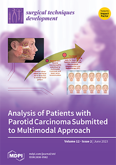
Parotid cancer, regardless of the histological subtype, is a clinical condition that, when treated surgically, may have associated damage to the functionality of the facial nerve. Moreover, surgical resection leaves functional sequelae such as facial paralysis, paresis of some branches, and aesthetic defects affecting the quality of life. Radiotherapy (RT) has shown improvement in local control and survival rates; however, its impact on the complete recovery of facial motricity remains controversial. The present study aimed to evaluate the impact of a multimodal approach on facial nerve functionality in patients diagnosed with parotid carcinoma who underwent parotidectomy and facial nerve microsurgical reconstruction, with or without adjuvant RT. View this paper
  • Issues are regarded as officially published after their release is announced to the table of contents alert mailing list.
  • You may sign up for e-mail alerts to receive table of contents of newly released issues.
  • PDF is the official format for papers published in both, html and pdf forms. To view the papers in pdf format, click on the "PDF Full-text" link, and use the free Adobe Reader to open them.
Order results
Result details
Select all
Export citation of selected articles as:
8 pages, 2116 KiB  
Case Report
Minimally Invasive Peritoneal Dialysis Catheter Insertion with Intraoperative Ultrasound-Doppler Measurement of Rectus Muscle Width and Inferior Epigastric Artery Localization
by Jurij Janež and Jan Grosek
Surg. Tech. Dev. 2023, 12(2), 99-106; https://doi.org/10.3390/std12020009 - 09 Jun 2023
Viewed by 1369
Abstract
Laparoscopic peritoneal dialysis catheter insertion is an established method for peritoneal dialysis catheter placement. One critical aspect of this procedure is rectus sheath tunneling. Proper peritoneal dialysis catheter incorporation within the abdominal wall is important for reducing pericatheter leaks and for proper peritoneal [...] Read more.
Laparoscopic peritoneal dialysis catheter insertion is an established method for peritoneal dialysis catheter placement. One critical aspect of this procedure is rectus sheath tunneling. Proper peritoneal dialysis catheter incorporation within the abdominal wall is important for reducing pericatheter leaks and for proper peritoneal dialysis functioning. Particularly, accurate positioning of the internal cuff within the rectus muscle sheath is crucial to prevent potential dialysate fluid leakage. In the present report, we describe the utilization of intraoperative ultrasound as a valuable tool for determining the ideal location of the internal cuff and assessing the course of the inferior epigastric artery during rectus sheath tunneling. This technique aims to optimize the position of the internal cuff and minimize complications such as vessel injury and pericatheter leaks. Furthermore, we demonstrate that post-catheter insertion, ultrasound imaging offers valuable insights into evaluating the appropriate placement of the internal cuff, external subcutaneous cuff, and potential bleeding within the abdominal wall. Full article
Show Figures

Figure 1

12 pages, 4870 KiB  
Technical Note
Old but Gold: The Surgeon’s Affair to Manage Inguinal Hernia
by Mario Giuffrida, Gabriela Elisa Nita and Federico Biolchini
Surg. Tech. Dev. 2023, 12(2), 87-98; https://doi.org/10.3390/std12020008 - 25 May 2023
Viewed by 2566
Abstract
Purpose: Inguinal hernia repair is a common surgical procedure. It was widely reported worldwide during the COVID-19 pandemic. To manage the lack of anesthesiologists, we have introduced a new protocol to manage inguinal hernia repair. Methods: This protocol is the result of a [...] Read more.
Purpose: Inguinal hernia repair is a common surgical procedure. It was widely reported worldwide during the COVID-19 pandemic. To manage the lack of anesthesiologists, we have introduced a new protocol to manage inguinal hernia repair. Methods: This protocol is the result of a strong collaboration between surgeons and anesthesiologists. It was based on EHS recommendations and the well-described percutaneous ilioinguinal–iliohypogastric and genitofemoral nerves block. Results: More than 400 patients have been treated at our institution. The application of the protocol has led to a sensible reduction in initially planned spinal anesthesia. The complications traditionally related to spinal anesthesia have not been reported in 80% of the patients. Only three patients required the infusion of atropine or flumazenil without the need to involve anesthesiologist. Conclusion: The application of our protocol seems promising. Preliminary results have shown the safety and efficacy of percutaneous ilioinguinal–iliohypogastric and genitofemoral nerves block. The combination of this kind of anesthesia with wound protector and adequate postoperative pain control can lead to a reproducible system avoiding the not strictly necessary presence of an anesthesiologist. The changes that have occurred in the healthcare system in recent years should be new opportunities for the improvement of resources and results. Full article
Show Figures

Figure 1

7 pages, 9373 KiB  
Technical Note
Posterior Cervical Unilateral Biportal Endoscopic One-Block Resection Technique for Cervical Ossified Ligamentum Flavum
by Rajeesh George and Pang Hung Wu
Surg. Tech. Dev. 2023, 12(2), 80-86; https://doi.org/10.3390/std12020007 - 09 May 2023
Viewed by 1235
Abstract
Background: There is sparse literature on the technique of single-stage anterior discectomy fusion and posterior decompression and flavectomy using the biportal endoscopic technique. Methods: Single-stage anterior discectomy fusion and posterior biportal endoscopic decompression and flavectomy apply to cervical myelopathy at a single level [...] Read more.
Background: There is sparse literature on the technique of single-stage anterior discectomy fusion and posterior decompression and flavectomy using the biportal endoscopic technique. Methods: Single-stage anterior discectomy fusion and posterior biportal endoscopic decompression and flavectomy apply to cervical myelopathy at a single level with an anterior disc and posterior ossified ligamentum flavum complex. Anterior discectomy and fusion were performed in the usual fashion in the supine position, and subsequently, posterior biportal endoscopic decompression was carried out after turning the patient prone. Conclusion: Single-stage anterior discectomy fusion and posterior biportal endoscopic decompression and flavectomy are good alternatives for circumferential decompression with myelopathy Full article
Show Figures

Figure 1

12 pages, 1167 KiB  
Article
Analysis of Facial Nerve Functionality and Survival Rates of Patients with Parotid Salivary Gland Carcinoma Submitted to Surgery, Facial Nerve Reconstruction, and Adjuvant Radiotherapy
by Wilber Edison Bernaola-Paredes, Franco Novelli, Estefani Albuja-Rivadeneira, Adriana Aparecida Flosi, Anna Victoria Garbelini Ribeiro, Helena Rubini Nogueira, Hugo Fontan Köhler, Clóvis Antonio Lopes Pinto, Kleber Arturo Vallejo-Rosero and Antonio Cassio Assis Pellizzon
Surg. Tech. Dev. 2023, 12(2), 68-79; https://doi.org/10.3390/std12020006 - 18 Apr 2023
Viewed by 1584
Abstract
Background and Objectives: Parotid cancer (PC), when treated surgically, may have associated damage to the functionality of the facial nerve. The role of radiotherapy in the recovery of facial motricity remains controversial. This study aimed to evaluate the impact of radiotherapy (RT) on [...] Read more.
Background and Objectives: Parotid cancer (PC), when treated surgically, may have associated damage to the functionality of the facial nerve. The role of radiotherapy in the recovery of facial motricity remains controversial. This study aimed to evaluate the impact of radiotherapy (RT) on facial nerve functionality in patients who underwent parotidectomy and facial nerve microsurgical reconstruction. Materials and Methods: Four groups of patients were composed: (a) those who underwent parotidectomy without facial nerve reconstruction and RT; (b) those with nerve reconstruction and without RT; (c) those without nerve reconstruction and RT; and (d) those with nerve reconstruction and RT. Results: 49 patients were male, and 43 were female. A total of 89 underwent parotidectomy, 45 partials, and 44 total. Thus, in nine patients, the sural nerve was used for microsurgical reconstruction. Moreover, 48 patients had a normal facial pattern, 15 with paresthesia, and 29 with permanent paralyses after the House–Brackmann (HB) scale evaluation. Conclusions: The evaluation of nerve functionality after parotidectomy by the House–Brackmann scale is a feasible way to evaluate facial motricity that has already decreased in these patients. Finally, longitudinal studies must be performed to clarify the role of each therapy in the multimodal approach and their clinical impact in facial nerve function. Full article
Show Figures

Graphical abstract

8 pages, 1602 KiB  
Technical Note
An Innovative Surgical Technique to Obtain an Adipose-Derived Stromal Cell-Rich Graft for the Treatment of Osteoarthritis: Technical Note
by Luca Ambrosio, Fabrizio Russo, Simone Catapano, Giuseppe Francesco Papalia, Gianluca Vadalà, Rocco Papalia and Vincenzo Denaro
Surg. Tech. Dev. 2023, 12(2), 60-67; https://doi.org/10.3390/std12020005 - 25 Mar 2023
Viewed by 1518
Abstract
Osteoarthritis (OA) is one of the main causes of disability worldwide and is caused by the progressive degeneration of joint tissues, ultimately leading to chronic pain and loss of function. Intraarticular delivery of mesenchymal stromal cells, such as adipose-derived stromal cells (ASCs), is [...] Read more.
Osteoarthritis (OA) is one of the main causes of disability worldwide and is caused by the progressive degeneration of joint tissues, ultimately leading to chronic pain and loss of function. Intraarticular delivery of mesenchymal stromal cells, such as adipose-derived stromal cells (ASCs), is being actively investigated due to their trophic properties observed in both preclinical and clinical studies. However, cell expansion and handling involve costly and time-consuming processes that limit their application. Recently, several devices and kits have been developed to isolate and process the stromal vascular fraction (SVF), a high biologically active compound of the adipose tissue, right at the patient’s bedside. In this study, we introduce a novel technique to obtain an SVF graft with a high content of ASCs for intraarticular injection directly from liposuction and with minimal equipment. In this technical note, we describe in detail the steps of the surgical technique as well as strategies to avoid common pitfalls and complications. Full article
Show Figures

Figure 1

Previous Issue
Back to TopTop